2010 Activity Report of BSRF 1. BSRF Commissioning Two rounds of dedicated synchrotron radiation running were offered in 2010. The first round was from 5 th July 2010 to 31 st July 2010; the second round was from 10 th September 2010 to 31 st September 2010. Generally, all the experimental stations of BSRF ran well with low fault rates. 177 proposals, 63 research institutes and 307 proposals, 92 research institutes were separately arranged in the two rounds of running. The research areas include Condensed Material Physics, Biology, Chemistry and Chemistry Engineering, Materials, Medical Science,Resources and Environment, National Defense Construction and so on. Many proposals have obtained the important experimental data. The two rounds of running have supplied experimental time not only to the fundamental researches and applied researches, but also to the Chinese National Programs for Science and Technology Development and key research projects. The second running was obvious better than before. The stability of the accelerator was improved greatly and no accident happened from 28 th September to 27 th October. It made a great effort to the SR running. Also some new experimental methods and techniques were used in this round of running, which were significant for the outcome of some important experiments. The workers of BEPC and BSRF gave up the vacation and kept on working during the Dragon Boat Festival and the National Day to ensure the normal running of SR. The Vacuum Ultraviolet experimental station (VUV), mid-energy X-ray experimental station, Soft X-ray Optics Station and Photoemission Spectroscopy Station and biological macromolecule station kept open to users in the coupling mode and have supplied experimental time for 55 proposals. The successful running in the coupling mode mode gained more beamtime for BSRF. Furthermore, it’s more convenient to arrange the beamtime for the users and significant to the urgently needed national research items.
Transcript
2010 Activity Report of BSRF
1. BSRF Commissioning
Two rounds of dedicated synchrotron radiation running were offered in 2010. The first round was from 5th July 2010 to 31st July 2010; the second round was from 10th September 2010 to 31st September 2010. Generally, all the experimental stations of BSRF ran well with low fault rates. 177 proposals, 63 research institutes and 307 proposals, 92 research institutes were separately arranged in the two rounds of running. The research areas include Condensed Material Physics, Biology, Chemistry and Chemistry Engineering, Materials, Medical Science,Resources and Environment, National Defense Construction and so on. Many proposals have obtained the important experimental data. The two rounds of running have supplied experimental time not only to the fundamental researches and applied researches, but also to the Chinese National Programs for Science and Technology Development and key research projects.
The second running was obvious better than before. The stability of the accelerator was improved greatly and no accident happened from 28th September to 27th October. It made a great effort to the SR running. Also some new experimental methods and techniques were used in this round of running, which were significant for the outcome of some important experiments. The workers of BEPC and BSRF gave up the vacation and kept on working during the Dragon Boat Festival and the National Day to ensure the normal running of SR.
The Vacuum Ultraviolet experimental station (VUV), mid-energy X-ray experimental station, Soft X-ray Optics Station and Photoemission Spectroscopy Station and biological macromolecule station kept open to users in the coupling mode and have supplied experimental time for 55 proposals. The successful running in the coupling mode mode gained more beamtime for BSRF. Furthermore, it’s more convenient to arrange the beamtime for the users and significant to the urgently needed national research items.
2. The project of BSRF Diffuse X-ray scattering station has mounted a cooling chamber (DE202
Expander Module from APD cryogenics Inc., sealed with a two-stage cryogenic refrigerator) on the diffractometer and tested to fulfill experiment setup for structure characterization of superconductor film around phase transition temperature. The temperature 14K was attained during the experiment.
X-ray absorption fine structure experimental station has developed mutifunctional in situ instruments, including Liquid helium temperature in situ XAFS cell, liquid nitrogen temperature XAFS cell, liquid sample cell, high pressure XAFS Instrument.
Small angle X-ray scattering experimental station has designed a rapid-mixing device for two solutions. The sample temperature can be adjusted between -10 and
80°C with a flowing medium and the flowing velocity of reactants can be controlled from 0.1 to 20 ml/min by using a peristaltic pump. The reaction time of the mixture can be acquired by irradiating X-ray to different positions as marked by the numerals in Fig.3. Therefore, it can be used for time-resolved SAXS experiment in some time scales.
X-ray imaging station has completed the project “Synchrotron Radiation nano-imaging facility” which was under the support of the important scientific equipment research project. During the dedicated operation for synchrotron radiation in Sep. and Oct. the resolution tests were implemented. The result indicate that the planar resolution of BSRF nano-image facility was achieved at 26nm, and the three-dimensional resolution was achieved at 30nm,
The micro-X-ray fluorescence analysis experimental station is optimized to
provide combinatorial μ-XRF and μ-XAS measurements with splendid performance using the double crystal monochromator and capillary focusing lens. The μ-XRF and μ-XAS techniques are switchable and both are available to users.
High pressure experimental station has installed a new 100W fiber laser in the Laser Heating System. With the help of the new laser, we have achieved 1700K under more than 100 GPa environment.
The mid-energy experimental station was renewed at the end of this year. A chamber filled with He was added for samples, such as wet sample, which can’t be measured in vacuum environment.
The Vacuum Ultraviolet experimental station has installed a new thermoelectric cooling unit. Thermal scan of CD is very useful in study of protein interactions. This station is now one of the two SRCD stations worldwide where such scans can be done.
X-ray diffraction experimental station has supplied two sets of in-situ temperature environmental systems (HTK16 & TTK450) to users, which can respectively
actualize sample temperature from -193°C to 450°C or from 25°C to 1600°C.
3. Researches Launched on BSRF
A lot of great achievements have been achieved by the users and workers of BSRF in 2010.
The research team led by Zhu Chen and Saijuan Chen of State Key Laboratory of Medical Genomics, Shanghai Institute of Hematology, Rui Jin Hospital affiliated to Shanghai Jiao Tong University School of Medicine has made BioXAS research on PML-RAR (promyelocytic leukemia protein-retinoic acid receptor) protein using the technique of XAFS experimental station. The research indicates that the change of the local structure from four coordinates to three coordinates after As3+ substitutes Zn2+ induces a lost of the original activity and function of the protein. This result plays an important role in the mechanism of curing APL (acute promyelocytic leukemia) by ATRA and As2O3 together and was published on “Science” in April, 2010.
An article “Large volume collapse observed in the phase transition in cubic PbCrO3 perovskite” was published by the research team led by Wansheng Xiao of Guangzhou Institute of Geochemistry, Chinese Academy of Sciences. Based on the cooperation with bInstitute of High Energy Physics, Chinese Academy of Sciences and Institute of Fluid Physics, China Academy of Engineering Physics, it’s the first
time that a cubic to cubic isostructural transition with near 10% volume collapse was found in the cubic perovskite sample. This result discovers a new field of the perovskite research, and will improve the progress of solid state physics, chemical, material science, geosciences.
A research approach to resolve the difficult problem of the popularization and application of X-ray tomographic phase-contrast imaging was proposed by the researchers of BSRF. This approach could not only significantly reduce delivered dose without the degradation of the image quality, but also has a much higher efficiency. It has removed the barrier existed in the application of X-ray CT technique in Medical science. The result was published in PNAS in August, 2010.
The research team of Maojun Yang from structural biology center of Tsinghua University has gained insight into the mysterious structure and function of MAGE proteins. The melanoma antigen (MAGE) family consists of more than 60 genes, many of which are cancer-testis antigens that are highly expressed in cancer and play a critical role in tumorigenesis. The team identified really interesting new gene (RING) domain proteins as binding partners for MAGE family proteins, and meanwhile presents the structure of one of the MAGE-RING complexes: MAGE G1-NSE1. In addition, they pointed that MAGE proteins could enhance the E3 ubiquitin ligase activity of the RING domain proteins. Their research has been published on September 24th, 2010 in Molecular Cell.
4. Operation of endstations
4.1 Photoemission Spectroscopy Station
(1)Progresses in Construction of Cluster type PES endstation
In the last year’s report, we have mainly described the major functionalities of
the will be built cluster system, its allocation and main components’ imaginary
layout. Since early 2009 to the middle of 2010 we have spent much time and effort
contacting with the high level suppliers who have experience to provide whole
solution and ability to integrate complex ultrahigh vacuum surface analysis systems
around the world, hoping to get an integrated system. At the end we decided to
integrate the system ourselves due to there are some important quality requirements
can’t be fulfilled. This means that we have to get all components from different
suppliers. This work has been done during the second half of the 2010. We have
signed more than ten contracts including the main ultrahigh vacuum chambers which
have a full functionality from entrance of sample to transfer it to analysis chamber.
The figure shows that it consists of a radial distribution chamber of 1200mm
diameter, thus allowing transfer sample as far as 600mm from its ports. There are
also electron energy analyzer of high resolution power, low temperature manipulator,
sample preparation chamber, PLD chamber, and so on. These all are expected to
arrive at lab before June 2011.
(2)Experiments using SR
Photoemission station started to run under parasitic mode since 2009, i.e. do
experiment when the storage runs for high energy physics under colliding mode.
Since Feb.-Jul. 2010, we have delivered more than 1000hrs of beamtime in parasitic
mode and three months of about 1500hrs under dedicated mode, in total of about
2500hrs.
Figure: Designed main ultrahigh vacuum chambers of the cluster system, including the radial distribution chamber in the center, plus the sample fast entrance chamber, sample storage chamber and analysis chamber around the RDC.
(3)Research Activities
Users publications using experimental data acquired in the PES station: Valence band of catalyst doped sodium alanate by X-ray photoelectron spectroscopy using synchrotron radiation , International Journal of Hydrogen Energy 35(2010)1213-1218; Effects of Annealing Atmosphere and Temperature on the Structure and Photoluminescence of ZnO films Prepared by Pulsed Laser Deposition, Chin. J. Luminesc.31(2010)613-618; Removal of oxidative carbonaceous fragments by annealing treatment studied by XANES, Nuclear Instruments and Methods in Physics Research A 619(2010)319-322; Evidence of Surface-Preferential Co Distribution in ZnO Nanocrystal and Its Effects on the Ferromagnetic Property, ACS Appl. Mater. Inter 2(2010)2053-2059; Direct Synthesis of Nickel(II) Tetraphenylporphyrin and Its Interaction with a Au(111) Surface: A Comprehensive Study, J. Phys. Chem. C 114(2010)9908-9916; Surface Modification Induced
Shielding Effects on Electron Orbital Coupling in Metallofullerene, Journal of Nanoscience and Nanotechnology 10(2010)8265; First Endohedral Metallofullerene-Containing Polymer: Preparation and Characterization of Gd@C82-Polystyrene, J. Phys. Chem. C 114(2010) 7631 -7636; Self-Construction of Core–Shell Structure by Metallofullerene-Containing Polymer, Journal of Nanoscience and Nanotechnology 10(2010); SRPES study of impregnated barium tungsten cathodes, Chin. J. Microwave 2010-08, P701-705.
4.2 Diffraction Station
In 2010, XRD experimental station was opened to users for two opening periods in
dedicated-mode. In the first opening period (from July 1 to July 31), XRD station provided
users with 450h of beam time to 12 research projects. In the second opening period (from
Sep. 15 to Oct. 31), total 895.5h of beam time were provided to 25 research projects. The
experimental modes included XRD/SAXS/XAFS and so on.
In these two operations, two sets of in-situ temperature environmental systems (HTK16
& TTK450) were normally supplied to users, which can respectively, actualize sample
temperature from -193°C to 450°C or from 25°C to 1600°C. The beam time with HTK16 &
TTK450 for experiments occupied 22.8% of the total beam times. In addition, because of
the variety of samples from users, the Pt heating strip in the furnace is easily eroded by
some sorts of samples. Therefore, a graphite heating strip with ceramic holder (see Fig. 1)
was also prepared for special need. This graphite heating element can work up to 1400°C
under higher vacuum environments.
FIG. 1. Graphite heating strip for HTK16 chamber. It is suitable for more samples especially the metallic powders. But a higher chamber vacuum is need and the utmost temperature is about 1400°C.
During the dedicated mode, because of the service time of the photon shutter at
beamline 4B9A exceeding 10 years and the frequently switching, an aging leakage of the
bellow was happened. After substituting a fluorescence block for the photon shutter
temporarily, the experimental measurements were restarted again. Now a new shutter has
been designed. In addition, other equipments, such as the optic supports and multipath
switch power-supply, were redesigned and will be put in use next year.
4.3 VUV beamline
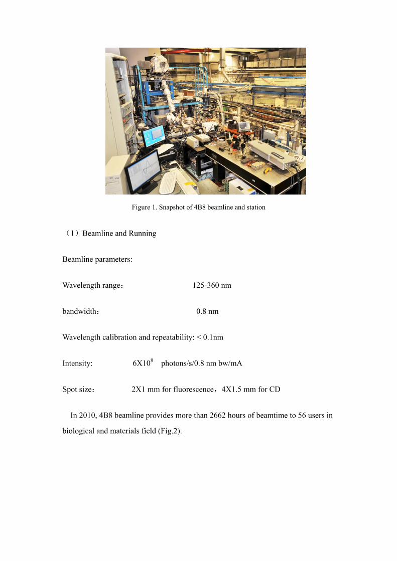
4B8 beamline (Fig.1) is optimized for VUV spectroscopies, especially for
circular dichroism(CD) and fluorescence detections. It is operational under both
synchrotron dedicated and high energy physics mode.
Figure 1. Snapshot of 4B8 beamline and station
(1)Beamline and Running
Beamline parameters:
Wavelength range: 125-360 nm
bandwidth: 0.8 nm
Wavelength calibration and repeatability: < 0.1nm
Intensity: 6X10⁸ photons/s/0.8 nm bw/mA
Spot size: 2X1 mm for fluorescence,4X1.5 mm for CD
In 2010, 4B8 beamline provides more than 2662 hours of beamtime to 56 users in
biological and materials field (Fig.2).
Figure 2. Statistic of beamtime allocation in 2010
(2) Fluorescence spectroscopy
Fluorescence lifetime detection is essential to fluorescence study. Synchrotron
radiation has been the favorable excitation source for fluorescence lifetime
measurement owing to its merits of short pulse, high repetition frequency and wide
tunable wavelength range. The pulse duration is 150 ps with pulse interval 800 ns
under single-bunch mode at the BSRF. Fluorescence lifetime measurement in time
domain requires the excitation from a well separated single-bunch using synchrotron
light sources. Under colliding mode of Beijing Electron Positron Collider II
(BEPCII), a hybrid filling pattern was realized such that a single-bunch was placed
in the middle of a large gap between two multi-bunch groups (Fig. 3). Detection of
fluorescence lifetime, based on the excitation of the light pulse from this designated
single-bunch, was established at beamline 4B8 of the Beijing Synchrotron Radiation
Facility (BSRF). The timing signal of the BEPCII was utilized as a trigger to gate
this fluorescence event. L-Tryptophan amino acid, a known lifetime standard, was
selected to assess the performance of lifetime measurement (Fig. 4). The measured
lifetime was consistent in both the colliding and the single-bunch mode with time
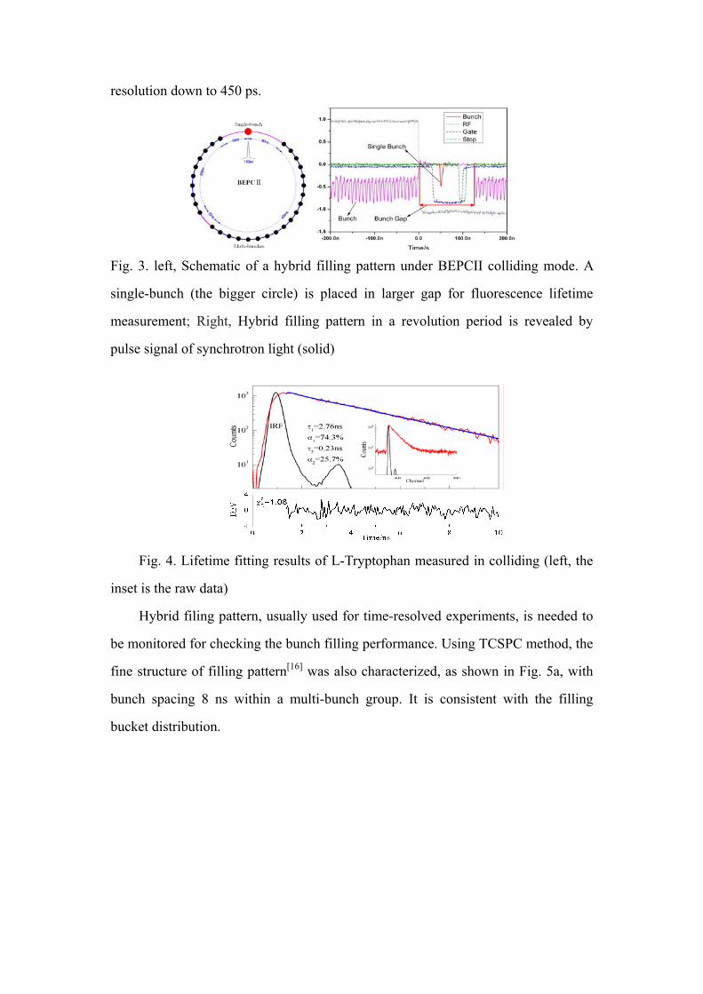
resolution down to 450 ps.
Fig. 3. left, Schematic of a hybrid filling pattern under BEPCII colliding mode. A
single-bunch (the bigger circle) is placed in larger gap for fluorescence lifetime
measurement; Right, Hybrid filling pattern in a revolution period is revealed by
pulse signal of synchrotron light (solid)
Fig. 4. Lifetime fitting results of L-Tryptophan measured in colliding (left, the
inset is the raw data)
Hybrid filing pattern, usually used for time-resolved experiments, is needed to
be monitored for checking the bunch filling performance. Using TCSPC method, the
fine structure of filling pattern[16] was also characterized, as shown in Fig. 5a, with
bunch spacing 8 ns within a multi-bunch group. It is consistent with the filling
bucket distribution.
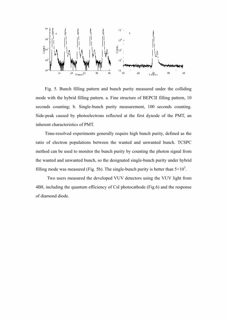
Fig. 5. Bunch filling pattern and bunch purity measured under the colliding
mode with the hybrid filling pattern. a. Fine structure of BEPCII filling pattern, 10
seconds counting; b. Single-bunch purity measurement, 100 seconds counting.
Side-peak caused by photoelectrons reflected at the first dynode of the PMT, an
inherent characteristics of PMT.
Time-resolved experiments generally require high bunch purity, defined as the
ratio of electron populations between the wanted and unwanted bunch. TCSPC
method can be used to monitor the bunch purity by counting the photon signal from
the wanted and unwanted bunch, so the designated single-bunch purity under hybrid
filling mode was measured (Fig. 5b). The single-bunch purity is better than 5×10³.
Two users measured the developed VUV detectors using the VUV light from
4B8, including the quantum efficiency of CsI photocathode (Fig.6) and the response
of diamond diode.
Figure 6 Measurement of the quantum efficiency of CsI photocathode
(3) Circular dichroism (CD) spectroscopy:
Figure 7. Calibration of thermoelectric controller and the thermal scan of protein.
Thermal scan of CD is very useful in study of protein interactions. A new thermoelectric cooling unit was installed and the temperature of the cell has been calibrated (Fig.7). 4B8 is now one of the two SRCD stations worldwide where such scans can be done.
To develop microluidic technique for dynamic study, the beam has been focused down to 75 microns vertically using a group of quartz lenses. The difference of CSA standard between the focused and the unfocused is negligible (Fig. 8).
`
Figure 8, left, spot size scan after focused; right, CSA measurement under the focused (red) and the unfocused beam.(green)
In order to study protein folding dynamics, a pump–probe instrumentation was set up such that a laser-induced temperature jump (T-jump) is used to pump the sample with middle-IR (MIR) absorption measurements as probe. Heating is achieved using output from a Nd:YAG laser incorporating optical parametric oscillator devices. A temperature jump of 15 degree has been reached within 60 nanoseconds (Fig.9).
Figure 9 T-jump up of D2O
4.4 Soft X-ray absorption spectroscopy station
In order to develop the application of light element X-ray absorption spectroscopy, lots of heuristic experiments were done and thus the understanding of the experiments conditions was known better. Based on the work mentioned above, Soft X-ray absorption spectroscopy station was established.
The station includes pre-pumping chamber, transfer and transport devices.
60ns
4ms
The sample bracket and holder were redesigned, which could improve the
vacuum level and save the time of sample-exchange.
An equipment to collect photoelectrons was installed on the re-focus mirror,
which can monitor signal I0 without lose of flux.
The voltage bias and its value for the nonconducting samples can be
decided by the situation of samples and light source, which is useful to
improve S/N.
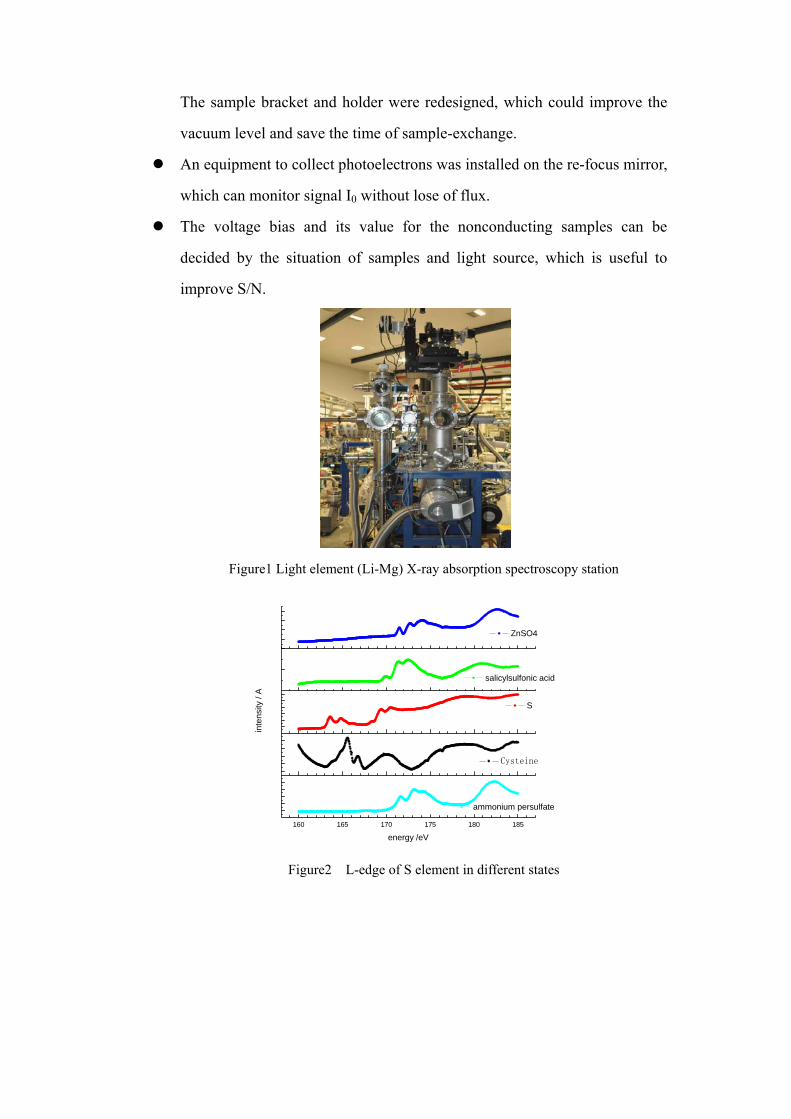
Figure1 Light element (Li-Mg) X-ray absorption spectroscopy station
160 165 170 175 180 185
ammonium persulfate
energy /eV
inte
nsity
/ A
Cysteine
S
salicylsulfonic acid
ZnSO4
Figure2 L-edge of S element in different states
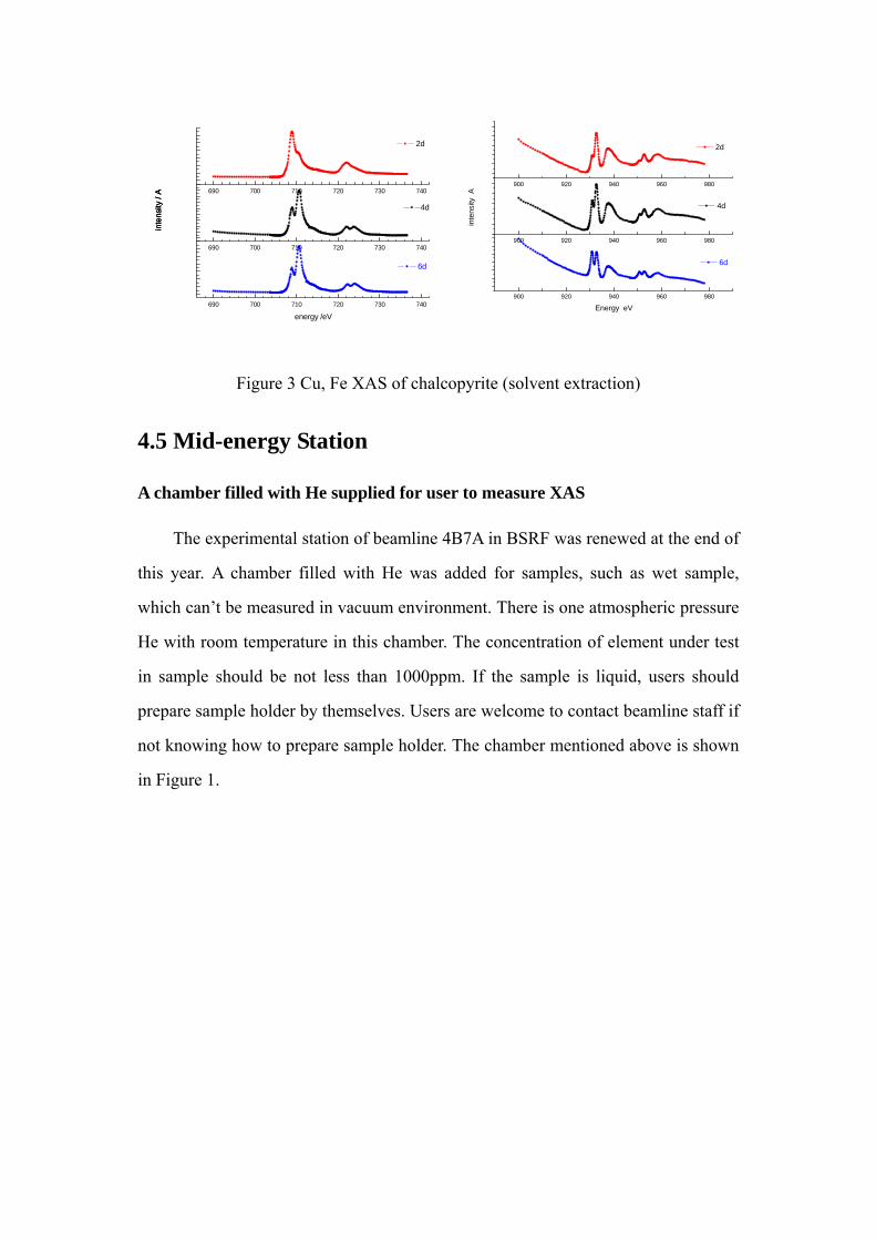
Figure 3 Cu, Fe XAS of chalcopyrite (solvent extraction)
4.5 Mid-energy Station
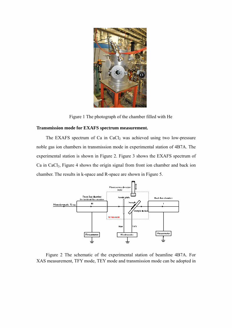
A chamber filled with He supplied for user to measure XAS
The experimental station of beamline 4B7A in BSRF was renewed at the end of
this year. A chamber filled with He was added for samples, such as wet sample,
which can’t be measured in vacuum environment. There is one atmospheric pressure
He with room temperature in this chamber. The concentration of element under test
in sample should be not less than 1000ppm. If the sample is liquid, users should
prepare sample holder by themselves. Users are welcome to contact beamline staff if
not knowing how to prepare sample holder. The chamber mentioned above is shown
in Figure 1.
900 920 940 960 980
900 920 940 960 980
900 920 940 960 980
Energy eV
6d
inte
nsity
A
4d
2d
690 700 710 720 730 740
690 700 710 720 730 740
690 700 710 720 730 740
inte
nsity
/ A
6d
energy /eV
inte
nsity
/ A
4d
inte
nsity
/ A
2d
Figure 1 The photograph of the chamber filled with He
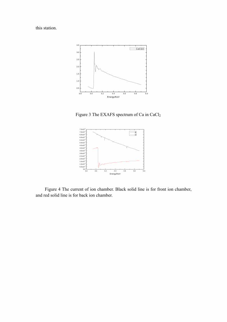
Transmission mode for EXAFS spectrum measurement.
The EXAFS spectrum of Ca in CaCl2 was achieved using two low-pressure
noble gas ion chambers in transmission mode in experimental station of 4B7A. The
experimental station is shown in Figure 2. Figure 3 shows the EXAFS spectrum of
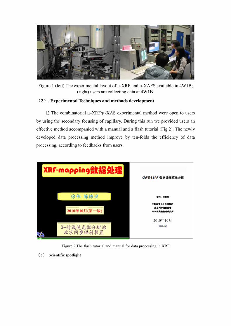
Ca in CaCl2, Figure 4 shows the origin signal from front ion chamber and back ion
chamber. The results in k-space and R-space are shown in Figure 5.
Figure 2 The schematic of the experimental station of beamline 4B7A. For XAS measurement, TFY mode, TEY mode and transmission mode can be adopted in
this station.
3.8 4.0 4.2 4.4 4.6 4.8 5.0
0.5
1.0
1.5
2.0
2.5
3.0
3.5
Energy/KeV
CaCl22
Figure 3 The EXAFS spectrum of Ca in CaCl2
3.8 4.0 4.2 4.4 4.6 4.8 5.00.0
5.0x10⁻⁹
1.0x10⁻⁸
1.5x10⁻⁸
2.0x10⁻⁸
2.5x10⁻⁸
3.0x10⁻⁸
3.5x10⁻⁸
4.0x10⁻⁸
4.5x10⁻⁸
5.0x10⁻⁸
5.5x10⁻⁸
6.0x10⁻⁸
6.5x10⁻⁸
7.0x10⁻⁸
7.5x10⁻⁸
Energy/KeV
B D
Figure 4 The current of ion chamber. Black solid line is for front ion chamber, and red solid line is for back ion chamber.
Figure 5 The results after treated with software WinXAS.
4.6 High Pressure Research Station A new 100W fiber laser was installed in the Laser Heating System. With the
help of the new laser, we have achieved 1700K under more than 100 GPa
environment.
A cleanup-pinhole was installed between the sample and the K-B mirror to
cut the tails of the focus beam spot. When using a pinhole with 20 μm diameter, we
can get a focus beam size with FWHM 16×7.5μm2 and the bottom size (<3% max
intensity) 28×20μm2. Helped with a 50 μm diameter pinhole, a focus beam spot with
26×7.5μm2 HWHM and 50×24μm2 bottom size is provided for the most diffraction
experiments.
Totally 26 research projects and 1573 hours beam time were arranged
during the dedicated synchrotron radiation runs cycle of 2010.
The RXD experiments of Gd, Fe2O3, WP4 and the high temperature ADXD
experiments of Gd2O3, La2O3, Nd2O3, YGG were carried out by high pressure
station, cooperated with Dr. Jung-Fu Lin from the University of Texas at Austin.
An article “Large volume collapse observed in the phase transition in cubic PbCrO3
perovskite” written by Xiao Wansheng et al. was published on PNAS (2010, 107, 14026-14029).
It’s the first time that a cubic to cubic isostructural transition with a 9.8% volume collapse was
found in the cubic perovskite sample. This result discovers a new field of the perovskite research,
and will improve the progress of solid state physics, chemical, material science, geosciences.
4.7 Fluorescence Analysis
(1) Experimental Activities and Instrument Improvement
In 2010 we have two dedicated synchrotron radiation runs. Totally 15 user
projects have been carried out in the first run from July.5th, 2010 to July.31st, 2010.
Using the double crystal monochromator and capillary focusing lens, the beamline is
optimized to provide combinatorial μ-XRF and μ-XAS measurements with splendid
performance: maximum flux as 1010phs/s@15KeV and detection limit as low as
ppm(μg/g). The strong intensity, focused fine-beam and improved signal-noise ratio
have significantly improved the performance of x-ray fluorescence analysis as
shown in user’s reports.
The next dedicated synchrotron radiation run is from Sept. 10th, 2010 to Oct.
31st, 2010. Totally 26 user projects have been carried out in this run. The μ-XRF and
μ-XAS techniques are switchable and both are available to users. The researches in
2010 involve various scientific projects in physics, chemistry, environmental
archaeology, public safety and other scientific area, etc. Owing to the improvement
of performance and expanded techniques, many unique and fundamental scientific
achievements are achieved.
Figure.1 (left) The experimental layout of μ-XRF and μ-XAFS available in 4W1B; (right) users are collecting data at 4W1B.
(2). Experimental Techniques and methods development
1) The combinatorial μ-XRF/μ-XAS experimental method were open to users
by using the secondary focusing of capillary. During this run we provided users an
effective method accompanied with a manual and a flash tutorial (Fig.2). The newly
developed data processing method improve by ten-folds the efficiency of data
processing, according to feedbacks from users.
Figure.2 The flash tutorial and manual for data processing in XRF
(3) Scientific spotlight
In 2010, the x-ray micro-fluorescence end-station allocated beam-time for 41
research projects. During this run several researches obtained significant results due
to the newly developed experimental techniques. Among those some spotlight
researches are briefly introduced hereby. The scientific archaeology group led by
Prof. Changsui Wang in the Graduate school of Chinese Academy of Sciences and
the environmental biology group led by Prof. Yingxu Chen in the Zhejiang
University, have overcome the limitation of conventional elemental analysis and
obtained splendid results by investigating the atomic speciation and local atomic
structure over the micrometer scale. (Fig.3 and Fig.4) All these breakthroughs are
realized thanks to the newly developed micro X-ray absorption spectroscopy.
Recently food safety has arisen to be one the key issues concerned by both
governments and civilians. Owing to the high brilliance and high spatial resolution,
the X-ray fluorescence end-station provides food sciences an effective method for
investigating the distribution of heavy metals in common food. By using the XRF
mapping analysis, the group leaded by Prof. Xinbin Feng from the Institute of
Geochemistry, Chinese Academy of Sciences, in collaboration with researchers from
Institute of High Energy Physics, Chinese Academy of Sciences, have found the
accumulation of heavy metals in the surface of rice. (Fig.5) The results have
revealed the reason accounting for the difficulty of acquiring XANES data on rice
and implied a profound breakthrough for the environmental and food safety studies.
A. The coloring mechanism of pigments in archaeological ceramics
Fig.3 The Cu-K edge micro-XAFS in the pigment of archaeological ceramics (courtesy of Prof. Changsui Wang, GUCAS)
B. The speciation of heavy metal in Chinese tea
Fig.4 The Pb-L3 edge micro-XAFS in Chinese tea (courtesy of Prof. Yingxu Chen, Zhejiang Univ.)
C. Distribution of Heavy metals in rices
Fig.5 The micro-XRF mapping of heavy metals in rice(courtesy of Prof.Xinbin Feng in collaboration with IHEP)
4.8 X-ray imaging station
For X-ray imaging station this year was extraordinary on which the project “Synchrotron Radiation nano-imaging facility” was accomplished. The project which was under the support of the important scientific equipment research project was beginning from 2007. The nano-image beamline had been built on 2009 see fig.1 and fig.2. Subsequently the work emphasis was on the adjustment of beamline and realization of the project parameters. During the dedicated operation for synchrotron radiation in Sep. and Oct. the resolution tests were implemented and the result indicate that the planar resolution of BSRF nano-image facility was achieved at 26nm, see fig.3 and fig.4, and the three-dimensional resolution was achieved at 30nm, see fig.5. Furthermore, the fluorescence test was implemented with the synchrotron radiation and the results indicated the resolution of X-ray probe was 20um and the test sensitivity was 50ppm.
The aforementioned test results indicated that the group has accomplished the project parameters through three year’s effort. Especially the planar resolution was transcend the design parameters and reached the top level of the world. Moreover, it’s the first time in the world to build a nano-image facility on the first-generation light sources.
Fig.1 The sketch of nano-image beamline and image set-up.
Fig. 2 The nano-resolution CT facility
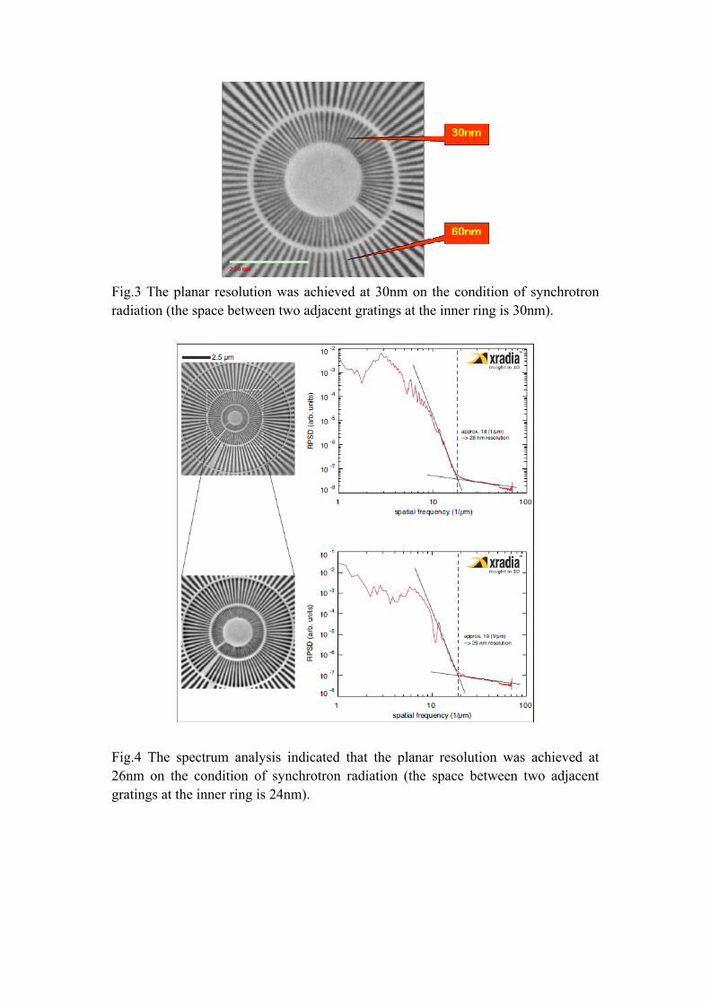
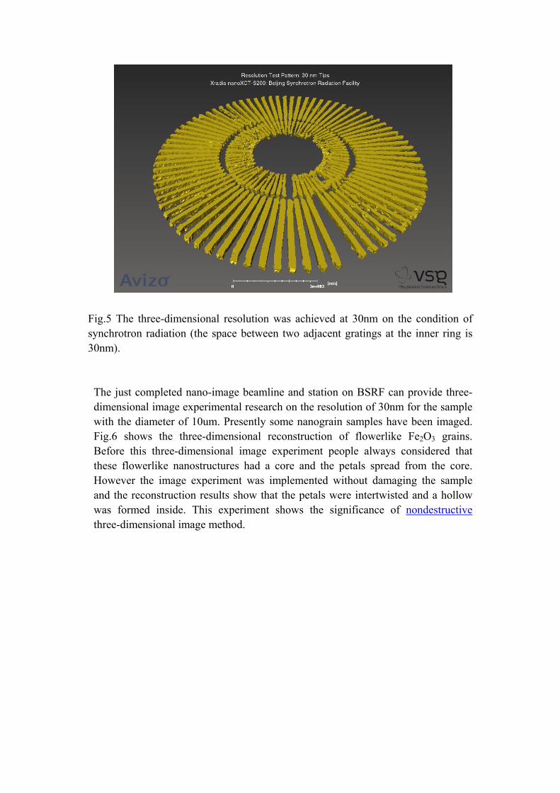
Fig.3 The planar resolution was achieved at 30nm on the condition of synchrotron radiation (the space between two adjacent gratings at the inner ring is 30nm).
Fig.4 The spectrum analysis indicated that the planar resolution was achieved at 26nm on the condition of synchrotron radiation (the space between two adjacent gratings at the inner ring is 24nm).
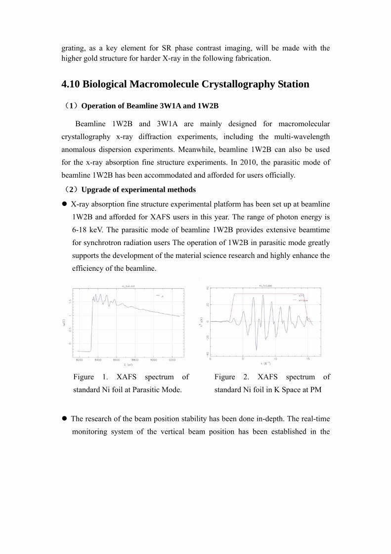
Fig.5 The three-dimensional resolution was achieved at 30nm on the condition of synchrotron radiation (the space between two adjacent gratings at the inner ring is 30nm).
The just completed nano-image beamline and station on BSRF can provide three- dimensional image experimental research on the resolution of 30nm for the sample with the diameter of 10um. Presently some nanograin samples have been imaged. Fig.6 shows the three-dimensional reconstruction of flowerlike Fe2O3 grains. Before this three-dimensional image experiment people always considered that these flowerlike nanostructures had a core and the petals spread from the core. However the image experiment was implemented without damaging the sample and the reconstruction results show that the petals were intertwisted and a hollow was formed inside. This experiment shows the significance of nondestructive three-dimensional image method.
Fig.6 The left is the three-dimensional reconstruction of flowerlike Fe2O3 grains and the right is the results of SEM.
With the consummation of the “Synchrotron Radiation nano-imaging facility” project, topography & imaging station arranged total 21 user’s projects with 318 beam hours during the dedicated operation for synchrotron radiation. There were 3 users engaged in the research of crystal topography, 1 user engaged in the research of imaging method and application and 8 users engaged in the research of nano-resolution CT.
4.9 LIGA and X-ray Lithography Stations
Fig1. The grating for X-ray phase contrast imaging
The Au grating with the height of 16 µm and width of 2µm (4µm period) for X-ray phase contrast imaging has been fabricated using nano X-ray lithography beamline at BSRF, and the analysis of property and the application for the grating will be done in the following work. Figure 1 shows the SEM structure of the Au grating. The
grating, as a key element for SR phase contrast imaging, will be made with the higher gold structure for harder X-ray in the following fabrication.
4.10 Biological Macromolecule Crystallography Station
(1)Operation of Beamline 3W1A and 1W2B
Beamline 1W2B and 3W1A are mainly designed for macromolecular crystallography x-ray diffraction experiments, including the multi-wavelength anomalous dispersion experiments. Meanwhile, beamline 1W2B can also be used for the x-ray absorption fine structure experiments. In 2010, the parasitic mode of beamline 1W2B has been accommodated and afforded for users officially.
(2)Upgrade of experimental methods
X-ray absorption fine structure experimental platform has been set up at beamline 1W2B and afforded for XAFS users in this year. The range of photon energy is 6-18 keV. The parasitic mode of beamline 1W2B provides extensive beamtime for synchrotron radiation users The operation of 1W2B in parasitic mode greatly supports the development of the material science research and highly enhance the efficiency of the beamline.
Figure 1. XAFS spectrum of standard Ni foil at Parasitic Mode.
Figure 2. XAFS spectrum of standard Ni foil in K Space at PM
The research of the beam position stability has been done in-depth. The real-time monitoring system of the vertical beam position has been established in the
whole beamline. The blade-type BPM and double-wire BPM are used to monitor the front-end and FOE beam position respectively. At the same time, the variation of the beam position at the experimental hatch has been monitored by the photodiode-type BPM, to evaluate the influence of the beam stability on the data qualities.
Figure 3. Real-time monitoring system of beam position at Beamline 1W2B
(3)Experimental devices and technology development
The beamline status monitoring system of beamline 1W2B has a stage success in 2010, which is used to monitor the important parameters of the beamline, such as the absolute positions of optical elements, vacuum states and cooling water flow. According to the history data of the beamline status, troubles can be solved immediately and fault diagnosis could be done more easily, which ensures the efficient operation of the Beamline 1W2B.
Database system of synchrotron radiation beamlines has been established in the first time at BSRF. As a pilot station, the information of the 1W2B beamline and experimental station is entered into the database in real time. Using the distributed management approach to publish the information by the web, the beamline remote monitoring, fault diagnosis and history status acquirement have been realized initially, which laid a solid foundation for the remote control
and highly automation of the beamline.
Figure 4. Web page of BSRF database system
4.11 Small Angle X-ray Scattering Station
Two dedicated-mode operations of 1W2A-SAXS station were carried out in
2010. During the first open period (from July, 5 to July, 31), 22 user’s projects came
from 14 institutions were performed in 498h of beam time. During the second open
period (from Sept. 10 to Oct. 31), SAXS station provided 1038h of beam time to 40
user’s projects distributed in 25 institutions. The experimental modes include SAXS,
WAXS and GISAXS. At the end of 2010, a parasitic mode to Beijing Positron and
Electron Collider (BEPC) has been tested for the 1W2A-SAXS station, it
demonstrates that the 1W2A-SAXS station can work well in the parasitic mode and
it will be opened to users next year.
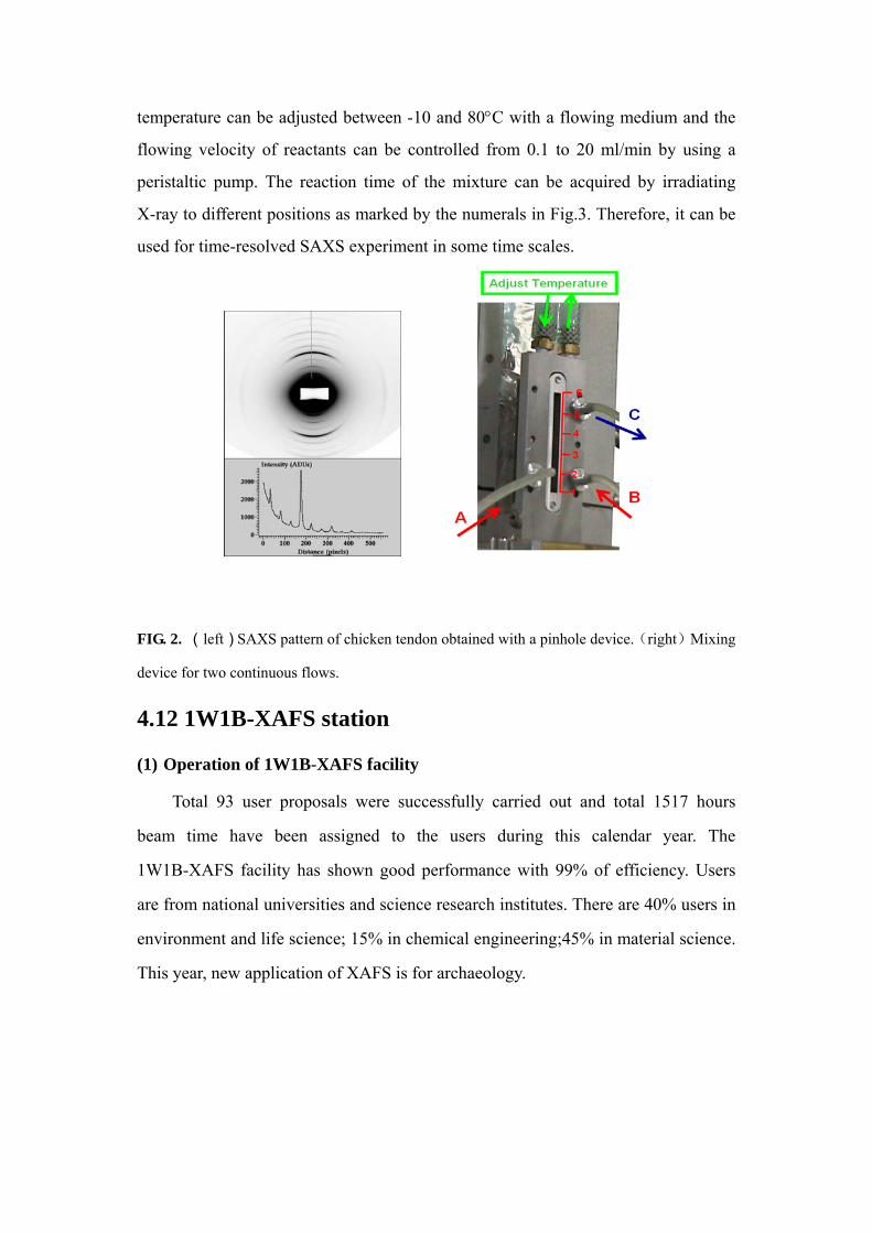
To get an “isotropic” x-ray source as possible, a pinhole device was tested at
beamline 1W2A. With the usage of this pinhole device, the SAXS pattern of a
chicken tendon is shown in Fig.2. It demonstrates that the single-to-noise ratio is
better, but the device needs to be improved again for an isotropic x-ray source. A
rapid-mixing device for two solutions was designed and shown in Fig.3. The sample
temperature can be adjusted between -10 and 80°C with a flowing medium and the
flowing velocity of reactants can be controlled from 0.1 to 20 ml/min by using a
peristaltic pump. The reaction time of the mixture can be acquired by irradiating
X-ray to different positions as marked by the numerals in Fig.3. Therefore, it can be
used for time-resolved SAXS experiment in some time scales.
FIG. 2. (left)SAXS pattern of chicken tendon obtained with a pinhole device.(right)Mixing
device for two continuous flows.
4.12 1W1B-XAFS station
(1) Operation of 1W1B-XAFS facility
Total 93 user proposals were successfully carried out and total 1517 hours
beam time have been assigned to the users during this calendar year. The
1W1B-XAFS facility has shown good performance with 99% of efficiency. Users
are from national universities and science research institutes. There are 40% users in
environment and life science; 15% in chemical engineering;45% in material science.
This year, new application of XAFS is for archaeology.
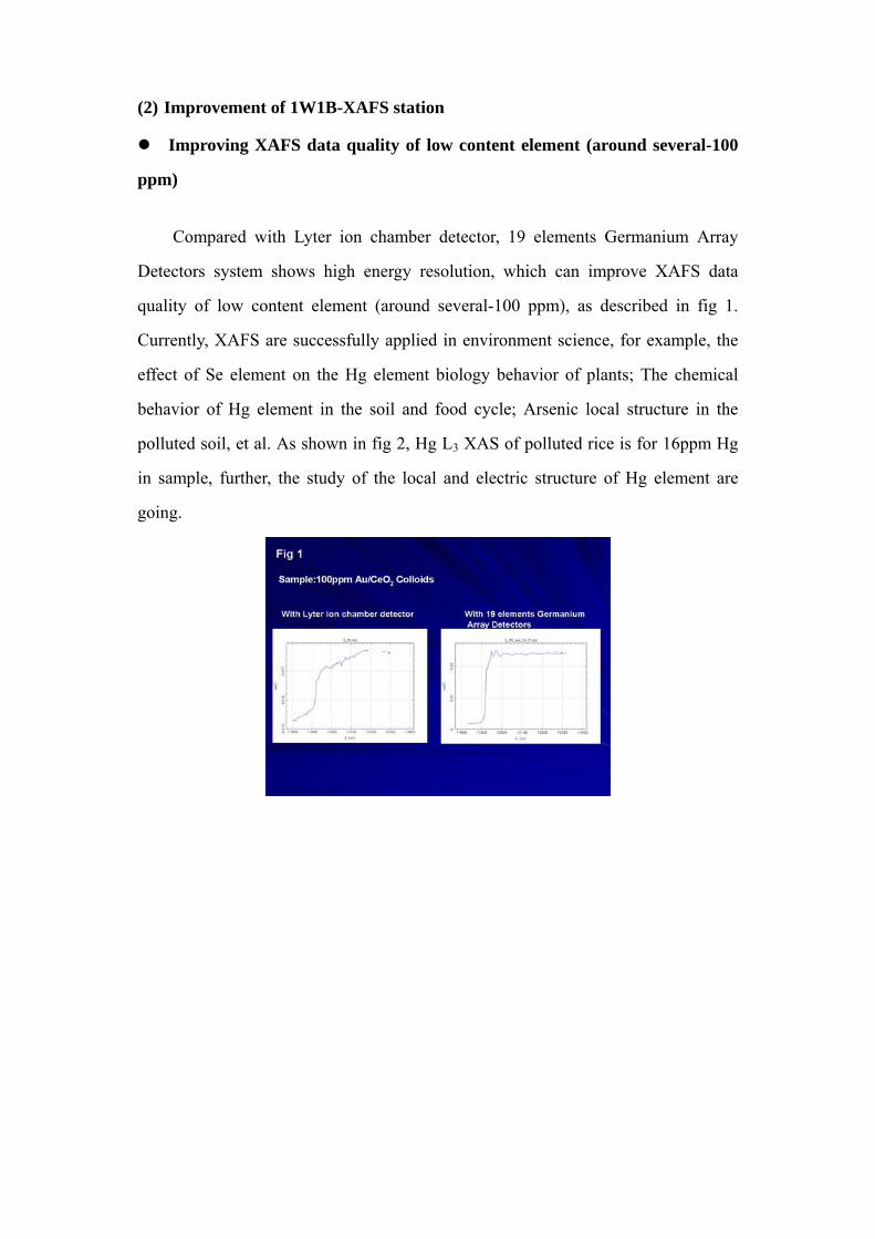
(2) Improvement of 1W1B-XAFS station
Improving XAFS data quality of low content element (around several-100
ppm)
Compared with Lyter ion chamber detector, 19 elements Germanium Array
Detectors system shows high energy resolution, which can improve XAFS data
quality of low content element (around several-100 ppm), as described in fig 1.
Currently, XAFS are successfully applied in environment science, for example, the
effect of Se element on the Hg element biology behavior of plants; The chemical
behavior of Hg element in the soil and food cycle; Arsenic local structure in the
polluted soil, et al. As shown in fig 2, Hg L3 XAS of polluted rice is for 16ppm Hg
in sample, further, the study of the local and electric structure of Hg element are
going.
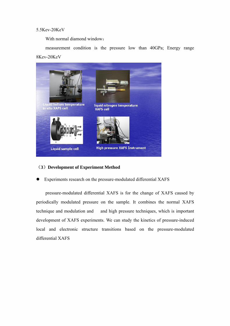
Mutifunctional In Situ Instruments
Liquid helium temperature in situ XAFS cell
the lowest temperature of the cell can be down to 8K.It has applied in the study
of isotopes effect in iron based superconductor,investigated the relationship
between the conductive property and local crystal lattice vibration.
Liquid nitrogen temperature XAFS cell
The cell is with convenient and simple design, based on the needs of life and
enviornment science. It is easy to change sample and can be done in transmission
and fluorescence mode XAFS.
Liquid sample cell
It is for XAFS measurement in aqueous solution and organic solvent. Also it is
very conveniently disassembly and assembly department and cleanout.Temperature
range from -40 to 250degree C.
High pressure XAFS Instrument
It is based on diamond anvil high pressure XAFS measurement, Now two mode
can be performed in 1W1B-XAFS station
With perforated diamond window:
measurement condition is the pressure low than 10GPa; Energy range
5.5Kev-20KeV
With normal diamond window:
measurement condition is the pressure low than 40GPa; Energy range
8Kev-20KeV
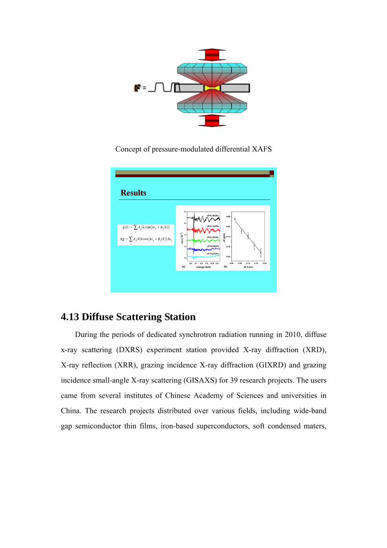
(3)Development of Experiment Method
Experiments research on the pressure-modulated differential XAFS
pressure-modulated differential XAFS is for the change of XAFS caused by
periodically modulated pressure on the sample. It combines the normal XAFS
technique and modulation and and high pressure techniques, which is important
development of XAFS experiments. We can study the kinetics of pressure-induced
local and electronic structure transitions based on the pressure-modulated
differential XAFS
Concept of pressure-modulated differential XAFS
4.13 Diffuse Scattering Station During the periods of dedicated synchrotron radiation running in 2010, diffuse
x-ray scattering (DXRS) experiment station provided X-ray diffraction (XRD),
X-ray reflection (XRR), grazing incidence X-ray diffraction (GIXRD) and grazing
incidence small-angle X-ray scattering (GISAXS) for 39 research projects. The users
came from several institutes of Chinese Academy of Sciences and universities in
China. The research projects distributed over various fields, including wide-band
gap semiconductor thin films, iron-based superconductors, soft condensed maters,
polymer thin films, nano-porous materials, nanostructures and so on.
In this year, the hutch facilities were upgraded in chief as the following aspects:
(1) To improve the mechanism of the device which was used for
sample-step-into-beam and aroused misoperation occasionally due to the coupling
between translation and rotation of the sample stage, a new device was designed and
employed. With this new type of device, the movements of sample rotation about
and translation along the axis of Phi are decoupled completely, which simplifies the
experiment process and lessens the probability of faulty operation. (2) To fulfill
experiment setup for structure characterization of superconductor film around phase
transition temperature, a cooling chamber (DE202 Expander Module from APD
cryogenics Inc., sealed with a two-stage cryogenic refrigerator) was mounted on the
diffractometer and tested during the second period of dedicated SR operation. The
temperature 14K was attained during the experiment. With this equipment of
cryostat,such experiments can be carried out as X-ray diffraction along (0 0 L),
ω-θ/2θ mapping, and reflectivity for single-crystal film and conventional X-ray
diffraction for poly-crystal.
Fig.1 X-ray diffraction experiment of superconductor-film sample at low
temperature. a) Photo of diffractometer with a cryostat mounted. b) X-ray diffraction
data along (0 0 L) obtained at different temperature.
Besides the routine work in facility maintenance, service for users and
equipment upgrade, DXRS station carried out its own researches on functional
materials of wide bandgap semiconductors and nanostructures. We studied the
microstructures of p-type transparent conducting oxide (p-TCO) thin films using
XRD, XRR and x-ray photoelectron spectroscopy (XPS). By characterizing the
structure-property relationship and its evolution with fabrication conditions, we
discovered a new origin mechanism of hole generation in p-TCO besides the
conventional acceptor doping. This mechanism can be described as this: the grain
boundaries and surface of p-TCO film adsorb oxygen; The adsorbed oxygen in the
GBs as well as at the surface depleted the free electrons, and form various
chemisorbed oxygen ions, which caused an upward band bending near the GBs and
finally resulted in holes accumulating, as schematically shown in Fig.2. This effect
leads to a better bulk p-type conductivity beside the grain boundaries, and even
generated the quasi-two-dimensional hole gas (2DHG). This mechanism is of
important significance in guiding the fabrication of p-type TCO, and will have an
active effect on the fabrication of wide band gap oxide semiconductor devices.
Fig. 2. The schematic of oxygen adsorption and the induced depletion region at the
grain boundaries (upper) and of the resulted p-type conductivity mechanism in the
doped and undoped SnO2 thin films (bottom);
DXRS station also made progresses in the fabrication and characterization of
nanostructures. We successfully prepared tin and copper nanorods and zinc
nanoplate, and then characterized their structures and morphologies using GIXRD
and GISAXS. Oxidization experiments were conducted too.