OBSTETRICAL ULTRASOUND IMAGING GUIDELINES Version 4.0; Effective 02-16-2015
MedSolutions, Inc. Clinical Decision Support Tool for Obstetrical Ultrasound Imaging
Common symptoms and symptom complexes are addressed by this tool. Imaging requests for patients with atypical symptoms or clinical presentations that are not specifically addressed will require physician review. Consultation with the
referring physician may provide additional insight.
This version incorporates MSI accepted revisions prior to 12/31/14
CPT® (Current Procedural Terminology) is a registered trademark of the American Medical Association (AMA). CPT® five digit codes, nomenclature and other data are copyright 5American Medical Association. All Rights Reserved. No fee schedules, basic units, relative values or related listings are included in the CPT® book. AMA does not directly or indirectly practice medicine or dispense medical services. AMA assumes no liability for the data contained herein or not contained herein.
MedSolutions, Inc. This tool addresses common symptoms and symptom complexes. Imaging requests for patients with atypicalClinical Decision Support Tool symptoms or clinical presentations that are not specifically addressed will require physician review. Diagnostic Strategies Consultation with the referring physician, specialist and/or patient’s Primary Care Physician (PCP) may provide additional insight.
V 4.0; Effective 02-16-2015 OB Ultrasound RETURN Page 2 of 68
OB-21~Preterm/Premature Rupture of the Membranes 38
OB-22~Second Trimester Screen 39
OB-23~Stillbirth History or Risk of Stillbirth 40
OB-24~Third Trimester Imaging 42
OB-25~Uncertain Dates, including: Cesarean Section History 43
OB-26~Unequal Fundal Size and Dates 45
OB-27~Uterine Anomalies or Adnexal/Pelvic Masses 46
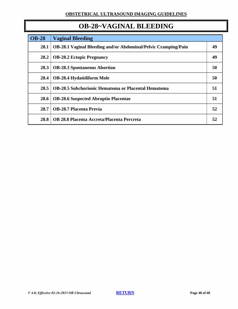
OB-28~VAGINAL BLEEDING 48
OB-29~Vasa Previa 53
OB-30~Procedure Coding Basics for Established Pregnancy 54
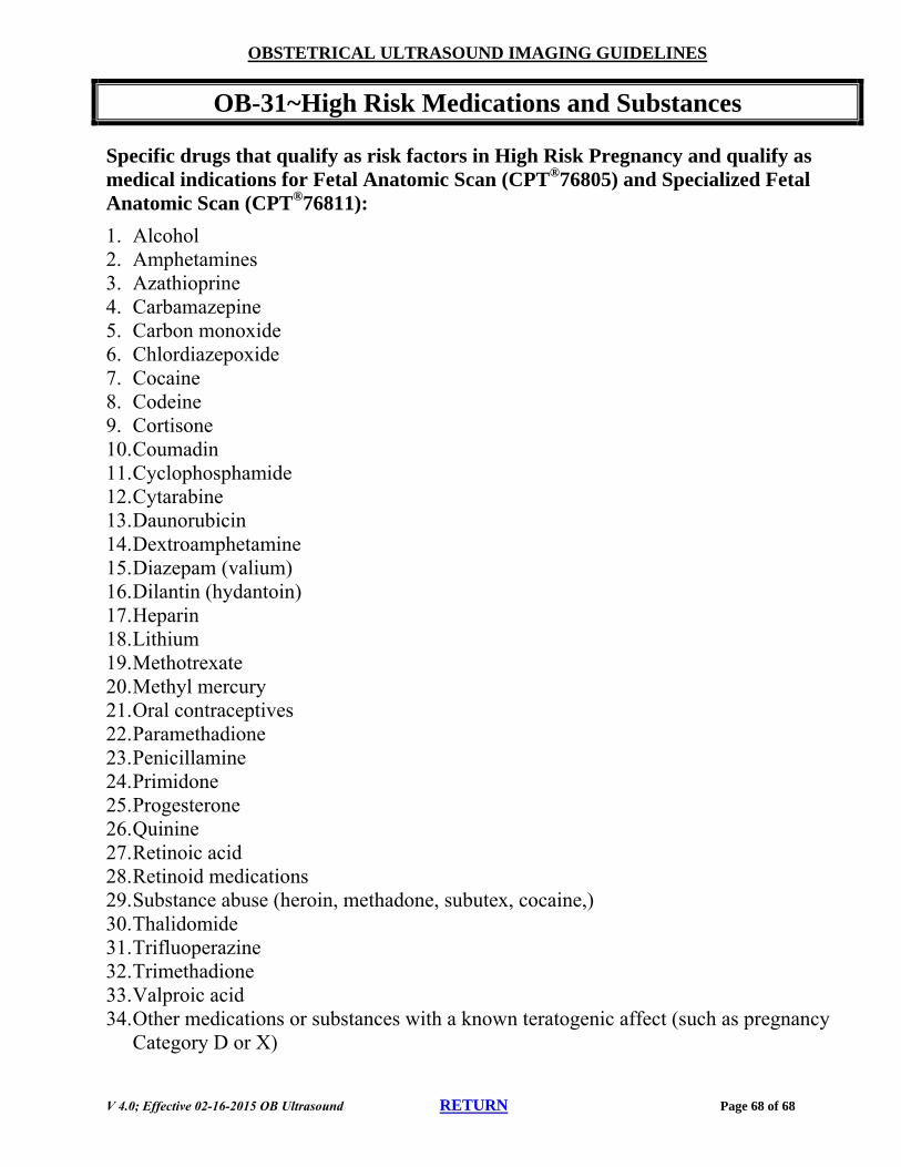
OB-31~High Risk Medications and Substances 68
V 4.0; Effective 02-16-2015 OB Ultrasound RETURN Page 3 of 68
ABBREVIATIONS and GLOSSARY for OB ULTRASOUND IMAGING GUIDELINES
ACOG American College of Obstetricians and Gynecologists
AFI amniotic fluid index
AFP alpha-fetoprotein
CST contraction stress test B-mode
(brightness) a two dimensional imaging procedure, B-mode ultrasound is the basis for all static and real time B-scan images
BPP Biophysical Profile. BPP combines data from two sources (ultrasound imaging and fetal heart rate monitoring), BPP measures the baby’s heart rate, muscle tone, movement, breathing, and amount of amniotic fluid
D & C dilatation and curettage
dichorionic twins
twins having distinct chorions (membrane that forms the fetal part of the placenta), including monozygotic twins (from one oocyte [egg]) separated within 72 hours of fertilization and all dizygotic twins (from two oocytes fertilized at the same time
Doppler involves measuring a change in frequency when the motion of vascular flow is measured
EDC Estimated Date of Confinement; determined from the first day of the last menstrual cycle
FHR fetal heart rate
hCG human chorionic gonadotropin
IDDM insulin-dependent diabetes mellitus
IUGR intrauterine growth restriction; an estimated or actual weight of the fetus below 10th percentile for gestational age
M-mode an ultrasound imaging technique in which structure movement can be depicted in a wave-like manner; primarily used in cardiac and fetal cardiac imaging
macrosomia estimated fetal weight of greater than 4000 or 4500 grams
monochorionic twins
twins developed from one oocyte (egg) developing with a single chorions (membrane that forms the fetal part of the placenta)
NICU Neonatal Intensive Care Unit
NST fetal non-stress test
oligohydramnios
diminished amniotic fluid volume (AFV) for gestational age; definitions include: 1.) amniotic fluid index (AFI) < 10 at < 30 weeks gestation, 2.) AFV < 500ml at 32-36 weeks gestation, 3.) maximum deepest pocket of < 2cm, and, 4.) AFI of < 5cm or < the 5th percentile for gestational age
PACS Picture Archiving and Communications System
polyhydramnios excessive amniotic fluid for gestational age; definitions include: 1.) amniotic fluid index (AFI) > 18 at < 30 weeks gestation, 2.) AFV > 1,700-1,900 ml during the last two months of pregnancy, 3.) AFI > 24cm
PROM preterm rupture of membranes
quad screen alpha-fetoprotein (AFP), estriol, human chorionic gonadotropin (hCG), inhibin A
real time scan
considered the most common type of ultrasound; a 2-dimensional scan that reflects structure and motion over time, scanning and display of images are run at a sufficiently rapid rate so that moving structures can be viewed moving at their natural rate; frame rates ≥ 15 frames per second are considered “real time”
V 4.0; Effective 02-16-2015 OB Ultrasound RETURN Page 4 of 68
OBSTETRICAL ULTRASOUND IMAGING GUIDELINES
GENERAL GUIDELINES
Required Documentation
The following information must be submitted with each request:
o Anticipated date of service o Expected date of delivery o Gestational age at date of service o Results of prior ultrasound studies if available
Inappropriate Uses of OB Ultrasound
Obstetrical ultrasound studies cannot be authorized for payment for individuals who do not have a positive pregnancy test.
Obstetrical ultrasound is not appropriate for the following: o sex determination only o to provide a keepsake or souvenir picture
V 4.0; Effective 02-16-2015 OB Ultrasound RETURN Page 5 of 68
OBSTETRICAL ULTRASOUND IMAGING GUIDELINES
OB-1~Abdominal Pain or Trauma
OB-1.1 Abdominal Pain or Trauma
For abdominal pain or trauma that presents without bleeding:
1. Ultrasound can be performed (CPT®76815 or CPT®76816, and/or CPT®76817
2. See also OB-28~Vaginal Bleeding
Blunt trauma in the first trimester (prior to 13 weeks) generally does not cause pregnancy loss with the exception of profound hypotension:
No imaging is indicated unless there is cramping and/or bleeding
Management of outpatient trauma implies that the trauma was not serious enough to be treated as inpatient. The major risk is abruption placentae: Monitor for uterine contractions for those >20 weeks which could include BPP (CPT®76818 or CPT®76819). Women having contractions less than one every 10 minutes during 4 hours of
monitoring are not at risk and do not need additional monitoring Women having contractions more frequently (more than one every 10 minutes during
4 hours of monitoring) are at risk and additional monitoring is indicated Ultrasound does not appear to be sensitive in predicting abruption, but an evaluation
with CPT®76805 (if not previously done) or CPT®76815, or CPT®76816 can be performed if a complete US was done previously.
No further imaging indicated unless cramping and/or bleeding is present
Reference
1. ACOG Educational Bulletin, Number 251, September 1998 (Reaffirmed 2010)
V 4.0; Effective 02-16-2015 OB Ultrasound RETURN Page 6 of 68
OBSTETRICAL ULTRASOUND IMAGING GUIDELINES
OB-2~Abnormal Fetal Position or Presentation
OB-2.1 Abnormal Fetal Position or Presentation
Confirmation of suspected abnormal fetal position or presentation (transverse or breech presentation): o An ultrasound can be performed at 35 weeks gestation or greater to determine fetal
position o Ultrasound to determine fetal position is not necessary prior to 35 weeks gestation
unless delivery is imminent or version is being considered
Coding Notes
Report one of the following: o CPT®76805 [plus CPT®76810 if more than one fetus] if a complete ultrasound has
not yet been performed, or o CPT®76815 or CPT®76816 if a complete ultrasound was performed previously
V 4.0; Effective 02-16-2015 OB Ultrasound RETURN Page 7 of 68
OBSTETRICAL ULTRASOUND IMAGING GUIDELINES
OB-3~Alloimmunization/Rh Isoimmunization/Other Alloimmunization/Other Causes of Fetal Anemia
Fetal anemia and hydrops may be a result of immune conditions, such as red-cell or Kell alloimmunization or non-immune hydrops caused by parvovirus B19 infection.
OB-3.1 Alloimmunization/RH Isoimmunization
Imaging for RH Isoimmunization:
1. Ultrasound (CPT®76815 or CPT®76816) every 2 to 4 weeks to assess fetal growth starting after performance of the fetal anatomic scan at 16-20 weeks
2. Weekly BPP (CPT®76818 or CPT®76819) or NST starting at 32 weeks or sooner depending on fetal condition
3.
Weekly fetal middle cerebral artery (MCA) Doppler (CPT®76821) starting at 20 weeks or sooner depending on fetal condition
Peak systolic velocity (PSV) of the fetal middle cerebral artery can be used as a substitute for amniocentesis to evaluate a fetus at risk for anemia due to Rhesus isoimmunization/alloimmunization
Because MCA-PSV increases across gestation, results should be adjusted for gestational age. Measurements can be initiated as early as 16 weeks of gestation if there is a past history of early severe fetal anemia, otherwise Doppler evaluation is begun later since intrauterine intravascular transfusions are very difficult before 20 weeks of gestation. The optimal interval between examinations has not been determined, but should be one to two weeks based on clinical experience and what is known about progression of fetal anemia in this setting.
OB-3.2 Other Alloimmunization
Requests will be forwarded to Medical Director review.
OB-3.3 Other Causes of Fetal Anemia
Parvovirus B-19 (Fifth Disease): ultrasound (CPT®76815 or CPT®76816) every 2 to 4 weeks to assess fetal growth starting at time of known exposure and continuing for 8-10 weeks post-exposure o Weekly BPP (CPT®76818 or CPT®76819) or NST starting at time of known
exposure and continuing for 8-10 weeks post-exposure.
V 4.0; Effective 02-16-2015 OB Ultrasound RETURN Page 8 of 68
o Weekly fetal middle cerebral artery (MCA) Doppler (CPT®76821) starting at time of known exposure and continuing for 8-10 weeks post-exposure
Twin anemia polycythemia sequence: ultrasound (CPT®76815 or CPT®76816) every 2 to 3 weeks to assess fetal growth. o Fetal middle cerebral artery (MCA) Doppler (CPT®76821) evaluation of each fetus
every 1-2 weeks.
Fetal hydrops associated with Polyhydramnios: if diagnosed with hydrops, image according to OB-3.1.
Practice Notes
The use of fetal middle cerebral artery Doppler (CPT®76821) for the diagnosis of fetal anemia due to other causes, such as feto-maternal hemorrhage and non-immune hydrops (other than Parvo), has yet to be determined.
Rhesus isoimmunization/alloimmunization is the process through which fetal Rh+ red blood cells enter the circulation of an Rh negative mother causing her to produce antibodies which can cross the placenta and destroy the red blood cells of the current Rh+ fetus in subsequent Rh+ pregnancies.
Twin anemia polycythemia sequence “TAPS” may occur spontaneously in up to 5% of monochorionic twins and may also develop after incomplete laser treatment in TTTS cases. As with TTTS the underlying mechanism is thought to be abnormal placental vascular anastomoses. One twin develops anemia and the other polycythemia. One of the features suggesting towards the diagnosis is discordance in fetal middle cerebral artery peak systolic velocity (MCA-PSV) measurements
References
1. Mari G, Deter RL, Carpenter RL, Rahman F, et al. Noninvasive diagnosis by Doppler ultrasonography of fetal anemia due to maternal red-cell alloimmunization. Collaborative Group for Doppler Assessment of the Blood Velocity in Anemic Fetuses. N Engl J Med. 2000;342(1):9.
2. Lopriore E, Slaghekke F, Oepkes D et-al. Clinical outcome in neonates with twin anemia-polycythemia sequence. Am. J. Obstet. Gynecol. 2010;203 (1): 54.e1-5.
4. Suzuki S. Twin anemia-polycythemia sequence with placental arterio-arterial anastomoses. Placenta. 2010;31 (7): 652.
5. Gucciardo L, Lewi L, Vaast P et-al. Twin anemia polycythemia sequence from a prenatal perspective. Prenat. Diagn. 2010;30 (5): 438-42.
6. Weingertner AS, Kohler A, Kohler M., et-al. Clinical and placental characteristics in four new cases of twin anemia-polycythemia sequence. Ultrasound Obstet Gynecol. 2010;35 (4): 490-4.
7. ACOG Bulletin No. 75. Management of Alloimmunization During Pregnancy. August 2006. 8. Lamont RF, Sobel JD, Vaisbuch E, Kusanovic JP, Mazaki-Tovi S, Uldbjerg N, Romero R.
Parvovirus B19 infection in human pregnancy. BJOG 2011; 118:1750186.
V 4.0; Effective 02-16-2015 OB Ultrasound RETURN Page 9 of 68
9. Reddy UM, Filly RA, Copel JA. Prenatal Imaging: Ultrasonography and Magnetic Resonance Imaging. Obstetrics and Gynecology 2008; 112:145
10. ACOG Bulletin No. 4. Prevention of Rh D Alloimmunization. May 1999.
V 4.0; Effective 02-16-2015 OB Ultrasound RETURN Page 10 of 68
OBSTETRICAL ULTRASOUND IMAGING GUIDELINES
OB-4~AMNIOTIC FLUID ABNORMALITIES
OB-4.1 Amniotic Fluid Abnormalities
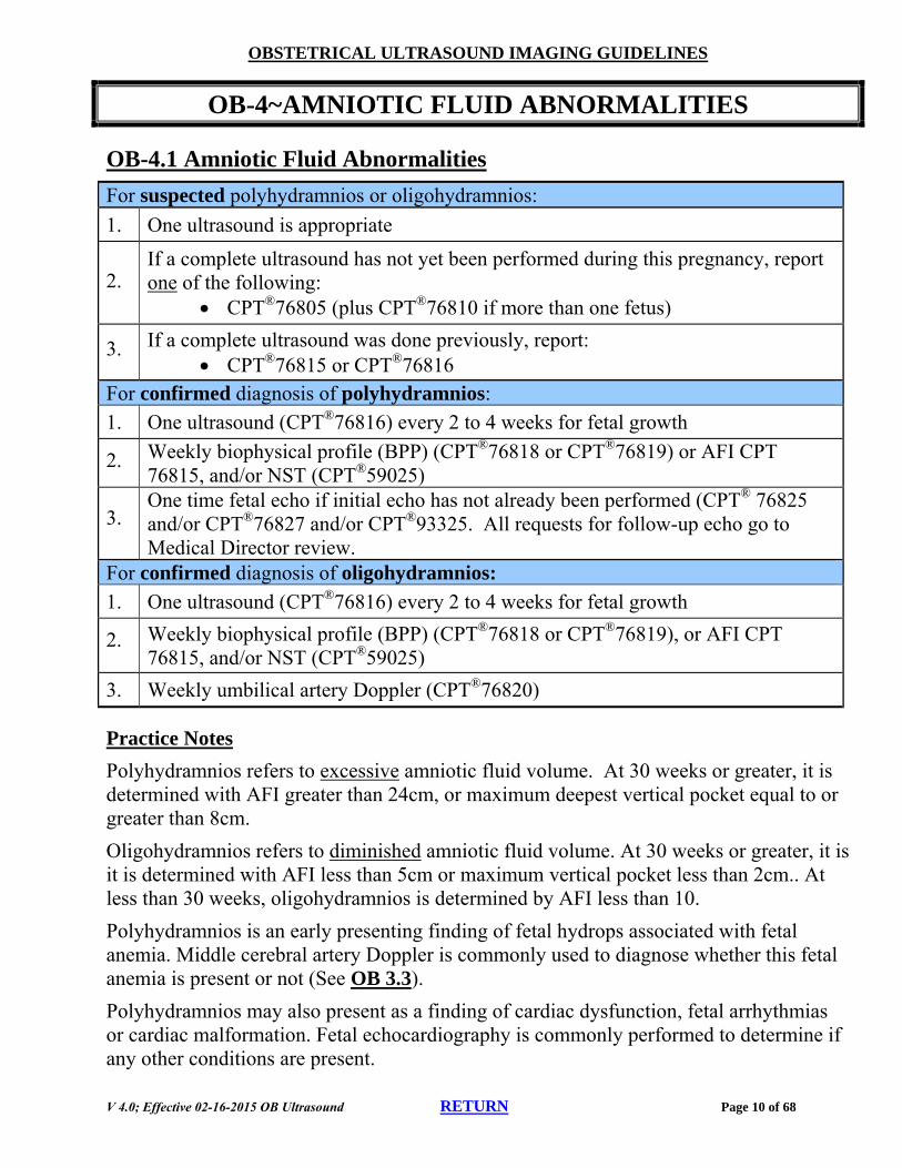
For suspected polyhydramnios or oligohydramnios:
1. One ultrasound is appropriate
2. If a complete ultrasound has not yet been performed during this pregnancy, report one of the following:
CPT®76805 (plus CPT®76810 if more than one fetus)
3. If a complete ultrasound was done previously, report: CPT®76815 or CPT®76816
For confirmed diagnosis of polyhydramnios:
1. One ultrasound (CPT®76816) every 2 to 4 weeks for fetal growth
2. Weekly biophysical profile (BPP) (CPT®76818 or CPT®76819) or AFI CPT 76815, and/or NST (CPT®59025)
3. One time fetal echo if initial echo has not already been performed (CPT® 76825 and/or CPT®76827 and/or CPT®93325. All requests for follow-up echo go to Medical Director review.
For confirmed diagnosis of oligohydramnios:
1. One ultrasound (CPT®76816) every 2 to 4 weeks for fetal growth
2. Weekly biophysical profile (BPP) (CPT®76818 or CPT®76819), or AFI CPT 76815, and/or NST (CPT®59025)
3. Weekly umbilical artery Doppler (CPT®76820)
Practice Notes
Polyhydramnios refers to excessive amniotic fluid volume. At 30 weeks or greater, it is determined with AFI greater than 24cm, or maximum deepest vertical pocket equal to or greater than 8cm.
Oligohydramnios refers to diminished amniotic fluid volume. At 30 weeks or greater, it is it is determined with AFI less than 5cm or maximum vertical pocket less than 2cm.. At less than 30 weeks, oligohydramnios is determined by AFI less than 10.
Polyhydramnios is an early presenting finding of fetal hydrops associated with fetal anemia. Middle cerebral artery Doppler is commonly used to diagnose whether this fetal anemia is present or not (See OB 3.3).
Polyhydramnios may also present as a finding of cardiac dysfunction, fetal arrhythmias or cardiac malformation. Fetal echocardiography is commonly performed to determine if any other conditions are present.
V 4.0; Effective 02-16-2015 OB Ultrasound RETURN Page 11 of 68
References
1. Am J Obstet Gynecol 1990;162:1168-1173 2. ACOG Practice Bulletin No.101: Ultrasonography in pregnancy. February 2009 3. American Pregnancy Association. Low amniotic fluid levels: Oligohydramnios. January 2007
http://www.americanpregnancy.org/pregnancycomplications/lowamnioticfluidoligohydramnios.htm. Accessed September 25, 2009
4. Chitkara U, Wilkins I, Lynch L, Mehalek K, Berkowitz RL. The Role of Sonography in Assessing Severity of Fetal Anemia in Rh- and Kell-Isoimmunized Pregnancies. Obstet Gynecol 1988;71:393-398.
V 4.0; Effective 02-16-2015 OB Ultrasound RETURN Page 12 of 68
OBSTETRICAL ULTRASOUND IMAGING GUIDELINES
OB-5~FETAL ANATOMIC SCAN
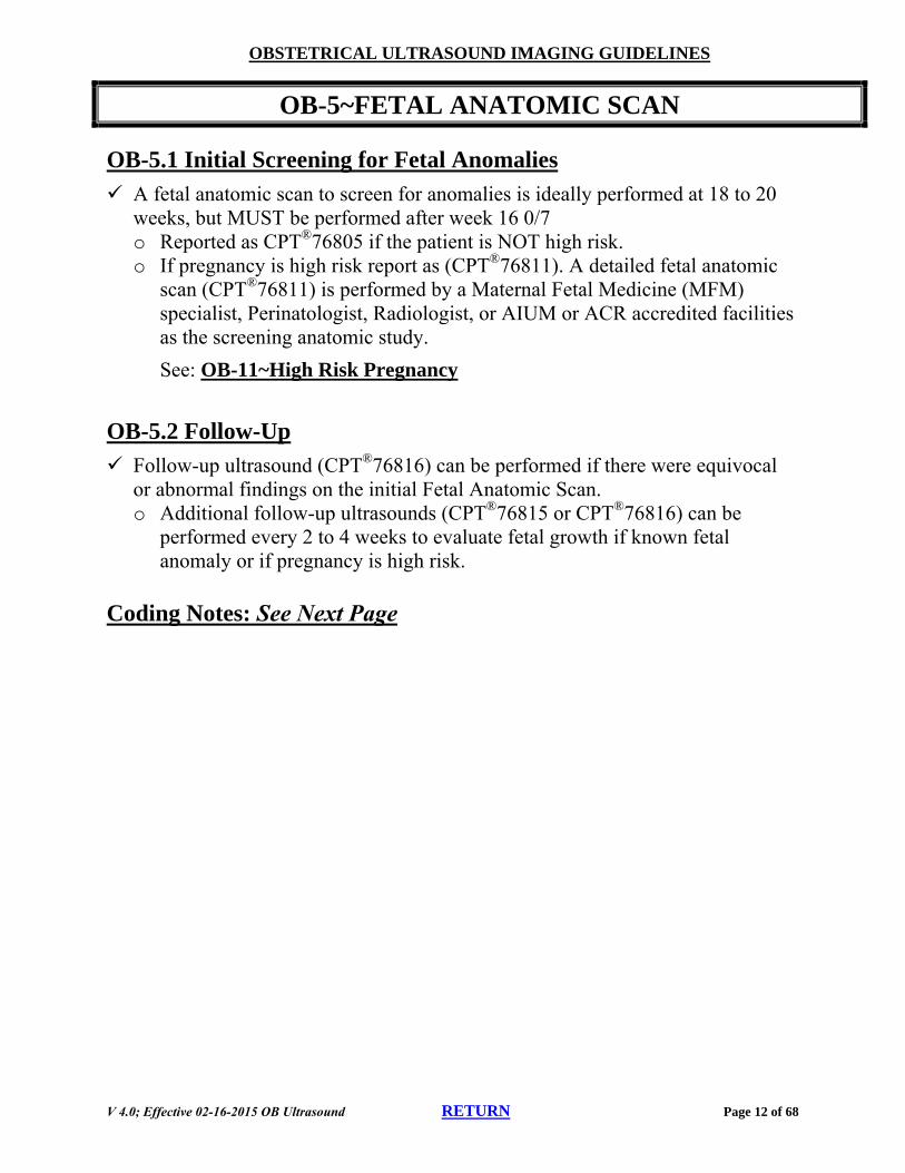
OB-5.1 Initial Screening for Fetal Anomalies
A fetal anatomic scan to screen for anomalies is ideally performed at 18 to 20 weeks, but MUST be performed after week 16 0/7 o Reported as CPT®76805 if the patient is NOT high risk. o If pregnancy is high risk report as (CPT®76811). A detailed fetal anatomic
scan (CPT®76811) is performed by a Maternal Fetal Medicine (MFM) specialist, Perinatologist, Radiologist, or AIUM or ACR accredited facilities as the screening anatomic study.
See: OB-11~High Risk Pregnancy
OB-5.2 Follow-Up
Follow-up ultrasound (CPT®76816) can be performed if there were equivocal or abnormal findings on the initial Fetal Anatomic Scan. o Additional follow-up ultrasounds (CPT®76815 or CPT®76816) can be
performed every 2 to 4 weeks to evaluate fetal growth if known fetal anomaly or if pregnancy is high risk.
Coding Notes: See Next Page
V 4.0; Effective 02-16-2015 OB Ultrasound RETURN Page 13 of 68
Coding Notes
References
1. Society of Maternal Fetal Medicine. White Paper on Ultrasound Code 76811. May 24, 2004. http://www.askleslie.net/rads/White_Paper_on_Ultrasound_Code_76811.pdf. Accessed May 10, 2010
1. ACOG practice bulletin no. 101: Ultrasonography in pregnancy. February 2009. Reaffirmed 2014
FETAL ANATOMIC SCAN - Coding Notes
CPT®76805
A complete transabdominal ultrasound (CPT®76805) is defined in CPT®
as including the following elements: Determination of the number of fetuses and amniotic/chorionic sacs Measurements appropriate for gestation (>14 weeks) Survey of intracranial/spinal/abdominal anatomy Four-chambered heart Umbilical cord insertion site Placenta location Amniotic fluid assessment Examination of maternal adnexa, when visible
CPT®76810 CPT®76810 is an add-on code used with the primary procedure CPT®76810 to report each additional fetus if there is a multiple gestation
CPT®76805
CPT®76810
CPT®76805 and CPT®76810 should only be reported once per pregnancy unless the mother changes to a new medical caregiver at a new office and there is a medical indication for ultrasound
CPT®76811
CPT®76812
CPT®76811 and CPT®76812 are defined in CPT® as including all of the requirements listed for procedures CPT®76805 and CPT®76810. The pregnancy must be considered “high risk” to support CPT®76811 and CPT®76812. In addition, the report must also document detailed anatomic evaluation of the following elements: Fetal brain/ventricles Face Heart/outflow tracts and chest anatomy Abdominal organ-specific anatomy Number/length/architecture of limbs Detailed evaluation of the umbilical cord and placenta Other fetal anatomy as clinically indicated
CPT®76812 CPT®76812 is an add-on code used with the primary procedure CPT®76812 to report each additional fetus in a multiple gestation.
CPT®76811 The reporting of CPT®76811 only once per pregnancy, per practice (per NPI) is appropriate.
V 4.0; Effective 02-16-2015 OB Ultrasound RETURN Page 14 of 68
OBSTETRICAL ULTRASOUND IMAGING GUIDELINES
OB-6~Fetal Distress/Decreased Fetal Movement
OB-6.1 Fetal Distress
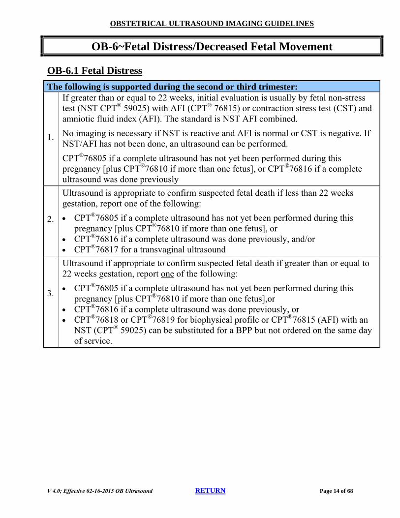
The following is supported during the second or third trimester:
1.
If greater than or equal to 22 weeks, initial evaluation is usually by fetal non-stress test (NST CPT® 59025) with AFI (CPT® 76815) or contraction stress test (CST) and amniotic fluid index (AFI). The standard is NST AFI combined.
No imaging is necessary if NST is reactive and AFI is normal or CST is negative. If NST/AFI has not been done, an ultrasound can be performed.
CPT®76805 if a complete ultrasound has not yet been performed during this pregnancy [plus CPT®76810 if more than one fetus], or CPT®76816 if a complete ultrasound was done previously
2.
Ultrasound is appropriate to confirm suspected fetal death if less than 22 weeks gestation, report one of the following:
CPT®76805 if a complete ultrasound has not yet been performed during this pregnancy [plus CPT®76810 if more than one fetus], or
CPT®76816 if a complete ultrasound was done previously, and/or CPT®76817 for a transvaginal ultrasound
3.
Ultrasound if appropriate to confirm suspected fetal death if greater than or equal to 22 weeks gestation, report one of the following:
CPT®76805 if a complete ultrasound has not yet been performed during this pregnancy [plus CPT®76810 if more than one fetus],or
CPT®76816 if a complete ultrasound was done previously, or CPT®76818 or CPT®76819 for biophysical profile or CPT®76815 (AFI) with an
NST (CPT® 59025) can be substituted for a BPP but not ordered on the same day of service.
V 4.0; Effective 02-16-2015 OB Ultrasound RETURN Page 15 of 68
OBSTETRICAL ULTRASOUND IMAGING GUIDELINES
OB-7~Fetal Echocardiography (ECHO)
OB-7.1 Indications for Fetal Conditions
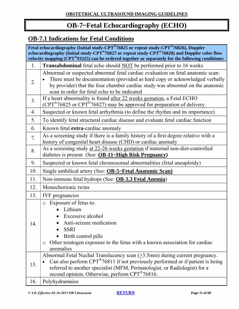
Fetal echocardiography (Initial study-CPT®76825 or repeat study-CPT®76826), Doppler echocardiography (Initial study-CPT®76827 or repeat study-CPT®76828) and Doppler color flow velocity mapping (CPT®93325) can be ordered together or separately for the following conditions:
1. Transabdominal fetal echo should NOT be performed prior to 16 weeks
2.
Abnormal or suspected abnormal fetal cardiac evaluation on fetal anatomic scan: There must be documentation (provided as hard copy or acknowledged verbally
by provider) that the four chamber cardiac study was abnormal on the anatomic scan in order for fetal echo to be indicated
3. If a heart abnormality is found after 22 weeks gestation, a Fetal ECHO (CPT®76825 or CPT®76827) may be approved for preparation of delivery.
4. Suspected or known fetal arrhythmia (to define the rhythm and its importance)
5. To identify fetal structural cardiac disease and evaluate fetal cardiac function
6. Known fetal extra-cardiac anomaly
7. As a screening study if there is a family history of a first degree relative with a history of congenital heart disease (CHD) or cardiac anomaly
8. As a screening study at 22-26 weeks gestation if maternal non-diet-controlled diabetes is present (See: OB-11~High Risk Pregnancy)
9. Suspected or known fetal chromosomal abnormalities (fetal aneuploidy)
10. Single umbilical artery (See: OB-5~Fetal Anatomic Scan)
o Exposure of fetus to: Lithium Excessive alcohol Anti-seizure medication SSRI Birth control pills
o Other teratogen exposure to the fetus with a known association for cardiac anomalies
15.
Abnormal Fetal Nuchal Translucency scan (>3.5mm) during current pregnancy. Can also perform CPT®76811 if not previously performed or if patient is being
referred to another specialist (MFM, Perinatologist, or Radiologist) for a second opinion. Otherwise, perform CPT®76816.
16. Polyhydramnios
V 4.0; Effective 02-16-2015 OB Ultrasound RETURN Page 16 of 68
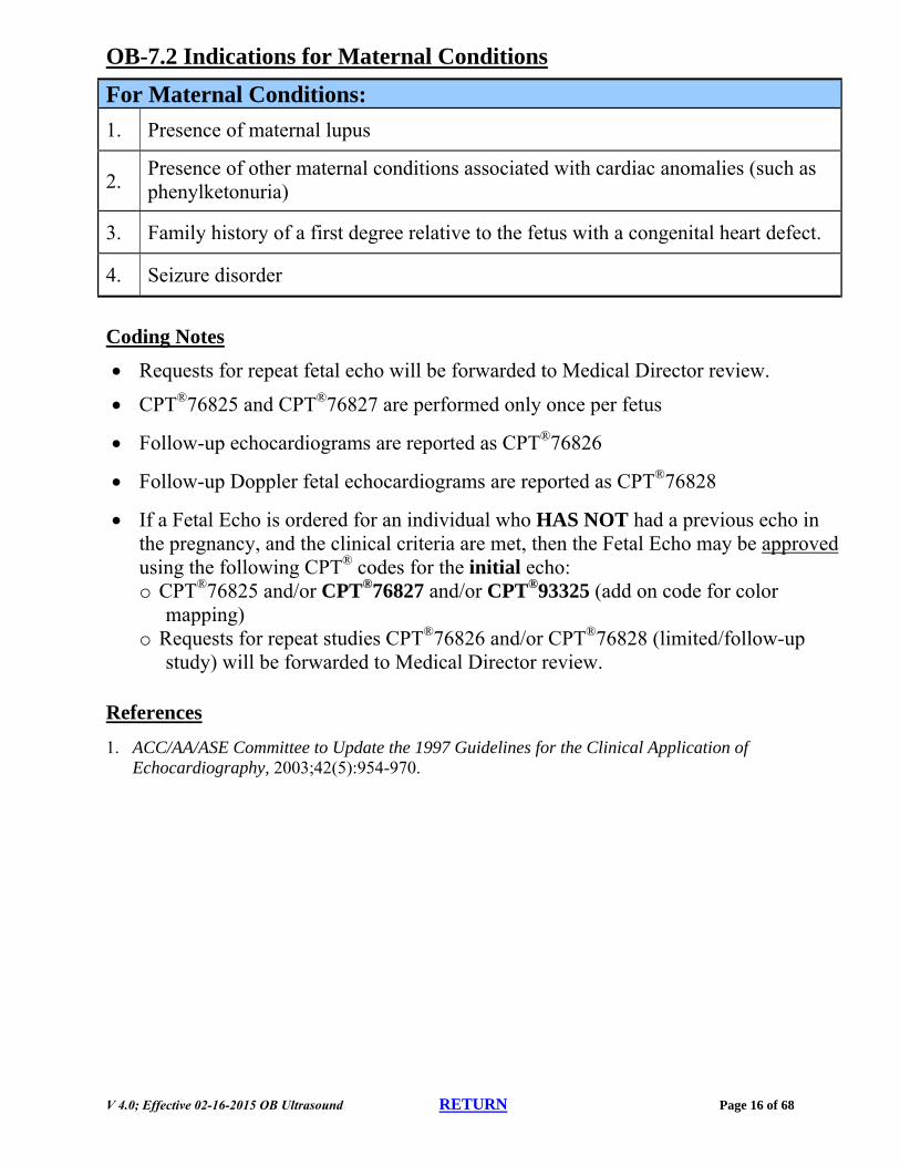
OB-7.2 Indications for Maternal Conditions
For Maternal Conditions:
1. Presence of maternal lupus
2. Presence of other maternal conditions associated with cardiac anomalies (such as phenylketonuria)
3. Family history of a first degree relative to the fetus with a congenital heart defect.
4. Seizure disorder
Coding Notes
Requests for repeat fetal echo will be forwarded to Medical Director review.
CPT®76825 and CPT®76827 are performed only once per fetus
Follow-up echocardiograms are reported as CPT®76826
Follow-up Doppler fetal echocardiograms are reported as CPT®76828
If a Fetal Echo is ordered for an individual who HAS NOT had a previous echo in the pregnancy, and the clinical criteria are met, then the Fetal Echo may be approved using the following CPT® codes for the initial echo: o CPT®76825 and/or CPT®76827 and/or CPT®93325 (add on code for color
mapping) o Requests for repeat studies CPT®76826 and/or CPT®76828 (limited/follow-up
study) will be forwarded to Medical Director review.
References
1. ACC/AA/ASE Committee to Update the 1997 Guidelines for the Clinical Application of Echocardiography, 2003;42(5):954-970.
V 4.0; Effective 02-16-2015 OB Ultrasound RETURN Page 17 of 68
OBSTETRICAL ULTRASOUND IMAGING GUIDELINES
OB-8~FETAL GROWTH PROBLEMS
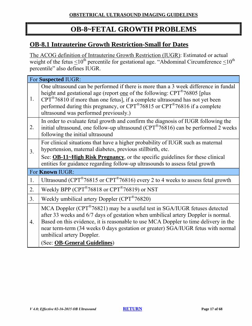
OB-8.1 Intrauterine Growth Restriction-Small for Dates
The ACOG definition of Intrauterine Growth Restriction (IUGR): Estimated or actual weight of the fetus <10th percentile for gestational age. “Abdominal Circumference <10th percentile” also defines IUGR.
For Suspected IUGR:
1.
One ultrasound can be performed if there is more than a 3 week difference in fundal height and gestational age (report one of the following: CPT®76805 [plus CPT®76810 if more than one fetus], if a complete ultrasound has not yet been performed during this pregnancy, or CPT®76815 or CPT®76816 if a complete ultrasound was performed previously.)
2. In order to evaluate fetal growth and confirm the diagnosis of IUGR following the initial ultrasound, one follow-up ultrasound (CPT®76816) can be performed 2 weeks following the initial ultrasound
3.
For clinical situations that have a higher probability of IUGR such as maternal hypertension, maternal diabetes, previous stillbirth, etc. See: OB-11~High Risk Pregnancy, or the specific guidelines for these clinical entities for guidance regarding follow-up ultrasounds to assess fetal growth
For Known IUGR:
1. Ultrasound (CPT®76815 or CPT®76816) every 2 to 4 weeks to assess fetal growth
2. Weekly BPP (CPT®76818 or CPT®76819) or NST
3. Weekly umbilical artery Doppler (CPT®76820)
4.
MCA Doppler (CPT®76821) may be a useful test in SGA/IUGR fetuses detected after 33 weeks and 6/7 days of gestation when umbilical artery Doppler is normal. Based on this evidence, it is reasonable to use MCA Doppler to time delivery in the near term-term (34 weeks 0 days gestation or greater) SGA/IUGR fetus with normal umbilical artery Doppler. (See: OB-General Guidelines)
V 4.0; Effective 02-16-2015 OB Ultrasound RETURN Page 18 of 68
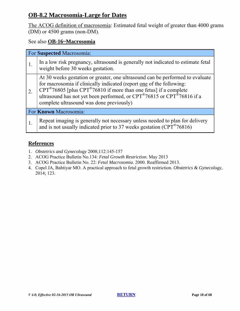
OB-8.2 Macrosomia-Large for Dates
The ACOG definition of macrosomia: Estimated fetal weight of greater than 4000 grams (DM) or 4500 grams (non-DM).
See also OB-16~Macrosomia
For Suspected Macrosomia:
1. In a low risk pregnancy, ultrasound is generally not indicated to estimate fetal weight before 30 weeks gestation.
2.
At 30 weeks gestation or greater, one ultrasound can be performed to evaluate for macrosomia if clinically indicated (report one of the following: CPT®76805 [plus CPT®76810 if more than one fetus] if a complete ultrasound has not yet been performed, or CPT®76815 or CPT®76816 if a complete ultrasound was done previously)
For Known Macrosomia:
1. Repeat imaging is generally not necessary unless needed to plan for delivery and is not usually indicated prior to 37 weeks gestation (CPT®76816)
References
1. Obstetrics and Gynecology 2008;112:145-157 2. ACOG Practice Bulletin No.134: Fetal Growth Restriction. May 2013 3. ACOG Practice Bulletin No. 22: Fetal Macrosomia. 2000. Reaffirmed 2013. 4. Copel JA, Bahtiyar MO. A practical approach to fetal growth restriction. Obstetrics & Gynecology,
2014; 123.
V 4.0; Effective 02-16-2015 OB Ultrasound RETURN Page 19 of 68
OBSTETRICAL ULTRASOUND IMAGING GUIDELINES
OB-9~FETAL HEART TONE
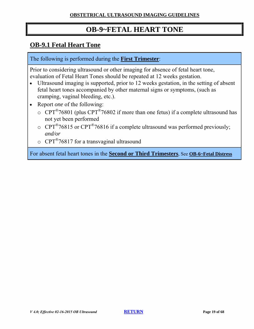
OB-9.1 Fetal Heart Tone
The following is performed during the First Trimester:
Prior to considering ultrasound or other imaging for absence of fetal heart tone, evaluation of Fetal Heart Tones should be repeated at 12 weeks gestation. Ultrasound imaging is supported, prior to 12 weeks gestation, in the setting of absent
fetal heart tones accompanied by other maternal signs or symptoms, (such as cramping, vaginal bleeding, etc.).
Report one of the following: o CPT®76801 (plus CPT®76802 if more than one fetus) if a complete ultrasound has
not yet been performed o CPT®76815 or CPT®76816 if a complete ultrasound was performed previously;
and/or o CPT®76817 for a transvaginal ultrasound
For absent fetal heart tones in the Second or Third Trimesters, See OB-6~Fetal Distress
V 4.0; Effective 02-16-2015 OB Ultrasound RETURN Page 20 of 68
OBSTETRICAL ULTRASOUND IMAGING GUIDELINES
OB-10~First Trimester Screening
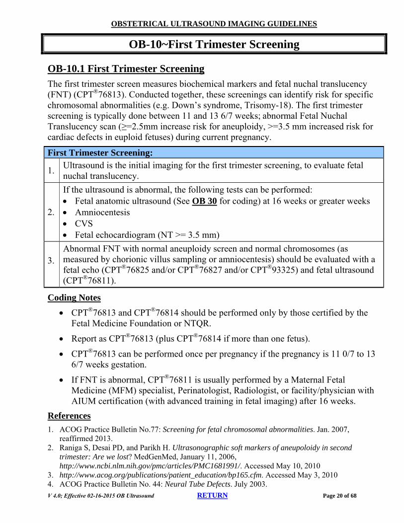
OB-10.1 First Trimester Screening
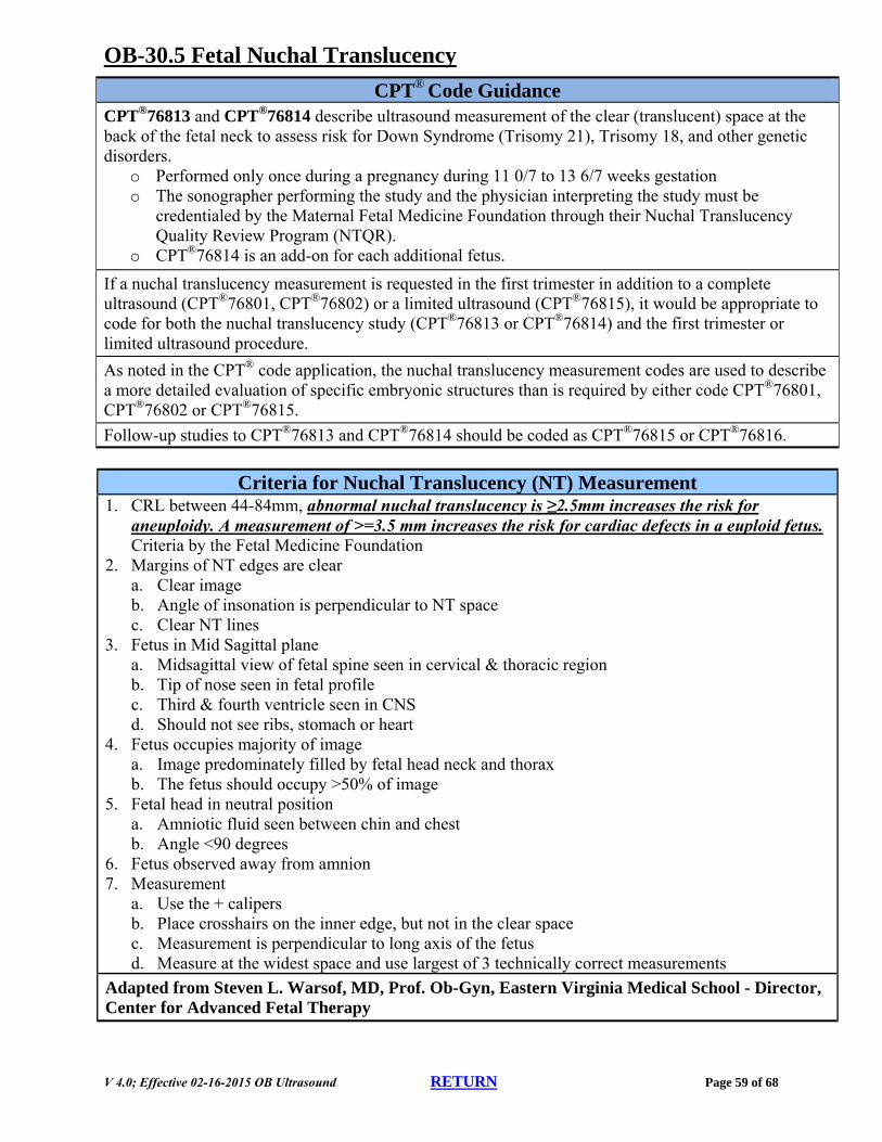
The first trimester screen measures biochemical markers and fetal nuchal translucency (FNT) (CPT®76813). Conducted together, these screenings can identify risk for specific chromosomal abnormalities (e.g. Down’s syndrome, Trisomy-18). The first trimester screening is typically done between 11 and 13 6/7 weeks; abnormal Fetal Nuchal Translucency scan (≥=2.5mm increase risk for aneuploidy, >=3.5 mm increased risk for cardiac defects in euploid fetuses) during current pregnancy.
First Trimester Screening:
1. Ultrasound is the initial imaging for the first trimester screening, to evaluate fetal nuchal translucency.
2.
If the ultrasound is abnormal, the following tests can be performed: Fetal anatomic ultrasound (See OB 30 for coding) at 16 weeks or greater weeks Amniocentesis CVS Fetal echocardiogram (NT >= 3.5 mm)
3. Abnormal FNT with normal aneuploidy screen and normal chromosomes (as measured by chorionic villus sampling or amniocentesis) should be evaluated with a fetal echo (CPT®76825 and/or CPT®76827 and/or CPT®93325) and fetal ultrasound (CPT®76811).
Coding Notes
CPT®76813 and CPT®76814 should be performed only by those certified by the Fetal Medicine Foundation or NTQR.
Report as CPT®76813 (plus CPT®76814 if more than one fetus).
CPT®76813 can be performed once per pregnancy if the pregnancy is 11 0/7 to 13 6/7 weeks gestation.
If FNT is abnormal, CPT®76811 is usually performed by a Maternal Fetal Medicine (MFM) specialist, Perinatologist, Radiologist, or facility/physician with AIUM certification (with advanced training in fetal imaging) after 16 weeks.
References
1. ACOG Practice Bulletin No.77: Screening for fetal chromosomal abnormalities. Jan. 2007, reaffirmed 2013.
2. Raniga S, Desai PD, and Parikh H. Ultrasonographic soft markers of aneupoloidy in second trimester: Are we lost? MedGenMed, January 11, 2006, http://www.ncbi.nlm.nih.gov/pmc/articles/PMC1681991/. Accessed May 10, 2010
3. http://www.acog.org/publications/patient_education/bp165.cfm. Accessed May 3, 2010 4. ACOG Practice Bulletin No. 44: Neural Tube Defects. July 2003.
V 4.0; Effective 02-16-2015 OB Ultrasound RETURN Page 21 of 68
OBSTETRICAL ULTRASOUND IMAGING GUIDELINES
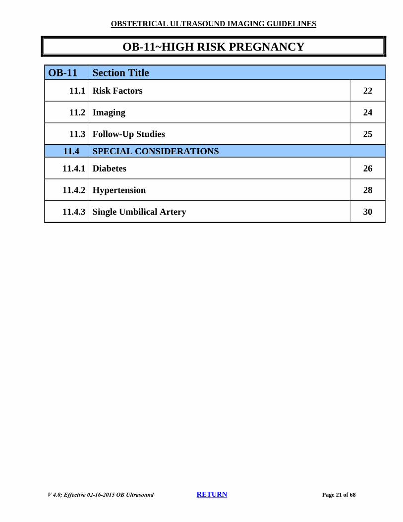
OB-11~HIGH RISK PREGNANCY
OB-11 Section Title
11.1 Risk Factors 22
11.2 Imaging 24
11.3 Follow-Up Studies 25
11.4 SPECIAL CONSIDERATIONS
11.4.1 Diabetes 26
11.4.2 Hypertension 28
11.4.3 Single Umbilical Artery 30
V 4.0; Effective 02-16-2015 OB Ultrasound RETURN Page 22 of 68
OBSTETRICAL ULTRASOUND IMAGING GUIDELINES
OB-11~HIGH RISK PREGNANCY
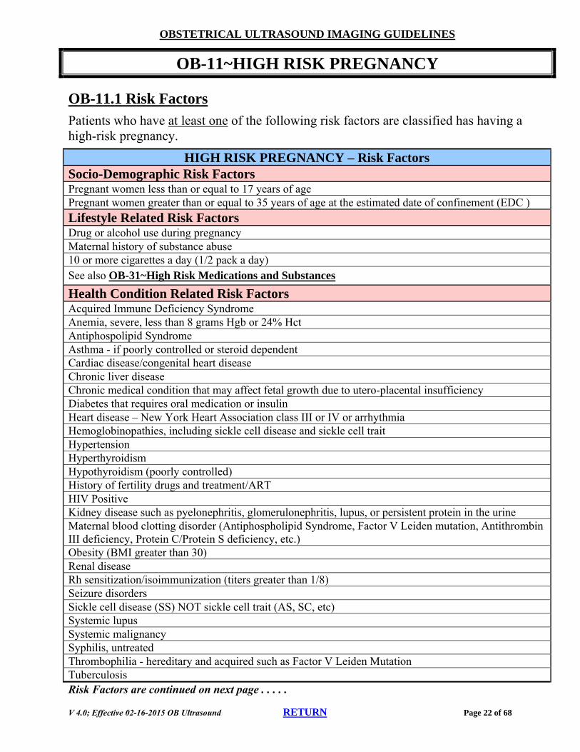
OB-11.1 Risk Factors
Patients who have at least one of the following risk factors are classified has having a high-risk pregnancy.
Risk Factors are continued on next page . . . . .
HIGH RISK PREGNANCY – Risk Factors Socio-Demographic Risk Factors Pregnant women less than or equal to 17 years of age Pregnant women greater than or equal to 35 years of age at the estimated date of confinement (EDC )
Lifestyle Related Risk Factors Drug or alcohol use during pregnancy Maternal history of substance abuse 10 or more cigarettes a day (1/2 pack a day) See also OB-31~High Risk Medications and Substances
Health Condition Related Risk Factors Acquired Immune Deficiency Syndrome Anemia, severe, less than 8 grams Hgb or 24% Hct Antiphospolipid Syndrome Asthma - if poorly controlled or steroid dependent Cardiac disease/congenital heart disease Chronic liver disease Chronic medical condition that may affect fetal growth due to utero-placental insufficiency Diabetes that requires oral medication or insulin Heart disease – New York Heart Association class III or IV or arrhythmia Hemoglobinopathies, including sickle cell disease and sickle cell trait Hypertension Hyperthyroidism Hypothyroidism (poorly controlled) History of fertility drugs and treatment/ART HIV Positive Kidney disease such as pyelonephritis, glomerulonephritis, lupus, or persistent protein in the urine Maternal blood clotting disorder (Antiphospholipid Syndrome, Factor V Leiden mutation, Antithrombin III deficiency, Protein C/Protein S deficiency, etc.) Obesity (BMI greater than 30) Renal disease Rh sensitization/isoimmunization (titers greater than 1/8) Seizure disorders Sickle cell disease (SS) NOT sickle cell trait (AS, SC, etc) Systemic lupus Systemic malignancy Syphilis, untreated Thrombophilia - hereditary and acquired such as Factor V Leiden Mutation Tuberculosis
V 4.0; Effective 02-16-2015 OB Ultrasound RETURN Page 23 of 68
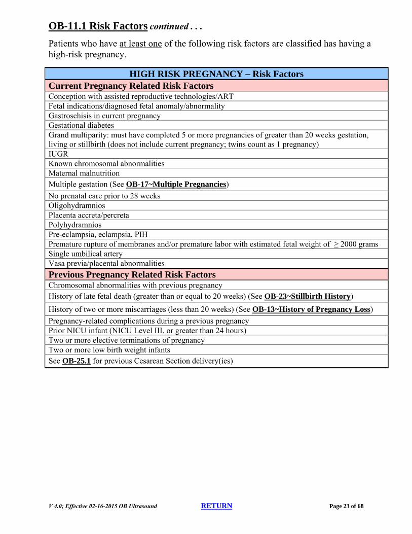
OB-11.1 Risk Factors continued . . .
Patients who have at least one of the following risk factors are classified has having a high-risk pregnancy.
HIGH RISK PREGNANCY – Risk Factors Current Pregnancy Related Risk Factors Conception with assisted reproductive technologies/ART Fetal indications/diagnosed fetal anomaly/abnormality Gastroschisis in current pregnancy Gestational diabetes Grand multiparity: must have completed 5 or more pregnancies of greater than 20 weeks gestation, living or stillbirth (does not include current pregnancy; twins count as 1 pregnancy) IUGR Known chromosomal abnormalities Maternal malnutrition Multiple gestation (See OB-17~Multiple Pregnancies)
No prenatal care prior to 28 weeks Oligohydramnios Placenta accreta/percreta Polyhydramnios Pre-eclampsia, eclampsia, PIH Premature rupture of membranes and/or premature labor with estimated fetal weight of ≥ 2000 grams Single umbilical artery Vasa previa/placental abnormalities
Previous Pregnancy Related Risk Factors Chromosomal abnormalities with previous pregnancy History of late fetal death (greater than or equal to 20 weeks) (See OB-23~Stillbirth History)
History of two or more miscarriages (less than 20 weeks) (See OB-13~History of Pregnancy Loss)
Pregnancy-related complications during a previous pregnancy Prior NICU infant (NICU Level III, or greater than 24 hours) Two or more elective terminations of pregnancy Two or more low birth weight infants See OB-25.1 for previous Cesarean Section delivery(ies)
V 4.0; Effective 02-16-2015 OB Ultrasound RETURN Page 24 of 68
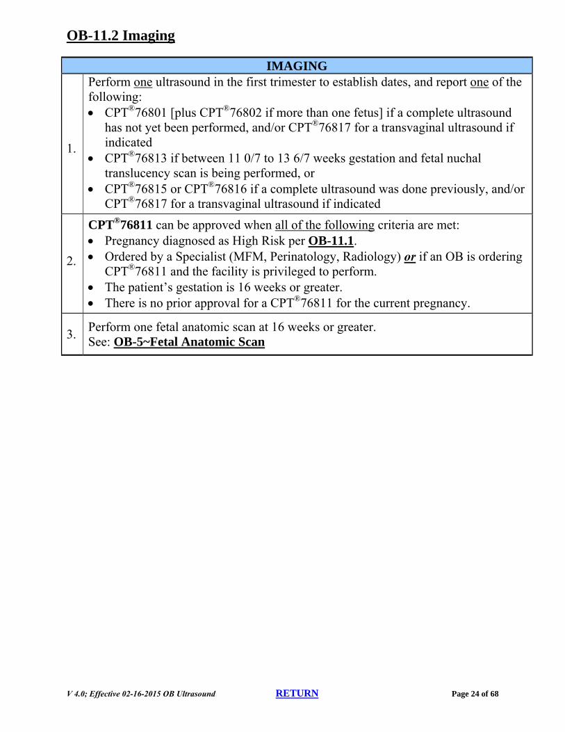
OB-11.2 Imaging
1.
Perform one ultrasound in the first trimester to establish dates, and report one of the following: CPT®76801 [plus CPT®76802 if more than one fetus] if a complete ultrasound
has not yet been performed, and/or CPT®76817 for a transvaginal ultrasound if indicated
CPT®76813 if between 11 0/7 to 13 6/7 weeks gestation and fetal nuchal translucency scan is being performed, or
CPT®76815 or CPT®76816 if a complete ultrasound was done previously, and/or CPT®76817 for a transvaginal ultrasound if indicated
2.
CPT®76811 can be approved when all of the following criteria are met: Pregnancy diagnosed as High Risk per OB-11.1. Ordered by a Specialist (MFM, Perinatology, Radiology) or if an OB is ordering
CPT®76811 and the facility is privileged to perform. The patient’s gestation is 16 weeks or greater. There is no prior approval for a CPT®76811 for the current pregnancy.
3. Perform one fetal anatomic scan at 16 weeks or greater. See: OB-5~Fetal Anatomic Scan
IMAGING
V 4.0; Effective 02-16-2015 OB Ultrasound RETURN Page 25 of 68
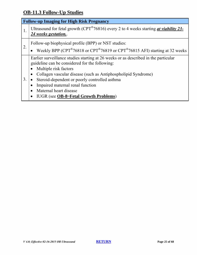
OB-11.3 Follow-Up Studies
Follow-up Imaging for High Risk Pregnancy
1. Ultrasound for fetal growth (CPT®76816) every 2 to 4 weeks starting at viability 23-24 weeks gestation..
2. Follow-up biophysical profile (BPP) or NST studies:
Weekly BPP (CPT®76818 or CPT®76819 or CPT®76815 AFI) starting at 32 weeks
3.
Earlier surveillance studies starting at 26 weeks or as described in the particular guideline can be considered for the following: Multiple risk factors Collagen vascular disease (such as Antiphospholipid Syndrome) Steroid-dependent or poorly controlled asthma Impaired maternal renal function Maternal heart disease IUGR (see OB-8~Fetal Growth Problems)
V 4.0; Effective 02-16-2015 OB Ultrasound RETURN Page 26 of 68
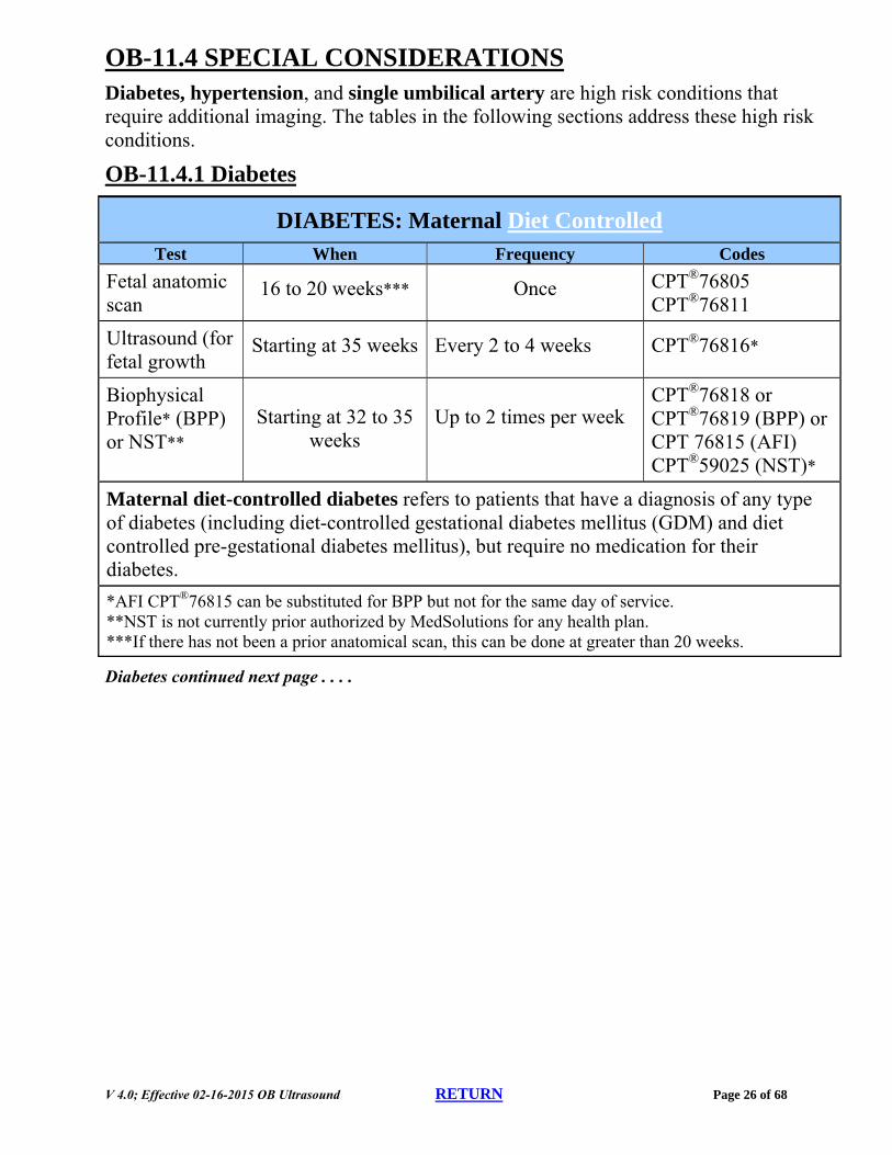
OB-11.4 SPECIAL CONSIDERATIONS Diabetes, hypertension, and single umbilical artery are high risk conditions that require additional imaging. The tables in the following sections address these high risk conditions.
OB-11.4.1 Diabetes
DIABETES: Maternal Diet Controlled Test When Frequency Codes
Fetal anatomic scan
16 to 20 weeks*** Once CPT®76805 CPT®76811
Ultrasound (for fetal growth
Starting at 35 weeks Every 2 to 4 weeks CPT®76816*
Biophysical Profile* (BPP) or NST**
Starting at 32 to 35 weeks
Up to 2 times per week CPT®76818 or CPT®76819 (BPP) or CPT 76815 (AFI) CPT®59025 (NST)*
Maternal diet-controlled diabetes refers to patients that have a diagnosis of any type of diabetes (including diet-controlled gestational diabetes mellitus (GDM) and diet controlled pre-gestational diabetes mellitus), but require no medication for their diabetes.
*AFI CPT®76815 can be substituted for BPP but not for the same day of service. **NST is not currently prior authorized by MedSolutions for any health plan. ***If there has not been a prior anatomical scan, this can be done at greater than 20 weeks.
Diabetes continued next page . . . .
V 4.0; Effective 02-16-2015 OB Ultrasound RETURN Page 27 of 68
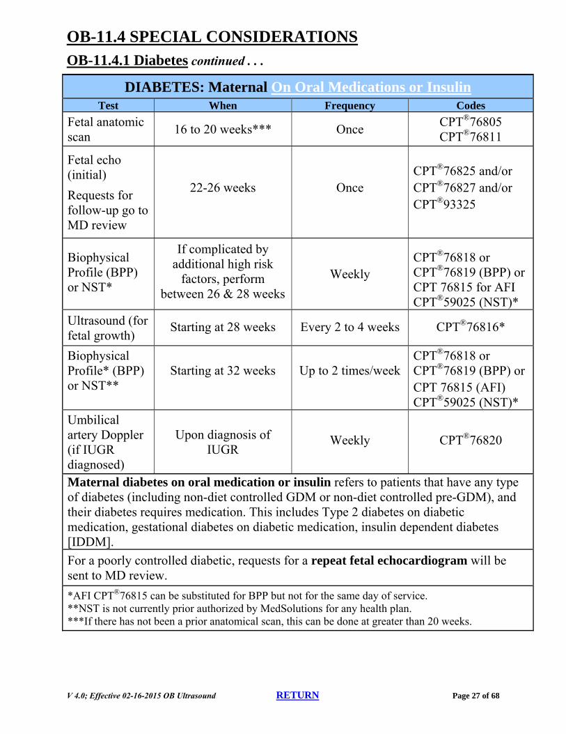
OB-11.4 SPECIAL CONSIDERATIONS OB-11.4.1 Diabetes continued . . .
DIABETES: Maternal On Oral Medications or Insulin Test When Frequency Codes
Fetal anatomic scan
16 to 20 weeks*** Once CPT®76805 CPT®76811
Fetal echo (initial)
Requests for follow-up go to MD review
22-26 weeks Once CPT®76825 and/or CPT®76827 and/or CPT®93325
Biophysical Profile (BPP) or NST*
If complicated by additional high risk
factors, perform between 26 & 28 weeks
Weekly CPT®76818 or CPT®76819 (BPP) or CPT 76815 for AFI CPT®59025 (NST)*
Ultrasound (for fetal growth)
Starting at 28 weeks Every 2 to 4 weeks CPT®76816*
Biophysical Profile* (BPP) or NST**
Starting at 32 weeks Up to 2 times/week CPT®76818 or CPT®76819 (BPP) or CPT 76815 (AFI) CPT®59025 (NST)*
Umbilical artery Doppler (if IUGR diagnosed)
Upon diagnosis of IUGR
Weekly CPT®76820
Maternal diabetes on oral medication or insulin refers to patients that have any type of diabetes (including non-diet controlled GDM or non-diet controlled pre-GDM), and their diabetes requires medication. This includes Type 2 diabetes on diabetic medication, gestational diabetes on diabetic medication, insulin dependent diabetes [IDDM].
For a poorly controlled diabetic, requests for a repeat fetal echocardiogram will be sent to MD review.
*AFI CPT®76815 can be substituted for BPP but not for the same day of service. **NST is not currently prior authorized by MedSolutions for any health plan. ***If there has not been a prior anatomical scan, this can be done at greater than 20 weeks.
V 4.0; Effective 02-16-2015 OB Ultrasound RETURN Page 28 of 68
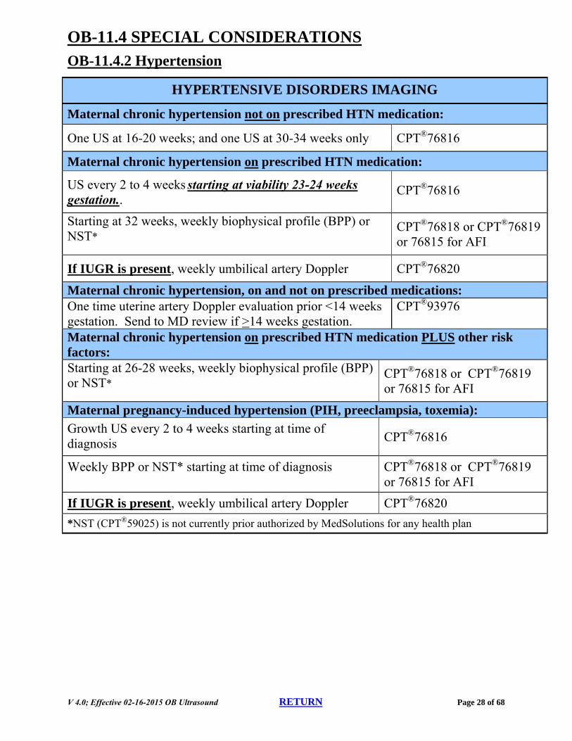
OB-11.4 SPECIAL CONSIDERATIONS OB-11.4.2 Hypertension
HYPERTENSIVE DISORDERS IMAGING
Maternal chronic hypertension not on prescribed HTN medication:
One US at 16-20 weeks; and one US at 30-34 weeks only CPT®76816
Maternal chronic hypertension on prescribed HTN medication:
US every 2 to 4 weeks starting at viability 23-24 weeks gestation..
CPT®76816
Starting at 32 weeks, weekly biophysical profile (BPP) or NST*
CPT®76818 or CPT®76819 or 76815 for AFI
If IUGR is present, weekly umbilical artery Doppler CPT®76820
Maternal chronic hypertension, on and not on prescribed medications: One time uterine artery Doppler evaluation prior <14 weeks gestation. Send to MD review if >14 weeks gestation.
CPT®93976
Maternal chronic hypertension on prescribed HTN medication PLUS other risk factors: Starting at 26-28 weeks, weekly biophysical profile (BPP) or NST*
Growth US every 2 to 4 weeks starting at time of diagnosis CPT®76816
Weekly BPP or NST* starting at time of diagnosis CPT®76818 or CPT®76819 or 76815 for AFI
If IUGR is present, weekly umbilical artery Doppler CPT®76820
*NST (CPT®59025) is not currently prior authorized by MedSolutions for any health plan
V 4.0; Effective 02-16-2015 OB Ultrasound RETURN Page 29 of 68
OB-11.4 SPECIAL CONSIDERATIONS
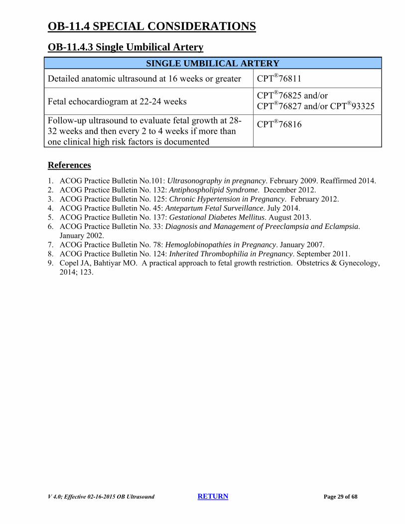
OB-11.4.3 Single Umbilical Artery
SINGLE UMBILICAL ARTERY
Detailed anatomic ultrasound at 16 weeks or greater CPT®76811
Fetal echocardiogram at 22-24 weeks CPT®76825 and/or CPT®76827 and/or CPT®93325
Follow-up ultrasound to evaluate fetal growth at 28-32 weeks and then every 2 to 4 weeks if more than one clinical high risk factors is documented
CPT®76816
References
1. ACOG Practice Bulletin No.101: Ultrasonography in pregnancy. February 2009. Reaffirmed 2014. 2. ACOG Practice Bulletin No. 132: Antiphospholipid Syndrome. December 2012. 3. ACOG Practice Bulletin No. 125: Chronic Hypertension in Pregnancy. February 2012. 4. ACOG Practice Bulletin No. 45: Antepartum Fetal Surveillance. July 2014. 5. ACOG Practice Bulletin No. 137: Gestational Diabetes Mellitus. August 2013. 6. ACOG Practice Bulletin No. 33: Diagnosis and Management of Preeclampsia and Eclampsia.
January 2002. 7. ACOG Practice Bulletin No. 78: Hemoglobinopathies in Pregnancy. January 2007. 8. ACOG Practice Bulletin No. 124: Inherited Thrombophilia in Pregnancy. September 2011. 9. Copel JA, Bahtiyar MO. A practical approach to fetal growth restriction. Obstetrics & Gynecology,
2014; 123.
V 4.0; Effective 02-16-2015 OB Ultrasound RETURN Page 30 of 68
OBSTETRICAL ULTRASOUND IMAGING GUIDELINES
OB-12~History of Infertility
OB-12.1 History of Infertility in Prior Pregnancy
Ultrasound imaging is supported if there is a history of infertility treatment (CPT®76801 [plus CPT®76802 if more than one fetus] and/or CPT®76817 for transvaginal ultrasound.
Repeat ultrasound is not usually necessary unless there are new clinical indications.
OB-12.2 Present Pregnancy with use of Fertility Drugs and Treatment (ART)
Follow high risk imaging, OB-11.2
V 4.0; Effective 02-16-2015 OB Ultrasound RETURN Page 31 of 68
OBSTETRICAL ULTRASOUND IMAGING GUIDELINES
OB-13~History of Pregnancy Loss
OB-13.1 History of Pregnancy Loss
History of Two or More Pregnancy Losses at Less than 19.6 Weeks: Ultrasound is supported: CPT®76801 [plus CPT®76802 if more than one fetus] and/or CPT®76817 for a
transvaginal ultrasound if a complete ultrasound has not yet been performed CPT®76815 or CPT®76816 if a complete ultrasound was done previously and/or
CPT®76817 for a transvaginal ultrasound
History of One or More Pregnancy Losses at >20 Weeks or Greater:
Ultrasound is supported: CPT®76805 [plus CPT®76810 if more than one fetus] if a complete ultrasound has not
yet been performed CPT®76815 or CPT®76816 if a complete ultrasound was done previously; and/or
CPT®76817 for a transvaginal ultrasound. See OB-23~Stillbirth History or Risk of Stillbirth
Reference
1. ACOG Practice Bulletin No. 102. Management of Stillbirth. March 2009. Reaffirmed 2012.
V 4.0; Effective 02-16-2015 OB Ultrasound RETURN Page 32 of 68
Ultrasound is supported at 16 weeks or greater for known or suspected incompetent cervix in order to evaluate cervical length: CPT®76805 [plus CPT®76810 if more than one fetus] and/or CPT®76817 if a complete ultrasound has not yet been performed during this pregnancy.
CPT®76816 if a complete ultrasound was done previously, and/or CPT®76817 for a transvaginal ultrasound). Ultrasound can be used earlier if there are known risk factors.
2.
Ultrasound (CPT®76816 and/or CPT®76817) can be performed every 2 to 4 weeks, starting at 16 weeks or greater for the following: Personal history of incompetent cervix Personal history of preterm delivery Personal history of preterm premature rupture of membranes Cerclage in place in current or past pregnancy Presence of uterine anomaly Current pregnancy is twins, triplets, or other multiple gestation Shorten cervix (25mm at less than 35 weeks gestation)
Preterm labor with current pregnancy
3.
Ultrasound (CPT®76817) can be performed every 2 to 4 weeks, starting at 16 weeks or greater for the following: Precipitous delivery Surgical trauma to cervix (e.g. conization [CKC—cold-knife conization] or Loop
Electrosurgical Excision Procedure [LEEP]) Overdilation of cervix during a termination of pregnancy Cervical obstetrical laceration from a previous delivery
4. If funneling or abnormally shorten cervix less than 25 mm <32 weeks gestation is found, ultrasound (CPT®76816 and/or CPT®76817 [for transvaginal ultrasound) can be performed weekly
OB-14.2 Personal History of Preterm Delivery
For individuals with a history of preterm delivery (<37 weeks gestation), see above, OB-14.1 Incompetent Cervix.
References
1. ACOG Practice Bulletin No. 142: Cerclage for the management of cervical insufficiency. February 2014.
V 4.0; Effective 02-16-2015 OB Ultrasound RETURN Page 33 of 68
2. Orzechowski KM, Boelig RC, Baxter JK, Berghella V. A universal transvaginal cervical length screening program for preterm birth prevention. Obstetrics and Gynecology, 2014; 124:520-525.
3. ACOG Practice Bulletin No.48: Cervical Insufficiency. Reaffirmed 2008 4. ACOG Practice Bulletin No. 127: Management of Preterm Labor. June 2012. 5. ACOG Practice Bulletin No. 130: Prediction and Prevention of Preterm Birth. October 2012.
Reaffirmed 2014.
V 4.0; Effective 02-16-2015 OB Ultrasound RETURN Page 34 of 68
OBSTETRICAL ULTRASOUND IMAGING GUIDELINES
OB-15~Intrauterine Device (IUD)
OB-15.1 Locate an Intrauterine Device – Any Trimester
Ultrasound can be performed to locate an intrauterine device (IUD) (CPT®76801 and/or 76817 if a complete ultrasound has not yet been performed)
o CPT®76815 or CPT®76816 if a complete ultrasound was done previously, and/or CPT®76817 for a transvaginal ultrasound.
OB-16~MACROSOMIA
See: OB-8~Fetal Growth Problems
V 4.0; Effective 02-16-2015 OB Ultrasound RETURN Page 35 of 68
OBSTETRICAL ULTRASOUND IMAGING GUIDELINES
OB-17~Multiple Pregnancies
OB-17.1 Multiple Pregnancies
For Suspected multiple pregnancies:
Ultrasound is appropriate to confirm suspected multiple pregnancy CPT®76801 or CPT®76805 if a complete ultrasound has not yet been performed during this pregnancy, CPT®76815 or CPT®76816 if a complete ultrasound was done previously, and/or CPT®76817 for a transvaginal ultrasound
For Known dichorionic multiple pregnancies: Follow-up ultrasounds for all known dichorionic multiple pregnancies (CPT®76815 or CPT®76816 and/or CPT®76817): Ultrasound (CPT®76815 or CPT®76816) every 4 weeks to assess fetal growth starting
at 28 weeks gestation Transvaginal ultrasound (CPT®76817) every 2-4 weeks to assess cervical length Weekly BPP (CPT®76818 or CPT®76819) or NST starting at 32 weeks
o Twice weekly BPP can be considered in rare clinical circumstances. These requests will be forwarded for Medical Director review.
If IUGR is diagnosed, weekly umbilical artery Doppler and/or Middle Cerebral Artery Doppler (CPT®76820 and/or CPT®76821)
If IVF dichorionic twins, report initial fetal echo as CPT®76825 and/or CPT®76827 and/or CPT®93325. Follow-up echo requests will be sent to Medical Director review.
If other high risk factors, see OB:11~High Risk Pregnancy.
Continued . . .
V 4.0; Effective 02-16-2015 OB Ultrasound RETURN Page 36 of 68
OB-17.1 Multiple Pregnancies Continued . . .
For Known diamniotic-monochorionic or monoamniotic-monochorionic multiple pregnancies:
1.
In addition to the imaging listed above for dichorionic pregnancies, diamniotic-monochorionic or monoamniotic-monochorionic twins also receive: Ultrasound (CPT®76815 or CPT®76816) every 2 to 4 weeks to assess fetal growth
starting at 16 weeks gestation Fetal middle cerebral artery (MCA) Doppler (CPT®76821) every 2 to 3 weeks
starting at 26 weeks may be considered to monitor for TTTS or TAPS. If needed earlier than 26 weeks, send to MD review.
If Twin to Twin Transfusion syndrome is suspected due to one twin failing to grow compared with the other twin, daily evaluation (CPT®76815 or CPT®76816, and/or CPT®76818 or CPT®76819) and/or umbilical artery Doppler (CPT®76820) can be performed to aid in planning imminent delivery.
If discordant twins (15% to 25% difference in actual weight between twins), twice weekly BPP plus ultrasound (CPT®76816) every 2 to 4 weeks, AND umbilical artery Doppler (CPT®78620) weekly
Daily fetal testing may be indicated if umbilical Doppler is abnormal. These requests will be forwarded for Medical Director review.
Fetal echo CPT®76825 and/or CPT®76827 and/or CPT®93325 for initial echo. For follow-up echo send to MD review.
2. Triplets or higher Multiple Pregnancy receive same imaging as diamniotic-monochorionic and monoamniotic-monochorionic twins. These requests will be forwarded for Medical Director review.
Reference
1. ACOG Practice Bulletin No. 56: Multiple Gestation: Complicated Twin, Triplet, and High Order Multi-Fetal Pregnancy. October 2004.
2. ACOG practice bulleten no.: 144. May 2014. Multi fetal gestations: twin triplet and higher order multi fetal pregnancies.
V 4.0; Effective 02-16-2015 OB Ultrasound RETURN Page 37 of 68
OBSTETRICAL ULTRASOUND IMAGING GUIDELINES
OB-18~Pelvic Mass or Neoplasm
See: OB-27~Uterine Anomalies or Abnormalities
OB-19~Polyhydramnios/Oligohydramnios
See: OB-4~Amniotic Fluid Abnormalities
OB-20~Post-Date Pregnancy
OB-20.1 Post Date Pregnancy
1. Follow-up ultrasound (CPT®76816) every 2 weeks (> 41weeks gestation) to evaluate fetal growth
2. CPT®76815 may be approved if ordered at 41 weeks or greater to evaluate
amniotic fluid index (AFI). A BPP (CPT®76818 or CPT®76819) should not be approved in addition to
CPT®76815 if the request is ONLY to evaluate AFI.
Practice Note
In post date pregnancy, uterine artery Doppler velocimetry (CPT®93976) has not been found to be useful.
References
1. ACOG Practice Bulletin 146. Management of Late-Term and Postterm Pregnancies. August, 2014.
V 4.0; Effective 02-16-2015 OB Ultrasound RETURN Page 38 of 68
OBSTETRICAL ULTRASOUND IMAGING GUIDELINES
OB-21~Preterm/Premature Rupture of Membranes
See also: OB-4~Amniotic Fluid Abnormalities
See Also: OB-14.2 Personal History of Preterm Delivery
OB-21.1 Preterm Premature Rupture of Membranes (PPROM)
Less than or equal to 36 6/7 weeks. Requests will be forwarded to Medical Director review. o This is likely a hospital admission for evaluation and monitoring until delivery.
OB-21.2 Premature Rupture of Membranes (PROM)
Greater than or equal to 37 weeks. Requests will be forwarded to Medical Director review.
Reference
1. ACOG Practice Bulletin 139. Premature Rupture of Membranes. October , 2013
V 4.0; Effective 02-16-2015 OB Ultrasound RETURN Page 39 of 68
OBSTETRICAL ULTRASOUND IMAGING GUIDELINES
OB-22~Second Trimester Screening
OB-22.1 Second Trimester Screening
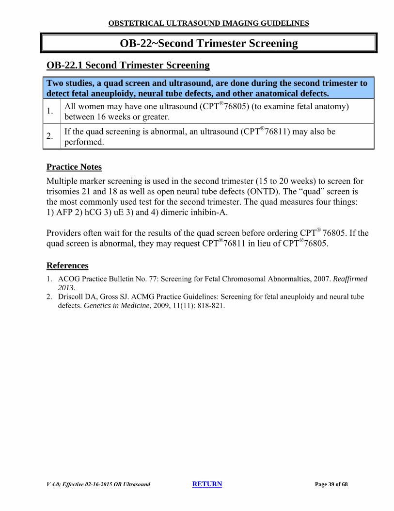
Two studies, a quad screen and ultrasound, are done during the second trimester to detect fetal aneuploidy, neural tube defects, and other anatomical defects.
1. All women may have one ultrasound (CPT®76805) (to examine fetal anatomy) between 16 weeks or greater.
2. If the quad screening is abnormal, an ultrasound (CPT®76811) may also be performed.
Practice Notes
Multiple marker screening is used in the second trimester (15 to 20 weeks) to screen for trisomies 21 and 18 as well as open neural tube defects (ONTD). The “quad” screen is the most commonly used test for the second trimester. The quad measures four things: 1) AFP 2) hCG 3) uE 3) and 4) dimeric inhibin-A. Providers often wait for the results of the quad screen before ordering CPT® 76805. If the quad screen is abnormal, they may request CPT®76811 in lieu of CPT®76805.
References
1. ACOG Practice Bulletin No. 77: Screening for Fetal Chromosomal Abnormalties, 2007. Reaffirmed 2013.
2. Driscoll DA, Gross SJ. ACMG Practice Guidelines: Screening for fetal aneuploidy and neural tube defects. Genetics in Medicine, 2009, 11(11): 818-821.
V 4.0; Effective 02-16-2015 OB Ultrasound RETURN Page 40 of 68
OBSTETRICAL ULTRASOUND IMAGING GUIDELINES
OB-23~Stillbirth History or Risk of Stillbirth
OB-23.1 Imaging
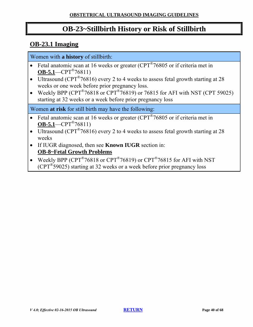
Women with a history of stillbirth:
Fetal anatomic scan at 16 weeks or greater (CPT®76805 or if criteria met in OB-5.1—CPT®76811)
Ultrasound (CPT®76816) every 2 to 4 weeks to assess fetal growth starting at 28 weeks or one week before prior pregnancy loss.
Weekly BPP (CPT®76818 or CPT®76819) or 76815 for AFI with NST (CPT 59025) starting at 32 weeks or a week before prior pregnancy loss
Women at risk for still birth may have the following:
Fetal anatomic scan at 16 weeks or greater (CPT®76805 or if criteria met in OB-5.1—CPT®76811)
Ultrasound (CPT®76816) every 2 to 4 weeks to assess fetal growth starting at 28 weeks
If IUGR diagnosed, then see Known IUGR section in: OB-8~Fetal Growth Problems
Weekly BPP (CPT®76818 or CPT®76819) or CPT®76815 for AFI with NST (CPT®59025) starting at 32 weeks or a week before prior pregnancy loss
V 4.0; Effective 02-16-2015 OB Ultrasound RETURN Page 41 of 68
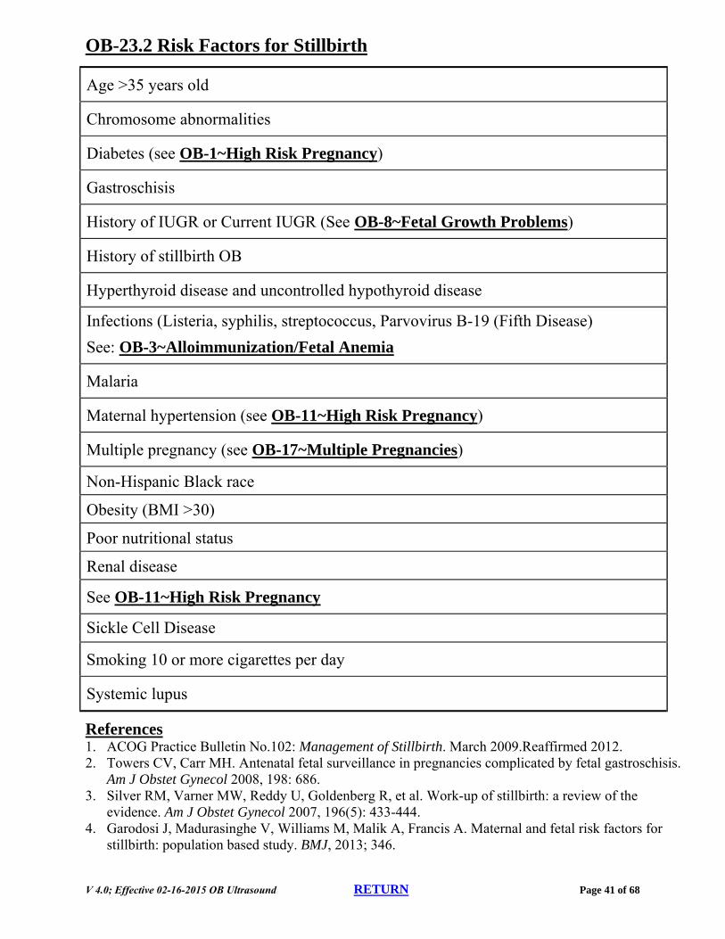
OB-23.2 Risk Factors for Stillbirth
Age >35 years old
Chromosome abnormalities
Diabetes (see OB-1~High Risk Pregnancy)
Gastroschisis
History of IUGR or Current IUGR (See OB-8~Fetal Growth Problems)
History of stillbirth OB
Hyperthyroid disease and uncontrolled hypothyroid disease
Maternal hypertension (see OB-11~High Risk Pregnancy)
Multiple pregnancy (see OB-17~Multiple Pregnancies)
Non-Hispanic Black race
Obesity (BMI >30)
Poor nutritional status
Renal disease
See OB-11~High Risk Pregnancy
Sickle Cell Disease
Smoking 10 or more cigarettes per day
Systemic lupus
References 1. ACOG Practice Bulletin No.102: Management of Stillbirth. March 2009.Reaffirmed 2012. 2. Towers CV, Carr MH. Antenatal fetal surveillance in pregnancies complicated by fetal gastroschisis.
Am J Obstet Gynecol 2008, 198: 686. 3. Silver RM, Varner MW, Reddy U, Goldenberg R, et al. Work-up of stillbirth: a review of the
evidence. Am J Obstet Gynecol 2007, 196(5): 433-444. 4. Garodosi J, Madurasinghe V, Williams M, Malik A, Francis A. Maternal and fetal risk factors for
stillbirth: population based study. BMJ, 2013; 346.
V 4.0; Effective 02-16-2015 OB Ultrasound RETURN Page 42 of 68
OBSTETRICAL ULTRASOUND IMAGING GUIDELINES
OB-24~Third Trimester Imaging
OB-24.1 Third Trimester Imaging - Ultrasound
1.
Imaging in the third trimester is indicated for bleeding, pain, absent fetal heart tone, decreased fetal movement and/or other high-risk indications.
(see OB-11~High Risk Pregnancy)
2. For suspected breach position, see: OB-2~Abnormal Fetal Position or Presentation
References
1. ACOG Practice Bulletin no. 101: Ultrasonography in pregnancy. February 2009. Reaffirmed 2014.
V 4.0; Effective 02-16-2015 OB Ultrasound RETURN Page 43 of 68
OBSTETRICAL ULTRASOUND IMAGING GUIDELINES
OB-25~Uncertain Dates
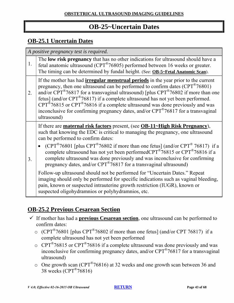
OB-25.1 Uncertain Dates
A positive pregnancy test is required.
1. The low risk pregnancy that has no other indications for ultrasound should have a fetal anatomic ultrasound (CPT®76805) performed between 16 weeks or greater. The timing can be determined by fundal height. (See: OB-5~Fetal Anatomic Scan).
2.
If the mother has had irregular menstrual periods in the year prior to the current pregnancy, then one ultrasound can be performed to confirm dates (CPT®76801) and/or CPT®76817 for a transvaginal ultrasound) [plus CPT®76802 if more than one fetus] (and/or CPT®76817) if a complete ultrasound has not yet been performed. CPT®76815 or CPT®76816 if a complete ultrasound was done previously and was inconclusive for confirming pregnancy dates, and/or CPT®76817 for a transvaginal ultrasound)
3.
If there are maternal risk factors present, (see OB-11~High Risk Pregnancy), such that knowing the EDC is critical to managing the pregnancy, one ultrasound can be performed to confirm dates:
(CPT®76801 [plus CPT®76802 if more than one fetus] (and/or CPT® 76817) if a complete ultrasound has not yet been performedCPT®76815 or CPT®76816 if a complete ultrasound was done previously and was inconclusive for confirming pregnancy dates, and/or CPT®76817 for a transvaginal ultrasound)
Follow-up ultrasound should not be performed for “Uncertain Dates.” Repeat imaging should only be performed for specific indications such as vaginal bleeding, pain, known or suspected intrauterine growth restriction (IUGR), known or suspected oligohydramnios or polyhydramnios, etc.
OB-25.2 Previous Cesarean Section
If mother has had a previous Cesarean section, one ultrasound can be performed to confirm dates:
o (CPT®76801 [plus CPT®76802 if more than one fetus] (and/or CPT 76817) if a complete ultrasound has not yet been performed
o CPT®76815 or CPT®76816 if a complete ultrasound was done previously and was inconclusive for confirming pregnancy dates, and/or CPT®76817 for a transvaginal ultrasound)
o One growth scan (CPT®76816) at 32 weeks and one growth scan between 36 and 38 weeks (CPT®76816)
V 4.0; Effective 02-16-2015 OB Ultrasound RETURN Page 44 of 68
References
1. ACOG Practice Bulletin No.101: Ultrasonography in pregnancy. February 2009. Reaffirmed 2014. 2. ACOG Committee Opinion, Number 297, Nonmedical Use of Obstetric Ultrasound. August 2004 3. ACR Practice Guideline for the Performance of Obstetrical Ultrasound, Effective 10/1/07 4. AIUM Practice Guideline for the Performance of Obstetric Ultrasound Examinations, 2007
V 4.0; Effective 02-16-2015 OB Ultrasound RETURN Page 45 of 68
OBSTETRICAL ULTRASOUND IMAGING GUIDELINES
OB-26~Unequal Fundal Size and Dates
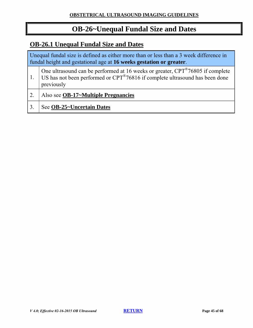
OB-26.1 Unequal Fundal Size and Dates
Unequal fundal size is defined as either more than or less than a 3 week difference in fundal height and gestational age at 16 weeks gestation or greater.
1. One ultrasound can be performed at 16 weeks or greater, CPT®76805 if complete US has not been performed or CPT®76816 if complete ultrasound has been done previously
2. Also see OB-17~Multiple Pregnancies
3. See OB-25~Uncertain Dates
V 4.0; Effective 02-16-2015 OB Ultrasound RETURN Page 46 of 68
OBSTETRICAL ULTRASOUND IMAGING GUIDELINES
OB-27~Uterine Anomalies or Adnexal/Pelvic Masses
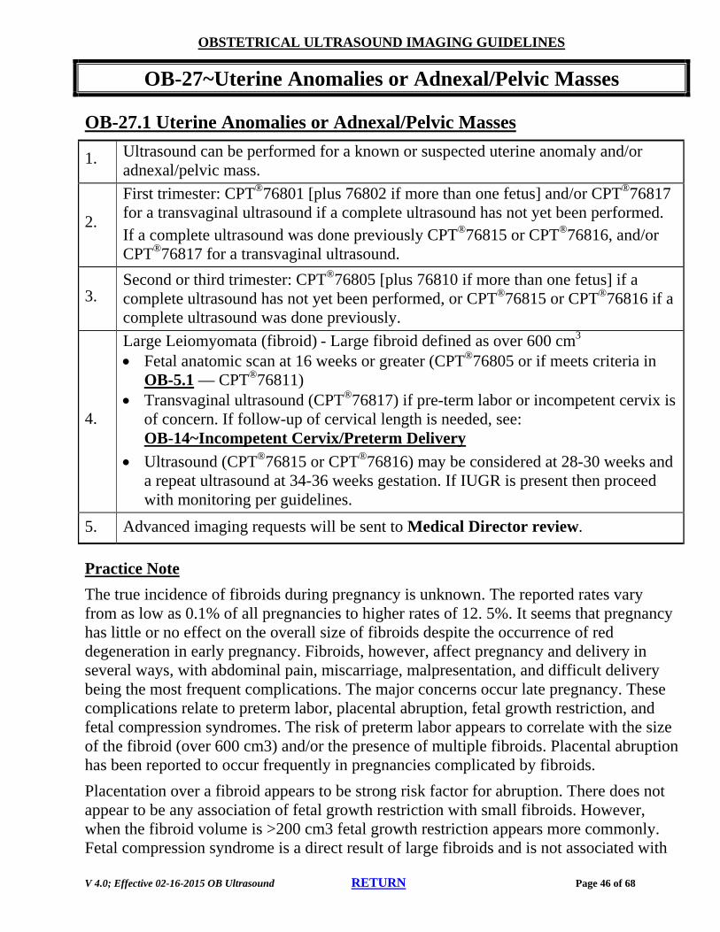
OB-27.1 Uterine Anomalies or Adnexal/Pelvic Masses
1. Ultrasound can be performed for a known or suspected uterine anomaly and/or adnexal/pelvic mass.
2.
First trimester: CPT®76801 [plus 76802 if more than one fetus] and/or CPT®76817 for a transvaginal ultrasound if a complete ultrasound has not yet been performed. If a complete ultrasound was done previously CPT®76815 or CPT®76816, and/or CPT®76817 for a transvaginal ultrasound.
3. Second or third trimester: CPT®76805 [plus 76810 if more than one fetus] if a complete ultrasound has not yet been performed, or CPT®76815 or CPT®76816 if a complete ultrasound was done previously.
4.
Large Leiomyomata (fibroid) - Large fibroid defined as over 600 cm3 Fetal anatomic scan at 16 weeks or greater (CPT®76805 or if meets criteria in
OB-5.1 — CPT®76811) Transvaginal ultrasound (CPT®76817) if pre-term labor or incompetent cervix is
of concern. If follow-up of cervical length is needed, see: OB-14~Incompetent Cervix/Preterm Delivery
Ultrasound (CPT®76815 or CPT®76816) may be considered at 28-30 weeks and a repeat ultrasound at 34-36 weeks gestation. If IUGR is present then proceed with monitoring per guidelines.
5. Advanced imaging requests will be sent to Medical Director review.
Practice Note
The true incidence of fibroids during pregnancy is unknown. The reported rates vary from as low as 0.1% of all pregnancies to higher rates of 12. 5%. It seems that pregnancy has little or no effect on the overall size of fibroids despite the occurrence of red degeneration in early pregnancy. Fibroids, however, affect pregnancy and delivery in several ways, with abdominal pain, miscarriage, malpresentation, and difficult delivery being the most frequent complications. The major concerns occur late pregnancy. These complications relate to preterm labor, placental abruption, fetal growth restriction, and fetal compression syndromes. The risk of preterm labor appears to correlate with the size of the fibroid (over 600 cm3) and/or the presence of multiple fibroids. Placental abruption has been reported to occur frequently in pregnancies complicated by fibroids.
Placentation over a fibroid appears to be strong risk factor for abruption. There does not appear to be any association of fetal growth restriction with small fibroids. However, when the fibroid volume is >200 cm3 fetal growth restriction appears more commonly. Fetal compression syndrome is a direct result of large fibroids and is not associated with
V 4.0; Effective 02-16-2015 OB Ultrasound RETURN Page 47 of 68
commonly found small fibroids. Finally, malposition or obstructed labor is associated with fibroids of the lower uterine segment.
2. Qidwai GI, Caughey AB, Jacoby AF. Obstetric outcomes in women with sonographically identified uterine leiomyomata. Obstet Gynecol 2006; 107:376.
3. Exacoustòs C, Rosati P. Ultrasound diagnosis of uterine myomas and complications in pregnancy. Obstet Gynecol 1993; 82:97.
4. Strobelt N, Ghidini A, Cavallone M, et al. Natural history of uterine leiomyomas in pregnancy. J Ultrasound Med 1994; 13:399.
5. Laughlin SK, Baird DD, Savitz DA, et al. Prevalence of uterine leiomyomas in the first trimester of pregnancy: an ultrasound-screening study. Obstet Gynecol 2009; 113:630.
6. Stout MJ, Odibo AO, Graseck AS, et al. Leiomyomas at routine second-trimester ultrasound examination and adverse obstetric outcomes. Obstet Gynecol 2010; 116:1056.
7. Terry KL, De Vivo I, Hankinson SE, Missmer SA. Reproductive characteristics and risk of uterine leiomyomata. Fertil Steril 2010; 94:2703.
8. Lev-Toaff AS, Coleman BG, Arger PH, et al. Leiomyomas in pregnancy: sonographic study. Radiology 1987; 164:375.
9. Rosati P, Exacoustòs C, Mancuso S. Longitudinal evaluation of uterine myoma growth during pregnancy. A sonographic study. J Ultrasound Med 1992; 11:511.
10. Klatsky PC, Tran ND, Caughey AB, Fujimoto VY. Fibroids and reproductive outcomes: a systematic literature review from conception to delivery. Am J Obstet Gynecol 2008; 198:357.
V 4.0; Effective 02-16-2015 OB Ultrasound RETURN Page 48 of 68
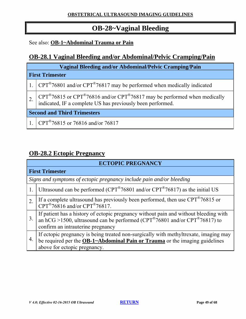
1. CPT®76801 and/or CPT®76817 may be performed when medically indicated
2. CPT®76815 or CPT®76816 and/or CPT®76817 may be performed when medically indicated, IF a complete US has previously been performed.
Second and Third Trimesters
1. CPT®76815 or 76816 and/or 76817
OB-28.2 Ectopic Pregnancy
ECTOPIC PREGNANCY
First Trimester
Signs and symptoms of ectopic pregnancy include pain and/or bleeding
1. Ultrasound can be performed (CPT®76801 and/or CPT®76817) as the initial US
2. If a complete ultrasound has previously been performed, then use CPT®76815 or CPT®76816 and/or CPT®76817.
3. If patient has a history of ectopic pregnancy without pain and without bleeding with an hCG >1500, ultrasound can be performed (CPT®76801 and/or CPT®76817) to confirm an intrauterine pregnancy
4. If ectopic pregnancy is being treated non-surgically with methyltrexate, imaging may be required per the OB-1~Abdominal Pain or Trauma or the imaging guidelines above for ectopic pregnancy.
V 4.0; Effective 02-16-2015 OB Ultrasound RETURN Page 50 of 68
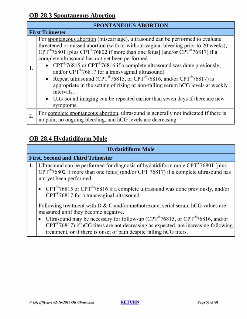
OB-28.3 Spontaneous Abortion
SPONTANEOUS ABORTION First Trimester
1.
For spontaneous abortion (miscarriage), ultrasound can be performed to evaluate threatened or missed abortion (with or without vaginal bleeding prior to 20 weeks), CPT®76801 [plus CPT®76802 if more than one fetus] (and/or CPT®76817) if a complete ultrasound has not yet been performed.
CPT®76815 or CPT®76816 if a complete ultrasound was done previously, and/or CPT®76817 for a transvaginal ultrasound)
Repeat ultrasound (CPT®76815, or CPT®76816, and/or CPT®76817) is appropriate in the setting of rising or non-falling serum hCG levels at weekly intervals.
Ultrasound imaging can be repeated earlier than seven days if there are new symptoms.
2. For complete spontaneous abortion, ultrasound is generally not indicated if there is no pain, no ongoing bleeding, and hCG levels are decreasing
OB-28.4 Hydatidiform Mole
Hydatidiform Mole
First, Second and Third Trimester 1. Ultrasound can be performed for diagnosis of hydatidiform mole CPT®76801 [plus
CPT®76802 if more than one fetus] (and/or CPT 76817) if a complete ultrasound has not yet been performed.
CPT®76815 or CPT®76816 if a complete ultrasound was done previously, and/or CPT®76817 for a transvaginal ultrasound.
Following treatment with D & C and/or methotrexate, serial serum hCG values are measured until they become negative. Ultrasound may be necessary for follow-up (CPT®76815, or CPT®76816, and/or
CPT®76817) if hCG titers are not decreasing as expected, are increasing following treatment, or if there is onset of pain despite falling hCG titers.
V 4.0; Effective 02-16-2015 OB Ultrasound RETURN Page 51 of 68
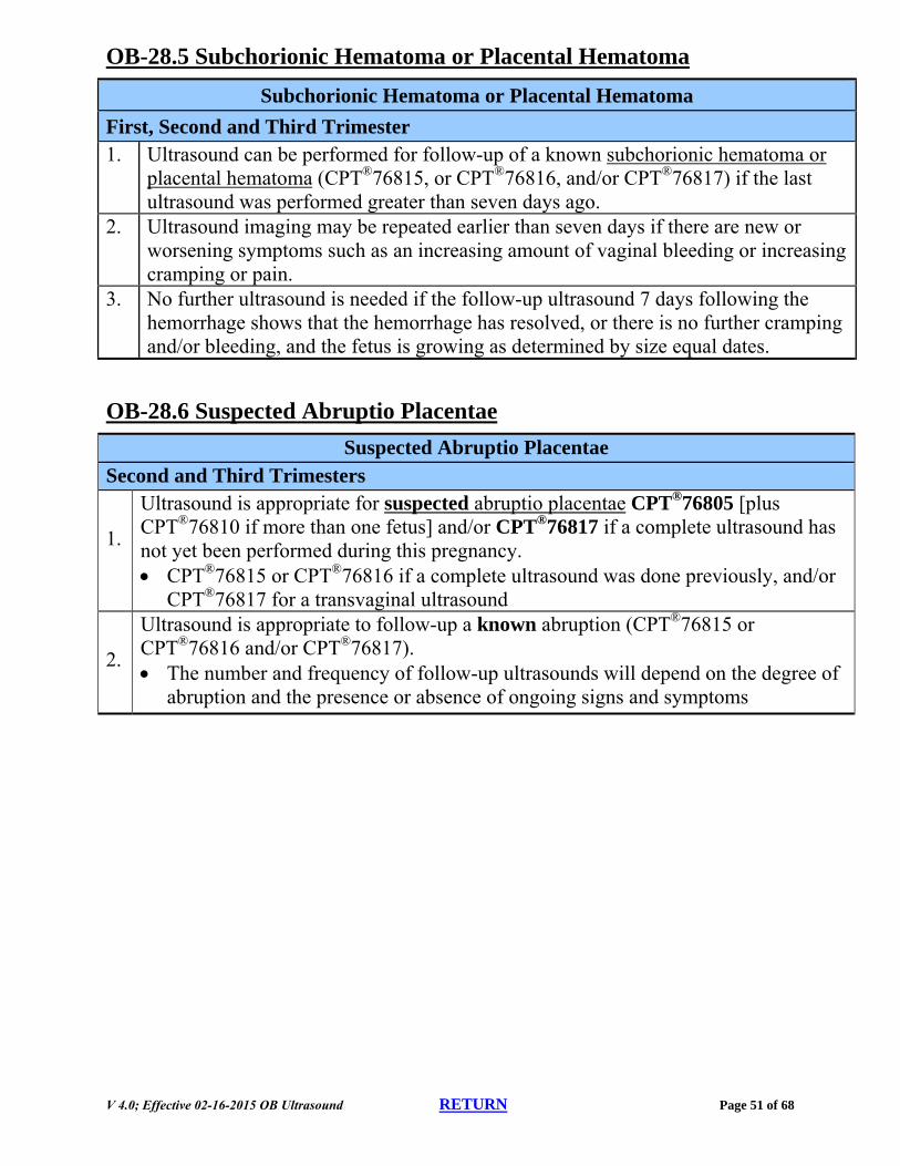
OB-28.5 Subchorionic Hematoma or Placental Hematoma
Subchorionic Hematoma or Placental Hematoma
First, Second and Third Trimester 1. Ultrasound can be performed for follow-up of a known subchorionic hematoma or
placental hematoma (CPT®76815, or CPT®76816, and/or CPT®76817) if the last ultrasound was performed greater than seven days ago.
2. Ultrasound imaging may be repeated earlier than seven days if there are new or worsening symptoms such as an increasing amount of vaginal bleeding or increasing cramping or pain.
3. No further ultrasound is needed if the follow-up ultrasound 7 days following the hemorrhage shows that the hemorrhage has resolved, or there is no further cramping and/or bleeding, and the fetus is growing as determined by size equal dates.
OB-28.6 Suspected Abruptio Placentae
Suspected Abruptio Placentae Second and Third Trimesters
1.
Ultrasound is appropriate for suspected abruptio placentae CPT®76805 [plus CPT®76810 if more than one fetus] and/or CPT®76817 if a complete ultrasound has not yet been performed during this pregnancy. CPT®76815 or CPT®76816 if a complete ultrasound was done previously, and/or
CPT®76817 for a transvaginal ultrasound
2.
Ultrasound is appropriate to follow-up a known abruption (CPT®76815 or CPT®76816 and/or CPT®76817). The number and frequency of follow-up ultrasounds will depend on the degree of
abruption and the presence or absence of ongoing signs and symptoms
V 4.0; Effective 02-16-2015 OB Ultrasound RETURN Page 52 of 68
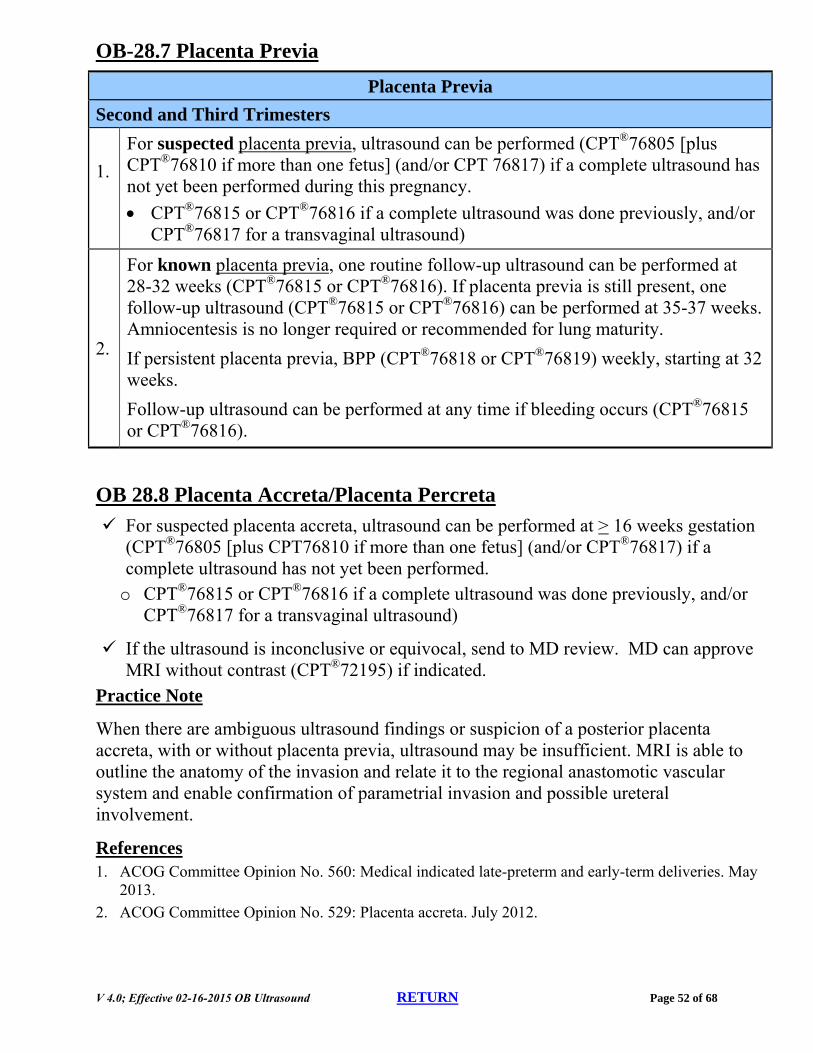
OB-28.7 Placenta Previa
Placenta Previa
Second and Third Trimesters
1.
For suspected placenta previa, ultrasound can be performed (CPT®76805 [plus CPT®76810 if more than one fetus] (and/or CPT 76817) if a complete ultrasound has not yet been performed during this pregnancy.
CPT®76815 or CPT®76816 if a complete ultrasound was done previously, and/or CPT®76817 for a transvaginal ultrasound)
2.
For known placenta previa, one routine follow-up ultrasound can be performed at 28-32 weeks (CPT®76815 or CPT®76816). If placenta previa is still present, one follow-up ultrasound (CPT®76815 or CPT®76816) can be performed at 35-37 weeks. Amniocentesis is no longer required or recommended for lung maturity.
If persistent placenta previa, BPP (CPT®76818 or CPT®76819) weekly, starting at 32 weeks.
Follow-up ultrasound can be performed at any time if bleeding occurs (CPT®76815 or CPT®76816).
OB 28.8 Placenta Accreta/Placenta Percreta
For suspected placenta accreta, ultrasound can be performed at > 16 weeks gestation (CPT®76805 [plus CPT76810 if more than one fetus] (and/or CPT®76817) if a complete ultrasound has not yet been performed.
o CPT®76815 or CPT®76816 if a complete ultrasound was done previously, and/or CPT®76817 for a transvaginal ultrasound)
If the ultrasound is inconclusive or equivocal, send to MD review. MD can approve MRI without contrast (CPT®72195) if indicated.
Practice Note
When there are ambiguous ultrasound findings or suspicion of a posterior placenta accreta, with or without placenta previa, ultrasound may be insufficient. MRI is able to outline the anatomy of the invasion and relate it to the regional anastomotic vascular system and enable confirmation of parametrial invasion and possible ureteral involvement.
References 1. ACOG Committee Opinion No. 560: Medical indicated late-preterm and early-term deliveries. May
2013.
2. ACOG Committee Opinion No. 529: Placenta accreta. July 2012.
V 4.0; Effective 02-16-2015 OB Ultrasound RETURN Page 53 of 68
OBSTETRICAL ULTRASOUND IMAGING GUIDELINES
OB-29~Vasa Previa
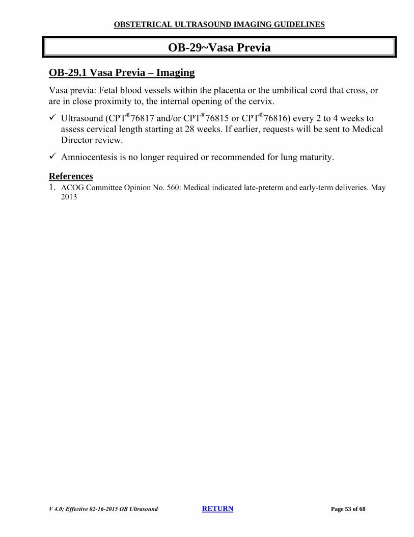
OB-29.1 Vasa Previa – Imaging
Vasa previa: Fetal blood vessels within the placenta or the umbilical cord that cross, or are in close proximity to, the internal opening of the cervix.
Ultrasound (CPT®76817 and/or CPT®76815 or CPT®76816) every 2 to 4 weeks to assess cervical length starting at 28 weeks. If earlier, requests will be sent to Medical Director review.
Amniocentesis is no longer required or recommended for lung maturity.
References 1. ACOG Committee Opinion No. 560: Medical indicated late-preterm and early-term deliveries. May
2013
V 4.0; Effective 02-16-2015 OB Ultrasound RETURN Page 54 of 68
OBSTETRICAL ULTRASOUND IMAGING GUIDELINES
OB-30~Procedure Coding Basics for Established Pregnancy
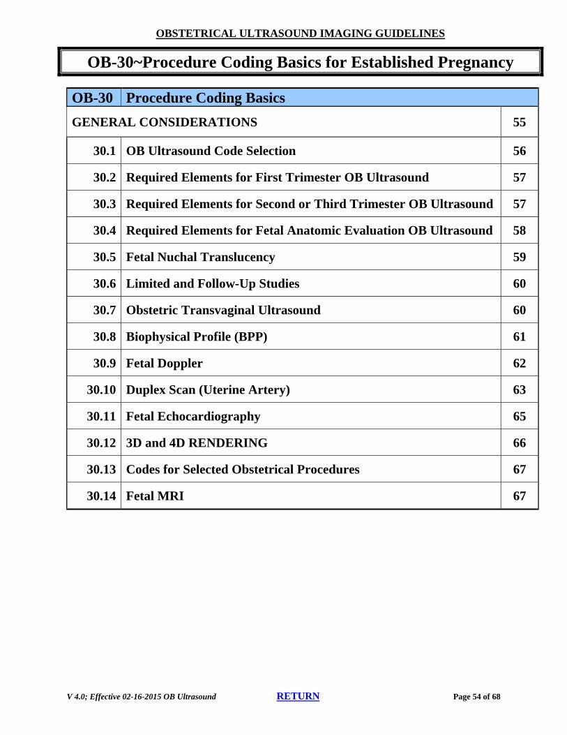
OB-30 Procedure Coding Basics
GENERAL CONSIDERATIONS 55
30.1 OB Ultrasound Code Selection 56
30.2 Required Elements for First Trimester OB Ultrasound 57
30.3 Required Elements for Second or Third Trimester OB Ultrasound 57
30.4 Required Elements for Fetal Anatomic Evaluation OB Ultrasound 58
30.5 Fetal Nuchal Translucency 59
30.6 Limited and Follow-Up Studies 60
30.7 Obstetric Transvaginal Ultrasound 60
30.8 Biophysical Profile (BPP) 61
30.9 Fetal Doppler 62
30.10 Duplex Scan (Uterine Artery) 63
30.11 Fetal Echocardiography 65
30.12 3D and 4D RENDERING 66
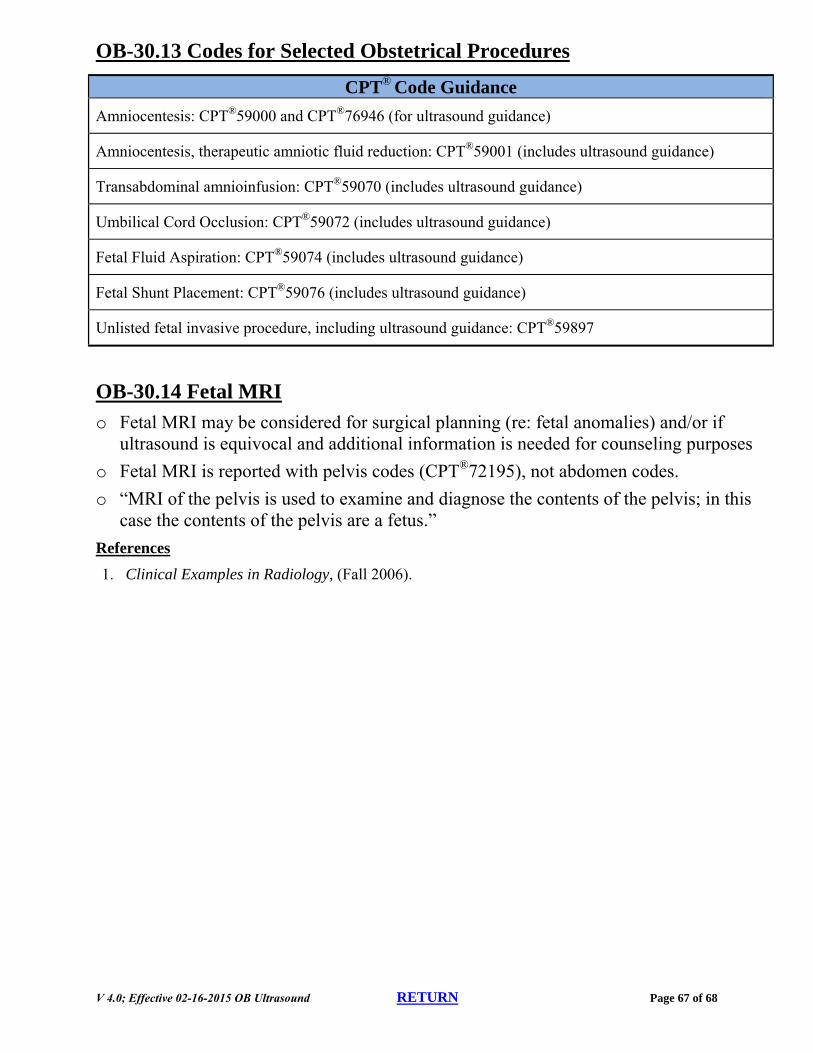
30.13 Codes for Selected Obstetrical Procedures 67
30.14 Fetal MRI 67
V 4.0; Effective 02-16-2015 OB Ultrasound RETURN Page 55 of 68
OBSTETRICAL ULTRASOUND IMAGING GUIDELINES
OBUS-30~Procedure Coding Basics for Established Pregnancy
GENERAL CONSIDERATIONS A Duplex scan describes:
1. An ultrasonic scanning procedure for characterizing the pattern and direction of blood flow in arteries and veins with the production of real time images integrating B-mode two dimensional vascular structure, and
2. Doppler spectral analysis, and
3. Color flow Doppler imaging
The use of a hand-held or any Doppler device that does not create a hard-copy output is considered part of the physical examination and is not separately billable. This exclusion includes devices that produce a record that does not permit analysis of bi-directional vascular flow.
The minimal use of color Doppler alone, when performed for anatomical structure identification, during a standard ultrasound procedure, is not separately reimbursable.
All obstetric ultrasound studies require permanently recorded images: o These images may be stored on film or in a Picture Archiving and Communication System
(PACS). o Obstetric ultrasound services may not be billed without image recording. o The use of a hand-held or any Doppler device that does not create a hard-copy output is
considered part of the physical examination and is not separately reimbursable.
Ultrasound procedure codes include the preparation of a required final written report which should be included in the patient’s medical record.
o Each procedure code has specific required elements which are described in this section. o The report should document the results of the evaluation of each element or the reason any
element is non-visualized. o Documentation of less than the required elements requires the billing of the “limited” code for
that anatomic region. o Only one (1) limited exam may be billed per encounter.
V 4.0; Effective 02-16-2015 OB Ultrasound RETURN Page 56 of 68
OB-30.1 OB Ultrasound Code Selection
It is not appropriate to report non-obstetrical pelvic ultrasound procedure codes (CPT®76830, CPT®76856, and CPT®76857) if pregnancy has already been diagnosed.
The CPT® code series, 76801-76815, contains what are considered the “normal OB codes”.
CPT® Code Guidance The OB ultrasound CPT® codes should be selected based on the following criteria. The length of gestation: CPT®76801 and CPT®76802 are reported for complete studies performed during the first trimester
(<14 weeks). CPT®76801 and CPT®76802 should only be used once per pregnancy unless the mother changes
to a new medical caregiver at a new office and there is a medical indication for ultrasound. CPT®76805 and CPT®76810 are used to report complete studies performed during the second and
third trimester. CPT®76805 and CPT®76810 should only be used once per pregnancy unless the mother changes
to a new medical caregiver at a new office and there is a medical indication for ultrasound.
The number of fetuses: CPT®76802, CPT®76810, CPT®76812, and CPT®76814 are “add-on” codes used to report each
additional fetus.
The imaging approach: CPT®76817 is used to report a transvaginal ultrasound. The other OB ultrasound codes are used for
transabdominal studies.
Whether the study is Complete or Limited: CPT®76815 and CPT®78616 are used to report limited or follow-up studies.
Whether a detailed fetal anatomic evaluation is performed: CPT®76811 and CPT®78612 describe an extensive fetal ultrasound evaluation and detailed anatomic
survey and are used only when the study includes this service. CPT®76812 is an add-on for each additional fetus. Any follow-up ultrasound for CPT®76811 should be coded as CPT®76816
V 4.0; Effective 02-16-2015 OB Ultrasound RETURN Page 57 of 68
OB-30.2 Required Elements for First Trimester OB Ultrasound
A complete first trimester transabdominal ultrasound (CPT®76801 and CPT®76802) is defined in CPT®
as including the following elements: o Determination of the number of gestational sacs and fetuses o Gestational sac/fetal measurements appropriate for gestation (<14 weeks) o Survey of visible fetal and placental anatomic structure o Qualitative assessment of amniotic fluid volume/gestational sac shape o Examination of maternal uterus and adnexa
CPT® Code Guidance It may not be possible to visualize the placenta during the early weeks of pregnancy. CPT®76801 and/or CPT®76802 may still be appropriately billed if the report documentation indicates placental anatomic structure could not be evaluated due to gestational age.
CPT®76802 is an ‘add-on’ code reported in conjunction with the ‘primary procedure’ CPT®76801 to report each additional gestation.
CPT®76801 and CPT®76802 should only be reported once per pregnancy unless the mother changes to a new medical caregiver at a new office and there is a medical indication for ultrasound. Follow-up studies to CPT®76801 and CPT®76802 should be reported as CPT®76815 or CPT®76816.
OB-30.3 Required Elements for Second or Third Trimester OB Ultrasound
CPT® Code Guidance A complete second or third trimester transabdominal ultrasound (CPT®76805 and CPT®76810) is defined in CPT® as including the following elements:
o Determination of the number of fetuses and amniotic/chorionic sacs o Measurements appropriate for gestation (>14 weeks) o Survey of intracranial/spinal/abdominal anatomy o Four-chambered heart o Umbilical cord insertion site o Placenta location o Amniotic fluid assessment o Examination of maternal adnexa, when visible
CPT®76810 is an ‘add-on’ code used with the ‘primary procedure’ CPT®76805 to report each additional gestation.
CPT®76805 and CPT®76810 should only be used once per pregnancy unless the mother changes to a new medical caregiver at a new office and there is a medical indication for ultrasound. Follow-up studies to CPT®76805 and CPT®76810 should be coded as CPT®76815 or CPT®76816.
V 4.0; Effective 02-16-2015 OB Ultrasound RETURN Page 58 of 68
OB-30.4 Required Elements for Fetal Anatomic Evaluation OB Ultrasound
CPT® Code Guidance Performance of the specialized fetal anatomic evaluation (CPT®76811 and CPT®76812) should be limited to those with special skills to perform this study, such as Maternal Fetal Medicine specialists, Perinatologists, and Radiologists (with advanced training in fetal imaging).
CPT®76811 and CPT®76812 are defined in CPT® as including all of the requirements listed for CPT®76805 and CPT®76810. In addition, the report must document detailed anatomic evaluation of the following elements:
o Fetal brain/ventricles o Face o Heart/outflow tracts and chest anatomy o Abdominal organ-specific anatomy o Number/length/architecture of limbs o Detailed evaluation of the umbilical cord and placenta o Other fetal anatomy as clinically indicated
CPT®76812 is an ‘add-on’ code used with the ‘primary procedure’ CPT®76811 to report each additional gestation.
o These studies are usually performed at 18 – 20 weeks and are most often completed at tertiary referral centers with perinatology departments.
o Only one medically indicated procedure CPT®76811 per pregnancy, per practice (per NPI) is appropriate. *Follow-up studies should be coded as CPT®76815 or CPT®76816
References
1. Society of Maternal Fetal Medicine. White Paper on Ultrasound Code 76811. May 24, 2004. http://www.askleslie.net/rads/White_Paper_on_Ultrasound_Code_76811.pdf. Accessed September 22, 2009
V 4.0; Effective 02-16-2015 OB Ultrasound RETURN Page 59 of 68
OB-30.5 Fetal Nuchal Translucency
CPT® Code Guidance CPT®76813 and CPT®76814 describe ultrasound measurement of the clear (translucent) space at the back of the fetal neck to assess risk for Down Syndrome (Trisomy 21), Trisomy 18, and other genetic disorders.
o Performed only once during a pregnancy during 11 0/7 to 13 6/7 weeks gestation o The sonographer performing the study and the physician interpreting the study must be
credentialed by the Maternal Fetal Medicine Foundation through their Nuchal Translucency Quality Review Program (NTQR).
o CPT®76814 is an add-on for each additional fetus.
If a nuchal translucency measurement is requested in the first trimester in addition to a complete ultrasound (CPT®76801, CPT®76802) or a limited ultrasound (CPT®76815), it would be appropriate to code for both the nuchal translucency study (CPT®76813 or CPT®76814) and the first trimester or limited ultrasound procedure.
As noted in the CPT® code application, the nuchal translucency measurement codes are used to describe a more detailed evaluation of specific embryonic structures than is required by either code CPT®76801, CPT®76802 or CPT®76815.
Follow-up studies to CPT®76813 and CPT®76814 should be coded as CPT®76815 or CPT®76816.
Criteria for Nuchal Translucency (NT) Measurement 1. CRL between 44-84mm, abnormal nuchal translucency is ≥2.5mm increases the risk for
aneuploidy. A measurement of >=3.5 mm increases the risk for cardiac defects in a euploid fetus. Criteria by the Fetal Medicine Foundation
2. Margins of NT edges are clear a. Clear image b. Angle of insonation is perpendicular to NT space c. Clear NT lines
3. Fetus in Mid Sagittal plane a. Midsagittal view of fetal spine seen in cervical & thoracic region b. Tip of nose seen in fetal profile c. Third & fourth ventricle seen in CNS d. Should not see ribs, stomach or heart
4. Fetus occupies majority of image a. Image predominately filled by fetal head neck and thorax b. The fetus should occupy >50% of image
5. Fetal head in neutral position a. Amniotic fluid seen between chin and chest b. Angle <90 degrees
6. Fetus observed away from amnion 7. Measurement
a. Use the + calipers b. Place crosshairs on the inner edge, but not in the clear space c. Measurement is perpendicular to long axis of the fetus d. Measure at the widest space and use largest of 3 technically correct measurements
Adapted from Steven L. Warsof, MD, Prof. Ob-Gyn, Eastern Virginia Medical School - Director, Center for Advanced Fetal Therapy
V 4.0; Effective 02-16-2015 OB Ultrasound RETURN Page 60 of 68
Practice Note “A nuchal translucency ultrasound consists of nuchal translucency measurement, a fetal crown rump length measurement and assessment of fetal viability. These are not the components of a 76801 (first trimester ultrasound), therefore a 76801 should not be billed unless you have received permission, in writing, from the health plan to use the 76801 in this manner.” (Society for Maternal Fetal Medicine’s White Paper on Nuchal Translucency Billing 2006/2007: http://www.ntqr.org/SM/Newsletters/NTExaminer_1.htm, November 10, 2006. Accessed September 30, 2010)
OB-30.6 Limited and Follow-Up Studies
CPT® Code Guidance CPT®76815 describes a limited or “quick look” study used to report one or more of the elements listed in the code definition, i.e. “fetal heart beat, placental location or fluid check (re: modified BPP which is NST with 76815)
o Reported only once, regardless of the number of fetuses, and only once per date of service o CPT®76815 should never be reported with complete studies CPT®76801/76802 and
CPT®76805/76810.
CPT®76816 describes a follow-up study designed to reassess fetal size or re-evaluate one or more abnormalities previously revealed on ultrasound.
o CPT®76816 should be reported once per fetus evaluated in follow-up. o Modifier -59 is appropriately used on subsequent codes. For example, a follow-up of a twin
pregnancy is reported: CPT®76816 and CPT®76816-59. o CPT®76816 should never be reported with complete studies CPT®76801, CPT®76802 and
CPT®76805, CPT®76810.
OBUS-30.7 Obstetric Transvaginal Ultrasound
CPT® Code Guidance
CPT®76817 is used to report an obstetrical transvaginal ultrasound.
CPT®76817 is reported only once regardless of the number of fetuses.
Although an obstetrical transvaginal ultrasound and transabdominal ultrasound can be performed at the same sitting and reported as two codes, there is rarely a medical indication to perform both studies at once.
V 4.0; Effective 02-16-2015 OB Ultrasound RETURN Page 61 of 68
OBUS-30.8 Biophysical Profile (BPP) The BPP combines data from ultrasound imaging and fetal heart rate (FHR) monitoring and is designed to predict the presence or absence of fetal asphyxia and, ultimately the risk of fetal death in the antenatal period (appropriately performed >22 weeks; should NOT be performed prior to the time when the fetus would be viable outside of the uterus).
o The following parameters are evaluated: Fetal breathing movements Gross fetal body movements Fetal tone Qualitative amniotic fluid volume 2 x 2 cm pocket Reactive FHR (non-stress testing portion) Performed if the ultrasound BPP is <8/8 or if
other clinical indicators are present (ie: contractions, pelvic pressure, symptoms of preterm labor or, labor)
CPT® Code Guidance CPT®76818 includes non-stress testing.
CPT®76819 does not include the non-stress testing portion.
If non-stress testing is performed without BPP, the appropriate code to use is CPT®59025 (Fetal non-stress test). CPT®59025 should not be reported with codes CPT®76818 or CPT®76819.
Although obstetrical ultrasound (CPT® codes 76805/76810/76815/76816/76820) and BPP (CPT®76818 and CPT®76819) can be performed at the same sitting and reported as two codes, it is generally not necessary to perform both studies at once.
o There are certain clinical circumstances in which it would be medically indicated to perform both studies at once.
o Each study must have a separate images, interpretations and reports
BPP and/or non-stress testing, performed on more than one fetus, should be reported separately. The use of modifier -59 on the second and subsequent studies is appropriate, depending on payer policy.
V 4.0; Effective 02-16-2015 OB Ultrasound RETURN Page 62 of 68
OB-30.9 Fetal Doppler MCA Doppler may be a useful test in SGA/IUGR fetuses detected after 33 weeks and 6/7 days of gestation when umbilical artery Doppler is normal. Based on this evidence it is reasonable to use MCA Doppler to time delivery in the near term-term (34 weeks 0 days gestation or greater) SGA/IUGR fetus with normal umbilical artery Doppler (see: OB-General Guidelines).
CPT® Code Guidance CPT®76820 describes Doppler velocimetry of the umbilical artery.
o Performed >22 weeks gestation o Utilized for known IUGR and known oligohydramnios o Known twin to twin transfusion or known discordant twins o Its use to predict preeclampsia, and stillbirth is considered investigational.
CPT®76821 describes Doppler velocimetry of the middle cerebral artery. o Performed as a substitute for amniocentesis to evaluate a fetus at risk for anemia due to Rhesus
isoimmunization/alloimmunization and Twin anemia polycythemia sequence. o Its use in the diagnosis of fetal anemia due to causes such as, feto-maternal hemorrhage, and
non-immune hydrops (other than Parvo) has yet to be determined.
Practice Notes
Middle Cerebral Artery Doppler (MCA): Doppler flow studies of the MCA are used in the assessment of the fetus at risk for anemia (see OB-3~Alloimmunization/Rh Isoimmuniazrion/Other/Fetal Anemia) and monochorionic twin pregnancies (see OB-17~Multiple Pregnancies)
In the preterm SGA fetus, middle cerebral artery (MCA) Doppler has limited accuracy to predict acidaemia and adverse outcome; it should not be used to time delivery. Most studies investigating MCA Doppler as a predictor of adverse outcome in preterm SGA fetuses have reported low predictive value, especially when umbilical artery Doppler is abnormal. In the largest study of predictors of neonatal outcome in SGA neonates of less than 33 weeks gestational age (n = 604), it was not a statistically significant predictor of outcome on logistic regression, although MCA PI < –2 SDs was associated with neonatal death (LR 1.12, 95% CI 1.04–1.21) and major morbidity (LR 1.12, 95% CI 1.1–1.33).
In the near-term SGA fetus with normal umbilical artery Doppler, an abnormal middle cerebral artery Doppler (PI < 5th centile) has moderate predictive value for acidosis at birth and should be used to time delivery. However, MCA Doppler may be a more useful test in SGA/IUGR fetuses detected after 34 weeks of gestation when umbilical artery Doppler is normal. Based on this evidence it is reasonable to use MCA Doppler to time delivery in the near term-term (34 weeks gestation or greater) SGA/IUGR fetus with normal umbilical artery Doppler.
References
1. Obstetrics and Gynecology 2008;112:145-157 2. Uterine artery Doppler flow studies in obstetric practice. Sciscione, Anthony C. et al.
American Journal of Obstetrics & Gynecology , Volume 201 , Issue 2 , 121 – 126. 3. First-trimester uterine artery Doppler and adverse pregnancy outcome: a meta-analysis
involving 55,974 women. Velauthar L, Plana MN, Kalidindi M, Zamora J, Thilaganathan B, Illanes SE, Khan KS, Aquilina J, Thangaratinam S. Ultrasound Obstet Gynecol. 2014;43(5):500.
V 4.0; Effective 02-16-2015 OB Ultrasound RETURN Page 63 of 68
OB-30.10 Duplex Scan (Uterine Artery) Uterine artery Duplex (Doppler) scan (CPT®93976), evaluation has been shown to predict adverse outcomes when utilized in the first and second trimester, prior to 16 weeks. The clinical utility however is limited to the first trimester when low dose Aspirin therapy can be instituted to decrease the risk of adverse outcomes (chronic hypertension, preeclampsia, and possibly IUGR). Provider certification, study technique, and abnormal test thresholds have been established by the Fetal Medicine Foundation (similar to certification process for Nuchal Translucency screening). The Society of Maternal Fetal Medicine (SMFM) has recommended the use of CPT®93976 only.
Treatment is now possible if started prior to 16 weeks gestation. Therefore, the use of Uterine Artery Doppler evaluation is now justified when utilized before 16 weeks gestation for patients with chronic hypertension or who are at risk for preeclampsia. The CPT® code recommended by SMFM is 93976 only. Send to Medical Director review if beyond 16 weeks gestation. One time only study.
CPT® Code Guidance CPT®93975 describes a complete duplex scan and should be reported if an organ is evaluated in its entirety. A complete study involves the evaluation of the inflow and outflow vessels of one or more organs.
CPT®93976 describes a limited duplex scan and should be reported when a complete study is not documented, for example, in the case of a follow-up study or a study of only the arterial flow.
CPT®93976 is used to report a fetal umbilical-placental flow study.
References
1. G, Capponi A, Arduini D, Romanini C. The value of fetal arterial and venous flows in predicting pH and blood gases measured in umbilical blood at cordocentesis in growth restricted fetuses. BJOG 1995;102:963–9.
2. Arduini D, Rizzo G, Romanini C. Changes in pulsatility index from fetal vessels preceding the onset of late decelerations in growth retarded fetuses. Obstet Gynecol 1992;79:605–10.
3. Dubiel M, Gudmundsson S, Gunnarsson G, Marsal K. Middle cerebral artery velocimetry as a predictor of hypoxemia in fetuses with increased resistance to blood flow in the umbilical artery. Early Hum Dev 1997;47:177–84.
4. Morris RK, Say R, Robson SC, Kleijen J, Khan KS. Systematic review of middle cerebral artery Doppler to predict fetal growth restriction/compromise of fetal wellbeing. Arch Dis Child Fetal Neonatal Ed 2008;93(Suppl 1):31–6.
5. Ozeren M, Dinc H, Ekmen U, Senekayli C, Aydemir V. Umbilical and middle cerebral artery Doppler indices in patients with preeclampsia. Eur J Obstet Gynecol Reprod Biol 1999;82:11–6.
6. Vergani P, Roncaglia N, Ghidini A, Cripper I, Cameroni I, Orsenigo F, et al. Can adverse neonatal outcome be predicted in late preterm or term fetal growth restriction? Ultrasound Obstet Gynecol 2010;36:166–70.
7. Fieni S, Gramellini D, Piantelli G. Lack of normalisation of middle cerebral artery velocity prior to fetal death before 30th week of gestation: a report of three cases. Ultrasound Obstet Gynecol 2004;24:474–5.
8. Baschat AA, Cosmi E, Bilardo CM, Wolf H, Berg C, Rigano S, et al. Predictors of neonatal outcome in early–onset placental dysfunction. Obstet Gynecol 2007;109:253–61.
9. Baschat AA, Gembruch U, Harman CR. The sequence of changes in Doppler and biophysical parameters as severe fetal growth restriction worsens. Ultrasound Obstet Gynecol 2001;18:571–7.
V 4.0; Effective 02-16-2015 OB Ultrasound RETURN Page 64 of 68
10. Oros D, Figueras F, Cruz–Martinez R, Meler E, Munmany M, Gratecos E. Longitudinal changes in uterine, umbilical and fetal cerebral Doppler indices in late–onset small–for–gestational age fetuses. Ultrasound Obstet Gynecol 2011;37:191–5.
11. Hershkovitz R, Kingdom JC, Geary M, Rodeck CH. Fetal cerebral blood flow redistribution in late gestation: identification of compromise in small fetuses with normal umbilical artery Doppler. Ultrasound Obstet Gynecol 2000;15:209–12.
12. Severi FM, Bocchi C, Visentin A, Falco P, Cobellis L, Florio P, et al. Uterine and fetal cerebral Doppler predict the outcome of third–trimester small–for–gestational age fetuses with normal umbilical artery Doppler. Ultrsound Obstet Gynecol 2002;19:225–8.
13. Reddy UM, Filly RA, Copel JA. Prenatal Imaging: Ultrasonography and Magnetic Resonance Imaging. Obstetrics and Gynecology 2008;112:145-157.
14. Sciscione AC, Hayes EJ. Uterine artery Doppler flow studies in obstetric practice. American J Obstet Gynecol, 2009; 121-126.
15. Stampalija T, Alfirevic Z, Gyte G. Doppler in obstetrics: evidence from randomized trials. Ultrasound Obstet Gynecol. 2010;36(6):779-80.
16. ACOG Practice Bulletin #134: Fetal Growth Restriction, May 2013. 17. ACOG Practice Bulletin no. 145: Antepartum Fetal Surveillance, July 2014. 18. Copel JA, Bahtiyar MO. A practical approach to fetal growth restriction. Obstetrics &
Gynecology, 2014; 123. 19. Uterine artery Doppler flow studies in obstetric practice. Sciscione, Anthony C. et al.
American Journal of Obstetrics & Gynecology , Volume 201 , Issue 2 , 121 – 126. 20. First-trimester uterine artery Doppler and adverse pregnancy outcome: a meta-analysis
involving 55,974 women. Velauthar L, Plana MN, Kalidindi M, Zamora J, Thilaganathan B, Illanes SE, Khan KS, Aquilina J, Thangaratinam S. Ultrasound Obstet Gynecol. 2014;43(5):500.
V 4.0; Effective 02-16-2015 OB Ultrasound RETURN Page 65 of 68
It is inappropriate to report codes CPT®76825 – CPT®76828 for the routine monitoring of fetal heart tones using a hand-held or any Doppler device that does not create a hard-copy output. Such fetal heart tone monitoring is considered part of the physical examination and is not separately billable
CPT®76825 describes fetal echocardiography, real time with image documentation (2D), with or without M-mode recording
CPT®76826: o is a follow-up or repeat fetal echocardiogram o should never be billed with CPT®76825 o should never be billed more than once on any date of service
CPT®76827 describes a complete Doppler echocardiography, fetal, pulsed wave and/or continuous wave with spectral display
CPT®76828: is a follow up or repeat Doppler fetal echocardiogram
CPT®93325 is used to report color mapping in conjunction with fetal echocardiography procedures CPT®76825 – CPT®76828.
V 4.0; Effective 02-16-2015 OB Ultrasound RETURN Page 66 of 68
OB-30.12 3D and 4D RENDERING There is currently insufficient data to generate appropriateness criteria for the use of 3D and 4D rendering in conjunction with ultrasound.
Current guidelines on ultrasonography in pregnancy from ACOG (2009) state: "The technical advantages of 3-dimensional ultrasonography include its ability to acquire and manipulate an infinite number of planes and to display ultrasound planes traditionally inaccessible by 2-dimensional ultrasonography. Despite these technical advantages, proof of a clinical advantage of 3-dimensional ultrasonography in prenatal diagnosis in general is still lacking. Potential areas of promise include fetal facial anomalies, neural tube defects, and skeletal malformations where 3-dimensional ultrasonography may be helpful in diagnosis as an adjunct to, but not a replacement for, 2-dimensional ultrasonography.”
Yagel et al (2009) described the state of the science of 3D/4D ultrasound (3D/4D US) applications in fetal medicine. They noted that 3D/4D US applications are many and varied. Their use in fetal medicine varies with the nature of the tissue to be imaged and the challenges each organ system presents, versus the advantages of each ultrasound application. The investigators stated that 3D/4D US has been extensively applied to the study of the fetus. Fetal applications include all types of anatomical assessment, morphometry and volumetry, as well as functional assessment. The authors concluded that 3D/4D US provides many advantages in fetal imaging; however, its contribution to improving the accuracy of fetal scanning over rates achieved with 2D US, remains to be established.
Clinical use of 3D ultrasound should be on an individual basis. There can be specific reasons that require 3D ultrasound when 2D cannot be utilized. Such as determination of fetal growth when there is absence of lower limbs / femurs. Since, the femur length is vital in determination of fetal weight and growth. Fractional limb volume measurement of the humerus is required to evaluate for IUGR. A second clinical scenario is seen with gastroschisis. Since the fetal abdomen is small due to the defect present, there is artificially high rate of IUGR. The cause of this is the use of the fetal abdominal circumference to determine growth. 3D Fractional limb volume measurement eliminates this issue and decreases false positives.
References
1. Lee W, Deter R, Sangi-Haghpeykar H, Yeo L, Romero R. Prospective validation of fetal weight estimation using fractional limb volume. Ultrasound Obstet Gynecol. 2013 Feb;41(2):198-203.
2. Obstetrics and Gynecology 2008; 112:145-157
3. ACOG Practice Bulletin No.101: Ultrasonography in pregnancy. February 2009. Reaffirmed 2014.
V 4.0; Effective 02-16-2015 OB Ultrasound RETURN Page 67 of 68
OB-30.13 Codes for Selected Obstetrical Procedures
CPT® Code Guidance
Amniocentesis: CPT®59000 and CPT®76946 (for ultrasound guidance)
Unlisted fetal invasive procedure, including ultrasound guidance: CPT®59897
OB-30.14 Fetal MRI
o Fetal MRI may be considered for surgical planning (re: fetal anomalies) and/or if ultrasound is equivocal and additional information is needed for counseling purposes
o Fetal MRI is reported with pelvis codes (CPT®72195), not abdomen codes.
o “MRI of the pelvis is used to examine and diagnose the contents of the pelvis; in this case the contents of the pelvis are a fetus.”
References
1. Clinical Examples in Radiology, (Fall 2006).
V 4.0; Effective 02-16-2015 OB Ultrasound RETURN Page 68 of 68
OBSTETRICAL ULTRASOUND IMAGING GUIDELINES
OB-31~High Risk Medications and Substances
Specific drugs that qualify as risk factors in High Risk Pregnancy and qualify as medical indications for Fetal Anatomic Scan (CPT®76805) and Specialized Fetal Anatomic Scan (CPT®76811):