37
2012 Update on Myeloid Neoplasms: Myelodysplastic Syndrome and Acute Myeloid Leukemia Ronald L. Weiss, M.D. University of Utah ARUP Laboratories August 16, 2012

2012 Update on Myeloid Neoplasms: Myelodysplastic Syndrome and Acute Myeloid
Leukemia
Ronald L. Weiss, M.D. University of Utah ARUP Laboratories
August 16, 2012

Speaker Profile
• Professor of Pathology, University of Utah School of Medicine
• Staff Hematopathologist, ARUP Laboratories • Disclosures
– Ownership/equity interest that is unrelated to this presentation
• AvanSci Bio, LLC; Board of Managers • DecipherGenX, Inc.; President/COO

Objectives
• Understand the pathophysiology of myelodysplastic syndromes and acute myeloid leukemia
• Review the current (WHO 2008) diagnostic criteria and classification of these disorders
• Understand the approach to the practical laboratory diagnosis of these disorders

2008 WHO Classification of the Myeloid Neoplasms
• Myeloproliferative Neoplasms (MPN) • Myeloid/Lymphoid neoplasms with eosinophilia
and abnormalities of PDGFRA, PDGFRB or FGFR1 • Myelodysplastic syndrome (MDS) • MDS/MPN
– Includes CMML and the provisional category of RARS-T
• Acute myeloid leukemia (AML)

Myelodysplastic Syndromes (MDS)

MDS, General Features
• Clonal stem cell disorder with ineffective hematopoiesis, cytopenias and dysplasia of one or more cell lineages
• Frequent cytogenetic and/or mutational abnormalities – 50% with abnormal karyotypes – 50% with somatic mutations (including 50% of normal
karyotype MDS cases) • Typical presentation: elderly adult with sustained,
unexplained anemia, leukopenia and/or thrombocytopenia
• De novo or secondary (usually therapy-related) • Risk-based rate of progression to AML

Revised (2008) WHO Classification of MDS
• Refractory cytopenia with unilineage displasia (RCUD) • Refractory anemia with ring sideroblasts (RARS) • RC with multilineage dysplasia (RCMD) • RA with excess blasts (RAEB-1, RAEB-2) • MDS with isolated del(5q) (5q- syndrome) • MDS, unclassifiable • Refractory cytopenia of childhood (RCC, provisional) • MDS, therapy-related

Approach to MDS Diagnosis
• Complete history, CBC with peripheral blood smear evaluation, bone marrow aspirate and biopsy, cytogenetics and FISH
• Minimum criteria: – Absence of de novo AML recurrent cytogenetic abnormalities – At least 2 of the following:
• Sustained, unexplained cytopenia(s) • Dysplasia in 2 or more lineages (except in RCUD, RARS and 5q-
syndrome) • Clonal cytogenetic abnormality(s) • ≥5% blasts in the bone marrow

Differential Diagnosis of MDS
• Megaloblastic anemias (folate and/or B12 deficiencies)
• Copper deficiency • Zinc toxicity • Arsenic toxicity • Chemotherapy-
associated effects • Chronic viral infections
(including HIV)
• Congenital dyserythropoietic anemia (especially in children and adolescents)
• Sideroblastic anemia (congenital, acquired)
• Aplastic anemia • Paroxysmal nocturnal
hemoglobinuria • Low blast count AML

Genetics of MDS
• Chromosomal copy number abnormalities (additions, deletions) especially of 5, 7, 8, 20
• Recurrent mutations in genes associated with: – DNA histone function (EZH2, ASXL1, UTX) – DNA methylation (DNMT3A, IDH1, IDH2, TET2) – Active areas of research on pathogenesis
pathways and potential therapeutic interventions
• Rare finding of FLT3 and/or NPM1 mutations

Progression to AML
• Clinical low-grade and high-grade forms of MDS – Either relatively stable disease (years), or develop clonal
cytogenetic evolution and progression to AML (months)
• International Prognostic Scoring System (IPSS) Risk Groups (n=4) provides some predictability – # of cytopenias, % of BM blasts, and karyotype – Risk of Progression to AML: Low (5.7 yr.), Intermediate-1
(3.5 yr.), Intermediate-2 (1.2 yr.), High (0.4 yr.) – But limited by heterogeneity within categories
• Genetic profiling to refine IPSS risk categories is underway

Treatment of MDS
• Only BMT is potentially curative, everything else is palliative at this point
• Treatment decisions depend upon many factors: – Age, performance status, IPSS risk stratification, patient
wishes, degree of cytopenias and required support (transfusions), is it de novo or secondary MDS
• Transfusion support, growth factors (especially EPO), iron chelation therapy (deferasirox), immunomodulators (lenalidomide) and/or DNA methyl transferase inhibitors (azacitidine, decitabine) – Lenalidomide is first-line treatment of the 5q- Syndrome,
and may be considered in selected other patients – DNMT inhibitors have shown promise in high-risk MDS

Myelodysplastic/ Myeloproliferative Neoplasms
What happened to CMML between WHO 2001 and WHO 2008?

MDS/MPN Overlap Syndromes
• Chronic myelomonocytic leukemia (CMML) • Atypical CML, BCR-ABL1 negative (aCML) • Juvenile myelomonocytic leukemia (JMML) • MDS/MPN, unclassifiable
– Includes the provisional entity of “Refractory Anemia with Ring Sideroblasts and thrombocytosis (RARS-T)”

CMML
• Diagnostic criteria: – Persistent monocytosis, absence of BCR-ABL1, no
rearrangement of PDGFRA or PDGFRB, <20% blasts in the PB/BM, and at least one of the following:
• Dysplasia in ≥1 cell line(s), clonal cytogenetic abnormality, OR unexplained monocytosis >3 months
– CMML-1= <5% PB blasts (includes promonocytes) and <10% BM blasts
– CMML-2= 5-19% PB blasts, 10-19% BM blasts or any blast count with Auer rods present
– WBC <13 x 109/L (CMML-MDS like), or ≥13 (CMML-MPN like)

Diagnostic work-up of CMML
• CBC and peripheral blood smear evaluation • BM aspirate/biopsy
– Morphologic evaluation for dysplasia, fibrosis – Flow cytometry to confirm % promonocyte and
monoblast phenotype – Cytogenetics and FISH
• Clonal karyotypic abnormalities other than t(9;22) • +8, -7 and/or del7q • Lack of BCR-ABL1, PDGFRA, PDGFRB

Somatic Mutations in CMML
• Common mutations of possible clinical significance: – TET2 (frequent marker of myelomonocytic clonal
dominance) – ASXL1 (associated with progression to AML?) – RUNX1 (progression to AML?) – EZH2 (poorer prognosis?) – CBL (myelomonocytic clones, uncertain?)
• Recent evidence points to abnormalities of DNA splicing pathways having a major role in CMML

Treatment and Prognosis
• Hydroxyurea or Azacitidine; Decitabine for CMML-2 • Only BMT is potentially curative • Poor prognostic risk factors (IPSS)
– CMML-2, WBC ≥13,000 (CMML MPN-like), hemoglobin <10g/L, platelet count <100 x 109/L
– Cytogenetics: • Low risk= NK, -Y only • High risk= +8, abnormalities of 7, complex karyotype • Intermediate risk= any other abnormality
• Poor overall survival (months-few years)
MDS/MPN, U
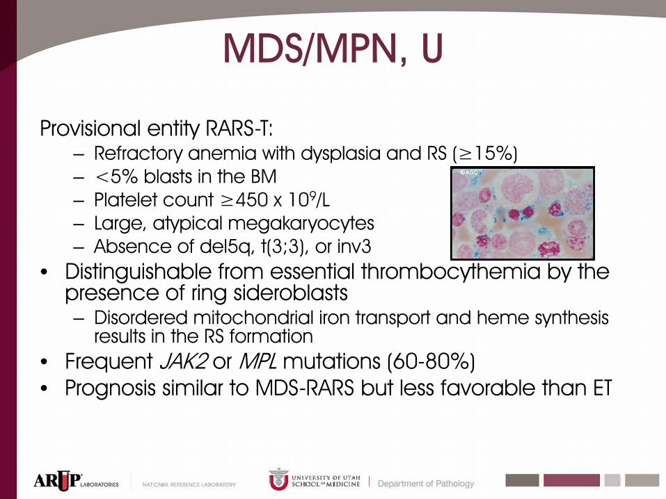
Provisional entity RARS-T: – Refractory anemia with dysplasia and RS (≥15%) – <5% blasts in the BM – Platelet count ≥450 x 109/L – Large, atypical megakaryocytes – Absence of del5q, t(3;3), or inv3
• Distinguishable from essential thrombocythemia by the presence of ring sideroblasts – Disordered mitochondrial iron transport and heme synthesis
results in the RS formation • Frequent JAK2 or MPL mutations (60-80%) • Prognosis similar to MDS-RARS but less favorable than ET

Acute Myeloid Leukemia
Genetic analysis and classification

Diagnosis & Classification Criteria
Morphology (PB, BM) Cytochemistry Immunophenotype Cytogenetics Karyotyping, FISH Mutation status PCR, NGS, expression arrays
Determining blast counts and lineage, maturational status, dysplasia, and effectiveness of hematopoiesis Identify recurring cytogenetic abnormalities Identify established mutational risk factors Establish the genetic baseline Impact therapeutic decision making

AML Diagnostic Criteria
• A clonal stem cell disorder with ≥20% blasts in the blood or bone marrow, OR the presence of one of the following recurring genetic abnormalities regardless of blast count: – t(8;21)(q22;q22); inv(16)(p13.1;q22) or
t(16;16)(p13.1;q22); and t(15;17)(q22;q12)

Genetic Analysis in AML
Typical evaluation panel: • Classic cytogenetics • FISH t(15;17) , t(9;22)
• Molecular Testing FLT3, NPM1, CEBPA, (DNMT3A, IDH1, IDH2) * Emerging/investigational mutational factors
inv16 t(8;21)
Core Binding
Factor AML
KIT

Sequencing the AML Genome?
• Whole genome sequencing of AML* leads to estimates of 500-1000 somatic mutations – Most are not considered to be primary “AML driver
mutations” – Sequencing does not capture gene expression
profiles or epigenetic changes that also contribute to leukemogenesis
* Mardis ER, et al. NEJM 2009;361:1058-66.

Integrated Genetic Profiling in AML
• Mutational analysis of 18 genes in 398 patients < 60 years old – 97.3% of the patients had at least one mutation
• The findings were then validated in an independent set of 104 patients
• Identified independent genetic predictors of outcome that improved risk stratification
• Improved outcomes seen with DNMT3A and NPM1 mutations, and MLL translocations
• Suggested a future role for AML mutational profiling as well as emphasizing the complex nature of the disease
Patel JP, et al. NEJM 2012;366:1079-89

AML and Related Neoplasms Classification
• AML with recurrent genetic abnormalities • AML with myelodysplasia-related changes • Therapy-related myeloid neoplasms • AML, NOS
– Includes the French-American-British (FAB) subtypes
• Myeloid sarcoma • Myeloid proliferations related to Down syndrome • Blastic plasmacytoid dendritic cell neoplasm

AML with Recurrent Genetic Abnormalities
• Additional entities introduced in the 2008 WHO classification – t(9;11)(p22;q23) MLLT3-MLL – t(6;9)(p23;q34) DEK-NUP214 – inv(3(q21;q26.2) or t(3;3)(q21;26.2) PRN1-EVI1 – t(1;22)(p13;q13) RBM15-MKL1 – Provisional entity: AML with mutated NPM1 – Provisional entity: AML with mutated CEBPA
• FLT3 mutations are frequently associated with other genetic abnormalities, and should be determined in cases of AML

Therapeutic & Prognostic Decision Making
• AML with t(15;17) – All-trans retinoic acid (ATRA), plus cytoreduction or arsenic trioxide
• Core Binding Factor (CBF) AML – Significantly higher response rates and long-term survival probabilities – Further evaluation for activating KIT mutations (in ~30%) open up the
potential for tyrosine-kinase inhibitor therapy • AML with NPM1 mutations
– Favorable prognosis – 25-35% of AML; 45-65% of NK-AML
• AML with FLT3 mutations (in 20% of AML) – Worse prognosis – Clinical evaluation for FLT3 inhibitor therapy or HSCT
• AML with CEBPA mutation (10-18% of NK-AML) – CEBPA double mutations (CEBPAdm) have a favorable prognosis

Monosomal Karyotype AML
• Newly recognized • Clone of at least 2 autosomal monosomies, or
a single autosomal monosomy (other than of X or Y) and at least one structural abnormality
• ~10-15% of AML, increases with age • Rarely seen with FLT3 or NPM1 , but frequently
seen with TP53 alterations • Poor response to treatment
– BMT recommended if possible

AML with Myelodysplasia-Related Changes
• Includes patients with: – AML arising in previous MDS or MPS/MPN – AML with specific MDS-associated cytogenetic
abnormality – AML with multi-lineage dysplasia (>50% of cells in ≥2
cell lineages) • Doesn’t include cases with prior cytotoxic therapy
for an unrelated disease, or cases with the “recurring” AML cytogenetic abnormalities
• More common in the elderly • ~25-30% of all AML cases (50% of adult AML?)

Therapy-related Myeloid Neoplasms
• Alkylators, radiotherapy, and/or topoisomerase II inhibitors; usually within 1-6 years
• Based upon blast count and morphology: – t-MDS – t-MDS/MPN – t-AML
• But, best considered as a single biologic disease with a dismal prognosis (5 year survival < 10%)
• ~90% have recurrent genetic abnormalities,

Genetics of Therapy-related Myeloid Neoplasms
• Genetic lesions similar to de novo MDS, but in different frequencies – TP53 (17p) mutations are infrequent in de novo, but
common (25-30%) in secondary MDS – RUNX1 (21q) and N/KRAS (1p/12p) mutations are more
common in secondary MDS than in de novo – Balanced translocations of RUNX1 (21q) and MLL
(11q23) are more common in radiation-induced MDS – del5 and del7 are more common in alkylator-induced
MDS • Similar to de novo AML genetics, but have worse
outcomes then the de novo counterparts – Except for inv(16), t(16;16) or t(15;17)

Myeloid Proliferations Related to Down Syndrome
• Transient abnormal myelopoiesis in 10% of DS – Median age of diagnosis = 5 days – Leukocytosis with increased blasts; variable RBC and platelet
values – Myeloid blasts with frequent CD7, CD56, CD41 and CD61
expression (megakaryoblast-like) – Trisomy 21 with GATA1 and JAK2 mutations – Usually resolves spontaneously, but 10-15% later develop a
myeloid neoplasm – Hepatic dysfunction/fibrosis is a poor prognostic sign
• Myeloid leukemia associated with DS – High risk of AML, especially AML-M7, in first 4 years of life – Usually preceded by myelodysplasia – Relatively good prognosis, especially in < 4 years of age
Myeloid Sarcoma
• Extramedullary proliferation of myeloblasts – Variable expression of CD13, CD14, CD33, CD34,
CD64, CD68, CD117, myeloperoxidase, and/or lysozyme
• May or may not be associated with concurrent disease of the bone marrow
• Diagnostic of AML – More common in pediatric AML – More common in monoblastic and
myelomonocytic leukemias

Blastic Plasmacytoid Dendritic Cell Neoplasm
• History of uncertain histiogenesis – “Blastic NK-cell lymphoma,” “CD4+/CD56+ hematodermic
neoplasm” • Confirmed to be of plasmacytoid dendritic cell origin,
related to myeloid cells • Cutaneous, bone marrow and blood involvement by
non-descript blasts • CD4, CD56 and CD123 expression, variable CD7 and
CD33, without B, T, NK or myelomonocytic specific markers
• ~60% have non-recurrent genetic abnormalities • Very aggressive clinical course • Only BMT offers a chance of long-term survival

Summary
• The myeloid neoplasms encompass a broad range of disorders
• Accurate diagnosis and treatment requires a combination of morphologic and genetic features
• New entities will continue to be added as we better understand the molecular genetic basis of pathogenesis

Questions?
Thank you for your attention


![Therapy-related Myeloid Neoplasms Following …Radiation therapy & t-MN • Have modern RT techniques [mega-voltage linear accelerators; intensity-modulated radiation therapy (IMRT)]](https://static.documents.pub/doc/80x56/5ed204196731c53a5734c2cd/therapy-related-myeloid-neoplasms-following-radiation-therapy-t-mn-a-have.jpg)