IDPH ESF-8 Plan: Pediatric and Neonatal Surge Annex Trauma and Blast Injury Care Guideline 2015 Purpose: To provide guidance to practitioners caring for pediatric patients during a disaster. Disclaimer: This guideline is not meant to be all inclusive, replace an existing policy and procedure at a hospital or substitute for clinical judgment. These guidelines may be modified at the discretion of the healthcare provider. Illinois EMSC www.luhs.org/emsc Initial Management of All Pediatric Trauma Patients Stabilize ABCs and c-spine (Airway, Breathing, and Circulation) IMMOBILIZE SPINE as indicated. Position for optimal airway and suction as needed. Position infants and children < 2 yrs supine on a backboard with a recess for the head or use a pad under the back from the shoulders to the buttocks. Consider needle decompression for signs of pneumothorax, hemothorax or tension pneumothorax Obtain weight (actual or use of weight/length based tool) Establish 2 peripheral IVs (consider large bore if possible) or if unable, establish intraosseous (IO) access Control any external bleeding Avoid removal of penetrating objects in the emergency department (should be performed in operating room due to risk of hemorrhage) Monitor Heart Rate (HR), Blood pressure (BP), Oxygen Saturation (SpO 2 ), mental status, temperature, perfusion, urine output, bedside glucose Perform detailed primary and secondary history & physical exam including mechanism of injury, Pediatric Trauma Score (PTS) and Pediatric Glasgow Coma Scale (PGCS) Consult pediatric care medical specialist for assistance with care of the acutely and critically injured patient (see below for Level I Trauma Criteria), to individualize the care of patient, if patient does not improve and needs to be admitted/transferred and as needed for further support and consult. Category 1 Trauma Criteria (minimum): All penetrating injuries to head, neck, torso, and/or groin Two or more body regions with potential threat to life or limb Combination trauma with ≥20% TBSA burn Limb paralysis and/or sensory deficit above the wrist and ankle Flail chest Amputation proximal to wrist or ankle Blunt or penetrating trauma with unstable vital signs AND/OR: o Hemodynamic compromise (Pediatric SBP≤80) o Respiratory compromised (Respiratory rate <10 or >29) o Altered mentation (PGCS≤10) Additional Blast Injury Considerations: o Classification of Blast Injuries (see page 8 for more detailed information on pediatric clinical presentations for common blast injuries and management of specific blast injuries) o Primary Results from impact of the over-pressurized blast wave Gas filled/ hollow structures most susceptible Examples: blast lung, tympanic membrane rupture, abdominal hemorrhage and perforation, eye globe rupture, traumatic brain injury (TBI) without physical signs of head injury o Secondary Results from flying debris and bomb fragments Entire body may be affected Examples: penetrating and blunt trauma injuries, eye penetration
Transcript
IDPH ESF-8 Plan: Pediatric and Neonatal Surge Annex Trauma and Blast Injury Care Guideline
2015
Purpose: To provide guidance to practitioners caring for pediatric patients during a disaster. Disclaimer: This guideline is not meant to be all inclusive, replace an existing policy and procedure at a hospital or substitute for clinical judgment. These guidelines may be modified at the discretion of the healthcare provider.
Illinois EMSC www.luhs.org/emsc
Initial Management of All Pediatric Trauma Patients
Stabilize ABCs and c-spine (Airway, Breathing, and Circulation)
IMMOBILIZE SPINE as indicated. Position for optimal airway and suction as needed. Position infants and children < 2 yrs supine on a backboard with a recess for the head or use a pad under the back from the shoulders to the buttocks.
Consider needle decompression for signs of pneumothorax, hemothorax or tension pneumothorax
Obtain weight (actual or use of weight/length based tool)
Establish 2 peripheral IVs (consider large bore if possible) or if unable, establish intraosseous (IO) access
Control any external bleeding
Avoid removal of penetrating objects in the emergency department (should be performed in operating room due to risk of hemorrhage)
Perform detailed primary and secondary history & physical exam including mechanism of injury, Pediatric Trauma Score (PTS) and Pediatric Glasgow Coma Scale (PGCS)
Consult pediatric care medical specialist for assistance with care of the acutely and critically injured patient (see below for Level I Trauma Criteria), to individualize the care of patient, if patient does not improve and needs to be admitted/transferred and as needed for further support and consult.
Category 1 Trauma Criteria (minimum):
All penetrating injuries to head, neck, torso, and/or groin
Two or more body regions with potential threat to life or limb
Combination trauma with ≥20% TBSA burn
Limb paralysis and/or sensory deficit above the wrist and ankle
Flail chest
Amputation proximal to wrist or ankle
Blunt or penetrating trauma with unstable vital signs AND/OR: o Hemodynamic compromise (Pediatric SBP≤80) o Respiratory compromised (Respiratory rate <10 or >29) o Altered mentation (PGCS≤10)
Additional Blast Injury Considerations: o Classification of Blast Injuries (see page 8 for more detailed information on pediatric clinical
presentations for common blast injuries and management of specific blast injuries) o Primary
Results from impact of the over-pressurized blast wave Gas filled/ hollow structures most susceptible Examples: blast lung, tympanic membrane rupture, abdominal hemorrhage and
perforation, eye globe rupture, traumatic brain injury (TBI) without physical signs of head injury
o Secondary Results from flying debris and bomb fragments Entire body may be affected Examples: penetrating and blunt trauma injuries, eye penetration
IDPH ESF-8 Plan: Pediatric and Neonatal Surge Annex Trauma and Blast Injury Care Guideline
2015
Illinois EMSC www.luhs.org/emsc
o Tertiary Results from victims being thrown by blast wind Entire body may be affected Examples: fractures, amputations, closed and open brain injury
o Quaternary All injuries, illnesses not due to Primary, Secondary or Tertiary mechanisms Entire body may be affected Example: crush injuries, burns, asphyxia, toxic exposures, exacerbation or
complications from existing or chronic conditions
IDPH ESF-8 Plan: Pediatric and Neonatal Surge Annex Trauma and Blast Injury Care Guideline
2015
Illinois EMSC www.luhs.org/emsc
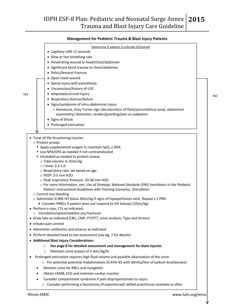
Management for Pediatric Trauma & Blast Injury Patients
Determine if patient is critically ill/injured
Capillary refill >2 seconds
Slow or fast breathing rate
Penetrating wound to head/chest/abdomen
Significant blunt trauma to chest/abdomen
Pelvic/femoral fracture
Open chest wound
Spinal injury with paresthesia
Unconscious/history of LOC
Amputation/crush injury
Respiratory distress/failure
Signs/symptoms of intra-abdominal injury:
o Hematuria, Grey Turner sign (discoloration of flank/periumbilical area), abdominal asymmetry/ distention, tender/guarding/pain on palpation
Signs of Shock
Prolonged extrication
YES NO
Treat all life threatening injuries: o Protect airway: Apply supplemental oxygen to maintain SpO2 ≥ 94% Use NPA/OPA as needed if not contraindicated Intubated as needed to protect airway o Tidal volume: 6-10mL/kg o I-time: 0.5-1.0 o Respiratory rate: set based on age o PEEP: 3-5 mm H2O o Peak Inspiratory Pressure: 20-30 mm H2O o For more information, see: Use of Strategic National Stockpile (SNS) Ventilators in the Pediatric
Patient: Instructional Guidelines with Training Scenarios, 2nd edition o Control any bleeding o Administer 0.9NS IVF bolus 20mL/kg if signs of hypoperfusion exist. Repeat x 2 PRN
Consider PRBCs if patient does not respond to IVF boluses (10mL/kg)
Perform x-rays, CTs as indicated: o Immobilize/splint/stabilize any fractures
Draw labs as indicated (CBC, CMP, PT/PTT, urine analysis, Type and Screen)
Initiate pain control
Administer antibiotics and tetanus as indicated
Perform detailed head to toe assessment (see pg. 7 for details)
Additional Blast Injury Considerations:
o See page 8 for detailed assessment and management for blast injuries
o Maintain urine output of 2-4mL/kg/hr
Prolonged extrication requires high fluid volume and possible alkanization of the urine
o For potential potential rhabdomylosis (0.45% NS with 50mEq/liter of sodium bicarbonate).
Monitor urine for RBCs and myoglobin
Obtain CKMB, ECG and maintain cardiac monitor
Consider compartment syndrome if pain disproportionate to injury
o Consider performing a fasciotomy (if experienced/ skilled practitioner available or after
consulting with Pediatric Care Medical Specialist)
IDPH ESF-8 Plan: Pediatric and Neonatal Surge Annex Trauma and Blast Injury Care Guideline
2015
Illinois EMSC www.luhs.org/emsc
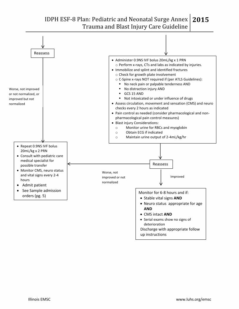
Administer 0.9NS IVF bolus 20mL/kg x 1 PRN o Perform x-rays, CTs and labs as indicated by injuries.
Immobilize and splint and identified fractures o Check for growth plate involvement o C-Spine x-rays NOT required if (per ATLS Guidelines): No neck pain or palpable tenderness AND No distraction injury AND GCS 15 AND Not intoxicated or under influence of drugs
Assess circulation, movement and sensation (CMS) and neuro checks every 2 hours as indicated
Pain control as needed (consider pharmacological and non-pharmacological pain control measures)
Blast injury Considerations: o Monitor urine for RBCs and myoglobin o Obtain ECG if indicated o Maintain urine output of 2-4mL/kg/hr
Reassess
Monitor for 6-8 hours and if:
Stable vital signs AND
Neuro status appropriate for age AND
CMS intact AND Serial exams show no signs of
deterioration
Discharge with appropriate follow up instructions
Worse, not
improved or not
normalized
Repeat 0.9NS IVF bolus 20mL/kg x 2 PRN
Consult with pediatric care medical specialist for possible transfer
Monitor CMS, neuro status and vital signs every 2-4 hours
Admit patient
See Sample admission orders (pg. 5)
Improved
Reassess
Worse, not improved
or not normalized, or
improved but not
normalized
IDPH ESF-8 Plan: Pediatric and Neonatal Surge Annex Trauma and Blast Injury Care Guideline
Assessment: □ Continuous cardiac monitoring □ Continuous pulse oximetry □ Blood pressure with all vital signs □ Routine I&O □ Strict I&O q 1 hour (maintain urine output at 2-4mL/kg/hr) □ Daily weight □ Seizure precautions □ Neuro checks ever_____ hours □ Perform CMS checks on extremities every ____hours to monitor for compartment syndrome/crush
syndrome
Tests:
Medications:
□ Fever/Pain Control: □ Acetaminophen (Tylenol) (15mg/kg/dose)_________mg PO/GT every 4 hrs PRN for
temperature ≥ 38.6°C/101.5°F or discomfort (max dose 3000mg/day) □ Acetaminophen (Tylenol) (20mg/kg/dose)_________mg PR every 4 hrs PRN for temperature ≥
38.6°C/101.5°F or discomfort (max dose 3000mg/day) □ Ibuprofen (Motrin) (10mg/kg/dose) _________mg PO/GT every 6 hours PRN for temperature
≥ 38.6°C/101.5°F or discomfort (for infants >5 months). Ensure adequate renal function before utilizing.
□ Morphine (0.1-0.2 mg/kg) _____ mg IV every 2-4 hours as needed (max 10mg/dose) □ Fentanyl _______mg IV every ______hours as needed.
□ Topical anesthetic for IV start and lab draws □ Apply topically once 30-90 minutes prior to procedure (maximum 1gm, 10 centimeter area squared, or application time of 2 hours)
IV Therapy:
□ Saline Lock □ NS bolus _______ mL IV to run over 1 – 2 hours □ LR bolus ______mL IV to run over 1-2 hours □ D5 0.45 NS with 20 mEq KCl/L to run at _________mL/hr (Ensure adequate renal function before
utilizing potassium)
IDPH ESF-8 Plan: Pediatric and Neonatal Surge Annex Trauma and Blast Injury Care Guideline
2015
Illinois EMSC www.luhs.org/emsc
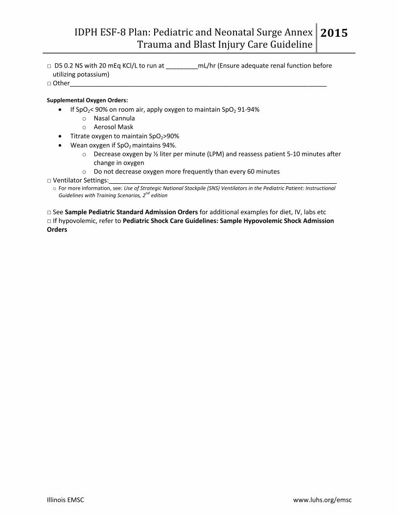
□ D5 0.2 NS with 20 mEq KCl/L to run at _________mL/hr (Ensure adequate renal function before utilizing potassium)
If SpO2< 90% on room air, apply oxygen to maintain SpO2 91-94% o Nasal Cannula o Aerosol Mask
Titrate oxygen to maintain SpO2>90%
Wean oxygen if SpO2 maintains 94%. o Decrease oxygen by ½ liter per minute (LPM) and reassess patient 5-10 minutes after
change in oxygen o Do not decrease oxygen more frequently than every 60 minutes
□ Ventilator Settings:________________________________________________________________ o For more information, see: Use of Strategic National Stockpile (SNS) Ventilators in the Pediatric Patient: Instructional
Guidelines with Training Scenarios, 2nd
edition
□ See Sample Pediatric Standard Admission Orders for additional examples for diet, IV, labs etc □ If hypovolemic, refer to Pediatric Shock Care Guidelines: Sample Hypovolemic Shock Admission Orders
IDPH ESF-8 Plan: Pediatric and Neonatal Surge Annex Trauma and Blast Injury Care Guideline
2015
Illinois EMSC www.luhs.org/emsc
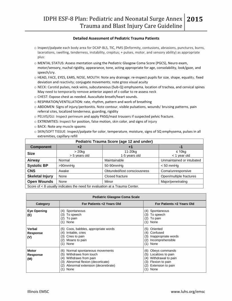
Detailed Assessment of Pediatric Trauma Patients
o Inspect/palpate each body area for DCAP-BLS, TIC, PMS (Deformity, contusions, abrasions, punctures, burns, lacerations, swelling, tenderness, instability, crepitus; + pulses, motor, and sensory ability) as appropriate plus:
o MENTAL STATUS: Assess mentation using the Pediatric Glasgow Coma Score (PGCS), Neuro exam, motor/sensory, nuchal rigidity, appearance, tone, acting appropriate for age, consolability, look/gaze, and speech/cry.
o HEAD, FACE, EYES, EARS, NOSE, MOUTH: Note any drainage; re-inspect pupils for size, shape, equality, fixed deviation and reactivity; conjugate movements; note gross visual acuity
o NECK: Carotid pulses, neck veins, subcutaneous (Sub-Q) emphysema, location of trachea, and cervical spines May need to temporarily remove anterior aspect of c-collar to re-assess neck
o CHEST: Expose chest as needed. Auscultate breath/heart sounds.
o RESPIRATION/VENTILLATION: rate, rhythm, pattern and work of breathing
o ABDOMEN: Signs of injury/peritonitis. Note contour, visible pulsations, wounds/ bruising patterns, pain referral sites, localized tenderness, guarding, rigidity
o PELVIS/GU: Inspect perineum and apply PASG/mast trousers if suspected pelvic fracture.
o EXTREMITIES: Inspect for position, false motion, skin color, and signs of injury
o BACK: Note any muscle spasms
o SKIN/SOFT TISSUE: Inspect/palpate for color, temperature, moisture, signs of SQ emphysema, pulses in all extremities, capillary refill
Pediatric Trauma Score (age 12 and under)
Component +2 +1 -1
Size > 20kg
> 5 years old 11-20kg
1-5 years old ≤ 10kg
< 1 year old
Airway Normal Maintainable Unmaintained or intubated
(6) Normal spontaneous movements (5) Withdraws from touch (4) Withdraws from pain (3) Abnormal flexion (decorticate) (2) Abnormal extension (decerebrate) (1) None
(6) Obeys commands (5) Localizes to pain (4) Withdrawal to pain (3) Flexion to pain (2) Extension to pain (1) None
IDPH ESF-8 Plan: Pediatric and Neonatal Surge Annex Trauma and Blast Injury Care Guideline
2015
Purpose: To provide guidance to practitioners caring for pediatric patients during a disaster. Disclaimer: This guideline is not meant to be all inclusive, replace an existing policy and procedure at a hospital or substitute for clinical judgment. These guidelines may be modified at the discretion of the healthcare provider.
Illinois EMSC www.luhs.org/emsc
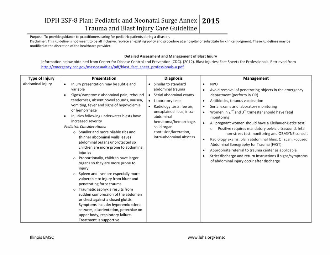
Detailed Assessment and Management of Blast Injury
Information below obtained from Center for Disease Control and Prevention (CDC). (2012). Blast Injuries: Fact Sheets for Professionals. Retrieved from http://emergency.cdc.gov/masscasualties/pdf/blast_fact_sheet_professionals-a.pdf
Type of Injury Presentation Diagnosis Management Abdominal injury Injury presentation may be subtle and
variable
Signs/symptoms: abdominal pain, rebound tenderness, absent bowel sounds, nausea, vomiting, fever and sighs of hypovolemia or hemorrhage
Injuries following underwater blasts have increased severity
Pediatric Considerations:
o Smaller and more pliable ribs and thinner abdominal walls leaves abdominal organs unprotected so children are more prone to abdominal injuries
o Proportionally, children have larger organs so they are more prone to injury
o Spleen and liver are especially more vulnerable to injury from blunt and penetrating force trauma.
o Traumatic asphyxia results from sudden compression of the abdomen or chest against a closed glottis. Symptoms include: hyperemic sclera, seizures, disorientation, petechiae on upper body, respiratory failure. Treatment is supportive.
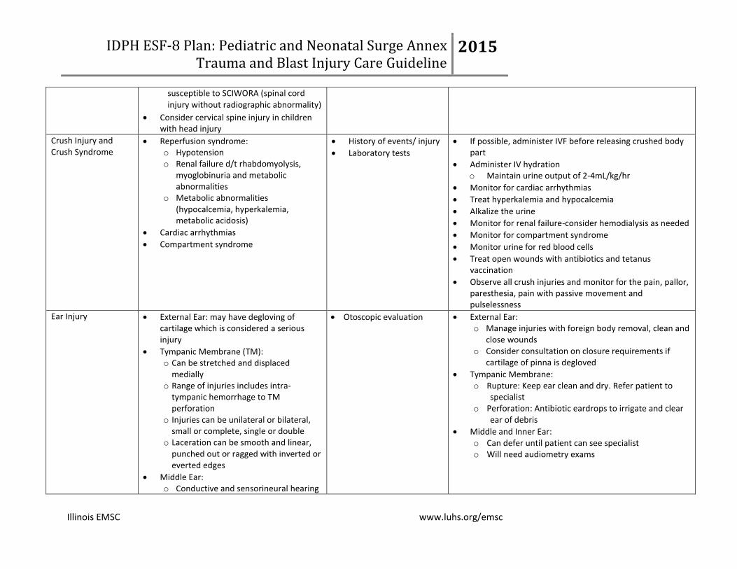
Extremity weakness or numbness Pediatric considerations:
o Traumatic brain injury (TBI) can occur in patients who have not had a loss in consciousness. Children may appear alert and awake initially but should be evaluated if they have any of the following symptoms:
Extremity Injuries Traumatic amputations: primarily occur through bony shaft rather than joint disarticulations
Fragments imbedded into extremity
Blunt force injuries
Crush injuries (see above for more information)
Document systemic musculoskeletal, neurological, and vascular states of each extremity
Document each open wound
Photograph if possible
Radiological exams as indicated
Perform thorough debridement
Antibiotics for all open fractures
Obviously contaminated wounds: o Irrigate with sterile saline; dress with Betadine
soaked sponges
Tetanus prophylaxis if indicated
Splint fractured extremities
Surgical management: o Initial debridement and bony stabilization should be
done in OR
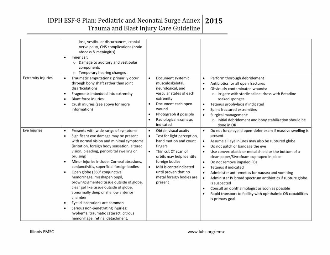
Eye Injuries Presents with wide range of symptoms
Significant eye damage may be present with normal vision and minimal symptoms (irritation, foreign body sensation, altered vision, bleeding, periorbital swelling or bruising)
Minor injuries include: Corneal abrasions, conjunctivitis, superficial foreign bodies
Open globe (360° conjunctival hemorrhage, misshapen pupil, brown/pigmented tissue outside of globe, clear gel like tissue outside of globe, abnormally deep or shallow anterior chamber
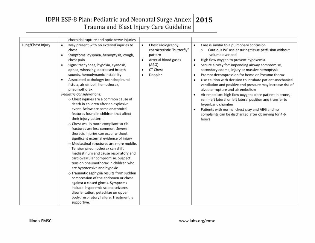
Associated pathology: bronchopleural fistula, air emboli, hemothorax, pneumothorax
Pediatric Considerations: o Chest injuries are a common cause of
death in children after an explosive event. Below are some anatomical features found in children that affect their injury pattern:
o Chest wall is more compliant so rib fractures are less common. Severe thoracic injuries can occur without significant external evidence of injury
o Mediastinal structures are more mobile. Tension pneumothorax can shift mediastinum and cause respiratory and cardiovascular compromise. Suspect tension pneumothorax in children who are hypotensive and hypoxic
o Traumatic asphyxia results from sudden compression of the abdomen or chest against a closed glottis. Symptoms include: hyperemic sclera, seizures, disorientation, petechiae on upper body, respiratory failure. Treatment is supportive.