15
2017 ANNUAL CANCER REPORT 1 UConn Health 2017 CANCER PROGRAM ANNUAL REPORT

2017 ANNUAL CANCER REPORT
1
UConn Health
2017
CANCER PROGRAM
ANNUAL REPORT
2017 ANNUAL CANCER REPORT
2
TABLE OF CONTENTS
2016 Cancer Committee Members………………………………………………………………………..
Reports:
Cancer Data Management………………………………………………………………………..
Top Ten Primary Sites of 2016…………………………………………………………………..
Top Five Primary Sites of 2016………………………………………………………………….
2016 Annual Primary Site Distribution Summary………………………………………
2016 Quality /Patient Improvement Study
2017 ANNUAL CANCER REPORT
3
2016 CANCER COMMITTEE MEMBERS/DEPARTMENTS Chairman: Dr. Susan Tannenbaum
Physician Members: Dr. Robert Dowsett Dr. Ellen Eisenberg Dr. Upendra Hegde Dr. Jayesh Kamath Dr. Douglas Gibson Dr. Melinda Sanders Dr. Pramod Srivastava Dr. Christina Stevenson
Non-Physicians: Sheri Amechi Sarah Loschiavo Marie Ziello Theresa Creamer Christopher Niemann Petra Rasor Caryl Ryan Morgan Hills Robin Schwartz Wendy Thibodeau Nancy Baccarro Alyce Ivey Christine Kaminski Leslie Bell Amber Tillinghast Wanita Thorpe Ellen Shaw

2017 ANNUAL CANCER REPORT
4
CANCER DATA MANAGEMENT
Cancer Data Management is a required component of all cancer programs accredited by the Commission
on Cancer (CoC). In 2016, the Cancer Registry accessioned 1,622 cases. Of this total, 1,010 were newly
diagnosed or analytic cases.
Cancer Data Management provides the means to collect demographics, staging, treatment, and follow-up
of each case of cancer seen at UConn Health. Data processed by the cancer registry is used to produce
data reports requested by administration and by the medical staff. All rules established by HIPAA are
observed.
There were 17,099 cases in the cancer registry database as of 3/30/17. The 2016 follow-up rate, which is
used in the calculation of survival data, was 92% for UConn. The nationwide follow-up rate is 90%.
Cancer Data Management is staffed by three full-time CTR’s and one full-time Oncology Data
Management Technician.
940984 1005
9611010
711 703745
652612
0
200
400
600
800
1000
1200
2012 2013 2014 2015 2016
Accessioned Cases 2012-2016N=8323
Analytic
Non-analytic

2017 ANNUAL CANCER REPORT
5
The mean age at diagnosis in 2016 was 64 years of age with patients ranging in age
from 7 to 90+ years. Malignancies occurred mostly in the 4th and 5th decade of life.
Geographically, the majority of the newly diagnosed patients resided in Hartford County.
In 2016, there were 763 patients from Hartford County. This represented 75% of the analytic
cases collected.
1145
89
209
296
182
138
39
10
50
100
150
200
250
300
350
0-29 30-39 40-49 50-59 60-69 70-79 80-89 90+ Unk
Years
2016 Age at Diagnosis (N=1010)
763
7735 55 25 11 26 3 13 2
0
100
200
300
400
500
600
700
800
900
2016 County at Diagnosis (N=1,010)
2017 ANNUAL CANCER REPORT
6
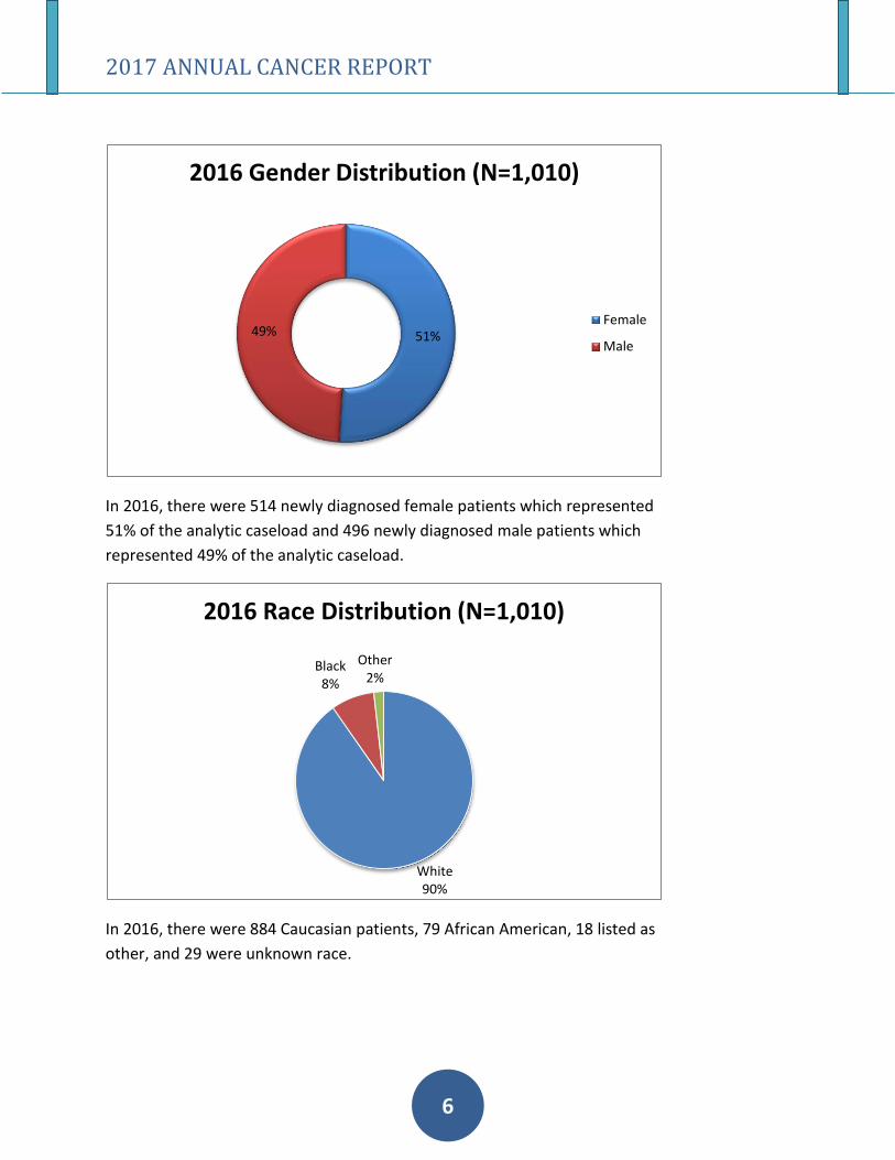
In 2016, there were 514 newly diagnosed female patients which represented
51% of the analytic caseload and 496 newly diagnosed male patients which
represented 49% of the analytic caseload.
In 2016, there were 884 Caucasian patients, 79 African American, 18 listed as
other, and 29 were unknown race.
51%49%
2016 Gender Distribution (N=1,010)
Female
Male
White90%
Black8%
Other 2%
2016 Race Distribution (N=1,010)
2017 ANNUAL CANCER REPORT
7
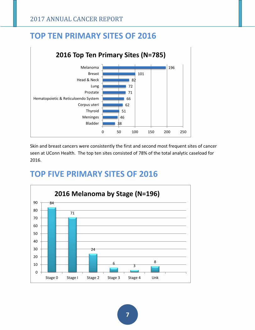
TOP TEN PRIMARY SITES OF 2016
Skin and breast cancers were consistently the first and second most frequent sites of cancer
seen at UConn Health. The top ten sites consisted of 78% of the total analytic caseload for
2016.
TOP FIVE PRIMARY SITES OF 2016
38
46
51
62
66
71
72
82
101
196
0 50 100 150 200 250
Bladder
Meninges
Thyroid
Corpus uteri
Hematopoietic & Reticuloendo System
Prostate
Lung
Head & Neck
Breast
Melanoma
2016 Top Ten Primary Sites (N=785)
84
71
24
63
8
0
10
20
30
40
50
60
70
80
90
Stage 0 Stage I Stage 2 Stage 3 Stage 4 Unk
2016 Melanoma by Stage (N=196)
2017 ANNUAL CANCER REPORT
8
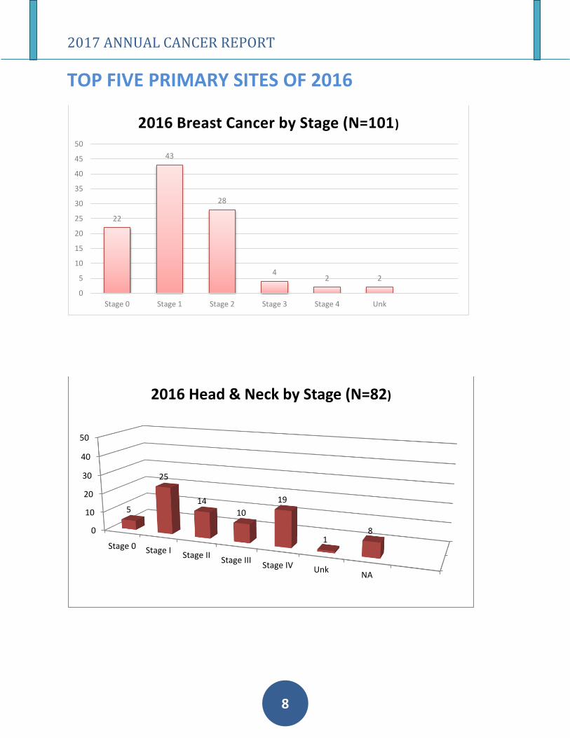
TOP FIVE PRIMARY SITES OF 2016
22
43
28
42 2
0
5
10
15
20
25
30
35
40
45
50
Stage 0 Stage 1 Stage 2 Stage 3 Stage 4 Unk
2016 Breast Cancer by Stage (N=101)
0
10
20
30
40
50
Stage 0 Stage IStage II
Stage IIIStage IV Unk
NA
5
25
14
10
19
18
2016 Head & Neck by Stage (N=82)

2017 ANNUAL CANCER REPORT
9
TOP FIVE PRIMARY SITES OF 2016
21
1
14
33
1
0
5
10
15
20
25
30
35
Stage I Stage II Stage III Stage IV Stage Unk
2016 Prostate Ca by Stage (N=71)
1
21
1
14
33
1 1
0
5
10
15
20
25
30
35
Stage 0 Stage I Stage II Stage III Stage IV Stage Unk NA
2016 Lung Cancer by Stage (N=72)

2017 ANNUAL CANCER REPORT
10
2016 Analytic Primary Site Distribution Summary
A total of 1,622 cases were accessioned into the Cancer Registry for 2016
There were 1,010 analytic and 612 non-analytic cases
Site Total Male Female Stg 0 Stg I Stg II Stg III Stg IV 88 Unk
Lip 2 1 1 0 1 0 0 0 0 1
Tongue 26 21 5 4 6 2 4 7 0 3
Salivary Glands 5 2 3 0 1 1 1 2 0 0
Floor of Mouth 4 0 4 0 1 3 0 0 0 0
Gum & Other 17 9 8 0 6 4 0 3 0 4
Tonsil 5 5 0 0 0 1 1 3 0 0
Oropharynx 2 2 0 0 0 0 1 1 0 0
Hypopharynx 2 2 0 0 0 1 0 1 0 0
Esophagus 4 3 1 0 0 1 2 0 0 1
Stomach 7 5 2 0 2 0 0 5 0 0
Small Intestine 4 1 3 0 1 0 1 0 1 1
Colon 34 20 14 5 7 7 9 5 1 0
Rectum & rectosigmoid 18 13 5 0 1 2 7 5 1 2
Anus 5 3 2 2 0 1 1 0 0 1
Liver & Intrahepatic Bile Duct 11 7 4 0 3 0 3 1 3 1
Gallbladder 1 0 1 0 0 0 1 0 0 0
Other Biliary 2 1 1 0 1 0 1 0 0 0
Pancreas 11 9 2 1 0 4 0 4 1 1
Retroperitoneum 1 1 0 0 0 1 0 0 0 0
Peritoneum, Omentum, & Mesentery2 0 2 0 0 0 1 0 1 0
Other Digestive Organs 2 2 0 0 0 0 0 0 2 0
Larynx 19 15 4 0 10 2 4 1 1 1
Lung & Bronchus 72 34 38 1 21 1 14 33 1 1
Soft Tissue 5 4 1 0 0 2 3 0 0 0
Melanoma- Skin 196 118 78 84 71 24 6 3 0 8
Other Non-Epithelial Skin 13 9 4 1 3 0 1 1 4 3
Breast 101 3 98 22 43 28 4 2 0 2
Cervix uteri 9 0 9 0 3 6 0 0 0 0
Corpus & Uterus, NOS 62 0 62 0 36 2 6 3 5 10
Ovary 12 0 12 0 4 0 3 4 1 0
Vulva 6 0 6 1 4 0 1 0 0 0
Other female genital organs 2 0 2 1 0 0 0 1 0 0
Prostate 71 71 0 0 15 41 7 6 0 2
Testis 2 2 0 0 1 0 0 0 0 1
Penis 4 4 0 1 1 2 0 0 0 0
Other Male Genital Organs 1 1 0 0 0 0 0 1 0 0
Urinary Bladder 38 22 16 15 11 6 1 3 0 2
Kidney & Renal Pelvis 18 10 8 0 11 2 2 3 0 0
Ureter 1 1 0 1 0 0 0 0 0 0
Other Urinary Organs 2 1 1 1 0 0 0 0 1 0
Brain 2 2 0 0 0 0 0 0 2 0
Cranial nerves Other Nervous System46 14 32 0 0 0 0 0 46 0
Thyroid 51 15 36 0 25 5 11 6 0 4
Other Endocrine including Thymus 18 8 10 0 0 0 0 0 18 0
Hodgkin Lymphoma 4 3 1 0 0 2 1 1 0 0
Non-Hodgkin Lymphoma 31 21 10 0 6 3 6 13 1 2
Myeloma 17 8 9 0 0 0 0 0 17 0
Leukemia 14 8 6 0 0 0 0 0 14 0
Mesothelioma 2 1 1 0 0 0 1 1 0 0
Kaposi Sarcoma 2 2 0 0 0 0 0 0 2 0
Miscellaneous 24 12 12 0 0 0 0 0 24 0
Total 1,010 496 514 140 295 154 104 119 147 51
2017 ANNUAL CANCER REPORT
11
INTRODUCTION
Endobronchial ultrasound (EBUS) was introduced in the last decade, enabling real-time
guidance of transbronchial needle aspiration (TBNA) of mediastinal and hilar structures and
parabronchial lung masses
The American College of Chest Physicians’ (CHEST) lung cancer guidelines (third edition)
summarized the data on EBUS-TBNA in the mediastinal staging of lung cancer and reported an
overall median sensitivity of 89% and a median negative predictive of 91%
Based on these findings, guidelines recommended ultrasound-guided, needle-based sampling
techniques over surgical staging as the first step in the mediastinal staging of lung cancer
OBJECTIVE
To determine the diagnostic yield of EBUS-TBNA in UConn
To establish the negative predictive value and sensitivity of EBUS-TBNA in UConn

2017 ANNUAL CANCER REPORT
12
METHODS
All patients who underwent convex and radial probe endobronchial ultrasound-guided
transbronchial needle aspiration from December 2014 to May 2015, were included in the study.
Electronic medical records were reviewed and demographic data were abstracted along with
clinical history and radiographic data.
Decision to proceed with EBUS-TBNA for investigation of lymphadenopathy, mediastinal mass
for both pathological tissue diagnosis of abnormal clinical and radiographic findings including
lymphadenopathy on CT imaging, FDG avidity on PET scanning, and mediastinal and hilar
pathologic nodal staging of lung cancer.
EBUS-TBNA PROCEDURE
All of the EBUS-TBNA procedures were conducted by a dedicated interventional pulmonologist
with or without fellows in training. All the patients were intubated and placed under general
anesthesia for the procedures. Conventional flexible bronchoscopy was first conducted to
examine the tracheobronchial tree.
DEFINITIONS
Reference standard
Cytologic analysis of EBUS-TBNA aspirates was compared with a reference standard of
definitive pathologic tissue diagnosis or a composite of at least 3 months of clinical follow-up
with radiographic imaging.
Definitive tissue sampling was defined by the cytologic evidence of lymphoid tissue,
granulomatous inflammation or tumor. The results were classified as malignant, benign
disease, normal/reactive hyperplasia, or inadequate sample. Sensitivity, specificity, negative
predictive value and diagnostic accuracy were determined for malignancy.
Diagnostic yield
Diagnostic yield was defined as frequency of a specific diagnosis in comparison to the same
diagnosis by reference standard.
RESULTS
There were 35 bronchoscopies with EBUS-TBNA utilizing both radial and convex probes
performed from December 2014 to May 2015 at the University of Connecticut Health Center

2017 ANNUAL CANCER REPORT
13
There were 3 that had no reference standards as the patient transferred care elsewhere or
refused further work up and were excluded from the analysis.
MALIGNANT
Twenty-six procedures were done due to high suspicion for malignant disease. Among the 26, 8
procedures utilized both radial and convex EBUS scopes; convex probe and routine
endobronchial biopsies were performed in 2 patients; only the convex probe was used in 13 of
the cases and 3 patients needed the radial probes alone.
There were 23 patients where the linear EBUS was used either alone or in combination with
other procedures. The diagnoses of the procedures are detailed below, relative to the
reference standard.
The diagnostic yield was 90.4%
One case had normal sized lymph nodes under endobronchial ultrasound and were not
biopsied, which turned out to have malignant disease on lymph node excision. One other
biopsy was negative but on repeat procedure at a different institution, it turned out to be
malignant disease.
Diagnosis Reference
standard
EBUS-TBNA
Squamous cell lung CA 2 2
Adenocarcinoma 4 4
Metastatic disease (other primary outside of lung) 6 4
Benign disease 6 6
Lymphoproliferative disease 1 1
Lung cancer staging 4 4
TOTAL 23 21

2017 ANNUAL CANCER REPORT
14
BENIGN DISEASE
Nine patients underwent biopsies for reasons other than suspicion for malignant disease.
There were 6 with granulomatous lymphadenitis with clinical and radiographic findings
consistent with sarcoidosis and no prior history of cancer. One had no reference standard and
was not included in the analysis. The diagnostic yield was 75%.
Diagnosis Reference Standard Diagnostic EBUS-TBNA
Granulomatous lymphadenitis 6 4
Reactive hyperplasia 1 1
Other (ILD) 1 1
TOTAL 8 6
RADIAL EBUS
There were 7 procedures using the radial EBUS probe to access peripheral lesions. Out of the 7,
only 3 were diagnostic (true positives), 2 were falsely negative and in 2 other cases, the lesions
could not be identified and so biopsies could not be done.
The 2 nodules that were falsely negative were measured at 1.0cm to 1.5cm at their narrowest
diameter by computed tomography (CT). The sizes of the 2 nodules that could not be identified
by radial EBUS were 1.2 and 1.3cm at their widest diameter by CT.
CONCLUSION
For suspicion of malignant disease,
Diagnostic yield is 90% NPV = 82% Sensitivity = 83%
Led to change in practice:
Lymph nodes >5mm under EBUS are now biopsied A different biopsy needle is being utilized for lymphadenopathy due to causes other than malignant disease
2017 ANNUAL CANCER REPORT
15
REFERENCES
Kennedy MR, Jimenez CA, Morice RC, et al. Factors influencing the diagnostic yield of
endobronchial ultrasound-guided transbronchial needle aspiration. J Bronchol Intervent
Pulmonol 2010; 17:202-208
Wahidi MM, et al. Technical Aspects of Endobronchial Ultrasound-Guided Transbronchial
Needle Aspiration CHEST Guideline and Expert Panel Report. CHEST 2016; 149(3):816-835


