94
2017 candidate guide DENTAL EXAM
2017
candidate guideDENTAL EXAM
Mission Statement
The mission of WREB is to develop and administer competency assessments for State agencies that license dental professionals.
Copyright 2017 WREB
All rights reserved. No part of this manual may be used or reproduced in any form or by any means without prior written permission of WREB.

This page intentionally left blank.

GENERAL INFORMATION ................................................................................................................. 1Welcome to the WREB Dental Exam ........................................................................................... 1WREB Exam Security and Identification Verification .................................................................... 1Malpractice Insurance ................................................................................................................... 2Exam Content ............................................................................................................................... 2Scoring Information ....................................................................................................................... 3Passing Requirements .................................................................................................................. 4Exam Results ................................................................................................................................ 4Provisional Scoring ....................................................................................................................... 4Testing Disabled Candidates ........................................................................................................ 5Dismissal for Improper Performance or Unethical Conduct .......................................................... 5Clinical Examination Overview ..................................................................................................... 6Schedule and Clinic Hours ........................................................................................................... 7Late Penalty .................................................................................................................................. 8Exam Personnel and Anonymity ................................................................................................... 8General Guidelines ....................................................................................................................... 9Infection Control Guidelines ........................................................................................................ 10Dental Assistants .........................................................................................................................11Equipment and Materials ............................................................................................................ 12Scoring Criteria and Patient Welfare .......................................................................................... 13Patient Selection ......................................................................................................................... 14Radiographs ............................................................................................................................... 16Authentication/Security ............................................................................................................... 17Alteration of Radiographs ........................................................................................................... 17Exam Preparation Material ......................................................................................................... 17
OPERATIVE ..................................................................................................................................... 24Operative Section Overview ....................................................................................................... 24Direct Posterior Class II (Composite or Amalgam) ..................................................................... 24Direct Anterior Class III (Composite) .......................................................................................... 25Indirect Posterior Class II (Cast Gold) ........................................................................................ 26Patient Acceptance ..................................................................................................................... 27Provisional Acceptance ............................................................................................................... 31Definitions ................................................................................................................................... 33Cavity Preparation ...................................................................................................................... 35Modification Procedure ............................................................................................................... 36“Dismissal for the Day” Approval ................................................................................................ 39The Finish Grade ........................................................................................................................ 39Releasing Your Patient ............................................................................................................... 40Reference Material ..................................................................................................................... 40Operative Scoring ....................................................................................................................... 41Direct Posterior Class II Composite Preparation Scoring Criteria Rating Scale ......................... 42Direct Anterior Class III Composite Preparation Scoring Criteria Rating Scale .......................... 43Direct Posterior Class II Amalgam Preparation Scoring Criteria Rating Scale ........................... 44Direct Finish Scoring Criteria Rating Scale ................................................................................. 45Posterior Composite Worksheet ............................................................................................ 47-48Anterior Composite Worksheet .............................................................................................. 49-50Amalgam Worksheet ............................................................................................................. 51-52Indirect Posterior Class II Preparation Scoring Criteria Rating Scale ......................................... 53Indirect Posterior Class II Finish Scoring Criteria Rating Scale .................................................. 54Cast Gold Worksheet ............................................................................................................. 55-56
TABLE OF CONTENTS
ENDODONTICS ............................................................................................................................... 57Endodontics Section Overview ................................................................................................... 57Tooth Selection ........................................................................................................................... 57Preparation of Teeth ................................................................................................................... 58Radiographs ............................................................................................................................... 60Submission of Arches, Radiographs and the Endodontic Worksheet ......................................... 62Rejected Teeth ............................................................................................................................ 63Back-Up Teeth for Alternate Submission .................................................................................... 64Exam Procedure ......................................................................................................................... 64Treatment ................................................................................................................................... 66After Treatment ........................................................................................................................... 67Definitions ................................................................................................................................... 68Reference Material ..................................................................................................................... 69Endodontic Scoring ..................................................................................................................... 70Endodontic Scoring Criteria ........................................................................................................ 71Endodontic Worksheet ................................................................................................................ 72
PERIODONTAL TREATMENT .......................................................................................................... 73Periodontal Section Overview ..................................................................................................... 73General Instructions .................................................................................................................... 73Patient Criteria ............................................................................................................................ 73Patient Acceptance ..................................................................................................................... 74Patient Unaccepted ................................................................................................................... 76Treatment ................................................................................................................................... 76Major Tissue Trauma .................................................................................................................. 76Treatment Grade ........................................................................................................................ 76Releasing Your Patient .............................................................................................................. 77Periodontal Treatment Scoring ................................................................................................... 78Periodontal Treatment Worksheet ......................................................................................... 79-80
END OF CLINICAL EXAMINATION ................................................................................................. 81
FREQUENTLY ASKED QUESTIONS ............................................................................................... 82

BE SURE TO VISIT US ONLINE at www.wreb.org for a complete preparation and understanding of the WREB examination process. This information supplements this Candidate Guide and is made available to you for a successful outcome!
Information for Dental Candidates
• Exam Locations, Schedule, and Fees
• Exam Procedures and Patient Requirements
• Online Application
• Exam Site Information
• CTP Exam Candidate Guide
• CTP Exam Candidate Tutorial
• Clinical Candidate Guide
• Dental Exam Candidate PreparationTutorial
• Exam Forms
• Special Accommodations Information
• Medical Emergency Cancellation Policy
• Request Score Reports/ Exam Information
• Appeals Process and Forms
• Frequently Asked Questions and Advice
General Information
• History of WREB
• WREB’s Mission Statement
• Member State Boards
• List of States Accepting WREB
• Frequently Asked Questions and Advice
Current Publications
• Current Newsletters
• Published Articles, Position Papers
Contact Us
WREB23460 North 19th Avenue, Suite 210
Phoenix, AZ 85027Telephone: (623) 209-5400Facsimile: (602) 371-8131
Email: [email protected]
Links
• Member State Boards
• States Accepting WREB
• Professional Organizations
• Credentialing Services
• Dental/Dental Hygiene Testing Agencies
• Prometric Test Centers for CTP Exam
• Dental Exam Site Schools
• Dental Hygiene Exam Site Schools
• Dental/Dental Hygiene Supplies & Equipment

This page intentionally left blank.

1
GENERAL INFORMATION
Welcome to the WREB Dental Exam
This Candidate Guide provides information needed for taking the dental exam. Study this Guide carefully. You may refer to this Guide during the exam. Please also visit the WREB website at www.wreb.org for a complete preparation and understanding of the WREB examination process.
The WREB examination (exam) has been developed, administered and reviewed in accordance with applicable guidelines from the American Dental Association, the American Association of Dental Boards, the American Psychological Association, the National Council on Measurement in Education and the American Educational Research Association. The exams were developed to provide a reliable clinical assessment for state boards’ use in making valid licensing decisions.
Since WREB member states cover a large geographical region and Candidates come from an even larger area, efforts have been made to make the exam unbiased with respect to regional practice and educational differences. WREB seeks educational diversity in the makeup of the exam review committees, including practitioners and educators who evaluate test content and develop the scoring criteria.
WREB Examiners are experienced practitioners from diverse backgrounds and locations. They are calibrated and tested prior to each exam. After the calibration training, Examiners are individually evaluated to assure they are able to grade according to the established criteria.
All official WREB documents contain the WREB logo. Schools or other individuals may prepare forms and schedules to assist Candidates. However, these documents are not authorized by WREB and may contain inaccurate information. WREB does not sponsor or endorse examination preparation courses.
You bear all risk for any misunderstanding resulting from the use of or reliance on unofficial information or material.
WREB Exam Security and Identification Verification
These instructions are your responsibility to read and understand the WREB exam security requirements.
Candidates MUST present acceptable and valid identification (ID), as described below, in order to be admitted to the WREB Dental exam. NOTE: If you have questions about the following identification requirements, you should contact the WREB Dental Department BEFORE attending the exam.
You must provide a personal photo during the exam registration process. This becomes a component of your individual Candidate Profile at WREB and will be included on all score reports to schools and state licensing boards. Your profile photo is used to create an individual WREB Candidate ID badge for the exam. This profile photo and the identification verification document will be validated at the exam by WREB personnel to verify your identity. Identification must be verified prior to admittance to any WREB clinical examination.
At the exam, you must appear in person and provide two (2) valid, non-expired forms of identification, one of which must be primary and one may be secondary.
2
Primary ID must have your photo and your signature. Acceptable forms of primary ID are:
• Government-issued driver’s license• Passport• Military ID• Alien registration card• Government-issued ID• Employee ID • School ID (must have either an expiration date – and be current or have a current date of
school year)
Secondary ID must have your name and signature. Acceptable forms of secondary ID are:
• Social Security card• Bank credit card• Bank ATM card • Library card
Make sure your IDs are current and indicate the same name you submitted to the WREB Office. This is very important for allowing you admittance to the examination.
At any time during the exam, you may be asked and should be prepared to present the primary ID and WREB Candidate ID badge to a School Coordinator, Site Coordinator, Auxiliary Coordinator or Floor Examiner.
Admittance to the exam does not imply that the identification you presented was valid. If it is determined that your ID was fraudulent or otherwise invalid, WREB will report to the appropriate governing agencies or board any Candidate or other individual who has misreported information or altered documentation in order to fraudulently attempt an exam. You are subject to dismissal from the clinical exam.
Malpractice Insurance
CNA Insurance Company, through the Professional Protector Plan in cooperation with WREB, will extend WREB professional liability coverage with the limit amounts of $1,000,000/$3,000,000 for the patient-based portion of the calendar year 2017 dental exam at no charge to the Candidates. WREB will forward the names and addresses of all candidates to CNA.
Exam Content
In addition to the evaluation of clinical abilities, diagnostic and professional judgment are also factors considered in the evaluation. For example, you are expected to know when a tooth requires a restoration, as well as the extent of restoration required.

3
For this exam, you are required to complete the following:
Operative – two restorative procedures. You will choose two restorative procedures, one of which must be a Direct Posterior Class II Composite. The second procedure can be one of the following: Direct Posterior Class II Composite Restoration, Direct Anterior Class III Composite Restoration, Direct Posterior Class II Amalgam Restoration, and Indirect Posterior Class II Cast Gold Restoration. Two Direct Posterior Class II Composite restorations are acceptable.
Endodontics – endodontic treatment on two extracted teeth; one anterior tooth and one canal on a posterior multi-canal tooth. Access and condensation are graded.
Periodontal Treatment – a Patient is submitted for approval, then root planing and scaling are completed and the Patient is submitted for grading.
Comprehensive Treatment Planning (CTP) – a computer-based examination using case materials provided by WREB. The exam is administered through Prometric Testing Centers.
Scoring Information
The WREB exam consists of two parts: the Clinical exam and the Comprehensive Treatment Planning (CTP) exam.
The Clinical exam consists of three (3) sections: Operative, Periodontal, and Endodontics. Operative and Endodontics are scored based on a Rating Scale of 1 to 5 where a final value of three (3.00) or higher is considered the passing level. The value of three (3.00) is defined to reflect minimally competent performance for all scoring criteria, and can be interpreted as corresponding to 75% in states where the passing level is legislated as 75%. The Operative and Endodontics sections are rated independently by three Examiners. Candidates receive the median (or middle) rating of the three ratings assigned by the Grading Examiners for each category. Median Examiner ratings are multiplied by assigned category weights. Weighted ratings (less any deductions) are added to obtain the score for each procedure. The procedure scores are then averaged to obtain the overall section score. Criteria definitions for rating scales, category weights, possible deductions and other scoring details are available on pgs. 41 and 70. Using the median rating precludes excessive influence by an Examiner whose opinion, in rare cases, may vary greatly from the consensus of the other two. For instance, if the three Grading Examiners assigned a 5, a 4 and a 1, the rating would be 4. Any procedure that is not brought to final completion will receive no points.
Periodontal scoring is expressed as a percentage with 75% or higher considered the passing level. Performance on the Periodontal section is rated independently by three Examiners. Periodontal Treatment scoring scale, percentages, possible deductions, and other scoring details can be found on pg. 78.
The Comprehensive Treatment Planning (CTP) examination is a computer-based examination administered by Prometric Testing Centers. The exam consists of three (3) patient cases of varying complexity, one of which is a pediatric patient. For each case, Candidates assess patient history, photographs, radiographs, and clinical information, create and submit a treatment plan, and then answer questions related to each case. Candidates are allowed three (3) hours to complete the CTP exam. Responses to each patient case are rated independently by three Examiners. Further information regarding the CTP exam can be found in the current Comprehensive Treatment Planning Candidate Guide available at www.wreb.org.

4
Passing Requirements
Successful completion of the exam requires passing all four sections (i.e., Operative, Periodontal, Endodontics, and CTP) within twelve (12) months. The twelve (12) month window begins with the first attempt at the clinical exam. The clinical exam must be attempted within the same exam year as the CTP exam. For example, if a 2017 CTP exam is taken (registered with a 2017 clinical exam), the first attempt at the clinical exam must be in 2017.
The three (3) clinical sections of the exam must be taken together. Failure to complete all three clinical sections results in failure of the exam. If two or more clinical sections are failed, the three (3) clinical sections must be retaken. Failure of one clinical section allows the opportunity to retake just the failed section within the twelve (12) month window. Exceptions to this policy will apply when the twelve (12) month period spans different testing years and significant changes to the exam occur.
If you fail the complete exam or any section of the exam three times, you are required to obtain formal remediation in the areas of failure prior to a fourth attempt. Upon failing a section a fourth time, additional remediation is required. WREB will specify the required hours of remediation. Individual states may have more stringent requirements. If you have failed the exam two or more times you should contact the state in which you are seeking licensure to obtain the state requirements.
Non-member state boards may have other scoring requirements or special restrictions. It is recommended that you check with the state board in the state where you plan to seek licensure.
Exam Results
It is WREB policy to notify you of exam results as soon as possible. Results will be posted online and can be accessed with your Candidate login and password. It is very important that you save your login information so that you may access your results. You will receive an email notice once your results are available. Exam results are confidential and will not be given over the telephone, the fax machine or by e-mail. They will only be posted to your secure WREB login online.
Notification to the Candidate of passing the WREB exam does not constitute licensure in any of the participating states. It is illegal to render Patient treatment until all state licensing requirements are met and the license certificate or letter is received from the state. A link to member states is on the WREB website.
If you do not pass the WREB exam you may elect to appeal your exam results. For information regarding the Appeal Procedure, contact the WREB office or visit the WREB website.
Provisional Scoring
Following the exam, provisional pass/fail results will be posted to Candidate profiles on wreb.org. These results are provisional until scores are reviewed and final results are posted by the WREB office. A change in outcome from provisional results to final results will not be considered a basis for appeal. WREB will make every effort to post provisional results for all Candidates, but there may be circumstances in which a Candidate's results will not be posted until the WREB office reviews and posts final official scores.
5
Testing Disabled Candidates
The WREB exam is designed to provide an equal opportunity for all Candidates to demonstrate their knowledge and ability. The exam is administered to ensure that it accurately reflects an individual’s aptitude, achievement level and clinical skills, rather than reflecting an individual’s impaired sensory, manual, or speaking skills, except where those skills are the factors the exam purports to measure.
WREB makes every reasonable effort to offer the exam in a manner which is accessible to persons with disabilities. If special accommodations are required, WREB attempts to make the necessary provisions unless providing such would fundamentally alter the measurement of skills and knowledge the exam is intended to test, would result in an undue burden, or would provide an unfair advantage to the disabled Candidate.
The appropriate professional (physician, psychologist, etc.) must complete a form obtained from the WREB website specifying what special accommodation is requested and attesting to the need for the accommodation. This must be received in the WREB office no later than 45 days prior to the exam.
WREB reserves the right to authorize the use of any accommodation to maintain the integrity and security of the exam.
Dismissal for Improper Performance or Unethical Conduct
Dismissal from the exam, failure of the exam, or reduction in an exam score may result from Improper Performance (as defined below) relative to procedural skills and clinical judgment, and/or Unethical Conduct (as defined below).
If a Candidate engages in Improper Performance or Unethical Conduct, the Candidate must obtain permission from the WREB Board of Directors before taking the exam at a later date.
Examples of Improper Performance include, but are not limited to:
• A case selection presenting conditions which jeopardizes successful Patient treatment within the parameters of the exam
• Disregard for Patient welfare and/or comfort• Failure to recognize or respond to systemic conditions which potentially jeopardize the
health of the Patient, Assistant or Examiners• Unprofessional, unkempt, or unclean appearance• Rude, abusive, or uncooperative behavior• Disregard for aseptic technique• Procedure generates excessive trauma to tissue• Performance grossly inadequate in the validated judgment of the Examiners• Failure to adhere to published WREB Guidelines
6
Examples of Unethical Conduct include, but are not limited to:
• Using unauthorized equipment at any time during the exam• Using unauthorized Assistants• Using unauthorized Patients• Altering Patient records or radiographs submitted in any format• Treating Patients outside clinic hours or receiving assistance from another practitioner• Altering Endodontic teeth• Dishonesty• Altering Candidate worksheet or treatment notes• Communicating written or electronic (computer) test item information to other Candidates
or individuals• Altering, omitting or attempting to disguise treatment performed on a patient• Any other behavior which compromises the standards of professional behavior
If a Candidate engages in Improper Performance or Unethical Conduct, in addition to dismissal from the exam, failure of the exam, or reduction in an exam score, WREB reserves the right to take any other reasonable action WREB deems appropriate, including, but not limited to reporting the Candidate to: (i) the various state licensing boards, (ii) the Candidate’s dental school, (iii) other dental or dental hygiene testing organizations or (iv) other professional organizations.
Clinical Examination Overview
The clinical examination (Operative, Endodontics and Periodontal) is a 2½ day exam. Your exam officially starts when you submit your first procedure for acceptance. In most cases, this will be when Endo sextants are submitted. If not performing Endo, at the time the first patient is submitted. Withdrawal for any reason after this point constitutes failure of the exam.
During the 2½ days you will be assigned a 4½ hour block for the Endodontics exam. About four weeks prior to your exam, you will receive a candidate schedule with your Endodontic block. The remainder of the time is open. You may schedule your operative and periodontal Patients as you wish during this open time. This allows flexibility in Patient scheduling and gives you an opportunity to schedule a backup Patient, if needed. The exam schedule is as follows:
Orientation Day 11:30 a.m. – 12:00 p.m.
1:30 – 2:30 p.m.
Following Orientation
Turn in endodontic models at location specified on Candidate schedule
Candidate Orientation at location specified on Candidate schedule
Pick up Candidate packets (ID required, refer to pgs. 1-2)
Clinic Days 1 and 2 8:00 a.m. – 4:30 p.m. Assigned endodontic block (4½ hours)Operative and Periodontal as desired
Clinic Day 3 8:00 – 11:00 a.m. Complete operative and Periodontal procedures, if necessary

7
It is not unusual to finish the exam by the end of the second clinical day. There is more than sufficient time to complete all procedures and to accommodate unexpected situations. The final half-day is provided for Candidates encountering unexpected circumstances that require extra time to complete procedures.
Schedule and Clinic Hours
The two Operative and Periodontal Treatment procedures may be performed anytime you are not specifically assigned to the Endodontics exam. If you complete the Endodontics procedure prior to the end of your assigned time you may return to the clinic and continue the clinical procedures.
Under certain circumstances, approval and completion of restorative procedures may be done on different days. However, to avoid penalty, you must perform the Periodontal Treatment procedure on the day it is approved. Refer to specific procedure sections of this Guide for more information.
Patients with procedures to be graded must be checked in by 4:30 p.m. on the first two days of the exam, and by 11:00 a.m. on the final day of the exam. After this time, 0.2 points are deducted from each procedure to be graded for each five minutes the Patient is late. If a Patient is 16 or more minutes late, the procedure will not be graded and no points will be earned.
WREB official time is based on the local time for each exam site. Cell phone time will be used to determine late penalties for operative and periodontal procedures. For the Endodontic exam, a separate, official clock will be designated in the lab.
All clinical procedures must be completed by 5:00 p.m. on Clinic Days 1 and 2. After 5:00 p.m., you are only permitted to:
• Place a temporary• Dismiss the Patient• Clean operatory unit• Leave the clinic
All Candidates and Patients must be out of the clinic by 5:30 p.m. or a late penalty will be deducted from the appropriate procedure score.
The Candidate Clinic at each exam site will open at the following times:
Day 1: Clinic opens at 7:00 a.m. Floor Examiners will be available at 7:30 a.m. Provisionally accepted patients may be examined for final approval by Floor Examiners. Patients may be submitted at 7:45 a.m. for Check-In. Examiners will begin seeing patients at 8:00 a.m. Candidates in the A Group may not submit Patients until 10:00 a.m. on Clinic Day 1.
Day 2: Clinic opens at 7:00 a.m. Floor Examiners will be available at 7:30 a.m. Patients may be submitted at 7:45 a.m. for Check-In. Examiners will begin seeing patients at 8:00 a.m.
Day 3: Clinic opens at 7:00 a.m. Floor Examiners will be available at 7:30 a.m. Patients may be submitted at 7:45 a.m. for Check-In. Examiners will begin seeing patients at 8:00 a.m. The exam ends at 11:00 a.m.
8
During the first hour of each day, you may set up your operatory and prepare your Patient for the day’s procedure. No local anesthetic is to be administered to patients until Floor Examiners are present at 7:30 a.m. and your patient's Medical History form is reviewed and initialed by a Floor Examiner. For Patient comfort, Patients should not be sent to the grading area until the time scheduled for Patient submission (7:45 a.m.).
Candidate operatories will be consolidated into one area on Clinic Day 3. Therefore, your operatory may be reassigned on this day. An announcement will be made during Candidate Orientation and signs will be posted advising where the clinic has been moved.
Late Penalty
Rated procedures (Operative and Endo)
1 to 5 minutes late: 0.2 deduction
6 to 10 minutes late: 0.4 deduction
11 to 15 minutes late: 0.6 deduction
16 or more minutes late: Procedure will not be graded. No points earned.
Percentage Procedures (Periodontal Treatment) (deducted from total possible for Periodontal Treatment)
1 to 5 minutes late: 4% deducted
6 to 10 minutes late: 8% deducted
11 to 15 minutes late: 12% deducted
16 or more minutes late: Procedure will not be graded. No points earned.
It is possible that the exam might be terminated in less than 2½ days due to a situation beyond the control of WREB, such as loss of power or act of nature. If this should occur, incomplete procedures cannot be carried over to a future exam. WREB cannot be held liable in these circumstances.
Exam Personnel and Anonymity
The WREB exam is conducted in a manner that is intended to provide total anonymity to remove possible bias from the scoring of Candidate work. All exam materials are numbered with a Candidate ID number. This number is randomly assigned prior to the exam and a sheet of badges with the number is provided at the exam. A badge must be worn at all times during the exam. Your name must not appear on any exam material including worksheets and radiographs. Only a Patient’s first name should be used on materials that are seen by Grading Examiners. Grading Examiners are separated from Candidates so there is no direct contact between Grading Examiners and Candidates. You will assist in keeping the exam anonymous by observing all signs and instructions.
9
WREB has two (2) categories of Examiners: Grading Examiners and Floor Examiners. Grading Examiners are segregated from Candidates during the examination. Patients are sent to a separate grading area for graded procedures. This allows the Grading Examiners to grade the procedure without knowledge of the Candidate.
Anonymity is preserved between the Grading Examiners and Candidates, not among Examiners themselves. There are occasions when fairness requires consultation among Examiners. Examiners are encouraged to consult whenever necessary. Examiner consultation generally benefits Candidates and should not be a reason for concern.
There are two (2) to four (4) Floor Examiners at each examination.
Floor Examiners do not serve in a grading capacity so there is no anonymity between Floor Examiners and Candidates. Floor Examiners serve as liaisons between Candidates and Grading Examiners to solve any problems that may arise during the exam. They are on the clinic floor to assist with questions or problems relating to the administration of the exam, and to approve certain phases of clinical procedures. Floor Examiners can help you by answering questions, clarifying exam procedures and acting as liaison between you and Grading Examiners. In addition, Floor Examiners can help with:
• Extra forms, such as Patient Medical History/Consent Form or Follow-Up Care Agreements• Providing Amalgam and Cast Gold Worksheets• Checking and signing Patient Medical History forms• Distributing communication forms from Grading Examiners • Checking in patients who have been provisionally accepted• Checking modification requests (See Operative Modification Procedure)• Managing a pulp exposure• Checking and initialing steps on worksheets
Any Floor Examiner in any area of the clinic can assist you. They are not assigned to specific areas. Ask the first Floor Examiner available for assistance.
You should always bring your worksheet when asking questions regarding procedures.
General Guidelines
A. Only Candidates, Patients and Assistants are allowed on the clinic floor. Candidate and Assistant identification badges must be visible on the chest or collar on the outer most layer (i.e., disposable gown) at all times during the exam. You will not be allowed in the Endodontic lab for your scheduled exam without showing your Candidate ID.
B. This exam uses the American System of tooth identification. Permanent teeth are recorded clockwise from the upper right quadrant to the lower right quadrant.
10
C. Worksheets must be completed in ink – not pencil. If you make an error prior to Patient acceptance, obtain a new worksheet (cross-outs are not accepted at acceptance). If you submit a worksheet that is not neat, clear and in ink, the Patient will be returned to you with a new worksheet to complete, resulting in lost time.
D. All electronic devices should be turned off or set to a mode that will not disturb other Candidates in the main clinic. Electronic devices, including cell phones, are prohibited in the Endodontic exam and the grading area. Patients with electronic devices will not be graded, but returned to you to leave the device, resulting in lost time.
E. Neither WREB nor any agency participating in the exam process accepts responsibility for treatment rendered to Patients during the exam. A Consent Form must be signed by Patients.
F. No surgical procedures may be done.G. Procedures presented for grading during the exam may be photographed or digitally
scanned by WREB personnel. These photographs are for use in training and calibrating Examiners. They have no relation to the grading process and cannot be released to Patients or Candidates.
H. The school provides information regarding the facility, supplies, hotels, commercial labs and other topics which can assist in preparing for the exam. This information is provided directly by the school; WREB is not responsible for its accuracy. Specific exam site information is available at www.wreb.org under "Exam Site Information."
I. Laboratory facilities are available at some schools if you wish to do your own lab work for the indirect procedure. A commercial lab may be used. However, no appellate procedure may be based on the performance of any commercial lab. Representatives of commercial laboratories are not allowed on the clinic floor during the exam. A designated location is set up outside the clinic for transfer of impressions and castings. See exam site information for details on laboratory facilities.
Infection Control Guidelines
Appropriate aseptic technique is an important component of the professional standard of dental care. You are expected to maintain acceptable standards during the exam. Failure to do so may result in a reduction of exam scores. The following are the minimally accepted standards:
• Appropriate attire is required while in the clinic. A lab coat, lab jacket or disposable gown are all acceptable if they are long sleeved. Scrubs may be worn under a lab coat, lab jacket or disposable gown. Color and style are not restricted. Your Candidate ID badge must be worn in a visible location on the outside of clinic attire. Clinic attire should not be worn outside the clinic if it has been contaminated.
• Clinic attire must be changed whenever visibly soiled. • Antiseptic soap is provided for hand washing.• Exam gloves must be worn during all Patient contact. When performing functions other
than direct Patient treatment, remove exam gloves or use over-gloves. Gloves must be changed between Patients and whenever the integrity of the glove is compromised. Schools provide gloves but cannot accommodate individual preferences. If you require a specific brand or size you should bring your own.
• Masks covering the nose and mouth must be worn during all procedures that generate aerosols. Schools provide masks but cannot accommodate individual preferences. If you have specific mask requirements you must provide your own. Masks must be changed whenever visibly soiled.

11
• Protective eyewear is required for you and your Assistant and must be worn during all procedures. You must provide your own eyewear. Use of a face shield is acceptable in lieu of eyewear.
• Protective eyewear is required for Patients (prescription glasses or safety glasses) during all Patient procedures, evaluation and grading. You are responsible for ensuring that your Patient is equipped with protective eyewear.
• Schools provide specific written instructions that must be carefully followed regarding:• Asepsis of the surfaces and equipment in the operatory to assure adequate disinfection
of all surfaces and equipment before and after each use.• Proper disposal of biohazardous waste.• Sterilization procedures for instruments. All instruments, including handpieces, are to
be sterilized between Patients. • “Sharps” containers are located throughout the clinic. All sharps must be disposed of
properly.• Food and beverages are prohibited in the clinic.
Dental Assistants
Dental Chair-Side Assistants may be used during clinical procedures. Dental Assistants may work with Floor Examiners on your behalf. Patients may be sent to the grading area by Dental Assistants if all paperwork is complete and instruments are present.
Assistants are not allowed to attend Candidate Orientation.
Only one Dental Assistant and only the one dental chair assigned to you can be used at any time.
Periodontal Treatment Assistants may not be dentists (including graduates of foreign dental schools), dental hygienists (including graduates of foreign dental hygiene schools), or dental hygiene students. Assistants may be Dental Assistants or dental students, if they are not in their final year of dental school. For purposes of the exam, WREB considers the final year of dental school as beginning September 1.
Operative Assistants may not be dentists (including graduates of foreign dental schools) or be in their final year of dental school. For purposes of the exam, WREB considers the final year of dental school as beginning September 1.
Operative Assistants may be Dental Assistants or Dental Hygienists, if they do not hold a permit to place and finish restorative materials.
Use of unauthorized Assistants is grounds for immediate dismissal from the exam.
A Dental Assistant Verification form, provided in your Candidate packet at the exam, must be completed and signed by you and your Assistant(s). This form must be completed and submitted to WREB at the end of the exam, even if an Assistant is not used.
Assistants are required to follow the same guidelines as Candidates. You are responsible for your Assistant(s)’ adherence to all guidelines.

12
Equipment and Materials
Equipment information specific to each school can be found in the "Exam Site Information" at www.wreb.org. Although schools supply some expendable materials, you are responsible for ensuring that you have all materials necessary to perform the required procedures, including high-speed and low-speed handpieces and periodontal scaling devices. Schools may have equipment available for rent if you choose not to bring your own. Information on rental equipment is included in the "Exam Site Information." Instruments must be acceptable even if rented.
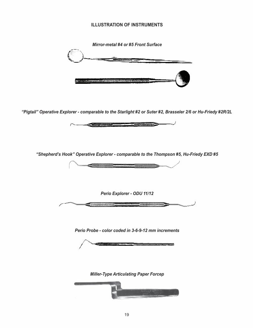
A. Special instruments for the Operative procedures are (illustrations, pg. 19): • New (unscratched) #4 or #5 metal front surface mouth mirror • New (sharp) pigtail explorer comparable to the Starlight #2, Suter #2, Brasseler 2/6
or Hu-Friedy 2R/2L• New (sharp) shepherd’s hook explorer comparable to the Thompson #5,
Hu-Friedy EXD #5• Miller-type Articulating Paper Forceps (not cotton pliers)
B. Special instruments for the Periodontal Treatment procedure are (illustrations, pg. 19): • New (unscratched) #4 or #5 metal front surface mouth mirror• New (sharp) ODU 11/12 explorer (may be American Eagle, Hartzell, Nordent, or
Hu-Friedy)• New periodontal probe, color coded with legible 3-6-9-12 mm markings (may be
American Eagle, Hu-Friedy, or Marquis)• It is recommended that you bring back-up instruments
C. A blood pressure measuring device is required.The schools have agreed to provide the following expendable materials: Anesthetic (local and topical), composite restorative materials, amalgam capsules, articulating paper, autoclave tape, cement, cotton pellets, cotton rolls, cotton swabs, cotton squares, instrument trays, deck paper, disinfectant, drinking cups, evacuator tips, face masks, facial tissue, floss, gloves, headrest covers, hemostatic agents, impression materials, mouthwash, needles (long and short), paper towels, Patient bibs, polishing materials for restoration, prophylaxis paste, retraction cord, rubber dams, rubber dam napkins, soap, standard saliva ejectors, trash bags and tray covers.
Materials provided are brands used by the school. If you wish to use a specific brand you must bring your own. You should provide any materials not specifically listed in the "Exam Site Information."
D. X-ray developer and fixer are supplied in the endodontic lab at schools with conventional radiographic facilities. Automatic and/or hand developers are provided by the school. A list of other materials provided in the lab can be found in the "Exam Site Information." You must supply any items needed to perform the endodontic procedure which are not on the list.
E. If using a sonic or ultrasonic device for periodontal treatment, you must provide your own and it must be adaptable to the hookups at the school. Information regarding hookups can be found in the "Exam Site Information."
F. You will be furnished with a dental chair, an operatory unit, and an operator’s stool. Personnel are available throughout the exam to resolve malfunctions of operatories and equipment provided by the school. If you have an equipment malfunction in the clinic

13
you should notify maintenance personnel and a Floor Examiner immediately. The Floor Examiner may determine that you are eligible for time compensation (on that day only) if the equipment malfunction cannot be resolved within 15 minutes. Time is not compensated for delays of less than 15 minutes. Time is determined from the point at which a Floor Examiner is notified. Many equipment malfunctions are due to improper use. You should become familiar with the equipment prior to the exam and follow all directions carefully. WREB cannot be responsible and will not compensate for time lost due to the malfunction of your personal equipment or rental equipment.
Scoring Criteria and Patient Welfare
Because WREB serves as a testing agency, not a teaching agency, performance that fails to meet examination standards does not always require immediate corrective action and may not present an immediate health concern for the Patient.
Patients participating in WREB exams may be released from the exam with restorations or treatments that received a failing score without Examiners requiring immediate correction of the condition. A failing score is an indication of not meeting exam criteria even though the restoration might still be serviceable. Only the most severe conditions, which could constitute an immediate threat to the Patient’s health, are identified by the Examiners with a Postoperative Care (PO) form. A Postoperative Care form is completed for the following situations:
• Soft tissue laceration or mutilation or major iatrogenic tissue trauma• Pulp exposure• Fractured direct restorations• Margins of restorations so defective that the tooth would be endangered if not treated prior
to the next regular recall exam• Contacts (interproximal) so defective that the tooth or periodontium would be endangered
if not treated prior to the next regular recall examAn Instructions to Candidate (IC) form may be completed by the Grading Examiners to request removal of caries, affected dentin or unsound demineralized enamel, to request additional radiographs, to request adjustment of occlusion or for any other communication that an Examiner determines appropriate.
Although the conditions that initiate a Postoperative Care or Instructions to Candidate form also may result in a low score in one or more of the scored categories, scoring is an independent event and is based only on the established criteria. Receiving either form is not an indication of procedure or exam failure. Absence of these forms does not assure satisfactory completion of any procedures. For example, it is possible that a rating of “2” is appropriate in a category because of elements in the criteria, but there is no immediate threat to the Patient’s health and no need for immediate exam site correction. No forms would be issued, even though the procedure score would be failing.
A Follow-Up Care Agreement form must be completed for each Patient. If a Patient is used for more than one procedure by the same Candidate, only one form needs to be completed with all procedures indicated on the form for that Patient. If a Patient is shared by one or more Candidates, each Candidate must complete a Follow-Up Care Agreement for that Patient. Prior to arriving at the exam, have a dentist accessible to the Patient (licensed in the state in which the Patient resides) who acknowledges the responsibility of providing any necessary postoperative care, sign the form. Give the yellow copy of the form to the Patient after they sign the form. The white copy

14
is turned in at the end of the exam in the Candidate Packet. If you are unable to have a licensed dentist sign the Follow-Up Care Agreement in advance (Patient is obtained during the exam), the form may be completed after the exam and either emailed, faxed or mailed to the WREB office. Final exam scores will not be released to the Candidate or any State Boards until the form is received.
Patient Selection
The following criteria apply to all Patients for the clinical exam:• The minimum Patient age for the Periodontal Treatment procedure is 18 years. There is
no minimum age for Operative procedures.• Patients cannot have completed more than two years of dental school.
Patient selection is an important factor in the clinical exam. You must provide a Patient or Patients for the Restorative and the Periodontal Treatment procedures.
Patient selection is your responsibility. WREB staff, the Boards of Dentistry of participating states, and dental schools are not able to supply Patients. You are graded on your ability to accurately determine and effectively interpret Patient qualification criteria. This is an integral part of the examination. Therefore, other professionals should not “prequalify” your Patient for the examination.
WREB strongly discourages the use of Patient procurement services. Patient procurement services are not allowed in the school during the examination. Use of such services is absolutely not necessary for success on the exam. Patient acceptance criteria are designed to standardize the exam, not as an obstacle to Patient procurement. Reading the criteria and understanding the broad range of Patients acceptable for the two operative restorations and the periodontal treatment procedure will enable you to evaluate your own Patients’ qualifications. The Patients accepted by WREB are Patients you routinely treat in a school dental clinic or a dental office. To increase the likelihood of success, WREB encourages you to procure patients for the exam whom you routinely treat in dental school or your dental office.
One Patient may be used for all three procedures if the criteria are met. Candidates may share a Patient if the criteria are met. Patients with a need for antibiotic prophylaxis may not be shared with other Candidates at the exam. You bear all risks and benefits associated with using the same Patient for more than one procedure or sharing a Patient with another Candidate.
If you share a Patient with another Candidate, each Candidate must submit the procedures separately for approval to start and for the preparation and finish grading.
If using more than one Patient, you may work on one Patient at your own operatory while another Patient is in the grading area. If a Patient is approved by the Grading Examiners, no appellate procedure may be based on the difficulty of the procedure submitted.
Incomplete procedures cannot be evaluated. Therefore, an additional consideration in your Patient selection is the cooperative attitude of the Patient. A Patient should not be selected who is apprehensive, hypersensitive or is unable to remain until the examination is completed. If your Patient is unable to be examined by three Examiners, you will fail that procedure.

15
Patient Medical History (sample form pgs. 21-22)• WREB accepts Patients with a blood pressure reading of 159/99 or below. A Patient with
blood pressure readings between 160/100 and 180/110 is accepted only with written consent of the Patient’s physician. WREB does not allow treatment of any Patient with a blood pressure reading greater than 180/110. Preoperative blood pressure and pulse must be taken on each Patient prior to acceptance and recorded on the Patient Medical History form.
• Obtain written clearance and/or antibiotic prophylaxis from a physician or dentist in the case of any significant medical problem. The medical clearance must indicate the specific medical concern. WREB adheres to the current American Heart Association Guidelines regarding required premedication. Patients with a need for antibiotic prophylaxis may not be shared with other Candidates at the exam.
• Any Patient who has received intravenous bisphosphonates for bone cancer or severe osteoporosis is not acceptable for the exam.
• Any Patient with diabetes controlled by insulin injection(s) or an insulin infusion device is not acceptable.
• Any Patient who has had a heart attack, stroke, or cardiac surgery within the past six (6) months is not acceptable.
• Any Patient who has clinical symptoms of active tuberculosis (clinical symptoms would include a productive cough or chest pain) is not acceptable.
• Any Patient with a known latex allergy is not acceptable.• Any Patient who has been diagnosed as HIV positive must present a medical consult with
permission to sit for the exam. • Any Patient who is known to be pregnant is not acceptable, except with the written consent
of the Patient’s health care provider.• Any Patient with problems which might be aggravated by the length or nature of the exam
may be rejected at the discretion of the Examiners.A legal consent is provided on the back of the Patient Medical History form and must be signed by the Patient. If a Patient is under the age of legal consent for the state in which the exam is given, the Consent Form must be signed by the parent or legal guardian of the underage Patient.
If you are using the same Patient for more than one procedure you may submit one Medical History and Consent Form for that Patient with all procedures indicated. Candidates who share a Patient must submit separate Medical History and Consent Forms for the procedure(s) performed on the Patient. The Patient must sign Medical History and Consent Forms for each Candidate who performs procedures on them.
Your Patient is essential to your success on the exam. Treat all Patients with care and compassion. Patients should receive nourishment during the exam. Special care must be taken when sharing Patients or using one Patient for multiple procedures to ensure the Patient receives adequate breaks and nourishment. Patients who are unable to be graded due to hypoglycemia or severe dehydration may result in a failing grade.
Patients should be given directions to the school, parking information, directions to the clinic and should be aware of the time commitment due to the nature of the exam and your exam schedule.
Patients should be prepared for temperature extremes in the clinic. Headphones, newspapers, books and magazines are permissible outside of the grading area. Electronic devices, including cell phones, are not allowed in the grading area.

16
Patient comfort should be considered and proper local anesthetic utilized as needed.
Any form of inhalation, parenteral or enteral sedation cannot be used during the exam. Patients must be ambulatory.
Radiographs
Preoperative radiographs are required for the two Restorative procedures, the Periodontal Treatment procedure and the Endodontic exam. Specific radiograph requirements for each procedure are outlined in each section of this Guide.
WREB accepts the use of conventional and digital radiographs as long as they are of diagnostic quality. Because schools differ in their radiographic facilities, please refer to the "Exam Site Information" for the site where you plan to take the exam to determine what is available (found on the website at www.wreb.org). Some exam sites will have only conventional facilities available, some will have only digital, and others will have both. It is important that you are prepared for what is available at the exam site you have selected. For the Endodontic exam, it is acceptable to submit radiographs in one format for preoperative images and a different format for postoperative images.
You should also read the exam site information carefully to determine if a digital site is equipped for secure transmission of images between different exam sites, or from your school to the exam site. It may be necessary to submit printed digital images. Depending on the facilities available, different portions of the information below will apply.
A. Digital Radiographs
All digital radiographs must be diagnostic. Examiners will view all images, printed or on monitors, as though they are mounted “button out.” Format your submissions accordingly.
Endodontic images, printed or on monitors, must include a 2.0 mm sphere for measuring.
• Digital Images on MonitorsOnly the radiographs being submitted for approval should be saved in the folder accessed by Examiners. All images submitted for a procedure must fit on one screen without overlap. The individual images should be no larger than three times the size of a conventional radiograph.
• Printed Digital ImagesPrinted digital images must be printed on high quality photographic paper. One printing is required for each submission. All printed images for each procedure must fit on one 8 ½” by 11” page without overlap and individual images should be no larger than three times the size of a conventional radiograph.
Printed digital images must include a label in legible print that includes Candidate ID, patient's first name, procedure, tooth number and surface.

17
B. Conventional Radiographs
• WREB accepts the use of conventional radiographs at all examination sites, as long as they are of diagnostic quality.
• Conventional films may be interpreted by Examiners using loupes with 2.5 X magnification or greater and backlighting (i.e., view box).
The use of image analysis tools, such as zoom and magnifier, will not be a part of an Examiner’s evaluation of digital images.
Perform all enhancement or edge sharpening prior to submitting images for Patient acceptance. It is your prerogative to use these feature(s) in digital or scanned conventional format to provide the best radiographic images for Examiner assessment.
Authentication/Security
All digital radiographs must be of diagnostic quality. Image capture stations are specified by the site. After capture transfer to the server, select images for uploading and enhance them as desired. The host site will provide specific radiographic personnel during Candidate screening and testing times. No individual other than the Candidate will be allowed to assist in image selection or editing for submission. A final archival disc will be provided to WREB by the host site for all digitally stored Candidate radiographs at completion of the exam.
You may submit digital radiographs from another dental school other than your exam site using equipment and information systems that conform to the DICOM Standard. Electronic transmission of digital radiographic images will be considered secure and authentic if they are received by designated exam personnel and never leave the DICOM secure format. If digital radiographs do not conform to DICOM Standard format, you may choose to take digital radiographs at the exam site, submit conventional films, or provide printed digital images.
Alteration of Radiographs
An altered radiograph is defined as a change to the proprietary tag of the format file. Intentionally performing any alteration, including but not limited to, cropping, compressing or “doctoring the image” as in a Photoshop®-type program is prohibited.
When applying for your exam online, you will electronically sign an affidavit that the radiographs submitted are original, unaltered films. (Periodontal films may be duplicates.)
Should analysis by WREB detect radiographic alteration of submitted digital images or conventional films, failure of the examination for unethical conduct will occur. If there is a question, you will be required to retake the radiographs with an observer present.
Exam Preparation Material
With this Candidate Guide, you should have received the following items:• Three (3) Follow-Up Care Agreement forms to be signed in advance by a dental care
provider and your Patients• Three (3) Patient Medical History and Consent Forms• An Endodontic Worksheet• A ziplock bag with a blank label
18
A Candidate preparation video is available on the WREB website.
Candidate Orientation is held the first day listed on the exam schedule. Following Candidate Orientation, you will receive your Candidate Packet containing:
• Worksheets for the Direct Composite procedures. Direct Amalgam and Indirect Posterior Class II (Cast Gold) worksheets are available on request at the end of Candidate Orientation. Please see a Floor Examiner.
• Worksheet for the Periodontal Treatment procedure• Candidate ID badges• Assistant ID badges• A Dental Assistant Verification form• Labels to use on Patient bibs when clinical procedures are graded• Three (3) Patient Information and Questionnaire pamphlets
Keep the packet envelope to submit required exam material to WREB personnel at the conclusion of the exam. Packets will be collected throughout the exam at the patient check-in desk outside the grading area.
19
ILLUSTRATION OF INSTRUMENTS
Mirror-metal #4 or #5 Front Surface
“Pigtail” Operative Explorer - comparable to the Starlight #2 or Suter #2, Brasseler 2/6 or Hu-Friedy #2R/2L
“Shepherd’s Hook” Operative Explorer - comparable to the Thompson #5, Hu-Friedy EXD #5
Miller-Type Articulating Paper Forcep
Perio Probe - color coded in 3-6-9-12 mm increments
Perio Explorer - ODU 11/12

20
This page intentionally left blank.

21
SAMPLEAnswer the following questions as completely and accurately as possible:
1. Are you taking any medication, pills or drugs (prescribed or not)? YES NO If yes, please list:
2. Do you have a sensitivity or allergy to latex? YES NO If yes, please list:
3. Are you allergic to any medicines? YES NO If yes, please list:
4. Have you ever received intravenous bisphosphonates for bone cancer or YES NO severe osteoporosis? If yes, please list:
5. Are you under the care of a physician at the present time or have you been treated by a YES NO physician in the past six months? If yes, for what condition?
6. Do you have, or have you been exposed to, any disease or condition not listed above that YES NO we should know about? If yes, please list:
7. Women only: Are you pregnant? YES NO If yes, expected due date:
Instructions to the Patient: Have you had or have you ever experienced any of the following conditions?Circle “YES” or “NO” to all questions.
Patient’s Initials:Instructions to Candidate:
Circle any “YES” answers. State in the lines below the significance (if any) and the steps taken for any alteration of procedure for this exam. Indicate the need and use for premedication, if necessary. Record all medication taken today on the back of the procedure worksheet. Attach any verification of the patient’s medical acceptability. A Floor Examiner must initial this form prior to the administration of local anesthetic and before the patient is sent to the grading area for “patient check-in.”
Consent Form on Reverse
Patient Blood Pressure Patient Pulse Floor Examiner Initials
PATIENT MEDICAL HISTORYAmalgam Anterior Composite
Posterior Composite 2Cast GoldPeriodontal Treatment
PATIENT’S FIRST NAME: CANDIDATE I.D. #:
DATE OF EXAMINATION: EXAM SITE:
A Heart Condition YES NO H Diabetes YES NOB Heart Surgery YES NO I Tuberculosis YES NOC Valve Replacement YES NO J Kidney/Renal Disease YES NOD Stroke YES NO K Hepatitis/Jaundice YES NOE High Blood Pressure YES NO L HIV Positive YES NOF Bleeding Disorder YES NO M Epilepsy/Seizures YES NOG Asthma/Lung/Respiratory Condition(s) YES NO N Joint Replacement YES NO
Use Ink
2017 - Revised
Posterior Composite 1
Front

22
SAMPLE
PATIENT CONSENT FORM AND ASSUMPTION OF RISK
Western Regional Examining Board, an Arizona non-profit corporation (“WREB”) is a national dental and dental hygiene testing agency required to test candidates’ clinical skills for the states that accept the results of WREB examinations. This involves doing certain types of dental procedures for volunteer patients.
The WREB examinations are typically administered at various dental schools and universities (“School” or “Schools”) around the country. You have agreed to volunteer as a patient for a candidate (the “Candidate”) that is taking a WREB examination. Other than administering an examination at a School, WREB has no relationship or affiliation with any of the Schools.
The Candidate has met the educational requirements necessary to take the exam, but WREB and the Schools have no knowledge regarding the Candidate’s skills or competence. The Candidate who is treating you may not be licensed in any of the member states of WREB. The Candidate will be performing a dental examination on you, including one or more procedures (collectively, the “Procedures”) as a part of the examination to determine if the Candidate is qualified to be licensed as a dentist or dental hygienist in a WREB state.
WREB and the Schools do not assume any responsibility for the treatment or Procedures you receive from the Candidate. If an injury occurs during the examination, neither WREB (including its examiners) nor the School (including anyone acting on its behalf) assumes any responsibility to provide follow up dental treatment. WREB and the Schools assume no responsibility for notifying you of any poor, substandard, or negligent work rendered by the Candidate. If you have any concerns regarding the quality of care administered by the Candidate, then you should see a licensed dentist.
By volunteering to be a patient for the Candidate during the WREB examination, you expressly acknowledge and agree that you are not and will not become a patient of record of the School solely due to the treatment or Procedures that you receive from the WREB Candidate during the examination. The School is merely a hosting site and is in no way responsible for supervising or overseeing the dental services provided by the WREB Candidate during the examination.
You hereby expressly agree to assume the risk for injuries of any kind that occur before, during, or after the WREB examination. You agree to indemnify WREB (including its examiners) and the School (including anyone acting on its behalf) against, and hold WREB (including its examiners) and the School (including anyone acting on its behalf) harmless from, any and all losses, claims, demands, damages, assessments, costs and expenses (including reasonable attorneys’ fees) of every kind, nature or description resulting from, arising out of or relating to your health care or condition before, during, or after the examination.
I hereby state that I have read and understand this Patient Consent Form and Assumption of Risk. I confirm that I have not completed more than two years of dental school, foreign or domestic. I consent to having radiographs and a dental examination made for me. I hereby consent to the Procedures. I realize that local anesthetics may have to be administered and I consent to the use of local anesthetics by the Candidate. I consent to having the WREB examiners take intraoral photographs of my teeth and gums for use in future examiner calibrations, provided my name is not associated with the photographs in any way. I understand that my medical history on the reverse side will be shared with examiners as required to determine eligibility for the exam and for reference in case of medical emergency.
I authorize Candidate ID #:__________, and his or her assistant, to perform a dental examination, (including the procedures), upon me.
Dental Procedure(s):
Must be at least 18 years of age for Periodontal Treatment
Address:
Zip:
Printed Name: Patient Signature (or Parent/Guardian if patient is a minor)
Phone:
Back

23
SAMPLEB. The patient is a “patient of record” at the Dental School and
will be provided follow-up care as necessary according to the guidelines of the School of Dentistry.
FOLLOW-UP CARE AGREEMENT
White Copy: Candidate File Yellow Copy: Patient
Amalgam Anterior Composite
Posterior Composite 2Cast GoldPeriodontal Treatment
PATIENT FULL NAME: CANDIDATE I.D. #:
DATE OF EXAMINATION: EXAM SITE:
The WREB dental exam is the process for determining if a Candidate has the clinical skills necessary to obtain a license to practice dentistry. Therefore, no guarantee can be made that the treatment performed during this exam will be adequate. If you need additional follow-up care related to the treatment received during the exam, you must visit a licensed dentist of your choice or you may use the referral below. Your candidate will provide you with a signed copy of this “Follow-Up Care Agreement” form.
I. PROVIDER’S ACCEPTANCE OF RESPONSIBILITY - Provider must be accessible to patient and licensed in the state in which the patient resides (option A or option B must be completed).
A. This is to acknowledge that I agree to provide any follow-up care required related to treatment rendered during the WREB dental exam. It is understood that this agreement expires sixty (60) days following the exam.
Name of Licensed Provider License Number
Address Telephone No.
City/State/Zip
Signature of Provider DateOR
II. PATIENT ACCEPTANCE I have read the above, and understand and accept that additional treatment related to services rendered
during this exam may be required. I understand that any necessary follow-up care is the responsibility of the licensed dentist (part A above) who signs this form. No school or exam location is responsible for providing follow-up care, unless that school or exam location has signed this “Follow-Up Care Agreement” (part B above), and acknowledges responsibility for follow-up care. I understand that there may be a fee involved in the follow-up care and that I will be responsible for that fee unless other arrangements have been made with the candidate. It is further understood that the provider listed above (part A or part B) has no obligation to provide care if not initiated within sixty (60) days after the exam.
Signature of Authorized School Official Date
Patient Signature (or Parent/Guardian if patient is a minor) Date
/
2017 - Revised
Posterior Composite 1
Use Ink

24
OPERATIVE
Operative Section Overview
You will provide patients and complete two restorative procedures, one of which must be a Direct Posterior Class II Composite. The second procedure can be one of the following four:
1. Direct Posterior Class II Composite Restoration (MO, DO or MOD) 2. Direct Anterior Class III Composite Restoration (ML, DL, MF, DF)3. Direct Posterior Class II Amalgam Restoration (MO, DO or MOD)4. Indirect Posterior Class II Cast Gold Restoration (up to and including a ¾ crown)
Two Direct Posterior Class II Composite restorations are acceptable. An Indirect Posterior Class II Composite Restoration is not allowed.
Rubber dam isolation is required for preparation grading and modification requests.
WREB scoring criteria (pgs. 42-45 and 53-54) accommodate Candidates with varying educational backgrounds coming from schools that may teach different procedural methods. WREB will score all operative procedures according to these scoring criteria.
Examiners may utilize 2.5 X magnification or greater for grading.
CASE SELECTION CRITERIA
Direct Posterior Class II (Composite or Amalgam)
A. The restoration must be a Class II restoration on any permanent posterior tooth except the mesial of a lower first premolar. A MOD on a lower first premolar is acceptable with a qualifying distal lesion.
B. Caries on an unrestored proximal surface is required. The caries must have clearly reached or penetrated the dentino-enamel junction (DEJ) on at least one of the two required radiographs. Refer to the illustrations on pg. 28. • All caries on the occlusal surface must be restored. You may do one preparation to
include all caries, or separate preparations if there is adequate, sound tooth structure between the carious lesions. Separate preparations must be restored with the same restorative material. Cusp tips are considered part of the occlusal surface.
• If there are qualifying carious lesions on both mesial and distal surfaces, both lesions must be restored. At your discretion, you may do separate preparations if they are separated by adequate, sound tooth structure. Separate preparations submitted on the same tooth will be graded as one submission. They must be restored with the same restorative material.
• Any proximal carious lesion on the approved tooth that reaches or penetrates the DEJ must be restored. If the tooth has a lesion that reaches or penetrates the DEJ on one proximal surface, and a second lesion on the other proximal surface that does not reach the DEJ (non-qualifying), you may treat or not treat the non-qualifying lesion at your discretion. If you choose to treat the non-qualifying lesion, request approval for the qualifying proximal lesion only; in the “Note to Examiners” on the worksheet write your intent to include the additional proximal lesion in your treatment.
25
• If there is a qualifying lesion on one proximal surface and the tooth also has a restoration with no recurrent caries, the restoration may remain if there is sound tooth structure between the preparation and the existing restoration.
C. A tooth with any temporary restoration, bonded facial veneer or orthodontic bracket is not acceptable.
D. There must be at least one pre-existing interproximal contact between the surface(s) with the qualifying carious lesion(s) and an adjacent tooth.
E. The proximal surface of the tooth adjacent to the planned restoration must be either an enamel surface or a permanent restoration. A temporary restoration or removable partial denture is not an acceptable adjacent surface. Caries may be present on the adjacent tooth as long as it does not compromise pre-existing interproximal contact or re-establishment of contact with the planned restoration.
F. The occlusal surface of the tooth must have some contact with the opposing dentition. Cusp tips are considered part of the occlusal surface. Occlusion against a stainless steel crown, complete denture, or partial denture (cast or acrylic) is acceptable. Teeth occluding with the tooth being restored may not have a temporary restoration on the occluding surface.
G. The tooth must be vital and asymptomatic with no clinical evidence of fistulae and no radiographic evidence of apical or pulpal pathology.
Direct Anterior Class III (Composite)
A. The restoration must be a Class III restoration on any permanent anterior tooth.B. The restoration may be a ML, DL, MF, or DF restoration. Usually lingual access is the
indicated approach for a Class III restoration. In rare instances, facial access may be indicated. If you feel that facial access is in the best interest of the Patient, you must provide a suitable rationale in “Note to Examiners” at Acceptance. If Examiners feel the proposed access is not appropriate, the submission may be rejected.
C. Caries on an unrestored proximal surface is required. The caries must have clearly reached or penetrated the dentino-enamel junction (DEJ) on the required radiograph. • Any carious lesion or existing restoration that communicates with the planned
restoration must be included in the preparation.• All caries on the surfaces approved must be restored (i.e., DL and separate
lingual pit).• If there are qualifying carious lesions on both mesial and distal surfaces, both lesions
must be restored. Separate preparations submitted on the same tooth will be graded as one submission. They must be restored with the same restorative material.
• A tooth with radiographic caries that extends apically beyond the cementoenamel junction (CEJ) is not acceptable.
D. A tooth with any temporary restoration, bonded facial veneer, or orthodontic bracket is not acceptable.
E. There must be pre-existing interproximal contact between all or part of the qualifying carious lesion and the adjacent tooth. Caries wholly gingival to and not involving any part of the proximal contact area is not acceptable, even if the caries reaches or penetrates the DEJ.

26
F. The proximal surface of the tooth adjacent to the planned restoration must be either an enamel surface or a permanent restoration. A temporary restoration or removable partial denture is not an acceptable adjacent surface. Caries may be present on the adjacent tooth as long as it does not compromise pre-existing interproximal contact or re-establishment of contact with the planned restoration.
G. Contact between the tooth to be restored and the opposing dentition is not required.H. The tooth must be vital and asymptomatic with no clinical evidence of fistulae and no
radiographic evidence of apical or pulpal pathology.
Indirect Posterior Class II (Cast Gold)
A. The restoration must be a Class II restoration (minimally an inlay up to and including a ¾ crown) on any permanent posterior tooth except the mesial of a lower first premolar. An MOD restoration on a lower first premolar is acceptable with a qualifying distal lesion.
B. Caries on an unrestored proximal surface is required unless there is an existing direct restoration showing sufficient breakdown to warrant a new restoration. The caries must have clearly reached or penetrated the dentino-enamel junction (DEJ) on at least one of the two required radiographs. • All caries on the occlusal surface must be restored. You may do one preparation to
include all caries, or separate preparations if there is adequate, sound tooth structure between the carious lesions. Separate preparations must be restored with the same restorative material. Cusp tips are considered part of the occlusal surface.
• If there are qualifying lesions on both mesial and distal surfaces, both lesions must be restored. At your discretion, you may do separate preparations if they are separated by adequate sound tooth structure. Separate preparations submitted on the same tooth will be graded as one submission. They must be restored with the same restorative material.
• Any proximal carious lesion on the approved tooth that reaches or penetrates the DEJ must be restored. If the tooth has a lesion that reaches or penetrates the DEJ on one proximal surface, and a second lesion on the other proximal surface that does not reach the DEJ (non-qualifying), you may treat or not treat the non-qualifying lesion at your discretion. If you choose to treat the non-qualifying lesion, request approval for the qualifying proximal lesion only; in the “Note to Examiners” on the worksheet write your intent to include the additional proximal lesion in your treatment.
• If there is a qualifying lesion on one proximal surface and the tooth also has a restoration with no recurrent caries, the restoration may remain if there is sound tooth structure between the preparation and the existing restoration.
C. A tooth with any temporary restoration, bonded veneer or orthodontic bracket is not acceptable. A tooth with an existing indirect restoration is also not acceptable (except as specified in item B, bullet 4 above).
D. There must be at least one pre-existing interproximal contact between the surface(s) with the qualifying carious lesion(s) and an adjacent tooth.
E. The proximal surface of the tooth adjacent to the planned restoration must be either an enamel surface or a permanent restoration. A temporary restoration or removable partial denture is not an acceptable adjacent surface. Caries may be present on the adjacent tooth as long as it does not compromise pre-existing interproximal contact or the re-establishment of contact with the planned restoration.
27
F. The occlusal surface of the tooth must have some contact with the opposing dentition. Cusp tips are considered part of the occlusal surface. Occlusion against a stainless steel crown, complete denture, or partial denture (cast or acrylic) is acceptable. Teeth opposing the planned restoration may not have a temporary restoration on the occluding surface.
G. The tooth must be vital and asymptomatic with no clinical evidence of fistulae and no radiographic evidence of apical or pulpal pathology.
Patient Acceptance
Prior to beginning any restorative procedure, your tooth selection (without rubber dam) must be approved by the Grading Examiners. Your Patient may be submitted for approval by either you or your dental Assistant, but you are responsible for all required paperwork and instruments being available and complete.
You may use the same Patient for both restorative procedures. Both procedures may be submitted for approval at the same time unless 1. they are on adjacent teeth or 2. they share opposing occlusion such that complete loss of occlusal contact will occur when one tooth is prepared. In either of these situations, one tooth must be prepared and restored before the second tooth can be approved. The second tooth may be approved at the same time that the first restored tooth is graded.
If neither of the above situations applies, you may submit both procedures for approval at the same time. You may also submit both preparations and both finished restorations at the same time.
No electronic devices, including cell phones, are allowed in the grading area. Patients with electronic devices will be required to return the device to the Candidate clinic.
To receive approval to begin treatment, send your Patient to the grading area with the following: (missing information, forms or instruments will delay the grading process)
A. Worksheet: Worksheets are color-coded (Composite-Tan, Class III Composite-Violet, Amalgam-Blue, Class II, Cast Gold-Gold). Instructions for completing these forms are the same for all restorative procedures. Using only blue or black ink (not pencil), complete the worksheet for the restoration to be done:• To avoid a wrong material penalty, verify that you are using the correct worksheet
for the procedure you intend to perform. Amalgam and Cast Gold Worksheets are available upon request from the Floor Examiners.
• Enter your Candidate ID number in the upper right corner.• Print your Patient’s first name only.• Indicate the tooth number (#1 through #32).• Mark the appropriate box for the surfaces to be restored.• Mark the acceptance box.
On the back of the worksheet, list all medications (type, concentration and dosage) your Patient has taken today. Also, in the appropriate space list the local anesthetic (type, concentration of vasoconstrictor [if used], and number of cartridges) you administer throughout the exam. Write “none” if no medications are taken or anesthetic administered.
B. Radiographs: WREB accepts the use of conventional or digital radiographs if they meet the criteria as specified in the section “Radiographs” on pgs. 16-17.

28
Minimally Qualifying LesionCaries has clearly reached the DEJ radiographically.
Qualifying LesionCaries has clearly penetrated the DEJ radiographically.
Does Not QualifyCaries has not clearly reached the DEJ radiographically.
Radiographic Criteria for Caries
29
For each restorative procedure, except the Class III Composite, two preoperative radiographs of the tooth to be restored are required: one bitewing and one periapical. The Class III Composite procedure requires only a periapical radiograph for acceptance. The radiographs must show the current condition of the tooth to be treated and must have been taken within the past six months. The radiographs (if conventional) must be original. Duplicate radiographs are not acceptable. Radiographs must show the apex and both interproximal surfaces of the tooth to be restored with both contacts clearly visible. These two features need not appear on the same radiograph. If necessary, one additional radiograph (bitewing or periapical) may be submitted for the Class III restoration. Radiographs will not be returned if a Patient is rejected. It is strongly recommended that duo-pak film (for conventional radiographs) be used during your initial Patient screening.
If using conventional radiographs, place them button-out in a mount and staple the mount to the back of the worksheet. Mounts will be provided upon request. If using digital radiographs, load or print them as if the button were out and mark the Patient’s left and right on the side of the radiograph. Staple printed digital radiographs to the back of the worksheet. The radiographs will be returned with your Patient, but they must be included in your Candidate Packet at the end of the examination.
If digital radiographs will be accessed by grading Examiners via computer, mark the box on the worksheet. Only the radiographs being submitted for approval should be saved in the folder accessed by Examiners. Additional radiographs should not be included as they cause confusion and may result in time lost. The file name for each tooth should include your Candidate ID number, the Patient’s first name only, the procedure, tooth number and surface to be treated. A sample file name for an Amalgam would be: A115 Tonya Amalgam #5DO.
Even if your two restorative procedures are performed in the same quadrant, separate bitewing and periapical radiographs must be available for each procedure. Both sets of radiographs must be originals, duplicate digital prints or duplicate storage of digital images. As mentioned, duo-pak film is strongly recommended for conventional radiographs.
If the submitted radiographs are incorrect, undiagnostic, or do not show the current condition of the tooth, the worksheet will be returned to you. You may then resubmit your Patient with the correct radiographs. There will be no point deduction for this error.
C. Patient Medical History/Consent Form: A Patient Medical History (including current blood pressure and pulse) and Consent Form must be completed for each Patient. Refer to the sample form on pgs. 21-22. If you use the same Patient for more than one procedure only one medical history is necessary. Mark the box on the upper right corner of the form for each procedure being submitted. Note that each procedure also must be listed on the Consent Form on the reverse side. Make sure your Patient signs the Patient Consent Form.The Patient Medical History form must be initialed by a Floor Examiner before administering local anesthetic or your Patient is sent to the grading area for approval. You should take both the medical history and the worksheet to a Floor Examiner; in some cases, the Floor Examiner also will sign the worksheet. When your Patient is submitted for approval, the Patient Medical History and Consent Form will be retained at the Patient Check-In desk; Grading Examiners will not see it.

30
D. Patient Tray: Make sure the following items are available on the Patient tray:• New #4 or #5 front-surface metal mouth mirror• New sharp pigtail explorer• New sharp shepherd’s hook explorer• Three 2” x 2” gauze pads
The mirror and explorers must be in an open autoclave bag. Place your paperwork (items A-D) on top of the tray. Instruments that fail to meet the requirements (new and sharp) may be returned to you for replacements resulting in time lost.
E. Patient Bib: Attach your Candidate ID label to the upper right corner (Patient’s right side) of the Patient bib.
F. Patient Eye Protection: Prescription glasses or safety glasses must be worn by all Patients while in the dental chair or in the grading area.
If your Patient is approved, he/she will return to you with the radiographs, your instruments and the worksheet initialed by one Grading Examiner next to “Accepted By,” indicating approval of your submission. Check the worksheet to be sure that the “Accepted By” line has been initialed and that any comments you made in the “Note to Examiners” have also been initialed. If you feel any initials are missing, notify a Floor Examiner before proceeding.
You may now proceed with treatment. Note that once the preparation is started, it must be completed and graded the same day. If the procedure is approved but will be performed on a subsequent day, you must receive Floor Examiner approval prior to releasing your Patient. Refer to “Dismissal for Day Approval” on pg. 39.
If your Patient is not approved, he/she will return with your instruments and the following:
• Pink copy of an “Unacceptable for Treatment” form indicating the reason the Patient was not approved.
• New Patient Medical History and Consent Form.
• New Worksheet with the box for 2nd (or 3rd) submission marked.
The worksheet and radiographs for the rejected submission will be retained in the grading area. While radiographs will not be returned to you, they will be available to the Grading Examiners if they are applicable to an alternate submission. In such a case, enter an explanatory note in the “Note to Examiners” on the new Worksheet (i.e., “rejected submission was a DO; resubmitting as an MOD”).
If your first submission is rejected, points will be deducted from the preparation score. You may submit alternate Patients (or the same Patient with a different restoration selected) until the criteria are met. A second unaccepted submission will result in an additional point deduction. No additional points will be lost for subsequent rejected submissions after the first two. NOTE: A rejected submission may not be resubmitted with new radiographs for the same restoration.
There may be a rare occasion when the treatment submitted meets the acceptance criteria listed, but is not approved by the Grading Examiners. If Examiners believe the submitted treatment is not in the best interest of the Patient or the examination process, the treatment will not be approved.
31
Provisional Acceptance
The following section applies to Candidates participating in the provisional acceptance process. If you are not participating in this process, please skip to Page 33.
Provisional acceptance, for the Operative Section only, is available to matriculating students at participating sites. For a complete list of participating sites, please visit our website at www.wreb.org. If your site is not listed, you will submit your patient at the exam as instructed under Patient Acceptance, pg. 27.
Provisional acceptance means your patient is radiographically accepted prior to the exam by WREB Grading Examiners. If provisionally accepted, all you will need is clinical confirmation by a Floor Examiner at the exam.
Preoperative radiographs for two operative procedures will be submitted as outlined below.
Submitting RadiographsRadiographs will be uploaded to WREB’s secure website by a designated staff member at the school. Uploads can only be done by the designated staff member during an assigned window. Windows begin approximately four (4) weeks prior to the exam, but you should verify with your school the exact dates for submission as some dates may vary. It is your responsibility to make an appointment with your school for submission within the window, and to verify that the information submitted is correct. Once the window has closed, no additional radiographs will be accepted. If you do not submit during the window, you will submit your patient(s) in the traditional manner at the clinical exam. Similarly, if after provisional acceptance, any information is found to be incorrect or must be changed on a submission (i.e. tooth number, procedure type), the provisional acceptance is void and the patient must be submitted in the traditional manner at the exam. You will be notified of results within one (1) week after the submission window closes.
You may submit one (1) submission per operative procedure for a total of two (2) submissions. No backup submissions are allowed and once a procedure is submitted, no changes will be allowed. Candidates are solely responsible for providing diagnostic quality radiographs, correct tooth numbers, and a diagnosis of the restorative procedures for all qualifying lesions on the teeth submitted for acceptance.
Requirements to submit:
• Your full name, Candidate ID number, and the last four digits of the social security number used on your online application for the WREB exam.
• For each radiograph, you will need: patient name, procedure, tooth number, and surfaces you plan to treat.
• Radiographs must be digital in jpg format. Scanned conventional radiographs will not be accepted.
• The radiographs must show the current condition of the tooth to be treated and must have been taken within the past six months.
• For each restorative procedure, except the Class III Composite, two preoperative radiographs of the tooth to be restored are required: one bitewing and one periapical. The Class III Composite procedure requires only a periapical radiograph for acceptance. Each radiograph must be uploaded as a separate jpg.

32
Once radiographs are submitted, a confirmation page will appear that lists your information, along with your patients’ information. You can print this page out for your records. You will also receive an email with this information.
After SubmissionRadiographs will be evaluated by calibrated Grading Examiners based on the Operative Case Selection Criteria found on pgs. 24-27. You will receive an email about one (1) week after the window closes notifying you of acceptance/rejection.
There is no penalty associated with provisional acceptance. If a submission is rejected, no penalty will apply. A patient who was rejected at provisional acceptance can be resubmitted with the same diagnosis (same procedure, teeth, surface(s)) at the exam site. These patients will proceed through the traditional acceptance process at the exam and will be subject to the patient rejection penalty. If you must use a different patient than one that was provisionally accepted, there is no penalty for resubmitting a new patient at the exam site. If you are submitting a new patient in the place of a provisionally accepted patient, please note the name of the original patient on the line provided on the worksheet.
At the ExamStarting at 7:30 a.m., Floor Examiners will review Patient Medical History forms and perform checks on patients who were provisionally accepted to verify they meet clinical criteria. Work on preparations should not begin until 8:00 a.m. Your patient is not approved for treatment until a Floor Examiner performs the clinical check, so DO NOT begin your preparation until you have a Floor Examiner initial the “Accepted by” line on your worksheet.
Floor Examiners will verify the following:
• Radiographic images for patient identity and tooth number(s) of pre-approved lesions as they are described on the correct operative worksheet.
• Preexisting interproximal contact between the surface(s) with the qualifying carious lesion(s) and the adjacent tooth.
• Caries on the adjacent tooth cannot compromise pre-existing interproximal contact or re-establishment of contact with the planned restoration.
• The tooth must have no clinical evidence of fistulae.• Class II
• The occlusal surface of the tooth must have some contact with the opposing dentition.• The lesion must be on a permanent posterior tooth and not only on the mesial of a
lower first premolar.• Class III
• Contact between the tooth to be restored and the opposing dentition is not required.• There must be preexisting interproximal contact between all or part of the qualifying
lesion and the adjacent tooth. Caries wholly gingival to and not involving any part of the proximal contact area is not acceptable.
Posterior Composite Worksheet Direct Posterior Class II
Candidate ID #:___________2nd Submission 3rd Submission
Patient’s First Name: ____________________ Tooth #:___________ DO MO MOD MO & DO Other
Use Ink
Todd 21
C219
Sheila If the patient above is replacing a provisionally accepted patient, please provide first name of patient being replaced.

33
Before calling the Floor Examiner to check your patient, the following should be ready:
• Completed worksheet as would be completed for traditional acceptance, see pg. 47.• Radiographs that were submitted for provisional acceptance should be up on the screen
for Floor Examiner reference.• Completed Patient Medical History Form. The Floor Examiner will not collect the form, but
will need to review it. You will submit the form to the check-in desk when your patient gets in line for prep grading (or a modification request).
• Patient Tray: Include all items listed on pg. 30.If the Floor Examiner finds the patient is not clinically acceptable, he/she will initial the “Referred for Clinical Review by” line on your worksheet and you will submit your patient to the grading area for Grading Examiner Review. Your patient will proceed through the traditional acceptance process and if found to be unacceptable, you will incur the patient rejection penalty as outlined on pg. 41.
If the Floor Examiner verifies that your patient meets all clinical acceptance criteria, you do not have to send the patient back to the grading area. The Floor Examiner will sign off on your worksheet on the “Accepted by” line and you may proceed with your preparation (provided it is at least 8:00 a.m.) of the lesion as outlined in this Guide.
Definitions
The following definitions are provided to assist you in more fully understanding scoring criteria and communications with Examiners:
Affected Dentin: A clinical diagnosis made by tactile sensation using light pressure with an explorer and encountering dentin that is slightly penetrable. (Light pressure with an explorer is the amount of pressure it takes to blanch your fingernail with an explorer.) Affected dentin has slight resistance to the perpendicular withdrawal of the explorer.
Caries Remaining: A clinical diagnosis made by tactile sensation using light pressure with an explorer and encountering dentin that is soft and penetrable. (Light pressure with an explorer is the amount of pressure it takes to blanch your fingernail with an explorer.) Caries has definite resistance to the perpendicular withdrawal of the explorer and may have a dry leathery appearance.
NOTE: If insufficient or improper extension of the preparation results in failure to access the entire lesion, a diagnosis of caries remaining can be supported from clinical or radiographic evidence even though the caries may not be accessible to direct tactile sensation.
Class II Slot Design: A conservative preparation created by the confluence of a gingival floor, axial wall, and proximal walls. It does not have a pulpal floor in its internal form. The proximal box has a definite axial wall that follows the external contours of the tooth to form definite buccal and lingual proximal line angles. A slot design may be indicated if, in your judgment, there is qualifying caries on the proximal surface but no lesion present clinically or radiographically on the occlusal surface.
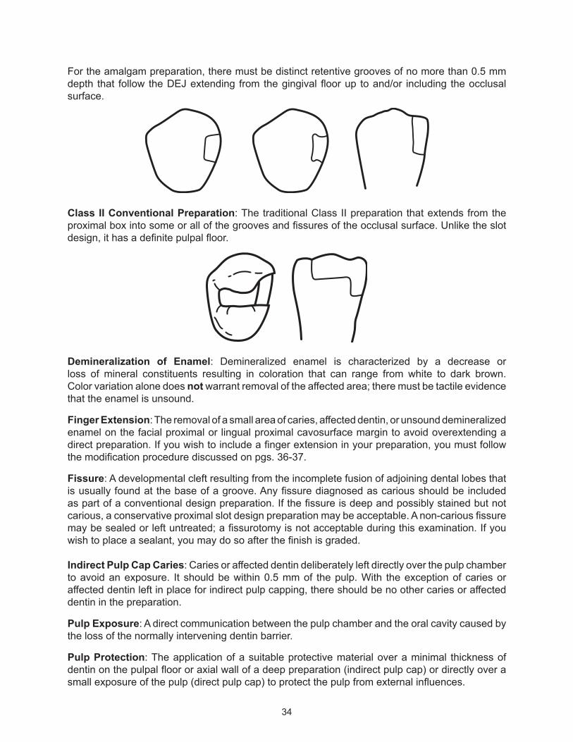
34
For the amalgam preparation, there must be distinct retentive grooves of no more than 0.5 mm depth that follow the DEJ extending from the gingival floor up to and/or including the occlusal surface.
Class II Conventional Preparation: The traditional Class II preparation that extends from the proximal box into some or all of the grooves and fissures of the occlusal surface. Unlike the slot design, it has a definite pulpal floor.
Demineralization of Enamel: Demineralized enamel is characterized by a decrease or loss of mineral constituents resulting in coloration that can range from white to dark brown. Color variation alone does not warrant removal of the affected area; there must be tactile evidence that the enamel is unsound.
Finger Extension: The removal of a small area of caries, affected dentin, or unsound demineralized enamel on the facial proximal or lingual proximal cavosurface margin to avoid overextending a direct preparation. If you wish to include a finger extension in your preparation, you must follow the modification procedure discussed on pgs. 36-37.
Fissure: A developmental cleft resulting from the incomplete fusion of adjoining dental lobes that is usually found at the base of a groove. Any fissure diagnosed as carious should be included as part of a conventional design preparation. If the fissure is deep and possibly stained but not carious, a conservative proximal slot design preparation may be acceptable. A non-carious fissure may be sealed or left untreated; a fissurotomy is not acceptable during this examination. If you wish to place a sealant, you may do so after the finish is graded.
Indirect Pulp Cap Caries: Caries or affected dentin deliberately left directly over the pulp chamber to avoid an exposure. It should be within 0.5 mm of the pulp. With the exception of caries or affected dentin left in place for indirect pulp capping, there should be no other caries or affected dentin in the preparation.
Pulp Exposure: A direct communication between the pulp chamber and the oral cavity caused by the loss of the normally intervening dentin barrier.
Pulp Protection: The application of a suitable protective material over a minimal thickness of dentin on the pulpal floor or axial wall of a deep preparation (indirect pulp cap) or directly over a small exposure of the pulp (direct pulp cap) to protect the pulp from external influences.
35
Sclerotic Dentin: A dentinal formation occurring ahead of the demineralization front of a slowly advancing carious lesion. It may be shiny and dark in color. It feels hard and impenetrable with an explorer.
Sealant: For purposes of the WREB exam, a sealant is considered to be a restorative material.
Tissue Trauma: Any undue iatrogenic damage to extraoral and/or intraoral tissues resulting in significant injury. Examples include lacerations greater than 3.0 mm, soft tissue burns, amputated papillae, and large tissue tags. Tissue trauma during an operative procedure is scored as part of the Finish, Function and Damage section of the finished restoration, even if the trauma is to tissue outside the immediate area of the restoration.
Unsound Demineralized Enamel: Demineralized enamel is considered unsound and should be removed if it is tactilely different from the adjacent unaffected enamel.
Cavity Preparation
WREB Examiners are calibrated to WREB preparation scoring criteria (see pgs. 42-44 and 53). Grading Examiners understand that some variations to outline and internal form may occur, but these should be small variations for the lesion treated. The management of major variations is covered in the Modification Procedure section.
It is imperative that all caries, affected dentin, and unsound demineralized enamel be totally removed. However, when caries is very deep (within 0.5 mm of the pulp chamber) the preferred treatment is to leave a small layer of caries and place an indirect pulp cap. Detection is typically accomplished with a sharp explorer to determine if softened dentin remains. All caries must be removed from the preparation, except that directly over the pulp chamber which if removed would result in a pulp exposure. If caries or affected dentin is intentionally left over the pulp, describe this in the “Note to Examiners” on the Worksheet.
Beveling for composite preparations is not a WREB requirement. However, if placed, bevels will be considered part of the outline and extension of the preparation.
If the preparation includes removal of a previous restoration, the entire previous restoration (including any base, sealant and/or liner) must be removed. If removal of previous pulp capping material is likely to expose the pulp, remove it to within 0.5 mm of the pulp and document this in “Note to Examiners” on your worksheet. Retentive pins may remain if they are adequately retained in dentin. Pins not adequately retained should be removed or made “flush” with the dentin surface of the preparation.
WREB strongly discourages the use of caries indicating solution. Examiners are trained to identify caries tactilely – not with indicating solution.
If a pulp exposure occurs, write Pulp Exposure in “Note to Examiners” under “Preparation Grade” on the worksheet and describe how you intend to manage the exposure. A rubber dam should be in place and a Floor Examiner must be called prior to placing pulp protection. The Floor Examiner will enter and initial a note on the worksheet, and direct you to place the pulp cap and complete the preparation. Any additional pulp protection will be placed after the preparation is graded. WREB considers all pulp exposures to be avoidable. There will be a deduction in score from the preparation points for any exposure, regardless of whether it is initially recognized by the Candidate or the Examiners.

36
For grading purposes, WREB differentiates between affected dentin and caries. Refer to the definitions on pg. 33. In the interest of Patient protection, all identified caries, affected dentin and unsound demineralized enamel will be removed prior to placement of the restoration.
Caries Remaining (other than the 0.5 mm of caries left for an indirect pulp cap) validated by two or more Grading Examiners is an automatic failure of the operative section. While it is most commonly diagnosed through direct access (as described on pg. 33), it may also be diagnosed from clinical or radiographic evidence that you have failed to completely access the lesion. Regardless of how it is diagnosed, you will be required to remove the caries. You may finish the restoration, although no points will be earned, or you may place a temporary and have the Patient contact the dentist on the Follow Up Care form for completion of the restoration. If you choose to finish the restoration, the Floor Examiner will check the final restoration. If remaining caries is identified by only one Grading Examiner you will be instructed to remove the caries, but since the finding was not validated by a second Grading Examiner, you will be allowed to finish the restoration for grading. When affected dentin or unsound demineralized enamel is documented by Grading Examiners you will be instructed to remove the affected dentin or unsound demineralized enamel and continue the procedure.
While WREB does not require placement of a base following the removal of deep caries, you are expected to place adequate pulp protection when indicated. With the exception of a direct pulp cap placed over an exposure (approved and initialed by a Floor Examiner), no pulp protection should be placed until after the preparation is graded.
Preparing a tooth without initial approval or preparing the wrong tooth results in failure of the entire operative section. Preparing a surface that has not been approved, without modification approval (e.g., including a lingual groove on a maxillary molar approved for Class II restoration), results in loss of all points for outline and extension and internal form. Restoring an operative procedure with a material other than what has been approved at acceptance (e.g., tooth approved for an amalgam and restored with composite or vice versa), results in loss of all points for the preparation and finish. If for any reason a Candidate submits a different procedure in lieu of a previously approved procedure, a 0.3 deduction will result.
Modification Procedure
Just as experienced practitioners often encounter unexpected circumstances that can modify treatment, you also may need to modify the outline, extension, and/or internal form of a planned preparation because of affected dentin, unsound demineralized enamel, or caries. (Occasionally, you may need a modification request for existing composite restorative material.) If you need to modify your preparation beyond the measurement criteria for a score of “5” you must communicate your intentions to Floor Examiners and Grading Examiners through a properly written Modification Request. A modification request should not be requested until the outline/extension and internal form are at the upper limit of the criteria for a score of “5.” Briefly describe on your worksheet under “Modification Request” the following:
• Type of modification (external outline, internal form, etc). External outline form modification includes the internal form that would normally support the new outline. Internal form modification relates to internal form only and has no effect on the preparation’s outline form.
• Location (proximal wall, pulpal floor, axial wall, etc.)

37
• Extent (amount of deviation from criteria for score of “5”)• Reason (caries, unsound demineralized enamel, affected dentin, restorative material)
Use the terms indicated on the last two pages of this Guide.
All requests for modification must be written in ink on the worksheet under “Modification Request.” All other notes (at acceptance, preparation and finish grading) must be written under “Note to Examiners” in the appropriate sections.
• Leave some caries, affected dentin, unsound demineralized enamel, or existing composite to show why the modification is being requested.
• If a planned variation in internal form is due to caries, the modification request should consider removal of caries only, not sound dentin.
• The extent of a modification request is referenced from the maximum extensions and depths listed in the preparation criteria for a score of 5 (pgs. 42-44 and 53). The Candidate’s preparation should reflect those maximum extensions prior to requesting a modification.
• Even though the facial extension of a Class III preparation need not break contact by criteria (pg. 43) any modification request involving the facial extension of this prep should be referenced from the point where facial contact is broken by 0.5 mm.
• Document the extent of the modification in multiples of 0.5 mm increments (i.e., 0.5 mm, 1.0 mm, etc.). Round up to the nearest 0.5 mm. This does not mean you request 0.5 mm modifications until the reason for modification no longer exists. Since space for listing modifications on the worksheet is limited, you are encouraged to initially specify the total extent of the modification required to remove the lesion.
• A rubber dam must be in place for all modification requests.• A planned “finger extension” (see definition) requires a modification request.
After entering your modification request on the worksheet, call a Floor Examiner. He/she may initial your modification note on the worksheet and instruct you to proceed. If the Floor Examiner feels the Grading Examiners should review the request, your Patient will be sent to the grading area with a Modification Request Form and a special gray card to indicate that only the modification request, not the completed preparation, should be evaluated.
After evaluation of the request by the Grading Examiners, the returned Modification Request Form will indicate if the modification requested was appropriate or not appropriate. The Floor Examiner will initial both pink and yellow copies of the form and return the pink copy to you. If you have requested multiple modifications, each numbered modification will be indicated as appropriate or not appropriate. There will also be at least two Grading Examiners’ initials on your worksheet. If any initials are missing, notify a Floor Examiner.
If the modification has been validated as appropriate, you may complete the preparation and submit for grading. The preparation (including any approved modification) will be graded according to WREB scoring criteria. If the modification is validated as not appropriate you should proceed with the preparation without the modification. There will be a deduction from the preparation score if any modification request is validated by Grading Examiners as not appropriate.
The Preparation GradeRubber dam isolation is required for preparation grading. The prepared tooth and at least one tooth on either side (excluding third molars), if present, must be isolated, clean, and dry. The rubber dam should be stabilized to withstand movement and time while your Patient is being evaluated. If an approximating tooth is partially erupted or otherwise cannot hold a rubber dam and you have varied your rubber dam placement as a result, the variation should be described in “Note to Examiners” under “Preparation Grade” on the worksheet.

38
When the preparation is ready to be graded, be sure that the tooth remains sufficiently anesthetized for Patient comfort during the evaluation process. Be sure to record the type and amount of anesthetic on the worksheet. Send your Patient to the grading area with the following:
A. Worksheet and attached radiographs with:• “Preparation Grade” box marked
B. Patient Tray with:• New #4 or #5 front-surface metal mouth mirror• New sharp pigtail explorer• New sharp shepherd’s hook explorer• Three 2" x 2" gauze pads
The mirror and explorers must be in an open autoclave bag. Place your worksheet on top of the tray.
C. Patient Bib: Attach your Candidate ID label to the upper right corner (Patient’s right side) of the Patient bib.
D. Patient Eye Protection: Prescription glasses or safety glasses must be worn by all Patients while in the grading area.
No electronic devices, including cell phones, are allowed in the grading area. Patients with electronic devices will be required to return the devices to the Candidate clinic.
After the preparation is graded, your Patient will return with the worksheet initialed by one Grading Examiner indicating that the preparation has been graded. At least three Grading Examiners must initial all notes in the “Note to Examiners” on the worksheet. If your worksheet does not have the required initials, notify a Floor Examiner before proceeding.
Adjustment of the approximating surface of an adjacent tooth may only be done after the preparation has been graded. Pulp protection also may only be done after the preparation has been graded (except for a direct pulp cap over an exposure).
For the Indirect (cast gold) preparation:You may take impressions only until 5:00 p.m. All Candidates and Patients must be out of the clinic by 5:30 p.m.
You may use laboratory facilities available at the school for casting the indirect restoration, or you may have a commercial laboratory fabricate the casting. You are responsible for the final indirect restoration whether it is done by you or a commercial laboratory.
Prior to cementation of the indirect restoration, you must have a Floor Examiner initial “Review of Tooth” on the Cast Gold worksheet. The review may be done at any stage of finish (from casting on the sprue to polished casting ready to cement) and is done without a rubber dam. You may proceed with finishing the casting in the mouth while waiting for a Floor Examiner to review the tooth. However, the casting must be removed from the mouth for a Floor Examiner to review. After reviewing the tooth, the Floor Examiner will initial “Review of Tooth” on the worksheet.

39
“Dismissal for the Day” Approval
Remember that any graded procedure that is started must be graded on the same day. If you received approval to start but have not begun the preparation, or if you received a preparation grade but wish to place the direct restoration on a subsequent day, you must see a Floor Examiner. A “Floor Examiner Check Sheet” will be completed and the pink and yellow copies given to you.
When you are ready to dismiss your Patient for the day, bring your worksheet to a Floor Examiner for approval. If appropriate, the Floor Examiner will sign “Dismissal for the Day” on the worksheet and your Patient may be dismissed. Dismissal approval must be completed by 5:00 p.m. However, if your Patient is detained in the grading area past 5:00 p.m. and “Dismissal for the Day” approval is necessary, it will be completed when the Patient has returned to the clinic.
If a Floor Examiner Check Sheet was issued, a Floor Examiner must evaluate your Patient prior to any treatment at the next appointment. At that next appointment, the Floor Examiner will initial both pink and yellow copies and return the pink copy to you. Failure to obtain the Floor Examiner’s initials will result in loss of all points for the procedure.
The Finish Grade
The finished restoration is graded without a rubber dam. The finish must be completed and graded the same day the restorative material is placed. Violation of this procedure will result in the loss of all points for the finish portion of the operative procedure.
Placing a material other than what was approved at acceptance will result in a loss of all points for preparation and finish.
A sealant or unfilled resin may not be placed over a composite restoration prior to finish grading. If you do so, your Patient will be returned to you and you will be asked to remove the sealant and then resubmit your Patient. After the finish is graded, you may apply a sealant to adjacent fissures and/or the restoration at your discretion.
When the restoration is ready to be graded, send your Patient to the grading area with the following:
A. Worksheet (and mounted radiographs) with the “Finish Restoration Grade” box marked on the worksheet.
B. Patient Tray with:• New #4 or #5 front-surface metal mouth mirror• New sharp pigtail explorer• New sharp shepherd’s hook explorer• Miller-type articulating paper forceps• Three 2” x 2” gauze pads
The instruments must be in an open autoclave bag. Place your paperwork and radiographs on top of the tray.
C. Patient Bib: Attach your Candidate ID label to the upper right corner (Patient’s right side) of the Patient bib.
D. Patient Eye Protection: Prescription glasses or safety glasses must be worn by all Patients while in the grading area.

40
Grading Examiners will check interproximal contacts with Floss Singles® and occlusion with Bausch® 40-micron articulating paper. Both are provided to the Examiners by WREB.
After the finish is graded, your Patient will return with the worksheet initialed by one Grading Examiner indicating that the finish has been graded. At least three Grading Examiners must initial all notes in the “Note to Examiners” on the worksheet. If your worksheet does not have the required initials, notify a Floor Examiner before proceeding.
Releasing Your Patient
Before releasing your Patient, do a final review of your worksheet to make sure that all necessary initials are present. The following initials are required:
• “Accepted By” (one Examiner)• “Preparation Graded” (one Examiner)• “Finish Graded” (one Examiner)• All “Note to Examiners” entries (one Examiner for Acceptance and three Examiners for
preparation and finish)If any initials are missing, notify a Floor Examiner. Missing initials not brought to the attention of a Floor Examiner cannot be grounds for an appeal.
Give your Patient the yellow copy of the Follow-Up Care Agreement form. Have him/her complete and turn in the Patient questionnaire. Ask a Floor Examiner to initial “Patient May Be Released From The Examination” line on the bottom of the worksheet. The Floor Examiner will verify that any follow-up requested by the Grading Examiners has been completed and will then initial the worksheet. Your Patient may then be dismissed. Do not dismiss your Patient without Floor Examiner permission.
Reference Material
Fundamentals of Operative Dentistry: A Contemporary Approach, 3rd edition (Summitt, Robbins, Hilton, & Schwartz), Quintessence Publishing Co.
Sturdevant’s Art and Science of Operative Dentistry, 5th edition (Roberson, Heymann, & Swift), Mosby Publishing Co.

41
UNUSUAL SITUATIONS
Preparing a tooth without approval. = Failure of the entire operative section
Preparing the wrong tooth. = Failure of the entire operative section
Failing to submit a Patient to the grading area for review of a modification request after instructed to do so by a Floor Examiner.
= Loss of all points for the preparation
Preparing the wrong surface or surface that has not been approved. (If the wrong surface is prepared, the original approved lesion must be included in the preparation.)
= Loss of all points for Outline and Extension and Internal Form
Restoring an operative procedure with a material other than what has been approved at acceptance (e.g., tooth approved for an amalgam and restored with composite or vice versa).
= Loss of all points for the preparation and finish
After Patient submission is approved, Candidate decides to submit a different tooth on the same or on a different Patient.
= 0.3 deduction from the applicable preparation score
OPERATIVE SCORING
Direct Restoration – Composite or AmalgamIndirect Restoration – Cast Gold
PREPARATION WEIGHTING
Outline and Extension: 46%Internal Form: 39%Operative Environment: 15%
FINISH WEIGHTING
Anatomical Form: 36.5%Margins: 36.5%Finish, Function and Damage: 27%
SCORE DEDUCTIONS
If two or more Examiners validate (document) any of the following errors, points are deducted from the appropriate preparation or finish score.
Patient Rejection: A 0.3 deduction per rejected submission from the applicable preparation score. Maximum 0.6 deduction.
Caries Remaining: Failure of entire operative section.
Pulp Exposure: Recognized by a Candidate or Floor Examiner or found during grading and validated by the Grading Examiners – a 0.5 deduction from the applicable preparation score.
Modification Request Not Appropriate: A 0.5 deduction for each modification request validated not appropriate. No maximum.
Late Penalties: 1 to 5 minutes late: 0.2 deduction; 6 to 10 minutes late: 0.4 deduction; 11 to 15 minutes late: 0.6 deduction; 16 or more minutes late: Procedure will not be graded. No points earned.
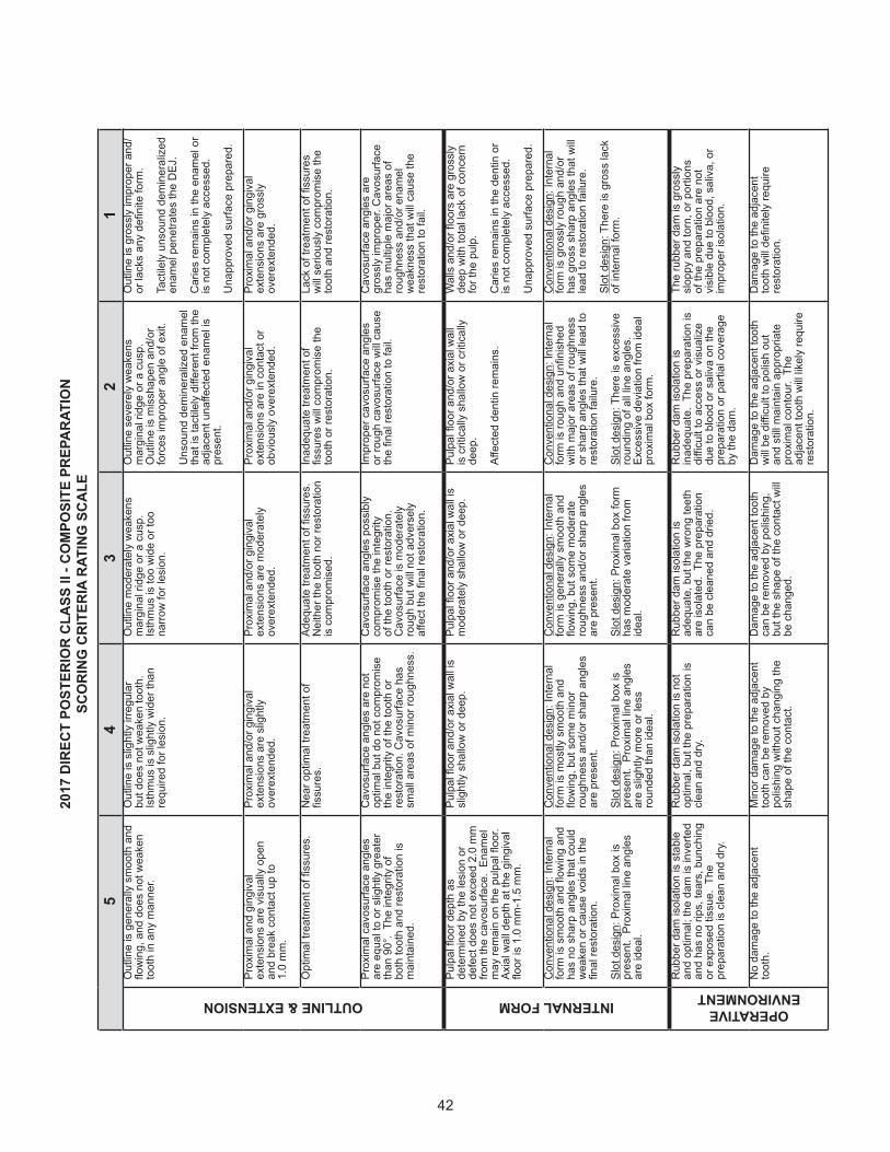
42
54
32
1
OUTLINE & EXTENSIONO
utlin
e is
gen
eral
ly s
moo
th a
nd
flow
ing,
and
doe
s no
t wea
ken
toot
h in
any
man
ner.
Out
line
is s
light
ly ir
regu
lar
but d
oes
not w
eake
n to
oth.
Is
thm
us is
slig
htly
wid
er th
an
requ
ired
for l
esio
n.
Out
line
mod
erat
ely
wea
kens
m
argi
nal r
idge
or a
cus
p.
Isth
mus
is to
o w
ide
or to
o na
rrow
for l
esio
n.
Out
line
seve
rely
wea
kens
m
argi
nal r
idge
or a
cus
p.
Out
line
is m
issh
apen
and
/or
forc
es im
prop
er a
ngle
of e
xit.
Uns
ound
dem
iner
aliz
ed e
nam
el
that
is ta
ctile
ly d
iffer
ent f
rom
the
adja
cent
una
ffect
ed e
nam
el is
pr
esen
t.
Out
line
is g
ross
ly im
prop
er a
nd/
or la
cks
any
defin
ite fo
rm.
Tact
ilely
uns
ound
dem
iner
aliz
ed
enam
el p
enet
rate
s th
e D
EJ.
Car
ies
rem
ains
in th
e en
amel
or
is n
ot c
ompl
etel
y ac
cess
ed.
Una
ppro
ved
surfa
ce p
repa
red.
Prox
imal
and
gin
giva
l ex
tens
ions
are
vis
ually
ope
n an
d br
eak
cont
act u
p to
1.0
mm
.
Prox
imal
and
/or g
ingi
val
exte
nsio
ns a
re s
light
ly
over
exte
nded
.
Prox
imal
and
/or g
ingi
val
exte
nsio
ns a
re m
oder
atel
y ov
erex
tend
ed.
Prox
imal
and
/or g
ingi
val
exte
nsio
ns a
re in
con
tact
or
obvi
ousl
y ov
erex
tend
ed.
Prox
imal
and
/or g
ingi
val
exte
nsio
ns a
re g
ross
ly
over
exte
nded
.
Opt
imal
trea
tmen
t of fi
ssur
es.
Nea
r opt
imal
trea
tmen
t of
fissu
res.
Adeq
uate
trea
tmen
t of fi
ssur
es.
Nei
ther
the
toot
h no
r res
tora
tion
is c
ompr
omis
ed.
Inad
equa
te tr
eatm
ent o
f fis
sure
s w
ill co
mpr
omis
e th
e to
oth
or re
stor
atio
n.
Lack
of t
reat
men
t of fi
ssur
es
will
serio
usly
com
prom
ise
the
toot
h an
d re
stor
atio
n.
Prox
imal
cav
osur
face
ang
les
are
equa
l to
or s
light
ly g
reat
er
than
90°
. Th
e in
tegr
ity o
f bo
th to
oth
and
rest
orat
ion
is
mai
ntai
ned.
Cav
osur
face
ang
les
are
not
optim
al b
ut d
o no
t com
prom
ise
the
inte
grity
of t
he to
oth
or
rest
orat
ion.
Cav
osur
face
has
sm
all a
reas
of m
inor
roug
hnes
s.
Cav
osur
face
ang
les
poss
ibly
co
mpr
omis
e th
e in
tegr
ity
of th
e to
oth
or re
stor
atio
n.
Cav
osur
face
is m
oder
atel
y ro
ugh
but w
ill no
t adv
erse
ly
affec
t the
fina
l res
tora
tion.
Impr
oper
cav
osur
face
ang
les
or ro
ugh
cavo
surfa
ce w
ill ca
use
the
final
rest
orat
ion
to fa
il.
Cav
osur
face
ang
les
are
gros
sly
impr
oper
. Cav
osur
face
ha
s m
ultip
le m
ajor
are
as o
f ro
ughn
ess
and/
or e
nam
el
wea
knes
s th
at w
ill ca
use
the
rest
orat
ion
to fa
il.
INTERNAL FORM
Pulp
al fl
oor d
epth
as
dete
rmin
ed b
y th
e le
sion
or
defe
ct d
oes
not e
xcee
d 2.
0 m
m
from
the
cavo
surfa
ce.
Enam
el
may
rem
ain
on th
e pu
lpal
floo
r.
Axia
l wal
l dep
th a
t the
gin
giva
l flo
or is
1.0
mm
-1.5
mm
.
Pulp
al fl
oor a
nd/o
r axi
al w
all i
s sl
ight
ly s
hallo
w o
r dee
p.Pu
lpal
floo
r and
/or a
xial
wal
l is
mod
erat
ely
shal
low
or d
eep.
Pulp
al fl
oor a
nd/o
r axi
al w
all
is c
ritic
ally
sha
llow
or c
ritic
ally
de
ep.
Affec
ted
dent
in re
mai
ns.
Wal
ls a
nd/o
r floo
rs a
re g
ross
ly
deep
with
tota
l lac
k of
con
cern
fo
r the
pul
p.
Car
ies
rem
ains
in th
e de
ntin
or
is n
ot c
ompl
etel
y ac
cess
ed.
Una
ppro
ved
surfa
ce p
repa
red.
Con
vent
iona
l des
ign:
Inte
rnal
fo
rm is
sm
ooth
and
flow
ing
and
has
no s
harp
ang
les
that
cou
ld
wea
ken
or c
ause
voi
ds in
the
final
rest
orat
ion.
Slot
des
ign:
Pro
xim
al b
ox is
pr
esen
t. P
roxi
mal
line
ang
les
are
idea
l.
Con
vent
iona
l des
ign:
Inte
rnal
fo
rm is
mos
tly s
moo
th a
nd
flow
ing,
but
som
e m
inor
ro
ughn
ess
and/
or s
harp
ang
les
are
pres
ent.
Slot
des
ign:
Pro
xim
al b
ox is
pr
esen
t. P
roxi
mal
line
ang
les
are
slig
htly
mor
e or
less
ro
unde
d th
an id
eal.
Con
vent
iona
l des
ign:
Inte
rnal
fo
rm is
gen
eral
ly s
moo
th a
nd
flow
ing,
but
som
e m
oder
ate
roug
hnes
s an
d/or
sha
rp a
ngle
s ar
e pr
esen
t.
Slot
des
ign:
Pro
xim
al b
ox fo
rm
has
mod
erat
e va
riatio
n fro
m
idea
l.
Con
vent
iona
l des
ign:
Inte
rnal
fo
rm is
roug
h an
d un
finis
hed
with
maj
or a
reas
of r
ough
ness
or
sha
rp a
ngle
s th
at w
ill le
ad to
re
stor
atio
n fa
ilure
.
Slot
des
ign:
The
re is
exc
essi
ve
roun
ding
of a
ll lin
e an
gles
. Ex
cess
ive
devi
atio
n fro
m id
eal
prox
imal
box
form
.
Con
vent
iona
l des
ign:
Inte
rnal
fo
rm is
gro
ssly
roug
h an
d/or
ha
s gr
oss
shar
p an
gles
that
will
lead
to re
stor
atio
n fa
ilure
.
Slot
des
ign:
The
re is
gro
ss la
ck
of in
tern
al fo
rm.
OPERATIVE ENVIRONMENT
Rub
ber d
am is
olat
ion
is s
tabl
e an
d op
timal
; the
dam
is in
verte
d an
d ha
s no
rips
, tea
rs, b
unch
ing
or e
xpos
ed ti
ssue
. Th
e pr
epar
atio
n is
cle
an a
nd d
ry.
Rub
ber d
am is
olat
ion
is n
ot
optim
al, b
ut th
e pr
epar
atio
n is
cl
ean
and
dry.
Rub
ber d
am is
olat
ion
is
adeq
uate
, but
the
wro
ng te
eth
are
isol
ated
. Th
e pr
epar
atio
n ca
n be
cle
aned
and
drie
d.
Rub
ber d
am is
olat
ion
is
inad
equa
te.
The
prep
arat
ion
is
diffi
cult
to a
cces
s or
vis
ualiz
e du
e to
blo
od o
r sal
iva
on th
e pr
epar
atio
n or
par
tial c
over
age
by th
e da
m.
The
rubb
er d
am is
gro
ssly
sl
oppy
and
torn
, or p
ortio
ns
of th
e pr
epar
atio
n ar
e no
t vi
sibl
e du
e to
blo
od, s
aliv
a, o
r im
prop
er is
olat
ion.
No
dam
age
to th
e ad
jace
nt
toot
h.M
inor
dam
age
to th
e ad
jace
nt
toot
h ca
n be
rem
oved
by
polis
hing
with
out c
hang
ing
the
shap
e of
the
cont
act.
Dam
age
to th
e ad
jace
nt to
oth
can
be re
mov
ed b
y po
lishi
ng,
but t
he s
hape
of t
he c
onta
ct w
ill be
cha
nged
.
Dam
age
to th
e ad
jace
nt to
oth
will
be d
ifficu
lt to
pol
ish
out
and
still
mai
ntai
n ap
prop
riate
pr
oxim
al c
onto
ur.
The
adja
cent
toot
h w
ill lik
ely
requ
ire
rest
orat
ion.
Dam
age
to th
e ad
jace
nt
toot
h w
ill de
finite
ly re
quire
re
stor
atio
n.
2017
DIR
ECT
POST
ERIO
R CL
ASS
II - C
OM
POSI
TE P
REPA
RATI
ON
SCO
RING
CRI
TERI
A RA
TING
SCA
LE

43
54
32
1OUTLINE & EXTENSION
Out
line
prov
ides
opt
imal
acc
ess
for c
arie
s re
mov
al a
nd in
serti
on o
f re
stor
ativ
e m
ater
ial.
Gin
giva
l ext
ensi
on is
vis
ually
ope
n up
to 0
.5 m
m. F
acia
l (or
ling
ual)
exte
nsio
n m
ay b
reak
pro
xim
al
cont
act u
p to
0.5
mm
.
Inci
sal c
onta
ct is
not
bro
ken.
Out
line
is s
light
ly o
ver o
r und
er
exte
nded
. O
utlin
e is
slig
htly
irr
egul
ar b
ut d
oes
not w
eake
n th
e to
oth.
Out
line
is m
oder
atel
y ov
er o
r un
der e
xten
ded.
Out
line
is
mod
erat
ely
irreg
ular
but
doe
s no
t w
eake
n th
e to
oth.
Gin
giva
l mar
gin
is m
oder
atel
y ov
erex
tend
ed.
Out
line
is s
ever
ely
over
or
unde
rext
ende
d.
Gin
giva
l wal
l is
in c
onta
ct o
r ob
viou
sly
over
exte
nded
.
Inci
sal e
xten
sion
has
bro
ken
cont
act.
Uns
ound
dem
iner
aliz
ed e
nam
el
that
is ta
ctile
ly d
iffer
ent f
rom
the
adja
cent
una
ffect
ed e
nam
el is
pr
esen
t.
Out
line
is g
ross
ly im
prop
er a
nd/o
r la
cks
any
defin
ite fo
rm.
Gin
giva
l wal
l is
gros
sly
over
exte
nded
.
Tact
ilely
uns
ound
dem
iner
aliz
ed
enam
el p
enet
rate
s th
e D
EJ.
Car
ies
rem
ains
in th
e en
amel
or i
s no
t com
plet
ely
acce
ssed
.
Una
ppro
ved
surfa
ce p
repa
red.
Cav
osur
face
form
s a
smoo
th
cont
inuo
us c
urve
with
no
shar
p an
gles
.
Cav
osur
face
is s
light
ly ir
regu
lar
and
roug
h; n
o sh
arp
angl
es.
Cav
osur
face
is m
oder
atel
y irr
egul
ar a
nd ro
ugh.
A fe
w s
harp
an
gles
are
pre
sent
.
Cav
osur
face
is s
ever
ely
irreg
ular
an
d/or
with
sha
rp a
ngle
s.C
avos
urfa
ce h
as m
ultip
le g
ross
irr
egul
ariti
es a
nd/o
r ena
mel
w
eakn
esse
s th
at w
ill ca
use
the
rest
orat
ion
to fa
il.
Ther
e ar
e no
acu
te c
avos
urfa
ce
angl
es.
Cav
osur
face
ang
les
are
not
optim
al b
ut d
o no
t com
prom
ise
the
inte
grity
of t
he to
oth
or re
stor
atio
n.
Cav
osur
face
ang
les
poss
ibly
co
mpr
omis
e th
e in
tegr
ity o
f the
to
oth
or re
stor
atio
n.
Cav
osur
face
ang
les
will
lead
to
enam
el fr
actu
re o
r fra
ctur
e of
the
rest
orat
ion.
Cav
osur
face
ang
les
are
gros
sly
inap
prop
riate
for t
he s
ituat
ion
and
will
lead
to fr
actu
re o
f the
re
stor
atio
n.
INTERNAL FORM
Axia
l wal
l fol
low
s ex
tern
al c
onto
ur
of to
oth.
Dep
th d
oes
not e
xcee
d 1.
0 m
m b
eyon
d th
e D
EJ.
Axia
l wal
l gen
eral
ly fo
llow
s ex
tern
al c
onto
ur o
f too
th.
Dep
th
does
not
exc
eed
1.5
mm
bey
ond
the
DEJ
.
Axia
l wal
l doe
s no
t fol
low
con
tour
of
toot
h. D
epth
doe
s no
t exc
eed
2.0
mm
bey
ond
the
DEJ
.
Axia
l wal
l dep
th e
xcee
ds 2
.0 m
m
beyo
nd th
e D
EJ.
Affec
ted
dent
in re
mai
ns.
Gro
ss re
mov
al o
f too
th s
truct
ure
jeop
ardi
zes
the
heal
th o
f the
to
oth.
Car
ies
rem
ains
in th
e de
ntin
or i
s no
t com
plet
ely
acce
ssed
.
Una
ppro
ved
surfa
ce p
repa
red.
Inte
rnal
line
ang
les
are
roun
ded
and
smoo
th.
Inte
rnal
wal
ls a
re
wel
l defi
ned.
Inte
rnal
wal
ls a
re w
ell d
efine
d an
d ro
unde
d, b
ut h
ave
som
e sl
ight
irr
egul
ariti
es.
Inte
rnal
wal
ls a
re ro
unde
d, b
ut
mod
erat
ely
roug
h, ir
regu
lar,
and
not d
efine
d. M
oder
atel
y sh
arp
line
angl
es a
re p
rese
nt.
Inte
rnal
wal
ls a
re s
ever
ely
irreg
ular
and
not
defi
ned.
Ang
le
of w
alls
und
erm
ines
ena
mel
, je
opar
dize
s in
cisa
l ang
le, o
r en
croa
ches
on
the
pulp
.
Gro
ssly
irre
gula
r and
sha
rp li
ne
angl
es s
how
tota
l dis
rega
rd fo
r th
e he
alth
of t
he to
oth.
OPERATIVE ENVIRONMENT
Rub
ber d
am is
olat
ion
is s
tabl
e an
d op
timal
; the
dam
is in
verte
d an
d ha
s no
rips
, tea
rs, b
unch
ing
or e
xpos
ed ti
ssue
. Th
e pr
epar
atio
n is
cle
an a
nd d
ry.
Rub
ber d
am is
olat
ion
is n
ot
optim
al, b
ut th
e pr
epar
atio
n is
cl
ean
and
dry.
Rub
ber d
am is
olat
ion
is a
dequ
ate,
bu
t the
wro
ng te
eth
are
isol
ated
. Th
e pr
epar
atio
n ca
n be
cle
aned
an
d dr
ied.
Rub
ber d
am is
olat
ion
is
inad
equa
te.
The
prep
arat
ion
is
diffi
cult
to a
cces
s or
vis
ualiz
e du
e to
blo
od o
r sal
iva
on th
e pr
epar
atio
n or
par
tial c
over
age
by
the
dam
.
The
rubb
er d
am is
gro
ssly
sl
oppy
and
torn
, or p
ortio
ns o
f th
e pr
epar
atio
n ar
e no
t vis
ible
du
e to
blo
od, s
aliv
a, o
r im
prop
er
isol
atio
n.
No
dam
age
to th
e ad
jace
nt to
oth.
Min
or d
amag
e to
the
adja
cent
to
oth
can
be re
mov
ed b
y po
lishi
ng w
ithou
t cha
ngin
g th
e sh
ape
of th
e co
ntac
t.
Dam
age
to th
e ad
jace
nt to
oth
can
be re
mov
ed b
y po
lishi
ng, b
ut
the
shap
e of
the
cont
act w
ill be
ch
ange
d.
Dam
age
to th
e ad
jace
nt to
oth
will
be d
ifficu
lt to
pol
ish
out a
nd s
till
mai
ntai
n ap
prop
riate
pro
xim
al
cont
our.
The
adj
acen
t too
th w
ill lik
ely
requ
ire re
stor
atio
n.
Dam
age
to th
e ad
jace
nt to
oth
will
defin
itely
requ
ire re
stor
atio
n.
2017
DIR
ECT
AN
TER
IOR
CLA
SS II
I - C
OM
POSI
TE P
REP
AR
ATIO
NSC
OR
ING
CR
ITER
IA R
ATIN
G S
CA
LE

44
54
32
1OUTLINE & EXTENSION
Out
line
is g
ener
ally
sm
ooth
and
flo
win
g, a
nd d
oes
not w
eake
n to
oth
in
any
man
ner.
Out
line
is s
light
ly ir
regu
lar b
ut d
oes
not w
eake
n to
oth.
Ist
hmus
is s
light
ly
wid
er th
an re
quire
d fo
r les
ion.
Out
line
mod
erat
ely
wea
kens
mar
gina
l rid
ge o
r a c
usp.
Ist
hmus
is to
o w
ide
or to
o na
rrow
for l
esio
n.
Out
line
seve
rely
wea
kens
mar
gina
l rid
ge o
r a c
usp.
Out
line
is m
issh
apen
an
d/or
forc
es im
prop
er a
ngle
of e
xit.
Uns
ound
dem
iner
aliz
ed e
nam
el th
at
is ta
ctile
ly d
iffer
ent f
rom
the
adja
cent
un
affec
ted
enam
el is
pre
sent
.
Out
line
is g
ross
ly im
prop
er a
nd/o
r la
cks
any
defin
ite fo
rm.
Tact
ilely
uns
ound
dem
iner
aliz
ed
enam
el p
enet
rate
s th
e D
EJ.
Car
ies
rem
ains
in th
e en
amel
or i
s no
t co
mpl
etel
y ac
cess
ed.
Una
ppro
ved
surfa
ce p
repa
red.
Pro
xim
al a
nd g
ingi
val e
xten
sion
s ar
e vi
sual
ly o
pen
and
brea
k co
ntac
t up
to
1.0
mm
.
Pro
xim
al a
nd/o
r gin
giva
l ext
ensi
ons
are
slig
htly
ove
rext
ende
d.P
roxi
mal
and
/or g
ingi
val e
xten
sion
s ar
e m
oder
atel
y ov
erex
tend
ed.
Pro
xim
al a
nd/o
r gin
giva
l ext
ensi
ons
are
in c
onta
ct o
r obv
ious
ly o
vere
xten
ded.
Pro
xim
al a
nd/o
r gin
giva
l ext
ensi
ons
are
gros
sly
over
exte
nded
.
Opt
imal
trea
tmen
t of fi
ssur
es.
Nea
r opt
imal
trea
tmen
t of fi
ssur
es.
Ade
quat
e tre
atm
ent o
f fiss
ures
. N
eith
er th
e to
oth
nor r
esto
ratio
n is
co
mpr
omis
ed.
Inad
equa
te tr
eatm
ent o
f fiss
ures
will
co
mpr
omis
e th
e to
oth
or re
stor
atio
n.La
ck o
f tre
atm
ent o
f fiss
ures
will
se
rious
ly c
ompr
omis
e th
e to
oth
and
rest
orat
ion.
Pro
xim
al c
avos
urfa
ce a
ngle
s ar
e ap
prox
imat
ely
90°.
The
inte
grity
of
bot
h to
oth
and
rest
orat
ion
is
mai
ntai
ned.
Cav
osur
face
ang
les
are
not o
ptim
al
but d
o no
t com
prom
ise
the
inte
grity
of
the
toot
h or
rest
orat
ion.
Cav
osur
face
ha
s sm
all a
reas
of m
inor
roug
hnes
s.
Cav
osur
face
ang
les
poss
ibly
co
mpr
omis
e th
e in
tegr
ity o
f the
to
oth
or re
stor
atio
n. C
avos
urfa
ce
is m
oder
atel
y ro
ugh
but w
ill n
ot
adve
rsel
y aff
ect t
he fi
nal r
esto
ratio
n.
Impr
oper
cav
osur
face
ang
les
or
roug
h ca
vosu
rface
will
cau
se th
e fin
al
rest
orat
ion
to fa
il.
Cav
osur
face
ang
les
are
gros
sly
impr
oper
. Cav
osur
face
has
mul
tiple
m
ajor
are
as o
f rou
ghne
ss a
nd/o
r en
amel
wea
knes
s th
at w
ill c
ause
the
rest
orat
ion
to fa
il.
INTERNAL FORM
Pro
xim
al w
alls
are
cle
arly
con
verg
ent
occl
usal
ly.
Pro
xim
al w
alls
are
bar
ely
conv
erge
nt
occl
usal
ly.
Pro
xim
al w
alls
are
par
alle
l or
dive
rgen
t in
one
area
.P
roxi
mal
wal
ls a
re c
ritic
ally
div
erge
nt
occl
usal
ly.
Pro
xim
al w
alls
are
gro
ssly
div
erge
nt
occl
usal
ly.
Pul
pal fl
oor i
s 1.
5 m
m-2
.0 m
m
from
the
cavo
surfa
ce a
nd p
rovi
des
adeq
uate
bul
k fo
r stre
ngth
of
rest
orat
ive
mat
eria
l. A
xial
wal
l dep
th a
t th
e gi
ngiv
al fl
oor i
s 1.
0 m
m-1
.5 m
m.
Axi
al w
all a
nd/o
r pul
pal fl
oor i
s sl
ight
ly s
hallo
w o
r dee
p, b
ut s
till
prov
ides
ade
quat
e bu
lk fo
r stre
ngth
of
rest
orat
ive
mat
eria
l.
Axi
al w
all a
nd/o
r pul
pal fl
oor i
s m
oder
atel
y sh
allo
w o
r dee
p, b
ut s
till
prov
ides
ade
quat
e bu
lk fo
r stre
ngth
of
rest
orat
ive
mat
eria
l.
Axi
al w
all a
nd/o
r pul
pal fl
oor i
s cr
itica
lly s
hallo
w o
r dee
p an
d do
es n
ot
prov
ide
adeq
uate
bul
k fo
r stre
ngth
of
rest
orat
ive
mat
eria
l.
Affe
cted
den
tin re
mai
ns.
Wal
ls a
nd/o
r floo
rs a
re g
ross
ly d
eep
with
tota
l lac
k of
con
cern
for t
he p
ulp.
Car
ies
rem
ains
in th
e de
ntin
or i
s no
t co
mpl
etel
y ac
cess
ed.
Una
ppro
ved
surfa
ce p
repa
red.
Con
vent
iona
l des
ign:
Inte
rnal
form
is
sm
ooth
and
has
no
shar
p an
gles
. R
eten
tive
groo
ves,
if p
lace
d, a
re
near
idea
l. A
xial
wal
l fol
low
s ex
tern
al
cont
our o
f the
toot
h.
Slo
t des
ign:
Pro
xim
al b
ox is
pre
sent
. A
xial
wal
l fol
low
s ex
tern
al c
onto
ur
of th
e to
oth.
Ret
entiv
e gr
oove
s ex
tend
from
gin
giva
l floo
r up
to a
nd/
or in
clud
ing
occl
usal
sur
face
, are
no
mor
e th
an 0
.5 m
m d
eep,
and
par
alle
l to
the
DE
J.
Con
vent
iona
l des
ign:
Inte
rnal
form
is
mos
tly s
moo
th, b
ut s
ome
min
or
roug
hnes
s an
d/or
sha
rp a
ngle
s ar
e pr
esen
t. R
eten
tive
groo
ves,
if
plac
ed, a
re a
dequ
ate.
Axi
al w
all
cont
our i
s ne
ar o
ptim
al.
Slo
t des
ign:
Pro
xim
al b
ox is
pre
sent
. A
xial
wal
l con
tour
is n
ear o
ptim
al.
Ret
entiv
e gr
oove
s ar
e m
inim
al a
nd
exte
nd u
p to
and
/or i
nclu
ding
the
occl
usal
sur
face
.
Con
vent
iona
l des
ign:
Inte
rnal
form
is
gen
eral
ly s
moo
th, b
ut s
ome
mod
erat
e ro
ughn
ess
and/
or s
harp
an
gles
are
pre
sent
. R
eten
tive
groo
ves,
if p
lace
d, a
re to
o de
ep o
r to
o sh
allo
w, o
r pla
ced
in a
n in
corr
ect
loca
tion.
Axi
al w
all c
onto
ur is
not
op
timal
.
Slo
t des
ign:
Pro
xim
al b
ox fo
rm is
qu
estio
nabl
e. A
xial
wal
l con
tour
is
not o
ptim
al.
Ret
entiv
e gr
oove
s ar
e to
o de
ep o
r too
sha
llow
and
/or p
lace
d in
an
inco
rrec
t pos
ition
.
Con
vent
iona
l des
ign:
Inte
rnal
form
is
roug
h an
d un
finis
hed
with
maj
or a
reas
of
roug
hnes
s or
sha
rp a
ngle
s th
at w
ill
lead
to re
stor
atio
n fa
ilure
. R
eten
tive
groo
ves,
if p
lace
d, a
re to
o de
ep o
r to
o sh
allo
w, o
r pla
ced
in a
n in
corr
ect
loca
tion,
and
will
com
prom
ise
the
toot
h or
rest
orat
ion.
Slo
t des
ign:
Pre
para
tion
has
scoo
ped
appe
aran
ce w
ith e
xces
sive
roun
ding
of
all
line
angl
es.
Ret
entiv
e gr
oove
s ar
e to
o de
ep, t
oo s
hallo
w, a
nd/o
r pl
aced
in a
n in
corr
ect l
ocat
ion,
and
will
co
mpr
omis
e th
e to
oth
or re
stor
atio
n.
Con
vent
iona
l des
ign:
Inte
rnal
form
is
gros
sly
roug
h an
d/or
has
gro
ss s
harp
an
gles
that
will
lead
to re
stor
atio
n fa
ilure
. G
ross
dis
rega
rd fo
r pro
per
plac
emen
t of r
eten
tive
feat
ures
w
ill c
ompr
omis
e th
e to
oth
and
rest
orat
ion.
Slo
t des
ign:
The
re is
gro
ss la
ck o
f in
tern
al fo
rm.
Ret
entiv
e gr
oove
s ar
e ab
sent
.
OPERATIVE ENVIRONMENT
Rub
ber d
am is
olat
ion
is s
tabl
e an
d op
timal
; the
dam
is in
verte
d an
d ha
s no
rips
, tea
rs, b
unch
ing
or e
xpos
ed
tissu
e. T
he p
repa
ratio
n is
cle
an a
nd
dry.
Rub
ber d
am is
olat
ion
is n
ot o
ptim
al,
but t
he p
repa
ratio
n is
cle
an a
nd d
ry.
Rub
ber d
am is
olat
ion
is a
dequ
ate,
bu
t the
wro
ng te
eth
are
isol
ated
. Th
e pr
epar
atio
n ca
n be
cle
aned
and
dr
ied.
Rub
ber d
am is
olat
ion
is in
adeq
uate
. Th
e pr
epar
atio
n is
diffi
cult
to a
cces
s or
vis
ualiz
e du
e to
blo
od o
r sal
iva
on
the
prep
arat
ion
or p
artia
l cov
erag
e by
th
e da
m.
The
rubb
er d
am is
gro
ssly
slo
ppy
and
torn
, or p
ortio
ns o
f the
pre
para
tion
are
not v
isib
le d
ue to
blo
od, s
aliv
a, o
r im
prop
er is
olat
ion.
No
dam
age
to th
e ad
jace
nt to
oth.
Min
or d
amag
e to
the
adja
cent
toot
h ca
n be
rem
oved
by
polis
hing
with
out
chan
ging
the
shap
e of
the
cont
act.
Dam
age
to th
e ad
jace
nt to
oth
can
be
rem
oved
by
polis
hing
, but
the
shap
e of
the
cont
act w
ill b
e ch
ange
d.
Dam
age
to th
e ad
jace
nt to
oth
will
be
diffi
cult
to p
olis
h ou
t and
stil
l mai
ntai
n ap
prop
riate
pro
xim
al c
onto
ur.
The
adja
cent
toot
h w
ill li
kely
requ
ire
rest
orat
ion.
Dam
age
to th
e ad
jace
nt to
oth
will
de
finite
ly re
quire
rest
orat
ion.
2017
DIR
EC
T P
OS
TER
IOR
CLA
SS
II -
AM
ALG
AM
PR
EPA
RAT
ION
SC
OR
ING
CR
ITE
RIA
RAT
ING
SC
ALE

45
54
32
1
ANATOMICAL FORM
Anat
omic
al fo
rm is
con
sist
ent
and
harm
onio
us w
ith
cont
iguo
us to
oth
stru
ctur
e.
Slig
ht v
aria
tion
in n
orm
al
anat
omic
al fo
rm is
pre
sent
.M
oder
ate
varia
tion
in n
orm
al
anat
omic
al fo
rm is
pre
sent
. M
argi
nal r
idge
is im
prop
erly
sh
aped
.
Anat
omic
al fo
rm is
impr
oper
. M
argi
nal r
idge
is p
oorly
sh
aped
.
Anat
omy
is to
o de
ep o
r too
fla
t.
Ther
e is
gro
ss la
ck o
f an
atom
ical
form
.
Prop
er p
roxi
mal
con
tour
and
sh
ape
are
rest
ored
.Th
ere
is s
light
var
iatio
n of
pr
oxim
al c
onto
ur a
nd s
hape
.Th
ere
is m
oder
ate
varia
tion
of p
roxi
mal
con
tour
and
sh
ape.
Prox
imal
con
tour
is p
oor.
Em
bras
ures
are
sev
erel
y ov
er o
r und
er c
onto
ured
.
Gro
ssly
impr
oper
pro
xim
al
cont
our o
r sha
pe.
Nor
mal
pro
xim
al c
onta
ct
area
and
pos
ition
are
re
stor
ed.
Con
tact
is v
isua
lly
clos
ed a
nd re
sist
s th
e pa
ssag
e of
ligh
tly w
axed
flo
ss.
Ther
e is
slig
ht v
aria
tion
of
norm
al c
onta
ct a
rea
and
posi
tion.
Con
tact
is v
isua
lly
clos
ed a
nd re
sist
s th
e pa
ssag
e of
ligh
tly w
axed
flo
ss.
Ther
e is
mod
erat
e va
riatio
n of
nor
mal
con
tact
are
a an
d po
sitio
n. L
ight
ly w
axed
flos
s w
ill pa
ss th
roug
h th
e co
ntac
t w
ith s
light
resi
stan
ce.
Con
tact
is v
isua
lly o
pen,
or
floss
will
not p
ass
thro
ugh
the
cont
act.
Con
tact
is g
ross
ly o
pen,
or
the
cont
act a
rea
is b
onde
d to
th
e ad
jace
nt to
oth.
MARGINS
Ther
e ar
e no
exc
esse
s or
de
ficie
ncie
s an
ywhe
re a
long
m
argi
ns.
Slig
ht m
argi
nal e
xces
ses
and/
or d
efici
enci
es a
re
pres
ent.
Mod
erat
e m
argi
nal e
xces
ses
and/
or d
efici
enci
es a
re
pres
ent.
A de
ep o
pen
mar
gin
is
pres
ent,
or c
ritic
al e
xces
ses
or d
efici
enci
es a
re p
rese
nt.
A m
argi
nal o
verh
ang
catc
hes
floss
.
Mul
tiple
ope
n m
argi
ns,
or g
ross
exc
esse
s or
de
ficie
ncie
s, a
re p
rese
nt.
A gr
oss
mar
gina
l ove
rhan
g sh
reds
flos
s.
FINISH, FUNCTION & DAMAGE
The
surfa
ce is
sm
ooth
with
no
pits
, voi
ds o
r irre
gula
ritie
s.Sl
ight
sur
face
irre
gula
ritie
s,
pitti
ng, o
r voi
ds a
re p
rese
nt.
Mod
erat
e su
rface
irr
egul
ariti
es, p
ittin
g, o
r voi
ds
are
pres
ent.
Crit
ical
sur
face
irre
gula
ritie
s,
pitti
ng, o
r voi
ds a
re p
rese
nt.
Gro
ss s
urfa
ce d
efec
ts
are
pres
ent a
nd/o
r the
re
stor
atio
n is
gro
ssly
fra
ctur
ed.
Occ
lusi
on is
rest
ored
to
prop
er c
entri
c w
ith n
o la
tera
l in
terfe
renc
es.
Ther
e is
sev
ere
hype
rocc
lusi
on in
cen
tric
or
late
ral e
xcur
sion
s. O
cclu
sal
cont
act m
arks
app
ear o
nly
on th
e re
stor
atio
n.
Occ
lusi
on is
gro
ssly
in
adeq
uate
.
Ther
e is
no
dam
age
to h
ard
or s
oft t
issu
e.M
inor
dam
age
to h
ard
or s
oft
tissu
e is
evi
dent
.M
oder
ate
dam
age
to h
ard
or
soft
tissu
e is
evi
dent
.Se
vere
dam
age
to h
ard
or
soft
tissu
e is
evi
dent
.G
ross
mut
ilatio
n of
har
d or
so
ft tis
sue
is e
vide
nt.
2017
DIR
ECT
FINI
SHSC
ORI
NG C
RITE
RIA
RATI
NG S
CALE

46
This page intentionally left blank.
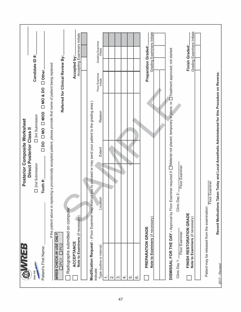
47
SAMPLE
Post
erio
r Com
posi
te W
orks
heet
D
irect
Pos
terio
r Cla
ss II
Can
dida
te ID
#:_
____
____
__
Gra
ding
Exa
min
ers
Initi
als
2nd
Subm
issi
on3r
d Su
bmis
sion
Mod
ifica
tion
Req
uest
- (F
loor
Exa
min
er m
ay in
stru
ct y
ou to
pro
ceed
or m
ay s
end
your
pat
ient
to th
e gr
adin
g ar
ea.)
Indi
cate
:
Patie
nt m
ay b
e re
leas
ed fr
om th
e ex
amin
atio
n:Fl
oor E
xam
iner
Fini
sh G
rade
d:FI
NIS
H R
ESTO
RAT
ION
GR
AD
E
N
ote
to E
xam
iner
s (if
nec
essa
ry)
Type
(out
line
or in
tern
al)
Loca
tion
Exte
ntR
easo
nFl
oor E
xam
iner
In
itial
sG
radi
ng E
xam
iner
In
itial
s
1. 2. 3. 4. 5. 6.
Rec
ord
Med
icat
ions
Tak
en T
oday
and
Loc
al A
nest
hetic
Adm
inis
tere
d fo
r thi
s Pr
oced
ure
on R
ever
se.
AC
CEP
TAN
CE
N
ote
to E
xam
iner
s (if
nec
essa
ry)
Acce
ptin
g Ex
amin
ers
Initi
als
Patie
nt’s
Firs
t Nam
e: _
____
____
____
____
___
Toot
h #:
____
____
___
DO
MO
MO
DM
O &
DO
Oth
er
2017
- R
evis
ed
DIS
MIS
SAL
FOR
TH
E D
AY -
Appr
oval
by
Floo
r Exa
min
er re
quire
d if:
Clin
ic D
ay 1
:Fl
oor E
xam
iner
Clin
ic D
ay 2
:Fl
oor E
xam
iner
Mat
eria
l not
pla
ced;
tem
pora
ry in
pla
ce, o
rTr
eatm
ent a
ppro
ved;
not
sta
rted
Rad
iogr
aphs
sub
mitt
ed o
n co
mpu
ter
PREP
AR
ATIO
N G
RA
DE
Not
e to
Exa
min
ers
(if n
eces
sary
) G
radi
ng E
xam
iner
s In
itial
sPr
epar
atio
n G
rade
d:
Acc
epte
d by
:
Ref
erre
d fo
r Clin
ical
Rev
iew
By:
PC1
PC2
WR
EB C
HEC
K-IN
USE
ON
LY
Use
Ink
If th
e pa
tient
abo
ve is
repl
acin
g a
prov
isio
nally
acc
epte
d pa
tient
, ple
ase
prov
ide
first
nam
e of
pat
ient
bei
ng re
plac
ed.
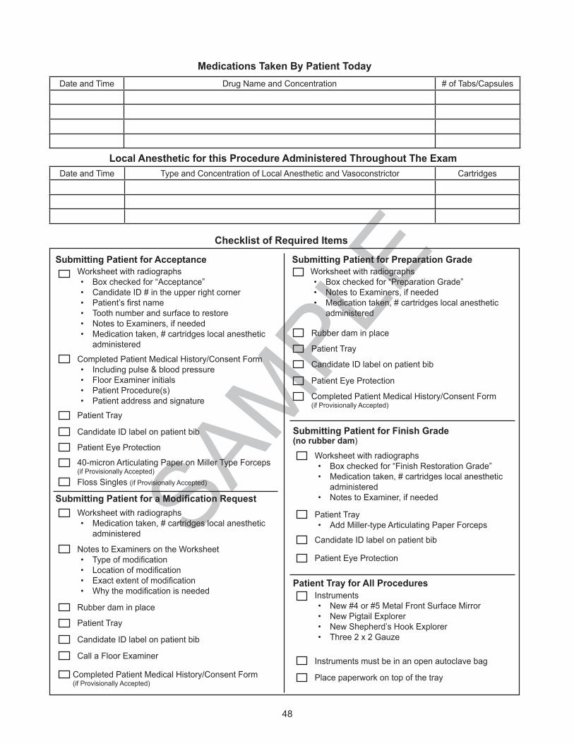
48
SAMPLE
Medications Taken By Patient Today Date and Time Drug Name and Concentration # of Tabs/Capsules
Local Anesthetic for this Procedure Administered Throughout The ExamDate and Time Type and Concentration of Local Anesthetic and Vasoconstrictor Cartridges
Checklist of Required Items
Patient Eye Protection
Rubber dam in place
Patient Tray
Candidate ID label on patient bib
Worksheet with radiographs• Box checked for “Preparation Grade”• Notes to Examiners, if needed• Medication taken, # cartridges local anesthetic
administered
Submitting Patient for Preparation Grade Submitting Patient for Acceptance
Patient Tray
Candidate ID label on patient bib
Patient Eye Protection
Completed Patient Medical History/Consent Form• Including pulse & blood pressure• Floor Examiner initials• Patient Procedure(s)• Patient address and signature
Worksheet with radiographs• Box checked for “Acceptance”• Candidate ID # in the upper right corner• Patient’sfirstname• Tooth number and surface to restore • Notes to Examiners, if needed• Medication taken, # cartridges local anesthetic
administered
Instruments must be in an open autoclave bag
Instruments• New #4 or #5 Metal Front Surface Mirror• New Pigtail Explorer• New Shepherd’s Hook Explorer• Three 2 x 2 Gauze
Place paperwork on top of the tray
Patient Tray for All Procedures
Submitting Patient for a Modification Request
Call a Floor Examiner
Worksheet with radiographs• Medication taken, # cartridges local anesthetic
administered
Candidate ID label on patient bib
Patient Tray
Rubber dam in place
Notes to Examiners on the Worksheet• Typeofmodification• Locationofmodification• Exactextentofmodification• Whythemodificationisneeded
Worksheet with radiographs• Box checked for “Finish Restoration Grade”• Medication taken, # cartridges local anesthetic
administered• Notes to Examiner, if needed
Patient Eye Protection
Patient Tray• Add Miller-type Articulating Paper Forceps
Candidate ID label on patient bib
Submitting Patient for Finish Grade(no rubber dam)
40-micron Articulating Paper on Miller Type Forceps (if Provisionally Accepted)
Completed Patient Medical History/Consent Form (if Provisionally Accepted)
Floss Singles (if Provisionally Accepted)
Completed Patient Medical History/Consent Form(if Provisionally Accepted)
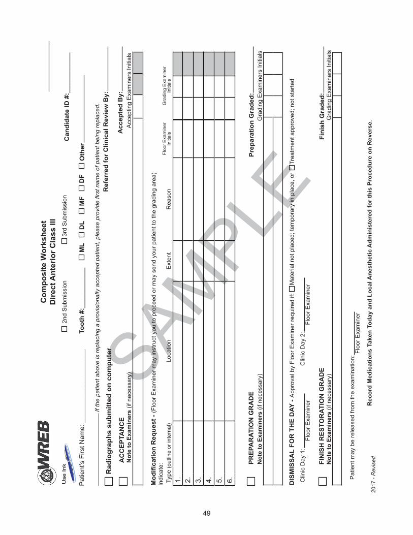
49
SAMPLE
Com
posi
te W
orks
heet
Dire
ct A
nter
ior C
lass
III
Can
dida
te ID
#:_
____
____
__
Gra
ding
Exa
min
ers
Initi
als
2nd
Sub
mis
sion
3rd
Sub
mis
sion
Mod
ifica
tion
Req
uest
- (F
loor
Exa
min
er m
ay in
stru
ct y
ou to
pro
ceed
or m
ay s
end
your
pat
ient
to th
e gr
adin
g ar
ea)
Indi
cate
:
Pat
ient
may
be
rele
ased
from
the
exam
inat
ion:
Floo
r Exa
min
er
PREP
AR
ATIO
N G
RA
DE
Not
e to
Exa
min
ers
(if n
eces
sary
) G
radi
ng E
xam
iner
s In
itial
sPr
epar
atio
n G
rade
d:
Fini
sh G
rade
d:
Pat
ient
’s F
irst N
ame:
___
____
____
____
____
_To
oth
#:__
____
____
_ M
LD
LM
FD
FO
ther
FIN
ISH
RES
TOR
ATIO
N G
RA
DE
Not
e to
Exa
min
ers
(if n
eces
sary
)
Type
(out
line
or in
tern
al)
Loca
tion
Ext
ent
Rea
son
Floo
r Exa
min
er
Initi
als
Gra
ding
Exa
min
er
Initi
als
1. 2. 3. 4. 5. 6.
Rec
ord
Med
icat
ions
Tak
en T
oday
and
Loc
al A
nest
hetic
Adm
inis
tere
d fo
r thi
s Pr
oced
ure
on R
ever
se.
2017
- R
evis
edAC
CEP
TAN
CE
N
ote
to E
xam
iner
s (if
nec
essa
ry)
Acc
eptin
g E
xam
iner
s In
itial
sA
ccep
ted
By:
DIS
MIS
SAL
FOR
TH
E D
AY -
App
rova
l by
Floo
r Exa
min
er re
quire
d if:
Clin
ic D
ay 1
:Fl
oor E
xam
iner
Clin
ic D
ay 2
:Fl
oor E
xam
iner
Mat
eria
l not
pla
ced;
tem
pora
ry in
pla
ce, o
rTr
eatm
ent a
ppro
ved;
not
sta
rted
Ref
erre
d fo
r Clin
ical
Rev
iew
By:
Rad
iogr
aphs
sub
mitt
ed o
n co
mpu
ter
Use
Ink
If th
e pa
tient
abo
ve is
repl
acin
g a
prov
isio
nally
acc
epte
d pa
tient
, ple
ase
prov
ide
first
nam
e of
pat
ient
bei
ng re
plac
ed.

50
SAMPLE
Medications Taken By Patient Today Date and Time Drug Name and Concentration # of Tabs/Capsules
Local Anesthetic for this Procedure Administered Throughout The ExamDate and Time Type and Concentration of Local Anesthetic and Vasoconstrictor Cartridges
Checklist of Required Items
Patient Eye Protection
Rubber dam in place
Patient Tray
Candidate ID label on patient bib
Worksheet with radiographs• Box checked for “Preparation Grade”• Notes to Examiners, if needed• Medication taken, # cartridges local anesthetic
administered
Submitting Patient for Preparation Grade Submitting Patient for Acceptance
Patient Tray
Candidate ID label on patient bib
Patient Eye Protection
Completed Patient Medical History/Consent Form• Including pulse & blood pressure• Floor Examiner initials• Patient Procedure(s)• Patient address and signature
Worksheet with radiographs• Box checked for “Acceptance”• Candidate ID # in the upper right corner• Patient’sfirstname• Tooth number and surface to restore • Notes to Examiners, if needed• Medication taken, # cartridges local anesthetic
administered
Instruments must be in an open autoclave bag
Instruments• New #4 or #5 Metal Front Surface Mirror• New Pigtail Explorer• New Shepherd’s Hook Explorer• Three 2 x 2 Gauze
Place paperwork on top of the tray
Patient Tray for All Procedures
Submitting Patient for a Modification Request
Call a Floor Examiner
Worksheet with radiographs• Medication taken, # cartridges local anesthetic
administered
Candidate ID label on patient bib
Patient Tray
Rubber dam in place
Notes to Examiners on the Worksheet• Typeofmodification• Locationofmodification• Exactextentofmodification• Whythemodificationisneeded
Worksheet with radiographs• Box checked for “Finish Restoration Grade”• Medication taken, # cartridges local anesthetic
administered• Notes to Examiner, if needed
Patient Eye Protection
Patient Tray• Add Miller-type Articulating Paper Forceps
Candidate ID label on patient bib
Submitting Patient for Finish Grade(no rubber dam)
40-micron Articulating Paper on Miller Type Forceps (if Provisionally Accepted)
Completed Patient Medical History/Consent Form (if Provisionally Accepted)
Floss Singles (if Provisionally Accepted)
Completed Patient Medical History/Consent Form(if Provisionally Accepted)

51
SAMPLE
Pat
ient
’s F
irst N
ame:
___
____
____
____
____
_
AC
CEP
TAN
CE
N
ote
to E
xam
iner
s (if
nec
essa
ry)
Acc
eptin
g E
xam
iner
s In
itial
s
Am
alga
m W
orks
heet
Dire
ct P
oste
rior C
lass
IIC
andi
date
ID #
:___
____
____
Rec
ord
Med
icat
ions
Tak
en T
oday
and
Loc
al A
nest
hetic
Adm
inis
tere
d fo
r thi
s Pr
oced
ure
on R
ever
se.G
radi
ng E
xam
iner
s In
itial
s
2nd
Sub
mis
sion
3rd
Sub
mis
sion
Type
(out
line
or in
tern
al)
Loca
tion
Ext
ent
Rea
son
Floo
r Exa
min
er
Initi
als
Gra
ding
Exa
min
er
Initi
als
1. 2. 3. 4. 5. 6.Mod
ifica
tion
Req
uest
- (F
loor
Exa
min
er m
ay in
stru
ct y
ou to
pro
ceed
or m
ay s
end
your
pat
ient
to th
e gr
adin
g ar
ea)
Indi
cate
:
Pat
ient
may
be
rele
ased
from
the
exam
inat
ion:
Floo
r Exa
min
er
Acc
epte
d by
:
PREP
AR
ATIO
N G
RA
DE
Not
e to
Exa
min
ers
(if n
eces
sary
) G
radi
ng E
xam
iner
s In
itial
sPr
epar
atio
n G
rade
d:
Fini
sh G
rade
d:
Toot
h #:
____
____
___
DO
MO
MO
DM
O &
DO
Oth
er
FIN
ISH
RES
TOR
ATIO
N G
RA
DE
Not
e to
Exa
min
ers
(if n
eces
sary
)
2017
- R
evis
ed
Ref
erre
d fo
r Clin
ical
Rev
iew
By:
DIS
MIS
SAL
FOR
TH
E D
AY -
App
rova
l by
Floo
r Exa
min
er re
quire
d if:
Clin
ic D
ay 1
:Fl
oor E
xam
iner
Clin
ic D
ay 2
:Fl
oor E
xam
iner
Mat
eria
l not
pla
ced;
tem
pora
ry in
pla
ce, o
rTr
eatm
ent a
ppro
ved;
not
sta
rted
If th
e pa
tient
abo
ve is
repl
acin
g a
prov
isio
nally
acc
epte
d pa
tient
, ple
ase
prov
ide
first
nam
e of
pat
ient
bei
ng re
plac
ed.
Rad
iogr
aphs
sub
mitt
ed o
n co
mpu
ter
Use
Ink
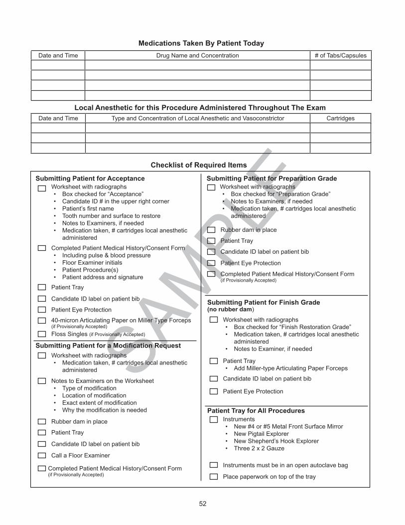
52
SAMPLE
Medications Taken By Patient Today Date and Time Drug Name and Concentration # of Tabs/Capsules
Local Anesthetic for this Procedure Administered Throughout The ExamDate and Time Type and Concentration of Local Anesthetic and Vasoconstrictor Cartridges
Checklist of Required Items
Patient Eye Protection
Rubber dam in place
Patient Tray
Candidate ID label on patient bib
Worksheet with radiographs• Box checked for “Preparation Grade”• Notes to Examiners, if needed• Medication taken, # cartridges local anesthetic
administered
Submitting Patient for Preparation Grade Submitting Patient for Acceptance
Patient Tray
Candidate ID label on patient bib
Patient Eye Protection
Completed Patient Medical History/Consent Form• Including pulse & blood pressure• Floor Examiner initials• Patient Procedure(s)• Patient address and signature
Worksheet with radiographs• Box checked for “Acceptance”• Candidate ID # in the upper right corner• Patient’sfirstname• Tooth number and surface to restore • Notes to Examiners, if needed• Medication taken, # cartridges local anesthetic
administered
Instruments must be in an open autoclave bag
Instruments• New #4 or #5 Metal Front Surface Mirror• New Pigtail Explorer• New Shepherd’s Hook Explorer• Three 2 x 2 Gauze
Place paperwork on top of the tray
Patient Tray for All Procedures
Submitting Patient for a Modification Request
Call a Floor Examiner
Worksheet with radiographs• Medication taken, # cartridges local anesthetic
administered
Candidate ID label on patient bib
Patient Tray
Rubber dam in place
Notes to Examiners on the Worksheet• Typeofmodification• Locationofmodification• Exactextentofmodification• Whythemodificationisneeded
Worksheet with radiographs• Box checked for “Finish Restoration Grade”• Medication taken, # cartridges local anesthetic
administered• Notes to Examiner, if needed
Patient Eye Protection
Patient Tray• Add Miller-type Articulating Paper Forceps
Candidate ID label on patient bib
Submitting Patient for Finish Grade(no rubber dam)
40-micron Articulating Paper on Miller Type Forceps (if Provisionally Accepted)
Completed Patient Medical History/Consent Form (if Provisionally Accepted)
Floss Singles (if Provisionally Accepted)
Completed Patient Medical History/Consent Form(if Provisionally Accepted)

53
54
32
1
OUTLINE & EXTENSION
Out
line
is g
ener
ally
sm
ooth
and
flo
win
g, a
nd d
oes
not w
eake
n to
oth
in a
ny m
anne
r.
Out
line
is s
light
ly ir
regu
lar b
ut
does
not
wea
ken
toot
h. I
sthm
us
is s
light
ly w
ider
than
requ
ired
for
lesi
on.
Out
line
mod
erat
ely
wea
kens
m
argi
nal r
idge
or a
cus
p. I
sthm
us
is to
o w
ide
or to
o na
rrow
for l
esio
n.
Out
line
seve
rely
wea
kens
mar
gina
l rid
ge o
r a c
usp.
Uns
ound
dem
iner
aliz
ed e
nam
el
that
is ta
ctile
ly d
iffer
ent f
rom
the
adja
cent
una
ffect
ed e
nam
el is
pr
esen
t.
Out
line
is g
ross
ly im
prop
er a
nd/o
r la
cks
any
defin
ite fo
rm.
Tact
ilely
uns
ound
dem
iner
aliz
ed
enam
el p
enet
rate
s th
e D
EJ.
Car
ies
rem
ains
in th
e en
amel
or i
s no
t com
plet
ely
acce
ssed
.
Una
ppro
ved
surfa
ce p
repa
red.
Prox
imal
and
gin
giva
l ext
ensi
ons
are
visu
ally
ope
n an
d br
eak
cont
act 0
.75
mm
-1.2
5 m
m.
The
ging
ival
mar
gin
has
a di
scer
nibl
e be
vel (
at le
ast 0
.5 m
m).
All
mar
gins
and
bev
els
are
smoo
th
and
wel
l defi
ned.
Prox
imal
and
/or g
ingi
val
exte
nsio
ns a
re s
light
ly
over
exte
nded
.
Mar
gins
are
slig
htly
irre
gula
r.
Beve
ls a
re le
ss th
an 0
.5 m
m o
r gr
eate
r tha
n 1.
0 m
m.
Prox
imal
and
/or g
ingi
val
exte
nsio
ns a
re m
oder
atel
y ov
erex
tend
ed.
Mar
gins
are
mod
erat
ely
irreg
ular
. Be
vels
are
mod
erat
ely
shal
low
or
deep
.
Prox
imal
and
/or g
ingi
val
exte
nsio
ns a
re in
con
tact
or
obvi
ousl
y ov
erex
tend
ed.
Mar
gins
are
crit
ical
ly ir
regu
lar o
r no
t defi
ned.
Bev
els
are
criti
cally
sh
allo
w o
r dee
p, o
r on
larg
e ar
eas
of u
nsup
porte
d en
amel
.
Prox
imal
and
/or g
ingi
val
exte
nsio
ns a
re g
ross
ly
over
exte
nded
.
Mar
gins
exh
ibit
gros
s la
ck o
f de
finiti
on.
Beve
ls a
re a
bsen
t.
Opt
imal
trea
tmen
t of fi
ssur
es.
Nea
r opt
imal
trea
tmen
t of fi
ssur
es.
Adeq
uate
trea
tmen
t of fi
ssur
es.
Nei
ther
the
toot
h no
r res
tora
tion
is
com
prom
ised
.
Inad
equa
te tr
eatm
ent o
f fiss
ures
w
ill co
mpr
omis
e th
e to
oth
or
rest
orat
ion.
Lack
of t
reat
men
t of fi
ssur
es w
ill se
rious
ly c
ompr
omis
e th
e to
oth
and
rest
orat
ion.
Prox
imal
cav
osur
face
ang
les
are
appr
oxim
atel
y 90
°.
Cav
osur
face
ang
les
are
not
optim
al b
ut d
o no
t com
prom
ise
the
inte
grity
of t
he to
oth
or re
stor
atio
n.
Cav
osur
face
ang
les
poss
ibly
co
mpr
omis
e th
e in
tegr
ity o
f the
to
oth
or re
stor
atio
n.
Impr
oper
cav
osur
face
ang
les
will
caus
e th
e fin
al re
stor
atio
n to
fail.
Cav
osur
face
ang
les
are
gros
sly
impr
oper
.
Idea
l con
serv
atio
n of
toot
h st
ruct
ure.
Slig
htly
inap
prop
riate
rem
oval
of
toot
h st
ruct
ure.
Mod
erat
ely
inap
prop
riate
rem
oval
of
toot
h st
ruct
ure.
Exce
ssiv
ely
inap
prop
riate
rem
oval
of
toot
h st
ruct
ure.
Gro
ssly
inap
prop
riate
rem
oval
of
toot
h st
ruct
ure.
INTERNAL FORM
Opt
imal
resi
stan
ce a
nd re
tent
ion
form
. Pa
ralle
lism
of w
alls
is id
eal;
ther
e ar
e no
und
ercu
ts p
rese
nt.
Res
ista
nce
and
rete
ntio
n fo
rm
is a
dequ
ate.
Wal
ls a
re s
light
ly
over
-tape
red.
No
unde
rcut
s ar
e pr
esen
t.
Res
ista
nce
and
rete
ntio
n fo
rm
is m
inim
ally
pre
sent
. W
alls
are
m
oder
atel
y ov
er-ta
pere
d, o
r a
smal
l und
ercu
t on
one
wal
l co
mpr
omis
es d
raw.
Res
ista
nce
and
rete
ntio
n fo
rm is
in
adeq
uate
. W
alls
are
exc
essi
vely
ov
er-ta
pere
d, o
r mod
erat
e un
derc
uts
are
pres
ent.
Com
plet
e se
atin
g or
rete
ntio
n of
the
rest
orat
ion
is c
ompr
omis
ed.
Res
ista
nce
and/
or re
tent
ion
form
is
com
plet
ely
abse
nt.
Wal
ls a
re
gros
sly
over
-tape
red
or g
ross
un
derc
uts
pres
ent.
Seat
ing
or
rete
ntio
n of
the
rest
orat
ion
is n
ot
poss
ible
.
Una
ppro
ved
surfa
ce p
repa
red.
Opt
imal
dep
th/re
duct
ion
of w
alls
co
nser
ves
toot
h st
ruct
ure
and
allo
ws
for a
dequ
ate
bulk
of
rest
orat
ive
mat
eria
l.
Slig
ht o
ver-r
educ
tion
or u
nder
-re
duct
ion
of w
alls
and
/or fl
oor i
s pr
esen
t.
Mod
erat
e ov
er-re
duct
ion
or u
nder
-re
duct
ion
of w
alls
and
/or fl
oor i
s pr
esen
t.
Crit
ical
ove
r-red
uctio
n or
und
er-
redu
ctio
n of
wal
ls a
nd/o
r floo
r is
pres
ent.
Exc
essi
ve d
epth
will
dam
age
the
pulp
.
Affec
ted
dent
in re
mai
ns.
Gro
ss o
ver-r
educ
tion
or u
nder
-re
duct
ion
of w
alls
and
/or fl
oor
is p
rese
nt. P
ulp
is d
efini
tely
co
mpr
omis
ed.
Car
ies
rem
ains
in th
e de
ntin
or i
s no
t com
plet
ely
acce
ssed
.Al
l wal
ls a
re s
moo
th.
Line
ang
les
are
clea
rly d
efine
d.W
alls
and
/or l
ine
angl
es a
re
slig
htly
irre
gula
r.W
alls
and
/or l
ine
angl
es a
re
mod
erat
ely
irreg
ular
.W
alls
and
/or l
ine
angl
es a
re ro
ugh
and
poor
ly d
efine
d.Th
ere
is g
ross
lack
of i
nter
nal
defin
ition
thro
ugho
ut p
repa
ratio
n.
OPERATIVEENVIRONMENT
Rub
ber d
am is
olat
ion
is s
tabl
e an
d op
timal
; the
dam
is in
verte
d an
d ha
s no
rips
, tea
rs, b
unch
ing
or
expo
sed
tissu
e. T
he p
repa
ratio
n is
cl
ean
and
dry.
Rub
ber d
am is
olat
ion
is n
ot
optim
al, b
ut th
e pr
epar
atio
n is
cl
ean
and
dry.
Rub
ber d
am is
olat
ion
is a
dequ
ate,
bu
t the
wro
ng te
eth
are
isol
ated
. Th
e pr
epar
atio
n ca
n be
cle
aned
an
d dr
ied.
Rub
ber d
am is
olat
ion
is
inad
equa
te.
The
prep
arat
ion
is
diffi
cult
to a
cces
s or
vis
ualiz
e du
e to
blo
od o
r sal
iva
on th
e pr
epar
atio
n or
par
tial c
over
age
by
the
dam
.
The
rubb
er d
am is
gro
ssly
slo
ppy
and
torn
, or p
ortio
ns o
f the
pr
epar
atio
n ar
e no
t vis
ible
due
to
bloo
d, s
aliv
a, o
r im
prop
er is
olat
ion.
No
dam
age
to th
e ad
jace
nt to
oth.
Min
or d
amag
e to
the
adja
cent
to
oth
can
be re
mov
ed b
y po
lishi
ng
with
out c
hang
ing
the
shap
e of
the
cont
act.
Dam
age
to th
e ad
jace
nt to
oth
can
be re
mov
ed b
y po
lishi
ng, b
ut
the
shap
e of
the
cont
act w
ill be
ch
ange
d.
Dam
age
to th
e ad
jace
nt to
oth
will
be d
ifficu
lt to
pol
ish
out a
nd s
till
mai
ntai
n ap
prop
riate
pro
xim
al
cont
our.
The
adj
acen
t too
th w
ill lik
ely
requ
ire re
stor
atio
n.
Dam
age
to th
e ad
jace
nt to
oth
will
defin
itely
requ
ire re
stor
atio
n.
2017
IND
IREC
T PO
STER
IOR
CLA
SS II
PR
EPA
RAT
ION
SCO
RIN
G C
RIT
ERIA
RAT
ING
SC
ALE

54
54
32
1
ANATOMICAL FORM
Anat
omic
al fo
rm is
con
sist
ent
and
harm
onio
us w
ith
cont
iguo
us to
oth
stru
ctur
e.
Slig
ht v
aria
tion
in n
orm
al
anat
omic
al fo
rm is
pre
sent
.M
oder
ate
varia
tion
in n
orm
al
anat
omic
al fo
rm is
pre
sent
. M
argi
nal r
idge
is im
prop
erly
sh
aped
.
Anat
omic
al fo
rm is
impr
oper
. M
argi
nal r
idge
is p
oorly
sh
aped
.
Anat
omy
is to
o de
ep o
r too
fla
t.
Ther
e is
gro
ss la
ck o
f an
atom
ical
form
.
Prop
er p
roxi
mal
con
tour
and
sh
ape
are
rest
ored
.Th
ere
is s
light
var
iatio
n of
pr
oxim
al c
onto
ur a
nd s
hape
.Th
ere
is m
oder
ate
varia
tion
of
prox
imal
con
tour
and
sha
pe.
Prox
imal
con
tour
is p
oor.
Em
bras
ures
are
sev
erel
y ov
er
or u
nder
con
tour
ed.
Gro
ssly
impr
oper
pro
xim
al
cont
our o
r sha
pe.
Nor
mal
pro
xim
al c
onta
ct a
rea
and
posi
tion
are
rest
ored
. C
onta
ct is
vis
ually
clo
sed
and
resi
sts
the
pass
age
of li
ghtly
w
axed
flos
s.
Ther
e is
slig
ht v
aria
tion
of
norm
al c
onta
ct a
rea
and
posi
tion.
Con
tact
is v
isua
lly
clos
ed a
nd re
sist
s th
e pa
ssag
e of
ligh
tly w
axed
flos
s.
Ther
e is
mod
erat
e va
riatio
n of
nor
mal
con
tact
are
a an
d po
sitio
n. L
ight
ly w
axed
flos
s w
ill pa
ss th
roug
h th
e co
ntac
t w
ith s
light
resi
stan
ce.
Con
tact
is v
isua
lly o
pen,
or
floss
will
not p
ass
thro
ugh
the
cont
act.
Con
tact
is g
ross
ly o
pen.
MARGINS
Res
tora
tion
is fu
lly s
eate
d w
ith
no e
xces
ses
or d
efici
enci
es
anyw
here
alo
ng th
e m
argi
ns.
Res
tora
tion
is s
eate
d.
Mar
gins
are
clo
sed,
but
sl
ight
mar
gina
l exc
esse
s or
de
ficie
ncie
s ar
e de
tect
able
w
ith a
n ex
plor
er.
Res
tora
tion
is s
eate
d.
Mod
erat
e m
argi
nal e
xces
ses
or d
efici
enci
es a
re d
etec
tabl
e w
ith a
n ex
plor
er.
Res
tora
tion
is n
ot s
eate
d.
Ther
e ar
e m
ultip
le a
reas
of
exp
osed
cem
ent,
open
m
argi
ns, a
nd/o
r mar
gina
l ex
cess
es.
A m
argi
nal o
verh
ang
catc
hes
floss
.
Res
tora
tion
is n
ot s
eate
d.
Ther
e ar
e gr
ossl
y ev
iden
t ar
eas
of e
xpos
ed c
emen
t, op
en m
argi
ns, a
nd/o
r m
argi
nal e
xces
ses.
A gr
oss
mar
gina
l ove
rhan
g sh
reds
flos
s.
FINISH, FUNCTION & DAMAGE
The
surfa
ce is
sm
ooth
with
no
pits
, voi
ds o
r irre
gula
ritie
s.Sl
ight
sur
face
irre
gula
ritie
s,
pitti
ng, o
r voi
ds a
re p
rese
nt.
Mod
erat
e su
rface
irr
egul
ariti
es, p
ittin
g, o
r voi
ds
are
pres
ent.
Sur
face
has
m
inor
cas
ting
irreg
ular
ities
.
Seve
re s
urfa
ce ir
regu
larit
ies,
pi
tting
, or v
oids
are
pre
sent
.G
ross
sur
face
def
ects
are
pr
esen
t.
Occ
lusi
on is
rest
ored
to
prop
er c
entri
c w
ith n
o la
tera
l in
terfe
renc
es.
Ther
e is
sev
ere
hype
rocc
lusi
on in
cen
tric
or
late
ral e
xcur
sion
s. O
cclu
sal
cont
act m
arks
app
ear o
nly
on
the
rest
orat
ion.
Occ
lusi
on is
gro
ssly
in
adeq
uate
.
Ther
e is
no
dam
age
to h
ard
or
soft
tissu
e.M
inor
dam
age
to h
ard
or s
oft
tissu
e is
evi
dent
.M
oder
ate
dam
age
to h
ard
or
soft
tissu
e is
evi
dent
.Se
vere
dam
age
to h
ard
or
soft
tissu
e is
evi
dent
.G
ross
mut
ilatio
n of
har
d or
so
ft tis
sue
is e
vide
nt.
2017
IND
IREC
T R
ESTO
RAT
ION
FIN
ISH
SCO
RIN
G C
RIT
ERIA
RAT
ING
SC
ALE
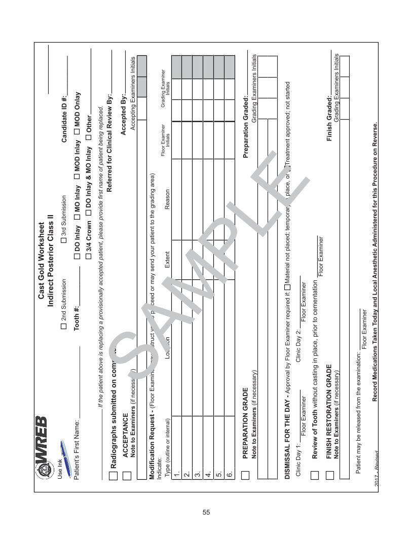
55
SAMPLE
Patie
nt’s
Firs
t Nam
e: _
____
____
____
____
___
Cast
Gol
d W
orks
heet
Indi
rect
Pos
terio
r Cla
ss II
Cand
idat
e ID
#:_
____
____
__
Gra
ding
Exa
min
ers
Initi
als
2nd
Subm
issi
on3r
d Su
bmis
sion
Mod
ifica
tion
Requ
est -
(Flo
or E
xam
iner
may
inst
ruct
you
to p
roce
ed o
r may
sen
d yo
ur p
atie
nt to
the
grad
ing
area
)In
dica
te:
Patie
nt m
ay b
e re
leas
ed fr
om th
e ex
amin
atio
n:Fl
oor E
xam
iner
PREP
ARAT
ION
GRA
DENo
te to
Exa
min
ers
(if n
eces
sary
) G
radi
ng E
xam
iner
s In
itial
sPr
epar
atio
n G
rade
d:
Fini
sh G
rade
d:
Toot
h #:
____
____
___
FINI
SH R
ESTO
RATI
ON
GRA
DE
Note
to E
xam
iner
s (if
nec
essa
ry)
DO In
lay
MO
Inla
yM
OD
Inla
yM
OD
Onl
ay
Oth
er3/
4 Cr
own
DO In
lay
& M
O In
lay
Revi
ew o
f Too
th w
ithou
t cas
ting
in p
lace
, prio
r to
cem
enta
tion
Floo
r Exa
min
er
Type
(out
line
or in
tern
al)
Loca
tion
Exte
ntR
easo
nFl
oor E
xam
iner
In
itial
sG
radi
ng E
xam
iner
In
itial
s
1. 2. 3. 4. 5. 6.
Reco
rd M
edic
atio
ns T
aken
Tod
ay a
nd L
ocal
Ane
sthe
tic A
dmin
iste
red
for t
his
Proc
edur
e on
Rev
erse
.20
17 -
Rev
isedAC
CEPT
ANCE
Note
to E
xam
iner
s (if
nec
essa
ry)
Acce
ptin
g Ex
amin
ers
Initi
als
Acce
pted
By:
Refe
rred
for C
linic
al R
evie
w B
y:
Clin
ic D
ay 1
:Fl
oor E
xam
iner
Clin
ic D
ay 2
:Fl
oor E
xam
iner
DISM
ISSA
L FO
R TH
E DA
Y - A
ppro
val b
y Fl
oor E
xam
iner
requ
ired
if:
Mat
eria
l not
pla
ced;
tem
pora
ry in
pla
ce, o
rTr
eatm
ent a
ppro
ved;
not
sta
rted
Radi
ogra
phs
subm
itted
on
com
pute
r
Use
Ink
If th
e pa
tient
abo
ve is
repl
acin
g a
prov
isio
nally
acc
epte
d pa
tient
, ple
ase
prov
ide
first
nam
e of
pat
ient
bei
ng re
plac
ed.
SAMPLE

56
SAMPLE
Medications Taken By Patient Today Date and Time Drug Name and Concentration # of Tabs/Capsules
Local Anesthetic for this Procedure Administered Throughout The ExamDate and Time Type and Concentration of Local Anesthetic and Vasoconstrictor Cartridges
Checklist of Required Items
Patient Eye Protection
Rubber dam in place
Patient Tray
Candidate ID label on patient bib
Worksheet with radiographs• Box checked for “Preparation Grade”• Notes to Examiners, if needed• Medication taken, # cartridges local anesthetic
administered
Submitting Patient for Preparation Grade Submitting Patient for Acceptance
Patient Tray
Candidate ID label on patient bib
Patient Eye Protection
Completed Patient Medical History/Consent Form• Including pulse & blood pressure• Floor Examiner initials• Patient Procedure(s)• Patient address and signature
Worksheet with radiographs• Box checked for “Acceptance”• Candidate ID # in the upper right corner• Patient’sfirstname• Tooth number and surface to restore • Notes to Examiners, if needed• Medication taken, # cartridges local anesthetic
administered
Instruments must be in an open autoclave bag
Instruments• New #4 or #5 Metal Front Surface Mirror• New Pigtail Explorer• New Shepherd’s Hook Explorer• Three 2 x 2 Gauze
Place paperwork on top of the tray
Patient Tray for All Procedures
Submitting Patient for a Modification Request
Call a Floor Examiner
Worksheet with radiographs• Medication taken, # cartridges local anesthetic
administered
Candidate ID label on patient bib
Patient Tray
Rubber dam in place
Notes to Examiners on the Worksheet• Typeofmodification• Locationofmodification• Exactextentofmodification• Whythemodificationisneeded
Worksheet with radiographs• Box checked for “Finish Restoration Grade”• Medication taken, # cartridges local anesthetic
administered• Notes to Examiner, if needed
Patient Eye Protection
Patient Tray• Add Miller-type Articulating Paper Forceps
Candidate ID label on patient bib
Submitting Patient for Finish Grade(no rubber dam)
40-micron Articulating Paper on Miller Type Forceps (if Provisionally Accepted)
Completed Patient Medical History/Consent Form (if Provisionally Accepted)
Floss Singles (if Provisionally Accepted)
Completed Patient Medical History/Consent Form(if Provisionally Accepted)

57
ENDODONTICS
Endodontics Section Overview
The exam consists of endodontic treatment on two extracted teeth: one anterior tooth and one multi-canal posterior tooth. You will be given four hours to complete the exam and will be allowed in the lab one-half hour before the exam to set up and to receive a setup check. Time is determined by the official WREB clock.
WREB examines Candidates with varying educational backgrounds, and schools may teach different endodontic procedures. WREB does not look for one standard procedure and scores according to the criteria on pg. 71.
The teeth will be mounted in a segmented arch. The arches are not included as exam material and you must purchase them separately. Acadental or Columbia carries segmented models approved for this exam. Please refer to the “Exam Site Information” (available at www.wreb.org) for your exam site prior to arrival to be sure your particular typodont is compatible with their manikin setup and if you are required to provide an articulator. WREB strongly encourages the use of the magnetic release model from either of these companies as it more easily facilitates sextant removal from the arch for radiographs. The model may be purchased at any time before the exam. All six sextants must be mounted during treatment. If you plan to mount back-up teeth, you will need to purchase additional sextants. (See pg. 64 “Back-Up Teeth for Alternate Submission.”)
The “Exam Site Information” (available at www.wreb.org) will have details on whether you may purchase the arches through the school. Arches may be purchased directly from Acadental at www.acadental.com/WREB or from Columbia at www.columbiadentoform.com.
A complete endodontic model is required. Each endodontic kit will contain:• One maxillary arch with three removable sextants• One mandibular arch with three removable sextants• Apex putty needed for mounting teeth• Fixing gel with mixing tips• Articulator, required at some sites (check your exam site’s information)
Tooth Selection
Any anterior or multi-canal posterior tooth with an intact crown may be used.• Anterior teeth are: 6, 7, 8, 9, 10, 11, 22, 23, 24, 25, 26 and 27• Posterior teeth are: 1, 2, 3, 4, 5, 12, 13, 14, 15, 16, 17, 18, 19, 20, 21, 28, 29, 30,
31 and 32Single canal posterior teeth will not be accepted.
You will be treating extracted teeth of your own selection. It is advisable to choose an easily treatable case.

58
To keep the teeth from becoming brittle and breaking during treatment, store them continuously in a liquid solution, described in “Preparation of Teeth,” on pgs. 58-60, before mounting in the sextant.
Verify that the tooth selected has the crown and all roots intact, fits in the sextant and can be mounted as listed in “Preparation of Teeth.” Teeth with long roots may not be appropriate for the sextants. WREB recommends a tooth no longer than 25 mm.
You may choose which canal to treat on the multi-canal posterior tooth. You will be required to designate which canal you plan to treat on your Endodontic Worksheet. Although only one canal of the posterior tooth needs to be treated, all canals must be identified as if all canals would be treated. Grading Examiners will use an endodontic explorer to check identify all canals.
On a posterior tooth occlusal caries that will involve the access prep should be restored with a radiopaque material. It must be obvious from the preoperative radiographs that any unrestored caries or restorative material does not violate the pulp chamber or obstruct the radiographic view of the pulp from either the buccal or the proximal view.
Craze lines, Class IV fractures and small enamel fractures are acceptable if they do not involve the pulp chamber and/or the access outline form.
Preparation of Teeth
Mount only one tooth per sextant. Selected teeth should be mounted to closely simulate their natural position in the mouth:
• Maxillary teeth must be mounted in maxillary sextants.• Mandibular teeth must be mounted in mandibular sextants. • Anterior teeth must be mounted in anterior sextants. It is sometimes necessary to mount
anterior teeth a little more upright than typodont teeth.• Posterior teeth must be mounted in the proper posterior sextants.
• The tooth cannot be rotated buccal-lingually or mesial-distally, or positioned from right side to left side or vice versa.
• Each tooth must have the facial of the tooth mounted to the facial side of the sextant, and be mounted in its proper position in the arch as shown in the following illustrations.
Note that only teeth 6, 7, 8, and 27 may be mounted in a socket other than their natural position in the arch. Teeth mounted in any other socket than indicated in the illustrations will be rejected.
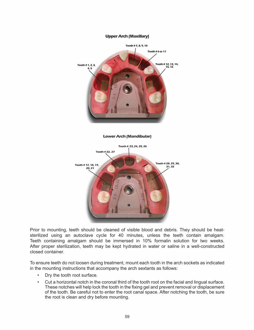
59
Prior to mounting, teeth should be cleaned of visible blood and debris. They should be heat-sterilized using an autoclave cycle for 40 minutes, unless the teeth contain amalgam. Teeth containing amalgam should be immersed in 10% formalin solution for two weeks. After proper sterilization, teeth may be kept hydrated in water or saline in a well-constructed closed container.
To ensure teeth do not loosen during treatment, mount each tooth in the arch sockets as indicated in the mounting instructions that accompany the arch sextants as follows:
• Dry the tooth root surface.• Cut a horizontal notch in the coronal third of the tooth root on the facial and lingual surface.
These notches will help lock the tooth in the fixing gel and prevent removal or displacement of the tooth. Be careful not to enter the root canal space. After notching the tooth, be sure the root is clean and dry before mounting.

60
• Place the apex putty in the bottom of the appropriate socket for the natural tooth selected. Place the tooth in the socket and align with the occlusal plane of the adjacent artificial teeth. The putty should extend around all root apices, laterally and apically. See illustration.
1. Apex putty must be at least 2.0 mm in depth beyond all root apices. 2. The apex putty should extend up the sides of the roots approximately ¼ of the distance from
root apex to the CEJ, but no more.
3. Fixing gel should cover the apex putty and the remaining space in the socket to the CEJ.
Teeth will be rejected if the putty is not visible radiographically at least 2.0 mm beyond the apex. The putty must allow the potential for a 2.0 mm overfill.
A tooth should be mounted at the same occlusal plane with proper alignment to the adjacent artificial teeth. However, if necessary to clearly see the apex putty 2.0 mm beyond the apices, a tooth may be up to 2.0 mm above the occlusal plane of the artificial teeth. If a mounted tooth has an improper buccal-lingual or mesial-distal alignment, it may make endo treatment more difficult.
Teeth will be rejected if there is a discrepancy greater than 2.0 mm above or below the occlusal plane of the adjacent artificial teeth.
Once the tooth and putty are positioned in the socket, fixing gel will be used to set the tooth. Before using, bleed the syringe to insure even extrusion of both materials, and then attach the mixing tip. Fill around the tooth until the fixing gel covers all visible apex putty, the entire socket area and is tight around the tooth’s entire root surface to the CEJ. To stabilize the tooth while adding the fixing gel and allowing it to set, you may want to use red rope wax to secure the tooth to the adjacent artificial tooth or use sticky wax on a toothpick secured to the artificial teeth on each side of the socket.
After the teeth have been mounted in the sextant, do not store in the formalin solution. A damp cloth (water only) will help keep the teeth from drying out.
Radiographs
Two preoperative radiographic views of each tooth submission must be submitted (one buccal and one proximal). The entire tooth from cusp tip/incisal edge to 2.0 mm beyond the apex must be visible on the film. As long as the entire tooth image is present, a cone cut does not render the radiograph undiagnostic.
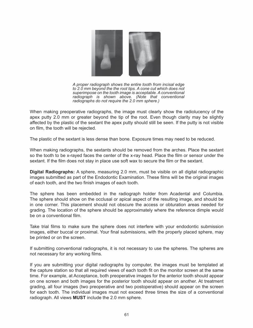
61
A proper radiograph shows the entire tooth from incisal edge to 2.0 mm beyond the the root tips. A cone cut which does not superimpose on the tooth image is acceptable. A conventional radiograph is shown above. (Note that conventional radiographs do not require the 2.0 mm sphere.)
When making preoperative radiographs, the image must clearly show the radiolucency of the apex putty 2.0 mm or greater beyond the tip of the root. Even though clarity may be slightly affected by the plastic of the sextant the apex putty should still be seen. If the putty is not visible on film, the tooth will be rejected.
The plastic of the sextant is less dense than bone. Exposure times may need to be reduced.
When making radiographs, the sextants should be removed from the arches. Place the sextant so the tooth to be x-rayed faces the center of the x-ray head. Place the film or sensor under the sextant. If the film does not stay in place use soft wax to secure the film or the sextant.
Digital Radiographs: A sphere, measuring 2.0 mm, must be visible on all digital radiographic images submitted as part of the Endodontic Examination. These films will be the original images of each tooth, and the two finish images of each tooth.
The sphere has been embedded in the radiograph holder from Acadental and Columbia. The sphere should show on the occlusal or apical aspect of the resulting image, and should be in one corner. This placement should not obscure the access or obturation areas needed for grading. The location of the sphere should be approximately where the reference dimple would be on a conventional film.
Take trial films to make sure the sphere does not interfere with your endodontic submission images, either buccal or proximal. Your final submissions, with the properly placed sphere, may be printed or on the screen.
If submitting conventional radiographs, it is not necessary to use the spheres. The spheres are not necessary for any working films.
If you are submitting your digital radiographs by computer, the images must be templated at the capture station so that all required views of each tooth fit on the monitor screen at the same time. For example, at Acceptance, both preoperative images for the anterior tooth should appear on one screen and both images for the posterior tooth should appear on another. At treatment grading, all four images (two preoperative and two postoperative) should appear on the screen for each tooth. The individual images must not exceed three times the size of a conventional radiograph. All views MUST include the 2.0 mm sphere.

62
The file name for each tooth should include your Candidate ID number, the tooth number and either “Anterior” or “Posterior.” A sample file name for an anterior tooth would be: B160 #8 Anterior.
A proper postoperative digital radiograph showing the entire tooth from incisal edge to apex with the 2.0 mm sphere placed so as not to interfere with the radiographic image.
Submission of Arches, Radiographs and the Endodontic Worksheet
Endodontic teeth and radiographs are collected prior to Candidate Orientation for evaluation by the Grading Examiners.
Only the sextants and accompanying radiographs for one anterior and one posterior tooth will be submitted before Candidate Orientation. At your scheduled exam, these sextants will be returned to you for treatment.
In the packet with this Candidate Guide, you received a ziplock bag with a blank label and an Endodontic Worksheet. Once you receive your schedule with your Candidate ID number, write your number on the label. The ziplock bag will be used to hold both sextants, conventional or printed digital radiographs and the worksheet.
Use a bur to inscribe your Candidate ID on the lingual side of the two sextants containing your first anterior and posterior teeth submissions.
Write your Candidate ID number on the worksheet (use only blue or black ink, do not use pencil). If you make an error prior to submitting your sextants for approval, obtain a new worksheet (cross-outs are not accepted at acceptance). If you submit a worksheet that is not neat, clear and in ink, your sextants will be returned to you with a new worksheet to complete which may result in loss of time. The date should be listed as the date you are submitting your sextants for approval. Write the anterior tooth number, the posterior tooth number and the posterior canal you plan to treat.
Just prior to turning in your sextants (before Candidate Orientation), place both sextants, the preoperative radiographs and the worksheet in the plastic bag. The sextants must be dry.
63
If submitting conventional films (2.0 mm sphere not required):
For each tooth you will submit two preoperative radiographs (one buccal view and one proximal view) in a four-film mount labeled only with your Candidate ID number, the tooth number, and “A” (for anterior) or “P” (for posterior). Your name should not appear on the film mounts, nor should film mounts be used that identify a school name or location. The two empty slots in the mount will be for your postoperative films.
If submitting printed digital films (2.0 mm sphere required):
For each tooth, you will submit a sheet with two preoperative radiographs (one buccal view and one proximal view) labeled only with your Candidate ID number, the tooth number and “A” (for anterior) and “P” (for posterior). Your name should not appear on the sheets. Printed digital images must be printed on high quality photographic paper and should be no larger than three times the size of a conventional radiograph.
If submitting digital radiographs in the computer (2.0 mm sphere required):
On your Candidate worksheet, check the box that the radiographs are available in digital format on the school’s computer. The file name for each tooth should include your Candidate ID number, the tooth number, and either “Anterior” or “Posterior.” A sample file name for an anterior tooth would be: B160 #8 Anterior. The films should be saved in a manner that will allow you to add the postoperative films, and submit the final films in a four mount template. Images should be no larger than three times the size of a conventional radiograph.
It is acceptable to submit radiographs in one format for preoperative images and a different format for postoperative images (i.e., printed digital for preoperative and conventional for postoperative).
Rejected Teeth
Teeth will be rejected if:• The crown is not intact. (NOTE: Craze lines, Class IV fractures and small enamel fractures
that do not involve the pulp chamber and/or the outline form of the access opening are acceptable.)
• There is evidence of occlusal or incisal reduction of the crown.• The tooth is improperly mounted: maxillary tooth in mandibular arch (or vice versa),
mounted in wrong socket, rotated buccal-lingually or mesial-distally, mounted on the wrong side of the arch.
• There is a height discrepancy greater than 2.0 mm above or below the occlusal plane of the adjacent artificial teeth.
• There is more than one natural tooth mounted in a sextant.• The radiographs are undiagnostic, or incorrect for the tooth; and/or the digital image is lacking
the 2.0 mm sphere, or the sphere interferes with the radiographic image. Score deduction will apply.
• The tooth has an existing restoration, base or caries that violates or radiographically obscures the pulp chamber, or the tooth has been restored with a full or partial coverage crown or onlay. (NOTE: A tooth with a restoration or base that obscures the radiographic visibility of the pulp chamber from either the buccal or the proximal view is unacceptable.)
• The posterior tooth is not multi-canal.• Apex putty is not visible 2.0 mm beyond all root apices.

64
If a submitted tooth is rejected by the Grading Examiners, it will be noted on your worksheet. If an alternate submission is required, you will be notified and allowed to submit a backup. There is no deadline for re-submissions. However, after 7:50 a.m. the first clinic morning, it may take two hours or more for the Grading Examiners to approve or reject a tooth. You may lose treatment time if a tooth has not been approved by the Grading Examiners prior to your scheduled Endodontics Exam. There will be no extension of exam time due to this approval process.
Rejected teeth may be removed from the sextant and resubmitted for acceptance if intially rejected for the following reasons:
• The tooth is improperly mounted: maxillary tooth in mandibular arch (or vice versa), mounted in wrong socket, rotated buccal-lingually or mesial-distally, mounted on the wrong side of the arch.
• There is a height discrepancy greater than 2.0 mm above or below the occlusal plane of the adjacent artificial teeth.
• There is more than one natural tooth mounted in a sextant.• Apex putty is not visible 2.0 mm beyond all root apices.• The radiographs are undiagnostic, or incorrect for the tooth; and/or the digital image is lacking
the 2.0 mm sphere, or the sphere interferes with the radiographic image. Score deduction will apply.
You are allowed up to three submissions for each anterior and each posterior tooth. Failure to submit an acceptable tooth in three attempts will result in loss of all points for the tooth affected (anterior or posterior).
Back-Up Teeth for Alternate Submission
You may mount additional teeth (back-up teeth) in additional sextants if you choose to do so. However, these sextants cannot be used to complete the arches. The two teeth to be treated are the only natural teeth that may be mounted in the manikin.
Back-up teeth should not be submitted for approval with the initial anterior and posterior teeth.Back-up teeth should be submitted only if a rejection notification is received.
Exam Procedure
Assistants are not permitted for this procedure. Written material may not be used to assist in treatment during the exam. You may use color code guides of endodontic file systems, but no other material. Candidates may not assist each other. This includes critiquing of another Candidate’s radiographs or discussion of treatment.
You are allowed to bring the 2017 Candidate Guide into the lab and refer to it during the exam. Notes, textbooks or other informational material must not be brought into the lab. All electronic devices must be turned off. No magnification other than loupes is allowed.
You must bring the upper and lower carrier trays with all necessary sextants to complete the arches.
65
Only the supplies listed in the “Exam Site Information” will be provided. You must provide all other supplies and equipment, including special equipment or mechanical files. Before entering the Endodontic Lab, make sure you have all necessary equipment and supplies.
You will be allowed to enter the lab at your assigned time. You must wear your Candidate badge in a visible location or you will not be allowed to enter. As you enter the lab, you will be assigned a station, identified by a green card with the station number. You are allowed a 30-minute setup period prior to the start of the Endodontic exam. During the first 15 minutes you are allowed to leave the lab to retrieve missing items as you prepare for the exam. During this 15 minute period you should confirm that you have all required instruments and materials, arrange materials and become familiar with the manikin setup procedure. Being prepared with all of the necessary materials will allow you to be ready to begin on time.
Fifteen minutes prior to the start of your Endodontic section you will be given the ziplock bag with your approved teeth/sextants, the preoperative radiographs and worksheet. Verify your Candidate ID number on the lingual side of each sextant, on the radiographs and on the worksheet. Acceptance of teeth to be treated will be initialed by a Grading Examiner on your worksheet. At this point, after you have been given the sextants to treat, you are strongly discouraged from leaving the lab. If you need to leave the lab, the arches, x-rays and worksheet must be placed in the ziplock bag and checked back in with the Endo Proctor. The arches will be returned to you when you return to the lab. If you leave the room from this point, without submitting the arches, etc. to the Endo Proctor, an automatic failure of the Endodontic exam will be assigned.
Once you have received your sextants, you should place them in the carrier tray to complete your arches and mount your arches in the manikin. Remember that the two teeth to be treated are the only natural teeth that may be mounted. The Endo Floor Examiner will perform a setup check and initial your Endodontic Worksheet. The following should be ready for the Endo Floor Examiner:
• Endo arches properly mounted in the manikin• Manikin in correct Patient treatment position with correct vertical dimension• Light on and mirror on tray• Endodontic Worksheet on tray
The rubber dam need not be in place for setup check. Both anterior and posterior teeth may be checked at the same time or separately.
Do not start treatment until you have Setup Check approval from the Endo Floor Examiner and the Endo Floor Examiner has announced that you may start the exam at the specified time.
If access is started without a Setup Check or prior to the announced start time, all Access points for the tooth are lost.
If the pulp chamber has been entered without a Setup Check or prior to the announced start time, all points for the tooth are lost.

66
Rubber dam placement is required before any treatment of the tooth is begun and must remain in place throughout the procedure.
Rubber dam placement must simulate proper placement on a Patient utilizing a rubber dam frame. Both teeth to be treated may be isolated at the same time, if feasible. Rubber dams may be removed only for making radiographs.
Performing the access opening or filing/preparation, or condensation of a canal without a rubber dam properly placed, is reason for dismissal from the endodontic exam with loss of all points for both teeth for the Endodontic procedures.
Single or multi-tooth isolation is acceptable. Placing the rubber dam clamp on an adjacent artificial tooth is recommended. Placing the rubber dam clamp on the natural tooth could cause the tooth to loosen. If a tooth loosens, notify the Endo Floor Examiner immediately.
You are expected to:• Follow universal precautions including radiation safety.• Work with arches mounted in proper Patient head simulation.• Receive a Setup Check approval before you begin.• Work with correct placement of a rubber dam during all aspects of treatment.
Violation of any of the above is grounds for dismissal and the loss of all points for the Endodontic section.
Working on the natural tooth or sextant in your hand and not properly mounted, at any time during the exam, is reason for dismissal from the Endodontic exam with loss of all points for both teeth for the endodontic procedures.
Treatment
AccessOn establishing outline form, restorative material, or caries should be treated as natural tooth structure.
Accessing an artifical sextant tooth will result in the deduction of all Access points. Treating a tooth other than the approved submission results in loss of all points for the tooth.
If the tooth fractures during treatment, you should complete the endodontic procedure.
If a crown fractures or any restorative material fractures during treatment, place the fractured pieces in the ziplock bag and turn them in with the treated tooth. Do not attempt to glue the pieces back together. If the access opening remains intact or can be determined by the Grading Examiners by putting pieces of the crown back together there will be no penalty assessed for the fracture. Make a note on the worksheet.
No occlusal or incisal reduction of clinical crowns is to be done, other than the normal access preparation outlined in the scoring criteria. Any other alteration will result in a loss of all Access points for that tooth.
67
CondensationInstrumentation technique, either mechanical or manual is at your discretion.
If a root fractures during treatment, Grading Examiners will score no higher than a 3.00 for condensation.
Any form of gutta-percha filling technique may be used, including any warm gutta-percha or carrier based thermoplasticized gutta-percha techniques, as well as thermoplastic synthetic polymer filling material.
You will perform endodontic treatment with gutta-percha fill to the facial cemento-enamel junction on the anterior tooth. There should be no fill coronal to the cemento-enamel junction.
You will perform endodontic access and treatment of the canal you selected on a posterior tooth. Fill to the canal orifice of the one treated posterior tooth canal. No filling material should overflow onto or cover the pulpal floor. Be sure the Grading Examiners can identify the orifices of all canals. You should treat no more than one canal per tooth. If, during treatment, two canals are found where you would expect to find only one (i.e., the distal root of a mandibular molar) immediately inform the Endo Floor Examiner and declare which canal will be treated. Make a note on the Endodontic Worksheet and have the Endo Floor Examiner initial it.
On the Endodontic Worksheet, you may make notes concerning treatment which you feel would be beneficial to the Grading Examiners, but the Endo Floor Examiner will not sign any note about treatment which he/she has not personally observed or has ample knowledge to confirm.
When making radiographs, the sextants may be removed from the arches as necessary. Only the final finish radiographs will be turned in. Taking excessive radiographs during the four-hour block consumes time and may result in late penalties. Plan carefully.
After Treatment
The two sextants with treated teeth, preoperative and postoperative radiographs in four-hole film mounts or a digital four bitewing image format and the Endodontic Worksheet must be turned in at the end of the exam time.
Postoperative radiographs of the final treatment should be taken with rubber dam and clamp removed. The radiographs should be from the same projection as the radiographs originally submitted: one from a buccal projection and one from a proximal projection. The two finish radiographs should be mounted with the corresponding preoperative radiographs.
You are responsible for submitting high-quality radiographs.
Allow sufficient time to submit dry radiographs. Radiographs submitted wet may be unreadable. You may want to consider bringing a hair dryer to the exam to facilitate the drying of radiographs.
Radiographs validated undiagnostic by the Grading Examiners will result in a deduction from the Endodontic score. (See pg. 70 for “Endodontic Scoring.”) If final radiographs are not submitted, there will be a deduction for each tooth. Radiographs are undiagnostic when they must be retaken to determine adequacy of treatment, or do not contain the 2.0 mm sphere or the sphere interferes with the radiographic image of a digitally stored or digital print postoperative image.

68
Schools will provide either automatic or hand developers. Be prepared to use either method of developing film. You must use the developing and fixing machines provided by the school in the endo lab. You may not leave the lab to develop films or provide your own developing equipment. You may provide your own self-developing film. Neither the school nor WREB can be held responsible for the quality of radiographs. Some schools will have only digital facilities. Be familiar with the Exam Site Information. There are often lines for the x-ray at the end of the four-hour exam. It is important that you schedule carefully or be prepared with self-developing film.
Postoperative digital images must be stored to the appropriate template as the preoperative images and contain the 2.0 mm sphere.
When turning your sextants in after treatment is complete:• Be sure the rubber dam and clamp are removed.• Remove the arches and carrier tray from the manikin.• Place both sextants with treated teeth, your worksheet, and the two preoperative and two
postoperative radiographs of each treated tooth in the ziplock bag.It is your responsibility to ensure that all of the materials listed above are turned in. Once you have left the Endo laboratory, you will be subject to failure of the Endodontic exam for items not turned in.
If you feel correspondence with Grading Examiners is necessary, you may use “Note to Examiners” on the worksheet.
Late penalties will be assessed to Candidates who exceed the four (4) hours allowed for the exam. You must have your endodontic sextants and radiographs turned in to avoid a late penalty. A reduction to the endo exam score will be assessed for each 5 minutes beyond the end of the time allowed. After 15 minutes, all points for the endodontic procedures will be lost. (See pg. 8 for “Late Penalty.”)
A random selection of teeth will be sectioned at the end of each exam. Any alteration of a tooth will result in failure of the entire exam and appropriate disciplinary action will be taken.
Examiners may remove the teeth from the sextants to look for irregularities.
Definitions
The following definitions are provided to assist you in more fully understanding scoring criteria and communications with Examiners:
Apical Perforation: Creating a new apical foramen.
Ledging: An irregularity created in the canal wall during filing.
Strip Perforation: A perforation on the lateral side of the root caused by transporting.
Transporting: Changing the position of the canal by straightening the walls during filing.

69
Unroofed Pulp Chamber: The dentin that covers the chamber incisally or occlusally, in which no ledges or overhangs are visible.
Zipping: Transporting the apical foramen.
Reference Material
WREB uses the basic endodontic access criteria from Pathways of the Pulp, 11th edition, (Stephen Cohen), Mosby Publishing Company.
Information regarding teeth cleaning and storage is based on excerpts from the CDC report, Guidelines for Infection Control in Dental Health-Care Settings --- 2003, December 19, 2003. The entire article is available at http://www.cdc.gov/mmwr/preview/mmwrhtml/rr5217a1.htm.
Other references are:
Endodontics, 6th edition, (Ingle and Bakland), Williams & Wilkins Publishing Company.
Principles and Practice of Endodontics, 4th edition, (Walton and Torabinejad), W.B. Saunders Publishing Co.
Elsevier: Problem Solving in Endodontics, fifth edition, James L. Gutmann, DDS, CertEndo, PhD(honoris causa), FACD, FICD, FADI and Paul E. Lovdahl, DDS, MSD, FACD, FADI.”
American Association of Endodontics. Spring 2010. Access Opening and Canal Location Endodontics - Colleagues for Excellence.
70
UNUSUAL SITUATIONS
Accessing an artificial sextant tooth. = Loss of all access points for the tooth
Treating a natural tooth other than the approved submission results.
= Loss of all points for the tooth
Alteration of the incisal or occlusal tooth surface.
= Loss of all access points for the tooth
ENDODONTIC SCORINGANTERIOR TOOTH
Access: 37.5%Condensation: 62.5%
POSTERIOR TOOTH
Access: 37.5%Condensation: 62.5%
Treatment GradeEach tooth will account for 50% of the total Endodontic section score.
Access (37.5%)Access must be appropriate for the entire tooth, even if treating only one canal.
Condensation (62.5%)Root fracture: the maximum on the rating scale that a Grading Examiner can assign for Condensation is a “3.00”.
ACCEPTANCE SCORE DEDUCTIONS
Submission Rejection: Rejection of first or second submission for either tooth (anterior or posterior): You are allowed to submit an alternate tooth. There is no point deduction, unless the rejection is for undiagnostic radiographs.
Rejection of a third submission for either tooth (anterior or posterior): No further submissions are accepted. No points are awarded for that tooth.
Undiagnostic Radiograph at Acceptance: Validated by two or more Grading Examiners, 0.3 will be deducted per tooth. Maximum 0.6 deduction. Score deduction will apply if the radiographs are undiagnostic, or incorrect for the tooth; and/or the digital image is lacking the 2.0 mm sphere, or the sphere interferes with the radiographic image.
TREATMENT SCORE DEDUCTIONS
Undiagnostic Radiograph at Treatment: Validated by two or more Grading Examiners, 0.2 will be deducted per tooth. Maximum 0.4 deduction. Radiographs are undiagnostic when they must be retaken to determine adequacy of treatment, or do not contain the 2.0 mm sphere or the sphere interferes with the radiographic image of a digitally stored or digital print postoperative image.
Missing Radiographs at Treatment (final radiographs not submitted): Validated by two or more Grading Examiners, 0.3 will be deducted per tooth. Maximum 0.6 deduction.
Late Penalties: 1 to 5 minutes late: 0.2 deduction; 6 to 10 minutes late: 0.4 deduction; 11 to 15 minutes late: 0.6 deduction; 16 or more minutes late: Procedure will not be graded. No points earned.

71
2017
EN
DO
DO
NTI
CSC
OR
ING
CR
ITER
IA R
ATIN
G S
CA
LE5
43
21
ACCESS OPENING
Out
line
Nea
r ide
al s
hape
, siz
e an
d lo
catio
n. F
or a
nter
iors
es
thet
ics
are
not a
ffect
ed.
If cr
own
is fr
actu
red,
acc
ess
is in
tact
or o
utlin
e an
d sh
ape
can
be d
eter
min
ed b
y pu
tting
pi
eces
bac
k to
geth
er.
Som
e va
riatio
n in
sha
pe,
size
and
/or l
ocat
ion.
May
be
slig
htly
ove
r or u
nder
ex
tend
ed.
For i
ncis
ors,
min
or
encr
oach
men
t on
inci
sal e
dge,
bu
t is
acce
ptab
le fo
r api
cal
inst
rum
enta
tion.
If cr
own
is fr
actu
red,
acc
ess
is in
tact
or o
utlin
e an
d sh
ape
can
be d
eter
min
ed b
y pu
tting
pi
eces
bac
k to
geth
er.
Sha
pe, s
ize
and/
or lo
catio
n ar
e fu
nctio
nal.
May
be
mod
erat
ely
over
or u
nder
ex
tend
ed.
For a
nter
iors
, en
croa
chm
ent o
n in
cisa
l edg
e is
mor
e th
an n
eces
sary
for
apic
al in
stru
men
tatio
n.
If cr
own
is fr
actu
red,
out
line
and
shap
e ca
n m
ostly
be
dete
rmin
ed.
Impr
oper
sha
pe, s
ize
and/
or
loca
tion
(pre
vent
s pr
oper
in
stru
men
tatio
n); o
r too
la
rge
(cro
wn
is c
ompr
omis
ed
by e
xces
sive
ext
ensi
on).
Fo
r ant
erio
rs, s
ever
e en
croa
chm
ent o
n th
e in
cisa
l ed
ge in
appr
opria
te fo
r api
cal
inst
rum
enta
tion.
If cr
own
is fr
actu
red,
out
line
and
shap
e ca
n pa
rtial
ly b
e de
term
ined
.
Gro
ssly
impr
oper
sha
pe, s
ize
or lo
catio
n; c
row
n se
vere
ly
com
prom
ised
by
gros
s ex
tens
ion.
For
ant
erio
rs,
inci
sal e
dge
is g
ross
ly v
iola
ted,
no
t nec
essa
ry fo
r api
cal
inst
rum
enta
tion.
If cr
own
is fr
actu
red,
out
line
and
shap
e ca
nnot
be
dete
rmin
ed.
Acc
ess
No
obst
ruct
ions
to c
anal
s.S
light
ove
r or u
nder
rem
oval
of
toot
h st
ruct
ure.
Slig
ht o
bstru
ctio
n pr
esen
t.
Mod
erat
e ov
er o
r und
er
rem
oval
of t
ooth
stru
ctur
e.
Mod
erat
e ob
stru
ctio
n pr
esen
t.
Exc
essi
ve o
ver o
r un
der r
emov
al o
f too
th
stru
ctur
e (p
reve
nts
prop
er
inst
rum
enta
tion)
.
Fille
d w
ith g
utta
per
cha
or
othe
r mat
eria
l pre
vent
ing
prop
er v
isua
lizat
ion
of a
cces
s.
Ext
erna
l cro
wn
shap
e al
tere
d.
Occ
lusa
l sur
face
redu
ced.
Cor
onal
or f
urca
l per
fora
tion.
Cha
mbe
r Roo
f/Pu
lp H
orn
Fully
rem
oved
.N
ot fu
lly re
mov
ed, a
min
or
toot
h le
dge.
Not
fully
rem
oved
, mod
erat
e to
oth
ledg
e.N
ot fu
lly re
mov
ed, e
xces
sive
to
oth
ledg
e. C
anal
acc
esse
d th
roug
h pu
lp h
orn
only
. R
oof
rem
ains
.
Not
ent
ered
and
can
al n
ot
acce
ssed
.
CONDENSATION
Fill
Gut
ta-p
erch
a fu
lly w
ithin
root
, le
ss th
an o
r equ
al to
1.0
mm
fro
m a
pica
l for
amen
.
Less
than
or e
qual
to 1
.0 m
m
of s
eale
r ext
rude
d be
yond
ap
ical
fora
men
.
Gut
ta-p
erch
a fu
lly w
ithin
root
, le
ss th
an o
r equ
al to
1.5
mm
fro
m a
pica
l for
amen
.
May
hav
e m
ore
than
1.0
mm
bu
t les
s th
an o
r equ
al to
3.0
m
m o
f sea
ler e
xtru
ded
beyo
nd
apic
al fo
ram
en.
Gut
ta-p
erch
a le
ss th
an o
r eq
ual t
o 2.
0 m
m fr
om a
pica
l fo
ram
en, s
hort
or lo
ng.
Sea
ler e
xtru
ded
mor
e th
an
3.0
mm
bey
ond
the
apic
al
fora
men
.
Gut
ta-p
erch
a le
ss th
an o
r eq
ual t
o 3.
0 m
m, s
hort
or lo
ng,
from
api
cal f
oram
en.
Gut
ta-p
erch
a m
ore
than
3.
0 m
m s
hort
or lo
ng fr
om
apic
al fo
ram
en o
r non
e pr
esen
t; or
an
unac
cept
able
m
ater
ial u
sed.
Den
sity
Api
cal ⅔
den
se a
nd w
ithou
t vo
ids.
Slig
ht v
oids
in th
e co
rona
l ⅓ o
f the
fill.
Api
cal ⅓
den
se a
nd w
ithou
t vo
ids.
Slig
ht v
oids
in th
e co
rona
l ⅔ o
f the
fill.
Slig
ht v
oids
in th
e ap
ical
⅓ o
r m
oder
ate
void
s in
the
coro
nal
2/3
of th
e fil
l.
Sig
nific
ant v
oid
in th
e fil
l.G
ross
voi
ds in
the
fill.
No
evid
ence
of g
utta
per
cha
cond
ensa
tion
or c
ompa
ctio
n.
Shap
e
Sm
ooth
and
tape
red
from
CE
J to
api
cal f
oram
en.
Sm
ooth
and
tape
red,
min
or
irreg
ular
ities
. M
inor
und
er o
r ov
er in
stru
men
tatio
n.
Tape
red
with
mod
erat
e irr
egul
ariti
es.
Mod
erat
e un
der o
r ove
r in
stru
men
tatio
n. A
pex
trans
porte
d bu
t les
s th
an
or e
qual
to 1
.0 m
m.
Tape
red
with
sig
nific
ant
irreg
ular
ities
. E
xces
sive
ove
r or
und
er in
stru
men
tatio
n.
Ape
x tra
nspo
rted
grea
ter t
han
1.0
mm
or l
ess
than
or e
qual
to
3.0
mm
, cre
atin
g an
arti
ficia
l ca
nal.
Roo
t per
fora
tion
due
to
strip
ping
.
Ape
x tra
nspo
rted
grea
ter t
han
3.0
mm
cre
atin
g an
arti
ficia
l ca
nal.
A se
para
ted
file
in th
e ca
nal w
ill b
e sc
ored
bas
ed o
n es
tabl
ishe
d W
RE
B c
riter
ia.
A ro
ot fr
actu
re c
an s
core
no
high
er th
an a
3 fo
r con
dens
atio
n.
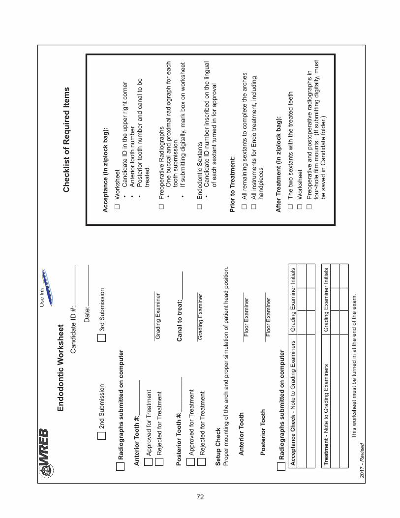
72
Endo
dont
ic W
orks
heet
2nd
Subm
issi
on3r
d Su
bmis
sion
Ant
erio
r Too
th #
:___
____
___
Appr
oved
for T
reat
men
tR
ejec
ted
for T
reat
men
t
Post
erio
r Too
th #
:___
____
___
Can
al to
trea
t:___
____
_
Appr
oved
for T
reat
men
t
Rej
ecte
d fo
r Tre
atm
ent
Acc
epta
nce
Che
ck -
Not
e to
Gra
ding
Exa
min
ers
Gra
ding
Exa
min
er In
itial
s
This
wor
kshe
et m
ust b
e tu
rned
in a
t the
end
of t
he e
xam
.
Gra
ding
Exa
min
er
Gra
ding
Exa
min
er
Setu
p C
heck
Prop
er m
ount
ing
of th
e ar
ch a
nd p
rope
r sim
ulat
ion
of p
atie
nt h
ead
posi
tion.
Ant
erio
r Too
thFl
oor E
xam
iner
Post
erio
r Too
thFl
oor E
xam
iner
Acc
epta
nce
(In z
iplo
ck b
ag):
□W
orks
heet
•C
andi
date
ID in
the
uppe
r rig
ht c
orne
r•
Ante
rior t
ooth
num
ber
•Po
ster
ior t
ooth
num
ber a
nd c
anal
to b
e tre
ated
□Pr
eope
rativ
e R
adio
grap
hs•
One
buc
cal a
nd p
roxi
mal
radi
ogra
ph fo
r eac
h to
oth
subm
issi
on•
If su
bmitt
ing
digi
tally
, mar
k bo
x on
wor
kshe
et
□En
dodo
ntic
Sex
tant
s•
Can
dida
te ID
num
ber i
nscr
ibed
on
the
lingu
al
of e
ach
sext
ant t
urne
d in
for a
ppro
val
Prio
r to
Trea
tmen
t:
□Al
l rem
aini
ng s
exta
nts
to c
ompl
ete
the
arch
es □
All i
nstru
men
ts fo
r End
o tre
atm
ent,
incl
udin
g ha
ndpi
eces
Afte
r Tre
atm
ent (
In z
iplo
ck b
ag):
□Th
e tw
o se
xtan
ts w
ith th
e tre
ated
teet
h □
Wor
kshe
et □
Preo
pera
tive
and
post
oper
ativ
e ra
diog
raph
s in
four-holefilmmounts.(Ifsubm
ittingdigitally,m
ust
be s
aved
in C
andi
date
fold
er.)
Che
cklis
t of R
equi
red
Item
s
2017
- R
evis
ed
Dat
e:
Can
dida
te ID
#:
Rad
iogr
aphs
sub
mitt
ed o
n co
mpu
ter
Rad
iogr
aphs
sub
mitt
ed o
n co
mpu
ter
Trea
tmen
t - N
ote
to G
radi
ng E
xam
iner
sG
radi
ng E
xam
iner
Initi
als
Use
Ink
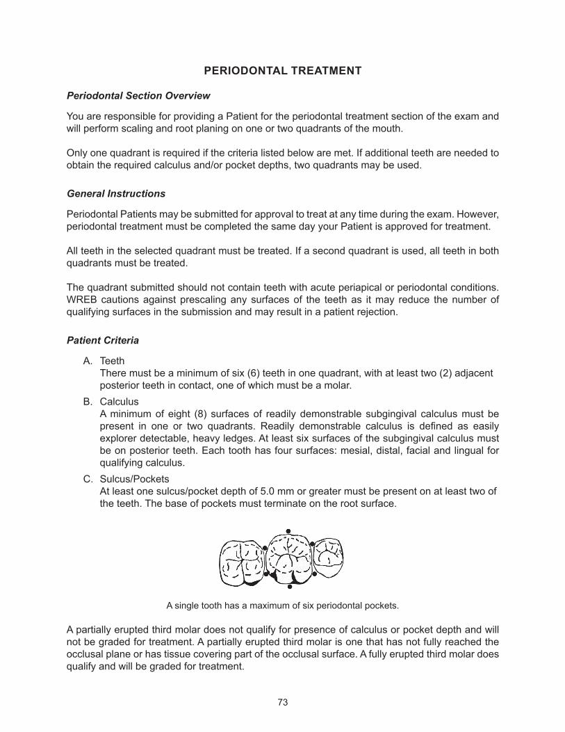
73
PERIODONTAL TREATMENT
Periodontal Section Overview
You are responsible for providing a Patient for the periodontal treatment section of the exam and will perform scaling and root planing on one or two quadrants of the mouth.
Only one quadrant is required if the criteria listed below are met. If additional teeth are needed to obtain the required calculus and/or pocket depths, two quadrants may be used.
General Instructions
Periodontal Patients may be submitted for approval to treat at any time during the exam. However, periodontal treatment must be completed the same day your Patient is approved for treatment.
All teeth in the selected quadrant must be treated. If a second quadrant is used, all teeth in both quadrants must be treated.
The quadrant submitted should not contain teeth with acute periapical or periodontal conditions. WREB cautions against prescaling any surfaces of the teeth as it may reduce the number of qualifying surfaces in the submission and may result in a patient rejection.
Patient Criteria
A. Teeth There must be a minimum of six (6) teeth in one quadrant, with at least two (2) adjacent posterior teeth in contact, one of which must be a molar.
B. Calculus A minimum of eight (8) surfaces of readily demonstrable subgingival calculus must be present in one or two quadrants. Readily demonstrable calculus is defined as easily explorer detectable, heavy ledges. At least six surfaces of the subgingival calculus must be on posterior teeth. Each tooth has four surfaces: mesial, distal, facial and lingual for qualifying calculus.
C. Sulcus/Pockets At least one sulcus/pocket depth of 5.0 mm or greater must be present on at least two of the teeth. The base of pockets must terminate on the root surface.
A single tooth has a maximum of six periodontal pockets.
A partially erupted third molar does not qualify for presence of calculus or pocket depth and will not be graded for treatment. A partially erupted third molar is one that has not fully reached the occlusal plane or has tissue covering part of the occlusal surface. A fully erupted third molar does qualify and will be graded for treatment.

74
Patient Acceptance
Prior to beginning treatment, the quadrant(s) must be approved by the Grading Examiners. You may submit your Patient or your Patient may be submitted by your dental Assistant if all paperwork is complete and the required instruments are present.
No electronic devices, including cell phones, are allowed in the grading area. Patients with electronic devices will be required to return the device to the Candidate clinic.
To facilitate Grading Examiner evaluation and for Patient comfort, anesthetize the quadrant(s) you are submitting for approval. Send your Patient to the grading area with:
A. Worksheet: (sample, pgs. 79-80) Use only blue or black ink. Do not use pencil.
Complete the original worksheet (not a copy). • Write your Patient’s first name only.• Write your Candidate ID number in the upper right corner. • Circle the quadrant(s) you are submitting.• List all teeth for the quadrant(s) you are submitting. • Indicate missing teeth with an "X" through the entire column.• Indicate the location of subgingival calculus by marking an “X” in the appropriate
boxes for all teeth in the quadrant(s).• Using a periodontal probe, measure the sulcus/pocket depths. Measurements should
be taken at the greatest depth for each area. For each tooth, record the pocket depths of 3.0 mm or greater in the spaces provided.
• Check the “Acceptance” box.• All medications your Patient has taken today, including type, concentration and amount
should be listed on the back of the worksheet. Cartridges of local anesthetic, as they are administered throughout the exam, also should be listed. If no medications are taken, write none.
B. Radiographs: Full mouth periapical radiographs including bitewings.Your Patient must have full-mouth periapical radiographs including bitewings. Posterior periapical radiographs should include root apices of any third molars when practical. A note to examiners on the worksheet, at acceptance, indicating patient intolerance in capturing the entire tooth on the radiograph(s) is acceptable. The radiographs must have been taken within the past three years. They must be dated. Original radiographs are preferred. Duplicates will be accepted if they are of diagnostic quality. Panographic films are not acceptable. Periodontal radiographs should be paper clipped to the back of the worksheet; please do not staple.
Radiographs must have your Candidate ID number and your Patient’s first name only on the film mount, template or print. Do not use film mounts that identify a school name or location. If incorrect, outdated or poor-quality radiographs are submitted, the radiographs and worksheet will be returned to you for correction. No points will be deducted.
Digital radiographs are accepted if they meet the criteria specified on pg.16. If submitting radiographs by computer, the file name should include your Candidate ID number, Patient first name and the word “Perio.”

75
C. Patient Medical History/Consent Form: The completed Patient Medical History and Consent Form signed (sample, pgs. 21-22).A Patient Medical History (including current blood pressure and pulse) and Consent Form must be completed for each Patient. If you use the same Patient for more than one procedure only one medical history is necessary. Mark the box on the upper right corner of the form for each procedure being submitted. Note that each procedure also must be listed on the Consent Form on the reverse side. Make sure your Patient signs the Consent Form. The Patient Medical History form must be reviewed and initialed by a Floor Examiner before your Patient is sent to the grading area for approval. Provide both the Periodontal Worksheet and Patient Medical History, including blood pressure and pulse, for a Floor Examiner to review. A Floor Examiner will also verify that your patient has completed the reverse of this document that constitutes the consent form and assumption of risk for you to perform the dental procedure described. When your Patient is submitted for approval, the Patient Medical History and Consent Form will be retained at the Patient Check-In desk; Examiners will not see it.
D. Patient Tray with:
• A new #4 or #5 metal front surface mirror• A new ODU 11/12 explorer• A new color coded 3-6-9-12 mm periodontal probe• Three 2" x 2" gauze pads
The instruments must be in an open autoclave bag. The paperwork should be placed on top of the tray.
E. Patient Bib: Attach your Candidate ID label to the upper right corner of the Patient’s bib (Patient’s right side).
F. Patient Eye Protection: Prescription glasses or safety glasses must be worn by all Patients.
Patient Approved – If your Patient is approved, the Patient will return with:• The worksheet initialed by one Grading Examiner next to “Acceptance”• Radiographs• Instruments
You may proceed with treatment.
Patient Not Approved – If your Patient is not approved, the Patient will return with: • A pink Unacceptable for Treatment form indicating the reason the Patient was not approved• Instruments• Radiographs• New Patient Medical History and Consent Form

76
Patient Unaccepted
If the first periodontal Patient submission does not meet the criteria listed on pg. 73, the Patient will not be approved by the Grading Examiners. A deduction will be applied to the periodontal treatment score. You may submit Patients for approval for treatment until the criteria are met. No additional deductions for subsequent rejected submissions will be assessed.
The worksheet for the Patient rejection will be retained in the grading area.
If your Patient was submitted with only one quadrant for acceptance and did not qualify, the same Patient may be resubmitted with an additional quadrant.
Treatment
Periodontal treatment must be completed the same day your Patient is approved.
If a Patient is approved, but treatment is not completed the same day, you will be allowed to resubmit the same Patient and have the submission re-approved, or submit an alternate submission on a different Patient. In either situation, there is a deduction from the periodontal treatment score.
You are evaluated on the thoroughness of calculus removal and root planing of all teeth in the quadrant(s) selected. Completely remove calculus and smooth root surfaces of all teeth in the quadrant(s).
Sonic or ultrasonic devices are acceptable, but rotary instruments and/or chemicals for calculus removal are prohibited.
Major Tissue Trauma
Major tissue trauma is defined as iatrogenic damage to extra-intraoral tissues resulting in significant injury to the Patient, such as lacerations, burns, amputated papillae or large tissue tags.
Grading Examiners compare the preoperative gingival condition to the postoperative gingival condition. Validated major tissue trauma by two or more Examiners results in loss of all points for the treatment procedure.
Treatment Grade
When treatment is completed, send the Patient to the grading area with:A. Worksheet: The worksheet with an “X” in the box for “Treatment Grade”B. Radiographs: Full mouth periapical radiographs including bitewingsC. Patient Tray with:
• New #4 or #5 metal front surface mirror• New ODU 11/12 explorer• New color coded, 3-6-9-12 mm periodontal probe• Three 2" x 2" gauze pads
The instruments must be in an open autoclave bag. The paperwork should be placed on top of the tray.
77
D. Patient Bib: Your Candidate ID label should be attached to the upper right corner of the Patient’s bib (Patient’s right side).
E. Patient Eye Protection: Prescription glasses or safety glasses must be worn by all Patients.
Patients are evaluated by three Grading Examiners and may be in the grading area for more than an hour. Consider Patient comfort and re-anesthetize, if necessary, before sending your Patient to the grading area.
Your Patient will return with the instrument tray, radiographs and the worksheet with “Treatment Grade” initialed by a Grading Examiner.
Releasing Your Patient
After the “Treatment Grade,” review the worksheet for all necessary initials. If the Grading Examiner initials are missing from the “Acceptance” or “Treatment Grade” notify a Floor Examiner.
Missing initials which are not brought to the attention of a Floor Examiner cannot be grounds for an appeal.
Give your Patient the yellow copy of the completed Follow-Up Care Agreement form for any postoperative care which may be necessary. Have your Patient fill out the Patient Questionnaire.The Floor Examiner will verify that any follow-up requested by the Grading Examiners has been completed and will then initial the worksheet. Your Patient may then be dismissed. Do not dismiss your Patient without Floor Examiner permission.

78
PERIODONTAL TREATMENT SCORING
Deductions
Patient Rejection – Validated by two or more Examiners – a 10% deduction from the total possible of 100% only applied to first patient.
Resubmission of Patient or submission of another Patient after receiving approval – a 10% deduction. If both a Patient rejection and a resubmission occur, only one 10% deduction will be taken.
Major Tissue Trauma – Validated by two or more Examiners results in loss of all points for the treatment procedure.
Late Penalties: Percentage Procedures (Periodontal Treatment) (deducted from total possible for Periodontal Perio Treatment): 1 to 5 minutes late: 4% deducted; 6 to 10 minutes late: 8% deducted; 11 to 15 minutes late: 12% deducted; 16 or more minutes late: Procedure will not be graded. No points earned.
Scoring
Validated calculus remaining is an error which is documented by at least two Grading Examiners and will be scored on the following scale:
Calculus Pieces Remaining Score0 100.00%1 87.50% 2 75.00%3 62.50% 4 50.00% 5 37.50% 6 25.00% 7 12.50% 8 0.00%

79
SAMPLE
Perio
dont
al T
reat
men
t Wor
kshe
et
Patie
nt’s
Firs
t Nam
e:__
____
____
____
____
__Ca
ndid
ate
ID #
:___
____
____
#___
__#_
____
#___
__#_
____
#___
__#_
____
#___
__#_
____
#___
__#_
____
#___
__#_
____
#___
__#_
____
#___
__#_
____
D F M L DF F MF
ML
L DL
CALC
ULUS
(Mar
k “X
” if p
rese
nt)
PRO
BING
DEPT
H(R
ecor
d 3m
m o
r gre
ater
)
Circ
le Q
uadr
ant(s
) Sel
ecte
d2n
d Su
bmis
sion
Upp
er R
ight
Upp
er L
eft
3rd
Subm
issi
onLo
wer
Rig
htLo
wer
Lef
t
TEET
H #‘
S
2017
- R
evis
ed
TREA
TMEN
T G
RADE
Trea
tmen
t Gra
ded:
No
te to
Exa
min
ers
(if n
eces
sary
)G
radi
ng E
xam
iner
Initi
als
ACCE
PTAN
CEAc
cept
ed b
y:
Note
to E
xam
iner
s (if
nec
essa
ry)
Gra
ding
Exa
min
er In
itial
s
Patie
nt m
ay b
e re
leas
ed fr
om th
e ex
amin
atio
n:Fl
oor E
xam
iner
Radi
ogra
phs
subm
itted
on
com
pute
r
Use I
nk

80
SAMPLE
Medications Taken By Patient Today Date and Time Drug Name and Concentration # of Tabs/Capsules
Checklist of Required Items
Local Anesthetic for this Procedure Administered Throughout The ExamDate and Time Type and Concentration of Local Anesthetic and Vasoconstrictor Cartridges
SUBMIT PATIENT WITH:
Periodontal Treatment Worksheet• Yourpatient’sfirstname• Your Candidate ID # in the upper right corner• Selected quadrant(s) circled• Teeth numbers indicated• Calculus indicated with “X” if present• Probing depths measured• Box checked for “Acceptance” or “Treatment Grade”• Medication taken and local anesthetic administered
Full Mouth Periapical Radiographs including Bitewings• If radiographs submitted digitally, mark box on worksheet
Place instruments in an open autoclave bag, with paperwork on top
Patient bib with appropriate Candidate ID # label on upper right-hand corner
Patient eye protection
Patient Medical History/Consent Form - Approval Only• Completed, including pulse and blood pressure• Patient address and signature• Floor Examiner initials
A Tray Containing the Following• New metal #4 or #5 front surface mirror• New ODU #11/12mm perio explorer• New 3-6-9-12mm perio probe• Three 2 X 2 gauze pads

81
END OF CLINICAL EXAMINATION
After all procedures have been completed, make sure:
• All paperwork for each procedure has the required signatures.• A Floor Examiner has initialed all operative worksheets as required.
If any signatures are missing, notify a Floor Examiner.
Be sure you have given your Patient(s) the yellow copy of the Follow-Up Care Agreement form. Make sure that the agreement is completely filled out and signed and dated by the patient, follow-up school of record and/or follow-up care provider.
Place the items listed below in your white Candidate Packet. If any of these items are missing, your results will be held until received by the WREB office.
• Operative Worksheets (composite, amalgam or cast gold) with bitewing and periapical radiographs
• Periodontal Treatment Worksheet• The Dental Assistant Verification form signed by you and your Assistant(s). If an Assistant
was not used for the operative and/or periodontal treatment procedure, mark the appropriate box indicating “no Assistant was used,” sign the form, and place in the Candidate Packet.
• The signed white copies of the Follow-Up Care Agreement forms for each Patient treated • Any pink copies of communication forms that were received from the Examiners
Do not seal your Candidate packet. Turn your packet into WREB staff at the entrance to the grading area.
Prior to the clinical exam, WREB will email a link to the Candidate Survey. We ask that you complete the Survey after the clinical exam.
It is WREB policy to notify Candidates of exam results as soon as possible after the conclusion of an exam. Results will be posted online and can be accessed with your Candidate login and password. You will receive an email notice once your results are available.
Do not call the WREB office for exam results. Exam results are confidential and will not be given over the telephone, fax machine or by e-mail. They will only be posted to your secure WREB login online.
82
FREQUENTLY ASKED QUESTIONS
1. May I use a foreign trained dentist as my dental Assistant?Operative Assistants may not be dentists (including graduates of foreign dental schools) or be in their final year of dental school. Operative assistants may be Dental Assistants or Dental Hygienists, if they do not hold a permit to place and finish restorative materials.
2. What is the minimum age a Patient can be? If my Patient is under 18, does the parent or guardian need to stay during the procedure?The minimum Patient age for the Periodontal Treatment procedure is 18 years. There is no minimum age for Operative procedures. A parent or guardian does not have to remain during the procedure.
3. When are my Assistant and my Patient allowed on the clinic floor to start the exam? When can I put my Patient in line for acceptance or grading? Assistants and Patients may enter the clinic with you at 7:00 a.m. on clinic days 1, 2 and 3. For Patient comfort, Patients should not be sent to the grading area any earlier than 7:45 a.m. The exam officially begins at 8:00 a.m. The Patient line will not move until 8:00 a.m. Candidates who are assigned Endodontics the first morning of the exam may not submit Patients until 10:00 a.m. (See details under “General Information-Schedule and Clinic Hours.”)
4. Do I have to have my Patient in line for grading by 10:30 a.m. on the last day of the exam?You have until 11:00 a.m. to have your Patient in line for grading on the last day. The first two days of the exam, your Patient must be in line for grading by 4:30 p.m. (See details under “General Information-Schedule and Clinic Hours.”)
5. Are translators allowed on the clinic floor? Translators will be allowed on the clinic floor or in the grading area only as needed. Translators will be asked to remain in the Patient waiting area until, or if their services are required.
6. What are Floor Examiners? Floor Examiners assist Candidates on the clinic floor: • Answer questions, clarify exam procedures • Act as liaisons between Candidates and Grading Examiners • Have extra forms for Candidates such as Patient Medical History and Follow-Up Care
Agreements• Sign Patient Medical History forms • Distribute forms from Examiners that affect Candidates and procedures • Check on modifications (see Operative-Modification Procedure) • Manage pulp exposures• Check and initial steps in the processes involved on worksheets. (See Operative-
Patient Acceptance or General Information-Exam Personnel and Anonymity.)

83
7. May I anesthetize my Patient before I send him/her to the grading area for approval to start?
For Periodontal Treatment Patients, you should anesthetize the quadrant(s) submitted for approval to facilitate Examiner evaluation and for Patient comfort. For operative Patient Check-In for acceptance, you may anesthetize Patients at your discretion.
8. May I submit both my operative restorations for approval at the same time? If the procedures are on the same Patient but not on adjacent teeth and accepting both would not cause the loss of occlusal contact, they may be submitted for approval at the same time. You may not submit patients with adjacent (consecutive tooth numbers) teeth for acceptance. (See details under “Operative-Patient Acceptance.”)
9. If I have both operative restorations approved to start, do I have to do both preps that day? You may do only one preparation if you choose. For the procedure that has been approved but not started, bring your worksheet to a Floor Examiner for the proper paperwork. (See details under “Operative-Dismissal for the Day Approval.”)
10. Do I have to work with a rubber dam? You do not have to work with a rubber dam, but a rubber dam is required when submitting a Patient for the preparation grade or when requesting a modification request for your patient on the candidate clinic floor. (See details under “Operative-Preparation Grade.”)
11. When do I call a Floor Examiner to check for a modification of outline or internal form?
When removal of caries, affected dentin, unsound demineralized enamel, or remaining restorative material will extend the outline and/or internal form of the preparation beyond the criteria for a “5”. (See details under “Operative-Modification Procedure.”)
12. How do I write a modification request? Write the type, location, extent, and reason (i.e., caries, affected dentin, unsound demineralized enamel, or remaining restorative material) for the “Modification Request(s)” in the spaces provided on the procedure worksheet. The space on the worksheet is limited, therefore, you are encouraged to write the total extent required to remove the lesion on your initial modification request(s) in 0.5 mm increments (i.e., 0.5 mm, 1.0 mm, 1.5 mm). A Floor Examiner will be available to answer any questions you may have.
13. When do I need original radiographs? And, when do I not? The two Operative procedures require original radiographs of the tooth taken within the prior six months. The radiographs must show the current condition of the tooth. Duplicates are not acceptable. Separate radiographs or images are needed for each procedure. The Periodontal Treatment procedure requires complete mouth periapical radiographs, including bitewings. The radiographs must have been taken within the past three years. Original radiographs are preferred, but duplicates are acceptable if they are of diagnostic quality. (See details under “Operative-Patient Acceptance” and “Periodontal Treatment-Patient Acceptance.”)

84
14. If WREB considers all exposures avoidable, how do I deal with an exposure or near exposure? The preferred procedure is to leave a small amount of caries or affected dentin (0.5 mm) over the pulp to avoid an exposure. Write in the “Note to Examiners” on the worksheet your intentions. All other caries in the preparation must be removed. If an exposure does occur, write in the “Note to Examiners” on the worksheet your intentions regarding the exposure and how it will be managed, place a rubber dam (if not already in place) and call a Floor Examiner. Upon verification of the exposure, a Floor Examiner will instruct you to place a pulp capping material over the exposure as soon as possible. (See details under “Operative-Cavity Preparation.”)
15. Can my Assistant dismiss my Patient while I’m in the Endodontics exam? Yes, if there is no follow up required when your Patient returns from the grading area. Remember, a Floor Examiner’s initials are required on worksheets for Patient release from the exam.
16. How many initials from Examiners do I need on my worksheet? It depends on what portion of the restoration you are doing. One initial is required at Acceptance, at least two initials if you have sent a note with a modification procedure and three initials are required if you have sent a Patient for grading. (See details and sample worksheet under “Operative.”)
17. When do I have to go to the Endodontics lab to do my Endodontics procedure? You may go to the endo lab anytime during your block time schedule. It is recommended that you are in the lab in the first 30 minutes to avoid any delay getting your “Setup Check.” You must turn in your two (2) endo models and radiographs at the end of the time block or you will receive a late penalty. There are no exceptions.
18. When do I take the Comprehensive Treatment Planning (CTP) computerized exam?
The CTP computerized exam can be taken at a Prometric Testing Center. Once you are enrolled in an exam, information will be emailed to you. This will include the time frame to take the exam, Prometric’s contact information to schedule your appointment, and your eligibility number.
19. Can I change my assigned time for the Endodontics exam? No. Once schedules are posted, they cannot be changed. Schedules are arranged in advance and in the best interest of all Candidates, taking into consideration space availability, supplies and exam materials. Schedules are made to give Candidates the optimum open block time and to maintain Patient flow in the grading area.
20. What identification do I need to provide at the exam? Candidates MUST present acceptable and valid identification in order to be admitted to the WREB dental exam. At the exam, you shall appear in person and provide two (2) valid, non-expired forms of identification. (See WREB Exam Security and Identification Verification under “General Information.”)

USEFUL PREPARATION TERMS WHEN COMMUNICATING WITH EXAMINERS
Class III
Lingual Access
Gingival Wall
Lingual Wall
Class III
Facial Access Lingual Access
Facial Wall
Incisal Wall
Gingival Wall
Axial Wall
Axial Wall
Incisal Wall
Facial Wall
Gingival Wall
Axial Wall
Incisal Wall
Diagrams not to scale.

USEFUL PREPARATION TERMS WHEN COMMUNICATING WITH EXAMINERS
Class IIPulpal Floor
Gingival FloorFinger Extension
Axial-Pulpal Line Angle
Distal Wall
Axial Wall
Axial-Gingival Line Angle
Buccal Occlusal Wall
Lingual Occlusal Wall
Distal Wall
Pulpal Floor
Buccal Proximal Wall
Lingual Proximal Wall
Gingival Floor
Diagrams not to scale.

2017 Dental Candidate GuideCopyright © 2017 WREB
23460 N. 19th Avenue, Suite 210Phoenix, Arizona 85027phone: 623-209-5400


