174

2018간세포암종
진료 가이드라인대한간암학회 - 국립암센터


3
2018 간세포암종 진료 가이드라인
2018 간세포암종 진료 가이드라인
목차
서론 ………………………………………………………………………………………………… 05
역학 ………………………………………………………………………………………………… 16
예방 ………………………………………………………………………………………………… 20
감시검사 …………………………………………………………………………………………… 26
진단 ………………………………………………………………………………………………… 30
병기 ………………………………………………………………………………………………… 41
치료 개관 …………………………………………………………………………………………… 44
간절제 ……………………………………………………………………………………………… 47
간이식 ……………………………………………………………………………………………… 52
국소치료 …………………………………………………………………………………………… 58
경동맥화학색전술 ………………………………………………………………………………… 64
체외 방사선치료 …………………………………………………………………………………… 70
전신치료 …………………………………………………………………………………………… 72
보조요법 …………………………………………………………………………………………… 77
이차 치료 …………………………………………………………………………………………… 78
선제적 항바이러스치료 …………………………………………………………………………… 91
암성 통증의 약물치료 ……………………………………………………………………………… 95
치료 후 반응평가 및 추적 ………………………………………………………………………… 97
주: 본 가이드라인은 간세포암종의 진료, 연구, 교육에 실제적 참고가 되도록 현재까지의 의학적 증거들을 전문가들이
검토한 후 증거중심의 의견을 정리한 것이다. 이에 대해 다른 견해가 있을 수 있으며 각 환자 진료에서 최선의 선택은 처해진
여러 여건에 따라 차이가 있을 수 있다. 본 가이드라인은 대한간암학회와 국립암센터에서 공동으로 제작하였으며 두 기관의
허락없이 수정, 변형, 무단 전제될 수 없다.


5
2018 간세포암종 진료 가이드라인
2018 간세포암종 진료 가이드라인
대한간암학회-국립암센터*
서론
개정 취지
2003년 대한간암연구회-국립암센터 간세포암종 진료 가이드라인이 처음 공표되고 2009년과
2014년 두 차례의 개정을 거친 이후 현재까지 간세포암종에 관한 국내외의 많은 새로운 연구
결과와 치료법들이 발표되었다. 서양과 다른 임상상을 보이는 아시아, 특히 우리나라 현실을 반영
할 수 있는 진단과 병기법, 그리고 이에 맞는 치료법에 관한 많은 연구들이 발표되고 지식이 축점
됨에 따라 새로운 연구결과에 근거한 대처방안들이 제시되고 있다. 이에 본 개정위원회는 지난
2014년 가이드라인 발표 이후 최근까지 알려진 새로운 연구결과들과 함께 전문가 의견을 종합
한 새로운 권고안을 도출하고자 가이드라인 개정작업을 2017년 여름에 착수하였다.
대상 집단
간세포암종이 의심되거나 처음으로 간세포암종 진단을 받은 환자가 본 가이드라인의 주된 대
상 환자 집단이다. 본 가이드라인에서의 치료는 처음 진단된 간세포암종 환자를 대상으로 한 초
치료법이 핵심이나 초치료 후 잔존암, 진행암 또는 재발암에 대해서도 처음으로 광범위한 검토
와 논의를 하여 권고사항을 마련하였다. 아울러 본 가이드라인은 예방, 감시검사, 치료개관, 기
저질환인 만성 간염에 대한 선제적 항바이러스제 치료와 암성 통증에 대한 관리, 치료 후 반응
평가법 등도 함께 기술하여 실제 임상에서 좀 더 유용하게 사용될 수 있게 하였다.
* 간세포암종 진료 가이드라인 개정위원회: 위원장 박중원; 분과위원장 이준성(내과), 서경석(외과), 정진욱(영상의학과), 성진실
(방사선종양학과); 위원 내과 김도영, 김지훈, 김형준, 김휘영, 박수영, 심주현, 이정훈, 임영석, 임호영, 장재영, 장정원, 전대원;
외과 고양석, 김경식, 김동식, 김성훈, 김성훈2, 김종만, 윤영철, 정동환, 조재영; 영상의학과 고영환, 김경민, 김소연, 김영환,
이인준, 이정민, 임현철, 조성기, 천호종, 최준일; 방사선종양학과 계철승, 김미숙, 김태현, 박희철, 배선현, 윤상민, 윤원섭, 장원일

6
The Korean Liver Cancer Association and National Cancer Center, Korea
독자층
본 가이드라인은 우리나라 간세포암종 환자 진단과 치료를 일선에서 담당하고 있는 진료의들
에게 유용한 임상 정보와 방향을 제공하고자 하였다. 또한, 수련 과정 중의 전공의 혹은 전임의
및 이들을 지도하는 교육자들에게도 구체적이고 실제적인 정보를 제공하고자 노력하였다.
집필진 및 기금에 관한 정보
대한간암학회와 국립암센터 합의에 따라 구성된 간세포암종 진료 가이드라인 개정위원회는
간장학을 전공하는 소화기내과 전문의, 종양내과 전문의, 외과 전문의, 영상의학과 전문의 및
방사선종양학과 전문의들로 구성되었으며 연구경비는 국립암센터(#1731510-1)가 지원하였다.
개정위원회의 각 위원은 각자 담당한 분야의 근거 자료 수집 및 분석과 원고 작성을 담당하였
으며 각 위원들의 이해관계상충보고는 별첨 1과 같다.
근거 수집을 위한 문헌 검색
2018 대한간암학회-국립암센터 간세포암종 진료 가이드라인 개정위원회(이하 개정위원회; 별
첨 2)는 최신 근거에 입각한 가이드라인 개정을 위해 2014년 가이드라인 발표 이후 최근까지
발표된 국내외의 간세포암종 관련 문헌을 MEDLINE 검색을 통해 수집하고 분석하였다. 관련 문
헌의 언어는 영어 및 한국어로 출판된 논문에 국한하였고, 검색어는 간세포암종 및 해당 세부
주제에 특정한 검색어를 포함하였다. 해당 세부 주제는 간세포암종의 역학, 예방, 진단 및 병기,
치료, 반응평가 등 임상적으로 중요한 항목을 모두 망라하였다.
체계적 문헌고찰 및 근거 수준, 권고 등급의 분류
근거를 위해 수집된 문헌은 체계적 고찰을 통해 분석하였고, 근거수준(levels of evidence)
은 수정된 GRADE 체계(Grading of Recommendations, Assessment, Development and
Evaluation)에 의해 분류하였다.1-4 (Table 1) 후속 연구를 통해 해당 근거 연구에 대한 평가가 바
뀔 가능성에 따라 바뀔 가능성이 가장 낮은, 즉 가장 높은 근거수준(A), 바뀔 가능성이 있는 중
간수준(B), 바뀔 가능성이 높은 가장 낮은 근거수준(C)으로 각각 정의하였다. 예컨대, 근거수준
A는 최소한 하나 이상의 무작위 대조연구로부터 얻은 근거수준과 유사하나 반드시 일치하지는
않는다. 가령, 향후 무작위 대조연구를 시행하는 것 자체가 어려워 현재 근거수준이 바뀌게 될

7
2018 간세포암종 진료 가이드라인
가능성이 없을 경우도 근거수준 A가 될 수 있다. 반면에 무작위 대조연구라도 대상환자 규모가
작고 추후 연구가 더 필요한 경우 혹은 초록으로만 발표된 경우 등에서는 근거 수준을 낮추었
다. 권고 등급(grades of recommendation)의 분류 역시 GRADE 체계를 채택하였는데, 각 근거
연구의 근거수준 자체뿐 아니라 해당 연구의 질(quality)적 측면, 연구결과의 임상적 파급효과
(patient-important outcome) 및 사회경제적 측면 등을 종합적으로 고려하여 강한 권고 등급(1)
과 약한 권고 등급(2)로 분류하였다. 따라서, 각 권고사항은 해당 근거 연구 수준(A–C) 및 그에
따른 권고 등급(1, 2)을 조합하여 다음과 같이 표기하였다: A1, A2, B1, B2, C1, C2 (Table 1). 본
가이드라인에서는 C2 등급의 권고는 최대한 배제하고자 노력하였다.
세부 주제 목록(List of the clinical questions)
개정위원회에서는 간세포암종 진료 가이드라인의 개정과 관련하여 4개 분과별로 세부 주제와
임상적 문제들을 선별하여(별첨 3) 각 항목에 대한 근거를 검토하고 각 분과위원회 토의와 전체
개정위원회 토의를 거쳐 권고사항을 제시하고자 하였다.
원고 검토
개정위원회 개시모임 이 후 각 분과별로 수 차례의 회의와 개정위원 전원이 참석한 3 차례의 회
의를 통해 만들어진 권고안을 다시 수 차례의 온라인 토의와 세 차례의 분과위원장 회의를 거
쳐 세부 검토를 하였다. 검토 과정은 원고 내용의 충실성뿐 아니라 방법론적 타당성을 AGREE II
(Appraisal of Guidelines for Research and Evaluation II)에 의거하여 평가하는 과정을 거쳤다.5, 6
이후 완성된 안을 자문위원회 및 공청회를 통해 검토한 후 개정위원회 분과장 회의에서 재차 수정
하였다. 자문위원회는 간암 임상 전문가 총 9명으로 구성되었다. 이러한 과정을 통해 만들어진 가
이드라인은 공청회와 대한간암학회 이사회 및 국립암센터 인준절차를 거쳤다. (별첨 4)
가이드라인의 공표
개정된 간세포암종 진료 가이드라인은 2018년 6월 15일 대한간학회-대한간암학회-한국간담
췌외과학회-대한간이식연구회 합동 ‘간 주간’ 학술대회에서 공표되었다. 한글판은 대한간암학
회 및 국립암센터 웹사이트 (http://www.klcsg.or.kr; http://ncc.re.kr)에서 확인할 수 있고, 영
문판은 별도 저널에 게재될 예정이다.

8
The Korean Liver Cancer Association and National Cancer Center, Korea
Table 1. Grading of Recommendations, Assessment, Development and Evaluation (GRADE)
Quality of evidence Criteria
High (A) Further research is unlikely to change confidence in the estimate of the clinical effect
Moderate (B) Further research may change confidence in the estimate of the clinical effect
Low (C) Further research is very likely to impact confidence on the estimate of clinical effect
Strength of recommendation Criteria
Strong (1)Factors influencing the strength of the recommendation included the quality of the evidence, presumed patient-important outcomes, and cost
Weak (2)Variability in preferences and values, or more uncertainty. Recommendation is made with less certainty, higher cost or resource consumption
NOTE. Of the quality levels of evidence, we excluded “very low quality (D)” in our guideline for convenience, which was originally included in the GRADE system and indicates that any estimate of effect being very uncertain.Evidence level was graded down if there is only an abstract, poor quality or inconsistency between studies; level was graded up if there is a large effect size.
재개정 계획
향후 간세포암종과 관련된 새로운 검사방법이나 약제, 치료법 등이 개발되고 새로운 중요한
연구결과가 밝혀져 가이드라인 개정이 우리나라 국민 건강증진에 필요하다고 판단되면 대한간
암학회와 국립암센터는 본 가이드라인을 일부 또는 전체를 재개정할 계획이다. 이에 대한 일정
은 필요시 다시 공지할 것이다.

9
2018 간세포암종 진료 가이드라인
Table 2. Recommendations of 2018 KLCA-NCC, Korea Practice Guidelines for Management of Hepatocellular Carcinoma
주제 권고사항
예방 1. 간세포암종 발생을 예방하기 위하여 모든 신생아(A1)와 감염될 위험이
있는 소아 및 성인에서 혈청 HBsAg, anti-HBs, IgG anti-HBc가 모두
음성이면 B형간염 예방접종을 시행한다 (B1).
2. 간세포암종 발생을 예방하기 위해 개인간 B형/C형 간염바이러스 전염
을 예방하고 (A1), 알코올 남용을 피하며, 비만, 당뇨와 같은 대사질환을
적절히 조절해야 한다 (C1).
3. 만성 바이러스간염 환자에서의 간세포암종 발생을 예방하기 위한 항바
이러스제 치료는 대한간학회의 만성 B형간염 및 만성 C형간염 진료 가이
드라인을 따른다 (A1).
4. 만성 B형간염 환자에서 지속적으로 바이러스가 억제되면(A1), 그리고
만성 C형간염 환자에서 인터페론 치료(A2), 또는 DAA 치료(C1) 후 SVR
이 획득되면 간세포암종 발생을 낮춘다.
5. 만성 B형간염과 연관된 간세포암종의 근치적 치료 후 항바이러스제 치
료는 간세포암종 재발을 감소시킬 수 있으므로 혈청 HBV DNA 양성인
만성 B형간염 환자는 치료한다 (B1).
6. 만성 C형간염과 연관된 간세포암종의 근치적 치료 후 DAA 치료와 간세
포암종 재발위험 증가 또는 감소 관련성은 아직 분명하지 않다 (C1).
7. 만성 간질환 환자에서 커피 음용은 간세포암종 발생 위험을 낮출 수 있
다 (B1).
감시검사 1. 간세포암종 고위험군[만성 B형간염(A1), 만성 C형간염(B1), 간경변증
(A1)] 환자에서 감시검사를 시행한다.
2. 간세포암종 감시검사로 간 초음파검사와 혈청 알파태아단백검사를 6개
월마다 정기적으로 시행한다 (A1).
3. 간 초음파검사를 적절히 시행할 수 없는 경우 대체 검사로서 역동적 조
영증강 CT 또는 역동적 조영증강 MRI 등을 시행할 수 있다 (C1).
진단 1. 간세포암종은 병리학적으로 진단하거나, 간세포암종의 고위험군(만성 B형
간염, 만성 C형간염, 간경변증)에서는 전형적 영상소견으로 진단할 수 있다
(A1).
2. 간세포암종의 고위험군에서 감시검사 중 간세포암종이 의심되는 1 cm 이상
결절은 진단을 위해 역동적 조영증강 CT, 또는 역동적 조영증강 MRI, 또는
간세포특이조영제 MRI (Gd-EOB-DTPA MRI)를 시행하여야 한다 (A1).

10
The Korean Liver Cancer Association and National Cancer Center, Korea
진단 처음 영상검사에서 정확한 진단을 할 수 없는 경우에는 추가로 역동적
조영증강 CT, 역동적 조영증강 MRI, 간세포특이조영제 MRI, 또는 혈관
내조영제 조영증강 초음파 등을 보완하여 진단할 수 있다 (B1).
3. 간세포암종 고위험군에서 감시검사 중 발견된 1cm 이상 결절이 아래와
같은 전형적 영상소견을 보이면, 간세포암종으로 진단할 수 있다.
(1) 역동적 조영증강 CT 또는 역동적 조영증강 MRI 영상에서 전형적
영상소견은 동맥기 조영증강과 문맥기 혹은 지연기 씻김현상으로 정
의한다 (A1).
(2) 간세포특이조영제 MRI 검사에서 전형적 영상소견은 동맥기 조영증
강과 문맥기, 지연기 혹은 간담도기의 씻김현상으로 정의한다. 단,
병변은 MRI의 T2 강조영상에서 매우 밝은 신호강도를 보이지 않아
야 하며, 확산강조영상이나 조영증강영상에서 과녁 모양을 보이지
않아야 한다 (B1).
4. 간세포암종 고위험군의 감시검사 중 발견된 1 cm 이상 결절 중 앞에서
언급한 전형적 영상소견을 보이지 않는 결절에 대해서는 보조적 영상소
견이 합당하다면 간세포암종 의증으로 진단할 수 있다 (B1); 보조적 영상
소견이란 MRI T2 강조영상에서의 중등도 신호강도, 확산강조영상에서의
고신호강도, 간담도기에서의 저신호강도, 추적검사에서 크기 증가들 중
하나 이상이 있으면서, 피막의 존재, 모자이크 모양, 결절 내 결절, 또는
종괴내 지방이나 출혈 등이 있는 경우이다.
5. 간세포암종 의증 결절은 6개월보다 짧은 간격의 추적검사 또는 생검을
하며(C1), 영상검사만으로 진단이 어려운 미확정결절의 경우, 추적검사
또는 생검을 한다 (B1).
6. 간세포암종 고위험군에서 감시검사 중 새로 발견된 1 cm 미만 결절은 6
개월보다 짧은 간격으로 추적 감시검사를 시행한다 (C1).
7. 간세포암종으로 진단받은 환자의 치료 후 추적검사에서 발견된 새로운
간종괴 혹은 크기가 증가되는 간종괴는 간세포암종의 전형적인 영상소견
이 보이지 않더라도 보조적 영상소견에 합당하다면 간세포암종으로 진단
할 수 있다 (C1).
8. 간세포암종 환자의 진단과 추적을 위해 필요한 CT 검사의 피폭량 제한
은 권장되지 않으나 불필요한 CT 검사는 반드시 회피해야 하며, 영상의
질이 떨어지지 않는 저선량 기법 적용 또는 대체 영상검사를 최대한 고려
한다 (C1).

11
2018 간세포암종 진료 가이드라인
병기 1. 본 가이드라인에서 간세포암종 병기는 modified UICC 병기를 기본으로
하며, BCLC 병기와 AJCC/UICC 병기를 보완적으로 사용한다 (B1).
2. 간절제 또는 간이식 등 간세포암종의 근치적 수술을 계획하는 경우 병기
평가를 위해 PET-CT를 시행할 수 있다 (C1).
3. 간세포암종의 간외전이가 의심되는 경우, 정확한 병기평가를 위해 흉부
CT, 골반 CT, 뼈스캔 등을 시행할 수 있다 (C1).
간절제 1. 문맥압항진증과 고빌리루빈혈증이 모두 없는 Child-Pugh 등급 A의 환
자에서 간에 국한된 단일 간세포암종은 간절제가 일차 치료법이다 (A1).
2. 경미한 문맥압항진증 또는 경미한 고빌리루빈혈증을 동반한 Child-
Pugh 등급 A 및 B7 등급의 간세포암종은 제한적 간절제를 선택적으로
시행할 수 있다 (C1).
3. 간기능이 잘 보존된 환자에서 간문맥, 간정맥, 담도 침습 등이 있더라도
주간문맥(main portal trunk) 침습이 없으면, 간에 국한된 3개 이하 간세
포암종은 간절제를 고려할 수도 있다 (C2).
4. 좌외측 구역과 전하방에 위치하여 접근이 용이한 간세포암종은 복강경
절제술을 고려할 수 있다 (B2).
간이식 1. 간세포암종 환자에서 간절제가 불가능하면서 영상학적 혈관침범과 원격
전이가 없는 5 cm 이하의 단일종괴 또는 3 cm 이하이며 3개 이하의 종
양(밀란척도)인 경우 간이식이 일차 치료법이다 (A1).
2. 간이식에 적응이 되는 간세포암종 환자 중 이식시기를 예측하기 어려운
경우 국소치료 또는 경동맥화학색전술 등을 먼저 시행하는 것이 추천된
다 (B1).
3. 간이식 적응증을 벗어나는 밀란척도 이상의 간세포암종 환자에서 국소
치료, 경동맥화학색전술, 혹은 기타치료 등에 의해 병기감소가 되면 간이
식을 고려할 수 있다 (C1).
4. 다른 효과적 치료법을 적용할 수 없는 경우, 분명한 혈관침범이 없고 간
외전이가 없는 간세포암종에서 간이식은 밀란척도 이상의 확대 기준을
적용할 수도 있다 (C2).
5. 간절제 이후 재발한 환자에서 구제간이식의 적응증은 일차 간이식에서
와 같다 (B1).

12
The Korean Liver Cancer Association and National Cancer Center, Korea
국소치료 1. 고주파열치료술(RFA)은 직경 3 cm 이하의 단일 간세포암종 치료로서
간절제와 비교하였을 때 생존율은 동등하고, 국소재발률은 높으며, 합병
증 발생률은 낮다 (A1).
2. 고주파열치료술은 종양괴사 효과나 생존율에서 에탄올주입술(PEI)보다
우수하다 (A1). 다만 직경 2 cm 이하의 간세포암종에서는 두 치료법의
결과가 유사하므로 고주파열치료술을 적용하기 어려운 경우 에탄올주입
술을 시행할 수 있다 (A2).
3. 수술적 치료를 적용하기 어려운 직경 3-5 cm 간세포암종에 대해 고주
파열치료술 단독치료에 비해 고주파열치료술과 경동맥화학색전술의 병
행치료는 생존율을 증가시킨다 (A2).
4. 간세포암종 치료에서 초단파소작술과 냉동소작술은 고주파열치료술
과 비교하여 유사한 생존율, 재발률, 합병증 발생률 등을 기대할 수 있다
(B2).
경동맥화학색전술
및
기타 경동맥
치료법
1. 간절제, 간이식, 국소치료를 적용하기 어려운 간세포암종 중 수행상태가
양호하고 주혈관침범이나 간외전이가 없을 때 통상적 경동맥화학색전술
(cTACE)이 추천된다 (A1).
2. cTACE는 항암효과를 극대화하고 간손상을 최소화하기 위해 가능한 한
선택적으로 종양의 영양동맥에 시행되어야 한다 (B1).
3. 간문맥침범이 있는 간세포암종 중 잔존 간기능이 좋고 종양이 간내 국소
적인 경우 cTACE 단독(B2) 또는 cTACE와 체외 방사선치료의 병행치료
(B1)를 시행할 수 있다.
4. 약물방출미세구를 이용한 경동맥화학색전술(DEB-TACE)은 cTACE와
비교하여 치료 효과는 유사하지만 색전후증후군이 적다 (B2).
5. 경동맥화학색전술 대상 환자들 중 간기능이 좋고 색전술후증후군의 경
감이 필요한 경우 경동맥방사선색전술(TARE)을 대체 치료법으로 고려할
수 있다 (B2).

13
2018 간세포암종 진료 가이드라인
체외 방사선치료 1. 간절제, 간이식, 국소치료 또는 경동맥화학색전술이 어려운 간세포암종에서
체외 방사선치료를 시행할 수 있다 (C1).
2. 체외 방사선치료는 간기능이 Child-Pugh 등급 A 또는 B7이고, 전산화 방사
선치료 계획에서 30 Gy 이하를 조사받는 체적이 전체 간부피의 40% 이상인
경우 시행한다 (B1).
3. 경동맥화학색전술에 불완전한 반응을 보이는 간세포암종에서 체외 방사선치
료를 시행할 수 있다 (B2).
4. 간문맥침범을 동반하는 간세포암종에서 체외 방사선치료를 병행할 수 있다
(B2).
5. 간세포암종 전이암의 완화 목적으로 체외 방사선치료를 시행할 수 있다 (B1).
6. 국소치료 후 재발한(불응성) 간세포암종에서 체외 방사선치료를 시행할 수 있
다 (C1).
전신치료 1. Child-Pugh 등급 A의 간기능과 ECOG 0-1의 양호한 전신상태를 가진
간세포암종 환자에서 국소 림프절, 폐 등의 간외전이가 있는 경우, 간혈
관침범이 있는 경우, 또는 다른 치료법들에 반응하지 않고 암이 진행하는
경우 소라페닙 치료를 한다 (A1). 소라페닙 치료 대상 환자에서 cTACE와
의 병행치료는 일반적으론 권장되지 않는다 (B1).
2. Child-Pugh 등급 A의 간기능과 ECOG 0-1의 양호한 전신상태를 가지
고 종양면적이 전체 간의 50% 미만인 간세포암종 환자에서, 국소 림프
절, 폐 등의 간외전이가 있는 경우, Vp3 이하 간문맥침범이 있는 경우,
또는 다른 치료법들에 반응하지 않고 암이 진행하는 경우 렌바티닙 치료
를 한다 (A2).
3. Child-Pugh 등급 B7 간기능과 ECOG 0-1의 양호한 전신상태를 가진
1항 종양 조건의 간세포암종 환자에서 소라페닙 치료를 시행할 수 있다
(C1).
보조요법 1. AJCC I, II 병기 간세포암종 환자에서 간절제, 고주파열치료술 또는 에탄
올주입술로써 근치적 치료가 된 후 사이토카인 유도 살해세포(CIK)를 이
용한 면역치료 보조요법을 시행할 수 있다 (B2).
2. 간세포암종의 근치적 치료 후 보조요법으로서 경동맥화학색전술, 소라
페닙, 세포독성화학요법 등은 권장되지 않는다 (B1).

14
The Korean Liver Cancer Association and National Cancer Center, Korea
1차 치료 실패 후의2차 치료
1. 간절제, 고주파열치료술, 또는 간이식으로 완치된 후 재발한 간세포암종
은 재발 시기와 환자의 잔여 간기능과 전신상태, 재발암의 크기, 위치, 개
수 등을 고려하여 재치료방법을 선택할 수 있다 (C1).
2. 절제불가능한 간세포암종에서 6개월 이내 2회 이상의 대응형(on-
demand) 통상적 TACE를 시행했으나 객관적 치료반응(완전반응 또는
부분반응)의 부재, 새로운 혈관침범, 또는 간외전이 중 하나 이상이 발
생한 경우는 TACE 불응성으로 판단하고 치료방법의 변경을 고려한다
(C1).
3. 최소 3주 이상 400mg 이상의 소라페닙 치료에도 간세포암종이 진행한
환자에서 간기능이 Child-Pugh A 이고 전신상태 ECOG 0-1 이면 레고
라페닙을 투여한다 (A1).
4. 소라페닙 치료에도 간세포암종이 진행하거나 소라페닙 부작용으로 중
단한 환자에서 간기능이 Child-Pugh A 이고 전신상태 ECOG 0-1 이면
니볼루맙을 투여할 수 있다 (B2).
5. 소라페닙 포함 2가지 이하의 전신치료에도 불구하고 간세포암종이 진
행한 환자에서 간기능이 Child-Pugh A 이고 전신상태 ECOG 0-1 이면
카보잔티닙을 투여한다 (B1).
6. 소라페닙 치료에도 간세포암종이 진행하거나 소라페닙 부작용으로 중단
한, 간기능이 Child-Pugh A이고 전신상태 ECOG 0-1이며 혈청 알파태
아단백 400 ng/ml 이상인 환자에서 라무시루맙을 투여할 수 있다 (B2).
7. 소라페닙, 렌바티닙, 레고라페닙, 니볼루맙, 카보잔티닙, 라무시루맙 등
과 같은 1차 및 2차 전신치료에 실패하거나 사용할 수 없는 진행성 간세
포암종 환자에서 양호한 간기능과 좋은 전신상태를 갖고 있는 경우 세포
독성화학요법을 시행할 수 있다 (C1).
8. 소라페닙, 렌바티닙, 레고라페닙, 니볼루맙, 카보잔티닙, 라무시루맙 등
과 같은 1차 및 2차 전신치료에 실패하거나 사용할 수 없는 진행성 간세
포암종 환자가 간문맥침범을 동반한 경우 잔존 간기능이 좋고 종양이 간
내 국한된 경우 간동맥주입화학요법(HAIC)을 고려할 수도 있다 (C2).

15
2018 간세포암종 진료 가이드라인
선제적 항바이러스제 치료
1. 세포독성화학요법 혹은 면역억제요법 시행 전에 B형간염 표면항원에 대
한 검사를 시행하여야 한다 (A1).
2. B형간염바이러스 보유자에서 간세포암종 치료에 세포독성화학요법을
하는 경우 B형간염바이러스의 재활성화를 예방하기 위해 선제적 항바이
러스제 치료를 시행한다 (A1). 한편, 경동맥화학색전술(B1), 간절제(B1),
간동맥주입화학요법(C1), 체외 방사선치료(C1), 또는 면역관문억제제치
료(C1)를 하는 경우 재활성화 예방을 위해 선제적 항바이러스제 치료를
할 수 있다.
C형간염바이러스 보유자에서 간세포암종 치료 시 DAA를 사용한 선제적
항바이러스제 치료는 아직 권고할 근거가 없다.
3. B형간염바이러스의 재활성화 환자에서 항바이러스제 선택은 대한간학
회의 만성 B형간염 진료 가이드라인을 따른다 (A1).
암성 통증의 약물치료
1. 간세포암종에서 약물을 이용한 통증 조절은 기저 간질환을 고려한 신중한
접근이 요구되며 진통제 사용 시 기저 간기능에 따라 약물을 선택하고 용량
과 투여 간격 조절을 고려한다 (C1).
2. 만성 간질환이 동반된 간세포암종 환자에서는 아세트아미노펜의 감량 투
여를 고려하며 (C1), 비스테로이드성 항염제(NSAID)의 사용은 매우 신중해
야 한다 (B1).
3. 만성 간질환이 동반된 간세포암종 환자에서는 약물의 대사와 간기능을 고
려하여 마약성 진통제의 선택 및 용량과 투여 간격 조절을 고려한다 (C1).
치료 후 반응평가 및 추적
1. 치료의 종양반응 평가는 치료 후 역동적 조영증강 CT 또는 MRI 소견의 종
양 크기 변화에 따른 RECIST 기준과 종양 생존 부위만을 고려한 mRECIST
기준을 병용한다 (B1).
2. 치료 후 완전반응에 이른 경우 첫 2년 내에는 2-6개월 간격으로 영상검사
(역동적 조영증강 CT/MRI, 간세포특이조영제 MRI) 및 혈청 종양표지자검
사 등을 통하여 재발 여부를 확인하고, 그 이후에도 개별 환자 조건에 따라
추적검사 간격을 조정하여 감시를 지속한다 (B1).

16
The Korean Liver Cancer Association and National Cancer Center, Korea
역학
역학 지표의 이해 (사망률 대 발병률, 조율 대 연령표준화율)
암 질병부담(disease burden)은 흔히 암 발병률(incidence) 및 암으로 인한 원인-특이적 사
망률(cause-specific mortality)로 표현된다. 이 두 가지 지표들 중 원인-특이적 사망률이 질병
부담 평가에 있어서 가장 중요하고 기본적인 척도로 이용된다. 특정 질병으로 인한 사망률은 사
회의 보건의료 정책과 연구의 우선순위를 결정하는데 가장 중요한 자료로 이용된다. 특정 질병
사망률의 추세(trend)에 대한 최신 자료들은 그런 정책과 연구의 시행이 질병부담을 경감시키는
데 적절히 작용하고 있는지를 확인하고, 새로운 조치가 필요한지를 가늠하는 지표가 된다.7, 8
사망률과 발병률은 조율(crude rate)과 연령표준화율(age-standardized rate)의 두 가지로 보
고된다. 우리나라의 암 사망률은 통계청이 조율과 연령표준화율(2005년 주민등록 연앙인구[年
央人口, mid-year population]로 보정) 모두 보고하고 있고, 암 발병률은 중앙암등록본부가 조
율과 연령표준화율(2000년 주민등록 연앙인구로 보정)로 보고하고 있다. 연령표준화율에서 보
정 기준으로 하는 인구집단이 무엇인지에 따른 분석 결과 차이는 별로 없는 것으로 알려져 있
다. 그러나, 조율과 연령표준화율은 서로 상반된 결과를 보이는 경우가 있어서 해석에 주의가
필요하다 (Fig. 1). 이는 특이 우리나라처럼 전체 인구집단이 빠르게 고령화 되는 경우에는 더
욱 그러하다. 미국 질병관리본부(Center for Disease Control and Prevention, CDC)에서는 평
가 척도를 사용할 목적이 무엇인지에 따라 조율과 연령표준화율 중 어느 것을 사용할 것인지
결정하도록 권고하고 있다 (https://www.cdc.gov/cancer/npcr/uscs/technical_notes/stat_
methods/rates.htm). 즉, 해당 질병의 사회적 부담과 그 질병을 극복하기 위해 소요될 자원의
규모를 측정하기 위해서는 조율을, 그리고 해당 질병의 국가/지역간 혹은 시기별 차이가 각 인
구집단의 연령분포에 의한 것인지를 확인하기 위해서는 연령표준화율을 사용하기를 권장한다.
이상의 배경으로, 본 가이드라인에서는 우리나라 간암 질병부담을 나타내는 지표로서 간
암 조사망률(crude death rate by liver cancer)를 가장 중요하게 간주하며, 조발병률(crude
incidence rate), 연령표준화사망률(age-standardized death rate), 및 연령표준화발병률(age-
standardized incidence rate)을 보조적인 지표로 고려한다.

17
2018 간세포암종 진료 가이드라인
간암 사망률 및 경제적 부담
우리나라 국민들의 가장 중요한 사망원인은 악성신생물(암)이다. 통계청 발표 사망원인 통계
에 의하면 2016년 암 사망률은 인구 10만 명당 153.0명으로서 1위를 차지했으며, 2위인 심장질
환 사망률 58.2명에 비해 거의 세배나 더 높았다. 2016년 간암 사망률은 21.5명으로서 폐암 사
망률 35.1명 다음으로서 암 사망률 2위를 기록했다. 그러나, 가장 왕성한 생산활동 연령층인 40
세-59세 사이에서는 간암으로 인한 사망률이 1위였다. 전체 연령층 중 간암 사망률은 남성에서
2위(31.5명), 여성에서는 3위(11.6명)였다.
우리나라에서 간암으로 인한 연간 경제적 부담은 2010년 총 3,114,000,000 US$(약 3조 4천억
원)로서 모든 암 중 1위를 차지하였고, 2000년 2,065,000,000 US$(약 2조 3천억원) 와 비교하
여 큰 폭으로 증가하였다.9 즉, 간암은 우리나라에서 모든 암 중 질병부담이 가장 높다.
간암 사망률 및 발병률의 추세
간암 연간 조사망률은 최근 30여년 동안 증가해 왔고, 이는 간암 질병부담이 더욱 심각해지
고 있는 이유가 된다. 즉, 10만명당 간암 연간 조사망률은 1984년 16.2명에서, 1999년 20.5명,
2002년 22.9명으로 가파르게 증가하다가, 그 이후 2015년까지 21명-23명 사이로 안정적인 정
체 상태를 유지하고 있다 (Fig. 1). 연간 절대 간암 사망자 수 또한 지난 20여년간 증가하여,
1999년 9,682명에서 2013년 11,405명으로 17.8% 증가하였다 (Fig. 2). 간암 연간 조발병률 역시
지난 20여년 동안 증가해 왔다. 전체 인구 10만명당 간암 연간 조발병률은 1999년 28.2명에서
지속적으로 증가하여 2010년 32.7명으로 정점을 찍은 후 2015년까지 31명-32명 정도로 안정적
인 정체 상태를 유지하고 있다.
지난 20여년간 간암 연간 조사망률 및 조발병률이 모두 증가해 온 반면에, 간암 연간 연령표
준화사망률 및 연령표준화발병률은 모두 감소추세를 보였다. 즉, 간암 연간 연령표준화사망률은
1999년 24.7명에서 2014년 16.4명으로, 연령표준화발병률은 1999년 33.8명에서 2014년 19.9명으
로 크게 감소하였다. 이처럼 간암 연간 사망률과 발병률에 대한 조율과 연령표준화율이 서로 상
반된 추세를 보이는 이유는 우리나라 인구집단과 간암 발병자들의 연령분포가 모두 빠르게 고령
화되고 있는데 기인한 것으로 추정된다. 즉, 1999년에 비해서 2014년에는 전체 인구집단의 평균

18
The Korean Liver Cancer Association and National Cancer Center, Korea
연령과 고령자 분포가 큰 폭으로 증가하였고, 간암 발병자들의 연령은 전체 인구집단의 연령보
다도 더 증가했기 때문에 연령표준화율이 크게 낮아진 것처럼 보이게 되는 것이다.
요약
이상의 자료를 요약하면, 우리나라에서 간암은 그 조사망률은 전체 연령대에서 2위지만 생산
활동 연령층에서 1위이며, 경제적 부담이 1위인 암이다. 간암의 연령표준화사망률과 발병률은
감소하고 있는 것으로 나타나지만, 이는 간암 질병부담이 줄어서가 아니라 전체 인구집단의 연
령분포가 고령화되었기 때문이다. 여전히 간암 조사망률과 조발병률은 소폭 증가 추세를 보이
고 있다. 이상의 자료들은 우리나라에서 간암이 가장 시급하게 극복해야 할 중요한 암임을 시사
한다.
Fig. 1. 연도별 간암 조사망률(crude death rate) 및 연령표준화사망률
(age-standardized death rate).

19
2018 간세포암종 진료 가이드라인
Fig. 2. 연도별 간이식 숫자 및 간암, 간질환 사망자 숫자 추세.

20
The Korean Liver Cancer Association and National Cancer Center, Korea
예방
간세포암종 원인 및 예방의 단계적 정의
간세포암종은 거의 전적으로 위험요인, 즉 만성 B형간염, 만성 C형간염 이나 간경변증 등을
가지고 있는 환자들에서 발생한다. 우리나라 간세포암종의 가장 중요한 원인은 만성 B형간염바
이러스(이하 HBV) 감염이다. 대한간암학회의 간세포암종 무작위 추출 조사사업 결과에 따르면
2008-2010년 사이에 간세포암종으로 진단된 환자들 중 62.2%가 HBV에, 10.4%가 C형간염바
이러스(이하 HCV)에 감염되어 있었고, 알코올성 및 원인 미상이 나머지 27.4%를 차지하였다.10
이 중 원인 미상은 지방간염이 주 기저질환일 것으로 추정된다. 간세포암종 환자들 중 약 90%
가 진단시점에서 간경변증 혹은 만성 B형간염을 가지고 있어서 근치적 치료가 어려운 경우가
많고, 치료 후 5년이나 10년 이상 경과되어도 재발위험이 지속되기 때문에, 환자들의 예후가 불
량하다. 중앙암등록본부가 2017년에 발표한 국가암등록통계에 의하면 간세포암종 환자들의 5
년 생존율은 33.6%, 10년 생존율은 20%로 낮다.11 따라서, 간세포암종이 발생하지 않도록 예방
조치를 하는 것이 매우 중요하다.
간세포암종이 발생하는 것을 막기 위한 1차 예방은 간세포암종의 원인을 원천적으로 차단하
는 것인데, HBV에 대한 백신접종을 통하여 감염되지 않도록 하거나 음주 제한으로 알코올성 간
경변증이 발생하는 것을 막는 것을 말한다. 2차 예방은 이미 간세포암종 원인을 가지고 있는 환
자들에서 간세포암종 발생 위험을 감소시키는 것이며, 만성 바이러스성 간염환자에서 바이러스
증식을 억제함으로써 간의 만성 염증 및 섬유화 진행을 막는 것을 말한다. 3차 예방은 이미 간
세포암종이 발생한 환자에서 근치적 치료로서 암을 완전히 제거한 후, 남아있는 간에 새로운 간
세포암종이 발생하는 것을 예방하기 위한 조치를 말한다.12
간세포암종의 1차 예방
우리나라에서 간세포암종 발생의 일차 예방으로는 HBV 감염을 예방하기 위한 보편적 예방접
종이 가장 중요하다.13 전세계적으로 HBV 전염의 대부분은 신생아 시기에 모자간 감염에 의해
이루어지므로, 가능한 한 출생 즉시, 늦어도 24시간 이내에 예방접종을 실시하여야 한다. 세계
보건기구에서는 산모의 HBV 보유 상태와 관계없이 모든 신생아들을 대상으로 HBV에 대한 예

21
2018 간세포암종 진료 가이드라인
방접종을 권유하고 있다.14 우리나라는 만성 B형간염 유병률이 약 4%에 이르는 유병지역으로서
성인도 전염될 위험이 높다. 따라서, HBV 표면항원에 대한 항체를 보유하고 있지 않고, 바이러
스에 노출된 적이 없는(HBsAg, anti-HBs, IgG anti-HBc 모두 음성) 성인은 HBV 예방접종을
맞도록 권유한다.15, 16 특히, HBV 감염의 고위험군 (만성 B형간염 환자의 가족, 보건의료 종사자,
HBV 유병률이 높은 지역으로 여행하는 여행객, 주사약물 남용자, 성생활 대상자가 여러 명인
경우, HBV 표면항체 형성이 되지 않은 성인 등)들은 더욱 그러하다.
HCV 감염을 예방할 수 있는 백신은 아직까지 개발되어 있지 않다. HCV는 거의 전적으로 오
염된 혈액을 통해서 전염되기 때문에, 비위생적인 침습적 시술들(소독안된 침시술, 부황, 문신,
또는 주사바늘 공유 등)을 피함으로써 감염을 예방해야 한다.
장기간에 걸친 과도한 알코올 섭취는 간경변증 및 간세포암종 발생의 독립적인 원인이며, 기
존에 만성 간질환이 있는 환자들에서의 간경변증 진행과 간세포암종 발생 위험을 더욱 증가시
킨다. 우리나라에서는 알코올성 간경변증은 B형 및 C형 만성 간염 다음으로 중요한 간세포암종
발생의 3대 원인이다. 따라서, 과도한 알코올 섭취 제한을 통하여 간세포암종 발생 위험을 낮추
려는 노력이 필요하다.
비만 및 당뇨병와 관련된 대사증후군 및 지방간질환도 간세포암종 발생을 증가시키는 것으로
알려져 있다.17, 18 따라서 간세포암종 발생을 막기 위해서는 비만과 대사증후군 해소 노력도 필
요하다. 간세포암종 발생 위험과 연관된 약제들 중 고지질혈증 치료제인 스타틴 (statin)제제에
대한 연구가 가장 많이 되어 있다. 대규모 메타분석에서 스타틴 사용은 37%의 간세포암종 발생
감소와 연관이 있는것으로 보고 되었으나,19 포함된 대다수의 연구가 후향적 관찰연구 였으며,
두개의 무작위 대조군 연구에서는 스타틴 제제의 간세포암종 발생 감소효과를 보여주지 못하였
다. 따라서 아직 스타틴 제제가 간세포암종 발생을 낮춘다고 하기에는 근거가 부족한 실정이며,
간세포암종 발병 고위험군인 간경변증 환자들에서 스타틴 제제의 장기 안전성이 아직 확실히
입증되어 있지 않기 때문에 주의가 필요하다.20 또한 2형 당뇨병 환자에서 메포민(metformin) 복
용이 간세포암종 발생을 낮춘다는 보고21도 있으나 좀 더 많은 연구가 필요하다.

22
The Korean Liver Cancer Association and National Cancer Center, Korea
항염증작용을 가지고 있는 아스피린과 혹은 항혈소판제제 역시 간세포암종 발생 위험을 낮추
는 것으로 대규모 전향적 인구기반 관찰연구에서 보고되었다 (RR 0.59; 95% CI, 0.45-0.77).22
국내에서 수행된 후향적 관찰연구에서도 비슷한 결과를 보고하였다.23 하지만 간세포암종의 가
장 강력한 위험군인 간경변증 환자들의 경우 출혈성 경향으로 항혈소판제제 사용이 일반 진료
에서 제한된다는 점을 고려할 때 항혈소판제제와 간세포암종 발생 위험 감소의 연관성은 관찰
적 연구에서 과평가 될 가능성이 있음을 주의하여야 한다.24
간세포암종 발생 위험을 줄일 수 있는 의학적 근거가 있는 음식으로는 커피가 유일하다. 최근
여러 메타분석 및 대규모 코호트연구 등에서 커피 음용은 소비량 및 기저 간질환 상태, 원인 등
과 관계없이 간세포암종 발생 위험을 의미있게 줄였다.25-28 대부분 연구에서 하루 커피 음용 양
은 2-3잔 이상 혹은 명확하지 않았다.
간세포암종의 2차 예방
만성 B형간염 혹은 C형간염 환자에서 지속적으로 높은 바이러스 혈증은 간세포암종 발생의
독립적인 위험요인이다. 따라서, 항바이러스제 치료로 HBV 혹은 HCV의 증식을 억제하면 간세
포암종의 발생을 감소시킬 수 있을 것으로 기대된다. 만성 B형간염 혹은 만성 C형간염에 대한
항바이러스치료는 대한간학회 진료가이드라인을 따른다.29, 30
만성 B형간염의 치료약제로는 경구 항바이러스제인 테노포비어(tenofovir) 제제나 엔테카비어
(entecavir)가 우선적으로 추천된다. 인터페론 치료가 만성 B형간염 환자에서 간세포암종의 발
생을 감소시키는지에 대한 무작위 대조군 연구결과는 없다. 만성 B형간염 환자들에서 최초의
경구 항바이러스제인 라미부딘(lamivudine)은 무작위 대조군 임상시험에서 간세포암종 발생을
감소시킬 수 있음이 입증되었다(32개월 추적; 라미부딘 vs. 대조군, 3.9% vs. 7.4%; P=0.047).31
대규모 관찰연구들에서 HBV 증식 억제능이 강한 항바이러스제인 엔테카비어와 테노포비어
제제의 장기 투약이 무치료 대조군에 비해 간세포암종 발생을 유의하게 감소시킴이 보고되었
다.32-34 하지만, 엔테카비어와 테노포비어로 인한 간세포암종 감소효과가 라미부딘에 비해 더
크지는 않으며, 장기간의 항바이러스제 투약에도 불구하고 간세포암종 발생 위험이 완전히 사
라지지는 않는 것으로 일관되게 보고되어 있다.35-38 결론적으로, 만성 B형간염에서 항바이러스

23
2018 간세포암종 진료 가이드라인
제 투여를 통한 간세포암종 2차 예방은 완전하지 않다.39
만성 C형간염 치료의 목표는 치료 종료 시점으로부터 12주 혹은 24주에 혈중 바이러스 미검
출 상태가 유지되는 지속바이러스반응(sustained virological response, 이하 SVR)을 달성하는
것이다. SVR 달성 후 HCV 재출현율은 장기적으로 1% 내외에 불과하기 때문에, 바이러스 완치
로 간주된다. SVR의 달성으로 간경변증으로의 진행 및 간세포암종의 발생을 예방할 수 있다.
하지만, 치료 전에 이미 진행된 간섬유화증을 가진 환자들에서는 지속바이러스반응 달성 이후
에도 간세포암종이 발생할 위험이 지속되기 때문에 정기적인 감시검사가 필요하다.34
메타분석연구들은 만성 C형간염에서 인터페론 치료가 무치료 대조군에 비하여 간세포암종
발생을 감소시킨다고 일관되게 보고하고 있다. 한 메타분석연구에서는 20개의 연구, 총 4700
명의 환자를 분석하였는데, 인터페론 치료군에서 간세포암종 발생이 의미있게 감소(RR 0.43;
95% CI, 0.33-0.56)하였으며, SVR군에서 비반응군에 비해 간세포암종 발생이 더 크게 감소(RR
0.35; 95% CI, 0.26-0.46)하였다.40 다른 메타분석에서도 30개의 연구, 총 약 25,000명의 환자
들을 분석한 결과 SVR을 달성한 군에서는 그렇지 않은 환자들에 비해서 간세포암종의 발생률
이 76% 감소하였다고 보고하였다.41 이런 결과는 간섬유화의 정도 혹은 간경변증의 존재 여부와
관계없이 일관되게 나타났다. 최근 HCV에 대한 경구용 직접 작용 항바이러스제들(direct acting
antivirals, 이하 DAA)이 속속 도입되고 있으며, SVR 달성률이 98-100%에 이르고 있다. 미국
보훈병원의 22,500명의 대규모 후향 코호트연구에서42 DAA 치료로써 SVR을 획득한 경우가 획
득하지 못한 경우 보다 간세포암종 발생 위험도가 0.28배로 의미있게 낮았다. 그러나 SVR이 획
득되었어도 간경변증이 있는 경우 비간경변증에 비해 간세포암종 발생 위험도가 4.73배 높았
다. 62,354명의 환자를 대상으로 미국 보훈병원자료를 분석한 또 다른 후향 연구에서는43, DAA
로 SVR에 도달한 경우 간세포암종의 발생을 71% 감소시켰다. DAA 치료와 인터페론간의 간세
포암종 발생 위험에 대한 비교를 시행한 메타분석에서는44 추적기간과 연령을 보정한 후, 간세
포암종의 발생과 재발률은 인터페론과 DAA사이에 차이가 없었다. 요약하면, 비록 대부분 짧은
관찰기간과 후향적 연구들이었다는 제한점이 있지만, DAA 치료로 SVR을 획득하면 간세포암종
의 발생이 감소하였다. 하지만, 앞으로 장기적인 전향적 추적 연구가 필요하다.

24
The Korean Liver Cancer Association and National Cancer Center, Korea
간세포암종의 3차 예방
간세포암종은 간절제로써 근치적 치료를 하더라도 5년 재발률이 약 50-70%에 이를 정도로 높기
때문에 3차 예방은 매우 중요하다. 간세포암종 근치적 치료 후 2년 이내의 재발은 기존 일차 종양의
전이일 가능성이 높으며, 과거 세포독성화학요법으로 예방을 시도하였으나 효과가 입증된 바 없다.34
만성 B형간염 혹은 C형간염 환자에서 간세포암종에 대한 근치적 치료 후 항바이러스제 치료
가 간세포암종 발생을 감소시킬 수 있는지에 대한 무작위 대조군 연구결과는 없다. 하지만, 많
은 관찰연구들은 HBV 관련 간세포암종에 대한 근치적 치료 후 경구용 항바이러스치료가 간세
포암종 재발을 50%까지 유의하게 감소시킬 수 있다고 보고하고 있다(hazard ratio [HR] 0.48;
95% CI, 0.32-0.70).45 근치적 치료 후(절제술, 고주파열치료술, 에탄올 주입법 등) 항바이러스
제 치료군과 비치료군간의 간세포암종 재발을 메타분석한 연구를 살펴보면 간세포암종의 재발
(55% vs. 58%; OR 0.59, 95% CI, 0.35-0.97; P=0.04), 간관련 사망(0% vs. 8%; OR 0.13; 95%
CI, 0.02-0.69, P=0.02), 전체 사망률(38% vs. 42%; OR 0.27; 95% CI, 0.14-0.50; p<0.001)
등이 유의하게 감소하였다.46, 47
HCV 관련 간세포암종에서 인터페론 치료가 근치적 치료 후 간세포암종 재발을 감소시킬 수 있
는지에 대한 한 메타분석연구에서 간절제나 국소치료를 받은 665명의 환자들이 포함되어 2-7년
간 관찰되었는데, SVR을 달성한 환자들의 간세포암종 재발률이 74% 감소하였고 사망률은 60%
감소하였다.48 다른 메타분석에서도 수술적 절제술 후 인터페론 치료군에서 비치료군에 비해 간세
포암종 재발이 유의하게 감소하였음을 보고하였다(각각 1, 2, 3, 5년; OR 0.52, 0.23, 0.41, 0.37).47
그러나 최근 사용되기 시작한 DAA 치료가 간세포암종 재발을 높이는 것 같다는 첫 보고가 있었
는데, HCV 연관 간세포암종 치료 후 완전 치료반응을 보인 58명의 환자들에서 중앙값 5.7개월 관
찰기간동안 간세포암종 재발률 27.6%가 관찰되었다.49 DAA에 의한 간세포암종의 재발을 일으키
는 기전으로 DAA로 인해서 면역학적 이상이 초래되는 것으로 제시되었다.50-53 이탈리아 연구에서
는 DAA 치료가 비록 단기간 연구였지만 간세포암종의 발생이나 재발을 감소시키지 못했다.54 그
러나 프랑스의 대규모 전향적 ANRS 코호트연구에서는55 간세포암종의 근치적 치료 후, DAA 치
료군과 비치료군의 간세포암종의 재발률은 차이를 보이지 않았고, 대상성 간경변증 코호트에서는
비치료군에서 재발률이 높았다. 또한, 간이식 수혜자에서도 DAA 치료군과 비치료군 사이에 간세

25
2018 간세포암종 진료 가이드라인
포암종의 발생률은 차이가 없었다. 전향적인 다기관 RESIST-HCV 코호트연구에서는56 간세포암
종 완치 후 DAA 치료 후 19 %에서 간세포암종이 재발하였으나 과거 비치료 환자들에서의 재발률
과 비교하여 높지 않다고 결론지었다. 고주파열치료술로 완치된 간세포암종 환자들에 대한 일본
의 소규모 후향적 연구에서57, DAA 치료군, 인터페론 치료군, 비치료군 중 DAA 치료군에서 간세
포암종 재발률이 제일 낮았으며(30% vs. 68% vs. 64%), DAA치료는 간세포암종의 재발과 연관이
없었다. 간세포암종의 근치적 치료를 받은 환자를 대상으로 한 일본의 다른 후향적 연구에서58 간
세포암종 첫치료 후 DAA 치료군과 항바이러스 비치료군 비교에서 간세포암종 재발률은 항바이러
스 비치료군에서 월등히 높았으며(치료 2년째, 25.0% vs. 46.5%, P = 0.003) DAA 치료는 간세포
암종의 재발위험도를 65% 감소시켰다. 요약하면 DAA 치료 중 혹은 치료 후 간세포암종의 재발은
일어날 수 있으나 간세포암종 완치 후 DAA 치료가 간세포암종 재발률을 증가시키지 않는 것으로
보인다.44 그러나 DAA 치료 후 재발 시기가 짧아지는 경향이 보고되었기에59 주의가 필요하며 간세포
암종 치료 후 DAA 치료와 재발과의 관계 규명을 위해 앞으로 대규모의 장기간 대조연구가 필요하다.
[권고사항]
1. 간세포암종 발생을 예방하기 위하여 모든 신생아(A1)와 감염될 위험이 있는 소아 및 성인에서
혈청 HBsAg, anti-HBs, IgG anti-HBc가 모두 음성이면 B형간염 예방접종을 시행한다 (B1).
2. 간세포암종 발생을 예방하기 위해 개인간 B형/C형 간염바이러스 전염을 예방하고(A1), 알코
올 남용을 피하며, 비만, 당뇨와 같은 대사질환을 적절히 조절해야 한다 (C1).
3. 만성 바이러스간염 환자에서의 간세포암종 발생을 예방하기 위한 항바이러스제 치료는 대한
간학회의 만성 B형간염 및 만성 C형간염 진료 가이드라인을 따른다 (A1).
4. 만성 B형간염 환자에서 지속적으로 바이러스가 억제되면(A1), 그리고 만성 C형간염 환자에
서 인터페론 치료(A2), 또는 DAA 치료(C1) 후 SVR이 획득되면 간세포암종 발생을 낮춘다.
5. 만성 B형간염과 연관된 간세포암종의 근치적 치료 후 항바이러스제 치료는 간세포암종 재발
을 감소시킬 수 있으므로 혈청 HBV DNA 양성인 만성 B형간염 환자는 치료한다 (B1).
6. 만성 C형간염과 연관된 간세포암종의 근치적 치료 후 DAA 치료와 간세포암종 재발 위험 증
가 또는 감소 관련성은 아직 분명하지 않다 (C1).
7. 만성 간질환 환자에서 커피 음용은 간세포암종 발생 위험을 낮출 수 있다 (B1).

26
The Korean Liver Cancer Association and National Cancer Center, Korea
감시검사
종양에 대한 감시검사(surveillance)를 시행하는 목적은 질환과 관련된 사망률을 감소시키고자
하는데 있다. 지금까지 간세포암종 감시검사가 사망률에 미치는 영향을 분석한 무작위 대조군
연구는 단지 2개에 불과하다. 1990년대 초 중국에서 HBV 보유자들 5,581명을 대상으로 시행된
연구에서 간세포암종의 감시검사로 간 초음파검사 없이 6개월 간격의 혈청 알파태아단백검사
(alpha-fetoprotein, 이하 AFP)만이 사용되었는데, 혈청 AFP는 대조군에 비해 간세포암종의
조기진단이 가능하였지만 전체 사망률은 감소시키지 못하였다. 이는 간세포암종이 진단되어도
효과적인 치료를 선택하지 못해 lead time 이득이 생존기간 연장으로 나타나지 못한 결과로 해석
되었다.60 그러나 총 18,816명의 B형간염 환자들을 대상으로 중국에서 시행된 다른 무작위 대조
연구결과 6개월 간격의 간 초음파와 혈청 AFP를 이용한 감시검사는 대조군에 비하여 사망률을
37% 감소시켰다. 또한 감시검사군의 환자들은 대조군 환자들보다 소간암의 진단율, 수술과 같은
근치적 치료가 가능한 병기의 간세포암종 진단 및 종양 진단 이후 환자들의 전체생존이 의미있게
개선되었다.61 이밖에도 몇 개의 후향적 연구들과 메타분석 결과를 종합하면 감시검사로 간세포
암종의 조기병기 진단 및 근치적 치료가 가능해지고 이는 환자 생존율의 증가로 이어짐을 보여
주어 고위험군에서 간세포암종 감시검사가 유용함을 일관되게 제시하였다.62-66
간세포암종의 위험군은 다른 암종과 달리 비교적 명확히 정의할 수 있으며, 간세포암종의 약
90%가 잘 알려진 위험인자와 연관이 되어 있기 때문에 대부분의 가이드라인들이 간세포암종의
고위험군에게 감시검사를 하도록 권고하고 있다.63 간세포암종 환자 중 약 80%는 기저 간경변
증을 수반하고 있기 때문에 원인에 상관없이 간경변증은 간세포암종 발생의 고위험군으로서 가
장 중요한 감시검사 대상이다. 간세포암종의 위험인자로 만성 B형간염은 우리나라를 비롯한 동
아시아 국가들의 간암환자의 약 70%를 차지하고, 만성 C형간염은 미국을 비롯한 서구국가들의
간암환자의 약 30%를 차지하며 진단 당시 간경변증이나 진행된 간섬유화를 수반하는 경우가
많다. 그러나 간절제술을 받았던 환자를 대상으로 한 국내 연구에서는 C형 간염과 관련된 간세
포암종도 간경변증이 동반되지 않았던 경우가 32.5%라서 서양의 유병률 보고보다 낮았다.67 이
들 인자 이외에도 나이, 음주, 남자, 당뇨, 가족력, 간경변증 또는 진행된 간섬유화 등이 간세포
암종의 위험인자로 알려져 있다.68, 69 이전에 발표된 간세포암종 감시검사에 대한 비용-효과 연

27
2018 간세포암종 진료 가이드라인
구에 따르면 연간 간세포암종 발생률이 1.5%를 넘는 간경변증 인구집단에서는 간세포암종 감
시검사가 비용대비 이득이 있는 것으로 분석되었다.70 그렇지만 만성 B형간염 환자에서 간세포
암종은 기저 간경변증 없이도 발생할 수 있기 때문에 B형간염 환자군에서는 연간발생률이 적
어도 0.2%를 넘을 때 간세포암종 감시검사 시행은 정당한 것으로 판단되었다.71 이 연간발생률
에 근거하면 원인에 관계없이 간경변증, 만성 B형간염, 진행된 섬유화 또는 간경변증을 동반하
고 있는 만성 C형간염이 고위험군으로서 간세포암종 감시검사의 주된 대상군으로 간주될 수 있
다. 다양한 원인의 간질환 자연경과에 대한 이전 연구들을 종합하여 분석한 결과에 의하면 간경
변증은 원인에 관계없이 간세포암종 발생의 가장 강력한 원인인자이며, 만성 B형간염 환자군과
간경변증 또는 진행된 간섬유화증이 동반되어 있는 만성 C형간염 환자군은 간세포암종 연간발
생률이 각각 0.2%, 1.5%를 초과하는 것으로 나타나 감시검사가 필요한 고위험군으로 분류되었
다.63, 71 그러나 바이러스 간염이 아닌 알코올성 간질환 및 다른 원인의 간질환에서 간경변증이
불분명한 경우에 간세포암종 감시검사의 유용성에 대해서는 아직까지 불확실하다. 또한 환자의
연령도 간세포암종 발생에 중요한 요소이지만 연령별로 감시검사 대상군을 달리하여 적용하는
것에 대한 근거도 아직 미약하다. 이미 간경변증이 발생한 환자들을 제외하고는, 지방간질환에
서 간세포암종 감시검사의 효과에 대한 정보가 부족하므로 간경변증이 없는 지방간 환자군에서
감시검사를 강력히 권고할만한 근거는 부족하다.
간세포암종 감시검사는 고위험군에게 정기적으로 간 초음파검사 단독 또는 혈청 AFP와 병
행하는 방법이 일반적이지만 권고된 감시검사법은 각 지역의 실정에 맞게 약간씩 달리 적용되
어 시행되고 있다. 간세포암종과 관련된 것으로 보고된 혈청 종양표지자들은 여러가지가 있지
만 이들 중에서 AFP보다 간세포암종 진단에 정확도가 높다고 알려진 생체 표지자는 사실상 없
기 때문에 그 동안 시행된 자료들도 거의 일률적으로 AFP에 국한된 정보만이 가능하다. 간세포
암종 감시검사로 간 초음파검사 단독 혹은 간 초음파검사에 혈청 AFP를 추가로 시행하는 방법
을 사용하였을 때 고위험군에서 조기병기의 간세포암종 발견에 대한 민감도는 약 60% 내외이
다.72-74 만성 B형간염 바이러스 보유자를 대상으로 감시검사의 목적으로 시행한 간 초음파검사
의 민감도와 특이도는 각각 65-80%와 90% 이상으로 이는 알파태아단백 등 혈청학적 검사보
다 간세포암종의 발견에 대한 민감도가 높은 것으로 보고되었다.66, 75 혈청 AFP와 간 초음파검
사 각각은 간세포암종 진단에 불완전한 방법이더라도 상호 보완적인 면이 있다.69 16개 기존 연

28
The Korean Liver Cancer Association and National Cancer Center, Korea
구들의 메타분석에서는 간 초음파검사와 혈청 AFP측정을 함께 시행하는 경우 민감도는 0.79
(95% CI, 0.57-0.91)로 간 초음파검사 단독으로 시행하였을 때와 비교하여 통계적으로 유의한
차이는 보이지 않았지만 간세포암종 진단율이 더 높은 것으로 나타났다(0.69; 95% CI, 0.46-
0.85).66 13개의 기존 관련 연구들의 메타분석 결과에서도 초음파검사를 단독으로 했을 때 간세
포암종 진단의 민감도가 63%였지만 간 초음파검사와 혈청 AFP를 병행했을 때 70%로 증가하
였다.62 간경변증 환자들을 대상으로 시행한 7개 관련 연구들의 메타분석 결과 조기병기의 간세
포암종 발견에 대한 민감도는 간 초음파검사와 혈청 AFP측정을 병행 또는 간초음파 검사 단독
으로 시행하였을 때 각각 63%, 45%로 간 초음파검사와 혈청 AFP를 함께 시행하는 경우 민감
도가 더 우수하였다.76 종양 감시검사법의 효율은 종양표지자의 기준치를 어떻게 하는가에 따
라 달라질 수 있고 한 지역의 인구집단내 간세포암종 유병률에 따라 다양하게 나타날 수 있다.
유병률이 상대적으로 낮은 유럽 국가에서는 간 초음파검사만을 검진방법으로 권고하고 있으나,
간세포암종의 유병률이 높은 우리나라와 일본에서는 간 초음파검사와 혈청 AFP 측정을 함께
하도록 권고하고 있다.77-79
종양 감시검사 주기는 종양배가시간, 근치적 치료가 불가능한 병기로의 이전(stage migration)
시간, 비용-대비 효과 및 환자의 생존증가에 대한 영향 등을 고려하여 결정할 필요가 있다. 간
세포암종 고위험군에서 효과적인 감시검사 주기는 아직 명확히 결정되지 않았지만 여러 가이드
라인에서 3~12개월(대부분 6개월)을 권고하고 있다.71, 77-80 간세포암종의 검진주기를 6개월과
12개월로 나누어 비교한 이탈리아 연구에서 6개월 감시검사 주기는 12개월 주기에 비해 단일결
절의 진단율을 향상시키지는 못하였고,81 3개월 및 6개월 주기의 집중 감시검사를 비교한 전향
적 무작위 연구에서도 소간세포암종 진단율은 차이를 보이지 않았다.82 그러나 또 다른 이탈리
아 후향적 관찰연구에서 6개월 간격으로 감시검사를 시행한 군은 12개월 주기의 군에 비해 초
기 소간세포암종 발견율과 환자 생존이 증가하였다.65 한편, 바이러스 간염환자들을 추적한 대
만연구에서도 4개월 주기 방법은 12개월 방법에 비하여 생존효과 증가는 미미하였지만 초기병
기의 소간세포암종 발견율이 더 우수하였다.83 또한 관련 연구들의 메타분석 결과에서도 6개월
주기 감시검사의 민감도는 70%로 12개월 주기의 감시검사 50%보다 더 높았다.62 간경변증 환
자들을 대상으로 한 비용-효과 연구에서 6개월 주기의 감시검사방법을 사용하였을 때 비용-
대비 더 효과적이고 환자들의 임상경과도 더 개선되는 것으로 나타났다.84 이전에 시행된 간세
29
2018 간세포암종 진료 가이드라인
포암종의 자연경과 관찰연구에서 5 cm 이하 종양의 배가시간은 약 4-7개월(범위: 136-204일)
로 추산되었다.85, 86 6개월 검사주기는 간세포암종에 대한 감시검사의 생존 이득 효과를 입증했
던 무작위 대조군 연구에서 채택하였던 검사주기였다.61 그러므로 이와 같은 연구결과들을 종합
해 볼때, 간세포암종 감시검사에 대해 6개월 검사주기를 선택하는 것이 보다 합리적인 방안으
로 생각된다.
간세포암종의 발생 빈도는 고위험군 내에서도 그 원인 질환 및 간경변증의 정도에 따라 달라지
게 되기 때문에, 다른 군보다 간세포암종 발생의 위험도가 더 높은 군들이 있을 수 있다. 임상적
으로 간세포암종이 강력히 의심되는 상황에서 간 초음파검사상 결절이 발견되지 않거나 혹은 나
쁜 음창 등의 이유로 인해 초음파검사가 불완전하게 시행되는 경우, 혈관조영제 조영증강 초음
파, 역동적 조영증강 CT 또는 MRI 등의 대체 영상검사방법을 고려해 볼 수 있다. 일본의 한 연
구에서 혈관조영제 조영증강 초음파검사는 간내 혈류공급 및 혈관침범을 평가할 수 있는 장점을
가지며 간세포암종 감시검사에 초음파검사 단독보다 비용-대비 효과적인 것으로 보고되었다.87
간세포암종 진단에서 일반적으로 CT가 초음파보다 진단적 정확도가 높음에도 불구하고 대상
성 간경변증 환자군을 대상으로 시행된 한 미국의 무작위 배정 연구에서는 6개월 주기의 간 초
음파검사가 1년 주기의 조영증강 CT 검사보다 간세포암종 감시검사로서 민감도가 더 높고 비용
이 더 적게 들었다.88 최근 국내에서 시행된 간경변증 환자 대상의 비교연구에서 간세포특이조
영제 MRI가 초음파검사에 비하여 간세포암종 발견율이 높았고 위양성 비율이 낮았다.89 그러나
이러한 대체 영상검사방법의 진단능에 대한 연구결과는 매우 제한적이므로 조심스럽게 해석해
야 한다. 진단적 환경에서 연구된 CT나 MRI의 간세포암종 진단능에 대한 여러 결과를 감시검사
환경에서 바로 적용할 수는 없으며, 이들 대체 영상검사방법의 위해성, 접근성과 비용-효과 측
면도 반드시 고려해야 할 점이다. 그러므로 향후 간세포암종의 감시검사로서 대체 검사방법의
올바른 적용을 위해서는 이들 검사법의 정확성과 더불어 비용, 그리고 잠재적 위해성 등에 대한
추가적인 검증 연구가 필요하다.

30
The Korean Liver Cancer Association and National Cancer Center, Korea
[권고사항]
1. 간세포암종 고위험군[만성 B형간염(A1), 만성 C형간염(B1), 간경변증(A1)] 환자에서 감시검
사를 시행한다.
2. 간세포암종 감시검사로 간 초음파검사와 혈청 알파태아단백검사를 6개월마다 정기적으로
시행한다 (A1).
3. 간 초음파검사를 적절히 시행할 수 없는 경우 대체 검사로서 역동적 조영증강 CT 또는 역
동적 조영증강 MRI 등을 시행할 수 있다 (C1).
진단
간세포암종은 침습적 방법인 생검을 통하여 병리학적으로 진단하거나, 간세포암종의 고위험
군(만성 B형간염, 만성 C형간염, 간경변증)에서는 비침습적 방법인 영상검사를 기반으로 임상
적으로 진단할 수 있다. 간세포암종의 고위험군에서 감시검사 중 발견된 1cm 이상의 결절은 진
단을 위해 역동적 조영증강 전산화단층촬영 (dynamic contrast-enhanced CT), 역동적 조영
증강 자기공명영상(MRI) 혹은 간세포특이조영제(gadolinium ethoxybenzyl diethylenetriamine
pentaacetic acid; Gd-EOB-DTPA) 조영증강 MRI (Gd-EOB-DTPA MRI)를 일차 영상검사로
시행하여야 한다. 진단적 영상검사의 목적은 간세포암종의 진단과 함께 병변의 범위를 결정하
여 병기를 결정하는 데 있다. 역동적 조영증강을 하지 않은 단일 조영 CT나 MRI 검사는 간세포
암종의 특징적인 혈관상 소견을 평가할 수 없기 때문에 임상적 진단도구로 이용할 수 없다.
최근 발표된 간세포암종의 영상검사진단능에 대한 메타분석 결과에 따르면, 역동적 조영증강
CT의 병변별 민감도는 76% (95% CI, 72-80%), MRI 83% (95% CI, 80-86%), 환자별 특이도
는 CT 91% (95% CI, 84-95%), MRI 89% (95% CI, 82-93%)였다.72 특히 간세포특이조영제를
사용하였을 경우에는 세포외액조영제를 사용한 역동적 MRI보다 높은 민감도(간세포특이조영제
85.6%; 95% CI, 81.1-87.7% vs. 세포외액조영제 77.5%; 95% CI, 73.1-79.3%)와 양성 예측도(간
세포특이조영제 94.2%; 95% CI, 90.9-96.3% vs. 세포외액조영제 83.6%; 95% CI, 77.2-87.5%)
를 보였다.90, 91

31
2018 간세포암종 진료 가이드라인
일차 영상검사에서 전형적인 소견을 보이지 않을 경우, 단계적으로 추가 영상검사를 시행하
면 민감도를 향상시킬 수가 있다.92, 93 추가 영상검사로는 역동적 조영증강 CT, 역동적 조영증
강 MRI 혹은 간세포특이조영제 MRI, 혈관내조영제 (blood pool agent) 조영증강 초음파를 사
용할 수 있다. 혈관내조영제 조영증강 초음파는 최근 대규모 연구에서 높은 특이도를 보였으
며,94 메타분석연구에서 84.4% (95% CI, 79.4-86.7%)의 민감도와 89.3% (95% CI, 85.7-92.5%)
의 양성 예측도를 보여, 역동적 조영증강 CT나 세포외액조영제를 사용한 MRI에 필적하는 결과
를 보였다.91 또한 최근의 연구결과에서 간세포암종 진단시 혈관내조영제 조영증강 초음파를 추
가 영상검사로 활용하였을 때 매우 높은 특이도를 보였다.95 그러나 전반적인 병변의 범위를 평
가(병기평가 등)하는 데는 다른 검사기법과 비교시 제한이 있어 추가 영상검사로 한정하여 시행
하는 것을 권고한다. 최근의 한 후향적 코호트연구는 초기 간세포암종 환자들에서 치료 전 역
동적 조영증강 CT 검사에 더하여 간세포특이조영제 MRI 검사를 하는 경우 CT 단일 검사에 비
하여 16.4%의 환자들에서 간내 작은 전이를 추가로 발견하였고, 13.3%의 환자들에서 병기 전
이(stage migration)가 이루어졌으며, 좀 더 정확한 종양 병기 진단으로 인해 치료 후 재발률을
28%, 사망률을 35% 감소시킬 수 있었음을 보고하였다.96
간세포암종의 영상진단은 1 cm 이상의 병변이 역동적 조영증강 CT, 역동적 조영증강 MRI 혹
은 간세포특이조영제 MRI에서 동맥기 조영증강과 문맥기, 지연기 혹은 간담도기에서 조영제 씻
김현상으로 정의되는 ‘전형적 영상소견’을 보일 때 가능하다. (Table 3), (Fig. 3) 간세포암종의
고위험군에서 감시검사 중 새로 발견된 1 cm 미만 결절은 6개월보다 짧은 간격으로 추적 초음
파검사를 시행한다.
간세포특이조영제 MRI 시행 시 조영제 씻김현상은 (i)문맥기, (ii)지연기, (iii)간담도기
(Hepatobiliary phase)의 저음영을 모두 포함한다. 기존의 가이드라인들이 채용하고 있는 역동
적 조영증강 CT 또는 MRI의 동맥기 조영증강 및 (i)문맥기 또는 (ii)지연기 조영제 씻김현상만을
진단 기준으로 하였을 때, 전향적 연구에서 밝혀진 민감도는 약 65-89%, 특이도는 91-100%
이다.92, 93 이 진단 기준은 매우 높은 특이도를 보이지만 낮은 민감도를 보이는 단점이 있는데,
특히 2 cm 미만의 병변에서 민감도는 41-62%에 불과하였다.97, 98 반면 간세포특이조영제 MRI
의 간담도기에서의 저음영을 씻김현상에 포함시키면 간세포암종 진단 민감도가 향상되었다.99-

32
The Korean Liver Cancer Association and National Cancer Center, Korea
101 따라서 간세포특이조영제 MRI가 보편화되고, 조기발견-조기치료를 지향하는 우리나라의 진
료 환경을 고려할 때, 간세포암종 진단의 민감도를 향상 시키기 위하여 조영제 씻김현상의 시기
를 (i)문맥기, (ii)지연기뿐만 아니라 (iii)간담도기로 확장하도록 한다. 그러나 세가지 씻김현상을
모두 사용할 경우, 진단의 특이도가 감소할 수 있는데 특이도를 낮추는 대표적 질환은 혈관종과
간내담관암종으로 알려져 있다.101 그러므로 MRI의 T2 강조영상에서 매우 밝은 신호강도를 보이
거나, 확산강조영상 또는 조영증강영상에서 과녁 모양을 보이는 경우에는 혈관종이나 간내담관
암종일 가능성이 있으므로 세가지 씻김현상 기준을 사용하여 간세포암종을 진단을 할 수 없다.
또한 우리나라에서는 호산구성 병변이 흔한데, 특히 간세포특이조영제 MRI에서 간세포암종으
로 오인될 수 있으므로 환자의 혈중 호산구 수치를 확인하는 것이 필요하겠다.102 혈관내조영제
조영증강 초음파를 시행하였을 경우에는 1 cm 이상의 병변이 동맥기 조영증강 및 60초 이후
지연기 경등도 씻김현상을 전형적 소견으로 정의한다.90
간세포암종이 의심되는 결절에서 상술한 ‘전형적 영상소견’을 보이지 않는 병변의 경우 보조
적 영상소견(Table 4)을 참고하여 ‘간세포암종 의증’으로 진단할 수 있다. 간세포암종 의증의 진
단 기준을 적용할 때에도 MRI T2 강조영상에서 매우 밝은 신호강도를 보이지 않아야 하며, 확
산강조영상이나 조영증강영상에서 과녁 모양을 보이지 않는 병변에 국한한다. 간세포암종 의증
은 American College of Radiology의 Liver Imaging Reporting And Data System (LI-RADS)의
LR-4 (probably HCC)에 해당하는 병변으로서 간세포암종으로 확진하기는 어려우나 간세포암종
일 가능성이 높은 병변으로90 6개월보다 짧은 간격의 추적검사를 하거나 생검을 해야 한다.

33
2018 간세포암종 진료 가이드라인
Fig. 3. Typical hallmarks of hepatocellular carcinoma in Gd-EOB-DTPA MRI

34
The Korean Liver Cancer Association and National Cancer Center, Korea
Table 3. Diagnosis of hepatocellular carcinoma
1. 영상진단: 간세포암종 고위험군(만성 B형간염, 만성 C형간염, 간경변증)에서 감시검사 중 발견
된 크기 1 cm 이상의 결절은
(1) 역동적 조영증강 CT, 또는 역동적 조영증강 MRI, 또는 간세포특이조영제 MRI에서 ‘전형적
영상소견’을 보이면, 간세포암종으로 진단할 수 있다.
일차 영상검사에서 정확한 진단을 할 수 없는 경우는 추가로 역동적 조영증강 CT, 역동적 조영
증강 MRI, 간세포특이조영제 MRI, 또는 혈관내조영제 조영증강 초음파를 시행하여 판단할 수
있다.
(2) ‘전형적 영상소견’이란 역동적 조영증강 CT, 역동적 조영증강 MRI, 또는 간세포특이조영제
MRI에서의 동맥기 조영증강과 문맥기, 지연기 혹은 간담도기의 조영제 씻김현상으로 정의한다.
단, MRI T2 강조영상에서 매우 밝은 신호강도를 보이지 않아야 하며, 확산강조영상이나 조영증
강영상에서 과녁 모양을 보이지 않는 병변에 국한한다.
(3) 혈관내조영제 조영증강 초음파를 시행하였을 경우, 동맥기 조영증강 및 60초 이후 지연기
경등도 씻김현상을 ‘전형적 영상소견’으로 정의한다.
(4) 상술한 ‘전형적 영상소견’을 보이지 않는 결절은 ‘보조적 영상소견’(Table 4)을 사용하여 ‘간
세포암종 의증’으로 진단할 수 있다. 단, MRI T2 강조영상에서 매우 밝은 신호강도를 보이지 않
아야 하며, 확산강조영상이나 조영증강영상에서 과녁 모양을 보이지 않는 병변에 국한한다.
2. 병리학적 진단
위 조건에 해당하지 않거나 간세포암종의 전형적인 영상소견을 보이지 않는 경우 생검을 시행한다.

35
2018 간세포암종 진료 가이드라인
Table 4. Ancillary imaging features
악성 종양의 가능성을
시사하는 소견
T2 강조영상의 중등도 신호강도, 확산강조영상에서의
고신호강도, 간담도기에서의 저신호강도, 추적검사에서
크기 증가*
간세포암종을 시사하는 소견피막의 존재, 모자이크 모양, 결절 내 결절, 종괴내 지방
이나 출혈
양성 종양을 시사하는 소견추적검사에서 2년이상 크기 변화 없음*, MRI의 T2 강조
영상에서 매우 밝은 신호강도, 종괴 효과 없음
*추적검사에서 크기 증가는 5 mm 이상의 결절에 대해서만 적용 가능하며 장경이 6개월에 50%,
일년에 100% 증가했을 때 유의한 크기 증가로 판단한다. 90
영상 기법이 발달하면서 간세포암종의 특징적 소견을 보이는 1 cm 미만의 작은 결절이 종종
발견되고 있으며, 현재 아시아권 가이드라인에서는 1 cm 미만 크기의 결절에서도 간세포암종
의 임상적 진단을 허용하고 있다.79, 103, 104 그러나 1 cm 미만의 간세포암종의 경우, 영상진단 검
사의 진단능이 1 cm 이상의 간세포암종에 비해 현저히 감소되는 것으로 알려져 있다 (CT 민감
도, <1 cm vs. ≥1 cm, 31% vs. 82%, P<0.001; MRI 민감도, 48% vs. 88%, P=0.02)90. 간세포
특이조영제 MRI에서도 1 cm 미만의 병변에 대한 민감도는 46%, 양성 예측도는 48%로 모두 1
cm 이상의 병변 (민감도 95%, 양성 예측도 78%)에 비해 현저히 낮았다.105 최근 연구에서 앞서
제시한 간세포암종 진단의 ‘보조적 영상소견’(Table 4)을 활용할 경우 진단의 민감도를 향상시
킬 수 있음이 보고되었다.105-109 그러나 1 cm 미만 병변의 경우 보수적 접근이 필요하며 6개월
미만의 간격으로 시행한 추적검사에서 크기 증가 혹은 패턴 변화가 일어나는지 신중하게 감시
하여야 한다.
간세포암종의 고위험군에서 일차 영상검사 및 추가 영상검사의 전형적 영상소견과 보조적 영
상소견에서 간세포암종 또는 의증의 진단기준을 만족하지 않는 경우 미확정결절(indeterminate
nodule)로 정의한다. 1-2 cm 사이의 미확정결절 90례를 추적 관찰한 연구에 따르면 미확정결
절은 간세포암종(빈도 14-23%)뿐만 아니라 비전형적 동맥-문맥 단락, 재생 결절, 이형성 결절

36
The Korean Liver Cancer Association and National Cancer Center, Korea
등으로 최종 확인되었다.110 따라서 고위험군의 미확정결절은 영상검사와 혈청 종양표지자의 추
적검사 등을 반복하여 종양의 크기 변화와 종양표지자의 증가 여부 등을 감시하거나, 생검을 시
도하여 병리학적 진단을 할 수 있다.
간세포암종을 병리학적으로 진단함에 있어, 생검을 통해 조직을 큰 위험 없이 얻을 수 있는
경우도 있지만, 대개의 간세포암종이 기저 간경변증을 동반하는 경우가 많아 간기능 저하에 따
른 출혈, 복수 등으로 인한 검사의 어려움, 암종 전파(seeding)의 위험성, 종양 표적(targeting)
의 어려움 등으로 인해 실제로 종양 생검을 시행할 수 없는 경우가 많다. 간경변증 환자에서 간
결절에 대한 병리학적 검사는 세침흡입세포검사(fine needle aspiration cytology), 세침흡입생
검(fine needle aspiration biopsy), 침핵생검(core needle biopsy) 등이 시행되나 조기 간세포
암종이나 형성이상결절 등의 병리학적 진단을 위해서는 침핵생검만이 권장되는데, 간세포암종
진단 민감도(sensitivity)는 종양의 위치, 크기, 분화도 등에 따라 차이를 보이며 전체적으로 약
72%로 보고되고 있는데, 2 cm 이하의 소간세포암종의 경우 그 민감도는 더 떨어진다.111, 112 특히
종양 표적(targeting) 자체가 어려운 경우까지 고려한다면 민감도는 더욱 떨어질 수 있다.111 세
포학적 검사인 세침흡입세포검사나 세침흡입생검은 grade 2 이상의 분화를 보이는 진행된 간
세포암종 진단에만 도움이 된다. 한편 생검을 통한 암종의 전파(seeding)는 0.6-5.1% 정도로
보고되므로 수술 또는 간이식술로써 완치 가능성이 높은 경우 생검의 당위성에 상당한 이의가
제기 되고 있다.113, 114 또한, 조기 간세포암종과 이형성 결절을 구분하는 기준인 간질조직 침윤
(stromal invasion)이 생검으로 판단이 어렵고 생검 자체의 위음성률도 33% 정도로 보고되고 있
어,111, 112 실제 임상에서는 대다수의 간세포암종 환자들이 임상적 진단기준에 따라 비침습적으로
진단된다. 그러나 영상검사만으로 간세포암종과 간세포암종-담관세포암종 혼합형(combined
HCC-CCA)115, 담관세포암종 또는 희귀 간암종 등을 구분할 수 없는 경우가 드물지 않게 있으
므로 앞서 기술한 ‘전형적 영상소견’에 벗어나는 종양이나 임상 경과에 맞지않는 비전형적 간내
종양은 생검이 필요하다. 간질조직 침윤 등으로 인해 병리적 진단이 어려운 경우 조기 간세포암
종 표지자군(marker panel; heat shock protein 70, glypican 3, glutamine synthetase)에 대한
면역염색을 수행함으로써 진단 정확성을 높힐 수 있다.116 또한 최선의 치료에도 불구하고 불응
하는 경우 감별진단을 위해서 생검은 고려되어야 한다.

37
2018 간세포암종 진료 가이드라인
혈청 AFP 등의 종양 표지자의 경우, 간세포암종 감시검사(surveillance)로는 사용될 수 있으나
간세포암종 진단에서의 역할은 위양성율 및 위음성율이 높기 때문에 제한적이다.117 작은 간세포
암종 중 약 35%에서 AFP 수치는 정상이고, AFP 수치 상승은 간세포암종 이외에 간염의 악화
또는 간세포의 활발한 재생시기 등의 비특이적인 경우에도 나타나므로 혈청 AFP 검사만으로
간세포암종을 진단할 수는 없다.74, 118, 119
간세포암종의 초진단이 아닌, 기존에 간세포암종으로 진단된 환자에서 발견되는 재발암의 영
상진단 기준은 아직 연구된 바가 없다. 하지만 기존에 간세포암종으로 진단받고 치료, 추적 중
인 환자에서는 아직 간세포암종이 발생하지 않은 환자 경우보다 간세포암종 발생의 가능성이
높으므로 영상진단의 민감도를 높일 필요가 있다. 따라서 기존에 간세포암종으로 진단받은 환
자의 추적검사에서 발견된 새로운 간종괴 혹은 크기가 증가되는 간종괴는 1 cm 미만 크기이거
나 간세포암종의 ‘전형적인 영상소견’을 보이지 않더라도 ‘보조적 영상소견’을 적극적으로 고려
하여 간세포암종으로 판단할 수 있다.
간세포암종 환자에서 컴퓨터단층촬영에 따른 방사선 피폭량과 위험도
방사선 피폭에 따른 암발생 위험은 2차 세계대전 종전 직후 일본 원폭피해자 연구를 통해
10-50 mSv 정도의 일회성 노출이나,120 방사선관련종사자 연구에서 장기적인 50-100 mSv
방사선 노출 때 암 발생 위험이 증가할 것으로 추정 되었다.121-123 International Commission
on Radiological Protection (ICRP) 보고에 따르면 방사선 피폭에 따른 암 발생은 linear-
nonthreshold dose-response에 따르는 것으로 볼 수 있다.124, 125 그러나 지금까지 환자에 대한
진단용 방사선 피폭과 관련된 직접적인 위험에 대한 연구는 알려진 바가 없다. 4상 역동적 조영
증강 간 CT에 의한 방사선 피폭량은 20-30 mSv 내외이며, 일본의 4상 역동적 조영증강 간 CT
의 dose reference level (상위 75%의 방사선량)이 27 mSv인데, 50세 남성에서 간 CT 검사에
따른 25 mSv 의료방사선이 피폭되었을 때 전리방사선의 생물학적 영향에 관한 위원회(BEIR)
VH-Phase2 보고를 근거로 그 위험을 산출하면 고형암과 백혈병의 평생 발생률(attributable
incidence)과 그에 따른 사망률(mortality risk)은 각각 0.148%, 0.09%로 추정된다.126, 127 그러나
ICRP 2007 권고에 따르면 "환자에 대한 의료용 피폭량을 제한할 경우 진단이나 치료 효율성이
저감되어 환자에게 이로움보다 해로움을 더할 수 있으므로 피폭량 한도는 권고하지 않는다. 그

38
The Korean Liver Cancer Association and National Cancer Center, Korea
러므로 방사선을 사용하는 검사와 치료 절차의 정당화와 방호 최적화를 강조한다" 고 명시하였
다.128 또한 CT에 의한 방사선 피폭은 고령 환자나 중증 환자 등 기대 잔여여명이 짧을수록 그
영향이 작다.129 이러한 이유와 근거에서 간세포암종 환자의 진단과 추적검사에서 필요한 CT 검
사의 피폭량 제한은 권장하지 않으나, 생존기간이 길것으로 추정되는 환자들에서 불필요한 CT
검사를 반드시 회피해야 하며 가능한 대체 검사를 고려한다. 최근 iterative reconstruction 혹은
낮은 관전압 등의 기법을 사용하여 CT에 의한 방사선 피폭량을 줄여도 CT 영상의 질이 떨어지
지 않는 다양한 저선량 기법들이 개발되고 있다.130, 131 따라서 방호 최적화를 위하여 이들 저선
량 기법 또는 MRI 등의 대체 영상검사의 사용을 적극적으로 고려할 수 있다.
[권고사항]
1. 간세포암종은 병리학적으로 진단하거나, 간세포암종의 고위험군(만성 B형간염, 만성 C형간
염, 간경변증)에서는 전형적 영상소견으로 진단할 수 있다 (A1).
2. 간세포암종의 고위험군에서 감시검사 중 간세포암종이 의심되는 1 cm 이상 결절은 진단
을 위해 역동적 조영증강 CT, 또는 역동적 조영증강 MRI, 또는 간세포특이조영제 MRI (Gd-
EOB-DTPA MRI)를 시행하여야 한다 (A1). 처음 영상검사에서 정확한 진단을 할 수 없는 경
우에는 추가로 역동적 조영증강 CT, 역동적 조영증강 MRI, 간세포특이조영제 MRI, 또는 혈
관내조영제 조영증강 초음파 등을 보완하여 진단할 수 있다 (B1).
3. 간세포암종 고위험군에서 감시검사 중 발견된 1 cm 이상 결절이 아래와 같은 전형적 영상소
견 을 보이면, 간세포암종으로 진단할 수 있다.
(1) 역동적 조영증강 CT 또는 역동적 조영증강 MRI 영상에서 전형적 영상소견은 동맥기 조영
증강과 문맥기 혹은 지연기 씻김현상으로 정의한다 (A1).
(2) 간세포특이조영제 MRI 검사에서 전형적 영상소견은 동맥기 조영증강과 문맥기, 지연기
혹은 간담도기의 씻김현상으로 정의한다. 단, 병변은 MRI의 T2 강조영상에서 매우 밝은
신호강도를 보이지 않아야 하며, 확산강조영상이나 조영증강영상에서 과녁 모양을 보이지
않아야 한다 (B1).
4. 간세포암종 고위험군의 감시검사 중 발견된 1 cm 이상 결절 중 앞에서 언급한 전형적 영상
소견을 보이지 않는 결절에 대해서는 보조적 영상소견이 합당하다면 간세포암종 의증으로 진

39
2018 간세포암종 진료 가이드라인
단할 수 있다 (B1); 보조적 영상소견이란 MRI T2 강조영상에서의 중등도 신호강도, 확산강조
영상에서의 고신호강도, 간담도기에서의 저신호강도, 추적검사에서 크기 증가들 중 하나 이
상이 있으면서, 피막의 존재, 모자이크 모양, 결절 내 결절, 또는 종괴내 지방이나 출혈 등이
있는 경우이다.
5. 간세포암종 의증 결절은 6개월보다 짧은 간격의 추적검사 또는 생검을 하며(C1), 영상검사만
으로 진단이 어려운 미확정결절의 경우, 추적검사 또는 생검을 한다 (B1).
6. 간세포암종 고위험군에서 감시검사 중 새로 발견된 1 cm 미만 결절은 6개월보다 짧은 간격
으로 추적 감시검사를 시행한다 (C1).
7. 간세포암종으로 진단받은 환자의 치료 후 추적검사에서 발견된 새로운 간종괴 혹은 크기가
증가되는 간종괴는 간세포암종의 전형적인 영상소견이 보이지 않더라도 보조적 영상소견에
합당하다면 간세포암종으로 진단할 수 있다 (C1).
8. 간세포암종 환자의 진단과 추적을 위해 필요한 CT 검사의 피폭량 제한은 권장되지 않으나
불필요한 CT 검사는 반드시 회피해야 하며, 영상의 질이 떨어지지 않는 저선량 기법 적용 또
는 대체 영상검사를 최대한 고려한다 (C1).

40
The Korean Liver Cancer Association and National Cancer Center, Korea
*간세포암종의 전형적 영상소견은 CT또는 MRI검사에서 동맥기 조영 증강과 문맥기, 지연기 혹은
간담도기의 조영제씻김현상으로 정의한다. 단, 병변은 MRI T2 강조 영상에서 매우 밝은 신호 강도
를 보이지 않아야 하며, 확산강조영상이나 조영증강영상에서 과녁 모양을 보이지 않는 병변에 국
한한다. 추가 영상검사로 시행한 혈관내조영제 조영증강 초음파의 경우 동맥기 조영 증강 및 60초
이후 지연기 경등도 씻김 현상을 전형적 영상소견으로 정의한다.
**보조적 영상소견은 악성 종양의 가능성을 시사하는 소견(MRI T2 강조 영상에서의 중등도 신호
강도, 확산강조영상에서의 고신호 강도, 간담도기에서의 저신호 강도, 또는 추적 검사에서 크기 증
가)들 중 하나 이상이 있으면서, 간세포암종을 시사하는 소견(피막의 존재, 모자이크모양, 결절 내
결절, 종괴 내 지방이나 출혈)이 있는 경우이다.
고위험군(CHB, CHC, LC) 감시검사 중새로 발견된 병변
일차 영상검사역동적 조영증강 CT,
또는 역동적 조영증강 MRI, 또는 간세포특이조영제 MRI
6개월 이내 추적 검사또는 생검
간세포암종 의증
간세포암종
간세포암종의전형적 영상 소견*
≥1cm
추가 영상검사역동적 조영증강 CT,
또는 역동적 조영증강 MRI, 또는 간세포특이조영제 MRI,
또는 혈관내조영제 조영증강 초음파
미확정 결절
6개월 이내 추적 감시검사
추적 검사또는 생검
간세포암종의보조적 영상 소견**
간세포암종의전형적 영상 소견*
No
No
No
Yes
Yes
Yes
Yes
No
Fig. 3. Diagnostic algorithm

41
2018 간세포암종 진료 가이드라인
병기
간세포암종 환자에서 병기는 임상경과를 예측하고 치료방법을 결정하는데 이용된다. 또한 병
기는 정보의 교환과 연구 디자인에도 유용하게 이용된다. 간세포암종은 대부분 간경변증 혹은
만성 간질환에서 발생하기 때문에 종양 병기뿐만 아니라 간기능도 중요한 예후지표다.132, 133 따
라서, 이상적인 병기 체계는 종양 병기와 간기능 정보들을 모두 포함하여야 한다. 그러나, 이런
요인으로 인해 간세포암종 병기 체계는 복잡해질 수밖에 없고, 따라서, 약 10여 가지의 병기 체
계들이 제안되어 있으나 어느 것이 가장 우수한지에 대해서는 전세계적으로 학자들의 의견이
일치되어 있지 않다.134
American Joint Committee on Cancer (AJCC)/ International Union Against Cancer (UICC)
병기 체계는 일반적으로 암의 병기 체계로서 세계적으로 가장 널리 이용되고 있으며(http://
www.uicc.org/uicc_old/resources/tnm), 여러 장기 고형암에서 일반화된 Tumor-Node-
Metastasis (TNM) 체계를 기본으로 이용한다. TNM 병기 체계는 수년단위로 개정되어 왔고 제8
판이 2017년에 제안되었다. 대한간암학회-국립암센터 간세포암종 가이드라인에서는 2003년부
터 일본 간암연구회에서 제안하였던 제5판 UICC 병기를 수정한 modified UICC (mUICC) 병기
135, 136를 사용해 왔기 때문에 국내 등록자료 이용의 일관성을 용이하게 한다.79 (Table 6) 그러나
mUICC 병기 체계는 국제적인 유효성 검증(validation)이 부족하며, AJCC/UICC TNM 병기 체계
와 차이가 있기 때문에 폭넓은 국제적인 정보 교류가 어렵다는 등의 제한점이 있다. 또한 개정
된 mUICC 병기 체계136는 담도침범과 혈관침범을 동일하게 병기에 적용하고 있는데 이에 대한
근거가 명확하지 않고 혈관침범의 경우와 비교하여 담도침범은 수술의 적응증 및 치료 후 예후
의 차이가 있어 이를 검증하는 연구가 필요한 실정이다. 암 병기를 결정하기 위해서는 간세포암
종 진단을 위한 역동적 조영증강 CT/MRI 또는 간세포특이조영제 MRI 검사 이외에 선택적으로
흉부 CT, 뼈 스캔, PET-CT 등을 시행할 수 있다. 그러나 병기에 따른 간외전이 빈도와 검사의
정확성 등을 고려할 때 초기 병기에서의 선택적 검사들은 제한적일 수 있다. 간외전이 여부 검
사는 환자의 병기와 증세 등을 고려하여 선택적으로 시행되어야 하며, 치료 방침 결정에 중요한
문맥압항진증 동반 유무를 확인하기 위해 위내시경검사가 필요하다.

42
The Korean Liver Cancer Association and National Cancer Center, Korea
AJCC/UICC 또는 mUICC 병기와 달리 Barcelona Clinic Liver Cancer (BCLC) 병기 체계는 암
병기와 간기능 정도, 그리고 전신수행능(performance status)의 세가지 요소를 포함하고 있다.
이 병기 체계는 치료방법과 연계되어 있고, 미국간학회 및 유럽간학회에서 채택되어 사용되고
있기 때문에 국제적인 정보교환이 용이하며 많은 연구들에서 검증되었다는 장점이 있다.77, 137 그
러나, 전신수행능 평가라는 상대적으로 주관적 요인이 포함되어 있고 Child-Pugh A-B등급을
한 병기로 분류함에 따라 다양한 간기능 상태가 포함될 수 있고, 병기에 따라 제안되는 치료방
법이 매우 단순화되어 있어서 그대로 따르지 못하는 경우에는 병기의 의미가 상실된다는 제한
점이 있다. B형간염 환자들이 대부분인 아시아인을 대상으로 Hong Kong Liver Cancer (HKLC)
Staging System이 개발되었는데, BCLC 병기의 중간 또는 진행병기의 환자들에서 BCLC 병기
에서 제안된 치료보다 더 적극적인 치료를 할 수 있는 환자들을 선별하여 생존율을 증가시킨다
고 보고하고 있다. 그러나 비아시아인과 다른 원인의 간암에 대하여 유효성 검증(validation)이
필요하다.138
정확한 암 병기를 결정하기 위해서는 간외전이 여부에 대한 검사가 필요하다. 간세포암종에
서 간외전이가 호발되는 부위는 폐, 림프절, 뼈, 부신, 복막 순으로 보고된다.139 간외전이 여부를
확인하기 위한 검사 적응증 및 방법에 대해서는 아직까지 정립된 바가 없으나, 최근 미국종합
암네트워크 (NCCN) 가이드라인에서 흉부 CT, 골반 CT 및 뼈 스캔을 시행할 것을 권고하고 있
다.140 PET-CT의 경우, 간외전이 발견에 있어서 유용하다는 후향적 연구와 메타분석이 보고된
바 있다.141-143 국내 전향적 연구에서 전이성 간세포암종 병변 35례를 대상으로 FDG-PET CT 결
과를 분석한 결과 85.7%의 민감도를 보였으며, 특히 간세포암종의 전이 중 흔한 부위인 폐 전
이는 80%, 뼈 전이는 100% 감지할 수 있었다.141 또한 FDG-PET CT를 시행받은 간세포암종 환
자 457명을 대상으로 한 국내 연구에 따르면, PET-CT를 시행한 후 BCLC A 병기 환자의 5%
(6/119)와 BCLC B 병기 환자의 1.4% (1/71)에서 간외전이가 발견되어 BCLC C로 병기 이동이
있었다.144 따라서 간절제 또는 간이식 등의 근치적 목적의 수술적 치료를 시행하는 간세포암종
환자에서는 선택적으로 PET-CT를 시행할 수 있다.

43
2018 간세포암종 진료 가이드라인
Table 6. Modified UICC stage*
Stage T N M
I T1 N0 M0
II T2 N0 M0
III T3 N0 M0
IV A T4 N0 M0
T1, T2, T3, T4 N1 M0
IV B T1, T2, T3, T4 N0, N1 M1
*Adopted from Liver Cancer Study Group of Japan135, 136
CriteriaT1 T2 T3 T4
(1) Number of tumors: solitary
(2) Diameter of the largest tumor ≤ 2 cm
(3) No vascular or bile duct invasion: Vp0, Vv0, B0
All three criteria are fulfilled
Two of the three criteria are fulfilled
One of the three criteria is fulfilled
None of the three criteria are fulfilled

44
The Korean Liver Cancer Association and National Cancer Center, Korea
[권고사항]
1. 본 가이드라인에서 간세포암종 병기는 modified UICC 병기를 기본으로 하며, BCLC 병기와
AJCC/UICC 병기를 보완적으로 사용한다 (B1).
2. 간세포암종 간절제 또는 간이식 등의 근치적 치료를 계획하는 경우 병기평가를 위해 PET-
CT를 시행할 수 있다 (C1).
3. 간세포암종의 간외전이가 의심되는 경우, 정확한 병기평가를 위해 흉부 CT, 골반 CT, 뼈스
캔 등을 시행할 수 있다 (C1).
치료 개관
간세포암종 환자에서 치료의 최종 목표는 환자의 암병기, 기저 간기능 및 수행능력에 따라 달
라질 수 있으나 전체적으로는 생존기간을 늘려 생존율을 높이는 데에 있으며 삶의 질 향상 또
한 고려되어야 한다. 이를 위해 소화기-간장학 내과, 종양내과, 외과, 영상의학과, 인터벤션영상
의학과, 방사선종양학과, 병리과 및 다른 여러 전문과들의 다학제적 치료 계획수립이 필요하다.
치료방법의 선택은 가능한 한 근거중심이어야 하며 최고이자 최선의 근거는 생존율을 확인하는 무
작위 전향적 대조연구(randomized controlled trial; 이하 RCT) 혹은 전향적 대조연구, 전향적 대규모
코호트연구 등을 대상으로 한 메타분석이다. 점차 이러한 연구들이 증가하고는 있느나 아직도 간세
포암종 치료에 관한 RCT 같은 최상의 증거는 부족한 실정이므로, 치료 계획수립에 있어 많은 부분을
중등도 정도의 증거에 근거하고 있다. 그러므로 치료법 적용에 많은 이해와 주의가 필요하다. 실제
임상에서 균형잡힌 다학제적 치료 계획수립이 어려운 이유는 환자 치료를 직접적으로 수행하는 각
과에서 주장하는 치료 적응증과 결과에서 객관성이 부족한 경우가 있기 때문이며, 이에 따라 본 가이
드라인 개정위원회와 같은 전문가 그룹의 집단토의를 통한 좀 더 객관적 평가가 필요하다.
본 가이드라인에서 제시되는 최상의 치료법은 근거중심의학의 결과이며 이를 위해서는 대상
환자에게 모든 가능한 치료방법이 동원될 수 있는 실질적 시설과 훈련된 인력이 갖춰진 상태를
전제해야 하며 환자의 경제적인 여건과 환자-보호자들의 순응 또한 뒷받침되어야 한다. 그러므

45
2018 간세포암종 진료 가이드라인
로 본 가이드라인에서는 앞서 언급한 여러 여건을 고려하여 mUICC 병기에 따른 최선의 치료법
과 차선의 치료법을 2014년에 처음으로 제시하였었고 이번 개정에서도 동일한 접근법을 취하
였다 (Fig. 4). 그러나 간세포암종 치료법 선택 기준은 병기 뿐만이 아니라 기저 간기능 상태, 수
행능력 등에 따라 달라질 수 있기에, 모든 경우의 수를 일일이 열거하여 요약할 수는 없었다. 간
세포암종 상태의 여러가지 경우에 대한 치료법 선택은 의학적 증거와 전문가 의견을 고려하여
권고사항으로 마련하였으며 각각의 치료법 편에서 자세히 기술하였다.
본 개관에서는 mUICC 병기별로 간기능이 우수하고 (Child-Pugh A 등급), 문맥압항진증 합병
증이 없고 수행능력이 좋은 (Eastern Cooperative Oncology Group performance 0-1) 간세포
암종 환자들을 대상으로 치료법을 간략히 도식화하여 치료 전반에 대한 이해를 돕고자 하였다.
본 가이드라인은 이번 개정에서 처음으로 2차 치료에 대해 별도로 다루고 있으며, 여기 치료 개
관은 첫 치료만을 제시한 것이다. 첫 치료후 남아있는 암이나, 재발되거나, 진행된 암에 대한 2
차 치료법들은 뒤에서 별도로 기술하고 권고사항을 마련하였다.
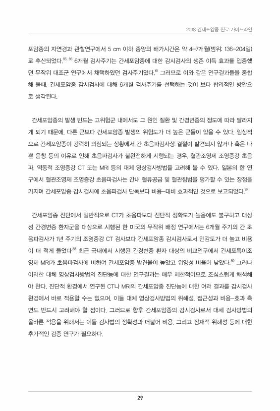
mUICC stage Best option Alternative option
I
Single/≤ 2 cm/VI-
ResectionRFA
TACEOther LRTEBRT
II
Single/>2 cm/VI-
ResectionLT (tumor size ≤ 5 cm)RFA (tumor size ≤ 3 cm)
TACE, TAREOther LRT (tumor size ≤ 3 cm)EBRT
II
Multiple/≤ 2 cm/VI-
LT (within Milan criteria)TACERFA (tumor number ≤ 3)
Resection (tumor number ≤ 2)Other LRT (tumor number ≤ 3)EBRT (tumor number ≤ 3)
Fig. 4. First-line treatment of 2018 KLCA-NCC, Korea Practice Guidelines for Patients with hepatocellular carcinoma, Child-Pugh class A, no portal hypertension, and ECOG 0-1
46
The Korean Liver Cancer Association and National Cancer Center, Korea
II
Single/≤ 2 cm/VI+
TACEEBRTSorafenibLenvatinib
Resection
III
Multiple/>2cm/VI-
TACELT (within Milan criteria)RFA (tumor number ≤ 3 and size ≤ 3cm)
Resection (tumor number ≤ 2)TAREEBRT (tumor number ≤ 3 and size ≤3cm)Other LRT (tumor number ≤ 3 and size ≤ 3cm)
III
Single/>2 cm/VI+
TACE+EBRTTACESorafenib Lenvatinib (tumor occupation <50%, Vp1-3)
ResectionEBRT
III
Multiple/≤ 2 cm/VI+
TACE+EBRTTACESorafenib, Lenvatinib
IVa
Multiple/>2 cm/VI+
SorafenibLenvatinib (tumor occupation <50%, Vp1-3) TACE+EBRT
TACE
IVa
Node+/no metastasis
SorafenibLenvatinib (tumor occupation <50%, Vp1-3)
TACEEBRT
IVb
Metastasis+
SorafenibLenvatinib (tumor occupation <50%, Vp1-3)
TACEEBRT
RFA, radiofrequency ablation; TACE, transarterial chemoembolization; LRT, loco-regional therapy including; Other LRT means percutaneous ethanol injection (PEI), microwave ablation (MWA), and cryoablation; TARE, transarterial radioembolization; EBRT, external beam radiation therapy; LT, liver transplantation; DDLT, deceased donor LT; LDLT, living donor LT; VI, vascular or bile duct invasion; ECOG, eastern cooperative oncology group; Vp, portal vein invasion

47
2018 간세포암종 진료 가이드라인
간절제
간절제는 간경변증이 없는 간에 국한된 단일 간세포암종 환자에서 1차 치료법이며145 간경변증이
있는 경우에도 잔존 간기능이 충분하다고 예상되는 경우 우선적으로 고려할 수 있다.146, 147 최근 수
술 전 검사 및 수술 술기의 발전, 수술 후 환자 관리의 경험 축적으로 간절제의 성적도 크게 향
상되었다.148 최근의 보고들에 따르면, 간세포암종 절제술 후 사망률은 1~3% 이하이며, 5년 생
존율은 46~69.5%, 5년 무병생존율은 23~56.3% 정도이다.149-152 간절제 후 간세포암종의 5년
재발률은 43.7~77% 정도이며 이들 중 80~95%가 간내에서 재발된다.153 간내 재발은 간내전이
(intrahepatic metastasis)와 다발성 암성변화(multicentric carcinogenesis)에 의한 새로운 간세
포암종(de novo HCC)으로 구분할 수 있다. 둘은 genomic hybridization, DNA fingerprinting,
DNA microarray, integration pattern of hepatitis B virus 등을 이용하여 감별할 수 있다.154 그
러나 실제 임상에서 두 경우를 감별할 수 있는 기준은 정의되어 있지 않으나, 일반적으로 2년을
경계로 2년 이후에 나타나는 후기 재발을 새로운 간세포암종(de novo HCC)으로 보는 경우가
많다.155 수술 후 재발에 관련된 위험인자는 종양관련 위험인자와 기저 간질환관련 위험인자로
나눌 수 있다. 종양관련 위험인자로는 크기와 개수 이외에도, 미세혈관 침습, 좋지 않은 암분화
도, 높은 혈청 알파태아단백(AFP) 또는 PIVKA-II 값, 18F-FDG PET 양성 등이 있으며, 기저 간
질환관련 위험인자로는 간경변증, 높은 혈청 HBV DNA 값, 활동성 간염 상태 등이 있다.141, 155-161
종양관련 위험인자는 주로 조기 재발과 연관되고, 기저 간질환 위험인자는 후기 재발과 관련성
이 높지만, 조기 재발과 후기 재발의 구분이 암종의 발생 기전 자체보다는 재발 시기만을 기준
으로 하기 때문에, 각 위험인자와 재발 시기의 상관관계가 뚜렷하지 않은 경우도 많다.
수술 후 추적관찰에서는 주로 CT, MRI 등의 영상의학적 검사와 혈청 종양표지자 검사가 추천
된다. 혈청 AFP는 전통적인 간세포암종의 대표적 종양표지자로서, 수술 전 상승되어 있었던 경
우에는 수술 후 간기능 정상화가 이뤄지면 추적에서도 재발 여부를 판단할 수 있는 유용한 지
표로 활용될 수 있다.162 PIVKA-II도 최근 진단 및 추적, 예후에 대해 그 효용성이 점차 높아지고
있는 종양표지자로서 AFP와 함께 임상에서 점차 활용 빈도가 높아지고 있다.156, 163
48
The Korean Liver Cancer Association and National Cancer Center, Korea
수술 전 평가
간절제의 안정성을 평가하기 위한 수술 전 검사로 오래 전부터 Child-Pugh 등급이 사용되어
왔다(Table 7).164 현재 대부분의 간절제는 ECOG 수행능력 0~2(Table 8)이며 Child-Pugh 등
급 A인 환자에서 시행되고 있지만 간경변증이 상당히 진행된 경우에도 A 등급일 수 있으므로
Child-Pugh 등급이 간절제의 안전성을 평가하는 충분한 검사법은 아니다.165, 166 그러므로 국내
여러 기관에서는 잔존 간기능을 예측하는 방법으로 일본에서 제시하였던 Indocyanine Green
15분 정체율(ICG-R15)을 수술 전 검사로서 시행하고 있다.167 우간절제 이상의 대량 간절제는
ICG-R15이 10% 이내의 환자에서만 권고되었지만 이후 연구에서는 14%까지의 환자에서도 우
간절제가 안전하게 시행될 수 있다고 보고되었다.168 반면 미국 및 유럽에서는 절제 가능성을 평
가하는 주요한 척도로 문맥압항진증과 혈청 빌리루빈치를 제시하였는데, 문액압항진증은 간정
맥압력차 10 mmHg 이상의 경우로 정의된다.169 임상적으로는 식도정맥류, 혹은 비장비대를 동
반한 혈소판감소증(100,000/mm3 이하), 복수 중 어느 하나가 있는 경우 문맥압항진증으로 정
의할 수 있고, 혈소판 감소증이 가장 중요한 임상적 인자로 여겨지고 있다.77 문맥압항진증이 있
는 경우에 간절제 후 합병증 발생률이 높고 장기적 예후가 불량하다고 보고되었지만,169-171 최근
일부 연구에서는 문맥압항진증이 동반된 환자에서도 문맥압항진증이 없는 환자와 동일한 장기
성적을 보고하고 있다.172-175 그러나 간경변증이 동반된 환자에서 간절제의 범위는 수술 후 간기
능 부전의 중요한 예측인자이기 때문에 경도의 문맥압항진증이 있는 환자에서는 대량간절제 미
만의 제한적 간절제를 시행해야 한다.
Table 7. Child-Pugh Classification
1 2 3
Albumin(g/dL) >3.5 2.8-3.5 <2.8
Bilirubin(mg/dL) <2.0 2.0-3.0 >3.0
Prothrombin time prolonged(s) 0-4 4-6 >6
Ascites none slight moderate
Encephalopathy (grade) none 1-2 3-4
Class A ≤ 6 points Class B = 7-9 points Class C ≥ 10 points

49
2018 간세포암종 진료 가이드라인
Table 8. Eastern Cooperative Oncology Group (ECOG) performance stage*
Grade ECOG
0 Fully active, able to carry on all pre-disease performance without restriction1 Restricted in physically strenuous activity but ambulatory and able to carry out work of a light or sedentary nature, e.g., light house work, office work2 Ambulatory and capable of all selfcare but unable to carry out any work activities. Up and about more than 50% of waking hours3 Capable of only limited selfcare, confined to bed or chair more than 50% of waking hours4 Completely disabled. Cannot carry on any selfcare. Totally confined to bed or chair5 Dead* Oken, M.M., et al. Toxicity And Response Criteria Of The Eastern Cooperative Oncology Group. Am J Clin Oncol 5:649-655, 1982.
간세포암종의 경우 대부분의 환자에서 만성 간질환을 동반한다. 수술 후 간기능 부전을 예방
하기 위해서는 간 예비기능 검사뿐만 아니라 수술 후 남는 잔존 간용적 (Future liver volume;
FLV)을 고려해야 한다. 정상간의 경우 전체간의 70-80%까지 절제할 수 있지만 만성 간질환을
갖고 있거나 간경변증이 동반된 환자의 경우 더 많은 간을 남겨야 한다. 간경변증이 동반된 환
자에서 안정적 간절제를 위한 잔존 간용적에 대한 연구는 많이 부족하지만 일반적으로 전체간
의 40% 이상의 간을 남기는 것을 권장한다.176 최근 간의 섬유화 정도를 비침습적으로 측정할
수 있는 새로운 검사가 개발되고 있다. 이 중 간섬유화스캔은 간절제 후 간 부전 및 재발을 예
측하는데 유용하다는 보고도 있다.177-180 절제 가능성을 알아보기 위해 시행되는 수술 전 영상검
사로는 역동적 조영증강 CT검사가 가장 기본적이다. 간세포특이조영제를 이용한 MRI의 경우
CT 검사에서 발견되지 않는 간세포암종 병변, 특히 1 cm 미만의 병변을 더 잘 발견하는 경향을
보였기에,181, 182 수술 전 절제 가능성 및 절제 범위를 결정하기 위한 검사로 유용할 수 있다. 간
세포암종 환자에서 수술을 해야되는 경우에는 간외전이 병소 유무를 알아보기 위해 추가 검사
를 선택적으로 시행한다. 18F-FDG 양전자방출단층촬영 (PET-CT)의 경우 간세포암종의 진단에
는 민감도가 떨어지지만141, 간외전이를 진단하는데 유용성이 증명되고 있어77, 183 수술 전 간외전
이를 발견하기 위한 검사로 시행할 수 있다. 이외에 흉부 CT와 뼈 스캔 등도 선택적으로 시행할
수 있다.184

50
The Korean Liver Cancer Association and National Cancer Center, Korea
간절제의 기본 원칙
최근 간절제가 보다 안전해진 것은 수술 중 출혈량이 줄어 수혈이 최소화 된 것에 크게 기인
한다. 수혈은 항암 면역기전을 저하시켜 수술 후 암의 재발을 증가시키는 것으로 알려져 있다.
간세포암종의 경우 최근 메타분석에서 수술 중 수혈이 간절제 후 합병증을 증가시키고 무병생
존율과 전체생존율을 감소시킨다고 보고하였다.185 선택적 간혈류차단술, 낮은 중심정맥압 유
지 및 정교한 간실질 박리 등에 의해 최근 간절제의 수혈률은 10% 이하이다.186 해부학적 절제
가 종양병리학적 관점에서는 절제연을 확보하고 미세전이를 제거하여 비해부학적 절제보다 성
적이 우수하다는 다양한 후향적 보고187-192 및 메타분석 보고193가 있다. 하지만, 최근 발표된 전
향적 무작위 연구결과를 보면 해부학적 절제는 2년 이내 조기 재발률만을 줄여주고, 5년 재발
률 및 생존율에는 영향이 없는 것으로 보고되었다.194 절제연에 암세포 침범이 남아 있지 않게
수술하는 것은 장기 예후에 절대적으로 중요하며, 전향적 무작위 연구에서는 2 cm 이상의 충
분한 절제연을 확보하는 것이 중요하다고 보고되었다.195 따라서, 가급적 충분한 절제연의 확보
와 해부학적 절제가 추천되지만, 간경변증이 동반된 환자에서는 수술 범위가 수술 후 합병증
과 밀접하게 연관되기 때문에 환자의 안전을 우선적으로 고려하여 적합한 수술 범위를 결정하
는 것이 무엇보다 중요하다.196-198 수술 후 예후를 향상시킬 목적으로 간절제 전 경동맥화학색전
술(TACE)을 시행하는 것은 추천되지 않는다.199, 200 간절제후 잔존 간용적은 수술 후 간기능 부
전의 중요한 예측인자이기 때문에 간경변증이 동반된 환자에서는 정상간에 비하여 보다 충분
한 잔존 간용적이 필요하다.201, 202 잔존 간용적이 충분하지 못할 것으로 예상되는 환자에서는 수
술 전 간문맥색전술이나 수술 중 간문맥결찰 등의 방법으로 잔존 간용적을 증가시켜 대량간절
제를 할 수도 있다.203-205 최근 간절제 방법으로 현수요법(hanging maneuver)이 종종 사용된다.
현수요법을 적용한 수술이 간세포암종의 치료성적에 영향을 미친다는 보고는 아직 없으나, 수
술 시간을 줄이고, 출혈을 감소시킨다는 보고가 있다.206 큰 간세포암종의 수술 시에는 전방접근
법(anterior approach)이 사용되기도 하는데, 전방접근법을 사용하면 대량 출혈 및 수혈의 빈도
가 적어지고, 생존율이 높아진다는 전향적 연구결과가 보고된 바 있으나,207 종양병리학적 이점
에 대해서는 추가적인 연구가 필요하다.
최소침습 간절제
복강경 간절제술은 기술적으로 빠르게 발전하고 그 적응증이 확대되고 있다. 전통적인 간절제

51
2018 간세포암종 진료 가이드라인
술과 비교하여 좌외측 및 전하방 구역에 대한 복강경 간절제술이 통증, 합병증 발생률 및 재원
기간 등이 더 우월하면서,208, 209 재발률 및 생존율의 차이는 없다는 보고가 많다.210, 211 복강경을
이용한 대량 간절제도 점차 늘고는 있으나, 아직은 경험 많은 의사들에게만 국한되어 시행되고
있으며, 그 성적에 대해서도 추가적인 연구가 필요할 것이다.212 최근 들어 로봇을 이용한 간절
제도 시도되고 있으나, 아직 제한적인 상태이며, 향후 전통적인 개복 수술 및 복강경 수술과의
비교 연구가 필요하다.213, 214
간절제의 적응증
간절제는 일반적으로 크기가 작은 1~2개 종양에서 시행될 때 양호한 예후를 보이며, 종양의
크기가 클수록 종양의 혈관침윤 빈도가 증가하고 불량한 예후를 보이는 것으로 보고 되었지만,
최근 연구에 의하면 종양의 크기가 10 cm 이상인 환자에서도 약 1/3에서는 미세혈관침윤이 관
찰되지 않았고, 이런 환자들에서는 수술 후 양호한 성적이 보고되었다.215, 216 따라서 수술 전 종
양의 크기만으로 절제 가능성을 판단하지 말아야 한다. 최근 수술 기술의 발달과 환자관리 능력
의 향상으로 고령 환자에서도 간절제 후 다른 연령과 비슷한 단기 및 장기 성적을 보이지만 나
이가 들수록 간 재생능력이 감소하기 때문으로 대량간절제는 신중히 고려되어야 한다.217-219
파열 간세포암종에서 간기능이 좋은 환자인 경우는 일차적으로 간절제를 시행하는 것이 효과
적인 치료방법이라는 보고도 있으나,220, 221 혈역학적으로 불안정한 경우에는 경동맥화학색전술
로 일차 지혈을 시키고 잔존 간기능을 정확히 평가한 후 정규 수술을 시행하는 것이 보다 효과
적이다.222 하지만 장기 성적은 파열되지 않은 간세포암종에 비해 떨어진다.223, 224 종양이 주간정
맥(major hepatic vein)이나 주간문맥(major portal vein)을 침범한 경우 일반적으로 간절제는
금기증으로 생각되나 주간문맥(main trunk or contralateral branch)의 침습을 제외한 경우, 간
섬유화가 적거나 종양 분화도 Edmondson-Steiner 등급이 낮은 환자에서는 간절제 후 5년 생
존율이 30% 이상이라는 보고도 있으며, 절제 후 사망률 3.7%, 중앙생존기간 19.9개월이라는 보
고도 있다.225-227 또한 국내 다기관 연구에 따르면, 담관 침습이 동반된 간세포암종의 간절제 후
5년 생존율도 32%로 우수한 편이었다.227, 228 따라서, 혈관이나 담관을 침습한 간세포암종이라도
환자의 상태가 양호하고 다른 치료방법이 없다면 수술적 절제를 고려할 수 있다.

52
The Korean Liver Cancer Association and National Cancer Center, Korea
[권고사항]
1. 문맥압항진증과 고빌리루빈혈증이 모두 없는 Child-Pugh 등급 A의 환자에서 간에 국한된
단일 간세포암종은 간절제가 일차 치료법이다 (A1).
2. 경미한 문맥압항진증 또는 경미한 고빌리루빈혈증을 동반한 Child-Pugh 등급 A 및 B7 등
급의 간세포암종은 제한적 간절제를 선택적으로 시행할 수 있다 (C1).
3. 간기능이 잘 보존된 환자에서 간문맥, 간정맥, 담도 침습 등이 있더라도 주간문맥(main portal
trunk) 침습이 없으면, 간에 국한된 3개 이하 간세포암종은 간절제를 고려할 수도 있다 (C2).
4. 좌외측 구역과 전하방에 위치하여 접근이 용이한 간세포암종은 복강경절제술을 고려할 수
있다 (B2).
간이식
단일결절로 5 cm 이하이거나, 다발성인 경우 결절이 3개 이하이고 각 결절이 3 cm 이하이면
서, 심한 간기능 장애를 동반한 경우 간이식은 일차 치료로 고려되어야 한다. 간이식은 간세포
암종을 포함하여 기저 간경변증까지 완전히 제거하고 새로운 간을 이식하기 때문에 이론적으로
가장 이상적인 치료법이다. 그러나 간이식 역사 초기에는 간세포암종 환자들에 대해 넓은 적응
증으로 시행된 결과 5년 생존율 40% 미만의 매우 불량한 성적을 보여 한 때 간세포암종은 간
이식의 상대적 금기증으로까지 여겨졌었다.229, 230 그 후 제한된 간이식 적응증으로 선별된 환자
에서 5년 무병생존율은 74%라는 우수한 결과가 보고되었다.231, 232 이태리 밀란(Milan) 그룹은 영
상검사에서 간외전이와 혈관침범이 없고, 단일결절인 경우 5 cm 이하, 다발성인 경우 결절이 3
개 이하이면서 각 결절이 3 cm 이하인 간세포암종 환자에서 간이식 후 4년 생존율 75%, 무병
생존율 83%라는 우수한 성적을 발표하여 간세포암종 환자에서의 간이식 기준을 제시하였다.233
밀란척도 발표 이후 15년간 발표된 90편의 논문으로부터 17,780명의 환자를 이용한 체계적 고
찰과 메타분석에서 밀란척도가 간이식 결과에 영향을 미치는 독립적 예후 인자로 확인되었으
며, 간세포암종이 없는 다른 적응증으로 간이식을 받은 환자와 비슷한 5년 생존율(65~78%)을
보였다.234-236

53
2018 간세포암종 진료 가이드라인
최근 영상기술의 빠른 발전으로 간세포암종의 비침습적 진단의 정확도가 높아지면서 밀란척
도가 만들어진 시점의 영상기술로는 확인되지 않던 작은 병변들이 관찰될 경우 밀란척도 내에
속하는지에 대한 판단에 혼란을 주는 경우가 있다. 22,392명의 환자를 이용한 메타분석에서는
가장 큰 간세포암종 결절의 직경이나 각 결절의 크기의 합이 예후에 가장 중요한 영향을 미쳤
고, 결절의 개수가 예후에 미치는 영향은 명확하지 않았으며,237 다른 연구에서는 CT/MRI에서
발견하지 못했으나 수술 후 병리학적 검사에서 확인된 병변은 간이식 예후에 별 영향을 미치지
않았다.238
이식 전 간세포암종 환자에서는 간이식을 위한 일반적인 전신검사 외에 간세포암종의 병기를
확인할 수 있는 검사를 시행한다. 간 자체에 대한 영상검사는 역동적 조영증강 CT 혹은 MRI를
시행하며 간외전이 여부를 확인하기 위해 흉부 CT와 복부 및 골반 CT 혹은 MRI를 반드시 시행
하고, 뇌 영상 및 골주사 및 18F-FDG PET-CT 검사를 시행할 수 있다.239 18F-FDG PET-CT에
서 간세포암종이 양성인 경우 세포분화도가 더 나쁘거나 미세혈관 침윤이 높아 음성인 경우보
다 무병생존율이 떨어지는 경향을 보이므로 종양생물학적 특성을 판단하는데 도움을 줄 수 있
다. 이식 대기 중 간세포암종의 진행 여부에 대한 최적의 평가 방법이나 간격에 대한 구체적인
연구나 합의는 없지만 3개월 간격으로 역동적 조영증강 CT 혹은 MRI와 혈청 AFP 측정을 하는
것이 가장 흔하다.240
뇌사자간이식
뇌사자간이식에서는 항상 공여 장기가 모자라기 때문에 많은 환자가 이식을 대기하고 있
는데, 특히 간세포암종 환자에서는 대기 등록 후 간이식까지의 기간이 문제가 된다. 미국의
UNOS(United Network for Organ Sharing)에서는 간이식 대기 우선순위를 결정하기 위해
MELD 점수를 도입하여, 간세포암종 환자에서는 단일결절로 2~5 cm 이하이거나 다발성인 경
우 결절이 3개 이하 각 결절이 3 cm 이하인 경우 처음 등록 후 6개월이 지난 후에도 간세포암
종이 밀란척도 범위에 있다면 MELD 점수 28점을 주고, 이식 대기 후 3개월마다 10%의 가산점
을 주어, 간세포암종 환자와 비간세포암종 환자의 대기 중 탈락 위험을 비슷하게 하려는 노력을
하고 있다.241, 242 그러나 우리나라는 국립장기이식관리센터에서 KONOS (Korean Network for
Organ Sharing) 등급제를 운영하였고 간세포암종 환자에 대한 가산점이 없었다. 이러한 문제를

54
The Korean Liver Cancer Association and National Cancer Center, Korea
해결하고자 2016년 6월부터 MELD 점수를 도입하여, 간세포암종이 밀란척도에 해당되는 경우,
대기자의 MELD 점수가 0~13인 경우 4점을 추가, MELD 점수 14~20인 경우 5점 추가, MELD
점수 21점 이상은 추가 점수를 부여하지 않도록 개정하였다.243 그럼에도 불구하고 우리나라에
서 대부분의 뇌사자 간이식은 MELD점수가 30점 이상인 경우에 시행되기 때문에 간세포암종
환자가 상대적으로 간을 배정 받기는 어렵다. MELD 점수 도입이 우리나라 간세포암종 환자에
게 어떠한 영향을 주는지 추후 연구가 필요하다.
가교 치료(Bridging therapy)
간이식에 적응이 되는 간세포암종 환자 중 이식시기를 예측하기 어려운 경우 국소치료 또는
경동맥화학색전술을 시행하는 경우가 드물지 않다. 간이식 대기 중 종양이 진행하여 간이식을
못하게 되는 이탈률(drop out rate)은 1년에 15–30%로 보고되며244, 245 국소치료를 통해 이탈률
을 0-25%로 감소시킨다고 보고되었다.246-248 종양진행을 막아서 이탈률을 감소시키기 위해 경
동맥화학색전술, 고주파열치료술 등의 국소치료를 시행할 수 있으며,246, 247, 249-251 간이식 대기중
에 6개월이 넘어갈 경우 국소치료가 도움이 된다.246 간이식 대기중에 AFP>15 ng/mL/month
증가한 경우 이식 후 재발 가능성이 높아서 낮은 생존율이 예상된다.252
간이식 전 치료가 간이식 후에 미치는 효과를 평가하는 것은 매우 어렵다. 지금까지 많은 연
구들은 이식 전 치료 여부에 따른 이식 후 생존율에 차이가 없다고 보고하고 있다.253-259 미국
의 OPTN/SRTR을 이용한 간이식 전의 경동맥화학색전술, 고주파열치료술 등의 국소치료를 받
은 환자들이 치료를 받지 않은 환자들보다 생존율이 더 높았고 이식 대기기간이 길수록 이식
후 생존율이 더 높았다.260 간이식 전에 이식 대기기간이 6개월부터 18개월 이내일 경우 간이식
후 좋은 결과를 보여준다고 하고 있으나, 261 이식 대기기간이 길어지는 경우 간세포암종의 진행
가능성이 높아서 6개월 이상 이식 대기가 예상되는 경우 가교 치료(bridging therapy)가 필요
하다.246, 261, 262 최근 미국에서 시행한 다기간 연구에서 밀란척도 이내 환자의 경우 국소치료 유
무가 간이식 후 간세포암종 재발에 영향을 주지 않아서 밀란척도 이내 환자의 경우 국소치료를
이용한 가교 치료는 간이식 후 간세포암종 재발 감소 및 환자 생존율 증가와 관련이 없었다.253
이식 전 국소치료 종류에 무관하게 국소치료 횟수가 3회 이상인 경우 이식 후 간세포암종 재발
가능성이 2배 이상 증가한다고 보고하였다.253 적출된 간 조직에서 국소치료로 완전 관해가 확

55
2018 간세포암종 진료 가이드라인
인된 경우 간이식 후 간세포암종 재발 가능성이 매우 낮았다.253
병기감소
간이식의 적응증을 벗어나는 간세포암종 환자에서 간이식을 목적으로 하는 병기감소
(downstaging) 치료에 대한 대규모 전향적 연구가 없으나 일부 소규모 전향적 연구에서 밀란
또는 UCSF척도에서 벗어나는 경우 이식 전 국소치료를 통해 밀란 또는 UCSF척도 내로 병기감
소(downstaging)가 된 경우 5년 생존율은 밀란척도 이내 또는 UCSF척도 이내의 환자들과 비
슷한 생존율을 보인다.251, 259, 263-267 경동맥화학색전술로 치료한 경우 간세포암종의 병기감소는
24~63%에서 가능하며,268-270 병기감소는 종양의 크기가 7 cm보다 작거나 종양 개수가 3개 이
하인 경우 더 효과적으로 알려져 있으나271 어떤 제한이 있는 것은 아니다.272 병기감소를 목적으
로 한 Yttrium90을 이용한 경동맥방사선색전술과 통상적 경동맥화학색전술을 이용한 방법간에
간이식 성적 차이는 없어 보이나273, 274 추가 연구가 필요하다.
생체간이식
장기기증에 대한 사회 인식 변화와 장기기증 활성화를 위한 여러 차례에 걸친 ‘장기 등 이식
에 관한 법률’ 개정에 힘입어 최근 국내에서도 뇌사자 장기기증 건수와 간이식 건수가 점차 증
가하고 있다.275, 276 그러나 뇌사자 장기기증이 절대적으로 부족한 우리나라에서는 생체간이식
이 주로 시행되고 있다. 2016년 한 해 동안 1,473 건의 간이식이 시행되었는데, 그 중 965 건
(65.5%)이 생체간이식이었고, 나머지 508 건(34.5%)이 뇌사자간이식 이었다. 최근 장기구득체
계 개선으로 국내 뇌사자 수는 증가하고 있고 이로 인해 간이식을 받아야하는 간이식대기자는
2013년 6,334명을 최고점으로 2016년 4,969명으로 감소하였다. 하지만 2016년 MELD 점수 도
입 이후 국내 뇌사 기증자로부터 기증된 간의 대부분은 MELD 30점 이상의 고위험 응급환자에
게 배정되어 간세포암종 환자에게 장기 배정 가산점을 부여하더라도 간세포암종 환자가 적절
한 시기에 뇌사자간이식을 받을 가능성이 상대적으로 낮다. 간세포암종 환자에서 기증형태에
따른 간이식 성적에 관해서는 아직 논란이 있다. 최근 633명의 생체간이식환자와 1,232명의 뇌
사자간이식환자에서 간세포암종의 간이식 후 성적에 관한 메타분석에 의하면 기증형태에 따른
간이식 후 전체생존율의 차이는 없었다.277 그러나 생체간이식의 무병생존율은 뇌사자간이식 성
적보다 나빴다. 하지만 간세포암종으로 생체간이식과 뇌사자간이식을 시행받은 환자 1,310명의

56
The Korean Liver Cancer Association and National Cancer Center, Korea
메타분석에서는 기증형태에 따른 생존율 및 무병생존율 모두에서 차이가 없었다.278 특히 간세
포암종 환자에서 기증형태에 따른 간이식 성적을 비교하려면 간이식 이후 성적을 비교하는 것
이 아니라, intention-to-treat 분석이 필요하다. 간세포암종을 가진 환자에서 생체간이식을 받
는 환자는 대기 시간이 짧고 대기 중 탈락 가능성이 거의 없지만, 뇌사자간이식의 경우 5-30%
의 대기중 탈락률이 보고되고 있다. 간세포암종 환자에서 intention-to-treat 분석을 이용한 기
증형태에 따른 간이식 성적 비교에서는 두 군간에 생존율 및 무병생존율에서 차이가 없었다.279,
280 간세포암종 환자에서 간이식 후 무병생존율의 차이는 기증형태보다는 종양의 병태생리, 이
식 전 관리와 대기기간의 차이에 의할 수 있다.281-283 간세포암종 환자에서 간이식 후 재발에 영
향을 미치는 인자를 올바르게 밝히기 위해서는 이러한 연구 비뚤림현상이나 종양병태생리를 감
안한 보다 잘 계획된 연구가 필요하다.
생체간이식 후 생존율은 밀란척도의 준용 여부에 따라 뇌사자간이식과 비슷한 생존율을 보이
기 때문에, 초기 생체간이식 프로그램에서는 뇌사자간이식 자료에 근거한 간이식 선정기준을
적용하여 왔다. 그러나, 뇌사자간이식 프로그램에서는 사회적 자원인 기증 장기의 이용-효율을
최대화하고자 선정기준을 정해왔기 때문에, 대부분 가족 내에서 이뤄지는 생체간이식에서도 이
와 동일한 선정 기준을 적용하는 것이 옳은 것인가 하는 비판이 있었다. 실제로 뇌사장기이식
이 상대적으로 드문 아시아권에서는 각 센터별로 적절한 생체간이식 선정기준을 정하여 이식을
수행하여왔는데, 한 전문병원에서는 종양의 크기가 5 cm 미만이고 혈중 AFP 수치가 400 ng/
mL 미만이면 종양의 개수에 관계없이 5년 환자생존율은 79.9%임을 보고하였고284, 다른 전문
병원에서는 영상검사상 혈관침윤이 없고, 혈중 AFP 수치가 400 ng/mL 미만일 때, 3년 생존율
이 86.2%임을 보고하였다.285 또 다른 전문병원에서는 혈중 AFP 수치가 100 ng/mL 미만이고,
종양의 크기가 5 cm 미만일 때 3년 무병생존율은 80.9%임을 보고하였고286, 모 전문병원에서
는 단일결절로 5 cm 미만, 6개 이하의 종양과 육안적으로 혈관침윤이 없는 경우 5년 생존율이
81.6%임을 보고하였다.287 일본의 한 병원에서는 종양의 크기가 5 cm 이하이면서 개수가 5개
이하일 때 3년 무병생존율이 94%이었고,288 다른 병원에서는 종양의 개수가 10개 이하이고, 혈
중 PIVKA-II 수치가 400 mAU/mL 이하일 때, 5년 생존율이 86.7%임을 보고하였다.289 일본의
또 다른 병원에서는 종양의 크기가 5 cm 이하이면서 혈중 PIVKA-II 수치가 300 mAU/mL 이
하일 때 5년 생존율이 86.7%임을 보고하였다.290 일본 49개 센터 653명의 간세포암종 간이식환

57
2018 간세포암종 진료 가이드라인
자 연구에서 밀란척도를 넘지만 혈중 AFP 수치가 200 ng/mL 이하이고 혈중 PIVKA-II 수치가
100 mAU/mL 이하인 경우에는 5년 무병생존율이 84.3%임을 보고하였다.291 이처럼 대부분 확
장된 척도들은 밀란척도에서 종양 크기와 개수를 수정한 것이다. 하지만 최근에는 선택 척도에
AFP과 PIVKA-II와 같은 생물학적 혈청표지자를 포함하고 있다.292 특히 최근 유럽의 다기관 연
구에 의하면 AFP를 포함한 척도가 크기와 개수에 기준하는 척도에 비해 간이식 후 종양 재발을
보다 잘 예측하며 밀란척도를 넘더라도 AFP를 포함하는 척도 기준에 포함될 경우 좋은 결과를
가져올 수 있다는 보고들이 있다.293, 294 이러한 성적을 종합하여 볼 때, 뇌사자간이식의 척도인
밀란척도를 넘는 경우에도 생체간이식은 수혜자와 기증자간의 특수한 상황을 고려하여야 한다.
따라서 다른 효과적 치료법을 적용할 수 없는 경우라면, 분명한 혈관침윤이 없고 간외전이가 없
는 경우, 종양의 크기와 수에 국한하지 않고 종양의 병태생리를 고려하여 적정한 수준의 재발위
험을 감수한 간이식을 고려할 수도 있다.
생체간이식에서는 간 기증자의 안전성이 무엇보다 중요한데, 국내 생체간이식 기증자들에 관
한 다양한 보고에 의하면 생체간이식 기증자 수술은 안전한 수술임을 알 수 있다.295-300 최근
832명 생체기증자를 포함하는 한국장기이식등록 연구결과에 의하면 주요합병증(담즙 누출, 담
도 협착, 간문맥 협착, 상처 열개, 폐부종 등)은 1.9% 였고 사망자는 없었다.301 그러나 세계적
으로는 생체간이식 기증자 수술과 연관된 약 0.2-0.3% 사망률이 보고되었으며, 중증합병증이
2% 내외에서 발생할 수 있다.302-305 따라서 생체간이식 기증자의 안전성을 동시에 고려할 때 숙
련된 기관에서 엄격한 기증자 선정기준을 적용하는 것이 필수적이다
간이식 후 면역억제요법
간세포암종 환자의 이식 후 면역억제를 위해 calcineurin inhibitor (cyclosporine, tacrolimus),
mammalian target of rapamycin inhibitor (mTORi) (sirolimus, everolimus)를 기본으로 하는 면
역억제제가 사용된다.306 최근 연구에서 mTORi의 사용이 간세포암종으로 간이식을 받은 환자에
게 재발감소 및 생존기간 연장에 도움을 줄 수 있다는 보고가 있으나 좀 더 많은 연구가 필요하
다.307
58
The Korean Liver Cancer Association and National Cancer Center, Korea
[권고사항]
1. 간세포암종 환자에서 간절제가 불가능하면서 영상학적 혈관침범과 원격전이가 없는 5 cm
이하의 단일종괴 또는 3 cm 이하이며 3개 이하의 종양(밀란척도)인 경우 간이식이 일차 치료
법이다 (A1).
2. 간이식에 적응이 되는 간세포암종 환자 중 이식시기를 예측하기 어려운 경우 국소치료 또는
경동맥화학색전술 등을 먼저 시행하는 것이 추천된다 (B1).
3. 간이식 적응증을 벗어나는 밀란척도 이상의 간세포암종 환자에서 국소치료, 경동맥화학색전
술, 혹은 기타치료 등에 의해 병기감소가 되면 간이식을 고려할 수 있다 (C1).
4. 다른 효과적 치료법을 적용할 수 없는 경우, 분명한 혈관침범이 없고 간외전이가 없는 간세
포암종에서 간이식은 밀란척도 이상의 확대 기준을 적용할 수도 있다 (C2).
5. 간절제 이후 재발한 환자에서 구제간이식의 적응증은 일차 간이식에서와 같다 (B1).
국소치료
국소치료(loco-regional therapy, LRT)는 수술과 비교해 시술이 간편하고 주변 간조직 손상
을 줄이면서 종양을 괴사시킬 수 있는 장점으로 인해 간세포암종의 비수술적 치료법으로 많
이 이용되고 있다. 큰 의미의 국소치료는 경동맥화학색전술도 포함하나 이 장에서는 협의의
국소치료에 국한하여 기술한다. 고주파열치료술(radiofrequency ablation, RFA)과 에탄올주
입술(percutaneous ethanol injection, PEI)이 표준적 국소치료고, 초단파소작술(microwave
ablation), 냉동소작술(cryoablation)이 최근 유효한 치료로 인정되며, 레이저소작술(laser
ablation), 고강도집속초음파치료술(high-intensity focused ultrasound, HIFU) 등은 임상적 시
도(clinical trial)로 분류된다.
적응증은 연구자나 시술법에 따라 차이가 있으나, 단발성 종양은 장경 5 cm 이하, 다발성 종
양은 개수가 3개 이하이고 각각의 장경이 3 cm 이하일 때 국소치료를 고려할 수 있다. 국소치
료를 보다 큰 종양에 적용하기 위한 노력도 계속되고 있으나, 치료 성공률은 아직까지 종양 크
기와 밀접한 관계가 있다. 교정 후 혈소판이 5만/mm3 이하이거나 프로트롬빈 시간이 50% 이

59
2018 간세포암종 진료 가이드라인
하일 때는 시술에 따른 출혈 위험성이 높으므로 시술을 피해야 한다.
고주파열치료술
고주파열치료술(RFA)은 현재 가장 자주 이용되는 간세포암종의 국소치료법이다. 고주파열치
료술은 종양 내에 삽입한 전극 주위로 고주파 교류(460-500 kHz) 전류를 흘려 분자들 간의 마
찰을 유도함으로써 종양과 그 주위 조직을 가열하여 괴사를 유도한다. 종양 조직은 그 온도가
45-50℃의 열에 3분 이상 노출 시, 그리고 60℃ 이상의 고열에 대해서는 거의 즉시 단백질의
변성과 세포막의 파괴로 인하여 응고성 괴사가 일어난다. 에탄올주입술에 비하여 고주파열치료
술은 적은 횟수의 시술로 종양의 완전괴사를 유도하며, 간세포암종의 크기가 2 cm 이하인 경우
높은 종양괴사율을 보인다.308-311 경피적 시술법이 주로 사용되나 경우에 따라서는 복강경 수술
또는 개복술을 통해 시행할 수 있다.
시술 후 1일-1주 이내 CT나 MRI로 평가한 초기 종양괴사율은 많은 연구에서 95% 이상이며
추가적인 고주파열치료술을 시행하여 이를 100%까지 높일 수 있다고 보고되었다.257, 310, 312 3년
국소재발률은 0.9-21.4%로 보고되었다.257, 312, 313 고주파열치료술 후 10년 국소재발률에 관해서
는 Shiina 등257의 보고에 의하면 3.2%이나, 김 등312에 의하면 38.2%로 보고되어 기관에 따라
큰 차이가 있다. 국소치료 후 생존과 관련된 독립인자는 초기 완전괴사, Child-Pugh 점수, 결절
의 수와 크기, 시술 전 혈청 AFP 수치 등이다. 국소치료가 가장 효과적인 간세포암종은 Child-
Pugh 등급 A이면서, 직경 2 cm 이하 단일결절이며 종양의 위치가 고주파열치료술을 시행하기
좋은 경우 고주파열치료술도 간절제와 유사한 결과를 보여 고주파열치료술을 일차 치료법으로
고려해야 한다는 주장도 있다.313,137
고주파열치료술 후 기대되는 간세포암종 환자의 장기 생존율은 종양 크기에 따라 차이가 있
으며, Child-Pugh 등급 A이면서 장경 2 cm 이하의 종양은 3년 생존율이 90% 내외, 5년 생존
율이 65-70%이고,257, 312, 313 장경 2-5 cm의 종양은 3년 생존율이 65-75%, 5년 생존율이 50%
내외로 보고되었다.257, 312 또한 Child-Pugh 등급 A 이면서 장경 3 cm 이하의 단일종괴에 대한
10년 생존율은 약 41.3%였다.257
60
The Korean Liver Cancer Association and National Cancer Center, Korea
고주파열치료술과 간절제를 비교한 연구는 대부분 무작위 대조연구가 아니고, 무작위 대조연
구의 경우 연구군이 작아 결론을 내리기에는 아직 부족한 실정이다.314 최근 발표된 무작위 대조
연구를 포함한 세 편의 무작위 대조연구에서 두 치료법 간에 뚜렷한 생존율 차이가 없었고315-
317, 차이가 난다고 보고된 무작위 대조연구의 경우 3 cm 이하 단일종괴군에 포함된 대상자수가
작고 고주파열치료술의 1년 생존율이 91%로 수술의 100%에 비해 지나치게 낮아서 신뢰하기 힘
들다318. 무작위 대조연구를 대상으로 한 메타분석연구에서 밀란척도 범위내 간세포암종 환자를
대상으로 했을 때 5년 생존율과 무재발률은 간절제군에서 유의하게 높았으나319, 3 cm 이하 단
일종괴 간세포암종 환자에서는 두 치료군 간 생존율에서 유의한 차이를 보이지 않았다.320 또 다
른 Child-Pugh 등급 A 간세포암종 환자를 대상으로 한 메타분석연구에서도 종양 크기 3 cm
미만군에서는 5년 생존율의 차이가 없었다.321 2 cm 이하의 단일 간세포암종 환자를 대상으로 한
시뮬레이션 연구에서도 일차 치료로 고주파열치료술을 시행한 군이 간절제를 선택한 군과 동등
한 장기 생존율을 보였다.322 최근 국내에서 발표된 전향적 대조군 연구에서 간절제와 고주파열치
료술 간에 무병생존율(disease-free survival)은 간절제가 더 우수하였으나 전체생존율에는 차이
가 없었으며323, 다른 비무작위 대조연구들에서도 3 cm 이하 간세포암종 치료에서 간절제와 고주
파열치료술 간에 유의한 생존율 차이가 없다고 보고하였다.324-326 간절제는 고주파열치료술에 비
해 합병증의 빈도가 좀 더 높고, 재원기간도 평균 8-9일 이상 더 긴 경향이 있었다.320
간세포암종이 직경 3 cm을 초과하게 되면 고주파열치료술의 국소재발률이 높아져서 30-
50%까지 보고되고 있다.312 이 때 국소재발률을 낮추고 생존율을 향상시키기 위해 경동맥화학색
전술과 병행치료가 시도되고 있다. 3 cm 이하의 3개 이하의 종양을 대상으로 비교하였을 때는
병행치료와 고주파열치료술 단독치료 사이에 생존율, 재발률은 모두 유의한 차이를 보이지 않
았다.327 반면, 종양의 크기가 3-5 cm인 경우에는 국소재발률과 생존율 모두 병행치료가 더 좋
은 것으로 보고되었다.328, 329 7편의 무작위 대조연구의 메타분석에서도 병행치료군에서 더 좋은
생존율을 보였고, 종양의 크기가 3 cm 이하인 하위군 비교에서만 유의한 생존율의 차이를 보
이지 않았다.330 고주파열치료술 단독치료와 경동맥화학색전술과의 병행치료를 비교한 8편의 무
작위 대조연구들의 메타분석들에서도 병행치료가 생존율과 재발률에서 더 좋은 결과를 보였고,
주요 합병증은 차이가 없었다.331, 332 이상의 결과를 고려할 때 3-5 cm 크기의 간세포암종 치료
에서 고주파열치료술과 경동맥화학색전술의 병합치료가 고주파열치료술 단독치료와 비교해 더

61
2018 간세포암종 진료 가이드라인
높은 생존율과 낮은 재발률을 보이고 합병증 발생률은 유의한 차이가 없다고 할 수 있다.
고주파열치료술의 제한점으로 간문(hilum) 주위나, 담도, 대장과 같은 주요 장기가 간세포암
종에 인접한 경우 시술 후 합병증의 위험성이 높고, 비교적 큰 혈관 주위에 종양이 인접한 경우
열 씻김현상(heat sink effect)으로 인하여 열 전달이 충분하지 않아 치료 효과가 떨어질 수 있
으며, 일반적으로 에탄올주입술에 비해 부작용이 상대적으로 많다.311, 333, 334 그러나 주변 장기와
시술 대상 병변 사이에 인공 복수를 위치 시킬 수 있는 경우에는 비교적 안전하고 효과적인 고
주파열치료술이 가능하다.335 고주파열치료술의 또 다른 한계는 2 cm 미만의 작은 간세포암종
이 통상적 초음파검사에서 잘 보이지 않을 수 있다는 점인데, 조영증강초음파나 항법융합영상
유도를 이용하여 통상적인 초음파 검사에서 보이지 않는 경우에도 시술이 가능해짐으로써 시술
적용 범위가 넓어졌다.336, 337
고주파열치료술의 합병증으로 인한 사망률은 0.1-0.5%이며, 주요 합병증은 5% 이내에서 발
생한다고 알려져 있다.313, 333, 334 주요 합병증은 종양전파, 혈액복막이나 혈흉, 간농양, 간실질 대
량경색, 장 천공, 기흉 등이다.257
결론적으로, 밀란척도내 간세포암종에서 간절제가 고주파열치료술과 비교하여 낮은 재발률을
보이고, 치료 후 합병증은 유의하게 높았으며, 생존율 차이에 대해서는 추가적 연구가 필요한
것으로 보인다. 단, 3 cm 이하 단일결절 간세포암종에서는 고주파열치료술이 간절제와 비교하
여 동등한 생존율을 보이고 국소재발률은 높으며 합병증 발생률은 낮아 종양의 위치가 고주파
열치료술을 시행하기 용이한 경우 간절제를 대신하여 사용할 수 있다.
에탄올주입술
에탄올주입술(Percutaneous ethanol injection, PEI)은 시술이 간편하고 부작용이 적다는 장점
으로 간세포암종 치료술로서 널리 사용되었으나, 여러 번 시술해야 하고 직경 3 cm을 초과하는
종양은 완전괴사가 어렵다는 단점이 있어 최근에는 고주파열치료술로 상당 부분 대체되었다.
에탄올주입술의 종양괴사율은 연구자에 따라 66-100%로 다양하게 보고되고 있다.309-311, 338 치
료 효과는 종양 크기가 중요하여 장경 2 cm 이하 종양에서는 90% 이상의 종양괴사율을 보이나

62
The Korean Liver Cancer Association and National Cancer Center, Korea
크기가 커질수록 괴사율이 떨어져서 3-5 cm에서는 약 50% 정도의 종양괴사율을 보인다. 국소
재발률은 약 24-34% 정도이다.339-341 에탄올주입술 후 기대되는 장기 생존율은 Child-Pugh 등
급 A이면서 장경 2 cm 이하의 단일 종양은 3년 생존율이 70-80% 이상, 5년 생존율이 50% 이
상, 장경 2-3 cm의 종양은 3년 생존율이 47-64%로 보고되었다.309, 338
간세포암종 환자에서 고주파열치료술과 에탄올주입술을 비교한 여러 무작위 대조연구 중에서
309-311, 338, 342, 343 이태리에서 발표된 무작위 대조연구342, 343를 제외하고는 고주파열치료술이 유의
하게 낮은 국소재발률을 보였고, 생존율도 고주파열치료술이 유의하게 높은 결과를 보였다. 특
히 4편의 무작위 대조연구를 메타분석한 연구에서 고주파열치료술의 3년 생존율이 에탄올주입
술에 비하여 유의하게 높은 것으로 보고되었다.344-347 그러나 직경 2 cm 이하의 간세포암종의
하위군에서는 유의한 생존율 차이를 보이지 않았다.346 이상의 연구결과를 볼 때 고주파열치료
군이 에탄올주입술군에 비해 국소 재발률은 더 낮고, 생존율은 더 높을 것으로 예상되나 추가
연구가 필요하며, 직경 2 cm 이하에서는 유사한 생존율을 보고하는 연구들도 있어 고주파열치
료술을 적용하기 어려운 경우 에탄올주입술을 고려해볼 수 있다348. 특히 혈관 주위에 종양이 위
치해 있는 경우, 고주파열치료술의 열 씻김현상(heat sink effect)을 극복하기 위하여 에탄올주
입술을 사용할 수 있으나, 간세포암종이 간 문부의 담도에 연해 있는 경우 고주파열치료술과 마
찬가지로 인접한 담도에 협착이 발생할 위험이 있는 것으로 알려져 있다.349, 350
초단파소작술 및 냉동소작술
근래에 간세포암종의 국소치료로 초단파소작술(microwave ablation) 및 냉동소작술
(cryoablation)의 이용이 증가하고 있다. 특히 초단파소작술은 조직의 전도 특성의 제한을 받지
않으며, 조직 온도를 100도 이상으로 빨리 증가시킬 수 있어 고주파열치료술에 비하여 열 씻김
현상에 영향을 덜 받고 더 큰 소작 범위를 만들 수 있는 장점들이 있다.351 이로 인하여 미국 및 유
럽에서는 2 cm 이상의 간세포암종의 국소치료에 고주파열치료술을 대신하여 초단파소작술을 사
용하는 경우가 흔하다. 냉동소작술은 얼음구(ice ball)가 초음파나 비조영 CT 또는 MR에서 매우
명확하게 잘 보이는 장점이 있어 시술 시 소작 범위의 모니터링이 용이하고, 시술 후 통증이 적
은 장점이 있다.351, 352 하지만 냉동소작술에 사용되는 프로브(probe)는 바늘 형태로 단일 프로브
로 큰 소작 범위을 만들기 어려워 대부분의 경우 여러 개의 프로브를 써야 하는 단점이 있다.
63
2018 간세포암종 진료 가이드라인
Child-Pugh 등급 A, B 환자에서 발생한 5 cm 이하, 3개 이하 간세포암종에서 고주파열치료
술군과 초단파소작술군을 비교한 무작위 대조연구에서 1, 3, 5년 생존율, 비재발률, 주요 합병
증은 두 군 간에 유의한 차이가 없었다.353 만성 간염 환자에서 3개 이하, 4 cm 이하 간세포암종
에 대한 고주파열치료술과 초단파소작술을 비교한 다기관 무작위 대조연구에서도 20개월 국소
종양진행률, 종양진행시간 및 20개월 생존율은 유의한 차이가 없었으나, 이 연구는 관찰 기간
이 짧은 제한이 있다.354 고주파열치료술과 다른 국소치료를 비교한 메타분석연구에서도 고주파
열치료술와 초단파소작술 간에 생존율, 주요 합병증 발생률에 유의한 차이가 없었다.347 Child-
Pugh 등급 class A, B이며 간경변이 있는 1개 또는 2개 간세포암종 환자에 대하여 냉동소작술
군과 고주파열치료군을 비교한 다기관 무작위 대조연구에서 1, 3, 5년 생존율, 무재발률 및 치
료 후 주요 합병증 발생률은 두 군 간에 유의한 차이를 보이지 않았다.355
이상의 제한된 무작위 대조연구와 메타분석연구결과들에서 초단파소작술과 냉동소작술은 고
주파열치료와 비교하여 생존율, 재발률, 주요 합병증 발생률에서 비슷한 결과를 보였다. 국내에
서는 고주파열치료술과 비교하여 초단파소작술과 냉동소작술의 비용이 더 큰 현실적인 문제가
있으며, 확실한 치료 효과과 합병증 등의 차이를 확인하기 위해서는 추가적인 대규모 전향적 무
작위 대조연구가 필요하다.
기타 임상적 시도 중인 국소치료법
고강도집속초음파치료(HIFU), 레이저치료술, 홀뮴주입술 등 여러 치료가 임상적으로 시도
(clinical trial) 되었으나 이러한 치료법은 아직까지 표준적 치료법들과의 대조연구가 거의 없어
향후 기술적 발전과 임상 연구결과를 더 지켜보아야 할 것으로 생각된다.
[권고사항]
1. 고주파열치료술(RFA)은 직경 3 cm 이하의 단일 간세포암종 치료로서 간절제와 비교하였을
때 생존율은 동등하고, 국소재발률은 높으며, 합병증 발생률은 낮다 (A1).
2. 고주파열치료술은 종양괴사 효과나 생존율에서 에탄올주입술(PEI)보다 우수하다(A1). 다만
직경 2 cm 이하의 간세포암종에서는 두 치료법의 결과가 유사하므로 고주파열치료술을 적용

64
The Korean Liver Cancer Association and National Cancer Center, Korea
하기 어려운 경우 에탄올주입술을 시행할 수 있다 (A2).
3. 수술적 치료를 적용하기 어려운 직경 3-5 cm 간세포암종에 대해 고주파열치료술 단독치료
에 비해 고주파열치료술과 경동맥화학색전술의 병행치료는 생존율을 증가시킨다 (A2).
4. 간세포암종 치료에서 초단파소작술과 냉동소작술은 고주파열치료술과 비교하여 유사한 생
존율, 재발률, 합병증 발생률 등을 기대할 수 있다 (B2).
경동맥화학색전술 및 기타 경동맥 치료법
간세포암종으로 진단받은 환자들 중 대다수는 문맥압항진증, 간기능 저하, 다발성 종양, 문맥
침습, 종양 주변의 충분한 절제구역 확보 불가능, 고령, 동반 질환 등의 이유로 비수술적 치료
를 받게 된다.356 이러한 환자들에서 전세계적으로 가장 흔히 시행되는 치료법이 경동맥화학색
전술(transarterial chemoembolization, TACE; 이하 TACE)이며 간세포암종에 대한 화학요법과
선택적 허혈에 의한 종양괴사 효과를 동시에 보고자 하는 치료법이다.275, 356, 357 TACE는 실제 임
상 상황에서 간세포암종의 1차 치료법으로 가장 많이 선택되고 있다.358 TACE는 리피오돌을 이
용하는 통상적 경동맥화학색전술(conventional TACE, cTACE)과 약물방출미세구를 이용하는
방법(drug eluting bead TACE, DEB-TACE)으로 크게 나눌 수 있다.359, 360 그러나 TACE는 항
암제를 투여하지 않고 단순한 색전 물질만 주입하는 경동맥색전술(transarterial embolization,
TAE)과 항암제만 동맥 내로 투여하고 동맥 색전을 시행하지 않는 간동맥주입화학요법(hepatic
arterial infusion chemotherapy, HAIC) 등과 구분하여야 한다.361, 362
통상적 경동맥화학색전술
통상적 경동맥화학색전술(cTACE)의 시술 방법은 화학요법제인 독소루비신, 시스플라틴 또는
마이토마이신 C를 리피오돌에 혼합하여 에멀젼의 형태로 종양의 영양동맥에 주입하고 이어서
색전물질(젤라틴 스폰지 입자, 폴리비닐알코올 입자, 미세구 등)로 동맥색전술을 시행하여 종양
의 허혈을 유발한다. 항암효과를 극대화하고 간손상을 최소화하기 위해서는 종양의 영양동맥
(feeding artery)에 대하여 가능한 한 선택적인 화학색전술을 시행해야 한다.363 영양동맥을 초
선택(super selection)하여 화학색전술을 시행하면 종양괴사율 및 국소치료 효과가 의미있게 향

65
2018 간세포암종 진료 가이드라인
상된다.364, 365 또한, cone-beam CT 기술을 활용하면 종양과 종양의 영양동맥을 보다 정확하
게 찾을 수 있어, 치료 효과의 증대를 기대할 수 있다.366-368 종양의 반응과 무관하게 1-2개월
의 일정한 간격으로 정해진 용량의 화학색전 물질을 사용하여 치료를 반복하는 것은 간손상을
초래할 가능성이 높으므로, 재발이나 잔류 종양이 확인된 후 추가치료를 계획하는 대응형(on-
demand) 치료방법이 바람직하다.
절제불가능 간세포암종 환자에서 cTACE의 종양반응, 진행까지 기간 및 생존연장 효과는 대
규모 코호트연구, 무작위 대조연구 및 이들의 메타분석을 통하여 확인되었다.133, 369-371 일본간암
연구회의 전향적 코호트연구결과보고에 따르면 cTACE를 시행한 환자 8,510명의 1, 3, 5, 7년
생존율은 각각 82%, 47%, 26%, 16%이었고, 종양의 크기가 5 cm 이상일 때 1, 3, 5년 생존율은
각각 63%, 30%, 16%이었다.372 일본과 한국의 27개 기관이 참여한 전향적 다기관 등록연구결과
에서는 mRECIST 방법으로 종양반응을 평가하였을 때 객관적 종양반응률이 73%이었고 2년 생
존율이 75%로 기존의 연구보다 높은 경향을 보였다.373 이러한 결과들은 지난 30여 년간 발표된
cTACE 연구결과를 계통적으로 고찰한 연구에 의해 다시 한번 뒷받침되고 있는 바, 이에 의하
면 cTACE 시행 환자의 1, 2, 3, 5년 생존율은 각각 70.3%, 51.8%, 40.4%, 32.4%로 기존 무작위
대조연구결과와 유사하였다.371
간세포암종의 간문맥침범은 우리나라 초진환자의 약 30%를 차지할 정도로 흔하다.357 간문맥
침범을 수반한 간세포암종에 대한 1차 치료법은 전신치료인 소라페닙 투여가 권장되나77 그 효
과가 충분하지 않으며 TACE 등과 같은 다른 치료법들과의 비교가 없었기에374 실제 임상에서는
보다 적극적인 치료법과 다양한 병합요법이 시도되고 있다.358 간문맥침범을 동반한 간세포암종
환자에서 간기능이 좋은 경우 TACE 시행 후 간기능 저하의 위험성은 낮고,375-378 반복적인 TACE
를 시행하였을 때 1년 생존율이 25~35%, 3년 생존율이 9~10%임이 보고되었다.372, 379, 380 간문
맥침범을 수반한 간세포암종의 TACE 치료군은 보존적 치료 대조군에 비해 더 높은 생존율을
보였다.380-382 이들 환자 중 간기능이 Child-Pugh A인 경우,375 종양범위가 간내 국한되어 있거
나 종양의 성장형태가 결절성인 경우,377, 379 혹은 간문맥침범이 문맥분지에 국한된 경우에378 더
좋은 예후를 보였다. 최근 간문맥침범을 수반한 간세포암종의 1차 치료법인 소라페닙과 TACE
단독요법, TACE와 체외방사선치료 병합요법을 비교한 국내 후향적 연구에서 TACE와 체외방

66
The Korean Liver Cancer Association and National Cancer Center, Korea
사선치료 병합요법군이 TACE 단독요법이나 소라페닙 치료군보다 더 긴 중앙생존기간을 보였
다.383 또한 간외전이가 있는 진행성 간세포암종에서 TACE에 의한 간내종양 치료가 환자의 생
존율 향상에 도움이 된다는 후향적 연구결과들이 있다.384-386 최근 국내 단일기관에서 간문맥침
범이 있으나 간내 국한된 간세포암종 환자를 대상으로 전향적 무작위 대조군연구를 수행하여
소라페닙 단독치료보다 cTACE와 체외 방사선치료의 병행치료가 전체생존율을 의미있게 증가
시켰으며, 객관적 종양반응율이 더 높았으며, 중앙 종양진행까지의 시간(time to progression)을
의미있게 증가시킴을 보고하였다.387
cTACE의 국소적 종양 치료 효과는 간세포암종의 크기, 수, 그리고 피막 형성이나 혈관침범을
비롯한 종양 성장 양상에 크게 좌우된다. 종양이 크고 다발성인 경우에는 반복적 치료에도 불구
하고 암이 완전히 소실되는 빈도가 낮지만, 종양이 작은 경우에는 미세도관으로 영양혈관을 초
선택하여 TACE를 시행함으로써 50% 이상에서 종양의 완전괴사를 유도할 수 있다.388 절제 가
능한 환자에서 1차 TACE 시행 후 절제술과 TACE를 비교한 국내의 전향적 코호트연구결과에
따르면 UICC T3 병기에서는 두 군 간에 비슷한 생존율을 보였고, T1 및 T2 병기에서는 TACE
가 성공적으로 시행되어 리피오돌이 종양에 잘 침착되었을 때 수술군과 비슷한 생존율을 보였
다.389 근치적 치료를 적용할 수 없었던 초기 BCLC 병기의 간세포암종에 대하여 cTACE를 시
행한 전향적 코호트연구에서 mRECIST로 평가한 1개월 종양 완전관해율이 67%, 3년 생존율이
80%에 달하였다는 보고도 있다.390 한편, 3 cm 이하의 단일 간세포암종에서 간절제, 고주파열
치료술 또는 cTACE의 5년 생존율을 비교한 후향적 연구에서 세 군 중에서 cTACE가 5년 생존
율 74.2%로 가장 낮았으나, 치료군 간의 간기능, 혈소판 수치, 정맥류 빈도 등의 차이를 통계적
으로 보정한 후에는 생존율 차이가 소실되었다.391 그러므로 앞 연구들의 선택 오류를 감안하더
라도 환자가 수술을 거부하거나 수술 위험성이 높은 경우, 또는 고주파열치료술이 적용되기 어
려운 경우에 TACE를 근치적 목적의 대체 치료로 고려할 수 있다.
cTACE의 가장 흔한 합병증은 발열, 통증 및 오심-구토의 복합적 증세인 색전후증후군(post-
embolization syndrome, PES)이며, 비가역적 간부전, 간경색, 간농양, 담도 손상 등의 심각한
간관련 합병증들이 발생할 수 있고, 패혈증, 폐지방색전, 담낭염, 담낭경색, 위장관 합병증 등
도 발생할 수 있다.392 합병증의 빈도와 중증도는 종양 크기, 간기능, 문맥침범 여부 및 치료범위

67
2018 간세포암종 진료 가이드라인
와 화학색전물질의 용량 등에 좌우된다. 대규모 계통적 고찰연구에 의하면 가장 흔한 부작용은
발열(57.8%)이었으며, 다음으로는 혈청간효소 상승(52.0%), 색전후증후군(47.7%), 복통(42.5%),
피로감(39.9%), 식욕부진(38%), 구토(34.2%), 오심(32.4%), 혈액학적/골수 독성(28.6%) 등의 순
이었고, 간부전은 1.0%의 환자에서만 발생하여 새로 발견된 안전성 문제는 없다고 하였다.371 최
근 시술 전후 dexamethasone 혹은 parecoxib와 같은 항염증성 약물을 투여하면 색전후증후
군을 유의하게 줄일 수 있다는 무작위 대조연구들이 발표되어393-395 임상적용을 고려할 수 있으
나 기저 바이러스 간염이나 당뇨병 악화 등의 부작용이 우려되기에 주의가 필요하다.
결론적으로 cTACE는 간기능과 수행능력이 유지되어 있고 혈관침범이 없는 결절성 간세포암
종에서 최상의 효과와 안전성을 기대할 수 있다. 무작위 대조연구의 대상에서 제외된 수행능력
저하, 주문맥 혈관침범, Child-Pugh 등급 C, 간외전이 등 나쁜 예후 인자를 가진 환자군에서
cTACE의 생존율 향상 효과와 적응증에 관해서는 추가적인 무작위 대조연구가 필요하다.
약물방출미세구를 이용한 경동맥화학색전술
종양혈관의 색전을 유발하고 고용량 항암제(doxorubicin)를 안정적으로 담을 수 있는 미세구
(microsphere)를 약물방출미세구(drug eluting bead)라고 한다. 항암제를 담은 약물방출미세구
로 종양의 영양동맥을 색전하면 종양 허혈이 유발되고, 종양혈관에 걸린 미세구에서 고농도의
항암제가 서서히 방출되어 종양 내 항암제 농도는 높아지며 전신 혈장의 항암제 농도는 오르지
않는 장점이 있다.396
약물방출미세구를 이용한 경동맥화학색전술(DEB-TACE)와 cTACE를 비교한 전향적 무작위
대조연구들에서는 종양반응, 재발기간, 및 생존기간 등에 있어서 의미있는 차이를 보이지 않았
다.397-399 현재까지 발표된 4개의 무작위 대조연구와 8개의 관찰연구를 대상으로 한 메타분석에
서도 1년, 2년, 3년 생존율, 종양반응률 및 부작용에서 DEB-TACE와 cTACE 사이의 차이는 나
타나지 않았다.400 다만, DEB-TACE가 시술 후 통증이 덜하고 빈도도 적었으며, 입원 기간도 1
일 정도 짧았다.399 DEB-TACE의 5년 생존율은 38.3% 및 22.5%로 보고되었다.401, 402
국내 환자 152명을 대상으로 한 전향적 다기관 등록 연구에서는 1개월 추적검사에서 완전 관

68
The Korean Liver Cancer Association and National Cancer Center, Korea
해 및 객관적 반응률이 40.1% 및 91.4%, 6개월 추적검사에서는 43.0% 및 55.4%를 보였다. 간
농양과 합병증에 의한 사망 증례는 없었다. 종양 크기에 따른 분석에서 2 cm 이하의 종양이
2-5 cm 크기의 종양에 비해 객관적 반응률이 낮은 경향을 보여 종양의 크기가 너무 작으면
DEB-TACE의 치료 효과가 cTACE에 비해 낮을 수 있음을 보여주었으며 이에 대한 추가적인
연구가 필요하다.403
결론적으로 DEB-TACE는 cTACE와 비교하여 장기 생존율은 동등하며, 색전후증후군이 적고
입원 기간이 단축되는 장점이 있으나, 비용-효과 측면과 작은 크기의 간세포암종에서 종양반응
이 떨어지는 경향을 고려하여 적절한 적응증을 찾는 노력이 요구된다.
Yttrium-90 (90Y) 미세구를 이용한 경동맥방사선색전술
경동맥방사선색전술(transarterial radioembolization; TARE)은 방사성동위원소를 포함하고 있
는 미세구(microsphere)를 간동맥으로 주입하여 체내 방사선 조사를 통해 종양을 치료하는 방
사선요법의 일종이다. 방사성동위원소로는 Yttrium-90 (90Y)가 가장 흔히 사용되는데, 90Y는 순
수한 고에너지의 베타선(β ray)을 방출하고 반감기는 2.67일이며 투과력은 평균 2.5 mm(최대
11mm)이다. 90Y를 운반하는 미세구는 직경 20-60 μm 정도 크기의 레진 또는 유리 미세구를 사
용한다. 주입된 미세구는 동맥의 색전효과는 최소화하면서 과혈관성 간세포암종에 높은 농도로
분포되어 방사선 조사에 의한 종양 치료 효과를 나타낸다. 치료 부위와 투여할 방사선량을 결정
하고, 간 이외의 다른 장기로의 유출 위험과 정도를 측정하기 위해 99m Tc-MAA를 이용한 사전
핵의학 영상검사가 필요하다. 특히 간-폐 단락(hepatopulmonary shunt)에 의한 폐선량이 중요
하며, 치료당 폐선량이 30Gy, 누적 폐선량이 50Gy가 넘지 않도록 조절하여야 한다.404
52명의 중등도 또는 진행성 간세포암종 환자에서 90Y 미세구를 이용한 TARE의 전향적 무대
조 2상 연구에서 객관적 종양반응률은 40.4%, 중앙생존기간은 15개월이었다.405 국내의 다기
관 전향적 연구에서는 BCLC 병기 B와 C 간세포암종 환자 40명의 3개월 객관적 종양반응률
이 57.5%이었고, 3년 생존율은 75%이었다.406 하지만 최근 진행성 간세포암종을 대상으로 한 2
개의 3상 무작위 대조연구에서 소라페닙과 비교하여 종양반응률이 높고 부작용은 적었으나 1
차 목표인 생존기간 연장효과를 보여주지는 못하였다.407, 408 90Y TARE와 cTACE 혹은 DEB-

69
2018 간세포암종 진료 가이드라인
TACE를 비교하는 소규모 무작위 대조연구 및 메타분석 결과는 연구마다 다르기는 하나 대체
로 생존율, 생존기간 및 안전성은 두 치료 간에 큰 차이가 없고 종양진행까지의 시간(time to
progression)은 90Y TARE가 더 긴 경향이 있었다.409-414
90Y TARE 후 가장 흔히 나타나는 부작용은 일시적인 피로감인데, TACE에 비해 색전효과가
작아 색전후증후군도 적은 편이며 문맥침범의 경우에도 비교적 안전하게 사용할 수 있는 것으
로 보고되었다. 약 20%에서 빌리루빈의 상승이 있었고 1개월 사망률은 0-3%이었다.405, 415, 416
간 이외의 장기로 주입되었을 때 방사성 폐렴이나 위장관 궤양 등 기존의 TACE보다 심한 합병
증이 발생할 수 있으므로 이에 대한 각별한 주의와 경험을 요한다.
결론적으로 90Y TARE는 현재까지의 무작위 대조연구에서 소라페닙이나 TACE와 같은 표준
치료에 비해 생존기간 연장 효과를 보여주지 못하였으나, 색전후증후군을 최소화하면서 TACE
에 비해 종양반응이나 종양진행까지의 시간 증가가 기대되므로 비용-효과 측면을 고려하여
TACE를 선별적으로 대체할 수도 있을 것이다. 또한 소라페닙과 TARE 병행치료에 대한 무작위
대조연구(NCT01556490)가 현재 진행 중이다.
[권고사항]
1. 간절제, 간이식, 국소치료를 적용하기 어려운 간세포암종 중 수행상태가 양호하고 주혈관침
범이나 간외전이가 없을 때 통상적 경동맥화학색전술(cTACE)이 추천된다 (A1).
2. cTACE는 항암효과를 극대화하고 간손상을 최소화하기 위해 가능한 한 선택적으로 종양의
영양동맥에 시행되어야 한다 (B1).
3. 간문맥침범이 있는 간세포암종 중 잔존 간기능이 좋고 종양이 간내 국소적인 경우 cTACE
단독(B2) 또는 cTACE와 체외 방사선치료의 병행치료(B1)를 시행할 수 있다.
4. 약물방출미세구를 이용한 경동맥화학색전술(DEB-TACE)은 cTACE와 비교하여 치료 효과
는 유사하지만 색전후증후군이 적다 (B2).
5. 경동맥화학색전술 대상 환자들 중 간기능이 좋고 색전술후증후군의 경감이 필요한 경우 경
동맥방사선색전술(TARE)을 대체 치료법으로 고려할 수 있다 (B2).

70
The Korean Liver Cancer Association and National Cancer Center, Korea
체외 방사선치료
간세포암종에 대한 체외 방사선치료 (external beam radiation therapy; EBRT)는 수술적 절제
가 불가능하거나 국소치료 등으로 근치적 치료가 되지 않는 환자에서 시행하고 있다.417 주로 간
기능이 Child-Pugh 등급 A 또는 B7인 경우 시행하고 있으며 40-90%의 종양반응률과 10-25
개월의 중앙생존기간을 보고하고 있다.418 체외 방사선치료에는 전산화단층촬영(computed
tomography)을 이용한 전산화 방사선치료 계획이 필요하며 3차원 치료 계획을 기본으로 한
선량-체적분석에서 간기능이 Child-Pugh 등급 A 또는 B7인 경우에는 30 Gy 이하가 조사
되는 체적이 전체 간부피의 40% 이상이 되도록 하여야 한다.419 또한 분할조사(fractionated
irradiation) 횟수가 10회 이하의 저분할 체외 방사선치료에서는 15 Gy 미만을 조사받는 정상
간 부피가 최소 700 mL 이상 되어야 하고, 종양을 제외 한 정상 간에 조사되는 평균 선량을 28
Gy (1회 당 2 Gy 조사한 것으로 환산한 생물학적 동등 선량) 이하로 제한하여야 한다.420, 421 간기
능이 Child-Pugh 등급 B8 이하인 경우에는 전산화 방사선치료계획 시행시에 Child-Pugh 등
급 A 또는 B7인 경우 보다 더 엄격한 선량체적기준을 적용해야 할 필요가 있다.422-424
간절제, 간이식, 국소치료가 어려운 간세포암종에서 체외 방사선치료를 시행할 수 있다. 저분
할방사선치료, 정위절제방사선치료, 입자방사선치료를 포함한 방사선치료의 3년 국소제어율
(local control rate) 및 생존율은 70-100%와 45-80%, 5년 국소제어율 및 생존율은 69-96%와
40-70%로 보고되었다.425-451 메타분석에서 경동맥화학색전술(TACE)과 체외 방사선치료의 병행
치료가 TACE 단독군에 비해 종양반응률이 통계적으로 유의하게 좋았고, 1년 및 3년 생존율도
유의하게 향상되었다.452 종양으로 인한 동정맥단락이 심하여 TACE가 어려웠던 경우에서 방사
선치료를 시행하여 약 20% 환자에서 혈관폐색이 유도되어 TACE가 가능하였다.453 TACE 후 불
완전한 치료반응을 보인 간세포암종에 체외 방사선치료를 추가하여 약 20-25%의 완전반응률
을 보고하기도 하였다.454, 455 TACE 2주 정도 경과 후 체외 방사선치료를 시작하는 순차적 병용
은 간기능 저하를 일으킬 수 있으나 Common Terminology Criteria of Adverse Event (CTCAE)
3등급 이상의 간기능 저하는 2.5%로 적었다.456
체외 방사선치료는 주요혈관 종양침범을 동반한 진행성 간세포암종에서 안전하게 시행할 수

71
2018 간세포암종 진료 가이드라인
있다. 체외 방사선치료 후 주요혈관 종양침범의 전체 종양반응률은 30-83%, 중앙생존기간은
7-34.4개월로 보고되었다.430, 433, 440, 453, 457-474 하대정맥 종양침범을 동반한 간세포암종에서도
TACE와 체외 방사선치료의 병행치료는 중앙생존기간이 11.7개월로 과거의 코호트 결과에 비해
유의하게 향상되었다.462 우리나라 다기관 후향적 코호트 분석에서 간문맥침범을 동반한 간세포
암종에 대해서 체외 방사선치료를 받은 환자의 67%가 TACE 혹은 간동맥주입화학요법과의 병
행치료가 시행되었다.475 최근의 메타분석에서 TACE 혹은 간동맥주입화학요법과 체외 방사선
치료의 병행치료군이 TACE 혹은 간동맥주입화학요법 단독군에 비해 간문맥침범에 대한 전체
종양반응률이 통계적으로 유의하게 좋았고, 전체생존율도 유의하게 향상되었다.476 간문맥침범
간세포암종 환자에서 소라페닙 단독군과 TACE와 체외 방사선치료의 병행치료군을 비교한 후
향적 분석연구383, 477, 478 및 최근 발표된 전향적 무작위 대조연구에서 병행치료군이 통계적으로
유의한 생존율 향상을 보였다. 383, 387, 477-479
간내 국소 진행된 간세포암종에서 TACE 혹은 간동맥주입화학요법과 체외 방사선치료의 병
행치료는 13-20개월의 중앙생존기간을 보고하였다.432, 459, 480 국소 진행된 간세포암종에서 체외
방사선치료를 시행하여 병기하강(downstaging)이 이루어진 경우에 수술적 치료를 고려할 수
있고, 체외 방사선치료 반응군에서 수술적 치료가 안전하고 효과적이었다고 보고되었다.481-483
또한, 간이식 대기 중 환자의 가교 치료로서 체외 방사선치료를 고려할 수 있다.484-486 또한 체외
방사선치료는 간절제, 고주파열치료술, 에탄올주입술 또는 TACE 등 치료 후 재발한 간세포암
종에 대하여 2차 치료로 시행될 수 있다.417, 426, 487-490
체외 방사선치료는 암성 통증 등 종양에 의한 증상 완화에도 효과적이다.418, 491, 492 간세포암종
의 담도 폐색으로 인해 황달이 발생한 경우 체외 방사선치료는 종양을 감소시켜 폐색과 황달을
완화시키며 생존기간의 연장을 기대할 수 있었다.493, 494 복부 림프절전이의 경우 체외 방사선치
료로 75-95%에서 종양반응률을 보였으며 생존기간의 연장도 보고되었다.495-501 부신전이에 대
한 체외 방사선치료 후 90% 이상에서 전이병변이 조절되었다.502 폐전이에 대한 체외 방사선치
료로 65-75%에서 종양반응률을 보였으며, 증상이 있던 환자의 90%에서 증상의 호전을 보였
다.499, 503 통증이 동반된 뼈전이에 대한 체외 방사선치료로 75–99%의 환자에서 통증이 완화됨
을 보고하였다.504-510 척수신경압박을 동반한 척추 전이에 대한 체외 방사선치료로 63–83% 환

72
The Korean Liver Cancer Association and National Cancer Center, Korea
자에서 신경 손상이 예방되었다.511-513 뇌전이의 증상 완화를 위해 체외 방사선치료를 시행할 수
있다.514-516
[권고사항]
1. 간절제, 간이식, 국소치료 또는 경동맥화학색전술이 어려운 간세포암종에서 체외 방사선치료
를 시행할 수 있다 (C1).
2. 체외 방사선치료는 간기능이 Child-Pugh 등급 A 또는 B7이고, 전산화 방사선치료 계획에
서 30 Gy 이하를 조사받는 체적이 전체 간부피의 40% 이상인 경우 시행한다 (B1).
3. 경동맥화학색전술에 불완전한 반응을 보이는 간세포암종에서 체외 방사선치료를 시행할 수
있다 (B2).
4. 간문맥침범을 동반하는 간세포암종에서 체외 방사선치료를 병행할 수 있다 (B2).
5. 간세포암종 전이암의 완화 목적으로 체외 방사선치료를 시행할 수 있다 (B1).
6. 국소치료 후 재발한(불응성) 간세포암종에서 체외 방사선치료를 시행할 수 있다 (C1).
전신치료
전신치료(systemic therapy)는 혈류를 통해 신체 전반과 목표 세포에 도달하는 물질을 사용
하는 치료방법을 통칭하는 용어이다. 암종의 성장, 전이 등에 관여하는 세포 내 신호전달체계
를 표적으로 하는 물질을 사용하는 방법을 분자표적치료(molecularly targeted therapy, MTT)
라고 통칭하며, 암종과 싸우는 내재적 면역체계를 도와서 암 치료를 하는 방법을 항암 면역치료
(immunotherapy)라고 통칭한다. 간세포암종의 전신치료 약물로는 과거부터 사용되었던 세포독
성화학요법제(cytotoxic chemotherapy), 최근에 개발된 분자표적치료제 및 면역치료제 중 하나
인 면역관문억제제(immune checkpoint inhibitor)가 있다.
소라페닙
소라페닙(sorafenib)은 vascular endothelial growth factor receptor 2 (VEGFR-2)와

73
2018 간세포암종 진료 가이드라인
platelet-derived growth factor receptor (PDGFR), Raf-1, c-kit 등의 수용체를 표적으로 하는
경구용 multi-tyrosine kinase inhibitor로서 진행된 간세포암종 환자에서 가장 처음 2007년에
생존율 증가가 검증된 분자표적치료제(molecularly targeted agent, MTA)이다. 서양의 다기관
3상 무작위 대조연구를 통해, 간문맥침범이 있거나 간외전이가 있는 진행성 간세포암종에서 소
라페닙으로 치료받은 환자들의 중앙생존기간은 10.7개월로 보존적 치료만 받은 대조군 환자들
의 7.9개월보다 유의하게 길었다(hazard ratio [HR] 0.69; 95% confidence interval [CI], 0.55-
0.87, P=0.0006).517 소라페닙은 종양진행까지의 시간(time-to-progression, TTP)도 연장시켜
서 소라페닙과 대조군의 TTP는 각각 5.5개월과 2.8개월이었다.517 우리나라 환자를 포함한 아시
아 태평양 지역의 진행성 간세포암종 환자들을 대상으로 한 3상 무작위 대조연구에서도 소라페
닙 치료를 받은 환자들의 중앙생존기간은 6.5개월로 보존적 치료군의 4.2개월보다 유의하게 길
었다(HR 0.68, 95% CI, 0.50-0.93, P=0.01).518 그 이후 진행성 간세포암종 환자를 대상으로 전
세계에서 시행된 다른 다섯 종류의 분자표적치료제 3상 무작위 대조연구와 한 종류의 2상 대조
연구에서 소라페닙 치료군은 공통적으로 중앙생존기간 10개월 전후(범위, 8.4-12.3개월)를 보고
하고 있다.519-523
두 개의 3상 연구 모두 Child-Pugh 등급 A의 양호한 간기능과 ECOG 수행능력 2 이하의 양
호한 전신상태를 가진 환자들을 대상으로 하였다. 다른 실제 임상기반 연구에서는 Child-Pugh
등급 B의 간기능을 가진 환자들에서도 Child-Pugh 등급 A인 환자들과 유사한 효과와 안전성
이 보고되었으나524-526 복수 유무나 Child-Pugh 점수에 따라 예후가 달랐다.527 최근 발표된 39
개국 3,371명의 소라페닙 투여자에 대한 대규모 관찰연구에서 전체적인 이상반응이나 약물관련
이상반응은 Child-Pugh 등급에 따라 차이가 없었다. 그러나 중대한 이상반응(serious adverse
events)은 Child-Pugh A 등급에 비해 B 등급에서 더 많이 발생하였고, 동일한 Child-Pugh 등
급 B에서도 Child-Pugh 점수가 7점(B7)인 경우에 비해 8점 또는 9점(B8-9)인 경우 더 많이 발
생하였다. 중앙생존기간도 Child-Pugh 등급 A(13.6개월), B7(6.2개월), B8(4.8개월), B9(3.7개월)
순으로 차이를 보였다.528 따라서, 간기능이 저하된 환자들에게서, 특히 Child-Pugh 등급 B7의
경우에는, 소라페닙을 조심스럽게 고려할 수 있겠으나, 향후 통제된 연구결과가 더 필요하다.
소라페닙이 임상에 도입된 이후 소라페닙 또는 다른 표적치료제들과 TACE와의 병행치료가

74
The Korean Liver Cancer Association and National Cancer Center, Korea
중간병기 혹은 진행병기의 간세포암종 환자들의 생존율을 증가시킬 수 있을 것이라는 예측에
따라 여러 임상 시도가 있었으나 모두 실패하였다.529 최근 국내 진행성 간세포암종 환자를 대
상으로 소라페닙 치료와 통상적 TACE를 동시에 시행한 병행치료군과 소라페닙 단독치료군 간
의 다기관 무작위 대조군연구에서 병행치료군은 중앙생존기간 12.8개월로 소라페닙 단독치료군
10.8개월보다 길었으나 통계적 유의성이 없어 임상 시도 1차 목표는 실패하였다 (HR 0.91; 95%
CI 0.69-1.21; P=0.290). 그러나 2차 목표인 무진행 생존기간(progression-free survival, PFS)
과 TTP, 종양반응 등은 병행치료군이 우수했으며, 사후검정(post-hoc) 분석에서 두 번 이상의
통상적 TACE를 시행할 수 있었던 환자들에서는 병행치료군이 중앙생존기간 18.6개월로 단독
치료군에 비해 유의한 생존기간 연장이 있었다 (HR 0.58; 95% CI, 0.40-0.82; P=0.006).530
소라페닙의 가장 흔한 부작용은 수족증후군(hand foot skin reaction, HFSR)과 설사이며 이
밖에도 피로감, 피부 발진, 고혈압, 성대부종(목소리 변성), 식욕감퇴, 체중 감소, 변비, 탈모 등
의 부작용이 생길 수 있다. 수족증후군은 소라페닙 복용 3개월이 지나면 더 이상 악화되지 않
는 경향이 있기에 치료 초기에 환자 교육과 관리를 통해 투약이 중단되지 않도록 하는 것이 중
요하다. 최근 수족증후군, 고혈압 등의 부작용이 오히려 소라페닙 효과를 예측할 수 있는 대리
지표 가능성이 제시되어 부작용 관리가 더욱 중요하다.531 Urea가 함유된 크림을 사용하여 손
발이 건조해지지 않도록 하고, 두터운 각질 (calluses)을 제거하며 쿠션이 좋은 편한 신발을 신
도록 한다. 뜨거운 물을 사용하는 것을 피하는 것이 좋고 필요 시 진통제를 복용하는 것도 도
움이 된다.532
소라페닙 치료에도 암이 진행한 환자에 대한 2차 치료제로서 레고라페닙, 니볼루맙, 카보잔티
닙, 라무시루맙 등이 임상시험에 성공하였으며 이에 대한 자세한 내용은 이차 치료 편에 기술하
였다.
렌바티닙
렌바티닙(lenvatinib)은 VEGFR-1/2/3, fibroblast growth factor receptor (FGFR)-1/2/3/4,
PDGFRα, RET, c-kit 등을 표적으로 하는 경구용 분자표적치료제이다. 최근 발표된 비열등성 3
상 무작위 대조연구에서 종양이 전체 간의 50% 미만을 차지하고, 담도 침범이 없으며, 주간문

75
2018 간세포암종 진료 가이드라인
맥을 침범하지 않은(Vp1-3) 진행성 간세포암종 환자에서 소라페닙에 열등하지 않은 생존기간
을 보여(HR 0.92, 95% CI 0.79-1.06)522 소라페닙 이후 10년 만에 간세포암종의 1차 전신치료
제로 성공하였다. 중앙생존기간은 렌바티닙 치료를 받은 환자들은 13.6개월(95% CI 12.1-14.9),
소라페닙 치료를 받은 환자들은 12.3개월(95% CI 10.4-13.9)이었다. 한편, 2차 평가변수인 PFS
은 렌바티닙군이 7.4개월로 소라페닙군의 3.7개월에 비해 유의하게 길었다(HR 0.66, 95% CI
0.57-0.77, P<0.00001). 또 다른 2차 평가변수인 TTP도 렌바티닙군이 8.9개월로 소라페닙군
의 3.7개월에 비해 유의하게 길었다(HR 0.63, 95% CI 0.53-0.73, P<0.0001). 종양평가에서는
RECIST v1.1로 독립적으로 분석한 객관적 반응률(objective response rate, ORR)의 경우 렌바
티닙군이 18.8%(완전반응 <1%, 부분반응 18%)로 소라페닙군의 6.5%(완전반응 <1%, 부분반응
6%)에 비해 유의하게 높았다(OR 3.34, 95% CI 2.17-5.14, P<0.0001).
반면 중대한 유해사례(serious adverse event, SAE)는 렌바티닙군이 43%로 소라페닙군의
30%에 비해 유의하게 많았다(OR 2.34, 95% CI 1.80-3.04, p<0.0001)522. 수족증후군은 렌바
티닙군이 27%로 소라페닙군의 54%에 비해 적었으나, 고혈압은 렌바티닙군이 42%로 소라페닙
군의 30%에 비해 많았다. 이외의 흔한 치료 관련 유해사례는 설사(39%), 식욕감소(34%), 체중
감소(31%), 피로(30%), 단백뇨(25%), 갑상선기능저하(16%) 등이 있었다.
한편 Child-Pugh 등급 B에서의 렌바티닙의 치료에 대한 연구결과는 아직 없으며, 렌바티닙
치료에도 간세포암종이 진행한 환자에 대한 2차 치료제는 현재까지 입증된 것이 없다.
니볼루맙
니볼루맙(nivolumab)은 면역관문억제제로서 programmed cell death receptor-1 (PD-1)을
억제하는 정맥주사용 재조합 사람 IgG4 단클론항체이다. 최근 발표된 진행성 간세포암종에서의
니볼루맙 치료 효과에 대한 무대조 1/2상 임상시험(checkmate 040) 결과에서 전체적인 객관적
반응률 20%(완전반응 1%, 부분반응 18%), 중앙 반응 지속기간(duration of response, DOR)이
9.9개월이었다.437 이들 환자 중 이전 소라페닙 사용력이 없는 환자들만을 분석했을 때, RECIST
v1.1로 분석한 객관적 반응률이 20%(완전반응 1.3%, 부분반응 18.8%)로 유사하였다. 이들의 중
앙생존기간은 28.6개월로 길어437 1차 치료제로서의 효과도 기대해볼 수 있겠다. 현재 진행성 간

76
The Korean Liver Cancer Association and National Cancer Center, Korea
세포암종의 1차 치료제로서 니볼루맙과 소라페닙의 효과를 비교하는 3상 다기관 무작위 대조군
연구(CheckMate-459, ClinicalTrials.gov 등록번호: NCT025276509)가 진행되고 있어서 그 결
과가 주목된다.
[권고사항]
1. Child-Pugh 등급 A의 간기능과 ECOG 0-1의 양호한 전신상태를 가진 간세포암종 환자에
서 국소 림프절, 폐 등의 간외전이가 있는 경우, 간혈관침범이 있는 경우, 또는 다른 치료법들
에 반응하지 않고 암이 진행하는 경우 소라페닙 치료를 한다 (A1). 소라페닙 치료 대상 환자
에서 cTACE와의 병행치료는 일반적으론 권장되지 않는다 (B1).
2. Child-Pugh 등급 A의 간기능과 ECOG 0-1의 양호한 전신상태를 가지고 종양면적이 전체
간의 50% 미만인 간세포암종 환자에서, 국소 림프절, 폐 등의 간외전이가 있는 경우, Vp3 이
하 간문맥침범이 있는 경우, 또는 다른 치료법들에 반응하지 않고 암이 진행하는 경우 렌바티
닙 치료를 한다 (A2).
3. Child-Pugh 등급 B7 간기능과 ECOG 0-1의 양호한 전신상태를 가진 1항 종양 조건의 간세
포암종 환자에서 소라페닙 치료를 시행할 수 있다 (C1).
77
2018 간세포암종 진료 가이드라인
보조요법
보조요법(adjuvant therapy)은 일반적으로 근치적 치료 후 재발을 막기위해 추가적으로 시행
하는 치료를 말하는데, 간세포암종의 경우 근치적 간절제 후 재발률이 5년에 약 50-70% 전후
로 높기에155, 533, 534 (간절제편 참조) 재발을 막기 위한 보조요법은 시급한 과제이다. 근치적 치료
후 보조요법으로 경동맥화학색전술,535 간동맥으로 방사성 동위원소인 Iodine-131을 주입하는
요법,536 vitamin K2537 나 vitamin A 유도체를538 이용한 보조요법 등이 연구되어 왔으나 아직까
지 임상적인 유용성이 입증되지 못했다.77 세포독성 전신화학요법과539 소라페닙540 역시 보조요
법으로 유용하다는 근거는 없다.
Takamaya등541이 환자에서 유래한 사이토카인 유도 살해세포(cytokine induced killer cell,
CIK)를 이용한 보조요법이 간세포암종 수술 후 3년 재발률을 대조군보다 15% 감소시켰다는 보
고를 한 후 이를 이용한 근치적 치료 후 보조요법에 관한 전향적 무작위 대조군 연구가 이루
어져 왔다.542-546 최근 국내에서 AJCC stage I, II 간세포암종 환자들에서 간절제 또는 국소치료
(고주파열치료, 에탄올주입술)로 완치된 후 시행된 무작위 대조군 3상 연구에서543 CIK 면역치
료 보조요법은 무재발 생존기간을 유의하게 증가시켰으며 (HR 0.63, 95% CI 0.43-0.94) 전체
적 생존기간도 유의하게 증가시켰다(HR 0.21, 95% CI 0.06-0.75). 본 3상 연구 이후 68.5개월
(Interquartile Range 45.0-82.2) 장기 추적에서도 CIK 보조요법군이 대조군에 비해 재발 또는
사망 위험을 의미있게 낮추고 (HR 0.67; 95% CI 0.48-0.94, P=0.009) 5년 무재발 생존율이
CIK 보조요법군 44.8%, 대조군 33.1%이었다.547 중국에서 시행된 3상 연구에서는 종양재발까지
기간(time to recurrence)을 유의하게 연장 시켜(13.6 개월 vs. 7.8 개월, P=0.01) 일차 목표를
달성하였지만, 무재발 생존이나 전체생존의 연장에 대한 분석에서는 유의성을 확인하지 못하였
다.542 근치적 치료 후 CIK를 이용한 보조요법에 대한 무작위 대조군 연구만을 대상으로 한 메타
분석에서 보조요법은 치료 후 3년까지의 무재발 생존과 전체적 생존을 유의하게 증가시킨다고
보고하였다.548
절제 가능한 병기에서 절제 전 신보조요법(neoadjuvant therapy)으로서 TACE를 시행할 수
있으나 절제술 단독치료와 비교하여 TACE를 먼저 시행하고 절제하는 것이 생존율이나 무질병

78
The Korean Liver Cancer Association and National Cancer Center, Korea
생존 (disease free survival)을 연장시킨다는 근거가 없다.549
[권고사항]
1. AJCC I, II 병기 간세포암종 환자에서 간절제, 고주파열치료술 또는 에탄올주입술로써 근치
적 치료 후 사이토카인 유도 살해세포(CIK)를 이용한 면역치료 보조요법을 시행할 수 있다
(B2).
2. 간세포암종의 근치적 치료 후 보조요법으로서 경동맥화학색전술이나 소라페닙, 세포독성화
학요법 등은 권장되지 않는다 (B1).
1차 치료 실패 후의 2차 치료
간절제, 간이식, 고주파열치료술로 완치 후 간내 재발한 간세포암종 환자에서의 생존율을 향상
시키는 2차 치료는 매우 중요하나, 이에 대한 가이드라인이나 각각의 치료 효과를 비교한 전향적
인 대조군연구는 전세계적으로 2차 전신치료제 이외에는 없는 실정이다. 그러나 실제 임상에서
간세포암종의 1차 치료 실패 후 2차 치료는 간세포암종의 특성상 매우 흔하기에 본 가이드라인
에서 처음으로 1차 치료 실패 후의 2차 치료에 대해 지금까지의 증거들을 조사하여 기술한다.
간절제 후 간내 재발 치료
간절제 후 간내 전이로 인한 국소재발 (dissemination) 또는 새 종양의 발생(de novo
carcinogenesis)으로 인한 재발 빈도는 수술 후 5년에 약 50-70% 전후이다. 기존 종양의 간내
전이로 인한 종양의 재발은 보통 간내 다결절 재발 (intrahepatic multiple recurrence)로 나타
나며 이런 경우 근치적 치료법이 불가능한 경우가 많으며 치료 후 재발 가능성이 높다. 이에 반
해 새로운 종양 발생 (de novo recurrence)의 경우 근치적 재수술 또는 국소치료술의 대상이
될 수 있다.155, 169, 533, 550-552 통상적으로 수술 후 2년 이내의 재발을 조기 재발(early recurrence),
2년 이후의 재발을 후기 재발(late recurrence)로 분류한다.155, 553 재발의 위험인자로는 종양관

79
2018 간세포암종 진료 가이드라인
련 위험인자와 기저 간질환관련 위험인자로 대별할 수 있다. 종양관련 위험인자는 종양의 크
기, 개수, 분화도, 혈관침범, 혈청 AFP 값(수술 전 상승된 경우), 충분한 절제연의 미확보, 비해
부학적 절제술(non-anatomical resection) 등이 해당되고, 주로 조기 재발과 연관된다.155, 159, 550,
551, 554, 555 기저 간질환 관련 위험인자는 만성 B형간염의 경우 수술 전후 높은 혈청 HBV DNA 농
도160, 556-558, 만성 C형간염의 경우 활동성 염증상태 지속 및 간섬유화 정도 등이 알려져 있고558,
559, 후기 재발과 관련성이 높다. 다수의 후향적 연구들에 따르면, 간내 재발에 대한 반복적인 간
절제는 전체 5년 생존율이 52%(범위 22-83%)인 효과적인 치료방법으로 인정받고 있다.153, 554,
560 구제간이식(salvage liver transplantation)은 반복 간절제에 비해 무병생존율 및 전체생존율
을 높이는 가장 효과적인 치료법 중 하나이지만551 수술과 관계된 합병증 발생이 의미있게 더 높
다.561 그러나 반복 간절제를 시행할 대상자는 이전의 간절제 후 잔존 간이 적어서 간기능 저하
가 우려되고 재발 가능성이 있으므로 실제 임상에서는 제한적이며562, 간이식은 기증자 문제 등
으로 좀 더 제한적이다. 반복 간절제가 불가능한 재발 간세포암종에 대해서는 고주파열치료술
및 경동맥화학색전술과 같은 비외과적 국소치료가 필요하다. 고주파열치료술은 작은 재발 간세
포암종에 대한 최소 침습치료로서 광범위하게 수행되어 왔다.563, 564 경동맥화학색전술은 특히
다발성 간내 재발에 대해 가장 널리 시행되는 치료법이다.564-566 수술 후 발생한 간내 재발암의
치료는 앞서 기술한 각각의 치료법의 효과를 비교한 메타분석567-571에서 각 치료법 간의 차이가
없는 것으로 보고되고 있어, 재발 당시의 환자의 잔여 간기능, 재발암의 위치, 수를 고려하여 적
절한 치료법의 선택이 필요하다.
고주파열치료술 후 간내 재발 치료
수술적 치료 대상이 되지 않아 고주파열치료술 또는 경피적 에탄올주입술 등의 국소치료를
시행한 경우 대체로 국소재발(local recurrence)이 수술에 비해 높게 보고되었다.318, 572 국소재발
의 정의는 국소치료 후 치료부위 또는 변연에서 종양이 재발한 것인데, 통상적으로는 완치 후
재발된 경우만을 뜻한다. 치료 후 2년까지의 국소재발률은 고주파열치료술 2-18%, 경피적 에
탄올주입술 11-45%이다.309-311, 338, 342 경피적 에탄올주입술은 병소 내에 주입한 에탄올의 확산이
종양 내 섬유성 격벽 또는 종양 피막 등에 의해 차단되어 치료 효과가 감소될 수 있고, 특히 장
경 3 cm 이상의 큰 병변에서 국소재발률이 43%에 달하여 주의를 요한다.573

80
The Korean Liver Cancer Association and National Cancer Center, Korea
국내 단일기관의 대규모 후향적 자료에 따르면 장경 5 cm 이하의 단일 종양 또는 3 cm 이하
의 3개의 결절 이하 간세포암종의 고주파열치료술 치료 후 5년의 간내 누적 재발률은 73.1%,
10년의 누적 재발률은 88.5% 였다.312 고주파열치료술은 간기능이 잘 보존된 작은 단일 종양, 특
히 장경 2 cm 이하에서 가장 결과가 좋아서 75년 생존율 70%를 보인다.313 고주파열치료술 이
후에 간내 재발한 경우 고주파열치료술을 재시행하여 완전반응에 이른 경우 생존율의 향상을
보일 수 있어 국소재발의 조기 발견이 중요하다.574 또한 고주파열치료술 후 재발암에 대해 간
절제나 구제간이식과 같은 수술적 치료가 고주파열치료술 재시행과 유사한 치료 효과를 보이며
575, 576 수술적치료나 고주파열치료술이 불가능한 경우 경동맥화학색전술을 시행할 수 있다.577
간이식 후 간내 재발 치료
Milan 기준 이내의 상태로 간이식을 시행하더라도 간세포암종의 재발률은 8-20%인 것으로
알려져 있다.578 면역억제제 사용에 따른 영향으로, 간이식 후 재발한 간세포암종의 예후는 불
량해서 재발 진단 후 중앙생존기간이 12개월 미만이며, 5년 생존율은 22%에 불과하다.578, 579 간
이식을 받은 119명의 간세포암종 환자에서 17.2개월의 중앙추적기간 중 16명 (13.4%)에서 재발
이 나타났고, 간내 재발이 가장 많았다.580 간이식을 받은 857명의 간세포암종 환자를 대상으
로 한 다른 연구에서도, 이식 후 중앙추적기간 15.8개월에 106명 (12.4%)에서 재발하였고 재발
후 중앙생존기간은 10.6개월이었다. 재발 부위는 폐 (55.7%), 이식 간 (37.8%), 복강 (37.7%), 뼈
(25.5%)의 순서였다.581 간이식 후 재발한 간세포암종 환자의 예후는 이식 전 병기나 적출한 간
의 병리 소견 뿐 아니라 이식 후 재발까지의 시간, 여러 장기의 침범 여부 등이 관여하고 재발
암에 대한 치료방법도 중요한 요소이므로 개별 환자에게 적절한 치료법을 적용하는 것이 필요
하다.582
간이식 후 간세포암종이 재발하더라도 근치적 치료가 가능한 경우에 생존률을 증가시킬 수
있다. 간세포암종으로 간이식을 받은 후 재발한 121명의 환자에서 38명(31.4%)이 간절제나 국
소치료를 받았고 51명(42.1%)은 완화적 치료를, 나머지 32명(26.4%)은 보존적 치료를 받았다.583
이들 중 근치적 치료를 받을 수 있었던 환자들의 중앙생존기간이 다른 치료를 받은 환자들보다
월등히 길었다. 일본에서 시행된 연구에서는 1996년부터 2007년까지 간세포암종으로 생체간
이식을 받은 101명을 대상으로 하였고, 이 중 재발한 17명의 치료 성적을 분석하였다. 9명의 환

81
2018 간세포암종 진료 가이드라인
자에게는 간 절제(6건), 폐 전이 절제(10건), 림프절 전이 절제(3건)의 수술적 치료를 시행하였
고 8명의 환자에게는 비수술적 치료를 시행하였다. 수술적 절제를 받은 환자들의 1, 3, 5년 생
존률은 각각 100%, 87.5%, 87.5%인 반면 비수술적 치료를 받은 환자들의 생존률은 각각 50%,
12.5%, 0%로 유의한 차이를 보였다.584
간이식 후 재발한 간세포암종이 간내에 국한되어 있고, 수술적 절제가 어려울 때 고주파열치
료술을 시행하면 좋은 예후를 기대할 수 있다. 간이식을 받은 486명의 환자 중 78명(16%)에서
간세포암종이 재발하여 15명은 간절제를, 11명은 고주파열치료술을 받을 수 있었고 나머지 52
명은 보존적 치료를 받았다. 수술군의 1, 3, 5년 생존률은 각각 92%, 51%, 35%였고 고주파열치
료군의 생존률은 87%, 51%, 28%로 두 군 사이에 차이가 없었다(P=0.879). 수술군의 1, 3, 5년
무재발 생존률은 83%, 16%, 16%였고 고주파열치료군의 무재발 생존률은 각각 76%, 22%, 0%
로 역시 두 군 사이에 차이가 없었다(P=0.745).585 간이식 후 재발하는 간세포암종은 다발성이
거나 간외전이를 동반하는 경우가 많기 때문에, 근치적인 간절제나 고주파열치료술을 적용하는
것은 흔하지 않다. 간이식 후 재발했을 때 경동맥화학색전술의 효과와 안전성에 대한 연구가 많
지 않으나, 간내 또는 간내와 간외 재발한 간세포암종 환자 14명을 대상으로 한 보고에서 경동
맥화학색전술 후 부분반응(partial response)은 57%에서, 불변(stable disease)은 28%에서 나
타났고 14%의 환자에서는 질환이 진행하였다. 경동맥화학색전술 치료를 받은 환자들의 재발 이
후 6, 12, 24개월 생존률은 각각 64.3%, 50%, 22.2%인 반면 전신 화학요법 등의 치료를 받은
환자 14명의 생존률은 35.7%, 21.4%, 10.7%였다(P=0.034).586 경동맥화학색전술 후 Child-Pugh
점수가 상승하지 않았을 뿐 아니라 심각한 이상 반응은 나타나지 않았고, 색전후증후군의 정
도 역시 간이식을 받지 않는 환자들에서와 다르지 않았다. 대만에서 시행된 연구에서는, 간이식
후 간세포암종이 다발성으로 재발한 11명의 환자가 경동맥화학색전술을 받았고 중앙생존기간은
6.6개월(0.3-12.7개월), 1년 생존률은 12.5%였다.587
간이식 후 광범위한 재발로 인해 간절제나 고주파열치료술, 경동맥화학색전술을 시도하지 못
할 때 또는 국소치료 후에도 진행하는 경우에 소라페닙을 사용할 수 있으나, 효과와 안전성
을 검증하기 위해 잘 고안된 무작위 대조군연구는 없다. 39명을 대상으로 한 환자-대조군연구
(case-control study)에서 24명은 최선의 지지요법을 받았고 15명은 소라페닙으로 치료받았는

82
The Korean Liver Cancer Association and National Cancer Center, Korea
데, 재발 시점으로부터 중앙생존기간이 소라페닙군에서 21.3개월로 지지요법군의 11.8개월보
다 유의하게 길었고(HR 5.2, P=0.0009), 소라페닙 투여에 따른 심각한 이상반응은 관찰되지 않
았다.588 그러나, 다른 연구에서는 간이식 후 소라페닙의 독성이 더 많이 발생한다고 보고하였
다.589 특히, 항암 효과를 높이기 위해 소라페닙과 mTOR 억제제인 everolimus를 병합 투여했을
때 위장관 출혈로 인한 사망 례가 보고되기도 하였다.590 또 다른 연구에서는 간이식 후 간세포
암종이 재발한 34명의 환자에서, 17명은 소라페닙 치료를 받을 수 있었고 나머지 17명은 보존적
치료를 받았는데 두 군의 3, 12개월 생존률은 각각 100%, 62%와 73%, 23%로 소라페닙 치료군
이 유의한 차이를 보였다. 이상 반응은 설사(18%), 트랜스아미나제 상승(11%), 피로(11%), 수족피
부반응(6%), 오심(6%)의 순서로 나타났다.591
[권고사항]
1. 간절제, 고주파열치료술, 또는 간이식으로 완치된 후 재발한 간세포암종은 재발 시기와 환자의
잔여 간기능과 전신상태, 재발암의 크기, 위치, 개수 등을 고려하여 재치료방법을 선택할 수
있다 (C1).
간세포암종의 경동맥화학색전술 불응성
통상적 TACE (cTACE)는 절제 불가능한 간세포암종에서 생존율 향상이 입증되어,133, 370
BCLC 중간 병기592 또는 간절제, 간이식, 국소치료를 적용하기 어려우면서 수행상태가 양호하
고 주혈관침범이나 간외전이가 없는 간세포암종 치료로 권고되고 있다.593-595 cTACE는 고식
적(palliative) 치료의 특성이 강하여 대부분 반복치료가 필요하나,596 반복에도 불구하고 질병
진행이 흔하여 TACE 불응성(refractoriness) 또는 실패(failure)의 개념이 등장하였다.370, 597 일
반적으로 TACE 불응성은 종양 특성상 TACE 치료반응이 낮은 경우이고 TACE 실패는 기술
적 문제 또는 잘못된 적응증을 지칭한다.598, 599 최근 TACE 불응성을 정의하기 위한 몇몇 연구
가 보고되었다. 국내 단일 기관 연구에서는 반복적인 cTACE 중 병기진행(stage progression,
이하 SP)을 cTACE 불응성의 대리지표(surrogate endpoint)로 설정하고 첫 cTACE 후 6개
월 동안 도합 3회의 cTACE가 필요하였거나 질병 진행이 발생한 경우 무병기진행 생존(SP-

83
2018 간세포암종 진료 가이드라인
free survival)이 불량해짐을 보여 이를 cTACE 불응성에 대한 예측인자로 제시하였다.600 이는
cTACE 시작 후 6개월 이내에 불응성을 판단하여 신속한 치료 변경을 가능하게 하나, cTACE
후 간기능의 변화가 고려되지 않았고, 널리 검증되지 않은 등의 한계가 있다. 오스트리아에
서 제안된 ART (Assessment for Retreatment with TACE) score는 cTACE 후 AST(aspartate
aminotransferase) 상승, Child-Pugh 점수의 상승, 영상의학적 종양반응의 부재 등을 조합한
점수 체계로, score가 2.5 이상인 경우 불량한 생존율과 두 번째 cTACE 이후 심각한 부작용을
보일 수 있으므로 sorafenib 등으로의 조기 전환을 권고하였다.601 프랑스에서는 첫 cTACE 전
BCLC 병기, AFP, cTACE 후 Child-Pugh 점수 변화, 영상의학적 치료반응 등을 조합한 ‘ABCR’
점수체계를 개발하여 ART score보다 예후예측능이 우월함을 보고하였고, ABCR≥4인 경우는
추가적인 cTACE를 지양할 것을 권고하였다.602 2012년 유럽 가이드라인은 최소 2회의 cTACE
에 무반응이면 ‘treatment stage migration’라 하여 sorafenib으로의 변경을 권고하였고,77 2014
년 대한간암학회 진료가이드라인에서는 반복적인 TACE 후 병기의 상향이동을 TACE 실패로
간주하여 sorafenib을 권고하였다.79 2014년 일본 간학회에서는 TACE 후 1-3개월 째 반응평가
에서 i) 50% 이상의 병변에서 불충분한 치료반응이 2회 이상, ii) 직전 TACE시행 전에 비해 종
양 개수의 증가가 2회 이상, iii) 새로운 혈관침범 또는 간외전이, iv) 종양표지자의 무반응 등을
TACE 실패로 정의하였다.595 TACE 불응성은 정의가 확립되어 있지 않으므로 치료 전략 수립의
근거가 될 연구가 아직 부족하다.
진행성 간세포암종에서 치료 효과가 입증된 sorafenib이 TACE 불응성을 극복하기 위한 대안
으로 제시되어 왔는데, 소라페닙 등 전신치료제는 TACE 불응성이 간외전이 등 병기의 상향 이
동으로 발현한 경우는 우선 고려해야 하겠으나 간내 재발을 포함한 전반적인 TACE 불응성에서
의 효과근거는 부족하다. 간접적인 근거로 sorafenib 3상 임상시험의 사후분석에서 과거 TACE
후 진행한 환자군에서 sorafenib치료군이 대조군에 비해 예후의 향상이 보고되었다.603 그러나
다른 국소치료법들과의 비교가 없어 소라페닙이 최선의 치료라고 하기 어렵다. 최근 일본의 후
향적 연구에서 상기 TACE 불응성 진단기준에 근거한 환자들을 대상으로 sorafenib으로 변경한
군과 TACE를 지속한 군을 비교하여 sorafenib군이 전체생존 및 종양진행까지의 시간 모두 우
월한 결과를 보고하였다.604 또한 TACE 불응성으로 판정된 환자들을 포함한 일본의 소규모 후
향적 연구에서 간동맥주입화학요법(hepatic arterial infusion chemotherapy, HAIC)이 종양반응

84
The Korean Liver Cancer Association and National Cancer Center, Korea
및 생존율에서 비교적 양호한 결과를 보고하였다.605 앞으로 최근 개발된 여러 전신치료제들의
TACE 불응성에 대한 치료적 역할이 규명되어야 할 것이다.
한편, cTACE가 조직 저산소증에 따른 허혈성 손상을 유발할 수 있어 항혈관생성물질로서의
역할을 갖는 sorafenib 등의 약제를 병용하여 치료 결과의 향상을 도모하기 위한 다수의 임상시
험이 진행 중이다.606 이들 연구의 상당수는 다양한 병기의 환자군을 포함하는데,607 TACE 불응
성만을 대상으로 디자인된 연구는 없는 실정이다. 현재까지 보고된 병행치료 연구들 중, 한 메
타분석에서는 병행치료가 종양진행까지의 시간은 연장시켰으나 전체생존율의 향상은 분명하지
않았고,608 약물방출미세구를 이용한 TACE (DEB-TACE)와 sorafenib 병행치료(SPACE 연구)
는 종양진행까지 시간 연장 효과를 입증하지 못하였다.609 최근 유럽에서 절제불가능한 간내 국
한된 간암에서 DEB-TACE와 sorafenib의 병행치료에 관한 다기관 3상 연구(TACE 2)가 보고
되었는데 양 군간 무진행생존은 유의한 차이가 없었다.610 아시아에서 수행된 절제불가능한 간내
국한된 간암에서 cTACE와 orantinib (TSU-68)의 병행치료에 관한 다기관 3상 연구(ORIENTAL
연구) 역시 일차 목표인 전체생존기간 향상을 입증하지 못하였다.611 따라서 현 시점에서 TACE
불응성에 대한 대안으로 TACE와 표적치료제의 병행치료를 권고하기에는 근거가 부족하다.
[권고사항]
1. 절제불가능한 간세포암종에서 6개월 이내 2회 이상의 대응형(on-demand) 통상적 TACE를
시행하였으나 객관적 치료반응(완전반응 또는 부분반응)의 부재, 새로운 혈관침범, 또는 간외
전이 중 하나 이상이 발생한 경우는 TACE 불응성으로 판단하고 치료방법의 변경을 고려한다
(C1).

85
2018 간세포암종 진료 가이드라인
소라페닙 치료 실패 후 2차 치료
소라페닙(sorafenib) 치료 중 기존 병변의 진행, 또는 새로운 간내 또는 간외 병변이 나타나면
소라페닙 치료에 실패한 것으로 간주하며 다양한 진행 양상은 예후와도 관련이 있다.612 실제 임
상에서 소라페닙의 중앙 투약기간은 12주 정도로 알려져 있는데,526, 613 종양의 진행이나 약물 부
작용, 간기능 악화 등의 여러 요인들이 소라페닙의 장기 투여를 어렵게 한다.
소라페닙 투여에도 종양이 진행하거나 부작용으로 복용을 중단한 간세포암종 환자에서 2차
전신치료제를 개발하기 위하여, fibroblast growth factor (FGF)와 VEGF 억제제인 brivanib614
mTOR 억제제 everolimus615, VEGF-2를 차단하는 단클론항체 라무시루맙(ramucirumab)616, 비
선택적 c-Met 억제제 Tivantinib617 등이 3상 다기관 무작위 대조군연구를 수행하였으나 모두가
위약군에 비해 생존율 증가를 입증하지 못해 실패하였다.
(1) 레고라페닙
레고라페닙(regorafenib)은 혈관신생, 종양발생, 전이, 종양면역 등에 관련된 신호를 차단하는
multikinase inhibitor인 경구용 분자표적치료제로, 소라페닙과 유사한 분자구조를 가졌으나 소
라페닙과 분자표적이 상당 부분 다른 것으로 알려져 있다.618-620 소라페닙 치료에도 간세포암종
이 진행하였지만, 간기능이 Child-Pugh A로 보존되고 ECOG 수행능력 0-1인 환자들에서 레
고라페닙의 2차 치료제로서의 효과와 안전성을 확인하기 위한 다국적 3상 무작위 대조군 임상
시험이 진행되었다. 대상 환자들은 소라페닙 치료의 마지막 28일 중 최소 20일 이상 매일 400
mg 이상 소라페닙을 복용하였고, 이상 반응으로 소라페닙을 중단하지 않았으나, 종양이 진행
된 경우에 임상시험에 참여하였는데 2:1의 비율로 레고라페닙군과 위약 대조군에 배정되었다.
레고라페닙을 복용한 환자들의 중앙생존기간은 10.6개월(95% CI 9.1-12.1)로 대조군의 7.8개
월(95% CI 6.3-8.8)보다 유의하게 길어(HR 0.63, 95% CI 0.50-0.79, P<0.001) 2차 전신치료
제로서 처음으로 생존 증가 입증에 성공하였다.621 레고라페닙군의 중앙 무진행생존(PFS)은 3.1
개월(95% CI 2.8-4.2)로 대조군의 1.5개월(95% CI 1.4-1.6)보다 유의하게 길었고(P<0.001),
mRECIST로 평가한 중앙 종양진행까지의 시간(TTP) 역시 각 군에서 3.2개월(95% CI 2.9-4.2),
1.5개월(95% CI 1.4-1.6)로 유의한 차이를 보였다(P<0.001). 레고라페닙과 관련된 3등급 이상
의 이상반응은 고혈압(15%), 수족피부반응(13%), 피로감(9%), 설사(3%)의 순서로 나타났는데,

86
The Korean Liver Cancer Association and National Cancer Center, Korea
레고라페닙의 평균 사용기간은 5.9개월(표준편차 6.0), 위약 3.3개월(표준편차 3.9)이었다.621
(2) 니볼루맙
니볼루맙(nivolumab)은 면역관문억제제로서 환자 T세포 표면의 programmed cell death
protein-1 (PD-1) 수용체에 결합하여 억제된 암세포 살상 기능을 회복시키는 정맥주사용 재조
합 사람 IgG4 단클론항체 PD-1 억제제이다. 최근 발표된 진행성 간세포암종에서의 니볼루맙
1/2상의 무대조 다국적 임상시험(CheckMate 040) 대상은 조직학적으로 확진되고, Child-Pugh
점수 6점 이하(dose-escalation 7점 이하)이며 전신수행능력 ECOG 0-1인 간세포암종 환자로
서 B형간염이 원인인 경우 혈청 HBV DNA는 100 IU/mL 미만이었다.622 본 연구의 하위그룹 분
석을 보면, 145명의 이전 소라페닙 치료에 실패하거나 부작용으로 치료를 중단한 환자 (소라페
닙 실패 132명, 부작용으로 중단 12명)에서 2주 간격으로 3 mg/kg의 용량으로 니볼루맙이 정
맥투여되었고, 일차 목표는 객관적 반응률(objective response rate), 이차 목표는 전체생존율
과 질환조절률(disease control rate) 등이었다. 객관적 반응률은 20%(95% CI 15-26)였고, 중앙
반응 지속기간 9.9개월, 12개월 생존율 60%(95% CI 51.4-67.5)였으며, 3/4 등급의 이상 반응
으로 피로감, 소양감, 발진, 설사 등이 2% 이하에서 나타났다.437, 622 미국 식약청은 CheckMate
040 무작위 1/2상 결과만으로 소라페닙 치료 후 2차 치료제로서 니볼루맵 사용을 조건부 신속
승인하여 현재 국내에서도 사용이 가능하나 2차 치료제로서의 최종 승인은 1차 치료제로서 니
볼루맙 효과를 검증하는 3상 다기관 무작위 대조군연구 CheckMate 459 (ClinicalTrials.gov
NCT025276509) 결과가 나오면 진행될 예정이다.
(3) 카보잔티닙
카보잔티닙(cabozantinib)은 MET, VEGFR2, RET 등을 동시에 차단하는 경구용 분자표적치
료제로서 진행성 간세포암종으로 소라페닙 치료에 실패한 환자들 중 간기능 Child-Pugh A와
ECOG 수행능력 0-1을 유지하는 환자를 대상으로 2차 또는 3차 치료제로서의 효과와 안전성을
확인하는 다국적 3상무작위 대조연구가 시행되었다. 대상 환자들은 임상시험에 참여하기 전,
소라페닙 포함 두가지 이하의 전신치료를 받았으나 질환 진행을 보인 환자들이었다. 연구의 일
차 목표는 전체생존(OS) 평가이었고 이차 목표는 무진행 생존(PFS)과 RECIST 1.1에 따른 객관
적 반응률(ORR)이었다. 전체 대상 환자들 중 27%는 소라페닙 포함 2가지 전신치료를 받았었는

87
2018 간세포암종 진료 가이드라인
데, 카보잔티닙 치료군의 중앙 전체생존은 10.2개월로 위약 대조군의 8.0개월보다 유의하게 길
어(HR 0.76, 95% CI 0.63-0.92, P=0.0049) 임상시험은 성공하였다. 하위그룹 분석에서 소라
페닙만 치료했었던 환자에서 카보잔티닙 중앙 전체생존은 11.3개월로 위약군 7.2개월에 비해 유
의한 생존기간 증가를 보였다 (stratified HR 0.70; 95% CI 0.55-0.88). 중앙 무진행생존 역시
카보잔티닙군에서 5.2개월로 대조군의 1.9개월보다 유의하게 길었고(HR 0.44, 95% CI 0.36-
0.52, P<0.001), 객관적 반응률 역시 카보잔티닙군이 의미있는 차이를 보였다 (4% vs. 0.4%,
P=0.0086). 카보잔티닙 투약기간 중앙값은 3.8개월이었으며 3등급 이상의 이상반응은 수족피
부반응 (17%), 고혈압 (16%), 트랜스아미나제 상승 (12%), 피로감 (10%), 설사 (10%) 등이었다.623
(4) 라무시루맙
라무시루맙(ramucirumab)은 VEGFR-2를 표적으로 하는 정맥주사용 단클론항체이다. 진행
성 간세포암종의 소라페닙 치료 이후 2차 치료제로서 시행한 다기관 3상 무작위 대조군연구
(REACH, NCT01140347)에서 위약군에 비해 전체생존 증가를 얻지 못해 임상연구는 실패하였
었다.616 그러나 혈청 AFP 400 ng/ml 이상인 환자만을 대상으로 한 post-hoc 하위그룹 분석에
서 위약군 4.2개월에 비해 라무시루맙군은 7.8개월의 의미있는 생존기간 증가를 보여(HR 0.67;
95% CI 0.51-0.90) 혈청 AFP이 높은 환자만을 대상으로 다기관 3상 2 대 1 무작위 대조군연
구(REACH-2, NCT02435433)를 다시 수행하였다. REACH-2 연구의 대상은 소라페닙 치료에
도 진행(PD)을 보였거나 부작용으로 치료를 중단한 진행성 간세포암종(병리 또는 영상 진단) 환
자들 중 Child-Pugh A, ECOG 0-1이며 혈청 AFP ≥400 ng/ml인 환자들이며 연구의 일차 목
표는 전체생존 평가이었다. 2주 간격으로 라무시루맙 8 mg/kg 정맥주입을 받은 군의 중앙 전
체생존은 8.5개월로 위약 대조군의 7.3개월보다 1.2개월 길어(HR 0.71, 95% CI 0.531-0.949,
P=0.0199) 1차 목표를 달성하였다. 중앙 무진행생존 역시 라무시루맙군에서 2.8개월로 대조
군 1.6개월에 비해 유의하게 길었고(HR 0.452, 95% CI 0.339-0.603, P<0.0001), 질환조절률
(DCR)도 59.9% 대 38.9%로 의미있는 차이(P=0.0006)를 보였으나 객관적 반응률(ORR)에서는
차이가 없었다. 라무시루맙 투약기간 중앙값은 12주이었으며 3등급 이상의 이상반응은 고혈압
(12.2%)이 가장 흔했으며 저나트륨혈증(5.6%) 등이 보고되었다.624

88
The Korean Liver Cancer Association and National Cancer Center, Korea
세포독성화학요법 및 간동맥주입화학요법
간세포암종 환자에서 소라페닙, 렌바티닙, 레고라페닙, 니볼루맙, 카보잔티닙, 라무시루맙 등
과 같은 1차 또는 2차 전신치료에 실패하거나, 이들을 사용할 수 없는 진행성 간세포암종 환
자에서 잔존 간기능이 좋은 경우 세포독성화학요법(cytotoxic chemotherapy)을 고려할 수 있
다.625-627 기존에 많이 사용된 약제는 doxorubicin이나, 반응률은 대개 20% 미만이었다.628-630
5-fluorouracil,631 gemcitabine,632, 633 oxaliplatin,634 capecitabine,635 irinotecan,636 octreotide,637,
638 interferon,639 tamoxifen640 등의 전신 요법도 생존율 향상을 증명하는 데에는 실패하였다. 단
일 약제로서 효과가 미미하기 때문에 복합화학요법이 많이 시도된다. Oxaliplatin을 포함하는
FOLFOX (oxaliplatin/fluorouracil/leucovorin) 복합화학요법이 가장 많이 연구되었다. FOLFOX
복합화학요법과 doxorubicin 단독 요법의 효과를 비교하기 위한 공개 다기관 무작위 대조연
구가(EACH study) 371명의 아시아 환자(중국(70%), 한국(14%), 태국(11%), 대만(5%))를 대상으
로 진행되었다. 일차 평가지표인 중앙생존기간을 유의하게 연장시키지는 못하였지만 (6.4개월
vs. 4.9개월; P=0.07), doxorubicin 단독 요법에 비하여 무진행 생존기간(2.93개월 vs. 1.77개
월, p<0.01)과, 불변(stable disease) 비율이 더 높았다(52.17% vs. 31.55%, p<0.001).641 EACH
study에서 중국 환자만을 따로 분석하였을 때, FOLFOX 복합화학요법은 doxorubicin 단독 요
법에 비하여 의미있게 생존기간이 길었다(5.9 개월 vs. 4.3개월, P=0.03).642
진행성 간세포암종 204명을 대상으로 oxaliplatin과 gemcitabine을 병용하는 GEMOX
(oxaliplatin and gemcitabine) 복합화학요법 다기관 후향적 연구에서 무진행 생존기간은 4.5개
월, 전체생존기간은 11.0개월이였다.643 또한 소라페닙과 같은 표적치료에 실패한 40명의 진행성
간세포암종 환자를 대상으로 2차 항암치료로써 GEMOX 복합항암요법의 후향적 연구에서 부분
반응 20%, 불변 46% 였다. 무진행 생존기간은 3.1개월, 중앙생존기간은 8.3개월이었다.644
Oxaliplatin이 포함된 총 17개의 임상 연구, 800명의 임상결과를 분석한 메타분석에서 부분반
응 16%, 중앙 무진행 생존기간 4.2개월, 중앙 전체생존기간은 9.3개월이었다.645 Liu 등은 중국
에서 진행된 임상결과를 포함하여 진행성 간세포암종 환자에서 oxaliplatin이 포함된 복합화학
요법의 임상결과에 대한 메타분석을 보고하였는데,646 부분반응 14%, 중앙 무진행 생존기간 4.7
개월, 중앙 전체생존기간은 9.5개월이었다.

89
2018 간세포암종 진료 가이드라인
간세포암종 환자의 대부분이 만성 간질환이나 간경변증을 동반하고 있고, 이는 항암제의 흡수
와 대사 등에 영향을 미쳐 충분한 용량의 항암제를 투여하는 것이 불가능한 경우가 많고, 항암
제에 의한 독성 발생 가능성이 높다.647 따라서 간세포암종에서 세포독성화학요법은 전신상태와
간기능이 양호한 환자들에게 제한적으로 사용되어야 할 것이며, 무의미하게 환자의 삶의 질을
저하시키지 않도록 경우에 따라 독성이 적은 약제를 사용하거나 독성이 강한 약제는 용량 감량
을 고려하는 등의 주의가 필요하다.
세포독성화학요법의 다른 형태인 간동맥주입화학요법(hepatic arterial infusion chemotherapy,
HAIC)은 세포독성 항암제를 간동맥에 직접 주입하여 간세포암종에 고농도의 항암제를 전달
하면서도 전신적인 부작용이 적게 발생할 수 있는 장점이 있다. 가장 많이 사용되는 약제는
5-fluorouracil 로, 단독 혹은 cisplatin 등과 병합으로 쓰인다. 선행 연구에서 진행성 간세포암종
에서 전반적인 반응률은 3.8~38.5%, 부분반응은 7~81%, 중앙 전체생존기간은 5~19.5개월이
었다.648-652 일본에서 28년간 14,246예의 HAIC의 치료 성적을 후향적으로 조사한 연구에서 5년
생존율은 32%, 중앙생존기간은 약 31개월로 조사되었으며, 통상적 경동맥화학색전술(cTACE)
의 성적과 비슷하였다.652 HAIC의 치료 성적에 영향을 미치는 중요한 인자는 잔존 간기능이며,
치료 4주 후에 평가한 Child-Pugh 점수가 상승하는 경우 예후가 좋지 않았다.653 소라페닙과
HAIC의 효능을 직접 비교한 전향적 연구는 없으나 후향적 연구에서 HAIC은 소라페닙에 비해
생존기간과 종양반응률이 더 우수하였다는 연구결과와,653-657 양군간에 생존기간은 차이가 없었
다는 서로 상반된 연구결과가 있다.658 다만 간문맥침범이 있는 진행성 간세포암종 환자만을 따
로 분석하였을 때, HAIC이 소라페닙에 비해 보다 좋은 결과를 보여 주었다.658 주간문맥을 침범
한 진행성 간세포암종 환자를 대상으로 HAIC과 TACE를 비교한 국내 다기관 후향적 연구에서
HAIC은 TACE에 비하여 종양반응 및 생존율이 높았다.119 적은 규모의 진행성 간세포암종 환자
를 대상으로 한 일본의 2상 무작위 대조연구에서 소라페닙과 HAIC 병행치료는 소라페닙 단독
치료군보다 생존율이 높았으나659 210명을 대상으로 진행된 3상 무작위 전향적 대조연구에서
소라페닙과 HAIC 병행치료는 소라페닙 단독치료군과 비교하여 생존율에 차이가 없어 1차 목표
달성에 실패하였다.660

90
The Korean Liver Cancer Association and National Cancer Center, Korea
[권고사항]
1. 최소 3주 이상 400mg 이상의 소라페닙 치료에도 간세포암종이 진행한 환자에서 간기능이
Child-Pugh A이고 전신상태 ECOG 0-1이면, 레고라페닙을 투여한다 (A1).
2. 소라페닙 치료에도 간세포암종이 진행하거나 소라페닙 부작용으로 중단한 환자에서 간기능
이 Child-Pugh A이고 전신상태 ECOG 0-1이면 니볼루맙을 투여할 수 있다 (B2).
3. 소라페닙 포함 2가지 이하의 전신치료에도 불구하고 간세포암종이 진행한 환자에서 간기능
이 Child-Pugh A 이고 전신상태 ECOG 0-1 이면 카보잔티닙을 투여한다 (B1).
4. 소라페닙 치료에도 간세포암종이 진행하거나 소라페닙 부작용으로 중단한, 간기능이 Child-
Pugh A이고 전신상태 ECOG 0-1이며 혈청 알파태아단백 400 ng/ml 이상인 환자에서 라무
시루맙을 투여할 수 있다 (B2).
5. 소라페닙, 렌바티닙, 레고라페닙, 니볼루맙, 카보잔티닙, 라무시루맙 등과 같은 1차 및 2차
전신치료에 실패하거나 사용할 수 없는 진행성 간세포암종 환자에서 양호한 간기능과 좋은
전신상태를 갖고 있는 경우 세포독성화학요법을 시행할 수 있다 (C1).
6. 소라페닙, 렌바티닙, 레고라페닙, 니볼루맙, 카보잔티닙, 라무시루맙 등과 같은 1차 및 2차 전신
치료에 실패하거나 사용할 수 없는 진행성 간세포암종 환자가 간문맥침범을 동반한 경우 잔존 간
기능이 좋고 종양이 간내 국한된 경우 간동맥주입화학요법(HAIC)을 고려할 수도 있다 (C2).

91
2018 간세포암종 진료 가이드라인
선제적 항바이러스제 치료
B형간염바이러스 보유자
간세포암종에서 세포독성화학요법 후 B형간염바이러스(hepatitis B virus, HBV) 재활성화율
은 30~60%로 다양하며,661, 662 이로 인한 사망률도 HBV가 재활성화된 환자의 30% 정도로 보
고되었다. 간세포암종 이외의 혈액암, 유방암 및 고형암이 발생한 HBV 보유자에서 면역억제제
혹은 세포독성 항암제 투약에 따른 혈청 HBV DNA 증가 혹은 생화학적 간기능검사의 이상을
동반하는 HBV의 재활성화는 20~50%에서 관찰된다.661, 663-666 따라서 B형간염 표면항원에 대
한 검사는 HBV 감염의 위험도가 높은 군에서는 세포독성화학요법 혹은 면역억제요법 시행 전
에 반드시 시행되어야 한다.667 HBV 보유자에 있어서 항바이러스제의 선제적(preemptive) 사용
은 세포독성화학요법 시작 시 혹은 면역억제제 투여기간 동안 투약되어야 하며 그 후 최소한 6
개월 이상 투약하여야 한다. 적절한 혈청 HBV DNA치에 대한 연구가 필요하나 항암제 투여 전
높은 HBV DNA치를 보인 환자에서는 항바이러스제 투여 중단 후 재발이 빈번하므로 세포독성
화학요법제 투약 전 혈청 HBV DNA가 2,000 IU/mL 이상인 군에서는 만성 B형간염 치료 목표
에 준하여 이에 도달할 때까지 치료를 지속하는 것을 고려한다.667 대부분의 선제적 치료 연구가
라미부딘(lamivudine)에 국한되어 있으나, 최근 개발된 다른 항바이러스제를 대체하여 사용할
수 있으며 선제요법 중 라미부딘에 내성이 발생할 경우에는 내성 치료 가이드라인에 따라 치료
한다.668, 669 특히, 치료 기간이 12개월 이상일 것으로 예측되는 군에서는 내성 발생을 고려하여
내성 발생이 적은 항바이러스제를 선택한다.670 인터페론은 골수억제 부작용과 일시적 간염 악
화에 대한 위험 때문에 선제적 치료제로 사용하지 않는다. B형간염 표면항원이 음성이며 anti-
HBc와 anti-HBs 양성인 군에서도 B형간염의 재활성화가 발생할 수 있으나 이는 매우 드물며
연구가 적은 실정으로 이에 대해 일괄적인 선제적 투약을 권유할 만한 증거는 없다.667
간세포암종 치료 중 HBV 재활성화에 대해 비교적 잘 알려진 연구는 경동맥화학색전술(TACE)
의 경우로서 대상 예의 4-40%에서 HBV 재활성화가 관찰된다.661, 662, 671-674 경구용 항바이러스
제인 라미부딘 선제요법을 시행한 군과 무투약 대조군으로 나누어 TACE 후 HBV의 재활성화
를 관찰한 연구에 따르면,672 HBV의 재활성화율은 각각 2.8%, 40.5%였으며, HBV 재활성화로
인한 간염은 각각 2.8%, 29.7%, HBV 감염으로 인한 간부전은 각각 0%, 8.1%로 의미있는 차

92
The Korean Liver Cancer Association and National Cancer Center, Korea
이가 보고되었다. 또 다른 무작위 연구에서는675 라미부딘 선제요법군에서 대조군에 비교하여
HBV DNA 미검출율이 높았으며(45.6% vs. 11.2%), 종양진행시간이 길게 나타났다(8.1 개월 vs.
4.1개월, P=0.006). 또한 엔테카비어로 선제요법을 시행한 군과 무투약군에 대한 관찰연구에
서는676 바이러스관련 이벤트(6.8% vs 54.4%, P=0.001) 와 간염의 급성악화가(0.0% vs 11.6%,
P=0.039) 의미있게 낮았다. 따라서 HBV 양성 간세포암종 환자의 TACE에는 항바이러스제의
선제적 투약을 고려할 수 있다. 그러나 TACE를 시행하는 경우에도 연구에 따라 항암제의 종류,
투여 간격, 치료 횟수 등이 동일하지 않으므로 이에 따른 B형간염의 재활성화도 차이가 있을 수
있어672-674, 항바이러스제 사용 시작의 기준이 되는 혈청 HBV DNA치 및 생화학적 간기능검사
수치 등에 대한 추가 연구가 필요하다.
간세포암종의 간동맥주입화학요법(HAIC) 치료 후 HBV 재활성화는 24~67%로 TACE보다 높
게 보고되었는데, 이는 HAIC의 치료 주기가 매주로서 치료 간격이 짧아 항암제 용량이 많기 때
문일 수 있다.662, 677, 678 그러나, TACE와 비교연구가 없고 대상 환자 수가 적어 TACE보다 재활
성화율이 더 높다는 근거를 뒷받침하기 위해서는 충분한 연구가 필요하다.
간세포암종의 수술적 절제술 후 HBV 보유자에서의 혈청 HBV DNA 증가 혹은 생화학적 간기
능검사의 이상을 동반하는 HBV 재활성화는 14~32%에서 관찰된다.679 경구용 항바이러스제인
텔비부딘을 수술한 당일부터 선제요법을 시행한 군과 무투약 대조군으로 나누어 HBV의 재활
성화를 관찰한 전향적 연구에서 HBV의 재활성화율은 각각 2.5%, 31.8%였으며, 무투약 대조군
의 57.1%가 절제술 후 1주 내 HBV 재활성화가 발생한 반면, 치료군은 수술 후 4주째 2.5%가 발
생하여, 수술적 절제술 환자에 선제적 항바이러스제 투약이 필요하다고 보고하였다.670 R0 절제
를 시행한 환자를 대상으로 아데포비어 치료군과 비치료군으로 각각 100명을 대상으로 한 무작
위 대조군 중국 연구에서680 치료군의 1, 3, 5년 무재발 생존율은 각각 85.0%, 50.3%, 46.1% 였
고, 비치료군은 84.0%, 37.9%, 27.1%으로 치료군에서 높았으며, 1년, 3년, 5년 전체생존율도 치
료군에서 높았다(96.0%, 77.6%, 63.1% vs. 94.0%, 67.4%, 41.5%). 아데포비어 치료군에서 재발
관련 상대위험도는 0.65, 사망관련 상대위험도는 0.42이었으며, 항바이러스 치료가 간세포암종
후기 재발의 의미있는 예측인자였다.

93
2018 간세포암종 진료 가이드라인
간세포암종의 체외 방사선치료 후 HBV 재활성화는 대조군과 라미부딘 선제적 투약군에서 각
각 21.8%와 0% 이고, 이로 인한 ALT상승률은 12.5%와 2.3%로 대조군에서 의미있게 높았음을
보고한 연구가 있다.681 최근 연구에서 TACE와 체외 방사선치료를 병행할 경우 단독 TACE 치료
에 비해 HBV 재활성화가 2배 이상 높아질 수 있다고 보고하였다.676 그러나, 체외 방사선치료와
관련된 HBV의 재활성화에 관한 연구가 적고, 전향적으로 통제된 연구가 없어 선제적 항바이러
스제 투약을 일반적으로 권고하기는 어렵다.
에탄올주입술과 고주파열치료술에 의한 B형간염 재활성화에 대한 연구 보고는 적지만 고주파열
치료술 후 HBV 재활성화는 5.6%~9.1%로 보고하고, 에탄올주입술 후 재활성화는 없었다.682, 683
국내 후향적 연구에서 진행성 간세포암종 환자의 소라페닙 치료와 관련된 HBV 재활성화는
없었다는 보고526와 HBV 재활성 위험이 증가하였다는 보고684가 있다.
한편 혈중 바이러스 농도가 높은 만성 B형간염 환자에서는 T세포가 소진해 있고 기능이
저하되어 있는데, 이러한 현상은 programmed death-1 (PD-1)과 같은 면역관문(immune
checkpoint)이 작동하기 때문이다.685 실제로, HBeAg 양성으로 혈중 바이러스 농도가 높은 환
자에서는 T세포 표면에 PD-1과 cytotoxic T-lymphocyte antigen 4 (CTLA-4)의 발현이 증가
되어 있다. 따라서, 면역관문억제제를 사용하여 PD-1을 차단하게 되면 CD8(+) T세포가 활성화
되고 간내에서 HBcAg 특이적인 인터페론 γ 생성이 증가하게 된다. 즉, 혈중 B형간염바이러스
양이 많은 간세포암종 환자에서 면역관문억제제 치료 시 T세포가 활성화되어 간 손상을 일으킬
우려가 있다. 이에 따라, 만성 B형간염 관련 간세포암종에서 니볼루맙 같은 면역관문억제제 치
료 시에는 혈중 HBV DNA 양이 적어야 하기에 효과적인 항바이러스제 치료를 병행해야 한다.
C형간염바이러스 보유자
C형간염바이러스(hepatitis C virus, HCV)에 의한 간세포암종의 경우 암치료 후 HCV 재활성
화나 이로 인한 간염 악화에 대한 보고는 거의 없다. 최근, 우리나라 HCV 또는 HBV 관련 간
세포암종 환자의 TACE 후 간염바이러스 재활성화, 간염 및 간부전 발생에 대한 후향적 관찰
연구에서 HCV군은 재활성화 26.5%, 간염 10.2%, 간부전은 0%이었고 HBV군은 각각 32.5%,
34.8%, 10.9% 이었다.686 두 군에서 TACE 후 재활성화의 차이는 없었지만, 간염과 간부전 발생
은 HCV 관련 간세포암종에서 의미있게 낮게 발생하였다.

94
The Korean Liver Cancer Association and National Cancer Center, Korea
HCV 관련 간세포암종 환자에서 DAA를 사용한 선제적 항바이러스치료에 대한 연구나 보고는
아직 없다. 간세포암종 치료 후 만성 C형간염에 대한 DAA 치료 중 혹은 치료 후 간세포암종의
재발은 일어날 수 있으나, DAA가 간세포암종 재발률을 증가시키지 않는 것으로 보인다.44 그러
나 DAA 치료 후 재발 시기가 짧아지는 경향이 보고되었기에59 주의가 필요하며 간세포암종 완
치 후 DAA 치료와 재발과의 관계는 아직 불분명하기에 규명을 위해 앞으로 장기간의 걸친 대
규모 전향적 대조연구가 필요하다(3차 예방편 참조).
[권고사항]
1. 세포독성화학요법 혹은 면역억제요법 시행 전에 B형간염 표면항원에 대한 검사를 시행하여
야 한다 (A1).
2. B형간염바이러스 보유자에서 간세포암종 치료에 세포독성화학요법을 하는 경우 B형간염바
이러스의 재활성화를 예방하기 위해 선제적 항바이러스제 치료를 시행 한다 (A1). 한편, 경동
맥화학색전술(B1), 간절제(B1), 간동맥주입화학요법(C1), 체외 방사선치료(C1), 또는 면역관
문억제제치료(C1)를 하는 경우 재활성화 예방을 위해 선제적 항바이러스제 치료를 할 수 있
다. C형간염바이러스 보유자에서 간세포암종 치료 시 DAA를 사용한 선제적 항바이러스제
치료는 아직 권고할 근거가 없다.
3. B형간염바이러스의 재활성화 환자에서 항바이러스제 선택은 대한간학회의 만성 B형간염 진
료 가이드라인을 따른다 (A1).

95
2018 간세포암종 진료 가이드라인
암성 통증의 약물치료
암 환자의 통증 유병률은 45%-53%정도로 알려져 있고687-689 통증 조절을 포함한 초기부터의
적극적 완화치료는 암 환자의 삶의 질을 향상시키며690-692 폐암 환자의 경우에는 생존기간도 향
상시킬 수 있다.693 간세포암종에 있어서 통증에 관한 연구는 드물지만 간세포암종 환자의 통증
유병률은 22%-66.8%로 보고되고 있어,688, 694, 695 간세포암종에서도 통증치료를 중요한 완화 치
료로 고려해야 한다. 간세포암종은 대부분 만성 간질환 및 간경변증을 동반하므로 간기능장애
정도에 따라 약물대사에 변화가 일어나게 되며696 진통제에 의한 부작용을 더 자주 더 심하게 겪
을 수 있다. 하지만, 간질환뿐 아니라697 간세포암종 환자에서 통증 치료에 대한 연구는 극히 부
족하므로 간세포암종의 통증 치료는 일반 암치료 원칙에698-700 준하나 환자의 간기능상태에 따
라 약제의 선택 및 용량, 투여 간격의 조절이 고려 될 필요가 있다.
암성 통증의 치료는 통증 강도와 조절 유무에 따라 비마약성 진통제로 시작해서 약한 마약성
진통제, 강한 마약성 진통제를 순차적으로 이용하는 전략이 보편적으로 이루어지고 있다.698-700
주로 사용되는 비마약성 진통제는 아세트아미노펜(acetaminophen)과 비스테로이드성 항염제
(non-steroidal anti-inflammatory drugs, NSAID)이며 경도의 통증(숫자통증등급 1-3)에 사용
된다. 중등도 통증(숫자통증 등급 4-6)에는 약한 마약성 진통제인 코데인(codeine), 하이드로코
데인(hydrocodein), 트라마돌(tramadol)이 주로 사용되고 중등도 또는 심한 통증 (숫자통증 등
급 7-10)에서는 강한 마약성 진통제로 모르핀(morphine), 옥시코돈(oxycodone), 하이드로몰폰
(hydromorphone), 펜타닐(fentanyl)과 그 유사체가 이용된다.
아세트아미노펜은 전격성 간부전을 일으킬 수 있는 약물이지만701, 702 하루 4 g 이하의 용량에
서는 임상적으로 유의한 간독성은 매우 드물다.703 다만 아세토아미노펜에 의한 간손상을 방지
하기 위해 다른 진통제가 아세토아미노펜과 함께 함유된 복합제의 경우 아세토아미노펜 함량을
1 단위(정, 캡슐)당 325 mg 이하로 제한한다.704 만성 음주자에서는 4 g 이하 용량에서도 아세트
아미노펜의 간부전 발생이 보고된 적이 있으나705 하루 4 g을 사용한 다른 연구에서 뚜렷한 간
독성의 증가는 없었고,706, 707 한 연구에서 작지만 유의한 ALT 상승이 보고되었다.708 간경변증 환
자에서 하루 2-3 g의 아세트아미노펜은 비대상성 변화와의 관련을 보이지 않았다.709 간경변증

96
The Korean Liver Cancer Association and National Cancer Center, Korea
환자에서는 아세트아미노펜의 반감기가 건강인에 비해 배로 증가하지만710 비대상성 간경변증
및 만성 간질환 환자에서도 4 g 이하의 아세트아미노펜은 의미있는 부작용을 일으키지 않았다
는 보고도 있다.710, 711 하지만 일반적으로, 간질환 환자에서는 대사 장애의 개연성과 반감기의 증
가가 예상되므로 아세트아미노펜의 일일 용량을 2-3 g으로 감량할 것을 권고한다.712, 713
비스테로이드성 항염제는 간질환 환자에서는 유리 형태의 약제가 증가되어 부작용과 독성을
일으킬 가능성이 높아지며,714 전체 약인성 간염 원인의 10%를 차지 한다고 알려져 있고715 간독
성이 보고되는 약제이다.701, 716 또한, 간경변증 환자에서는 신독성,717 위궤양이나 출혈,718, 719 간경
변증의 비대상성 악화709 등의 부작용을 초래 할 수 있어 가급적 사용을 피해야 한다.
간은 마약성 진통제의 주요한 대사 기관이므로 간기능 저하가 동반된 간세포암종 환자에서
대사와 분비 장애로 부작용 증가 가능성이 있으며 마약성 진통제는 간경변증 환자에서 간성 혼
수의 주요한 원인인자이다.711 따라서 마약성 진통제 각각의 간 대사 특성을 고려하여 약제를 선
택하고 용량이나 간격을 조절하는 것이 필요하다.713, 720 모르핀은 자체로 진통효과를 나타내며
간의 포합작용으로 대사된 후 90% 이상이 신장으로 배설되지만 간경변증 환자에서는 정상인
보다 2배 정도 반감기가 길어지며721, 722 생체이용률이 간세포암종(68%)에서 정상인(17%)보다 4
배 증가한다.723 코데인은 모르핀으로 대사된 후 진통 작용을 나타내므로 간질환 환자에서 혈중
농도를 예상하기 어렵고 천장효과가 있어 충분한 진통효과를 얻기 전에 부작용을 초래 할 수
있다. 하이드로코돈도 하이드로몰폰으로 대사된 후 효과를 나타내므로 약제의 혈중농도 예측
이 어려울 수 있다. 트라마돌은 코데인에 비해 10배정도 수용기 친화력이 낮고 말초 통증 경로
를 통해 효과를 나타내므로 대체로 간질환 환자에서 부작용이 적을 것으로 생각되나 전이암을
포함한 간세포암종 환자에서 반감기가 3배 길어 진다는 보고가 있다.724 옥시코돈은 진통작용을
하는 옥시몰폰을 비롯한 여러 산물로 대사되므로 대사체의 혈중농도가 다양할 수 있어 진통 효
과를 예측하기 어려울 수 있고 간이식환자 연구에서 이식 후에 비해 이식 전에 반감기가 길고
제거율은 낮고 호흡억제 작용이 강하게 나타난다고 보고되었다.725 하이드로몰폰은 자체로 진통
효과를 나타내며 포합 반응에 의해 대사-분비되어 간기능 장애에 의한 영향이 상대적으로 크지
않아 간기능 저하 환자에서도 반감기의 변화가 없다고 보고되었다.726 펜타닐은 사이토크롬에
의해 대사되나 독성 대사체를 발생 시키지 않고 간경변증 환자에서 혈중 농도의 변화가 없으며

97
2018 간세포암종 진료 가이드라인
신기능 장애에 대해서도 영향이 크지 않다.713, 720, 727
[권고사항]
1. 간세포암종에서 약물을 이용한 통증 조절은 기저 간질환을 고려한 신중한 접근이 요구되며
진통제 사용시 기저 간기능에 따라 약물을 선택하고 용량과 투여 간격 조절을 고려한다 (C1).
2. 만성 간질환이 동반된 간세포암종 환자에서는 아세트아미노펜의 감량 투여를 고려하며
(C1), 비스테로이드성 항염제(NSAID)의 사용은 매우 신중해야 한다 (B1).
3. 만성 간질환이 동반된 간세포암종 환자에서는 약물의 대사와 간기능을 고려하여 마약성 진
통제의 선택 및 용량과 투여 간격 조절을 고려한다 (C1).
치료 후 반응평가 및 추적
종양반응
간세포암종 치료 연구의 주된 일차목적은 전체생존기간(overall survival, OS) 우월성 검증이
다. 그러나 종양반응(tumor response)과 종양진행까지의 시간(time to progression, TTP)도 치
료 효과를 보는 대리 지표로 사용되어 왔다. 종양학에서 종양치료 후 종양반응 평가는 전통적
으로 1979년 세계보건기구(WHO)에서 정한 정의를 이용해 왔다. (Table 9)728 그러나 WHO 정의
를 사용하면서 몇 가지 문제점이 제기되었는데, 연구자간에 종양 크기 변화, 특히 종양의 단경
과 숫자 변화를 측정하는 방법에 차이가 있었으며, 진행(progression, PD)의 정의에 한 개의 종
양 크기 변화로 정의하는 연구자가 있는 반면 모든 종양 크기 변화의 합계로 정의하는 연구자
도 있어 통일되지 못한 결과가 나타났다. 또한 최근 CT, MRI 등의 영상기술 발전으로 가능해
진 3차원적인 영상에 의한 종양 크기 변화를 적절하게 반영하지 못하는 단점도 있었다. 이를 극
복하기 위해 2000년 Response evaluation criteria in solid tumors (RECIST) 정의와 2009년
RECIST 1.1이 발표되어 표적 및 비표적 병변들 모두에서 치료반응을 평가한 뒤 이를 바탕으로
전체치료반응(overall response) 평가를 권고하고 있다.729, 730 그러나 이러한 기준들은 일차적으

98
The Korean Liver Cancer Association and National Cancer Center, Korea
로 세포독성화학요법에 대한 평가에 사용되도록 고안된 것으로서, 종양의 크기 변화를 가져오
지 않는 치료법들의 치료반응은 평가할 수 없거나 최고의 치료성적이 불변(stable disease, SD)
인 경우에 어떻게 평가하는가 문제가 제기되었다. 특히 분자표적치료제나 경동맥화학색전술로
치료한 경우 종양 크기 변화가 별로 없으므로 RECIST 평가가 부적절하다.731 또한 몇몇 임상연
구에서는 RECIST 기준이 중재시술 및 새로운 분자표적치료제로 인한 종양괴사를 잘 반영하지
못한다는 점이 알려졌다.517, 732 이론적으로 간세포암종의 적절한 종양평가를 위해서는 잔존 종양
부위는 CT나 MRI를 통하여 평가되어야 하며 역동적 영상검사의 동맥기에 조영증강 되는 부위
로 정의되어야 한다. 국소치료 후 발생하는 광범위한 종양괴사가 병변의 직경 감소와 항상 일치
하지는 않는 간세포암종 특성 때문에 유럽간학회(European Association for the Study of the
Liver, EASL)에서 종양의 괴사 정도를 포함하는 새로운 간세포암종 치료반응 정의를 발표하였
고,733이어서 modified RECIST(mRECIST)도 제안되었다.553, 734 이러한 제안은 표적병변의 잔존
종양 직경을 반응평가로 사용되어야 한다는 사실에 근거한 것이다. 추가적으로 혈관침범, 림프
절 평가, 복수 및 흉수의 평가와 새로운 병변 등에 대한 기준 등이 mRECIST에서 수정되었고 이
차이들을 정리하였다. 그러나 mRECIST기준은 살아있는 종양부위 판정을 위한 CT/MRI 영상검
사 질과 판독의사 주관에 영향을 받는다는 약점이 있다.
최근 등장한 면역치료제의 반응평가의 경우, 면역치료제를 투여 후 실제 종양이 반응을 보이
기 전에 종양 크기가 일시적으로 증가하는 가진행(pseudoprogression)이 알려져 있으며, 기존
의 RECIST로 평가하는 경우 성급하게 진행(progressive disease, PD)으로 평가하여 치료 기회
를 잃게 되는 문제가 발생할 수 있다. 이에 최근 면역치료제 치료 후 반응평가를 위한 iRECIST
가 제청되었는데 RECIST와 주된 차이점은 PD를 unconfirmed PD (UPD)와 confirmed PD
(CPD)로 나누어, 최초로 PD가 의심되는 경우 UPD로, 이후 추적검사에서 크기가 계속 증가되거
나 새로운 병변이 계속 출현하는 경우 CPD로 정의한다. 향후 지속적으로 개발될 새로운 면역치
료제의 평가를 위해서 이러한 새 반응평가법에 대한 검증 및 개량이 필요하다.735
이런 종양반응 평가법들은 여러 후향적 연구에서 간세포암종 환자의 예후를 반영하는 것으로
평가되었으나, 향후 많은 수의 환자들을 대상으로 한 전향적 연구에서 효용성이 밝혀져야 한다.
현재 어느 치료반응평가방법이 더 우월한지 명확하지 않으므로 간세포암종의 병기와 치료법에

99
2018 간세포암종 진료 가이드라인
따라 적절한 종양반응 평가법을 선택하여 환자의 치료 결정에 참조할 필요가 있다. 혈청 종양표
지자의 경우, 종양의 크기를 측정하기 어려운 경우 치료반응평가에 유용한 경우가 있으며, 혈청
AFP 재상승은 AST/ALT 상승이 없으면서 뚜렷한 영상의학적 재발 소견 없이 증가한 경우 재발
진단에 도움을 줄 수는 있으나162 혈청 종양표지자로만 치료반응을 평가하여서는 안된다.736
[권고사항]
1. 치료의 종양반응 평가는 치료 후 역동적 조영증강 CT 또는 MRI 소견의 종양 크기 변화에 따
른 RECIST 기준과 종양 생존 부위만을 고려한 mRECIST 기준을 병용한다 (B1).
Table 9. Assessment of tumor response*
Target lesions Response RECIST mRECIST
CR Disappearance of all target lesions
Disappearance of any intratumoral arterial enhancement in all target lesions
PR
At least a 30% decrease in the sum of the diameters of target lesions, taking as reference the baseline sum of the diameters of target lesions
At least a 30% decrease in the sum of the diameters of viable (enhancement in the arterial phase) target lesions, taking as reference the baseline sum of the diameters of target lesions
SDAny cases that do not qualify for either PR or PD
Any cases that do not qualify for either PR or PD
PD
An increase of at least 20% in the sum of the diameters of target lesions, taking as reference the smallest sum of the diameters of target lesions recorded since treatment started
An increase of at least 20% in the sum of the diameters of viable (enhancing) target lesions, taking as reference the smallest sum of the diameters of viable (enhancing) target lesions recorded since treatment started

100
The Korean Liver Cancer Association and National Cancer Center, Korea
Non-target lesions Response
CR Disappearance of all non-target lesions
Disappearance of any intratumoral arterial enhancement in all non-target lesions
IR/SD Persistence of one or more non-target lesions
Persistence of intratumoral arterial enhancement in one or more non-target lesions
PD
Appearance of one or more new lesions and/or unequivocal progression of existing non-target lesions
Appearance of one or more new lesions and/or unequivocal progression of existing non-target lesions
mRECIST recommendations
Pleural effusion and ascites
Cytopathologic confirmation of the neoplastic nature of any effusion that appears or worsens during treatment is required to declare PD.
Porta hepatis lymph node
Lymph nodes detected at the porta hepatis can be considered malignant if the lymph node short axis is at least 2 cm.
Portal vein invasion Malignant portal vein invasion should be considered as a non-measurable lesion and thus included in the non-target lesion group.
New Lesion
A new lesion can be classified as HCC if its longest diameter is at least 1 cm and the enhancement pattern is typical for HCC. A lesion with atypical radiological pattern can be diagnosed as HCC by evidence of at least 1 cm interval growth.
Overall Response Assessment in mRECIST
Target Lesions Nontarget Lesions New Lesions Overall Response
CR CR No CR
CR IR/SD No PR
PR Non-PD No PR
SD Non-PD No SD
PD Any Yes or no PD
Any PD Yes or no PD
Any Any Yes PD
RECIST, Response Evaluation Criteria In Solid Tumors; mRECIST, modified Response Evaluation Criteria In Solid Tumors; CR, complete response; PR, partial response; IR,incomplete response; SD, stable disease; PD, progressive disease.*Adapted from J Hepatol 2012;56:908-943 77 and Semin Liver Dis 2010;30:52-60734

101
2018 간세포암종 진료 가이드라인
완전반응 후 추적
간세포암종에서 치료 후 완전반응(complete response, CR) 이후의 추적(follow-up after CR)
에 대한 연구는 매우 부족한 실정이다. 실제 진료에서는 근치적 목적으로 시행하는 간절제, 간
이식술, 경피적 국소치료 후 완전반응에 이른 경우 추적은 역동적 조영증강 CT 또는 MRI 및 혈
청 종양표지자 검사와 함께 환자의 잔존 간기능 평가를 위한 생화학적 검사를 병행하여야 하며
추적 관찰 간격은 각 치료방법에 따른 재발위험도, 치료 전 재발위험인자의 유무 등을 감안하여
결정한다.
근치적 치료 후 재발의 대부분은 2년 이내에 발생하므로 이를 감안하여 추적검사의 간격을 설
정한다. 재발을 빨리 발견할수록 근치적 치료법을 다시 적용할 수 있는 가능성이 늘어나므로 추
적검사를 자주 시행하는 것이 중요하다.737 그러나 환자의 간세포암종의 병기, 위험인자, 기저 간
기능 상태에 따라 재발위험도가 다양하므로 타당한 권고안을 제시하는 것은 현실적으로 어렵
다. 대체로 첫 2년간은 2-6개월 간격으로 역동적 조영증강 CT 또는 MRI 또는 간세포특이조영
제 MRI와 혈청 종양표지자를 이용하여 추적하는 것이 권고되고, 2년까지 재발이 없는 경우 6개
월 간격으로 추적하는 것이 선호된다.71, 77, 738 간세포암종 환자의 추적검사에서 필요한 CT 검사
의 피폭량 제한은 권장하지 않으나, 생존기간이 길것으로 추정되는 환자들에서 불필요한 CT 검
사를 반드시 회피해야 하며 가능한 대체 검사를 고려한다. 아울러, 종양 자체의 위험인자 및 기
저 간질환에 따른 위험인자에 따라 개개인의 위험도를 산정하여739, 740 추적 간격을 신중히 결정
해야 할 것이다.741
[권고사항]
1. 치료 후 완전반응(complete response)에 이른 경우 첫 2년 내에는 2-6개월 간격으로 영상
검사(역동적 조영증강 CT/MRI, 간세포특이조영제 MRI) 및 혈청 종양표지자검사 등을 통하여
재발 여부를 확인하고, 그 이후에도 개별 환자 조건에 따라 추적검사 간격을 조정하여 감시를
지속한다 (B1).

102
The Korean Liver Cancer Association and National Cancer Center, Korea
별첨 1. 개정위원회 위원들의 이해관계상충 정보 (conflict of interest disclosure)
최근 2년의 이해관계상충 정보는 아래와 같음
고양석: 밝힐 내용 없음.
고영환: 밝힐 내용 없음.
김경민: 밝힐 내용 없음.
김경식: 한국 카비프레지니우스 자문위원 및 후원 강의; 삼양 바이오팜 연구 자문
김도영: 밝힐 내용 없음.
김동식: 한국아스텔라스, 한국팜비오, 한미약품, SK케미칼, 동아에스티의 후원 연구
김미숙: 밝힐 내용 없음.
김성훈(국립암센터): 밝힐 내용 없음.
김성훈(원주세브란스): 밝힐 내용 없음.
김소연: Bayer Healthcare, Samsung Medison 후원 강의; Bayer Healthcare, Samsung
Medison 자문위원; Samsung Medison 제약회사 임상연구 참여
김영환: 밝힐 내용 없음.
김종만: 밝힐 내용 없음.
김지훈: Gilead, BMS, MSD, Abbvie, 종근당, 유한양행, 동아 ST, 한국 메라리니, 대웅, 한국테
바 후원강의, Gilead, AbbVie, 종근당, 동아 ST 자문, Gilead, BMS, MSD, Abbvie, 종근당, 유한
양행, 동아 ST, 대웅, KOWA 후원연구
김태현: 밝힐 내용 없음.
김형준: 밝힐 내용 없음.
김휘영: 밝힐 내용 없음.
박수영: 밝힐 내용 없음.
박중원: Bayer Healthcare, Eisai, Ono-BMS 후원 강의; Ono-BMS, Eisai, Midatech, Roche,
Bayer Healthcare, Cue, Genetech 자문위원, Bayer Healthcare, Ono-BMS, Eisai, Roche,
Exelixis, Kowa, AstraZenca, Blueprint 임상연구 참여
박희철: 밝힐 내용 없음.
배선현: 밝힐 내용 없음.

103
2018 간세포암종 진료 가이드라인
서경석: 밝힐 내용 없음.
성진실: 밝힐 내용 없음.
심주현: 밝힐 내용 없음.
윤상민: 밝힐 내용 없음.
윤영철: 밝힐 내용 없음.
윤원섭: 밝힐 내용 없음.
이인준: 밝힐 내용 없음.
이정민: Acuzen, Starmed Ltd., Cannon Medical, RF MEDICAL, GE Healthcare 연구비,
Bayer Healthcare, Guerbet, Philips, Samsung Medison 연구비 및 후원 강의
이정훈: 밝힐 내용 없음.
이준성: 밝힐 내용 없음.
임영석: Bayer Healthcare, 한국 BMS 제약, Gilead Sciences 후원 연구
임현철: STARmed 후원 강의, NeuWave 임상연구참여
임호영: Bayer healthcare 자문위원, Bayer, Roche, Blueprint, AstraZeneca, Medimmune,
MSD, Novartis, H3 biomedicine Inc, Lilly, Ono, BMS 임상연구 참여
장재영: 밝힐 내용 없음.
장정원: 밝힐 내용 없음.
전대원: BMS, 유한, 동아, 대웅, 브레인온, 바이톡스텍 후원, Roche 연구, Gilead, 유한, BMS,
셀트리온, 동아 후원 강의
정동환: 밝힐 내용 없음.
정진욱: Guerbet 전임상연구 수행, Guerbet 후원 강의, BTG 임상연구 참여
조성기: 밝힐 내용 없음.
조재영: 밝힐 내용 없음.
천호종: 제일약품 Uni-DEB 임상시험 책임자, 엔게인 EGgel 임상시험 책임자 및 후원강의, BTG
후원강의, DC bead Korean registry 및 Sirtex Korean registry 임상연구 참여
최준일: Bayer Healthcare 후원 강의, Guerbet, Samsung Medison 임상연구 참여

104
The Korean Liver Cancer Association and National Cancer Center, Korea
별첨 2. 2018 간세포암종 진료 가이드라인 개정위원회 위원 구성
구분 성명 소속
개정위원장 박중원 국립암센터 간암센터 내과
내과 위원장 이준성 인제의대 일산백병원 내과
부위원장 장재영 순천향의대 서울병원 내과
간사 김휘영 이화의대 목동병원 내과
위원 김도영 연세의대 세브란스병원 내과
김지훈 고려의대 구로병원 내과
김형준 중앙대학교병원 내과
박수영 경북대학교병원 내과
심주현 울산의대 서울아산병원 내과
이정훈 서울대학교병원 내과
임영석 울산의대 서울아산병원 내과
임호영 성균관의대 삼성서울병원 내과
장정원 가톨릭의대 서울성모병원 내과
전대원 한양대학교병원 내과
외과 위원장 서경석 서울대학교병원 외과
부위원장 김경식 연세의대 세브란스병원 외과
간사 김성훈 원주세브란스기독병원 외과
위원 고양석 화순전남대학교병원 외과
김동식 고려의대 안암병원 외과
김성훈 국립암센터 외과
김종만 성균관의대 삼성서울병원 외과
윤영철 가톨릭의대 인천성모병원 외과
정동환 울산의대 외과
조재영 분당서울대병원 외과

105
2018 간세포암종 진료 가이드라인
영상의학과 위원장 정진욱 서울대학교병원 영상의학과
위원 고영환 국립암센터 영상의학과
김경민 연세의대 세브란스병원 영상의학과
김소연 울산의대 서울아산병원 영상의학과
김영환 에스포항병원 영상의학과
이인준 국립암센터 영상의학과
이정민 서울대학교병원 영상의학과
임현철 성균관의대 삼성서울병원 영상의학과
조성기 성균관의대 삼성서울병원 영상의학과
천호종 가톨릭의대 서울성모병원 영상의학과
최준일 가톨릭의대 서울성모병원 영상의학과
방사선종양학과 위원장 성진실 연세의대 세브란스병원 방사선종양학과
위원 계철승 가톨릭의대 인천성모병원 방사선종양학과
김미숙 원자력병원 방사선종양학과
김태현 국립암센터 방사선종양학과
박희철 성균관의대 삼성서울병원 방사선종양학과
배선현 순천향의대 부천병원 방사선종양학과
윤상민 울산의대 서울아산병원 방사선종양학과
윤원섭 고려의대 안산병원 방사선종양학과
장원일 원자력병원 방사선종양학과

106
The Korean Liver Cancer Association and National Cancer Center, Korea
별첨 3. List of clinical key questions
내과
1. 1차, 2차, 3차 예방으로 간세포암종 발생을 감소시킬 수 있는가?
P: 예방조치 대상 일반인 (1차 예방), 간세포암종 발생 위험군 (2차 예방) 및 재발위험이군 (3차 예방)
I : 예방조치를 시행한 군
C: 예방조치를 시행하지 않은 군
O: 간세포암 발생률 (1차 및 2차 예방), 재발률 (3차 예방), 생존율
1-1. 만성 C형간염에서 DAA가 간세포암종 발생을 감소시키는가?
P: 만성 C형 간염 환자군
I : DAA 치료군
C: DAA 미치료군
O: 간세포암종 발생율
2. 간세포암종 감시검사로 고위험군의 사망률을 발생을 감소시킬 수 있는가?
P: 간암 고위험 군
I : 간암 감시검사를 시행한 군
C: 간암 감시검사를 시행하지 않은 군
O: 간세포암 관련 사망률
3. 영상검사로 확진 되지 못한 결절 (indeterminate nodule)은 어떻게 해야 하는가?
P: 간세포암종으로 확진할 수 없는 미확정결절이 있는 환자
I : 생검을 통한 병리학적 진단
C: 영상검사와 종양표지자의 추적검사 등을 반복
O: 진단의 정확성
4. 간세포암종 진단 후 간외전이 여부는 어떤 방법으로 검사해야 하는가?
P: 간세포암종으로 진단받은 환자
I : 추가영상 시행함
C: 추가영상 시행하지 않음
O: 간 외 전이 여부 및 정확한 병기평가

107
2018 간세포암종 진료 가이드라인
5. 우리나라에 적합한 간세포암종 병기법은 무엇인가?
P: 간세포암종 병기법
I : mUICC 병기
C: 비 mUICC 병기
O: 예후 예측과 치료계획의 정확도
6. 간세포암종 치료 후 적절한 반응평가 방법은 어떤 기준을 사용할 것인가?
P: 간세포암종 환자
I : 종양반응 평가법 (WHO criteria, RECIST, mRECIST, RECIST 1.1, iRECIST, CHO criteria)
C: 생존율 survival rate
O: 상관관계
7. 국소치료, 간절제, 간이식 등 근치적 치료 후 얼마 간격으로, 어떤 방법으로 재발 여부를 추적해야
하는가?
P: 근치 치료 간세포암종 환자
I : 역동적 조영증강 영상검사
C: 대체 간격 (3개월/6개월/9개월/12개월) 검사
O: 간세포암 발생률, 생존율
8. 근치적 간절제 혹은 국소치료 후 추가적인 항암 보조요법 혹은 면역 치료는 필요한가 ?
P: 근치적 간절제 혹은 국소치료를 받은 환자
I : 항암치료, 면역치료 등 추가적 보조 요법
C: 추가 보조 요법 없이 추적 관찰
O: 재발률 감소, 생존율 향상
9. 간세포암종 완치 후 DAA가 간세포암종 재발을 증가 시키는가?
P: 간세포암종 치료 후 완치군
I : DAA 치료군
C: DAA 미치료군
O: 간세포암종 재발률

108
The Korean Liver Cancer Association and National Cancer Center, Korea
10. 국소치료, 간절제, 간이식 등 근치적 치료 후 재발한 간세포암종에서 적합한 2차 치료법은 무엇인가?
P: 근치적 치료 후 재발한 간세포암종
I : 수술 (간절제, 간이식) 치료군
C: 비수술 (고주파열치료술, 경동맥화학색전술, 소라페닙) 치료군
O: 생존율
11. TACE refractoriness의 정의와 이들 환자에서 2차 치료법은 무엇인가?
P: 절제/이식 불가능한 간세포암종으로 TACE를 받은 환자
I : Sorafenib, HAIC, TACE+sorafenib
C: TACE 지속 또는 best supportive care
O: 생존율
12. 소라페닙 이외에 진행성 간세포암종 환자에게 일차적으로 사용될 수 있는 분자표적 치료제 및 면
역치료제는 무엇이며 효과는 어떠한가?
P: 진행성 간세포암종 환자
I : 분자표적 치료제 및 면역치료제
C: 위약 또는 표준치료(소라페닙)
O: 총생존기간
13. 소라페닙 치료 실패 환자에서 효과적인 2차 표적치료제는 무엇인가?
P: 간세포암종으로 소라페닙 치료를 받았으나 실패한 환자
I : regorafenib, nivolumab, cabozantinib
C: 보존적 치료
O: 생존율
14. 진행성 간세포암종에서 소라페닙과 국소치료 병용의 효과와 안전성은 어떠한가?
P: 진행성 간세포암종 환자
I : 소라페닙과 국소치료 병용
C: 소라페닙 단독치료
O: 생존율과 안전성

109
2018 간세포암종 진료 가이드라인
외과
1. 간절제는 어떠한 경우에 간세포암종의 일차 치료로 적절한가?
P: HCC patients
I : liver resection
C: Other treatment modalities
O: OS
2. 간문맥 고혈압 혹은 고빌리루빈혈증을 동반한 간세포암종에서 간절제는 적절한가?
P: HCC patients with portal hypertension or hyperbilirubinemia
I : liver resection
C: Other treatment modalities
O: OS, quality of life
3. 진행된 간세포암종 환자에서 간절제는 유용한가?
P: advanced stage HCC patients
I : liver resection
C: TACE, RT, sorafenib
O: DFS, OS
4. 복강경 간절제는 어떤 경우에 시행할 수 있는가?
P: HCC patients
I : Laparoscopic liver resection
C: Conventional open liver resection
O: DFS, OS, complications, quality of life
5 간이식은 어떠한 경우에 간세포암종의 일차 치료로 적절한가?
P: HCC patients
I : liver tranplantation
C: TACE, RT, sorafenib
O: OS

110
The Korean Liver Cancer Association and National Cancer Center, Korea
6. 간이식 적응 간세포암종에서 가교치료는 언제 시행하는 것이 적절한가?
P: HCC patients within Milan criteria
I : local ablation treatment or TACE
C: conservative treatment
O: DFS, OS
7. 진행된 간세포암종 환자에서 병기감소 후 간이식은 유용한가?
P: advanced stage HCC patients
I : liver transplantation after downstaging
C: TACE, RT, sorafenib
O: DFS, OS
8. 혈관침범이 없고 간외전이가 없는 밀란척도 이상의 간세포암종 환자에서 간이식은 유용한가?
P: HCC patients above Milan criteria without vascular invasion or extra-hepatic metastasis
I : liver transplantation
C: TACE, RT, Sorafenib
O : DFS, OS
9. 간절제 후 재발한 간세포암종 환자에서 구제간이식의 유용한가?
P: Recurred HCC patients after liver resection
I : Salvage liver transplantation
C: liver resection, ablation therapy, TACE
O : DFS, OS
영상의학과
1. 간세포암종이 의심되는 환자에서 진단적 검사로 어떤 검사방법이 적절한가?
P: 간세포암종이 의심되는 환자
I : 역동적 조영증강 CT
C: 역동적 조영증강 MRI , 간세포특이조영제 MRI, 조영증강 초음파
O: 민감도, 특이도

111
2018 간세포암종 진료 가이드라인
2. 간세포암종이 의심되는 환자에서 영상진단의 기준은 무엇인가?
P: 간세포암종이 의심되는 환자
I : 동맥기 조영증강/ 문맥기 혹은 지연기 씻김 소견
C: 보조적 영상소견들
O: 민감도, 특이도
3. 간세포암종이 의심되는 환자에서 1cm 미만의 결절에 대해 간세포암종을 진단할 수 있는가?
P: 간세포암종이 의심되는 환자
I : 1cm미만의 간세포암종
C: 1cm이상의 간세포암종
O: 민감도, 특이도
4. 기존에 진단된 간세포암 환자에서 영상진단의 기준은 초진단의 경우와 같은가?
P: 기존에 진단된 간세포암 환자
I : 초진단과 같은 영상진단 기준 적용
C: 초진단과 다른 영상진단 기준 적용
O: 진단 정확도
5. 간세포암 환자에서 CT를 시행할 때 피폭량을 고려해야 하는가?
P: 간세포암 환자
I : CT를 시행함
C: CT 시행하지 않음
O: 위험-이익 분석
6. HCC에 대해 고주파열치료는 수술적 절제술과 생존률 면에서 유사한 결과를 예상할 수 있는가?
P: HCC patients
I : RFA
C: Hepatic resection
O: OS, PFS, TTP, complications

112
The Korean Liver Cancer Association and National Cancer Center, Korea
7. 고주파열치료는 에탄올주입술에 비해 우월한가?
P: HCC patients
I : RFA
C: Ethanol
O: OS, PFS, TTP, complications
8. HCC에 대해 고주파열치료와 TACE의 병합치료는 고주파열치료 단독 치료에 비해 더 우월한가?
P: HCC patients
I : RFA + TACE
C: RFA alone
O: OS, PFS, TTP, complications
9. HCC에 대해 cryoablation, microwave ablation은 RFA와 비교해 유용한 국소치료법인가?
P: HCC patients
I : Cryoablation, microwave ablation
C: RFA, ethanol ablation
O: OS, PFS, TTP, complications
10. TACE는 어떠한 경우에 간세포암의 초치료로 적절한가?
P: HCC patients
I : TACE
C: Other treatment modalities
O: OS
11. Advanced stage에서 TACE를 시행하는 것은 적절한가?
P: Advanced stage HCC patients
I : TACE
C: Conservative treatment, systemic chemotherapy
O: OS, quality of life

113
2018 간세포암종 진료 가이드라인
12. 간세포암에 대한 TACE 시술 시 superselective TACE는 유용한가?
P: HCC patients
I : Selective TACE
C: Nonselective TACE
O: Tumor response, OS
13. DEB-TACE는 어떠한 경우에 적응 가능한가? 통상적 TACE와 비교하여 어떠한 이점이 있으며,
표준치료로서 권고될 수 있는가?
P: HCC patients
I : DEB-TACE
C: Conventional TACE
O: OS, PFS, TTP, complications, cost
14. TARE를 (TACE를 대체하는) 표준치료로 고려할 수 있는가?
P: HCC patients
I : TARE
C: TACE
O: OS, PFS, TTP, complications, cost
15. 간절제 후 재발한 HCC의 치료에 TACE는 유용한가?
P: Recurred HCC following hepatectomy
I : TACE
C: RFA, Surgery
O: OS, PFS, TTP, complications
방사선종양학과
1. 간절제 또는 국소치료가 불가능한 간세포암종에 대해서 체외 방사선치료 (저분할방사선치료, 정위
절제방사선치료, 입자방사선치료를 포함한 방사선치료)를 시행할 수 있는가?
P: 간절제 또는 국소치료가 불가능한 간세포암종
I : 입자방사선치료, 저분할방사선치료, 정위절제방사선치료를 포함한 체외 방사선치료 (External
beam radiation therapy including Particle radiotherapy, hypofractionated radiotherapy, or

114
The Korean Liver Cancer Association and National Cancer Center, Korea
stereotactic body radiotherapy)
C: 경동맥화학색전술 (Transarterial chemoembolization)
O: 치료성적 (Overall survival, Local control, Progression free survival, Toxicity)
2. 체외 방사선치료는 어떤 경우에 안전하게 시행할 수 있는가? 적응증은 무엇인가?
P: 간세포암종 환자 (HCC patients)
I : 체외 방사선치료 (External beam radiation therapy)
C: 선량-체적 인자 (Dose volumetric parameters)
O: 방사선 유발 간독성 (Radiation induced liver toxicity)
3. 경동맥화학색전술로 불완전한 효과가 예상되는 간세포암종에서 체외 방사선치료의 병용이 효과적인가?
P: 국소 진행된 간세포암종 환자 (Locally advanced HCC patients)
I : 경동맥화학색전술과 체외 방사선치료 병용 (Combined treatment with transarterial
chemoembolization and external beam radiation therapy)
C: 경동맥화학색전술 단독 (Transarterial chemoembolization alone)
O: 생존율 (Overall survival)
4. 주요혈관침범을 동반한 간세포암종에서 체외 방사선치료를 시행할 수 있는가?
P: 주요혈관침범을 동반한 간세포암 환자 (HCC patients with macrovascular invasion)
I : 체외 방사선치료 (External beam radiation therapy)
C: 표적치료제 (Sorafenib)
O: 생존율 (Overall survival)
5. 간세포암종의 원발 부위 통증 및 전이암으로 인한 증상을 완화하기 위해 체외 방사선치료를 시행
할 수 있는가?
P: 간세포암종의 원발 부위 통증 및 전이암을 동반한 환자 (Patients with symptomatic HCC or
metastatic disease)
I : 체외 방사선치료 (External beam radiation therapy)
C: 보존적 치료, 전신치료 (Supportive care or systemic treatment)
O: 증상완화/국소제어율 (Symptom palliation/ Local control)

115
2018 간세포암종 진료 가이드라인
6. 진행성 간세포암종에서 체외 방사선치료는 수술적 치료를 위한 병기하강(down staging)의 역할을
할 수 있는가?
P: 국소 진행된 간세포암종 환자(Locally advanced HCC patients)
I : 체외 방사선치료 (External beam radiation therapy)
C: 표적치료제 (Sorafenib)
O: 안전성/생존율 (Safety/ Overall survival)
7. 간절제, 고주파열치료술, 에탄올주입술 또는 경동맥화학색전술 후에 재발(불응성)한 간세포암종에
서 체외 방사선치료를 시행할 수 있는가?
P: 국소치료 후 재발한(불응성) 간세포암종 (Recurrent or refractory HCC after locoregional treatment)
I : 체외 방사선치료 (External beam radiation therapy)
C: 국소치료 (간절제, 고주파열치료술, 에탄올주입술 또는 경동맥화학색전술) 반복 (Repeated
resection, radiofrequency ablation, Ethanol injection, or transarterial chemoembolization)
O: 치료성적 (Overall survival, Local control, Progression free survival, Toxicity)

116
The Korean Liver Cancer Association and National Cancer Center, Korea
별첨 4. 2018 대한간암학회-국립암센터 간세포암종 진료 가이드라인 개정 경과
2003년 7월 5일 2003 대한간암연구회-국립암센터 간세포암종 진료 가이드라인 제정
2009년 6월 27일 2009 대한간암연구회-국립암센터 간세포암종 진료 가이드라인 개정
2014년 6월 14일 2014 대한간암학회-국립암센터 간세포암종 진료 가이드라인 개정
2015년 5월 Gut and Liver 및 Korean Journal of Radiology에 2014 가이드라인 영문판 개재
2017년 6월 대한간암학회(회장 박중원)의 간세포암종 진료 가이드라인 개정 제의에 대해 국립암센터 동의 후 국립암센터 연구비 마련(과제책임자 박중원, 과제번호 1731510-1 ) 및 가이드라인 개정위원회 구성(별첨 2)
2017년 6월 26일 2018 가이드라인 개정위원회 개시모임 개최(서울대병원 삼성암연구동 1층 강당)
2017년 7월 – 10월 각 분과별로 수 차례 회의 후 분과 초안을 마련
2017년 10월 28일 가이드라인 개정위원회 1차 총회 (서울대병원 삼성암연구동 1층 강당)
2017년 11월 각 분과별로 1차 총회에서 토의된 내용에 대해 회의 후 수정
2017년 11월 25일 가이드라인 개정위원회 2차 총회 (서울대병원 삼성암연구동 1층 강당)
2017년 12월 – 2018년 1월 각 분과별로 2차 총회에서 토의된 내용에 대해 회의 후 수정
2018년 1월 27일 가이드라인 개정위원회 3차 총회 (서울대병원 삼성암연구동 1층 강당)
2018년 2월 – 3월 각 분과별로 3차 총회에서 토의된 내용에 대해 회의 후 수정된 안을 온라인으로 회람 및 재수정
2018년 3월 22일 1차 가이드라인 분과장 회의 (광화문 달개비)
2018년 4월 26일 2차 가이드라인 분과장 회의 (광화문 달개비)
2018년 5월 가이드라인 자문위원 리뷰
2018년 5월 31일 공청회 (서울대병원 의생명연구원 지하1층 강당)
2018년 6월 3차 가이드라인 분과장 온라인 회의
2018년 6월 대한간암학회 임원회 및 국립암센터에 의해 최종안 승인
2018년 6월 15일 2018 대한간암학회-국립암센터 간세포암종 진료 가이드라인 한글판 공표 (Liver Week, 인천 그랜드하이얏트호텔)
* 감사의 글: 본 원고 교정은 국립암센터 소화기내과 전임의들이, 병기 그림 개선은 류준선박사가 기여하였음.
자문위원: 최병인(중앙대 영상의학과), 유병철(건국대 내과), 한광협(연세대 내과), 백승운(성균관대
내과), 변관수(고려대 내과), 이원재(성균관대 영상의학과), 김태유(서울대 내과), 박영년(연세대 병리과),
황신(울산의대 외과)

117
2018 간세포암종 진료 가이드라인
참고문헌
1. Guyatt GH, Oxman AD, Vist GE, et al. GRADE: an emerging consensus on rating quality of evidence and strength of recommendations. BMJ 2008;336:924-6.
2. Guyatt GH, Oxman AD, Kunz R, et al. What is "quality of evidence" and why is it important to clinicians? BMJ 2008;336:995-8.
3. Guyatt GH, Oxman AD, Kunz R, et al. Going from evidence to recommendations. BMJ 2008;336:1049-51.
4. Schunemann HJ, Oxman AD, Brozek J, et al. Grading quality of evidence and strength of recommendations for diagnostic tests and strategies. BMJ 2008;336:1106-10.
5. Brouwers MC, Kho ME, Browman GP, et al. Development of the AGREE II, part 1: performance, usefulness and areas for improvement. CMAJ 2010;182:1045-52.
6. Brouwers MC, Kho ME, Browman GP, et al. Development of the AGREE II, part 2: assessment of validity of items and tools to support application. CMAJ 2010;182:E472-8.
7. Lozano R, Naghavi M, Foreman K, et al. Global and regional mortality from 235 causes of death for 20 age groups in 1990 and 2010: a systematic analysis for the Global Burden of Disease Study 2010. Lancet 2012;380:2095-128.
8. GBD Mortality and Causes of Death Collaborators. Global, regional, and national age-sex specific all-cause and cause-specific mortality for 240 causes of death, 1990-2013: a systematic analysis for the Global Burden of Disease Study 2013. Lancet 2015;385:117-71.
9. Lee KS, Chang HS, Lee SM, et al. Economic Burden of Cancer in Korea during 2000-2010. Cancer Res Treat 2015;47:387-98.
10. Kim BH, Lim YS, Kim EY, et al. Temporal improvement in survival of patients with hepatocellular carcinoma in a hepatitis B virus-endemic population. J Gastroenterol Hepatol 2018;33:475-483.
11. Korean Central Cancer Registry. Annual report of Korean Central Cancer Registry(2015). 2017.
12. Fujiwara N, Friedman SL, Goossens N, et al. Risk factors and prevention of hepatocellular carcinoma in the era of precision medicine. J Hepatol 2018;68:526-549.
13. Chang MH, You SL, Chen CJ, et al. Decreased incidence of hepatocellular carcinoma in hepatitis B vaccinees: a 20-year follow-up study. J Natl Cancer Inst 2009;101:1348-55.
14. World Health Organization. Hepatitis B vaccines: WHO position paper. 2017.15. European Association for the Study of the Liver. EASL clinical practice guidelines:
Management of chronic hepatitis B virus infection. J Hepatol 2012;57:167-85.16. Sarin SK, Kumar M, Lau GK, et al. Asian-Pacific clinical practice guidelines on the management
of hepatitis B: a 2015 update. Hepatol Int 2016;10:1-98.

118
The Korean Liver Cancer Association and National Cancer Center, Korea
17. Marrero JA, Fontana RJ, Fu S, et al. Alcohol, tobacco and obesity are synergistic risk factors for hepatocellular carcinoma. J Hepatol 2005;42:218-24.
18. El-Serag HB, Tran T, Everhart JE. Diabetes increases the risk of chronic liver disease and hepatocellular carcinoma. Gastroenterology 2004;126:460-8.
19. Singh S, Singh PP, Singh AG, et al. Statins are associated with a reduced risk of hepatocellular cancer: a systematic review and meta-analysis. Gastroenterology 2013;144:323-32.
20. Choi J, Roberts LR. Statins and metformin for chemoprevention of hepatocellular carcinoma. Clinical Liver Disease 2016;8:48-52.
21. Chen HP, Shieh JJ, Chang CC, et al. Metformin decreases hepatocellular carcinoma risk in a dose-dependent manner: population-based and in vitro studies. Gut 2013;62:606-15.
22. Sahasrabuddhe VV, Gunja MZ, Graubard BI, et al. Nonsteroidal anti-inflammatory drug use, chronic liver disease, and hepatocellular carcinoma. J Natl Cancer Inst 2012;104:1808-14.
23. Lee M, Chung GE, Lee JH, et al. Antiplatelet therapy and the risk of hepatocellular carcinoma in chronic hepatitis B patients on antiviral treatment. Hepatology 2017;66:1556-1569.
24. Singh S, Singh PP, Roberts LR, et al. Chemopreventive strategies in hepatocellular carcinoma. Nat Rev Gastroenterol Hepatol 2014;11:45-54.
25. Bravi F, Bosetti C, Tavani A, et al. Coffee reduces risk for hepatocellular carcinoma: an updated meta-analysis. Clin Gastroenterol Hepatol 2013;11:1413-1421 e1.
26. Inoue M, Yoshimi I, Sobue T, et al. Influence of coffee drinking on subsequent risk of hepatocellular carcinoma: a prospective study in Japan. J Natl Cancer Inst 2005;97:293-300.
27. Gelatti U, Covolo L, Franceschini M, et al. Coffee consumption reduces the risk of hepatocellular carcinoma independently of its aetiology: a case-control study. J Hepatol 2005;42:528-34.
28. Setiawan VW, Wilkens LR, Lu SC, et al. Association of coffee intake with reduced incidence of liver cancer and death from chronic liver disease in the US multiethnic cohort. Gastroenterology 2015;148:118-25; quiz e15.
29. Korean Association for the Study of the Liver. KASL clinical practice guidelines: management of chronic hepatitis B. Clin Mol Hepatol 2016;22:18-75.
30. Korean Association for the Study of the Liver. KASL clinical practice guidelines: management of hepatitis C. Clin Mol Hepatol 2016;22:76-139.
31. Liaw YF, Sung JJ, Chow WC, et al. Lamivudine for patients with chronic hepatitis B and advanced liver disease. N Engl J Med 2004;351:1521-31.
32. Wong GL, Chan HL, Mak CW, et al. Entecavir treatment reduces hepatic events and deaths in chronic hepatitis B patients With liver cirrhosis. Hepatology 2013;58:1537-47.
33. Kim WR, Loomba R, Berg T, et al. Impact of long-term tenofovir disoproxil fumarate on incidence of hepatocellular carcinoma in patients with chronic hepatitis B. Cancer

119
2018 간세포암종 진료 가이드라인
2015;121:3631-8.34. Colombo M, Iavarone M. Role of antiviral treatment for HCC prevention. Best Pract Res Clin
Gastroenterol 2014;28:771-81.35. Lim YS, Han S, Heo NY, et al. Mortality, liver transplantation, and hepatocellular
carcinoma among patients with chronic hepatitis B treated with entecavir vs lamivudine. Gastroenterology 2014;147:152-61.
36. Thiele M, Gluud LL, Dahl EK, et al. Antiviral therapy for prevention of hepatocellular carcinoma and mortality in chronic hepatitis B: systematic review and meta-analysis. BMJ Open 2013;3.
37. Lok AS, McMahon BJ, Brown RS, Jr., et al. Antiviral therapy for chronic hepatitis B viral infection in adults: A systematic review and meta-analysis. Hepatology 2016;63:284-306.
38. Singal AK, Salameh H, Kuo YF, et al. Meta-analysis: the impact of oral anti-viral agents on the incidence of hepatocellular carcinoma in chronic hepatitis B. Aliment Pharmacol Ther 2013;38:98-106.
39. Choi J, Han S, Kim N, et al. Increasing burden of liver cancer despite extensive use of antiviral agents in a hepatitis B virus-endemic population. Hepatology 2017;66:1454-1463.
40. Singal AG, Volk ML, Jensen D, et al. A sustained viral response is associated with reduced liver-related morbidity and mortality in patients with hepatitis C virus. Clin Gastroenterol Hepatol 2010;8:280-8, 288 e1.
41. Morgan RL, Baack B, Smith BD, et al. Eradication of hepatitis C virus infection and the development of hepatocellular carcinoma: a meta-analysis of observational studies. Ann Intern Med 2013;158:329-37.
42. Kanwal F, Kramer J, Asch SM, et al. Risk of Hepatocellular Cancer in HCV Patients Treated With Direct-Acting Antiviral Agents. Gastroenterology 2017;153:996-1005 e1.
43. Ioannou GN, Green PK, Berry K. HCV eradication induced by direct-acting antiviral agents reduces the risk of hepatocellular carcinoma. J Hepatol 2017.
44. Waziry R, Hajarizadeh B, Grebely J, et al. Hepatocellular carcinoma risk following direct-acting antiviral HCV therapy: A systematic review, meta-analyses, and meta-regression. J Hepatol 2017;67:1204-1212.
45. Yin J, Li N, Han Y, et al. Effect of antiviral treatment with nucleotide/nucleoside analogs on postoperative prognosis of hepatitis B virus-related hepatocellular carcinoma: a two-stage longitudinal clinical study. J Clin Oncol 2013;31:3647-55.
46. Wong JS, Wong GL, Tsoi KK, et al. Meta-analysis: the efficacy of anti-viral therapy in prevention of recurrence after curative treatment of chronic hepatitis B-related hepatocellular carcinoma. Aliment Pharmacol Ther 2011;33:1104-12.
47. Miao RY, Zhao HT, Yang HY, et al. Postoperative adjuvant antiviral therapy for hepatitis

120
The Korean Liver Cancer Association and National Cancer Center, Korea
B/C virus-related hepatocellular carcinoma: a meta-analysis. World J Gastroenterol 2010;16:2931-42.
48. Singal AK, Freeman DH, Jr., Anand BS. Meta-analysis: interferon improves outcomes following ablation or resection of hepatocellular carcinoma. Aliment Pharmacol Ther 2010;32:851-8.
49. Reig M, Marino Z, Perello C, et al. Unexpected early tumor recurrence in patients with hepatitis C virus -related hepatocellular carcinoma undergoing interferon-free therapy: a note of caution. J Hepatol 2016;65:719-726.
50. Martin B, Hennecke N, Lohmann V, et al. Restoration of HCV-specific CD8+ T cell function by interferon-free therapy. J Hepatol 2014;61:538-43.
51. Burchill MA, Golden-Mason L, Wind-Rotolo M, et al. Memory re-differentiation and reduced lymphocyte activation in chronic HCV-infected patients receiving direct-acting antivirals. J Viral Hepat 2015;22:983-91.
52. Spaan M, van Oord G, Kreefft K, et al. Immunological Analysis During Interferon-Free Therapy for Chronic Hepatitis C Virus Infection Reveals Modulation of the Natural Killer Cell Compartment. J Infect Dis 2016;213:216-23.
53. Serti E, Chepa-Lotrea X, Kim YJ, et al. Successful Interferon-Free Therapy of Chronic Hepatitis C Virus Infection Normalizes Natural Killer Cell Function. Gastroenterology 2015;149:190-200 e2.
54. Conti F, Buonfiglioli F, Scuteri A, et al. Early occurrence and recurrence of hepatocellular carcinoma in HCV-related cirrhosis treated with direct-acting antivirals. J Hepatol 2016;65:727-33.
55. [email protected] AcsgohcEa. Lack of evidence of an effect of direct-acting antivirals on the recurrence of hepatocellular carcinoma: Data from three ANRS cohorts. J Hepatol 2016;65:734-40.
56. Cabibbo G, Petta S, Calvaruso V, et al. Is early recurrence of hepatocellular carcinoma in HCV cirrhotic patients affected by treatment with direct-acting antivirals? A prospective multicentre study. Aliment Pharmacol Ther 2017;46:688-695.
57. Minami T, Tateishi R, Nakagomi R, et al. The impact of direct-acting antivirals on early tumor recurrence after radiofrequency ablation in hepatitis C-related hepatocellular carcinoma. J Hepatol 2016;65:1272-1273.
58. Ikeda K, Kawamura Y, Kobayashi M, et al. Direct-Acting Antivirals Decreased Tumor Recurrence After Initial Treatment of Hepatitis C Virus-Related Hepatocellular Carcinoma. Dig Dis Sci 2017;62:2932-2942.
59. Singal A, Hoteit M, John B, et al. Direct Acting Antiviral Thearpy is Associated with Shorter Time to HCC Recurrence but not Increased Risk of Recurrence. Hepatology 2017;66:729A.
60. Chen JG, Parkin DM, Chen QG, et al. Screening for liver cancer: results of a randomised controlled trial in Qidong, China. J Med Screen 2003;10:204-9.

121
2018 간세포암종 진료 가이드라인
61. Zhang BH, Yang BH, Tang ZY. Randomized controlled trial of screening for hepatocellular carcinoma. J Cancer Res Clin Oncol 2004;130:417-22.
62. Singal A, Volk ML, Waljee A, et al. Meta-analysis: surveillance with ultrasound for early-stage hepatocellular carcinoma in patients with cirrhosis. Aliment Pharmacol Ther 2009;30:37-47.
63. Zhao C, Nguyen MH. Hepatocellular Carcinoma Screening and Surveillance: Practice Guidelines and Real-Life Practice. J Clin Gastroenterol 2016;50:120-33.
64. Sangiovanni A, Del Ninno E, Fasani P, et al. Increased survival of cirrhotic patients with a hepatocellular carcinoma detected during surveillance. Gastroenterology 2004;126:1005-14.
65. Santi V, Trevisani F, Gramenzi A, et al. Semiannual surveillance is superior to annual surveillance for the detection of early hepatocellular carcinoma and patient survival. J Hepatol 2010;53:291-7.
66. Kim DY, Kim HJ, Jeong SE, et al. The Korean guideline for hepatocellular carcinoma surveillance. J Korean Med Assoc 2015;58:385-397.
67. Shim CW, Park JW, Kim SH, et al. Noncirrhotic hepatocellular carcinoma: etiology and occult hepatitis B virus infection in a hepatitis B virus-endemic area. Therap Adv Gastroenterol 2017;10:529-536.
68. Hartke J, Johnson M, Ghabril M. The diagnosis and treatment of hepatocellular carcinoma. Semin Diagn Pathol 2017;34:153-159.
69. Dulku G, Dhillon R, Goodwin M, et al. The role of imaging in the surveillance and diagnosis of hepatocellular cancer. J Med Imaging Radiat Oncol 2017;61:171-179.
70. Sarasin FP, Giostra E, Hadengue A. Cost-effectiveness of screening for detection of small hepatocellular carcinoma in western patients with Child-Pugh class A cirrhosis. Am J Med 1996;101:422-34.
71. Bruix J, Sherman M, American Association for the Study of Liver Diseases. Management of hepatocellular carcinoma: an update. Hepatology 2011;53:1020-1022
72. Chou R, Cuevas C, Fu R, et al. Imaging Techniques for the Diagnosis of Hepatocellular Carcinoma: A Systematic Review and Meta-analysis. Ann Intern Med 2015;162:697-711.
73. Singal AG, Nehra M, Adams-Huet B, et al. Detection of hepatocellular carcinoma at advanced stages among patients in the HALT-C trial: where did surveillance fail? Am J Gastroenterol 2013;108:425-32.
74. Colli A, Fraquelli M, Casazza G, et al. Accuracy of ultrasonography, spiral CT, magnetic resonance, and alpha-fetoprotein in diagnosing hepatocellular carcinoma: a systematic review. Am J Gastroenterol 2006;101:513-523
75. Aghoram R, Cai P, Dickinson JA. Alpha-foetoprotein and/or liver ultrasonography for screening of hepatocellular carcinoma in patients with chronic hepatitis B. Cochrane Database Syst Rev 2012:CD002799.

122
The Korean Liver Cancer Association and National Cancer Center, Korea
76. Tzartzeva K, Obi J, Rich NE, et al. Surveillance Imaging and Alpha Fetoprotein for Early Detection of Hepatocellular Carcinoma in Patients With Cirrhosis: A Meta-analysis. Gastroenterology 2018.
77. European Association for the Study of the Liver, European Organisation for Research and Treatment of Cancer. EASL-EORTC clinical practice guidelines: management of hepatocellular carcinoma. J Hepatol 2012;56:908-43.
78. Kokudo N, Makuuchi M. Evidence-based clinical practice guidelines for hepatocellular carcinoma in Japan: the J-HCC guidelines. J Gastroenterol 2009;44 Suppl 19:119-21.
79. Korean Liver Cancer Study Group, National Cancer Center Korea. 2014 KLCSG-NCC Korea Practice Guideline for the Management of Hepatocellular Carcinoma. Gut Liver 2015;9:267-317.
80. Korean Liver Cancer Study Group, National Cancer Center Korea. [Practice guidelines for management of hepatocellular carcinoma 2009]. Korean J Hepatol 2009;15:391-423.
81. Santagostino E, Colombo M, Rivi M, et al. A 6-month versus a 12-month surveillance for hepatocellular carcinoma in 559 hemophiliacs infected with the hepatitis C virus. Blood 2003;102:78-82.
82. Trinchet JC, Chaffaut C, Bourcier V, et al. Ultrasonographic surveillance of hepatocellular carcinoma in cirrhosis: a randomized trial comparing 3- and 6-month periodicities. Hepatology 2011;54:1987-97.
83. Wang JH, Chang KC, Kee KM, et al. Hepatocellular carcinoma surveillance at 4- vs. 12-month intervals for patients with chronic viral hepatitis: a randomized study in community. Am J Gastroenterol 2013;108:416-24.
84. Andersson KL, Salomon JA, Goldie SJ, et al. Cost effectiveness of alternative surveillance strategies for hepatocellular carcinoma in patients with cirrhosis. Clin Gastroenterol Hepatol 2008;6:1418-24.
85. Barbara L, Benzi G, Gaiani S, et al. Natural history of small untreated hepatocellular carcinoma in cirrhosis: a multivariate analysis of prognostic factors of tumor growth rate and patient survival. Hepatology 1992;16:132-7.
86. Sheu JC, Sung JL, Chen DS, et al. Growth rate of asymptomatic hepatocellular carcinoma and its clinical implications. Gastroenterology 1985;89:259-66.
87. Tanaka H, Iijima H, Nouso K, et al. Cost-effectiveness analysis on the surveillance for hepatocellular carcinoma in liver cirrhosis patients using contrast-enhanced ultrasonography. Hepatol Res 2012;42:376-84.
88. Pocha C, Dieperink E, McMaken KA, et al. Surveillance for hepatocellular cancer with ultrasonography vs. computed tomography -- a randomised study. Aliment Pharmacol Ther 2013;38:303-12.
89. Kim SY, An J, Lim YS, et al. MRI With Liver-Specific Contrast for Surveillance of Patients With

123
2018 간세포암종 진료 가이드라인
Cirrhosis at High Risk of Hepatocellular Carcinoma. JAMA Oncol 2017;3:456-463.90. Lee YJ, Lee JM, Lee JS, et al. Hepatocellular carcinoma: diagnostic performance of
multidetector CT and MR imaging-a systematic review and meta-analysis. Radiology 2015;275:97-109.
91. Hanna RF, Miloushev VZ, Tang A, et al. Comparative 13-year meta-analysis of the sensitivity and positive predictive value of ultrasound, CT, and MRI for detecting hepatocellular carcinoma. Abdom Radiol (NY) 2016;41:71-90.
92. Sangiovanni A, Manini MA, Iavarone M, et al. The diagnostic and economic impact of contrast imaging techniques in the diagnosis of small hepatocellular carcinoma in cirrhosis. Gut 2010;59:638-44.
93. Khalili K, Kim TK, Jang HJ, et al. Optimization of imaging diagnosis of 1-2 cm hepatocellular carcinoma: an analysis of diagnostic performance and resource utilization. J Hepatol 2011;54:723-8.
94. Terzi E, Iavarone M, Pompili M, et al. Contrast ultrasound LI-RADS LR-5 identifies hepatocellular carcinoma in cirrhosis in a multicenter restropective study of 1,006 nodules. J Hepatol 2018;68:485-492.
95. Aube C, Oberti F, Lonjon J, et al. EASL and AASLD recommendations for the diagnosis of HCC to the test of daily practice. Liver Int 2017;37:1515-1525.
96. Kim HD, Lim YS, Han S, et al. Evaluation of early-stage hepatocellular carcinoma by magnetic resonance imaging with gadoxetic acid detects additional lesions and increases overall survival. Gastroenterology 2015;148:1371-82.
97. Yoon SH, Lee JM, So YH, et al. Multiphasic MDCT enhancement pattern of hepatocellular carcinoma smaller than 3 cm in diameter: tumor size and cellular differentiation. AJR Am J Roentgenol 2009;193:W482-9.
98. Bolondi L, Gaiani S, Celli N, et al. Characterization of small nodules in cirrhosis by assessment of vascularity: the problem of hypovascular hepatocellular carcinoma. Hepatology 2005;42:27-34.
99. Kierans AS, Kang SK, Rosenkrantz AB. The Diagnostic Performance of Dynamic Contrast-enhanced MR Imaging for Detection of Small Hepatocellular Carcinoma Measuring Up to 2 cm: A Meta-Analysis. Radiology 2016;278:82-94.
100. Choi SH, Byun JH, Lim YS, et al. Diagnostic criteria for hepatocellular carcinoma 3 cm with hepatocyte-specific contrast-enhanced magnetic resonance imaging. J Hepatol 2016;64:1099-1107.
101. Joo I, Lee JM, Lee DH, et al. Noninvasive diagnosis of hepatocellular carcinoma on gadoxetic acid-enhanced MRI: can hypointensity on the hepatobiliary phase be used as an alternative to washout? Eur Radiol 2015;25:2859-68.

124
The Korean Liver Cancer Association and National Cancer Center, Korea
102. Ahn SJ, Choi JY, Kim KA, et al. Focal eosinophilic infiltration of the liver: gadoxetic acid-enhanced magnetic resonance imaging and diffusion-weighted imaging. J Comput Assist Tomogr 2011;35:81-5.
103. Kudo M, Matsui O, Izumi N, et al. Surveillance and diagnostic algorithm for hepatocellular carcinoma proposed by the Liver Cancer Study Group of Japan: 2014 update. Oncology 2014;87 Suppl 1:7-21.
104. Omata M, Cheng AL, Kokudo N, et al. Asia-Pacific clinical practice guidelines on the management of hepatocellular carcinoma: a 2017 update. Hepatol Int 2017;11:317-370.
105. Yu MH, Kim JH, Yoon JH, et al. Small (</=1-cm) hepatocellular carcinoma: diagnostic performance and imaging features at gadoxetic acid-enhanced MR imaging. Radiology 2014;271:748-60.
106. Kim JE, Kim SH, Lee SJ, et al. Hypervascular hepatocellular carcinoma 1 cm or smaller in patients with chronic liver disease: characterization with gadoxetic acid-enhanced MRI that includes diffusion-weighted imaging. AJR Am J Roentgenol 2011;196:W758-65.
107. Park MJ, Kim YK, Lee MW, et al. Small hepatocellular carcinomas: improved sensitivity by combining gadoxetic acid-enhanced and diffusion-weighted MR imaging patterns. Radiology 2012;264:761-70.
108. Park MJ, Kim YK, Lee MH, et al. Validation of diagnostic criteria using gadoxetic acid-enhanced and diffusion-weighted MR imaging for small hepatocellular carcinoma (<= 2.0 cm) in patients with hepatitis-induced liver cirrhosis. Acta Radiol 2013;54:127-36.
109. Jang KM, Kim SH, Kim YK, et al. Imaging features of subcentimeter hypointense nodules on gadoxetic acid-enhanced hepatobiliary phase MR imaging that progress to hypervascular hepatocellular carcinoma in patients with chronic liver disease. Acta Radiol 2015;56:526-35.
110. Khalili K, Kim TK, Jang HJ, et al. Indeterminate 1-2-cm nodules found on hepatocellular carcinoma surveillance: biopsy for all, some, or none? Hepatology 2011;54:2048-54.
111. Roskams T, Kojiro M. Pathology of early hepatocellular carcinoma: conventional and molecular diagnosis. Semin Liver Dis 2010;30:17-25.
112. Forner A, Vilana R, Ayuso C, et al. Diagnosis of hepatic nodules 20 mm or smaller in cirrhosis: Prospective validation of the noninvasive diagnostic criteria for hepatocellular carcinoma. Hepatology 2008;47:97-104.
113. Stigliano R, Marelli L, Yu D, et al. Seeding following percutaneous diagnostic and therapeutic approaches for hepatocellular carcinoma. What is the risk and the outcome? Seeding risk for percutaneous approach of HCC. Cancer Treat Rev 2007;33:437-47.
114. Silva MA, Hegab B, Hyde C, et al. Needle track seeding following biopsy of liver lesions in the diagnosis of hepatocellular cancer: a systematic review and meta-analysis. Gut 2008;57:1592-6.

125
2018 간세포암종 진료 가이드라인
115. Brunt E, Aishima S, Clavien PA, et al. cHCC-CCA: Consensus terminology for primary liver carcinomas with both hepatocytic and cholangiocytic differentation. Hepatology 2018.
116. Tremosini S, Forner A, Boix L, et al. Prospective validation of an immunohistochemical panel (glypican 3, heat shock protein 70 and glutamine synthetase) in liver biopsies for diagnosis of very early hepatocellular carcinoma. Gut 2012;61:1481-7.
117. Tateishi R, Yoshida H, Matsuyama Y, et al. Diagnostic accuracy of tumor markers for hepatocellular carcinoma: a systematic review. Hepatol Int 2008;2:17-30.
118. Wong RJ, Ahmed A, Gish RG. Elevated alpha-fetoprotein: differential diagnosis - hepatocellular carcinoma and other disorders. Clin Liver Dis 2015;19:309-23.
119. Lok AS, Sterling RK, Everhart JE, et al. Des-gamma-carboxy prothrombin and alpha-fetoprotein as biomarkers for the early detection of hepatocellular carcinoma. Gastroenterology 2010;138:493-502.
120. Pierce DA, Preston DL. Radiation-related cancer risks at low doses among atomic bomb survivors. Radiat Res 2000;154:178-86.
121. Preston DL, Shimizu Y, Pierce DA, et al. Studies of mortality of atomic bomb survivors. Report 13: Solid cancer and noncancer disease mortality: 1950-1997. Radiat Res 2003;160:381-407.
122. Sont WN, Zielinski JM, Ashmore JP, et al. First analysis of cancer incidence and occupational radiation exposure based on the National Dose Registry of Canada. Am J Epidemiol 2001;153:309-18.
123. Cardis E, Vrijheid M, Blettner M, et al. Risk of cancer after low doses of ionising radiation: retrospective cohort study in 15 countries. BMJ 2005;331:77.
124. Gilbert ES. Invited commentary: studies of workers exposed to low doses of radiation. Am J Epidemiol 2001;153:319-22; discussion 323-4.
125. Upton AC, National Coluncil on Radiation Protection and Measurements Scientific Committee. The state of the art in the 1990's: NCRP Report No. 136 on the scientific bases for linearity in the dose-response relationship for ionizing radiation. Health Phys 2003;85:15-22.
126. Huda W, Ogden KM, Khorasani MR. Converting dose-length product to effective dose at CT. Radiology 2008;248:995-1003.
127. National Research Council, Committee to Assess Health Risks from Exposure to Low Levels of ionizing Radiation. Health Risks from Exposure to Low Levels of Ionizing Radiation: BEIR VII Phase 2. Washington, DC: National Academy Press, 2006.
128. The 2007 Recommendations of the International Commission on Radiological Protection. ICRP publication 103. Ann ICRP 2007;37:1-332.
129. Brenner DJ, Shuryak I, Einstein AJ. Impact of reduced patient life expectancy on potential cancer risks from radiologic imaging. Radiology 2011;261:193-8.
130. Takahashi H, Okada M, Hyodo T, et al. Can low-dose CT with iterative reconstruction reduce
126
The Korean Liver Cancer Association and National Cancer Center, Korea
both the radiation dose and the amount of iodine contrast medium in a dynamic CT study of the liver? Eur J Radiol 2014;83:684-91.
131. Pregler B, Beyer LP, Teufel A, et al. Low Tube Voltage Liver MDCT with Sinogram-Affirmed Iterative Reconstructions for the Detection of Hepatocellular Carcinoma. Sci Rep 2017;7:9460.
132. D'Amico G, Garcia-Tsao G, Pagliaro L. Natural history and prognostic indicators of survival in cirrhosis: a systematic review of 118 studies. J Hepatol 2006;44:217-31.
133. Llovet JM, Bruix J. Systematic review of randomized trials for unresectable hepatocellular carcinoma: Chemoembolization improves survival. Hepatology 2003;37:429-42.
134. Meier V, Ramadori G. Clinical staging of hepatocellular carcinoma. Dig Dis 2009;27:131-41.135. Ueno S, Tanabe G, Nuruki K, et al. Prognostic performance of the new classification of
primary liver cancer of Japan (4th edition) for patients with hepatocellular carcinoma: a validation analysis. Hepatol Res 2002;24:395-403.
136. Nihon Kangan K. General rules for the clinical and pathological study of primary liver cancer. Tokyo: Kanehara & Co., 2010.
137. Bruix J, Sherman M, Practice Guidelines Committee of American Association for the Study of Liver Diseases. Management of hepatocellular carcinoma. Hepatology 2005;42:1208-36.
138. Yau T, Tang VY, Yao TJ, et al. Development of Hong Kong Liver Cancer staging system with treatment stratification for patients with hepatocellular carcinoma. Gastroenterology 2014;146:1691-700 e3.
139. Uchino K, Tateishi R, Shiina S, et al. Hepatocellular carcinoma with extrahepatic metastasis: clinical features and prognostic factors. Cancer 2011;117:4475-83.
140. Network. NCC. Hepatobiliary Cancers (Version 1.2017).141. Park JW, Kim JH, Kim SK, et al. A prospective evaluation of 18F-FDG and 11C-acetate PET/CT for
detection of primary and metastatic hepatocellular carcinoma. J Nucl Med 2008;49:1912-21.142. Lee JE, Jang JY, Jeong SW, et al. Diagnostic value for extrahepatic metastases of hepatocellular
carcinoma in positron emission tomography/computed tomography scan. World J Gastroenterol 2012;18:2979-87.
143. Sugiyama M, Sakahara H, Torizuka T, et al. 18F-FDG PET in the detection of extrahepatic metastases from hepatocellular carcinoma. J Gastroenterol 2004;39:961-8.
144. Cho Y, Lee DH, Lee YB, et al. Does 18F-FDG positron emission tomography-computed tomography have a role in initial staging of hepatocellular carcinoma? PLoS One 2014;9:e105679.
145. Lang H, Sotiropoulos GC, Domland M, et al. Liver resection for hepatocellular carcinoma in non-cirrhotic liver without underlying viral hepatitis. Br J Surg 2005;92:198-202.
146. Capussotti L, Muratore A, Massucco P, et al. Major liver resections for hepatocellular

127
2018 간세포암종 진료 가이드라인
carcinoma on cirrhosis: early and long-term outcomes. Liver Transpl 2004;10:S64-8.147. Poon RT, Fan ST, Lo CM, et al. Improving survival results after resection of hepatocellular
carcinoma: a prospective study of 377 patients over 10 years. Ann Surg 2001;234:63-70.148. Andreou A, Vauthey JN, Cherqui D, et al. Improved long-term survival after major resection
for hepatocellular carcinoma: a multicenter analysis based on a new definition of major hepatectomy. J Gastrointest Surg 2013;17:66-77; discussion p 77.
149. Huang J, Zhang Y, Peng Z, et al. A modified TNM-7 staging system to better predict the survival in patients with hepatocellular carcinoma after hepatectomy. J Cancer Res Clin Oncol 2013;139:1709-19.
150. Lee EC, Kim SH, Park H, et al. Survival analysis after liver resection for hepatocellular carcinoma: A consecutive cohort of 1002 patients. J Gastroenterol Hepatol 2017;32:1055-1063.
151. Torzilli G, Belghiti J, Kokudo N, et al. A snapshot of the effective indications and results of surgery for hepatocellular carcinoma in tertiary referral centers: is it adherent to the EASL/AASLD recommendations?: an observational study of the HCC East-West study group. Ann Surg 2013;257:929-37.
152. Kim JH, Choi DW, Kim SB. Safety and long-term outcome following major hepatectomy for hepatocellular carcinoma combined with compensated liver cirrhosis. Journal of Korean Surgical Society 2006;70:444-450.
153. Minagawa M, Makuuchi M, Takayama T, et al. Selection criteria for repeat hepatectomy in patients with recurrent hepatocellular carcinoma. Ann Surg 2003;238:703-10.
154. Finkelstein SD, Marsh W, Demetris AJ, et al. Microdissection-based allelotyping discriminates de novo tumor from intrahepatic spread in hepatocellular carcinoma. Hepatology 2003;37:871-9.
155. Imamura H, Matsuyama Y, Tanaka E, et al. Risk factors contributing to early and late phase intrahepatic recurrence of hepatocellular carcinoma after hepatectomy. J Hepatol 2003;38:200-7.
156. Kaibori M, Ishizaki M, Matsui K, et al. Predictors of microvascular invasion before hepatectomy for hepatocellular carcinoma. J Surg Oncol 2010;102:462-8.
157. Li SH, Guo ZX, Xiao CZ, et al. Risk factors for early and late intrahepatic recurrence in patients with single hepatocellular carcinoma without macrovascular invasion after curative resection. Asian Pac J Cancer Prev 2013;14:4759-63.
158. Nathan H, Schulick RD, Choti MA, et al. Predictors of survival after resection of early hepatocellular carcinoma. Ann Surg 2009;249:799-805.
159. Portolani N, Coniglio A, Ghidoni S, et al. Early and late recurrence after liver resection for hepatocellular carcinoma: prognostic and therapeutic implications. Ann Surg 2006;243:229-35.

128
The Korean Liver Cancer Association and National Cancer Center, Korea
160. Wu JC, Huang YH, Chau GY, et al. Risk factors for early and late recurrence in hepatitis B-related hepatocellular carcinoma. J Hepatol 2009;51:890-7.
161. Zhou L, Rui JA, Wang SB, et al. Prognostic factors of solitary large hepatocellular carcinoma: the importance of differentiation grade. Eur J Surg Oncol 2011;37:521-5.
162. Kim YI, Kim HS, Park JW. Higher Ratio of Serum Alpha-Fetoprotein Could Predict Outcomes in Patients with Hepatitis B Virus-Associated Hepatocellular Carcinoma and Normal Alanine Aminotransferase. PLoS One 2016;11:e0157299.
163. Kim do Y, Paik YH, Ahn SH, et al. PIVKA-II is a useful tumor marker for recurrent hepatocellular carcinoma after surgical resection. Oncology 2007;72 Suppl 1:52-7.
164. Pugh RN, Murray-Lyon IM, Dawson JL, et al. Transection of the oesophagus for bleeding oesophageal varices. Br J Surg 1973;60:646-9.
165. Belghiti J, Hiramatsu K, Benoist S, et al. Seven hundred forty-seven hepatectomies in the 1990s: an update to evaluate the actual risk of liver resection. J Am Coll Surg 2000;191:38-46.
166. Farges O, Malassagne B, Flejou JF, et al. Risk of major liver resection in patients with underlying chronic liver disease: a reappraisal. Ann Surg 1999;229:210-5.
167. Makuuchi M, Sano K. The surgical approach to HCC: our progress and results in Japan. Liver Transpl 2004;10:S46-52.
168. Fan ST, Lai EC, Lo CM, et al. Hospital mortality of major hepatectomy for hepatocellular carcinoma associated with cirrhosis. Arch Surg 1995;130:198-203.
169. Llovet JM, Fuster J, Bruix J. Intention-to-treat analysis of surgical treatment for early hepatocellular carcinoma: resection versus transplantation. Hepatology 1999;30:1434-40.
170. An M, Park JW, Shin JA, et al. [The adverse effect of indirectly diagnosed portal hypertension on the complications and prognosis after hepatic resection of hepatocellular carcinoma]. Korean J Hepatol 2006;12:553-61.
171. Choi GH, Park JY, Hwang HK, et al. Predictive factors for long-term survival in patients with clinically significant portal hypertension following resection of hepatocellular carcinoma. Liver Int 2011;31:485-93.
172. Capussotti L, Ferrero A, Vigano L, et al. Portal hypertension: contraindication to liver surgery? World J Surg 2006;30:992-9.
173. Cucchetti A, Ercolani G, Vivarelli M, et al. Is portal hypertension a contraindication to hepatic resection? Ann Surg 2009;250:922-8.
174. He W, Zeng Q, Zheng Y, et al. The role of clinically significant portal hypertension in hepatic resection for hepatocellular carcinoma patients: a propensity score matching analysis. BMC Cancer 2015;15:263.
175. Ishizawa T, Hasegawa K, Aoki T, et al. Neither multiple tumors nor portal hypertension are surgical contraindications for hepatocellular carcinoma. Gastroenterology 2008;134:1908-16.

129
2018 간세포암종 진료 가이드라인
176. Guglielmi A, Ruzzenente A, Conci S, et al. How much remnant is enough in liver resection? Dig Surg 2012;29:6-17.
177. Cescon M, Colecchia A, Cucchetti A, et al. Value of transient elastography measured with FibroScan in predicting the outcome of hepatic resection for hepatocellular carcinoma. Ann Surg 2012;256:706-12; discussion 712-3.
178. Hu H, Han H, Han XK, et al. Nomogram for individualised prediction of liver failure risk after hepatectomy in patients with resectable hepatocellular carcinoma: the evidence from ultrasound data. Eur Radiol 2018;28:877-885.
179. Kim SU, Ahn SH, Park JY, et al. Prediction of postoperative hepatic insufficiency by liver stiffness measurement (FibroScan((R))) before curative resection of hepatocellular carcinoma: a pilot study. Hepatol Int 2008;2:471-7.
180. Wong JS, Wong GL, Chan AW, et al. Liver stiffness measurement by transient elastography as a predictor on posthepatectomy outcomes. Ann Surg 2013;257:922-8.
181. Hammerstingl R, Huppertz A, Breuer J, et al. Diagnostic efficacy of gadoxetic acid (Primovist)-enhanced MRI and spiral CT for a therapeutic strategy: comparison with intraoperative and histopathologic findings in focal liver lesions. Eur Radiol 2008;18:457-67.
182. Kim SH, Kim SH, Lee J, et al. Gadoxetic acid-enhanced MRI versus triple-phase MDCT for the preoperative detection of hepatocellular carcinoma. AJR Am J Roentgenol 2009;192:1675-81.
183. Lin CY, Chen JH, Liang JA, et al. 18F-FDG PET or PET/CT for detecting extrahepatic metastases or recurrent hepatocellular carcinoma: a systematic review and meta-analysis. Eur J Radiol 2012;81:2417-22.
184. Koneru B, Teperman LW, Manzarbeitia C, et al. A multicenter evaluation of utility of chest computed tomography and bone scans in liver transplant candidates with stages I and II hepatoma. Ann Surg 2005;241:622-8.
185. Liu L, Wang Z, Jiang S, et al. Perioperative allogenenic blood transfusion is associated with worse clinical outcomes for hepatocellular carcinoma: a meta-analysis. PLoS One 2013;8:e64261.
186. Tsujita E, Taketomi A, Kitagawa D, et al. Selective hepatic vascular exclusion for the hepatic resection of HCC. Hepatogastroenterology 2007;54:527-30.
187. Cucchetti A, Qiao GL, Cescon M, et al. Anatomic versus nonanatomic resection in cirrhotic patients with early hepatocellular carcinoma. Surgery 2014;155:512-521.
188. Ishii M, Mizuguchi T, Kawamoto M, et al. Propensity score analysis demonstrated the prognostic advantage of anatomical liver resection in hepatocellular carcinoma. World J Gastroenterol 2014;20:3335-42.
189. Kaibori M, Kon M, Kitawaki T, et al. Comparison of anatomic and non-anatomic hepatic resection for hepatocellular carcinoma. J Hepatobiliary Pancreat Sci 2017;24:616-626.

130
The Korean Liver Cancer Association and National Cancer Center, Korea
190. Kudo A, Tanaka S, Ban D, et al. Anatomic resection reduces the recurrence of solitary hepatocellular carcinoma </=5 cm without macrovascular invasion. Am J Surg 2014;207:863-9.
191. Sakoda M, Ueno S, Iino S, et al. Survival Benefits of Small Anatomical Resection of the Liver for Patients with Hepatocellular Carcinoma and Impaired Liver Function, Based on New-Era Imaging Studies. J Cancer 2016;7:1029-36.
192. Zhao H, Chen C, Gu S, et al. Anatomical versus non-anatomical resection for solitary hepatocellular carcinoma without macroscopic vascular invasion: A propensity score matching analysis. J Gastroenterol Hepatol 2017;32:870-878.
193. Huang X, Lu S. A Meta-analysis comparing the effect of anatomical resection vs. non-anatomical resection on the long-term outcomes for patients undergoing hepatic resection for hepatocellular carcinoma. HPB (Oxford) 2017;19:843-849.
194. Feng X, Su Y, Zheng S, et al. A double blinded prospective randomized trial comparing the effect of anatomic versus non-anatomic resection on hepatocellular carcinoma recurrence. HPB (Oxford) 2017;19:667-674.
195. Shi M, Guo RP, Lin XJ, et al. Partial hepatectomy with wide versus narrow resection margin for solitary hepatocellular carcinoma: a prospective randomized trial. Ann Surg 2007;245:36-43.
196. Eguchi S, Kanematsu T, Arii S, et al. Comparison of the outcomes between an anatomical subsegmentectomy and a non-anatomical minor hepatectomy for single hepatocellular carcinomas based on a Japanese nationwide survey. Surgery 2008;143:469-75.
197. Suh KS. Systematic hepatectomy for small hepatocellular carcinoma in Korea. J Hepatobiliary Pancreat Surg 2005;12:365-70.
198. Wakai T, Shirai Y, Sakata J, et al. Anatomic resection independently improves long-term survival in patients with T1-T2 hepatocellular carcinoma. Ann Surg Oncol 2007;14:1356-65.
199. Kim IS, Lim YS, Yoon HK, et al. The effect of preoperative transarterial chemoembolization on the patient’s outcome in resectable hepatocellular carcinoma. Korean J Med 2005;69:614-621.
200. Wu CC, Ho YZ, Ho WL, et al. Preoperative transcatheter arterial chemoembolization for resectable large hepatocellular carcinoma: a reappraisal. Br J Surg 1995;82:122-6.
201. Hayashi H, Beppu T, Okabe H, et al. Functional assessment versus conventional volumetric assessment in the prediction of operative outcomes after major hepatectomy. Surgery 2015;157:20-26.
202. Nishio T, Taura K, Koyama Y, et al. Prediction of posthepatectomy liver failure based on liver stiffness measurement in patients with hepatocellular carcinoma. Surgery 2016;159:399-408.
203. Beppu T, Okabe H, Okuda K, et al. Portal Vein Embolization Followed by Right-Side Hemihepatectomy for Hepatocellular Carcinoma Patients: A Japanese Multi-Institutional Study. J Am Coll Surg 2016;222:1138-1148 e2.

131
2018 간세포암종 진료 가이드라인
204. Pandanaboyana S, Bell R, Hidalgo E, et al. A systematic review and meta-analysis of portal vein ligation versus portal vein embolization for elective liver resection. Surgery 2015;157:690-698.
205. Schadde E, Raptis DA, Schnitzbauer AA, et al. Prediction of Mortality After ALPPS Stage-1: An Analysis of 320 Patients From the International ALPPS Registry. Ann Surg 2015;262:780-5; discussion 785-6.
206. Belghiti J, Guevara OA, Noun R, et al. Liver hanging maneuver: a safe approach to right hepatectomy without liver mobilization. J Am Coll Surg 2001;193:109-11.
207. Liu CL, Fan ST, Cheung ST, et al. Anterior approach versus conventional approach right hepatic resection for large hepatocellular carcinoma: a prospective randomized controlled study. Ann Surg 2006;244:194-203.
208. Jin B, Chen MT, Fei YT, et al. Safety and efficacy for laparoscopic versus open hepatectomy: A meta-analysis. Surg Oncol 2017.
209. Wong-Lun-Hing EM, van Dam RM, van Breukelen GJ, et al. Randomized clinical trial of open versus laparoscopic left lateral hepatic sectionectomy within an enhanced recovery after surgery programme (ORANGE II study). Br J Surg 2017;104:525-535.
210. Cherqui D. Laparoscopic liver resection: A new paradigm in the management of hepatocellular carcinoma? J Hepatol 2015;63:540-2.
211. Takahara T, Wakabayashi G, Konno H, et al. Comparison of laparoscopic major hepatectomy with propensity score matched open cases from the National Clinical Database in Japan. J Hepatobiliary Pancreat Sci 2016;23:721-734.
212. Chana P, Burns EM, Arora S, et al. A Systematic Review of the Impact of Dedicated Emergency Surgical Services on Patient Outcomes. Ann Surg 2016;263:20-7.
213. Chen PD, Wu CY, Hu RH, et al. Robotic Versus Open Hepatectomy for Hepatocellular Carcinoma: A Matched Comparison. Ann Surg Oncol 2017;24:1021-1028.
214. Lai EC, Tang CN. Long-term Survival Analysis of Robotic Versus Conventional Laparoscopic Hepatectomy for Hepatocellular Carcinoma: A Comparative Study. Surg Laparosc Endosc Percutan Tech 2016;26:162-6.
215. Hwang S, Lee YJ, Kim KH, et al. Long-Term Outcome After Resection of Huge Hepatocellular Carcinoma >/= 10 cm: Single-Institution Experience with 471 Patients. World J Surg 2015;39:2519-28.
216. Zhou YM, Li B, Xu DH, et al. Safety and efficacy of partial hepatectomy for huge (>/=10 cm) hepatocellular carcinoma: a systematic review. Med Sci Monit 2011;17:RA76-83.
217. Iakova P, Awad SS, Timchenko NA. Aging reduces proliferative capacities of liver by switching pathways of C/EBPalpha growth arrest. Cell 2003;113:495-506.
218. Mullen JT, Ribero D, Reddy SK, et al. Hepatic insufficiency and mortality in 1,059 noncirrhotic

132
The Korean Liver Cancer Association and National Cancer Center, Korea
patients undergoing major hepatectomy. J Am Coll Surg 2007;204:854-62; discussion 862-4.219. Nishikawa H, Kimura T, Kita R, et al. Treatment for Hepatocellular Carcinoma in Elderly
Patients: A Literature Review. J Cancer 2013;4:635-643.220. Chan WH, Hung CF, Pan KT, et al. Impact of spontaneous tumor rupture on prognosis of
patients with T4 hepatocellular carcinoma. J Surg Oncol 2016;113:789-95.221. Sada H, Ohira M, Kobayashi T, et al. An Analysis of Surgical Treatment for the Spontaneous
Rupture of Hepatocellular Carcinoma. Dig Surg 2016;33:43-50.222. Schwarz L, Bubenheim M, Zemour J, et al. Bleeding Recurrence and Mortality Following
Interventional Management of Spontaneous HCC Rupture: Results of a Multicenter European Study. World J Surg 2018;42:225-232.
223. Aoki T, Kokudo N, Matsuyama Y, et al. Prognostic impact of spontaneous tumor rupture in patients with hepatocellular carcinoma: an analysis of 1160 cases from a nationwide survey. Ann Surg 2014;259:532-42.
224. Li J, Huang L, Liu CF, et al. Risk factors and surgical outcomes for spontaneous rupture of BCLC stages A and B hepatocellular carcinoma: a case-control study. World J Gastroenterol 2014;20:9121-7.
225. Kokudo T, Hasegawa K, Matsuyama Y, et al. Survival benefit of liver resection for hepatocellular carcinoma associated with portal vein invasion: A Japanese nationwide survey. J Clin Oncol 2016;34.
226. Kokudo T, Hasegawa K, Matsuyama Y, et al. Liver Resection for Hepatocellular Carcinoma Associated With Hepatic Vein Invasion: A Japanese Nationwide Survey. Hepatology 2017;66:510-517.
227. Lee JM, Jang BK, Lee YJ, et al. Survival outcomes of hepatic resection compared with transarterial chemoembolization or sorafenib for hepatocellular carcinoma with portal vein tumor thrombosis. Clin Mol Hepatol 2016;22:160-7.
228. Moon DB, Hwang S, Wang HJ, et al. Surgical outcomes of hepatocellular carcinoma with bile duct tumor thrombus: a Korean multicenter study. World J Surg 2013;37:443-51.
229. Koneru B, Cassavilla A, Bowman J, et al. Liver transplantation for malignant tumors. Gastroenterol Clin North Am 1988;17:177-93.
230. Iwatsuki S, Starzl TE, Sheahan DG, et al. Hepatic resection versus transplantation for hepatocellular carcinoma. Ann Surg 1991;214:221-8; discussion 228-9.
231. Bismuth H, Chiche L, Adam R, et al. Liver resection versus transplantation for hepatocellular carcinoma in cirrhotic patients. Ann Surg 1993;218:145-51.
232. Bismuth H, Majno PE, Adam R. Liver transplantation for hepatocellular carcinoma. Semin Liver Dis 1999;19:311-22.
233. Mazzaferro V, Regalia E, Doci R, et al. Liver transplantation for the treatment of small
133
2018 간세포암종 진료 가이드라인
hepatocellular carcinomas in patients with cirrhosis. N Engl J Med 1996;334:693-9.234. Mazzaferro V, Bhoori S, Sposito C, et al. Milan criteria in liver transplantation for
hepatocellular carcinoma: an evidence-based analysis of 15 years of experience. Liver Transpl 2011;17 Suppl 2:S44-57.
235. ELTR - European Liver Transplant Registry. Volume 2014, 2014.236. OPTN - Organ Procurement and Transplantation Network. Volume 2014, 2014.237. Germani G, Gurusamy K, Garcovich M, et al. Which matters most: number of tumors, size of
the largest tumor, or total tumor volume? Liver Transpl 2011;17 Suppl 2:S58-66.238. Sugimachi K, Shirabe K, Taketomi A, et al. Prognostic significance of preoperative imaging in
recipients of living donor liver transplantation for hepatocellular carcinoma. Transplantation 2011;91:570-4.
239. Lee JM, Trevisani F, Vilgrain V, et al. Imaging diagnosis and staging of hepatocellular carcinoma. Liver Transpl 2011;17 Suppl 2:S34-43.
240. Clavien PA, Lesurtel M, Bossuyt PM, et al. Recommendations for liver transplantation for hepatocellular carcinoma: an international consensus conference report. Lancet Oncol 2012;13:e11-22.
241. Elwir S, Lake J. Current Status of Liver Allocation in the United States. Gastroenterol Hepatol (N Y) 2016;12:166-70.
242. JB; A, United Network for Organ Sharing. Changes to OPTN by laws and policies from actions at November board of directors meeting. 2016.
243. Korean Network for Organ Sharing. Volume 2017, 2017.244. Freeman RB, Edwards EB, Harper AM. Waiting list removal rates among patients with chronic
and malignant liver diseases. Am J Transplant 2006;6:1416-21.245. Yao FY, Ferrell L, Bass NM, et al. Liver transplantation for hepatocellular carcinoma: expansion
of the tumor size limits does not adversely impact survival. Hepatology 2001;33:1394-403.246. Llovet JM, Mas X, Aponte JJ, et al. Cost effectiveness of adjuvant therapy for hepatocellular
carcinoma during the waiting list for liver transplantation. Gut 2002;50:123-128.247. Mazzaferro V, Battiston C, Perrone S, et al. Radiofrequency ablation of small hepatocellular
carcinoma in cirrhotic patients awaiting liver transplantation: a prospective study. Ann Surg 2004;240:900-9.
248. Pomfret EA, Washburn K, Wald C, et al. Report of a national conference on liver allocation in patients with hepatocellular carcinoma in the United States. Liver Transpl 2010;16:262-78.
249. Lee MW, Raman SS, Asvadi NH, et al. Radiofrequency ablation of hepatocellular carcinoma as bridge therapy to liver transplantation: A 10-year intention-to-treat analysis. Hepatology 2017;65:1979-1990.
250. Yao FY, Bass NM, Nikolai B, et al. Liver transplantation for hepatocellular carcinoma: analysis

134
The Korean Liver Cancer Association and National Cancer Center, Korea
of survival according to the intention-to-treat principle and dropout from the waiting list. Liver Transpl 2002;8:873-83.
251. Yao FY, Kerlan RK, Jr., Hirose R, et al. Excellent outcome following down-staging of hepatocellular carcinoma prior to liver transplantation: an intention-to-treat analysis. Hepatology 2008;48:819-27.
252. Vibert E, Azoulay D, Hoti E, et al. Progression of alphafetoprotein before liver transplantation for hepatocellular carcinoma in cirrhotic patients: a critical factor. Am J Transplant 2010;10:129-37.
253. Agopian VG, Harlander-Locke MP, Ruiz RM, et al. Impact of Pretransplant Bridging Locoregional Therapy for Patients With Hepatocellular Carcinoma Within Milan Criteria Undergoing Liver Transplantation: Analysis of 3601 Patients From the US Multicenter HCC Transplant Consortium. Ann Surg 2017;266:525-535.
254. Decaens T, Roudot-Thoraval F, Bresson-Hadni S, et al. Impact of pretransplantation transarterial chemoembolization on survival and recurrence after liver transplantation for hepatocellular carcinoma. Liver Transpl 2005;11:767-75.
255. Lesurtel M, Mullhaupt B, Pestalozzi BC, et al. Transarterial chemoembolization as a bridge to liver transplantation for hepatocellular carcinoma: an evidence-based analysis. Am J Transplant 2006;6:2644-50.
256. Pelletier SJ, Fu S, Thyagarajan V, et al. An intention-to-treat analysis of liver transplantation for hepatocellular carcinoma using organ procurement transplant network data. Liver Transpl 2009;15:859-68.
257. Shiina S, Tateishi R, Arano T, et al. Radiofrequency ablation for hepatocellular carcinoma: 10-year outcome and prognostic factors. Am J Gastroenterol 2012;107:569-77; quiz 578.
258. Si T, Chen Y, Ma D, et al. Transarterial chemoembolization prior to liver transplantation for patients with hepatocellular carcinoma: A meta-analysis. J Gastroenterol Hepatol 2017;32:1286-1294.
259. Porrett PM, Peterman H, Rosen M, et al. Lack of benefit of pre-transplant locoregional hepatic therapy for hepatocellular cancer in the current MELD era. Liver Transpl 2006;12:665-73.
260. Xing MZ, Kim HS. Independent prognostic factors for posttransplant survival in hepatocellular carcinoma patients undergoing liver transplantation. Cancer Medicine 2017;6:26-35.
261. Mehta N, Heimbach J, Lee D, et al. Wait Time of Less Than 6 and Greater Than 18 Months Predicts Hepatocellular Carcinoma Recurrence After Liver Transplantation: Proposing a Wait Time "Sweet Spot". Transplantation 2017;101:2071-2078.
262. Kollmann D, Selzner N, Selzner M. Bridging to liver transplantation in HCC patients. Langenbecks Arch Surg 2017;402:863-871.
263. Chapman WC, Garcia-Aroz S, Vachharajani N, et al. Liver Transplantation for Advanced

135
2018 간세포암종 진료 가이드라인
Hepatocellular Carcinoma after Downstaging Without Up-Front Stage Restrictions. J Am Coll Surg 2017;224:610-621.
264. Kim JH, Sinn DH, Gwak GY, et al. Factors determining long-term outcomes of hepatocellular carcinoma within the Milan criteria: liver transplantation versus locoregional therapy: A retrospective cohort study. Medicine (Baltimore) 2016;95:e4735.
265. Massarollo PC, Coppini AZ, Salzedas-Netto AA, et al. Favorable Long-term Outcome in Patients Submitted to Liver Transplantation After Downstaging of Hepatocellular Carcinoma According to a Brazilian Selection Protocol. Transplant Proc 2016;48:2338-2340.
266. Ravaioli M, Grazi GL, Piscaglia F, et al. Liver transplantation for hepatocellular carcinoma: results of down-staging in patients initially outside the Milan selection criteria. Am J Transplant 2008;8:2547-57.
267. Yao FY, Mehta N, Flemming J, et al. Downstaging of hepatocellular cancer before liver transplant: long-term outcome compared to tumors within Milan criteria. Hepatology 2015;61:1968-77.
268. Chapman WC, Majella Doyle MB, Stuart JE, et al. Outcomes of neoadjuvant transarterial chemoembolization to downstage hepatocellular carcinoma before liver transplantation. Ann Surg 2008;248:617-25.
269. De Luna W, Sze DY, Ahmed A, et al. Transarterial chemoinfusion for hepatocellular carcinoma as downstaging therapy and a bridge toward liver transplantation. Am J Transplant 2009;9:1158-68.
270. Roayaie S, Frischer JS, Emre SH, et al. Long-term results with multimodal adjuvant therapy and liver transplantation for the treatment of hepatocellular carcinomas larger than 5 centimeters. Ann Surg 2002;235:533-9.
271. Yao FY, Hirose R, LaBerge JM, et al. A prospective study on downstaging of hepatocellular carcinoma prior to liver transplantation. Liver Transpl 2005;11:1505-14.
272. Bhoori S, Sposito C, Germini A, et al. The challenges of liver transplantation for hepatocellular carcinoma on cirrhosis. Transpl Int 2010;23:712-22.
273. Lewandowski RJ, Kulik LM, Riaz A, et al. A comparative analysis of transarterial downstaging for hepatocellular carcinoma: chemoembolization versus radioembolization. Am J Transplant 2009;9:1920-8.
274. Parikh ND, Waljee AK, Singal AG. Downstaging hepatocellular carcinoma: A systematic review and pooled analysis. Liver Transpl 2015;21:1142-52.
275. Bargellini I, Florio F, Golfieri R, et al. Trends in utilization of transarterial treatments for hepatocellular carcinoma: results of a survey by the Italian Society of Interventional Radiology. Cardiovasc Intervent Radiol 2014;37:438-44.
276. Korean Organ Donation Agency. Volume 2017, 2017.

136
The Korean Liver Cancer Association and National Cancer Center, Korea
277. Grant RC, Sandhu L, Dixon PR, et al. Living vs. deceased donor liver transplantation for hepatocellular carcinoma: a systematic review and meta-analysis. Clin Transplant 2013;27:140-7.
278. Liang W, Wu L, Ling X, et al. Living donor liver transplantation versus deceased donor liver transplantation for hepatocellular carcinoma: a meta-analysis. Liver Transpl 2012;18:1226-36.
279. Azoulay D, Audureau E, Bhangui P, et al. Living or Brain-dead Donor Liver Transplantation for Hepatocellular Carcinoma: A Multicenter, Western, Intent-to-treat Cohort Study. Ann Surg 2017;266:1035-1044.
280. Bhangui P, Vibert E, Majno P, et al. Intention-to-Treat Analysis of Liver Transplantation for Hepatocellular Carcinoma: Living Versus Deceased Donor Transplantation. Hepatology 2011;53:1570-1579.
281. Kulik LM, Fisher RA, Rodrigo DR, et al. Outcomes of living and deceased donor liver transplant recipients with hepatocellular carcinoma: results of the A2ALL cohort. Am J Transplant 2012;12:2997-3007.
282. Kulik L, Abecassis M. Living donor liver transplantation for hepatocellular carcinoma. Gastroenterology 2004;127:S277-82.
283. Sarasin FP, Majno PE, Llovet JM, et al. Living donor liver transplantation for early hepatocellular carcinoma: A life-expectancy and cost-effectiveness perspective. Hepatology 2001;33:1073-9.
284. Kwon CH, Kim DJ, Han YS, et al. HCC in living donor liver transplantation: can we expand the Milan criteria? Dig Dis 2007;25:313-9.
285. Suh KS, Cho EH, Lee HW, et al. Liver transplantation for hepatocellular carcinoma in patients who do not meet the Milan criteria. Dig Dis 2007;25:329-33.
286. Choi HJ, Kim DG, Na GH, et al. Clinical outcome in patients with hepatocellular carcinoma after living-donor liver transplantation. World J Gastroenterol 2013;19:4737-44.
287. Lee SG, Hwang S, Moon DB, et al. Expanded indication criteria of living donor liver transplantation for hepatocellular carcinoma at one large-volume center. Liver Transpl 2008;14:935-45.
288. Sugawara Y, Tamura S, Makuuchi M. Living donor liver transplantation for hepatocellular carcinoma: Tokyo University series. Dig Dis 2007;25:310-2.
289. Ito T, Takada Y, Ueda M, et al. Expansion of selection criteria for patients with hepatocellular carcinoma in living donor liver transplantation. Liver Transpl 2007;13:1637-44.
290. Taketomi A, Sanefuji K, Soejima Y, et al. Impact of des-gamma-carboxy prothrombin and tumor size on the recurrence of hepatocellular carcinoma after living donor liver transplantation. Transplantation 2009;87:531-7.
291. Todo S, Furukawa H, Tada M, et al. Extending indication: role of living donor liver

137
2018 간세포암종 진료 가이드라인
transplantation for hepatocellular carcinoma. Liver Transpl 2007;13:S48-54.292. Lee JH, Cho Y, Kim HY, et al. Serum Tumor Markers Provide Refined Prognostication in
Selecting Liver Transplantation Candidate for Hepatocellular Carcinoma Patients Beyond the Milan Criteria. Ann Surg 2016;263:842-50.
293. Duvoux C, Roudot-Thoraval F, Decaens T, et al. Liver transplantation for hepatocellular carcinoma: a model including alpha-fetoprotein improves the performance of Milan criteria. Gastroenterology 2012;143:986-94 e3; quiz e14-5.
294. Notarpaolo A, Layese R, Magistri P, et al. Validation of the AFP model as a predictor of HCC recurrence in patients with viral hepatitis-related cirrhosis who had received a liver transplant for HCC. J Hepatol 2017;66:552-559.
295. Kim SH, Kim YK. Improving outcomes of living-donor right hepatectomy. Br J Surg 2013;100:528-34.
296. Kim SJ, Na GH, Choi HJ, et al. Surgical outcome of right liver donors in living donor liver transplantation: single-center experience with 500 cases. J Gastrointest Surg 2012;16:1160-70.
297. Shin M, Song S, Kim JM, et al. Donor morbidity including biliary complications in living-donor liver transplantation: single-center analysis of 827 cases. Transplantation 2012;93:942-8.
298. Kim KH, Jung DH, Park KM, et al. Comparison of open and laparoscopic live donor left lateral sectionectomy. Br J Surg 2011;98:1302-8.
299. Hwang S, Lee SG, Lee YJ, et al. Lessons learned from 1,000 living donor liver transplantations in a single center: how to make living donations safe. Liver Transpl 2006;12:920-7.
300. Yi NJ, Suh KS, Cho JY, et al. Three-quarters of right liver donors experienced postoperative complications. Liver Transpl 2007;13:797-806.
301. Lee JG, Lee KW, Kwon CHD, et al. Donor safety in living donor liver transplantation: The Korean organ transplantation registry study. Liver Transpl 2017;23:999-1006.
302. Chan SC, Chan AC, Sharr WW, et al. Perpetuating proficiency in donor right hepatectomy for living donor liver transplantation. Asian J Surg 2013.
303. Ghobrial RM, Freise CE, Trotter JF, et al. Donor morbidity after living donation for liver transplantation. Gastroenterology 2008;135:468-76.
304. Siegler M, Simmerling MC, Siegler JH, et al. Recipient deaths during donor surgery: a new ethical problem in living donor liver transplantation (LDLT). Liver Transpl 2006;12:358-60.
305. Brown RS, Jr. Live donors in liver transplantation. Gastroenterology 2008;134:1802-13.306. Schwartz M, Roayaie S, Llovet J. How should patients with hepatocellular carcinoma
recurrence after liver transplantation be treated? J Hepatol 2005;43:584-9.307. Liang W, Wang D, Ling X, et al. Sirolimus-based immunosuppression in liver transplantation
for hepatocellular carcinoma: a meta-analysis. Liver Transpl 2012;18:62-9.308. Lencioni R, Cioni D, Crocetti L, et al. Early-stage hepatocellular carcinoma in patients

138
The Korean Liver Cancer Association and National Cancer Center, Korea
with cirrhosis: long-term results of percutaneous image-guided radiofrequency ablation. Radiology 2005;234:961-7.
309. Lin SM, Lin CJ, Lin CC, et al. Radiofrequency ablation improves prognosis compared with ethanol injection for hepatocellular carcinoma < or =4 cm. Gastroenterology 2004;127:1714-23.
310. Shiina S, Teratani T, Obi S, et al. A randomized controlled trial of radiofrequency ablation with ethanol injection for small hepatocellular carcinoma. Gastroenterology 2005;129:122-30.
311. Lencioni RA, Allgaier HP, Cioni D, et al. Small hepatocellular carcinoma in cirrhosis: randomized comparison of radio-frequency thermal ablation versus percutaneous ethanol injection. Radiology 2003;228:235-40.
312. Kim YS, Lim HK, Rhim H, et al. Ten-year outcomes of percutaneous radiofrequency ablation as first-line therapy of early hepatocellular carcinoma: analysis of prognostic factors. J Hepatol 2013;58:89-97.
313. Livraghi T, Meloni F, Di Stasi M, et al. Sustained complete response and complications rates after radiofrequency ablation of very early hepatocellular carcinoma in cirrhosis: Is resection still the treatment of choice? Hepatology 2008;47:82-9.
314. Majumdar A, Roccarina D, Thorburn D, et al. Management of people with early-or very early-stage hepatocellular carcinoma. The Cochrane Library 2017.
315. Ng KKC, Chok KSH, Chan ACY, et al. Randomized clinical trial of hepatic resection versus radiofrequency ablation for early-stage hepatocellular carcinoma. Br J Surg 2017;104:1775-1784.
316. Chen MS, Li JQ, Zheng Y, et al. A prospective randomized trial comparing percutaneous local ablative therapy and partial hepatectomy for small hepatocellular carcinoma. Ann Surg 2006;243:321-8.
317. Feng K, Yan J, Li X, et al. A randomized controlled trial of radiofrequency ablation and surgical resection in the treatment of small hepatocellular carcinoma. J Hepatol 2012;57:794-802.
318. Huang J, Yan L, Cheng Z, et al. A randomized trial comparing radiofrequency ablation and surgical resection for HCC conforming to the Milan criteria. Ann Surg 2010;252:903-12.
319. Xu X-L, Liu X-D, Liang M, et al. Radiofrequency Ablation versus Hepatic Resection for Small Hepatocellular Carcinoma: Systematic Review of Randomized Controlled Trials with Meta-Analysis and Trial Sequential Analysis. Radiology 2017:162756.
320. Qi X, Tang Y, An D, et al. Radiofrequency ablation versus hepatic resection for small hepatocellular carcinoma: a meta-analysis of randomized controlled trials. J Clin Gastroenterol 2014;48:450-7.
321. Jia JB, Zhang D, Ludwig JM, et al. Radiofrequency ablation versus resection for hepatocellular carcinoma in patients with Child-Pugh A liver cirrhosis: a meta-analysis. Clin Radiol 2017;72:1066-1075.

139
2018 간세포암종 진료 가이드라인
322. Cho YK, Kim JK, Kim WT, et al. Hepatic resection versus radiofrequency ablation for very early stage hepatocellular carcinoma: a Markov model analysis. Hepatology 2010;51:1284-90.
323. Lee HW, Lee JM, Yoon JH, et al. A prospective randomized study comparing radiofrequency ablation and hepatic resection for hepatocellular carcinoma. Ann Surg Treat Res 2018;94:74-82.
324. Yang H-J, Lee J-H, Lee DH, et al. Small single-nodule hepatocellular carcinoma: comparison of transarterial chemoembolization, radiofrequency ablation, and hepatic resection by using inverse probability weighting. Radiology 2014;271:909-918.
325. Kang TW, Kim JM, Rhim H, et al. Small hepatocellular carcinoma: radiofrequency ablation versus nonanatomic resection—propensity score analyses of long-term outcomes. Radiology 2015;275:908-919.
326. Kim GA, Shim J, Kim MJ, et al. Radiofrequency ablation as an alternative to hepatic resection for single small hepatocellular carcinomas. Br J Surg 2016;103:126-135.
327. Shibata T, Isoda H, Hirokawa Y, et al. Small hepatocellular carcinoma: is radiofrequency ablation combined with transcatheter arterial chemoembolization more effective than radiofrequency ablation alone for treatment? Radiology 2009;252:905-913.
328. Morimoto M, Numata K, Kondou M, et al. Midterm outcomes in patients with intermediate-sized hepatocellular carcinoma: a randomized controlled trial for determining the efficacy of radiofrequency ablation combined with transcatheter arterial chemoembolization. Cancer 2010;116:5452-60.
329. Peng Z-W, Zhang Y-J, Chen M-S, et al. Radiofrequency ablation with or without transcatheter arterial chemoembolization in the treatment of hepatocellular carcinoma: a prospective randomized trial. J Clin Oncol 2012;31:426-432.
330. Lu Z, Wen F, Guo Q, et al. Radiofrequency ablation plus chemoembolization versus radiofrequency ablation alone for hepatocellular carcinoma: a meta-analysis of randomized-controlled trials. Eur J Gastroenterol Hepatol 2013;25:187-94.
331. Wang X, Hu Y, Ren M, et al. Efficacy and Safety of Radiofrequency Ablation Combined with Transcatheter Arterial Chemoembolization for Hepatocellular Carcinomas Compared with Radiofrequency Ablation Alone: A Time-to-Event Meta-Analysis. Korean J Radiol 2016;17:93-102.
332. Ni JY, Liu SS, Xu LF, et al. Meta-analysis of radiofrequency ablation in combination with transarterial chemoembolization for hepatocellular carcinoma. World J Gastroenterol 2013;19:3872-82.
333. de Baere T, Risse O, Kuoch V, et al. Adverse events during radiofrequency treatment of 582 hepatic tumors. AJR Am J Roentgenol 2003;181:695-700.
334. Rhim H, Yoon KH, Lee JM, et al. Major complications after radio-frequency thermal ablation of hepatic tumors: spectrum of imaging findings. Radiographics 2003;23:123-34; discussion 134-6.

140
The Korean Liver Cancer Association and National Cancer Center, Korea
335. Song I, Rhim H, Lim HK, et al. Percutaneous radiofrequency ablation of hepatocellular carcinoma abutting the diaphragm and gastrointestinal tracts with the use of artificial ascites: safety and technical efficacy in 143 patients. Eur Radiol 2009;19:2630-40.
336. Lee MW, Rhim H, Cha DI, et al. Planning US for percutaneous radiofrequency ablation of small hepatocellular carcinomas (1-3 cm): value of fusion imaging with conventional US and CT/MR images. J Vasc Interv Radiol 2013;24:958-65.
337. Kudo M, Hatanaka K, Maekawa K. Newly developed novel ultrasound technique, defect reperfusion ultrasound imaging, using sonazoid in the management of hepatocellular carcinoma. Oncology 2010;78 Suppl 1:40-5.
338. Lin SM, Lin CJ, Lin CC, et al. Randomised controlled trial comparing percutaneous radiofrequency thermal ablation, percutaneous ethanol injection, and percutaneous acetic acid injection to treat hepatocellular carcinoma of 3 cm or less. Gut 2005;54:1151-6.
339. Ishii H, Okada S, Nose H, et al. Local recurrence of hepatocellular carcinoma after percutaneous ethanol injection. Cancer 1996;77:1792-6.
340. Vilana R, Bruix J, Bru C, et al. Tumor size determines the efficacy of percutaneous ethanol injection for the treatment of small hepatocellular carcinoma. Hepatology 1992;16:353-7.
341. Livraghi T, Bolondi L, Lazzaroni S, et al. Percutaneous ethanol injection in the treatment of hepatocellular carcinoma in cirrhosis. A study on 207 patients. Cancer 1992;69:925-9.
342. Brunello F, Veltri A, Carucci P, et al. Radiofrequency ablation versus ethanol injection for early hepatocellular carcinoma: A randomized controlled trial. Scand J Gastroenterol 2008;43:727-35.
343. Giorgio A, Di Sarno A, De Stefano G, et al. Percutaneous radiofrequency ablation of hepatocellular carcinoma compared to percutaneous ethanol injection in treatment of cirrhotic patients: an Italian randomized controlled trial. Anticancer research 2011;31:2291-2295.
344. Cho YK, Kim JK, Kim MY, et al. Systematic review of randomized trials for hepatocellular carcinoma treated with percutaneous ablation therapies. Hepatology 2009;49:453-9.
345. Shen A, Zhang H, Tang C, et al. Systematic review of radiofrequency ablation versus percutaneous ethanol injection for small hepatocellular carcinoma up to 3 cm. J Gastroenterol Hepatol 2013;28:793-800.
346. Yang B, Zan RY, Wang SY, et al. Radiofrequency ablation versus percutaneous ethanol injection for hepatocellular carcinoma: a meta-analysis of randomized controlled trials. World J Surg Oncol 2015;13:96.
347. Luo W, Zhang Y, He G, et al. Effects of radiofrequency ablation versus other ablating techniques on hepatocellular carcinomas: a systematic review and meta-analysis. World J Surg Oncol 2017;15:126.
348. Ebara M, Okabe S, Kita K, et al. Percutaneous ethanol injection for small hepatocellular

141
2018 간세포암종 진료 가이드라인
carcinoma: therapeutic efficacy based on 20-year observation. J Hepatol 2005;43:458-64.349. Cha DI, Lee MW, Rhim H, et al. Therapeutic efficacy and safety of percutaneous ethanol
injection with or without combined radiofrequency ablation for hepatocellular carcinomas in high risk locations. Korean J Radiol 2013;14:240-7.
350. Lencioni R, Llovet JM. Percutaneous ethanol injection for hepatocellular carcinoma: alive or dead? J Hepatol 2005;43:377-80.
351. Ahmed M, Brace CL, Lee FT, Jr., et al. Principles of and advances in percutaneous ablation. Radiology 2011;258:351-69.
352. Shi Y, Zhai B. A Recent Advance in Image-Guided Locoregional Therapy for Hepatocellular Carcinoma. Gastrointest Tumors 2016;3:90-102.
353. Yu J, Yu X-l, Han Z-y, et al. Percutaneous cooled-probe microwave versus radiofrequency ablation in early-stage hepatocellular carcinoma: a phase III randomised controlled trial. Gut 2016:gutjnl-2016-312629.
354. Vietti VN, Duran R, Guiu B, et al. Microwave ablation and radiofrequency ablation for the treatment of hepatocellular carcinoma: Result of the first prospective randomized controlled trial. Ann Oncol 2017;28.
355. Wang C, Wang H, Yang W, et al. Multicenter randomized controlled trial of percutaneous cryoablation versus radiofrequency ablation in hepatocellular carcinoma. Hepatology 2015;61:1579-1590.
356. Sotiropoulos GC, Lang H, Frilling A, et al. Resectability of hepatocellular carcinoma: evaluation of 333 consecutive cases at a single hepatobiliary specialty center and systematic review of the literature. Hepatogastroenterology 2006;53:322-9.
357. Kwak HW, Park JW, Nam BH, et al. Clinical outcomes of a cohort series of patients with hepatocellular carcinoma in a hepatitis B virus-endemic area. J Gastroenterol Hepatol 2014;29:820-9.
358. Park JW, Chen M, Colombo M, et al. Global patterns of hepatocellular carcinoma management from diagnosis to death: the BRIDGE Study. Liver Int 2015;35:2155-66.
359. Gaba RC. Chemoembolization practice patterns and technical methods among interventional radiologists: results of an online survey. AJR Am J Roentgenol 2012;198:692-9.
360. Satake M, Uchida H, Arai Y, et al. Transcatheter arterial chemoembolization (TACE) with lipiodol to treat hepatocellular carcinoma: survey results from the TACE study group of Japan. Cardiovasc Intervent Radiol 2008;31:756-61.
361. Brown DB, Gould JE, Gervais DA, et al. Transcatheter therapy for hepatic malignancy: standardization of terminology and reporting criteria. J Vasc Interv Radiol 2009;20:S425-34.
362. Bruix J, Sala M, Llovet JM. Chemoembolization for hepatocellular carcinoma. Gastroenterology 2004;127:S179-88.

142
The Korean Liver Cancer Association and National Cancer Center, Korea
363. Matsui O, Kadoya M, Yoshikawa J, et al. Small hepatocellular carcinoma: treatment with subsegmental transcatheter arterial embolization. Radiology 1993;188:79-83.
364. Golfieri R, Cappelli A, Cucchetti A, et al. Efficacy of selective transarterial chemoembolization in inducing tumor necrosis in small (<5 cm) hepatocellular carcinomas. Hepatology 2011;53:1580-9.
365. Golfieri R, Renzulli M, Mosconi C, et al. Hepatocellular carcinoma responding to superselective transarterial chemoembolization: an issue of nodule dimension? J Vasc Interv Radiol 2013;24:509-17.
366. Iwazawa J, Ohue S, Hashimoto N, et al. Survival after C-arm CT-assisted chemoembolization of unresectable hepatocellular carcinoma. Eur J Radiol 2012;81:3985-92.
367. Miyayama S, Yamashiro M, Hashimoto M, et al. Comparison of local control in transcatheter arterial chemoembolization of hepatocellular carcinoma </=6 cm with or without intraprocedural monitoring of the embolized area using cone-beam computed tomography. Cardiovasc Intervent Radiol 2014;37:388-95.
368. Pung L, Ahmad M, Mueller K, et al. The Role of Cone-Beam CT in Transcatheter Arterial Chemoembolization for Hepatocellular Carcinoma: A Systematic Review and Meta-analysis. J Vasc Interv Radiol 2017;28:334-341.
369. Llovet JM, Real MI, Montana X, et al. Arterial embolisation or chemoembolisation versus symptomatic treatment in patients with unresectable hepatocellular carcinoma: a randomised controlled trial. Lancet 2002;359:1734-9.
370. Lo CM, Ngan H, Tso WK, et al. Randomized controlled trial of transarterial lipiodol chemoembolization for unresectable hepatocellular carcinoma. Hepatology 2002;35:1164-71.
371. Lencioni R, de Baere T, Soulen MC, et al. Lipiodol transarterial chemoembolization for hepatocellular carcinoma: A systematic review of efficacy and safety data. Hepatology 2016;64:106-16.
372. Takayasu K, Arii S, Ikai I, et al. Prospective cohort study of transarterial chemoembolization for unresectable hepatocellular carcinoma in 8510 patients. Gastroenterology 2006;131:461-9.
373. Ikeda M, Arai Y, Park SJ, et al. Prospective study of transcatheter arterial chemoembolization for unresectable hepatocellular carcinoma: an Asian cooperative study between Japan and Korea. J Vasc Interv Radiol 2013;24:490-500.
374. Heimbach JK, Kulik LM, Finn RS, et al. AASLD guidelines for the treatment of hepatocellular carcinoma. Hepatology 2018;67:358-380.
375. Chung GE, Lee JH, Kim HY, et al. Transarterial chemoembolization can be safely performed in patients with hepatocellular carcinoma invading the main portal vein and may improve the overall survival. Radiology 2011;258:627-34.
376. Georgiades CS, Hong K, D'Angelo M, et al. Safety and efficacy of transarterial

143
2018 간세포암종 진료 가이드라인
chemoembolization in patients with unresectable hepatocellular carcinoma and portal vein thrombosis. J Vasc Interv Radiol 2005;16:1653-9.
377. Lee HS, Kim JS, Choi IJ, et al. The safety and efficacy of transcatheter arterial chemoembolization in the treatment of patients with hepatocellular carcinoma and main portal vein obstruction. A prospective controlled study. Cancer 1997;79:2087-94.
378. Silva JP, Berger NG, Tsai S, et al. Transarterial chemoembolization in hepatocellular carcinoma with portal vein tumor thrombosis: a systematic review and meta-analysis. HPB (Oxford) 2017;19:659-666.
379. Chung JW, Park JH, Han JK, et al. Hepatocellular carcinoma and portal vein invasion: results of treatment with transcatheter oily chemoembolization. AJR Am J Roentgenol 1995;165:315-21.
380. Luo J, Guo RP, Lai EC, et al. Transarterial chemoembolization for unresectable hepatocellular carcinoma with portal vein tumor thrombosis: a prospective comparative study. Ann Surg Oncol 2011;18:413-20.
381. Niu ZJ, Ma YL, Kang P, et al. Transarterial chemoembolization compared with conservative treatment for advanced hepatocellular carcinoma with portal vein tumor thrombus: using a new classification. Med Oncol 2012;29:2992-7.
382. Xue TC, Xie XY, Zhang L, et al. Transarterial chemoembolization for hepatocellular carcinoma with portal vein tumor thrombus: a meta-analysis. BMC Gastroenterol 2013;13:60.
383. Kim GA, Shim JH, Yoon SM, et al. Comparison of chemoembolization with and without radiation therapy and sorafenib for advanced hepatocellular carcinoma with portal vein tumor thrombosis: a propensity score analysis. J Vasc Interv Radiol 2015;26:320-9 e6.
384. Jung SM, Jang JW, You CR, et al. Role of intrahepatic tumor control in the prognosis of patients with hepatocellular carcinoma and extrahepatic metastases. J Gastroenterol Hepatol 2012;27:684-9.
385. Lee IC, Huo TI, Huang YH, et al. Transarterial chemoembolization can prolong survival for patients with metastatic hepatocellular carcinoma: a propensity score matching analysis. Hepatol Int 2012;6:753-62.
386. Yoo DJ, Kim KM, Jin YJ, et al. Clinical outcome of 251 patients with extrahepatic metastasis at initial diagnosis of hepatocellular carcinoma: does transarterial chemoembolization improve survival in these patients? J Gastroenterol Hepatol 2011;26:145-54.
387. Yoon SM, Ryoo BY, Lee SJ, et al. Efficacy and Safety of Transarterial Chemoembolization Plus External Beam Radiotherapy vs Sorafenib in Hepatocellular Carcinoma With Macroscopic Vascular Invasion: A Randomized Clinical Trial. JAMA Oncol 2018.
388. Miyayama S, Matsui O, Yamashiro M, et al. Ultraselective transcatheter arterial chemoembolization with a 2-f tip microcatheter for small hepatocellular carcinomas: relationship between local tumor recurrence and visualization of the portal vein with iodized

144
The Korean Liver Cancer Association and National Cancer Center, Korea
oil. J Vasc Interv Radiol 2007;18:365-76.389. Lee HS, Kim KM, Yoon JH, et al. Therapeutic efficacy of transcatheter arterial
chemoembolization as compared with hepatic resection in hepatocellular carcinoma patients with compensated liver function in a hepatitis B virus-endemic area: a prospective cohort study. J Clin Oncol 2002;20:4459-65.
390. Bargellini I, Sacco R, Bozzi E, et al. Transarterial chemoembolization in very early and early-stage hepatocellular carcinoma patients excluded from curative treatment: a prospective cohort study. Eur J Radiol 2012;81:1173-8.
391. Yang HJ, Lee JH, Lee DH, et al. Small single-nodule hepatocellular carcinoma: comparison of transarterial chemoembolization, radiofrequency ablation, and hepatic resection by using inverse probability weighting. Radiology 2014;271:909-18.
392. Chung JW, Park JH, Han JK, et al. Hepatic tumors: predisposing factors for complications of transcatheter oily chemoembolization. Radiology 1996;198:33-40.
393. Ogasawara S, Chiba T, Ooka Y, et al. A randomized placebo-controlled trial of prophylactic dexamethasone for transcatheter arterial chemoembolization. Hepatology 2017.
394. Yang H, Seon J, Sung PS, et al. Dexamethasone Prophylaxis to Alleviate Postembolization Syndrome after Transarterial Chemoembolization for Hepatocellular Carcinoma: A Randomized, Double-Blinded, Placebo-Controlled Study. J Vasc Interv Radiol 2017;28:1503-1511 e2.
395. Lv N, Kong Y, Mu L, et al. Effect of perioperative parecoxib sodium on postoperative pain control for transcatheter arterial chemoembolization for inoperable hepatocellular carcinoma: a prospective randomized trial. Eur Radiol 2016;26:3492-9.
396. Varela M, Real MI, Burrel M, et al. Chemoembolization of hepatocellular carcinoma with drug eluting beads: efficacy and doxorubicin pharmacokinetics. J Hepatol 2007;46:474-81.
397. Lammer J, Malagari K, Vogl T, et al. Prospective randomized study of doxorubicin-eluting-bead embolization in the treatment of hepatocellular carcinoma: results of the PRECISION V study. Cardiovasc Intervent Radiol 2010;33:41-52.
398. Sacco R, Bargellini I, Bertini M, et al. Conventional versus doxorubicin-eluting bead transarterial chemoembolization for hepatocellular carcinoma. J Vasc Interv Radiol 2011;22:1545-52.
399. Golfieri R, Giampalma E, Renzulli M, et al. Randomised controlled trial of doxorubicin-eluting beads vs conventional chemoembolisation for hepatocellular carcinoma. Br J Cancer 2014;111:255-64.
400. Facciorusso A, Di Maso M, Muscatiello N. Drug-eluting beads versus conventional chemoembolization for the treatment of unresectable hepatocellular carcinoma: A meta-analysis. Dig Liver Dis 2016;48:571-7.

145
2018 간세포암종 진료 가이드라인
401. Burrel M, Reig M, Forner A, et al. Survival of patients with hepatocellular carcinoma treated by transarterial chemoembolisation (TACE) using Drug Eluting Beads. Implications for clinical practice and trial design. J Hepatol 2012;56:1330-5.
402. Malagari K, Pomoni M, Moschouris H, et al. Chemoembolization with doxorubicin-eluting beads for unresectable hepatocellular carcinoma: five-year survival analysis. Cardiovasc Intervent Radiol 2012;35:1119-28.
403. Lee M, Chung JW, Lee KH, et al. Korean Multicenter Registry of Transcatheter Arterial Chemoembolization with Drug-Eluting Embolic Agents for Nodular Hepatocellular Carcinomas: Six-Month Outcome Analysis. J Vasc Interv Radiol 2017;28:502-512.
404. Salem R, Thurston KG. Radioembolization with 90Yttrium microspheres: a state-of-the-art brachytherapy treatment for primary and secondary liver malignancies. Part 1: Technical and methodologic considerations. J Vasc Interv Radiol 2006;17:1251-78.
405. Mazzaferro V, Sposito C, Bhoori S, et al. Yttrium-90 radioembolization for intermediate-advanced hepatocellular carcinoma: a phase 2 study. Hepatology 2013;57:1826-37.
406. Kim DY, Park BJ, Kim YH, et al. Radioembolization With Yttrium-90 Resin Microspheres in Hepatocellular Carcinoma: A Multicenter Prospective Study. Am J Clin Oncol 2015;38:495-501.
407. Chow PK, Gandhi M. Phase III multicenter open-label randomized controlled trial of selective internal radiation therapy (SIRT) versus sorafenib in locally advanced hepatocellular carcinoma: the SIRveNIB study. J Clin Oncol 2017;35:4002.
408. Vilgrain V, Pereira H, Assenat E, et al. Efficacy and safety of selective internal radiotherapy with yttrium-90 resin microspheres compared with sorafenib in locally advanced and inoperable hepatocellular carcinoma (SARAH): an open-label randomised controlled phase 3 trial. Lancet Oncol 2017;18:1624-1636.
409. Salem R, Gordon AC, Mouli S, et al. Y90 Radioembolization Significantly Prolongs Time to Progression Compared With Chemoembolization in Patients With Hepatocellular Carcinoma. Gastroenterology 2016;151:1155-1163 e2.
410. Kolligs FT, Bilbao JI, Jakobs T, et al. Pilot randomized trial of selective internal radiation therapy vs. chemoembolization in unresectable hepatocellular carcinoma. Liver Int 2015;35:1715-21.
411. Zhang Y, Li Y, Ji H, et al. Transarterial Y90 radioembolization versus chemoembolization for patients with hepatocellular carcinoma: A meta-analysis. Biosci Trends 2015;9:289-98.
412. Facciorusso A, Serviddio G, Muscatiello N. Transarterial radioembolization vs chemoembolization for hepatocarcinoma patients: A systematic review and meta-analysis. World J Hepatol 2016;8:770-8.
413. Lobo L, Yakoub D, Picado O, et al. Unresectable Hepatocellular Carcinoma: Radioembolization

146
The Korean Liver Cancer Association and National Cancer Center, Korea
Versus Chemoembolization: A Systematic Review and Meta-analysis. Cardiovasc Intervent Radiol 2016;39:1580-1588.
414. Pitton MB, Kloeckner R, Ruckes C, et al. Randomized comparison of selective internal radiotherapy (SIRT) versus drug-eluting bead transarterial chemoembolization (DEB-TACE) for the treatment of hepatocellular carcinoma. Cardiovasc Intervent Radiol 2015;38:352-60.
415. Salem R, Lewandowski RJ, Mulcahy MF, et al. Radioembolization for hepatocellular carcinoma using Yttrium-90 microspheres: a comprehensive report of long-term outcomes. Gastroenterology 2010;138:52-64.
416. Sangro B, Carpanese L, Cianni R, et al. Survival after yttrium-90 resin microsphere radioembolization of hepatocellular carcinoma across Barcelona clinic liver cancer stages: a European evaluation. Hepatology 2011;54:868-78.
417. Cha H, Park HC, Yu JI, et al. Clinical Practice Patterns of Radiotherapy in Patients with Hepatocellular Carcinoma: A Korean Radiation Oncology Group Study (KROG 14-07). Cancer Res Treat 2017;49:61-69.
418. Hawkins MA, Dawson LA. Radiation therapy for hepatocellular carcinoma: from palliation to cure. Cancer 2006;106:1653-63.
419. Kim TH, Kim DY, Park JW, et al. Dose-volumetric parameters predicting radiation-induced hepatic toxicity in unresectable hepatocellular carcinoma patients treated with three-dimensional conformal radiotherapy. Int J Radiat Oncol Biol Phys 2007;67:225-31.
420. Schefter TE, Kavanagh BD, Timmerman RD, et al. A phase I trial of stereotactic body radiation therapy (SBRT) for liver metastases. Int J Radiat Oncol Biol Phys 2005;62:1371-8.
421. Pan CC, Kavanagh BD, Dawson LA, et al. Radiation-associated liver injury. Int J Radiat Oncol Biol Phys 2010;76:S94-100.
422. Culleton S, Jiang H, Haddad CR, et al. Outcomes following definitive stereotactic body radiotherapy for patients with Child-Pugh B or C hepatocellular carcinoma. Radiother Oncol 2014;111:412-7.
423. Feng M, Suresh K, Schipper MJ, et al. Individualized Adaptive Stereotactic Body Radiotherapy for Liver Tumors in Patients at High Risk for Liver Damage: A Phase 2 Clinical Trial. JAMA Oncol 2018;4:40-47.
424. Mizumoto M, Okumura T, Hashimoto T, et al. Proton beam therapy for hepatocellular carcinoma: a comparison of three treatment protocols. Int J Radiat Oncol Biol Phys 2011;81:1039-45.
425. Andolino DL, Johnson CS, Maluccio M, et al. Stereotactic body radiotherapy for primary hepatocellular carcinoma. Int J Radiat Oncol Biol Phys 2011;81:e447-53.
426. Huang WY, Jen YM, Lee MS, et al. Stereotactic body radiation therapy in recurrent hepatocellular carcinoma. Int J Radiat Oncol Biol Phys 2012;84:355-61.

147
2018 간세포암종 진료 가이드라인
427. Kang JK, Kim MS, Cho CK, et al. Stereotactic body radiation therapy for inoperable hepatocellular carcinoma as a local salvage treatment after incomplete transarterial chemoembolization. Cancer 2012;118:5424-31.
428. Honda Y, Kimura T, Aikata H, et al. Stereotactic body radiation therapy combined with transcatheter arterial chemoembolization for small hepatocellular carcinoma. J Gastroenterol Hepatol 2013;28:530-6.
429. Sanuki N, Takeda A, Oku Y, et al. Stereotactic body radiotherapy for small hepatocellular carcinoma: a retrospective outcome analysis in 185 patients. Acta Oncol 2014;53:399-404.
430. Bae SH, Kim MS, Cho CK, et al. Feasibility and efficacy of stereotactic ablative radiotherapy for Barcelona Clinic Liver Cancer-C stage hepatocellular carcinoma. J Korean Med Sci 2013;28:213-9.
431. Yoon SM, Lim YS, Park MJ, et al. Stereotactic body radiation therapy as an alternative treatment for small hepatocellular carcinoma. PLoS One 2013;8:e79854.
432. Lo CH, Huang WY, Lee MS, et al. Stereotactic ablative radiotherapy for unresectable hepatocellular carcinoma patients who failed or were unsuitable for transarterial chemoembolization. Eur J Gastroenterol Hepatol 2014;26:345-52.
433. Xi M, Zhang L, Zhao L, et al. Effectiveness of stereotactic body radiotherapy for hepatocellular carcinoma with portal vein and/or inferior vena cava tumor thrombosis. PLoS One 2013;8:e63864.
434. Weiner AA, Olsen J, Ma D, et al. Stereotactic body radiotherapy for primary hepatic malignancies - Report of a phase I/II institutional study. Radiother Oncol 2016;121:79-85.
435. Takeda A, Sanuki N, Tsurugai Y, et al. Phase 2 study of stereotactic body radiotherapy and optional transarterial chemoembolization for solitary hepatocellular carcinoma not amenable to resection and radiofrequency ablation. Cancer 2016;122:2041-9.
436. Wahl DR, Stenmark MH, Tao Y, et al. Outcomes After Stereotactic Body Radiotherapy or Radiofrequency Ablation for Hepatocellular Carcinoma. J Clin Oncol 2016;34:452-9.
437. Sangro B, Melero I, Yau T, et al. Nivolumab in Sorafenib-Naive and -Experienced Patients With Advanced Hepatocellular Carcinoma: CheckMate 040 Study. The International Liver Cancer Association's 11th Annual Conference. Seoul 2017.
438. Kawashima M, Furuse J, Nishio T, et al. Phase II study of radiotherapy employing proton beam for hepatocellular carcinoma. J Clin Oncol 2005;23:1839-46.
439. Fukumitsu N, Sugahara S, Nakayama H, et al. A prospective study of hypofractionated proton beam therapy for patients with hepatocellular carcinoma. Int J Radiat Oncol Biol Phys 2009;74:831-6.
440. Sugahara S, Nakayama H, Fukuda K, et al. Proton-beam therapy for hepatocellular carcinoma associated with portal vein tumor thrombosis. Strahlenther Onkol 2009;185:782-8.

148
The Korean Liver Cancer Association and National Cancer Center, Korea
441. Nakayama H, Sugahara S, Tokita M, et al. Proton beam therapy for hepatocellular carcinoma: the University of Tsukuba experience. Cancer 2009;115:5499-506.
442. Komatsu S, Fukumoto T, Demizu Y, et al. Clinical results and risk factors of proton and carbon ion therapy for hepatocellular carcinoma. Cancer 2011;117:4890-904.
443. Bush DA, Kayali Z, Grove R, et al. The safety and efficacy of high-dose proton beam radiotherapy for hepatocellular carcinoma: a phase 2 prospective trial. Cancer 2011;117:3053-9.
444. Kim TH, Park JW, Kim YJ, et al. Phase I dose-escalation study of proton beam therapy for inoperable hepatocellular carcinoma. Cancer Res Treat 2015;47:34-45.
445. Hong TS, Wo JY, Yeap BY, et al. Multi-Institutional Phase II Study of High-Dose Hypofractionated Proton Beam Therapy in Patients With Localized, Unresectable Hepatocellular Carcinoma and Intrahepatic Cholangiocarcinoma. J Clin Oncol 2016;34:460-8.
446. Bush DA, Smith JC, Slater JD, et al. Randomized Clinical Trial Comparing Proton Beam Radiation Therapy with Transarterial Chemoembolization for Hepatocellular Carcinoma: Results of an Interim Analysis. Int J Radiat Oncol Biol Phys 2016;95:477-82.
447. Kasuya G, Kato H, Yasuda S, et al. Progressive hypofractionated carbon-ion radiotherapy for hepatocellular carcinoma: Combined analyses of 2 prospective trials. Cancer 2017;123:3955-3965.
448. Fukuda K, Okumura T, Abei M, et al. Long-term outcomes of proton beam therapy in patients with previously untreated hepatocellular carcinoma. Cancer Sci 2017;108:497-503.
449. Oshiro Y, Mizumoto M, Okumura T, et al. Analysis of repeated proton beam therapy for patients with hepatocellular carcinoma. Radiother Oncol 2017;123:240-245.
450. Kim TH, Park JW, Kim BH, et al. Optimal time of tumour response evaluation and effectiveness of hypofractionated proton beam therapy for inoperable or recurrent hepatocellular carcinoma. Oncotarget 2018;9:4034-4043.
451. Su TS, Liang P, Liang J, et al. Long-Term Survival Analysis of Stereotactic Ablative Radiotherapy Versus Liver Resection for Small Hepatocellular Carcinoma. Int J Radiat Oncol Biol Phys 2017;98:639-646.
452. Huo YR, Eslick GD. Transcatheter Arterial Chemoembolization Plus Radiotherapy Compared With Chemoembolization Alone for Hepatocellular Carcinoma: A Systematic Review and Meta-analysis. JAMA Oncol 2015;1:756-65.
453. Hsu HC, Chen TY, Chiu KW, et al. Three-dimensional conformal radiotherapy for the treatment of arteriovenous shunting in patients with hepatocellular carcinoma. Br J Radiol 2007;80:38-42.
454. Oh D, Lim DH, Park HC, et al. Early three-dimensional conformal radiotherapy for patients with unresectable hepatocellular carcinoma after incomplete transcatheter arterial chemoembolization: a prospective evaluation of efficacy and toxicity. Am J Clin Oncol

149
2018 간세포암종 진료 가이드라인
2010;33:370-5.455. Choi C, Koom WS, Kim TH, et al. A prospective phase 2 multicenter study for the efficacy of
radiation therapy following incomplete transarterial chemoembolization in unresectable hepatocellular carcinoma. Int J Radiat Oncol Biol Phys 2014;90:1051-60.
456. Yu JI, Park HC, Lim DH, et al. Scheduled interval trans-catheter arterial chemoembolization followed by radiation therapy in patients with unresectable hepatocellular carcinoma. J Korean Med Sci 2012;27:736-43.
457. Yamada K, Izaki K, Sugimoto K, et al. Prospective trial of combined transcatheter arterial chemoembolization and three-dimensional conformal radiotherapy for portal vein tumor thrombus in patients with unresectable hepatocellular carcinoma. Int J Radiat Oncol Biol Phys 2003;57:113-9.
458. Kim DY, Park W, Lim DH, et al. Three-dimensional conformal radiotherapy for portal vein thrombosis of hepatocellular carcinoma. Cancer 2005;103:2419-26.
459. Han KH, Seong J, Kim JK, et al. Pilot clinical trial of localized concurrent chemoradiation therapy for locally advanced hepatocellular carcinoma with portal vein thrombosis. Cancer 2008;113:995-1003.
460. Katamura Y, Aikata H, Takaki S, et al. Intra-arterial 5-fluorouracil/interferon combination therapy for advanced hepatocellular carcinoma with or without three-dimensional conformal radiotherapy for portal vein tumor thrombosis. J Gastroenterol 2009;44:492-502.
461. Shirai S, Sato M, Suwa K, et al. Feasibility and efficacy of single photon emission computed tomography-based three-dimensional conformal radiotherapy for hepatocellular carcinoma 8 cm or more with portal vein tumor thrombus in combination with transcatheter arterial chemoembolization. Int J Radiat Oncol Biol Phys 2010;76:1037-44.
462. Koo JE, Kim JH, Lim YS, et al. Combination of transarterial chemoembolization and three-dimensional conformal radiotherapy for hepatocellular carcinoma with inferior vena cava tumor thrombus. Int J Radiat Oncol Biol Phys 2010;78:180-7.
463. Yu JI, Park HC, Lim DH, et al. Prognostic index for portal vein tumor thrombosis in patients with hepatocellular carcinoma treated with radiation therapy. J Korean Med Sci 2011;26:1014-22.
464. Chuma M, Taguchi H, Yamamoto Y, et al. Efficacy of therapy for advanced hepatocellular carcinoma: intra-arterial 5-fluorouracil and subcutaneous interferon with image-guided radiation. J Gastroenterol Hepatol 2011;26:1123-32.
465. Yoon SM, Lim YS, Won HJ, et al. Radiotherapy plus transarterial chemoembolization for hepatocellular carcinoma invading the portal vein: long-term patient outcomes. Int J Radiat Oncol Biol Phys 2012;82:2004-11.
466. Hou JZ, Zeng ZC, Zhang JY, et al. Influence of tumor thrombus location on the outcome of

150
The Korean Liver Cancer Association and National Cancer Center, Korea
external-beam radiation therapy in advanced hepatocellular carcinoma with macrovascular invasion. Int J Radiat Oncol Biol Phys 2012;84:362-8.
467. Tang QH, Li AJ, Yang GM, et al. Surgical resection versus conformal radiotherapy combined with TACE for resectable hepatocellular carcinoma with portal vein tumor thrombus: a comparative study. World J Surg 2013;37:1362-70.
468. Park MS, Kim SU, Park JY, et al. Combination treatment of localized concurrent chemoradiation therapy and transarterial chemoembolization in locally advanced hepatocellular carcinoma with intrahepatic metastasis. Cancer Chemother Pharmacol 2013;71:165-73.
469. Tanaka Y, Nakazawa T, Komori S, et al. Radiotherapy for patients with unresectable advanced hepatocellular carcinoma with invasion to intrahepatic large vessels: efficacy and outcomes. J Gastroenterol Hepatol 2014;29:352-7.
470. Yu JI, Yoon SM, Park HC, et al. Multicenter validation study of a prognostic index for portal vein tumor thrombosis in hepatocellular carcinoma. Cancer Res Treat 2014;46:348-57.
471. Lee SU, Park JW, Kim TH, et al. Effectiveness and safety of proton beam therapy for advanced hepatocellular carcinoma with portal vein tumor thrombosis. Strahlenther Onkol 2014;190:806-14.
472. Duan F, Yu W, Wang Y, et al. Trans-arterial chemoembolization and external beam radiation therapy for treatment of hepatocellular carcinoma with a tumor thrombus in the inferior vena cava and right atrium. Cancer Imaging 2015;15:7.
473. Yu JI, Park JW, Park HC, et al. Clinical impact of combined transarterial chemoembolization and radiotherapy for advanced hepatocellular carcinoma with portal vein tumor thrombosis: An external validation study. Radiother Oncol 2016;118:408-15.
474. Kim DY, Park JW, Kim TH, et al. Risk-adapted simultaneous integrated boost-proton beam therapy (SIB-PBT) for advanced hepatocellular carcinoma with tumour vascular thrombosis. Radiother Oncol 2017;122:122-129.
475. Im JH, Yoon SM, Park HC, et al. Radiotherapeutic strategies for hepatocellular carcinoma with portal vein tumour thrombosis in a hepatitis B endemic area. Liver Int 2017;37:90-100.
476. Zhao Q, Zhu K, Yue J, et al. Comparison of intra-arterial chemoembolization with and without radiotherapy for advanced hepatocellular carcinoma with portal vein tumor thrombosis: a meta-analysis. Ther Clin Risk Manag 2017;13:21-31.
477. Nakazawa T, Hidaka H, Shibuya A, et al. Overall survival in response to sorafenib versus radiotherapy in unresectable hepatocellular carcinoma with major portal vein tumor thrombosis: propensity score analysis. BMC Gastroenterol 2014;14:84.
478. Cho JY, Paik YH, Park HC, et al. The feasibility of combined transcatheter arterial chemoembolization and radiotherapy for advanced hepatocellular carcinoma. Liver Int

151
2018 간세포암종 진료 가이드라인
2014;34:795-801.479. Yoon SM LS, Kim JH, Shin JH, An J, Lee HC, Ryoo BY, Lim YS. Effect of transarterial
chemoembolization plus external beam radiotherapy on survival of patients with hepatocellular carcinoma showing macroscopic vascular invasion compared with sorafenib: A randomized trial. J Clin Oncol 2018;36:suppl 4S; abstr 210.
480. Skinner HD, Sharp HJ, Kaseb AO, et al. Radiation treatment outcomes for unresectable hepatocellular carcinoma. Acta Oncol 2011;50:1191-8.
481. Lee HS, Choi GH, Choi JS, et al. Surgical resection after down-staging of locally advanced hepatocellular carcinoma by localized concurrent chemoradiotherapy. Ann Surg Oncol 2014;21:3646-53.
482. Li N, Feng S, Xue J, et al. Hepatocellular carcinoma with main portal vein tumor thrombus: a comparative study comparing hepatectomy with or without neoadjuvant radiotherapy. HPB (Oxford) 2016;18:549-56.
483. Hamaoka M, Kobayashi T, Kuroda S, et al. Hepatectomy after down-staging of hepatocellular carcinoma with portal vein tumor thrombus using chemoradiotherapy: A retrospective cohort study. Int J Surg 2017;44:223-228.
484. Katz AW, Chawla S, Qu Z, et al. Stereotactic hypofractionated radiation therapy as a bridge to transplantation for hepatocellular carcinoma: clinical outcome and pathologic correlation. Int J Radiat Oncol Biol Phys 2012;83:895-900.
485. Mannina EM, Cardenes HR, Lasley FD, et al. Role of Stereotactic Body Radiation Therapy Before Orthotopic Liver Transplantation: Retrospective Evaluation of Pathologic Response and Outcomes. Int J Radiat Oncol Biol Phys 2017;97:931-938.
486. Sapisochin G, Barry A, Doherty M, et al. Stereotactic body radiotherapy vs. TACE or RFA as a bridge to transplant in patients with hepatocellular carcinoma. An intention-to-treat analysis. J Hepatol 2017;67:92-99.
487. Park W, Lim DH, Paik SW, et al. Local radiotherapy for patients with unresectable hepatocellular carcinoma. Int J Radiat Oncol Biol Phys 2005;61:1143-50.
488. Kim TH, Kim DY, Park JW, et al. Three-dimensional conformal radiotherapy of unresectable hepatocellular carcinoma patients for whom transcatheter arterial chemoembolization was ineffective or unsuitable. Am J Clin Oncol 2006;29:568-75.
489. Seong J, Park HC, Han KH, et al. Local radiotherapy for unresectable hepatocellular carcinoma patients who failed with transcatheter arterial chemoembolization. Int J Radiat Oncol Biol Phys 2000;47:1331-5.
490. Bae SH, Park HC, Lim DH, et al. Salvage treatment with hypofractionated radiotherapy in patients with recurrent small hepatocellular carcinoma. Int J Radiat Oncol Biol Phys 2012;82:e603-7.

152
The Korean Liver Cancer Association and National Cancer Center, Korea
491. Dawson LA, McGinn CJ, Lawrence TS. Conformal chemoradiation for primary and metastatic liver malignancies. Semin Surg Oncol 2003;21:249-55.
492. Soliman H, Ringash J, Jiang H, et al. Phase II trial of palliative radiotherapy for hepatocellular carcinoma and liver metastases. J Clin Oncol 2013;31:3980-6.
493. Cheng SH, Lin YM, Chuang VP, et al. A pilot study of three-dimensional conformal radiotherapy in unresectable hepatocellular carcinoma. J Gastroenterol Hepatol 1999;14:1025-33.
494. Huang JF, Wang LY, Lin ZY, et al. Incidence and clinical outcome of icteric type hepatocellular carcinoma. J Gastroenterol Hepatol 2002;17:190-5.
495. Yoon SM, Kim JH, Choi EK, et al. Radioresponse of hepatocellular carcinoma-treatment of lymph node metastasis. Cancer Res Treat 2004;36:79-84.
496. Zeng ZC, Tang ZY, Fan J, et al. Consideration of role of radiotherapy for lymph node metastases in patients with HCC: retrospective analysis for prognostic factors from 125 patients. Int J Radiat Oncol Biol Phys 2005;63:1067-76.
497. Park YJ, Lim DH, Paik SW, et al. Radiation therapy for abdominal lymph node metastasis from hepatocellular carcinoma. J Gastroenterol 2006;41:1099-106.
498. Yamashita H, Nakagawa K, Shiraishi K, et al. Radiotherapy for lymph node metastases in patients with hepatocellular carcinoma: retrospective study. J Gastroenterol Hepatol 2007;22:523-7.
499. Jang JW, Kay CS, You CR, et al. Simultaneous multitarget irradiation using helical tomotherapy for advanced hepatocellular carcinoma with multiple extrahepatic metastases. Int J Radiat Oncol Biol Phys 2009;74:412-8.
500. Yeung R, Hamm J, Liu M, et al. Institutional analysis of stereotactic body radiotherapy (SBRT) for oligometastatic lymph node metastases. Radiat Oncol 2017;12:105.
501. Kim Y, Park HC, Yoon SM, et al. Prognostic group stratification and nomogram for predicting overall survival in patients who received radiotherapy for abdominal lymph node metastasis from hepatocellular carcinoma: a multi-institutional retrospective study (KROG 15-02). Oncotarget 2017;8:94450-94461.
502. Jung J, Yoon SM, Park HC, et al. Radiotherapy for Adrenal Metastasis from Hepatocellular Carcinoma: A Multi-Institutional Retrospective Study (KROG 13-05). PLoS One 2016;11:e0152642.
503. Jiang W, Zeng ZC, Zhang JY, et al. Palliative radiation therapy for pulmonary metastases from hepatocellular carcinoma. Clin Exp Metastasis 2012;29:197-205.
504. Taki Y, Yamaoka Y, Takayasu T, et al. Bone metastases of hepatocellular carcinoma after liver resection. J Surg Oncol 1992;50:12-8.
505. Murakami R, Baba Y, Furusawa M, et al. Short communication: the value of embolization
153
2018 간세포암종 진료 가이드라인
therapy in painful osseous metastases from hepatocellular carcinomas; comparative study with radiation therapy. Br J Radiol 1996;69:1042-4.
506. Kaizu T, Karasawa K, Tanaka Y, et al. Radiotherapy for osseous metastases from hepatocellular carcinoma: a retrospective study of 57 patients. Am J Gastroenterol 1998;93:2167-71.
507. Seong J, Koom WS, Park HC. Radiotherapy for painful bone metastases from hepatocellular carcinoma. Liver Int 2005;25:261-5.
508. He J, Zeng ZC, Tang ZY, et al. Clinical features and prognostic factors in patients with bone metastases from hepatocellular carcinoma receiving external beam radiotherapy. Cancer 2009;115:2710-20.
509. Sakaguchi M, Maebayashi T, Aizawa T, et al. Radiation Therapy and Palliative Care Prolongs the Survival of Hepatocellular Carcinoma Patients with Bone Metastases. Intern Med 2016;55:1077-83.
510. Jung IH, Yoon SM, Kwak J, et al. High-dose radiotherapy is associated with better local control of bone metastasis from hepatocellular carcinoma. Oncotarget 2017;8:15182-15192.
511. Nakamura N, Igaki H, Yamashita H, et al. A retrospective study of radiotherapy for spinal bone metastases from hepatocellular carcinoma (HCC). Jpn J Clin Oncol 2007;37:38-43.
512. Chang UK, Kim MS, Han CJ, et al. Clinical result of stereotactic radiosurgery for spinal metastasis from hepatocellular carcinoma: comparison with conventional radiation therapy. J Neurooncol 2014;119:141-8.
513. Rades D, Dahlke M, Janssen S, et al. Radiation Therapy for Metastatic Spinal Cord Compression in Patients with Hepatocellular Carcinoma. In Vivo 2015;29:749-52.
514. Choi HJ, Cho BC, Sohn JH, et al. Brain metastases from hepatocellular carcinoma: prognostic factors and outcome: brain metastasis from HCC. J Neurooncol 2009;91:307-13.
515. Park Y, Kim KS, Kim K, et al. Nomogram prediction of survival in patients with brain metastases from hepatocellular carcinoma treated with whole-brain radiotherapy: a multicenter retrospective study. J Neurooncol 2015;125:377-83.
516. Wang S, Wang A, Lin J, et al. Brain metastases from hepatocellular carcinoma: recent advances and future avenues. Oncotarget 2017;8:25814-25829.
517. Llovet JM, Ricci S, Mazzaferro V, et al. Sorafenib in advanced hepatocellular carcinoma. N Engl J Med 2008;359:378-90.
518. Cheng AL, Kang YK, Chen Z, et al. Efficacy and safety of sorafenib in patients in the Asia-Pacific region with advanced hepatocellular carcinoma: a phase III randomised, double-blind, placebo-controlled trial. Lancet Oncol 2009;10:25-34.
519. Cainap C, Qin S, Huang WT, et al. Linifanib versus Sorafenib in patients with advanced hepatocellular carcinoma: results of a randomized phase III trial. J Clin Oncol 2015;33:172-9.
520. Cheng AL, Thongprasert S, Lim HY, et al. Randomized, open-label phase 2 study comparing

154
The Korean Liver Cancer Association and National Cancer Center, Korea
frontline dovitinib versus sorafenib in patients with advanced hepatocellular carcinoma. Hepatology 2016;64:774-84.
521. Johnson PJ, Qin S, Park JW, et al. Brivanib versus sorafenib as first-line therapy in patients with unresectable, advanced hepatocellular carcinoma: results from the randomized phase III BRISK-FL study. J Clin Oncol 2013;31:3517-24.
522. Kudo M, Finn RS, Qin S, et al. Lenvatinib versus sorafenib in first-line treatment of patients with unresectable hepatocellular carcinoma: a randomised phase 3 non-inferiority trial. Lancet 2018.
523. Zhu AX, Rosmorduc O, Evans TR, et al. SEARCH: a phase III, randomized, double-blind, placebo-controlled trial of sorafenib plus erlotinib in patients with advanced hepatocellular carcinoma. J Clin Oncol 2015;33:559-66.
524. Furuse J, Ishii H, Nakachi K, et al. Phase I study of sorafenib in Japanese patients with hepatocellular carcinoma. Cancer Sci 2008;99:159-65.
525. Kim JE, Ryoo BY, Ryu MH, et al. Sorafenib for hepatocellular carcinoma according to Child-Pugh class of liver function. Cancer Chemother Pharmacol 2011;68:1285-90.
526. Shim JH, Park JW, Choi JI, et al. Practical efficacy of sorafenib monotherapy for advanced hepatocellular carcinoma patients in a Hepatitis B virus-endemic area. J Cancer Res Clin Oncol 2009;135:617-25.
527. Kim HY, Park JW, Joo J, et al. Worse outcome of sorafenib therapy associated with ascites and Child-Pugh score in advanced hepatocellular carcinoma. J Gastroenterol Hepatol 2013;28:1756-61.
528. Marrero JA, Kudo M, Venook AP, et al. Observational registry of sorafenib use in clinical practice across Child-Pugh subgroups: The GIDEON study. J Hepatol 2016;65:1140-1147.
529. Kudo M, Arizumi T. Transarterial Chemoembolization in Combination with a Molecular Targeted Agent: Lessons Learned from Negative Trials (Post-TACE, BRISK-TA, SPACE, ORIENTAL, and TACE-2). Oncology 2017;93 Suppl 1:127-134.
530. Park JW, Kim YJ, Kim DY, et al. Sorafenib with versus without concurrent conventional transarterial chemoembolization (cTACE) in patients with advanced hepatocellular carcinoma (HCC): Results from a multicenter, open-label, randomized, controlled phase III STAH trial. J Hepatol 2018;68:S2 GS-003.
531. Granito A, Marinelli S, Negrini G, et al. Prognostic significance of adverse events in patients with hepatocellular carcinoma treated with sorafenib. Therap Adv Gastroenterol 2016;9:240-9.
532. Brose MS, Frenette CT, Keefe SM, et al. Management of sorafenib-related adverse events: a clinician's perspective. Semin Oncol 2014;41 Suppl 2:S1-S16.
533. Roayaie S, Blume IN, Thung SN, et al. A system of classifying microvascular invasion to predict outcome after resection in patients with hepatocellular carcinoma. Gastroenterology

155
2018 간세포암종 진료 가이드라인
2009;137:850-5.534. Belghiti J, Panis Y, Farges O, et al. Intrahepatic recurrence after resection of hepatocellular
carcinoma complicating cirrhosis. Ann Surg 1991;214:114-7.535. Liao M, Zhu Z, Wang H, et al. Adjuvant transarterial chemoembolization for patients after
curative resection of hepatocellular carcinoma: a meta-analysis. Scand J Gastroenterol 2017;52:624-634.
536. Hong Y, Wu LP, Ye F, et al. Adjuvant Intrahepatic Injection Iodine-131-Lipiodol Improves Prognosis of Patients with Hepatocellular Carcinoma After Resection: a Meta-Analysis. Indian J Surg 2015;77:1227-32.
537. Riaz IB, Riaz H, Riaz T, et al. Role of vitamin K2 in preventing the recurrence of hepatocellular carcinoma after curative treatment: a meta-analysis of randomized controlled trials. BMC Gastroenterol 2012;12:170.
538. Chu KJ, Lai EC, Yao XP, et al. Vitamin analogues in chemoprevention of hepatocellular carcinoma after resection or ablation--a systematic review and meta-analysis. Asian J Surg 2010;33:120-6.
539. Zhong J, Xiang B, Ma L, et al. Conventional oral systemic chemotherapy for postoperative hepatocellular carcinoma: A systematic review. Mol Clin Oncol 2014;2:1091-1096.
540. Bruix J, Takayama T, Mazzaferro V, et al. Adjuvant sorafenib for hepatocellular carcinoma after resection or ablation (STORM): a phase 3, randomised, double-blind, placebo-controlled trial. Lancet Oncol 2015;16:1344-54.
541. Takayama T, Sekine T, Makuuchi M, et al. Adoptive immunotherapy to lower postsurgical recurrence rates of hepatocellular carcinoma: a randomised trial. Lancet 2000;356:802-7.
542. Xu L, Wang J, Kim Y, et al. A randomized controlled trial on patients with or without adjuvant autologous cytokine-induced killer cells after curative resection for hepatocellular carcinoma. Oncoimmunology 2016;5:e1083671.
543. Lee JH, Lee JH, Lim YS, et al. Adjuvant immunotherapy with autologous cytokine-induced killer cells for hepatocellular carcinoma. Gastroenterology 2015;148:1383-91 e6.
544. Yu X, Zhao H, Liu L, et al. A randomized phase II study of autologous cytokine-induced killer cells in treatment of hepatocellular carcinoma. J Clin Immunol 2014;34:194-203.
545. Hui D, Qiang L, Jian W, et al. A randomized, controlled trial of postoperative adjuvant cytokine-induced killer cells immunotherapy after radical resection of hepatocellular carcinoma. Dig Liver Dis 2009;41:36-41.
546. Weng DS, Zhou J, Zhou QM, et al. Minimally invasive treatment combined with cytokine-induced killer cells therapy lower the short-term recurrence rates of hepatocellular carcinomas. J Immunother 2008;31:63-71.
547. Lee J-H, Lee J, Lim Y-S, et al. Sustained efficacy of adjuvant immunotherapy with cytokine-

156
The Korean Liver Cancer Association and National Cancer Center, Korea
induced killer cells for hepatocellular carcinoma: an extended 5-year follow-up. J Hepatol 2018;68:S37 GS-008.
548. Wang H, Liu A, Bo W, et al. Adjuvant immunotherapy with autologous cytokine-induced killer cells for hepatocellular carcinoma patients after curative resection, a systematic review and meta-analysis. Dig Liver Dis 2016;48:1275-1282.
549. Jianyong L, Jinjing Z, Lunan Y, et al. Preoperative adjuvant transarterial chemoembolization cannot improve the long term outcome of radical therapies for hepatocellular carcinoma. Sci Rep 2017;7:41624.
550. Okada S, Shimada K, Yamamoto J, et al. Predictive factors for postoperative recurrence of hepatocellular carcinoma. Gastroenterology 1994;106:1618-24.
551. Shirabe K, Kanematsu T, Matsumata T, et al. Factors linked to early recurrence of small hepatocellular carcinoma after hepatectomy: univariate and multivariate analyses. Hepatology 1991;14:802-5.
552. Nakashima O, Kojiro M. Recurrence of hepatocellular carcinoma: multicentric occurrence or intrahepatic metastasis? A viewpoint in terms of pathology. J Hepatobiliary Pancreat Surg 2001;8:404-9.
553. Llovet JM, Di Bisceglie AM, Bruix J, et al. Design and endpoints of clinical trials in hepatocellular carcinoma. J Natl Cancer Inst 2008;100:698-711.
554. Poon RT, Fan ST, Lo CM, et al. Intrahepatic recurrence after curative resection of hepatocellular carcinoma: long-term results of treatment and prognostic factors. Ann Surg 1999;229:216-22.
555. Adachi E, Maeda T, Matsumata T, et al. Risk factors for intrahepatic recurrence in human small hepatocellular carcinoma. Gastroenterology 1995;108:768-75.
556. Kim BK, Park JY, Kim DY, et al. Persistent hepatitis B viral replication affects recurrence of hepatocellular carcinoma after curative resection. Liver Int 2008;28:393-401.
557. Ohkubo K, Kato Y, Ichikawa T, et al. Viral load is a significant prognostic factor for hepatitis B virus-associated hepatocellular carcinoma. Cancer 2002;94:2663-8.
558. Hung IF, Poon RT, Lai CL, et al. Recurrence of hepatitis B-related hepatocellular carcinoma is associated with high viral load at the time of resection. Am J Gastroenterol 2008;103:1663-73.
559. Kubo S, Yamamoto T, Ikebe T, et al. Relationship between multicentric occurrence of hepatocellular carcinoma and histology of noncancerous hepatic tissue in patients with chronic hepatitis C. Jpn J Cancer Res 1999;90:1076-80.
560. Chan DL, Morris DL, Chua TC. Clinical efficacy and predictors of outcomes of repeat hepatectomy for recurrent hepatocellular carcinoma - a systematic review. Surg Oncol 2013;22:e23-30.
561. Chan DL, Alzahrani NA, Morris DL, et al. Systematic review of efficacy and outcomes of

157
2018 간세포암종 진료 가이드라인
salvage liver transplantation after primary hepatic resection for hepatocellular carcinoma. J Gastroenterol Hepatol 2014;29:31-41.
562. Poon RT, Fan ST, O'Suilleabhain CB, et al. Aggressive management of patients with extrahepatic and intrahepatic recurrences of hepatocellular carcinoma by combined resection and locoregional therapy. J Am Coll Surg 2002;195:311-8.
563. Choi D, Lim HK, Rhim H, et al. Percutaneous radiofrequency ablation for recurrent hepatocellular carcinoma after hepatectomy: long-term results and prognostic factors. Ann Surg Oncol 2007;14:2319-29.
564. Peng ZW, Zhang YJ, Liang HH, et al. Recurrent hepatocellular carcinoma treated with sequential transcatheter arterial chemoembolization and RF ablation versus RF ablation alone: a prospective randomized trial. Radiology 2012;262:689-700.
565. Shim JH, Kim KM, Lee YJ, et al. Complete necrosis after transarterial chemoembolization could predict prolonged survival in patients with recurrent intrahepatic hepatocellular carcinoma after curative resection. Ann Surg Oncol 2010;17:869-77.
566. Shimada K, Sakamoto Y, Esaki M, et al. Analysis of prognostic factors affecting survival after initial recurrence and treatment efficacy for recurrence in patients undergoing potentially curative hepatectomy for hepatocellular carcinoma. Ann Surg Oncol 2007;14:2337-47.
567. Gavriilidis P, Askari A, Azoulay D. Survival following redo hepatectomy vs radiofrequency ablation for recurrent hepatocellular carcinoma: a systematic review and meta-analysis. HPB (Oxford) 2017;19:3-9.
568. Wang DY, Liu L, Qi XS, et al. Hepatic Re-resection Versus Transarterial Chemoembolization for the Treatment of Recurrent Hepatocellular Carcinoma after Initial Resection: a Systematic Review and Meta-analysis. Asian Pac J Cancer Prev 2015;16:5573-8.
569. Zhang CS, Zhang JL, Li XH, et al. Is radiofrequency ablation equal to surgical re-resection for recurrent hepatocellular carcinoma meeting the Milan criteria? A meta-analysis. J BUON 2015;20:223-30.
570. Cai H, Kong W, Zhou T, et al. Radiofrequency ablation versus reresection in treating recurrent hepatoc ellular carcinoma: a meta-analysis. Medicine (Baltimore) 2014;93:e122.
571. Erridge S, Pucher PH, Markar SR, et al. Meta-analysis of determinants of survival following treatment of recurrent hepatocellular carcinoma. Br J Surg 2017;104:1433-1442.
572. Zhou Y, Zhao Y, Li B, et al. Meta-analysis of radiofrequency ablation versus hepatic resection for small hepatocellular carcinoma. BMC Gastroenterol 2010;10:78.
573. Khan KN, Yatsuhashi H, Yamasaki K, et al. Prospective analysis of risk factors for early intrahepatic recurrence of hepatocellular carcinoma following ethanol injection. J Hepatol 2000;32:269-78.
574. Rossi S, Ravetta V, Rosa L, et al. Repeated radiofrequency ablation for management of

158
The Korean Liver Cancer Association and National Cancer Center, Korea
patients with cirrhosis with small hepatocellular carcinomas: a long-term cohort study. Hepatology 2011;53:136-47.
575. Imai K, Beppu T, Chikamoto A, et al. Salvage treatment for local recurrence of hepatocellular carcinoma after local ablation therapy. Hepatol Res 2014;44:E335-45.
576. Xie X, Jiang C, Peng Z, et al. Local Recurrence after Radiofrequency Ablation of Hepatocellular Carcinoma: Treatment Choice and Outcome. J Gastrointest Surg 2015;19:1466-75.
577. Okuwaki Y, Nakazawa T, Kokubu S, et al. Repeat radiofrequency ablation provides survival benefit in patients with intrahepatic distant recurrence of hepatocellular carcinoma. Am J Gastroenterol 2009;104:2747-53.
578. Roayaie S, Schwartz JD, Sung MW, et al. Recurrence of hepatocellular carcinoma after liver transplant: patterns and prognosis. Liver Transpl 2004;10:534-40.
579. Hollebecque A, Decaens T, Boleslawski E, et al. Natural history and therapeutic management of recurrent hepatocellular carcinoma after liver transplantation. Gastroenterol Clin Biol 2009;33:361-9.
580. Kim YS, Lim HK, Rhim H, et al. Recurrence of hepatocellular carcinoma after liver transplantation: patterns and prognostic factors based on clinical and radiologic features. AJR Am J Roentgenol 2007;189:352-8.
581. Bodzin AS, Lunsford KE, Markovic D, et al. Predicting Mortality in Patients Developing Recurrent Hepatocellular Carcinoma After Liver Transplantation: Impact of Treatment Modality and Recurrence Characteristics. Ann Surg 2017;266:118-125.
582. Roh YN, David Kwon CH, Song S, et al. The prognosis and treatment outcomes of patients with recurrent hepatocellular carcinoma after liver transplantation. Clin Transplant 2014;28:141-8.
583. Sapisochin G, Goldaracena N, Astete S, et al. Benefit of Treating Hepatocellular Carcinoma Recurrence after Liver Transplantation and Analysis of Prognostic Factors for Survival in a Large Euro-American Series. Ann Surg Oncol 2015;22:2286-94.
584. Taketomi A, Fukuhara T, Morita K, et al. Improved results of a surgical resection for the recurrence of hepatocellular carcinoma after living donor liver transplantation. Ann Surg Oncol 2010;17:2283-9.
585. Huang J, Yan L, Wu H, et al. Is radiofrequency ablation applicable for recurrent hepatocellular carcinoma after liver transplantation? J Surg Res 2016;200:122-30.
586. Zhou B, Shan H, Zhu KS, et al. Chemoembolization with lobaplatin mixed with iodized oil for unresectable recurrent hepatocellular carcinoma after orthotopic liver transplantation. J Vasc Interv Radiol 2010;21:333-8.
587. Cheng YC, Chen TW, Fan HL, et al. Transarterial chemoembolization for intrahepatic multiple recurrent HCC after liver resection or transplantation. Ann Transplant 2014;19:309-16.

159
2018 간세포암종 진료 가이드라인
588. Sposito C, Mariani L, Germini A, et al. Comparative efficacy of sorafenib versus best supportive care in recurrent hepatocellular carcinoma after liver transplantation: a case-control study. J Hepatol 2013;59:59-66.
589. Staufer K, Fischer L, Seegers B, et al. High toxicity of sorafenib for recurrent hepatocellular carcinoma after liver transplantation. Transpl Int 2012;25:1158-64.
590. Bhoori S, Toffanin S, Sposito C, et al. Personalized molecular targeted therapy in advanced, recurrent hepatocellular carcinoma after liver transplantation: a proof of principle. J Hepatol 2010;52:771-5.
591. Waghray A, Balci B, El-Gazzaz G, et al. Safety and efficacy of sorafenib for the treatment of recurrent hepatocellular carcinoma after liver transplantation. Clin Transplant 2013;27:555-61.
592. European Association For The Study Of The L, European Organisation For R, Treatment Of C. EASL-EORTC clinical practice guidelines: management of hepatocellular carcinoma. J Hepatol 2012;56:908-43.
593. Korean Liver Cancer Study G, National Cancer Center K. 2014 Korean Liver Cancer Study Group-National Cancer Center Korea practice guideline for the management of hepatocellular carcinoma. Korean J Radiol 2015;16:465-522.
594. Korean Liver Cancer Study G, National Cancer Center K. 2014 KLCSG-NCC Korea Practice Guideline for the Management of Hepatocellular Carcinoma. Gut Liver 2015;9:267-317.
595. Kudo M, Izumi N, Kokudo N, et al. Management of hepatocellular carcinoma in Japan: Consensus-Based Clinical Practice Guidelines proposed by the Japan Society of Hepatology (JSH) 2010 updated version. Dig Dis 2011;29:339-64.
596. Vogl TJ, Trapp M, Schroeder H, et al. Transarterial chemoembolization for hepatocellular carcinoma: volumetric and morphologic CT criteria for assessment of prognosis and therapeutic success-results from a liver transplantation center. Radiology 2000;214:349-57.
597. Lencioni R. Loco-regional treatment of hepatocellular carcinoma. Hepatology 2010;52:762-73.598. Yamanaka K, Hatano E, Kitamura K, et al. Early evaluation of transcatheter arterial
chemoembolization-refractory hepatocellular carcinoma. J Gastroenterol 2012;47:343-6.599. Park JW, Amarapurkar D, Chao Y, et al. Consensus recommendations and review by an
International Expert Panel on Interventions in Hepatocellular Carcinoma (EPOIHCC). Liver Int 2013;33:327-37.
600. Kim HY, Park JW, Joo J, et al. Severity and timing of progression predict refractoriness to transarterial chemoembolization in hepatocellular carcinoma. J Gastroenterol Hepatol 2012;27:1051-6.
601. Sieghart W, Hucke F, Pinter M, et al. The ART of decision making: retreatment with transarterial chemoembolization in patients with hepatocellular carcinoma. Hepatology 2013;57:2261-73.

160
The Korean Liver Cancer Association and National Cancer Center, Korea
602. Adhoute X, Penaranda G, Naude S, et al. Retreatment with TACE: the ABCR SCORE, an aid to the decision-making process. J Hepatol 2015;62:855-62.
603. Bruix J, Raoul JL, Sherman M, et al. Efficacy and safety of sorafenib in patients with advanced hepatocellular carcinoma: subanalyses of a phase III trial. J Hepatol 2012;57:821-9.
604. Ogasawara S, Chiba T, Ooka Y, et al. Efficacy of sorafenib in intermediate-stage hepatocellular carcinoma patients refractory to transarterial chemoembolization. Oncology 2014;87:330-41.
605. Takaki H, Yamakado K, Tsurusaki M, et al. Hepatic arterial infusion chemotherapy with fine-powder cisplatin and iodized-oil suspension in patients with intermediate-stage and advanced-stage (Barcelona Clinic Liver Cancer stage-B or stage-C) hepatocellular carcinoma: multicenter phase-II clinical study. Int J Clin Oncol 2015;20:745-54.
606. Lin J, Wu L, Bai X, et al. Combination treatment including targeted therapy for advanced hepatocellular carcinoma. Oncotarget 2016;7:71036-71051.
607. Kim HY, Park JW. Clinical trials of combined molecular targeted therapy and locoregional therapy in hepatocellular carcinoma: past, present, and future. Liver Cancer 2014;3:9-17.
608. Liu L, Chen H, Wang M, et al. Combination therapy of sorafenib and TACE for unresectable HCC: a systematic review and meta-analysis. PLoS One 2014;9:e91124.
609. Lencioni R, Llovet JM, Han G, et al. Sorafenib or placebo plus TACE with doxorubicin-eluting beads for intermediate stage HCC: The SPACE trial. J Hepatol 2016;64:1090-1098.
610. Meyer T, Fox R, Ma YT, et al. Sorafenib in combination with transarterial chemoembolisation in patients with unresectable hepatocellular carcinoma (TACE 2): a randomised placebo-controlled, double-blind, phase 3 trial. Lancet Gastroenterol Hepatol 2017;2:565-575.
611. Kudo M, Cheng AL, Park JW, et al. Orantinib versus placebo combined with transcatheter arterial chemoembolisation in patients with unresectable hepatocellular carcinoma (ORIENTAL): a randomised, double-blind, placebo-controlled, multicentre, phase 3 study. Lancet Gastroenterol Hepatol 2018;3:37-46.
612. Reig M, Rimola J, Torres F, et al. Postprogression survival of patients with advanced hepatocellular carcinoma: rationale for second-line trial design. Hepatology 2013;58:2023-31.
613. Lencioni R, Kudo M, Ye SL, et al. GIDEON (Global Investigation of therapeutic DEcisions in hepatocellular carcinoma and Of its treatment with sorafeNib): second interim analysis. Int J Clin Pract 2014;68:609-17.
614. Llovet JM, Decaens T, Raoul JL, et al. Brivanib in patients with advanced hepatocellular carcinoma who were intolerant to sorafenib or for whom sorafenib failed: results from the randomized phase III BRISK-PS study. J Clin Oncol 2013;31:3509-16.
615. Zhu AX, Kudo M, Assenat E, et al. Effect of everolimus on survival in advanced hepatocellular carcinoma after failure of sorafenib: the EVOLVE-1 randomized clinical trial. JAMA 2014;312:57-67.

161
2018 간세포암종 진료 가이드라인
616. Zhu AX, Park JO, Ryoo BY, et al. Ramucirumab versus placebo as second-line treatment in patients with advanced hepatocellular carcinoma following first-line therapy with sorafenib (REACH): a randomised, double-blind, multicentre, phase 3 trial. Lancet Oncol 2015;16:859-70.
617. Rimassa L, Assenat E, Peck-Radosavljevic M, et al. Second-line tivantinib (ARQ 197) vs placebo in patients (Pts) with MET-high hepatocellular carcinoma (HCC): Results of the METIV-HCC phase III trial. J Clin Oncol 2017;35:4000-4000.
618. Wilhelm SM, Dumas J, Adnane L, et al. Regorafenib (BAY 73-4506): a new oral multikinase inhibitor of angiogenic, stromal and oncogenic receptor tyrosine kinases with potent preclinical antitumor activity. Int J Cancer 2011;129:245-55.
619. Abou-Elkacem L, Arns S, Brix G, et al. Regorafenib inhibits growth, angiogenesis, and metastasis in a highly aggressive, orthotopic colon cancer model. Mol Cancer Ther 2013;12:1322-31.
620. Wilhelm SM, Carter C, Tang L, et al. BAY 43-9006 exhibits broad spectrum oral antitumor activity and targets the RAF/MEK/ERK pathway and receptor tyrosine kinases involved in tumor progression and angiogenesis. Cancer Res 2004;64:7099-109.
621. Bruix J, Qin S, Merle P, et al. Regorafenib for patients with hepatocellular carcinoma who progressed on sorafenib treatment (RESORCE): a randomised, double-blind, placebo-controlled, phase 3 trial. Lancet 2017;389:56-66.
622. El-Khoueiry AB, Sangro B, Yau T, et al. Nivolumab in patients with advanced hepatocellular carcinoma (CheckMate 040): an open-label, non-comparative, phase 1/2 dose escalation and expansion trial. Lancet 2017;389:2492-2502.
623. Abou-Alfa GK, Meyer T, Cheng A-L, et al. Cabozantinib (C) versus placebo (P) in patients (pts) with advanced hepatocellular carcinoma (HCC) who have received prior sorafenib: Results from the randomized phase III CELESTIAL trial. Journal of Clinical Oncology 2018;36:207.
624. Zhu AX, Kang Y-K, Yen C-J, et al. REACH-2: A randomized, double-blind, placebo-controlled phase 3 study of ramucirumab versus placebo as second-line treatment in patients with advanced hepatocellular carcinoma (HCC) and elevated baseline alpha-fetoprotein (AFP) following first-line sorafenib., In ASCO 2018 2018.
625. Brandi G, de Rosa F, Agostini V, et al. Metronomic capecitabine in advanced hepatocellular carcinoma patients: a phase II study. Oncologist 2013;18:1256-7.
626. Mir O, Coriat R, Boudou-Rouquette P, et al. Gemcitabine and oxaliplatin as second-line treatment in patients with hepatocellular carcinoma pre-treated with sorafenib. Med Oncol 2012;29:2793-9.
627. Lee JE, Bae SH, Choi JY, et al. Epirubicin, cisplatin, 5-FU combination chemotherapy in sorafenib-refractory metastatic hepatocellular carcinoma. World J Gastroenterol 2014;20:235-41.

162
The Korean Liver Cancer Association and National Cancer Center, Korea
628. Chlebowski RT, Brzechwa-Adjukiewicz A, Cowden A, et al. Doxorubicin (75 mg/m2) for hepatocellular carcinoma: clinical and pharmacokinetic results. Cancer Treat Rep 1984;68:487-91.
629. Choi TK, Lee NW, Wong J. Chemotherapy for advanced hepatocellular carcinoma. Adriamycin versus quadruple chemotherapy. Cancer 1984;53:401-5.
630. Sciarrino E, Simonetti RG, Le Moli S, et al. Adriamycin treatment for hepatocellular carcinoma. Experience with 109 patients. Cancer 1985;56:2751-5.
631. Tetef M, Doroshow J, Akman S, et al. 5-Fluorouracil and high-dose calcium leucovorin for hepatocellular carcinoma: a phase II trial. Cancer Invest 1995;13:460-3.
632. Yang TS, Lin YC, Chen JS, et al. Phase II study of gemcitabine in patients with advanced hepatocellular carcinoma. Cancer 2000;89:750-6.
633. Guan Z, Wang Y, Maoleekoonpairoj S, et al. Prospective randomised phase II study of gemcitabine at standard or fixed dose rate schedule in unresectable hepatocellular carcinoma. Br J Cancer 2003;89:1865-9.
634. Yen Y, Lim DW, Chung V, et al. Phase II study of oxaliplatin in patients with unresectable, metastatic, or recurrent hepatocellular cancer: a California Cancer Consortium Trial. Am J Clin Oncol 2008;31:317-22.
635. Patt YZ, Hassan MM, Aguayo A, et al. Oral capecitabine for the treatment of hepatocellular carcinoma, cholangiocarcinoma, and gallbladder carcinoma. Cancer 2004;101:578-86.
636. Boige V, Taieb J, Hebbar M, et al. Irinotecan as first-line chemotherapy in patients with advanced hepatocellular carcinoma: a multicenter phase II study with dose adjustment according to baseline serum bilirubin level. Eur J Cancer 2006;42:456-9.
637. Yuen MF, Poon RT, Lai CL, et al. A randomized placebo-controlled study of long-acting octreotide for the treatment of advanced hepatocellular carcinoma. Hepatology 2002;36:687-91.
638. Barbare JC, Bouche O, Bonnetain F, et al. Treatment of advanced hepatocellular carcinoma with long-acting octreotide: a phase III multicentre, randomised, double blind placebo-controlled study. Eur J Cancer 2009;45:1788-97.
639. Llovet JM, Sala M, Castells L, et al. Randomized controlled trial of interferon treatment for advanced hepatocellular carcinoma. Hepatology 2000;31:54-8.
640. Barbare JC, Bouche O, Bonnetain F, et al. Randomized controlled trial of tamoxifen in advanced hepatocellular carcinoma. J Clin Oncol 2005;23:4338-46.
641. Qin S, Bai Y, Lim HY, et al. Randomized, multicenter, open-label study of oxaliplatin plus fluorouracil/leucovorin versus doxorubicin as palliative chemotherapy in patients with advanced hepatocellular carcinoma from Asia. J Clin Oncol 2013;31:3501-8.
642. Qin S, Cheng Y, Liang J, et al. Efficacy and safety of the FOLFOX4 regimen versus doxorubicin

163
2018 간세포암종 진료 가이드라인
in Chinese patients with advanced hepatocellular carcinoma: a subgroup analysis of the EACH study. Oncologist 2014;19:1169-78.
643. Zaanan A, Williet N, Hebbar M, et al. Gemcitabine plus oxaliplatin in advanced hepatocellular carcinoma: a large multicenter AGEO study. J Hepatol 2013;58:81-8.
644. Patrikidou A, Sinapi I, Regnault H, et al. Gemcitabine and oxaliplatin chemotherapy for advanced hepatocellular carcinoma after failure of anti-angiogenic therapies. Invest New Drugs 2014;32:1028-35.
645. Petrelli F, Coinu A, Borgonovo K, et al. Oxaliplatin-based chemotherapy: a new option in advanced hepatocellular carcinoma. a systematic review and pooled analysis. Clin Oncol (R Coll Radiol) 2014;26:488-96.
646. Chiu CH, Liu YH, Wang YC, et al. In vitro activity of SecA inhibitors in combination with carbapenems against carbapenem-hydrolysing class D beta-lactamase-producing Acinetobacter baumannii. J Antimicrob Chemother 2016;71:3441-3448.
647. Thomas MB. Systemic therapy for hepatocellular carcinoma. Cancer J 2008;14:123-7.648. Lim TY, Cheong JY, Cho SW, et al. [Effect of low dose 5-fluorouracil and cisplatin intra-
arterial infusion chemotherapy in advanced hepatocellular carcinoma with decompensated cirrhosis]. Korean J Hepatol 2006;12:65-73.
649. Woo HY, Bae SH, Park JY, et al. A randomized comparative study of high-dose and low-dose hepatic arterial infusion chemotherapy for intractable, advanced hepatocellular carcinoma. Cancer Chemother Pharmacol 2010;65:373-82.
650. Hamada A, Yamakado K, Nakatsuka A, et al. Hepatic arterial infusion chemotherapy with use of an implanted port system in patients with advanced hepatocellular carcinoma: prognostic factors. J Vasc Interv Radiol 2004;15:835-41.
651. Ueshima K, Kudo M, Takita M, et al. Hepatic arterial infusion chemotherapy using low-dose 5-fluorouracil and cisplatin for advanced hepatocellular carcinoma. Oncology 2010;78 Suppl 1:148-53.
652. Kudo M, Izumi N, Sakamoto M, et al. Survival Analysis over 28 Years of 173,378 Patients with Hepatocellular Carcinoma in Japan. Liver Cancer 2016;5:190-7.
653. Terashima T, Yamashita T, Arai K, et al. Beneficial Effect of Maintaining Hepatic Reserve during Chemotherapy on the Outcomes of Patients with Hepatocellular Carcinoma. Liver Cancer 2017;6:236-249.
654. Kawaoka T, Aikata H, Hyogo H, et al. Comparison of hepatic arterial infusion chemotherapy versus sorafenib monotherapy in patients with advanced hepatocellular carcinoma. J Dig Dis 2015;16:505-12.
655. Terashima T, Yamashita T, Arai K, et al. Feasibility and efficacy of hepatic arterial infusion chemotherapy for advanced hepatocellular carcinoma after sorafenib. Hepatol Res 2013.

164
The Korean Liver Cancer Association and National Cancer Center, Korea
656. Jeong SW, Jang JY, Lee JE, et al. The efficacy of hepatic arterial infusion chemotherapy as an alternative to sorafenib in advanced hepatocellular carcinoma. Asia Pac J Clin Oncol 2012;8:164-71.
657. Song DS, Song MJ, Bae SH, et al. A comparative study between sorafenib and hepatic arterial infusion chemotherapy for advanced hepatocellular carcinoma with portal vein tumor thrombosis. J Gastroenterol 2015;50:445-54.
658. Fukubayashi K, Tanaka M, Izumi K, et al. Evaluation of sorafenib treatment and hepatic arterial infusion chemotherapy for advanced hepatocellular carcinoma: a comparative study using the propensity score matching method. Cancer Med 2015;4:1214-23.
659. Ikeda M, Shimizu S, Sato T, et al. Sorafenib plus hepatic arterial infusion chemotherapy with cisplatin versus sorafenib for advanced hepatocellular carcinoma: randomized phase II trial. Ann Oncol 2016;27:2090-2096.
660. Kudo M, Ueshima K, Yokosuka O, et al. Prospective Randomized Controlled Phase III Trial Comparing the Efficacy of Sorafenib versus Sorafenib in Combination with Low-Dose Cisplatin/Fluorouracil Hepatic Arterial Infusion Chemotherapy in Patients with Advanced Hepatocellular Carcinoma. J Hepatol;64:S209-S210.
661. Yeo W, Lam KC, Zee B, et al. Hepatitis B reactivation in patients with hepatocellular carcinoma undergoing systemic chemotherapy. Ann Oncol 2004;15:1661-6.
662. Nagamatsu H, Itano S, Nagaoka S, et al. Prophylactic lamivudine administration prevents exacerbation of liver damage in HBe antigen positive patients with hepatocellular carcinoma undergoing transhepatic arterial infusion chemotherapy. Am J Gastroenterol 2004;99:2369-75.
663. Lalazar G, Rund D, Shouval D. Screening, prevention and treatment of viral hepatitis B reactivation in patients with haematological malignancies. Br J Haematol 2007;136:699-712.
664. Mindikoglu AL, Regev A, Schiff ER. Hepatitis B virus reactivation after cytotoxic chemotherapy: the disease and its prevention. Clin Gastroenterol Hepatol 2006;4:1076-81.
665. Lau GK, He ML, Fong DY, et al. Preemptive use of lamivudine reduces hepatitis B exacerbation after allogeneic hematopoietic cell transplantation. Hepatology 2002;36:702-9.
666. Yeo W, Chan PK, Zhong S, et al. Frequency of hepatitis B virus reactivation in cancer patients undergoing cytotoxic chemotherapy: a prospective study of 626 patients with identification of risk factors. J Med Virol 2000;62:299-307.
667. Lok AS, McMahon BJ. Chronic hepatitis B. Hepatology 2007;45:507-539.668. Wu XY, Li X, Chen ZH, et al. An optimized antiviral modification strategy for prevention of
hepatitis B reactivation in patients undergoing prophylactic lamivudine and chemotherapy: a pilot study. Tumour Biol 2013;34:909-18.
669. Cortelezzi A, Vigano M, Zilioli VR, et al. Adefovir added to lamivudine for hepatitis B recurrent infection in refractory B-cell chronic lymphocytic leukemia on prolonged therapy with

165
2018 간세포암종 진료 가이드라인
Campath-1H. J Clin Virol 2006;35:467-9.670. Huang L, Li J, Yan J, et al. Antiviral therapy decreases viral reactivation in patients with
hepatitis B virus-related hepatocellular carcinoma undergoing hepatectomy: a randomized controlled trial. J Viral Hepat 2013;20:336-42.
671. Lao XM, Luo G, Ye LT, et al. Effects of antiviral therapy on hepatitis B virus reactivation and liver function after resection or chemoembolization for hepatocellular carcinoma. Liver Int 2013;33:595-604.
672. Lao XM, Wang D, Shi M, et al. Changes in hepatitis B virus DNA levels and liver function after transcatheter arterial chemoembolization of hepatocellular carcinoma. Hepatol Res 2011;41:553-63.
673. Firpi RJ, Nelson DR. Management of viral hepatitis in hematologic malignancies. Blood Rev 2008;22:117-26.
674. Jang JW, Choi JY, Bae SH, et al. A randomized controlled study of preemptive lamivudine in patients receiving transarterial chemo-lipiodolization. Hepatology 2006;43:233-40.
675. Park JW, Park KW, Cho SH, et al. Risk of hepatitis B exacerbation is low after transcatheter arterial chemoembolization therapy for patients with HBV-related hepatocellular carcinoma: report of a prospective study. Am J Gastroenterol 2005;100:2194-200.
676. Jang JW, Kwon JH, You CR, et al. Risk of HBV reactivation according to viral status and treatment intensity in patients with hepatocellular carcinoma. Antivir Ther 2011;16:969-77.
677. Tamori A, Nishiguchi S, Tanaka M, et al. Lamivudine therapy for hepatitis B virus reactivation in a patient receiving intra-arterial chemotherapy for advanced hepatocellular carcinoma. Hepatol Res 2003;26:77-80.
678. Nagamatsu H, Kumashiro R, Itano S, et al. Investigation of associating factors in exacerbation of liver damage after chemotherapy in patients with HBV-related HCC. Hepatol Res 2003;26:293-301.
679. Kubo S, Nishiguchi S, Hamba H, et al. Reactivation of viral replication after liver resection in patients infected with hepatitis B virus. Ann Surg 2001;233:139-45.
680. Huang G, Lau WY, Wang ZG, et al. Antiviral therapy improves postoperative survival in patients with hepatocellular carcinoma: a randomized controlled trial. Ann Surg 2015;261:56-66.
681. Kim JH, Park JW, Kim TH, et al. Hepatitis B virus reactivation after three-dimensional conformal radiotherapy in patients with hepatitis B virus-related hepatocellular carcinoma. Int J Radiat Oncol Biol Phys 2007;69:813-9.
682. Dan JQ ZY, Huang JT, Chen MS, Gao HJ, Peng ZW, Xu L, Lau WY. Hepatitis B virus reactivation after radiofrequency ablation or hepatic resection for HBV-related small hepatocellular carcinoma: a retrospective study. 2013.
683. Yoshida H, Yoshida H, Goto E, et al. Safety and efficacy of lamivudine after radiofrequency

166
The Korean Liver Cancer Association and National Cancer Center, Korea
ablation in patients with hepatitis B virus-related hepatocellular carcinoma. Hepatol Int 2008;2:89-94.
684. Suh SJ, Yim HJ, Seo JH, et al. The risk of hepatitis B virus reactivation is considerably high during sorafenib therapy in patients with advanced hepatocellular carcinoma. J Hepatol 2017;66:S449-S450.
685. Lin CL, Kao JH. Review article: novel therapies for hepatitis B virus cure - advances and perspectives. Aliment Pharmacol Ther 2016;44:213-22.
686. Sung PS, Bae SH, Jang JW, et al. Differences in the patterns and outcomes of enhanced viral replication between hepatitis C virus and hepatitis B virus in patients with hepatocellular carcinoma during transarterial chemolipiodolization. The Korean journal of hepatology 2011;17:299.
687. Hong SH, Roh SY, Kim SY, et al. Change in cancer pain management in Korea between 2001 and 2006: results of two nationwide surveys. J Pain Symptom Manage 2011;41:93-103.
688. Kim JY, Jang WY, Hur MH, et al. Prevalence and management of pain by different age groups of Korean cancer patients. Am J Hosp Palliat Care 2013;30:393-8.
689. van den Beuken-van Everdingen MH, de Rijke JM, Kessels AG, et al. Prevalence of pain in patients with cancer: a systematic review of the past 40 years. Ann Oncol 2007;18:1437-49.
690. Grudzen CR, Richardson LD, Johnson PN, et al. Emergency Department-Initiated Palliative Care in Advanced Cancer: A Randomized Clinical Trial. JAMA Oncol 2016.
691. Bakitas MA, Tosteson TD, Li Z, et al. Early Versus Delayed Initiation of Concurrent Palliative Oncology Care: Patient Outcomes in the ENABLE III Randomized Controlled Trial. J Clin Oncol 2015;33:1438-45.
692. Zimmermann C, Swami N, Krzyzanowska M, et al. Early palliative care for patients with advanced cancer: a cluster-randomised controlled trial. Lancet 2014;383:1721-30.
693. Temel JS, Greer JA, Muzikansky A, et al. Early palliative care for patients with metastatic non-small-cell lung cancer. N Engl J Med 2010;363:733-42.
694. Carr BI, Pujol L. Pain at presentation and survival in hepatocellular carcinoma. J Pain 2010;11:988-93.
695. Ryu E, Kim K, Cho MS, et al. Symptom clusters and quality of life in Korean patients with hepatocellular carcinoma. Cancer Nurs 2010;33:3-10.
696. Verbeeck RK. Pharmacokinetics and dosage adjustment in patients with hepatic dysfunction. Eur J Clin Pharmacol 2008;64:1147-61.
697. Radner H, Ramiro S, Buchbinder R, et al. Pain management for inflammatory arthritis (rheumatoid arthritis, psoriatic arthritis, ankylosing spondylitis and other spondylarthritis) and gastrointestinal or liver comorbidity. Cochrane Database Syst Rev 2012;1:CD008951.
698. Organization WH. Cancer pain relief: with a guide to opioid availability: World Health

167
2018 간세포암종 진료 가이드라인
Organization, 1996.699. Welfare. MoH. Cancer pain management guideline. Seoul: Ministry of Health & Welfare,
2012.700. Network NCC. NCCN clinical practice guideline in oncology: adult cancer pain. Vol. 1. Fort
Washington: National Comprehensive Cancer Network, 2013.701. Rossi S, Assis DN, Awsare M, et al. Use of over-the-counter analgesics in patients with chronic
liver disease: physicians' recommendations. Drug Saf 2008;31:261-70.702. Larson AM, Polson J, Fontana RJ, et al. Acetaminophen-induced acute liver failure: results of
a United States multicenter, prospective study. Hepatology 2005;42:1364-72.703. US Department of Health and Human Services UFaDAF. Drugs: acetaminophen information,
2013.704. UFaDAF UDoHaHS. Drugs: acetaminophen information, 2017.705. Mofredj A, Cadranel JF, Darchy B, et al. [Hepatotoxicity caused by therapeutic doses of
paracetamol in alcoholics. Report of 2 cases of fatal hepatitis in cirrhosis]. Ann Med Interne (Paris) 1999;150:507-11.
706. Dart RC, Bailey E. Does therapeutic use of acetaminophen cause acute liver failure? Pharmacotherapy 2007;27:1219-30.
707. Kuffner EK, Green JL, Bogdan GM, et al. The effect of acetaminophen (four grams a day for three consecutive days) on hepatic tests in alcoholic patients--a multicenter randomized study. BMC Med 2007;5:13.
708. Heard K, Green JL, Bailey JE, et al. A randomized trial to determine the change in alanine aminotransferase during 10 days of paracetamol (acetaminophen) administration in subjects who consume moderate amounts of alcohol. Aliment Pharmacol Ther 2007;26:283-90.
709. Khalid SK, Lane J, Navarro V, et al. Use of over-the-counter analgesics is not associated with acute decompensation in patients with cirrhosis. Clin Gastroenterol Hepatol 2009;7:994-9; quiz 913-4.
710. Villeneuve JP, Raymond G, Bruneau J, et al. [Pharmacokinetics and metabolism of acetaminophen in normal, alcoholic and cirrhotic subjects]. Gastroenterol Clin Biol 1983;7:898-902.
711. Hirschfield GM, Kumagi T, Heathcote EJ. Preventative hepatology: minimising symptoms and optimising care. Liver Int 2008;28:922-34.
712. Benson GD, Koff RS, Tolman KG. The therapeutic use of acetaminophen in patients with liver disease. Am J Ther 2005;12:133-41.
713. Chandok N, Watt KD. Pain management in the cirrhotic patient: the clinical challenge. Mayo Clin Proc 2010;85:451-8.
714. Williams RL, Upton RA, Cello JP, et al. Naproxen disposition in patients with alcoholic

168
The Korean Liver Cancer Association and National Cancer Center, Korea
cirrhosis. Eur J Clin Pharmacol 1984;27:291-6.715. Bessone F. Non-steroidal anti-inflammatory drugs: What is the actual risk of liver damage?
World J Gastroenterol 2010;16:5651-61.716. Riley TR, 3rd, Smith JP. Ibuprofen-induced hepatotoxicity in patients with chronic hepatitis C:
a case series. Am J Gastroenterol 1998;93:1563-5.717. Ackerman Z, Cominelli F, Reynolds TB. Effect of misoprostol on ibuprofen-induced renal
dysfunction in patients with decompensated cirrhosis: results of a double-blind placebo-controlled parallel group study. Am J Gastroenterol 2002;97:2033-9.
718. Castro-Fernandez M, Sanchez-Munoz D, Galan-Jurado MV, et al. [Influence of nonsteroidal antiinflammatory drugs in gastrointestinal bleeding due to gastroduodenal ulcers or erosions in patients with liver cirrhosis]. Gastroenterol Hepatol 2006;29:11-4.
719. Lee YC, Chang CH, Lin JW, et al. Non-steroidal anti-inflammatory drugs use and risk of upper gastrointestinal adverse events in cirrhotic patients. Liver Int 2012;32:859-66.
720. Smith HS. Opioid metabolism. Mayo Clin Proc 2009;84:613-24.721. Hasselstrom J, Eriksson S, Persson A, et al. The metabolism and bioavailability of morphine in
patients with severe liver cirrhosis. Br J Clin Pharmacol 1990;29:289-97.722. Tegeder I, Lotsch J, Geisslinger G. Pharmacokinetics of opioids in liver disease. Clin
Pharmacokinet 1999;37:17-40.723. Kotb HI, El-Kady SA, Emara SE, et al. Pharmacokinetics of controlled release morphine (MST)
in patients with liver carcinoma. Br J Anaesth 2005;94:95-9.724. Kotb HI, Fouad IA, Fares KM, et al. Pharmacokinetics of oral tramadol in patients with liver
cancer. J Opioid Manag 2008;4:99-104.725. Tallgren M, Olkkola KT, Seppala T, et al. Pharmacokinetics and ventilatory effects of
oxycodone before and after liver transplantation. Clin Pharmacol Ther 1997;61:655-61.726. Durnin C, Hind ID, Ghani SP, et al. Pharmacokinetics of oral immediate-release
hydromorphone (Dilaudid IR) in subjects with moderate hepatic impairment. Proc West Pharmacol Soc 2001;44:83-4.
727. Haberer JP, Schoeffler P, Couderc E, et al. Fentanyl pharmacokinetics in anaesthetized patients with cirrhosis. Br J Anaesth 1982;54:1267-70.
728. Miller AB, Hoogstraten B, Staquet M, et al. Reporting results of cancer treatment. Cancer 1981;47:207-14.
729. Therasse P, Arbuck SG, Eisenhauer EA, et al. New guidelines to evaluate the response to treatment in solid tumors. European Organization for Research and Treatment of Cancer, National Cancer Institute of the United States, National Cancer Institute of Canada. J Natl Cancer Inst 2000;92:205-16.
730. Eisenhauer EA, Therasse P, Bogaerts J, et al. New response evaluation criteria in solid

169
2018 간세포암종 진료 가이드라인
tumours: revised RECIST guideline (version 1.1). Eur J Cancer 2009;45:228-47.731. Facciuto ME, Rochon C, Pandey M, et al. Surgical dilemma: liver resection or liver
transplantation for hepatocellular carcinoma and cirrhosis. Intention-to-treat analysis in patients within and outwith Milan criteria. HPB (Oxford) 2009;11:398-404.
732. Forner A, Ayuso C, Varela M, et al. Evaluation of tumor response after locoregional therapies in hepatocellular carcinoma: are response evaluation criteria in solid tumors reliable? Cancer 2009;115:616-23.
733. Bruix J, Sherman M, Llovet JM, et al. Clinical management of hepatocellular carcinoma. Conclusions of the Barcelona-2000 EASL conference. European Association for the Study of the Liver. J Hepatol 2001;35:421-30.
734. Lencioni R, Llovet JM. Modified RECIST (mRECIST) assessment for hepatocellular carcinoma. Semin Liver Dis 2010;30:52-60.
735. Seymour L, Bogaerts J, Perrone A, et al. iRECIST: guidelines for response criteria for use in trials testing immunotherapeutics. Lancet Oncol 2017;18:e143-e152.
736. Yamamoto K, Imamura H, Matsuyama Y, et al. AFP, AFP-L3, DCP, and GP73 as markers for monitoring treatment response and recurrence and as surrogate markers of clinicopathological variables of HCC. J Gastroenterol 2010;45:1272-82.
737. Poon RT. Differentiating early and late recurrences after resection of HCC in cirrhotic patients: implications on surveillance, prevention, and treatment strategies. Ann Surg Oncol 2009;16:792-4.
738. Liu D, Chan AC, Fong DY, et al. Evidence-Based Surveillance Imaging Schedule After Liver Transplantation for Hepatocellular Carcinoma Recurrence. Transplantation 2017;101:107-111.
739. Hyder O, Dodson RM, Weiss M, et al. Trends and patterns of utilization in post-treatment surveillance imaging among patients treated for hepatocellular carcinoma. J Gastrointest Surg 2013;17:1774-1783.
740. Zheng J, Chou JF, Gonen M, et al. Prediction of Hepatocellular Carcinoma Recurrence Beyond Milan Criteria After Resection: Validation of a Clinical Risk Score in an International Cohort. Ann Surg 2017;266:693-701.
741. Dioguardi Burgio M, Ronot M, Fuks D, et al. Follow-up Imaging After Liver Transplantation Should Take Into Consideration Primary Hepatocellular Carcinoma Characteristics. Transplantation 2015;99:1613-8.

2018 간세포암종 진료 가이드라인
발 행 인 박중원
편 집 인 박중원
발 행 처 대한간암학회•국립암센터
발 행 일 2018년 6월
2차 인쇄일 2018년 10월
인 쇄 처 (주)아임스인터내셔널
(05854) 서울특별시 송파구 법원로 128, A동 1204-1205호
•Tel : (02) 2052-6660 •Fax : (02) 2052-6667
•E-mail : [email protected]


![· Á Á Á X ] v v P ] o o X } u t ó ï ì ñ } µ v Ç Z } U K v o l U t/ ñ ð ò ñ ì ò ì ô r ó ó õ ...](https://static.documents.pub/doc/80x56/5b09ad677f8b9ac90f8e73aa/x-v-v-p-o-o-x-u-t-v-z-u-k-v-o-l-u-t-r-.jpg)
![Groove Dance Competition and Convention - v Z µ o …...u ] ] } v Z µ o v Z P µ o ] } v ~ ó ï î ñ ó õ X ì ó ï ó Á Á Á X' } } À } u ] ] } v X } u > h W ^ u í ï U](https://static.documents.pub/doc/80x56/5f4330f29ba7db3c6914d49b/groove-dance-competition-and-convention-v-z-o-u-v-z-o-v-z-p-.jpg)


![5HOLDEOH 6XVWDLQDEOH $GDSWDEOH - Aquatech...K v r & } µ } ] v ] À U v } v µ P U W í ñ ï í ó ó î ð r ó ð ò r ñ ï ì ì } µ Á µ Z X } u Á Á Á X µ Z X } u + À](https://static.documents.pub/doc/80x56/5ff9dd2afdbdfb242362f29f/5holdeoh-6xvwdlqdeoh-gdswdeoh-aquatech-k-v-r-v-u-v-.jpg)


