Page 1
J
B
P
K
I
H
S
Journal
Of
BP Koirala Institute of
Health Sciences
2018, Volume 1, Issue 1
January - June
ISSN (Online)
ISSN (PRINT)
Free full text available on
http://journal.bpkihs.edu
http://journal.bpkihs.edu
A Peer Reviewed Official
Bio-Medical Publication of
BP Koirala Institute of Health Sciences
Previously Health Renaissance
Page 2
JBPKIHS 2018; 1(1)
BPKIHS, Dharan, Nepal
JBPKIHS Journal of BP Koirala Institute of Health Sciences
A Peer Reviewed Official Bio-Medical publication of BP Koirala Institute of Health Sciences
EDITORIAL BOARD
Chief Editor
Dhana Ratna Shakya
Professor, Department of Psychiatry, BPKIHS
Joint Editor
Pashupati Chaudhary
Professor and Head, Department of Orthopedics, BPKIHS
Web Editor
Shankar Prasad Shah
Associate Professor, Department of Otorhinolaryngology and Head & Neck Surgery, BPKIHS
Members
Madhab Lamsal, Professor and Head, Department of Biochemistry, BPKIHS
Ram Sharan Mehta, Professor, Department of Medical Surgical Nursing, BPKIHS
Ashish Shrestha, Additional Professor and Head, Department of Community Dentistry, BPKIHS
Bajrang Prasad Shah, Associate Professor, Department of Otorhinolaryngology and HNS, BPKIHS
Manoj Bhattarai, Associate Professor, Department of Radio-diagnosis & Medical Imaging, BPKIHS
Dhan Keshar Khadka, Associate Professor, Department of Dermatology & Venereology, BPKIHS
Dipesh Raj Pandey, Associate Professor, Department of Pharmacology & Therapeutics, BPKIHS
Suchana Marahatta, Assistant Professor, Department of Dermatology & Venereology, BPKIHS
Rajesh Gyawali, Assistant Professor, Department of Orthodontics, BPKIHS
Statistical Consultants
Surya Raj Niraula, Professor, Department of S P H & Community Medicine, BPKIHS
Dharani Dhar Baral, Assistant Professor, Department of S P H & Community Medicine, BPKIHS
Advisory Board
Professor Badri Prasad Badhu, Nepal
Professor Bishwanath Yadav, Nepal
Professor Chandra Bhushan Jha, Nepal
Professor Chandra Shekhar Agrawal, Nepal
Professor Gajendra Prasad Rauniar, Nepal
Professor Madan Prasad Upadhyaya, Nepal
Professor Narendra Bhatta, Nepal
Professor Paras Kumar Pokharel, Nepal
Professor Prahlad Karki, Nepal
Professor Ramesh Kanta Adhikari, Nepal
Professor Rupa Rajbhandari Singh, Nepal
Professor Sanjib Kumar Sharma, Nepal
Professor Sudha Agrawal, Nepal
Professor Anurag Shrivastava, India
Professor K K Deepak, India
Professor K K Verma, India
Professor Mukesh Tripathi, India
Professor O P Kalra, India
Professor R M Pandey, India
Dr. P T Jayawickramarajah, Sri Lanka
Page 3
JBPKIHS 2018; 1(1)
BPKIHS, Dharan, Nepal
ABOUT THE JOURNAL
Mission and Scope: Journal of BPKIHS (JBPKIHS) is a scientific, biomedical publication of B. P.
Koirala Institute of Health Sciences, Dharan, Nepal. JBPKIHS is published as a continuation of
Health Renaissance as per the decision of the 23rd Academic Committee meeting of BPKIHS held
on the 1st February, 2016. The change coincides with a print and online redesign and integration into a
more cohesive global online network. It is to emphasize that the mission and scope are essentially the
same as that of Health Renaissance for which it had stood for and worked towards over the past
thirteen years.
The main mission of the journal is to act as a means of for improving the quality of health care and
medical education, particularly in the context of developing countries with limited resources. It aims
to achieve the above mission by providing a standard platform to the physicians, scientists,
administrators and educators all over the world in various fields of health profession and medical
education for sharing their experiences and views and for disseminating the results of scientific
researches related to these fields.
JBPKIHS would be of interest to all those who are involved in patient care, biomedical research,
education of health professionals and administration of health services and community at large. The
journal accepts original articles, review articles, case reports, brief communications and letters to the
editor. The journal agrees to use the "Uniform Requirements for Manuscripts submitted to Biomedical
Journals". All materials submitted to this Journal should confirm to these requirements. Detailed
guidelines for submitting a manuscript to the journal have been given at the end of this issue. Authors
are requested to follow these guidelines carefully while preparing the manuscript, for quicker
acceptance and publication of the same in the journal.
Publication and Subscription Details: Two issues of the journal are published in a calendar year.
While all efforts have been made by the editors and publisher to ensure that no inaccuracies or
misleading information/opinions or statements appear in the articles or in advertisements being
published in this journal; it is, however, clarified that all such information, statements or opinions
appearing in this journal are those of contributors and / or of advertisers, and they themselves are
solely responsible for the correctness of the same. The opinions expressed or statements made herein
may not necessarily be the opinion and views of the editorial board of the journal or those of B. P.
Koirala Institute of Health Sciences, Nepal. B. P. Koirala Institute of Health Sciences, Nepal or the
editors take no responsibility or liability, whatsoever, for the consequences of any form arising out of
any of the articles / advertisements included in this journal.
While all efforts are made for ensuring the accuracy of schedules or protocols of various modalities of
treatment that appear in this journal, the readers are, however, advised to re-verify and get familiar
with them, especially for newer or unfamiliar drugs / protocols / procedures etc. before starting
practicing them.
B. P. Koirala Institute of Health Sciences, Nepal, assumes no responsibility for the accuracy of the
editorial contained in this journal and such editorial materials do not represent the official policy or
recommendations of the university. The appearance of advertisements in this journal does not
constitute a guarantee or endorsement by the editorial board of B. P. Koirala Institute of health
Sciences of quality or value of any advertised product or service, or the claims made for them by
advertisers.
Copyright: B. P. Koirala Institute of Health Sciences, Dharan, Nepal. The expressions and opinions
in the articles are solely of the authors and do not represent those of the editorial board of the B. P.
Koirala Institute of Health Sciences. Advertisements, if any, published in the journal cannot be
considered as endorsed by the editorial board or the B. P. Koirala Institute of Health Sciences.
JBPKIHS: Published by B. P. Koirala Institute of Health Sciences, Dharan, Nepal.
Page 4
JBPKIHS 2018; 1(1)
BPKIHS, Dharan, Nepal
TABLE OF CONTENTS
EDITORIAL
Issue of Mental health at our work place .......................................................................... 1 DR Shakya
A change in name: Health Renaissance is now Journal of BPKIHS .............................. 5 BK Bhattarai
ORIGINAL ARTICLE
Blunt trauma head injuries and time to death in the cases autopsied at a tertiary care
centre .................................................................................................................................... 7 B Sah, BN Yadav, S Jha
Effectiveness of education intervention programme on life support measures for the
nurses working in emergency unit of BPKIHS: a pre-experimental study ................. 14
U Yadav, RS Mehta
Endoscopic medial maxillectomy for sinonasal inverted papilloma ............................. 20
ST Chettri, S Karki, SP Shah, BP Sah, S Manandar, D Kandel, S Mishra, RK Jaiswal, N Panthi
Pattern of hematological malignancies diagnosed by peripheral smear examination . 25
P Paudyal, A Pradhan, S Pokharel, N Shah, B Pradhan, P Poudel
Randomized controlled trial comparing cefazolin with ceftriaxone in perioperative
prophylaxis in orthopaedic surgeries .............................................................................. 36 RPS Kalawar, BP Shrestha, GP Khanal, P Chaudhary, R Rijal, R Maharjan, SR Paneru
Questionnaire survey on methods of determining the relationship of the mandibular
canal and third molars ...................................................................................................... 44 A Mainali, A Vaidya
Vaginal administration of isosorbide mononitrate for cervical ripening prior to
induction of labor for postdated pregnancy: a randomized controlled trial ............... 51 R Rai, P Basnet, A Thakur, T Pradhan
Ziehl Neelsen vs. Auramine staining technique for detection of acid fast bacilli ........ 59 R Gurung, R Shrestha, N Poudyal, SK Bhattacharya
Histopathological spectrum of upper gastrointestinal endoscopic biopsies ................. 67 S Hirachand, RR Sthapit, P Gurung, S Pradhanang, R Thapa, M Sedhai, S Regmi
CASE REPORT
Pentazocine induced ulcers: a presentation of drug abuse ............................................ 75 N Shah, R Paudel
Wernicke’s encephalopathy: a case report ..................................................................... 78 BR Adhikari, N Sapkota, R Gautam, M Basnet, P Koirala, S Limbu
CALL FOR PAPERS ........................................................................................................ 82
Page 5
Editorial
1
Issue of Mental health at our work place
DR Shakya, Department of Psychiatry
BP Koirala Institute of Health Sciences, Dharan
A significant part of a day and entire life
of an individual of this era is spent in his/
her work place, station or field.1 There are
factors in particular office, workplace or
field which exert protective effect to the
holistic health of the related stakeholders
and empower them. Similarly, there may
be some risk factors leading to ill health,
lost peace and failure in life. These factors
not only affect body, but also mind and
brain, mental direction, psychological state
and mental health as a whole. The
circumstances, environment and
exchanges in the work place interact not
only psycho-socially, but also biologically
in a complex way, either to protect or risk
an individual from ill health. Mental
illness affects a significant proportion of
any population2 but many of them remain
undetected and unattended.3
The morbidity, disability and mortality due
to suicide and neglect of overall health
because of psychiatric illness remain high.
The unattended/ untreated illness also
results in reduced working capacity and
skills, decline in economical status and
overall productivity. The cost incurred due
to chronic illness also contributes to
economic drop. Mental disorder spares
none of us; staff, workers, teachers,
students, farmers, managers, leaders,
policy makers and all. It affects though in
some varying proportions depending on
the risk and protective factors within and
around us, including those in our
workplace. Hence, there is a need for an
employee, employer, manager and
organization to closely observe this
interaction. Are we conscious and
concerned enough about this issue? Here,
we intend to raise and draw attention of
related stakeholders towards this often
forgotten (in our context) but important
issue.
Our institute, B. P. Koirala Institute of
Health Sciences (BPKIHS) was
established on Jan 18, 1993 with main
objective of developing socially
responsible and competent health
workforce, providing health care and
involving in innovative health research.4
This has been work place directly to a total
of 1648 staff including faculty, other
teachers, administrative and other staff
(1008 on permanent, 99 performance, 541
contract basis) and an educational centre
for a total of 1598 students of various
programs and levels in this academic year
Address for correspondence
Dr. Dhana Ratna Shakya, MD
Professor
Department of Psychiatry
BP Koirala Institute of Health Sciences, Dharan
Email: [email protected]
Page 6
Shakya DR
Issue of mental health at our work place
JBPKIHS 2018; 1(1):1-4
2
of 2017/18 AD (BPKIHS, December
2017).5 These stakeholders; not only the
buildings, roads, lands here are the basis of
the direction of, move towards, progress
and achievement of these objectives. Only
with this complementing appreciation and
accordingly the behavior among the
stakeholders (authority, staff, workers,
teachers, students), we will be able to
achieve the goal.
Medical and education fields, both are
sensitive and stressful. Now, it is high time
to look into our local contexts,
circumstances and stressor status of both
medical and non-medical staff of this
institute. We, as a part of this institute,
leading in the country should strive to set
an example in the direction of mental
health friendly work place. For this need
of the era, let’s unite and work step by
step.
First thing, we need to start in this
direction, is the information and data
regarding overall health status including
mental disorder and stressor, health
indicators, local contexts and
circumstances, risk and protective factors.
Realistic analysis, sincere approach and
appropriate management are as equally
important as timely increment of salary for
the rise of happiness index and quality of
life here. Time has come now for all the
related sides; the institute, authority,
teachers, staff and students to take their
respective role of resource mobilization,
coordination, research conduction and
participation for generation of such
operational research data.
Second equally important component is
mental health friendly policy and plans.
Whole country is involved these days in
writing and revising the Constitution of
Nepal. Let’s not forget that only with
mental peace and health of its
stakeholders, we will be able to move this
institute forward.
It is not only important to bear respective
role at institute level, but also at individual
level. Lets we teachers, health
professionals, staff, students, all service
providers introspect ourselves whether we
have open and healthy communication,
respectful and civilized manners,
empathetic and supportive behaviors
among ourselves and with service users.1
Are we encouraging behaviors or cultures
inviting ill health, like rampant use of
alcohol, cannabis or displaying wasteful
expenses in parties and celebrations? Are
we secluding ourselves and our children
sparing from our healthy cultures,
Page 7
Shakya DR
Issue of mental health at our work place
JBPKIHS 2018; 1(1):1-4
3
festivals, occasions and traditions, and
indiscriminately indulging in internet and
social media in the name of modernity and
advancement?
No one from outside will bother whether
our working environment, residential
settings and places are safe (e.g. humps
with no coloring and adequate light),
healthy, peaceful; free from pollution
(noise, air, water, soil) or having adequate
lighting and comfortable temperature etc.
and free from or with minimum of
occupational hazards. There is no
alternative to our own sincere concern.
We have enough evidences indicating that
many and many people are affected by
mental agony, ailments and stress. We are
not the exception.2,3 But, are we well
informed, aware, alert and concerned
about this? Let’s consider and accept this
fact and develop positive attitude. We need
regular awareness raising programs for all
stakeholders. Let’s review whether we
have a mechanism, process and unit to
ensure that our needy people (with stress,
problem, issue or disorder) are
appropriately heard, attended and helped.
Are we adopting compatible view to our
colleagues in our own work place
struggling or recuperating from stress and
mental illness?
Continuous review is required regarding
whether our departments, units and offices
are compatible to the interest, skill,
subjects, training, post/ designation of its
staff. Transparent review on the equitable
distribution and provision of opportunity
for training, education and career
development is paramount for both
individual and academic organization. At
organizational levels, let’s consider
whether our work place, burden and
schedules are overburdened and stressful
or whether less stimulating, too boring or
too leisurely. At individual level, let’s be
watchful whether any of us are displaying
warning signs of stress, ill health or mental
disorder. Let’s help each other and
facilitate seek help from the expert. It’s the
high time now to think sincerely about
mental health at workplace.6 May we not
be left behind!
The intention of raising this issue here in
this journal is to support brain storming,
introspection, reflection, and motivation
for clear direction, expression and
execution of mental health friendly
workplace philosophy.
Page 8
Shakya DR
Issue of mental health at our work place
JBPKIHS 2018; 1(1):1-4
4
References
1. Gray P. Mental health in Workplace.
Mental Health Foundation, Victoria
Street, London, UK. 1999. P. 1.
2. Murray CL, Lopez AD. The Global
Burden of Disease: a Comprehensive
Assessment of Mortality and Disability
from Diseases, Injuries, and Risk
Factors in 1990 and Projected.
Cambridge, Mass: Harvard University
Press; 1996.
3. Wang PS, Aguilar-Gaxiola S, Alonso
J, et al. Use of mental health services
for anxiety, mood, and substance
disorders in 17 countries in the WHO
world mental health surveys. Lancet.
2007; 370: 841-50.
4. B. P. Koirala Institute of Health
Sciences. Available at-
http://bpkihs.edu/introduction.html,
Accessed at December 24, 2017.
5. B. P. Koirala Institute of Health
Sciences. Annual Report 2016-17 and
Plan of Action 2017-18. BPKIHS,
Dharan, Nepal. 2017.
6. World Health Organization. World
Mental Health Day 2017. Available at-
www.who.int/mental_health/world-
mental-health-day/2017/en/. Accessed
at September 11, 2017.
Page 9
Editorial
5
A change in name: Health Renaissance is now Journal of BPKIHS
BK Bhattarai
Department of Anaesthesiology and Critical Care
BP Koirala Institute of Health Sciences, Dharan
As you must have noticed that the name of
our journal has changed; we are now
Journal of B. P. Koirala Institute of Health
Sciences (JBPKIHS in short) in place of
Health Renaissance from the issues of 2016
onwards as per the decision of the senate of
the B. P. Koirala Institute of Health
Sciences. We are aware that brand identity
is considered important in the present day
world. But, often rebranding is attempted to
better reach the customers. As such there
seems no problem with the name; but it is
believed by many of us that the masthead
Health Renaissance despite connoting much
wider perspectives with holistic health
concepts could not continue to get enough
contributions from authors to substantiate
the title. Our predecessor editorial teams
attempted to approach various indexing
bodies for indexing the journal; they have
received feedbacks from such bodies also
suggesting that our articles have not
substantiated the name of the journal. Many
contributing scholars of the journal have
also suggested for a change in the name of
the journal. And now, ultimately the name
has been changed.
A feeling exists among many scholars
associated with BPKIHS that BPKIHS itself
has remained a brand name in Nepal and
some parts of neighboring South Asia but
has not been able to maintain its journal’s
brand name up to the mark. Now, since the
journal name includes BPKIHS in it, we
may expect it to better reflect the publisher
and thereby, help add its publicity and
authenticity.
We know that biomedical journals are
considered quite influential in shaping
clinical practices, public health policies and
researches. Despite popular belief, many
journals, including the influential ones, have
become non-influential in due course of
time. The entire biomedical field warrants
keeping the journal literature up-to-date. In
order to keep up with the time, many
journals resort to various changes, including
the journal’s title. Indeed journals are more
permanent entities than papers but they are
also subject to changes and sometimes so to
major extents. A study has shown that only
seven out of 27 general medical journals
could continuously publish with their initial
name from starting of the circulation in a
span of 50 years.1 Recently, the American
Address for correspondence
Dr. Balkrishna Bhattarai
Professor
Department of Anaesthesiology and Critical Care
BP Koirala Institute of Health Sciences, Dharan
Email: [email protected]
Page 10
Bhattarai B
A change in name: Health Renaissance is now journal of BPKIHS
JBPKIHS 201; 14(1):5-6
6
Medical Association changed the names of
its nine research journals with the prefix
“Archives of” with “JAMA” in the name
(for example, Archives of Psychiatry was
changed to JAMA Psychiatry).2
We find that journals generally change their
names for different reasons; major ones
such as: merger of journal, split of journal
or major change in the scope of journal; or
minor incremental adjustments such as
audience, frequencies or format of
publications. But, it is important to
remember that change of journal name have
different implications for the entire
academic community and even may be
perceived negatively by many.3,4 The
change of name of the journal can confuse
the librarians, the authors and the readers an
can ultimately lead to the loss of continuity.5
Further, there can be erroneous referencing
and loss of citations that can snowball
through the layers of scientific communities
magnifying the problems even further.6 We
must remain cautious about these
possibilities.
We look forward to publishing scientifically
useful and interesting articles in health
sciences from Nepal and abroad. I would
like to inform all concerned that there has
been no change in the scope of the journal
and request the contributors to submit their
manuscripts to JBPKIHS for publishing.
Our being the last editorial team of Health
Renaissance would like to thank the
previous editorial teams for their
contributions in bringing the journal to the
level that we took over. I would like to
thank the members of our editorial team for
the effort in attempting to make the journal
title page impressive.
References
1. Ionnidis JPA, Belbasis L, Evangelou E.
Fifty-year fate and impact of general
medical journals. Plos ONE 2010; 5:
e12531. (Accessed from
www.plosone.org)
2. Coyle JT. Much more than a name
change. JAMA Psychiatry 2013; 70:8
3. Monroe FC. Title changes: another
view. Serials Librarian 1992; 23: 71-83,
4. Afes VB, Wrynn PE. Biomedical
journal title changes: reasons, trend and
impact. Bull Med Libr Assoc. 1993; 81:
48-53
5. Tempest D. The effect of journal title
changes on impact factors. Learned
Publishing 2005; 18: 57-62
6. Hugget S. What is in a name? Journal
rebranding and its consequences on
citations. Research Trends 2011; 34.
Page 11
Original Article
7
Blunt trauma head injuries and time to death in the cases autopsied at a tertiary
care centre
B Sah, BN Yadav, S Jha
Department of Forensic Medicine and Toxicology
BP Koirala Institute of Health Sciences, Dharan
Abstract
Background: In developing countries, accident rates in general and traumatic head injuries
in particular are increasing as traffic increases besides other factors like industrialization, falls
and ballistic trauma. Most injury related deaths and disabilities are preventable.
Objective: To find out the relationship between the extent and severity of fatal blunt trauma
injuries in head region with duration of survival (time to death), place of death,
hospitalization status and intoxication status.
Methods: This was a hospital based, cross sectional and analytical study done on the cases
brought for postmortem examination at a mortuary of B. P. Koirala Institute of Health
Sciences, Dharan, Nepal over one year period (13th April 2012 to 13th April 2013).
Appropriate statistical test was used to compare the Injury Severity Score (ISS) with duration
of survival, place of death, hospitalization status and intoxication status.
Result: Significant difference was present between ISS of hospitalized cases and not
hospitalized cases, of cases who died within half an hour (spot death) and between half hour
and 6 hour (death at emergency) but there was no significant difference among other different
cases who were hospitalized and between intoxicated and not intoxicated at the incident.
Conclusion: This study has shown the time to death in blunt trauma head injury cases with
higher ISS is less as compared to those with less ISS. The ISS is also significantly different
for hospitalized and not hospitalized cases. This shows us to focus more on preventive
strategies of such injuries.
Keywords: Autopsies, Blunt Head injuries, Injury Severity Score
Introduction
Injuries are the third leading cause of death
worldwide, causing more than five million
deaths annually.1 Injuries constitute the
leading cause of death among children,
adolescents and young adults aged 1 to 44
years.2 Indeed, almost 50 percent of all
injury related deaths are among 15-44
Address for correspondence
Dr. Bikash Sah
Department of Forensic Medicine and Toxicology
BP Koirala Institute of Health Sciences, Dharan
Email: [email protected]
Page 12
Sah et al.
Blunt trauma head injuries and time to death in the cases
autopsied at a tertiary care centre
JBPKIHS 2018;1(1):7-13
8
years age group.3 Each year, injury
accounts for more than five million deaths
globally. The overall burden of injury in
terms of morbidity and mortality is
underestimated; while ignoring the number
of survivors of injuries, many of whom
suffer life-long health consequences.
Traffic collisions, falls, drowning, burns
and deliberate acts of violence against
oneself or others are among the causes of
these injuries. In developing countries;
accident rates in general and traumatic
head injury in particular are increasing as
traffic increases besides other factors like:
industrialization, falls and ballistic trauma.
Most injury related deaths and disabilities
are preventable.4 This study is done with
the view to guide policy makers for
prioritizing between preventive strategies
and therapeutic strategies. For this
purpose, this study is done with objective
to find out the relationship between the
extent and severity of fatal blunt trauma
injuries in head region with duration of
survival (time to death), place of death,
hospitalization status and intoxication
status from detail of death scene
investigations, history, medical case sheets
of hospitalized cases and of the medico-
legal autopsy findings. This will also
enhance the knowledge of the medical
faculty in the field of early diagnosis and
management of such injuries.
Materials and Methods
This was a hospital based, cross sectional
and analytical study done on the cases
brought for postmortem examination at a
mortuary of B. P. Koirala Institute of
Health Sciences, Dharan, Nepal over one
year period (13th April 2012 to 13th April
2013). A routine medico-legal autopsy of
these cases was performed and the injuries
were noted. The injuries in all the body
parts were noted and allotted the
Abbreviated Injury Scale (AIS) score as
described in the Abbreviated Injury Scale
2005, Update 20085 scale book published
by the Association for the Advancement of
Automotive Medicine. The injuries with
their respective scores were entered into a
simplified chart; the 3 highest AIS scores
in the 3 among the 6 different body
regions were squared and were added to
obtain the ISS of the case. If the AIS score
in any of the 6 body regions was 6, then
the ISS was automatically scored 75.
Normal distribution of ISS was checked
and then appropriate statistical test was
used to compare the ISS with duration of
survival, place of death, hospitalization
status and intoxication status. The
probability of significance was set at 5%
and 95% confidence limits.
Inclusion and exclusion criteria:
Cases with head injury produced by blunt
trauma were included in the study while
the cases with unclear cause of trauma and
Page 13
Sah et al.
Blunt trauma head injuries and time to death in the cases
autopsied at a tertiary care centre
JBPKIHS 2018;1(1):7-13
9
decomposed body cases were excluded
from the study.
Data Collection and Statistical Method:
Data were collected systematically in a
detailed proforma developed for the
postmortem evaluation of blunt trauma
injuries. The detailed information’s about
the cases were collected from different
sources including the inquest report and
other relevant papers brought by the
investigating officer, interviewing the
investigating officer, the relatives,
neighbors, friends or other persons
accompanying the dead body, autopsy
examination findings, relevant clinical
history and findings found upon admission
in hospital and subsequently. All collected
data were compiled and entered into the
Excel (Microsoft). Statistical Package for
Social Sciences (SPSS) version 11.0 was
used for analysis. Observations were
recorded, analyzed and discussed. Ethical
clearance was taken from the Ethical
Committee of B. P. Koirala Institute of
Health Sciences.
Results
Out of 496 autopsies, 76 cases were of
fatal blunt trauma head injury and different
causes of these blunt trauma are shown in
the table I. The duration of survival for
those who died at the spot was less than 30
minutes, who died at emergency was
between 30 minutes and 6 hours, who died
at ward or Intensive Care Unit (ICU) was
between 6 hours to 3 days and who died
after discharge was more than 3 days as
shown in table III. A Shapiro-Wilk’s test
(p<0.05)6,7 and a visual inspection of
histogram, normal Q-Q plot and box plot
showed that Injury Severity Score (ISS)
for head injury cases with a skewness of
0.5 (Standard Error: 0.276) and a kurtosis
of -1.451 (Standard Error: 0.545)8,9,10 was
not normally distributed. Mann Whitney U
test was used to compare the ISS with
respect to different factors and it was
found that there was significant difference
between ISS of those who were
hospitalized and those who were not
hospitalized as shown in table II. There
was no significant difference between ISS
of those who were intoxicated to those
who were not intoxicated as shown in table
II.
Table I: Cause of Trauma
Cause of Trauma Number (%)
Physical Assault by
Blunt Weapon
8 (10.5)
Collapse of Roof 1 (1.3)
Fall from Height 13 (17.1)
Road Traffic Accident 54 (71.1)
Page 14
Sah et al.
Blunt trauma head injuries and time to death in the cases
autopsied at a tertiary care centre
JBPKIHS 2018;1(1):7-13
10
Table II: Comparison of ISS with Hospitalization and Intoxication Status
Head Injury Cases Number (%) Median ISS (IQR)* P value**
Hospitalized 36 (47.4) 20 (17-28.25) 0.001
Not-Hospitalized 40 (52.6) 75 (33-75)
Intoxicated at Incident 26 (34.2) 37 (19.25-75) 0.282
Not-Intoxicated at Incident
Unknown
49 (64.5)
1 (1.3)
26 (17.5-62.5)
75
*ISS is Injury Severity Score and IQR is Interquartile range
**Test of significance is 0.05
Hospitalized cases were further subdivided
into the cases who died at emergency, at
ward or ICU and after discharged. It was
found that there was no significant
difference between ISS of emergency fatal
cases and of ward/ICU fatal cases,
between ISS of ward/ICU fatal cases and
of discharged fatal cases but there was
significant difference between ISS of who
died within 30 minutes at the spot and of
those who died at emergency as shown in
table III. The median ISS with interquartile
range (IQR) for all cases was 31.5 (18.5-
75) whereas median ISS with IQR for the
cases who were hospitalized and who were
not hospitalized, who were intoxicated and
who were not intoxicated at the time of
incident is shown in table II and median
ISS with IQR for the cases with different
duration of survival and different place of
death is shown in table III.
Table III: Comparison of ISS with Duration of Survival and Place of Death
Duration of Survival Place of Death Number (%) Median ISS* (IQR)* P value**
Less than 1/2 Hour Spot Death 39 (51.3) 75 (33-75)
20 (17-27.5)
0.001
½ Hour - 6 Hours Emergency 17 (22.4)
½ Hour - 6 Hours Emergency 17 (22.4) 20 (17-27.5)
17 (17-41.5)
0.891
6 Hours -72 Hours Ward/ICU* 9 (11.8)
6 Hours -72 Hours Ward/ICU 9 (11.8) 17 (17-41.5)
20.5
(16.25-30.75)
0.967
>72 Hours Discharged 10 (13.2)
Unknown Spot Death 1 (1.3) 75
*ISS is Injury Severity Score, IQR is Interquartile range and ICU is Intensive Care Unit
Page 15
Sah et al.
Blunt trauma head injuries and time to death in the cases
autopsied at a tertiary care centre
JBPKIHS 2018;1(1):7-13
11
**Test of significance is 0.05
Discussion
This study done with objective to find out
the relationship between ISS with other
factors especially time to death has shown
that the injury severity score for the cases
who died prior to hospitalization is
significantly different from that of
hospitalized cases and this finding is
supported by the study done in Singapore11
where there was a significant difference
between the ISS of those who died pre
hospital compared to those who died in
hospital. In the same study, the mean
injury severity score was 38.7, 42% of the
victims were pronounced dead at the time
of accident, 15% in the emergency, 2% in
the operating theater and 41% in ward
which is also similar to our findings.11
Common causes of trauma in our study are
road traffic accidents and fall from height
which is similar to the study done by
Ghimire A et al.12
In our study, the portion of death at the
spot that is 52.6% and the findings of
significant difference between ISS of spot
death and that of death at emergency but
of no significant difference among ISS of
death at emergency, at ward or ICU and
after discharge support us to give more
priority to the preventive strategies over
the therapeutic measures. This view of our
study findings is strongly supported by the
findings in a study done by Muhammad
Tahir Khadim et al13 where out of 57 head
injury cases, 40 (70.2%) injured persons
died on spots and 17 (29.8%) were
received alive in various nearby hospitals.
Seven (12.3%) patients died within 5
hours, 2 (3.5%) between 5-10 hrs, 4 (7%)
could stay alive for 21-24 hrs and 1 (1.8%)
each for 2 days, 5 days, 10 days and 14
days respectively. Akash Jhanjee14 found
that 19.67% were spot dead and brought
dead each whereas 59.02% succumbed to
their injuries after some duration of
hospital stay which is different from our
study finding. In the same study14, it was
found that in victims with low ISS (21-30
and 31-40, ISS score ranges), survival was
more as compared to the victims with high
ISS (51-60, 61-70 and 71-75, ISS score
ranges) which is similar to our study.
Majority of the victims with associated
body injuries to two or more body regions
were spot dead (18 cases) and brought
dead (17 cases) while remaining cases had
very short survival period.14 Victims with
associated injuries of the chest had long
survival period as compared to victims
with associated head injuries. Mean ISS14
was 44 whereas in our study median ISS is
31.5. In a study from Malaysia15, it was
also found that victims with low ISS had a
longer survival period as compared to
those with high ISS which is similar to our
study finding. It was also noted that
Page 16
Sah et al.
Blunt trauma head injuries and time to death in the cases
autopsied at a tertiary care centre
JBPKIHS 2018;1(1):7-13
12
victims with two or more region injuries
either were spot dead or brought dead.15
Results of the study emphasize the need to
improve the pre-hospital care with
provision of trauma services at site and to
establish neurosurgical facilities with
trauma registry. Limitation of our study is
to involve only the cases who are brought
to our institute and only of one year.
Conclusion
This study has shown the time to death in
blunt trauma head injury cases with higher
ISS is less as compared to those with less
ISS. The ISS is also significantly different
for hospitalized and not hospitalized cases
but not significantly different for the cases
who were hospitalized depending upon
their duration of survival and place of
death. This shows that there is a need to
give priority to preventive measures for
such injuries.
Acknowledgement
I would like to thank Dr. Nuwadatta
Subedi, Dr. Sharmila Gurung, Dr. Sanjay
Sah, Dr. Abdul Sami Khan, Dr. Prakash
Chandra Panjiyar, Mr. Navin Sah, Mr.
Rampriti Sah, Mr. Ugranarayan Yadav,
Mr. Jitendra Uraw, Mr. Jay Prakash Uraw
and Mr. Ram Prasad Uraw for their help
during my study. My heartfelt thank is for
the deceased’s on whom this study was
done and their relatives who gave me
permission for this study.
References
1. Murray CJ, Lopez AD. Alternative
projections of mortality and disability
by cause 1990–2020: Global Burden of
Disease Study. The Lancet. 1997 May
24; 349(9064): 1498-504.
2. Fingerhut LA, warner M, Injury
Chartbook, Health, United States
1996-1997, Hyattsville, MD, National
Center for Health Statistics, 1997.
3. Joshi SK. A study of injuries and
violence related articles in Nepal. J
Nepal Med. Assoc. 2009 Dec 5; 48(3):
209-16.
4. Epidemiological Study on Injury and
Violence in Nepal, Conducted by
Nepal Health Research Council
(NHRC) Ramshah Path Kathmandu.
2009.
5. Association for the Advancement of
Automotive Medicine. Abbreviated
Injury Scale 2005, Update 2008.
Barrington, IL: Association for the
Advancement of Automotive
Medicine; 2008.
6. Shapiro SS, Wilk MB. An analysis of
variance test for normality (complete
samples). Biometrika. 1965 Dec 1;
52(3/4): 591-611.
7. Razali NM, Wah YB. Power
comparisons of Shapiro-Wilk,
Kolmogorov-Smirnov, Lilliefors and
Anderson-Darling tests. Journal of
Statistical Modeling and Analytics.
2011; 2(1): 21-33.
8. Cramer D. Fundamental statistics for
social research. Step-by-step
calculations and computer techniques
using SPSS for Windows. London and
New York: Routledge. 1998.
Page 17
Sah et al.
Blunt trauma head injuries and time to death in the cases
autopsied at a tertiary care centre
JBPKIHS 2018;1(1):7-13
13
9. Cramer D, Howitt DL. The Sage
dictionary of statistics: a practical
resource for students in the social
sciences. Sage; 2004 May 18.
10. Doane DP, Seward LE. Measuring
skewness: a forgotten statistic. Journal
of Statistics Education. 2011; 19(2): 1-
8.
11. Wong E, Leong MK., Anantharaman
V, Raman L, Wee KP, Chao TC. Road
traffic accident mortality in Singapore.
The Journal of Emergency Medicine.
Elsevier BV; 2002 Feb; 22(2): 139–46.
Available from:
http://dx.doi.org/10.1016/s0736-
4679(01)00455-3
12. Ghimire A, Nagesh S, Jha N, Niraula
S, Devkota S. An epidemiological
study of injury among urban
population. Kathmandu University
Medical Journal. Nepal Journals
Online (JOL); 2010 Feb 19; 7(4).
Available from:
http://dx.doi.org/10.3126/kumj.v7i4.27
62
13. Khadim MT, Hassan U, Ishtiaq S,
Sarfraz T. Patterns of fatal head
injuries due to road traffic accidents-
autopsy findings at AFIP Rawalpindi,
Pakistan. Pakistan Armed Forces
Medical Journal. 2011 Jun 30(2).
14. Jhanjee A. A postmortem study of
abdominal and pelvic trauma in central
Delhi. Anil Aggrawal's Internet
Journal of Forensic Medicine and
Toxicology. 2000; 1(2).
15. Mansar AH, Muhammad Aadeel T,
Osman K, AW SI. An epidemiological
study of abdominal and pelvic injury
trauma in post-mortem cases at
hospital Kuala Lumpur between the
years of 2002-2003. Journal Sains
Kesihatan Malaysia. 2008; 6(2): 65-73.
Page 18
Original Article
14
Effectiveness of education intervention programme on life support measures for
the nurses working in emergency unit of BPKIHS: a pre-experimental study
U Yadav, RS Mehta
BP Koirala Institute of Health Sciences, Nepal
Abstract
Introduction: Lack of resuscitation skills of nurses in basic life support (BLS) and advanced
life support (ALS) has been identified as a contributing factor to poor outcomes of cardiac
arrest victims.
Objective: To assess the effectiveness of education intervention programme to improve the
knowledge and thereby the quality of Emergency service; especially in the area of Basic Life
Support, Advance Life Support and Triage system.
Method: Pre-experimental research design was used to conduct the study among the nurses
working in Emergency unit of BP Koirala Institute of Health Sciences where CPR is very
commonly performed. Using convenient sampling technique, a total of 24 nurses agreed to
participate and to give consent were included in the study. The theoretical, demonstration and
re-demonstration sessions were arranged, involving the trained doctors and nurses during the
three hours educational programme. Post-test was carried out after education intervention
programme. The 2010 BLS and ALS guidelines were used as guide for the study contents.
The collected data were analyzed using SPSS-15 software.
Result: It was found that there is significant increase in knowledge after education
intervention in the components of life support measures (BLS/ALS) i.e. ratio of chest
compression to ventilation in BLS (P= 0.001), correct sequence of CPR (p< 0.001), rate of
chest compression in ALS (P= 0.001), the depth of chest compression in adult CPR (p<
0.001), and position of chest compression in CPR (P= 0.016). The participating nurses well
appreciated the programme and requested to continue in future for all the nurses.
Conclusion: Educational intervention programme certainly improves the knowledge of the
working nurses, and thereby the quality of Emergency service; especially in the areas of
Basic Life Support, Advance Life Support and Triage System.
Key words: Nurses, Basic Life support, advanced life support, Resuscitation
Introduction
It is well known that in the event of a
person suffering a cardiac arrest,
successful outcome is dependent on the
__________________________________________
Address for correspondence
Mr. Upendra Yadav
Department of Child Health Nursing
College of Nursing, BPKIHS, Dharan
Email: [email protected]
Page 19
Yadav et al.
Effectiveness of education intervention programme on life support measures for the
nurses working in emergency unit of BPKIHS: a pre-experimental study
JBPKIHS 2018;1(1):14-19
15
time taken for resuscitation to commence.1
In cases of in-hospital cardiac arrest, the
most important predictor of a successful
outcome is the ‘time to defibrillation’
interval.2 Although all health care
providers in contact with patients should
be proficient at basic life support (BLS),
nurses in particular should be competent at
BLS, being the health care providers most
likely to be the first respondents to an in-
hospital cardiac arrest.3 BLS proficiency
includes the use of an automated external
defibrillator (AED)4 and it is, therefore,
expected that nurses trained in BLS should
be able to use this device. BLS knowledge
and skills tend to degrade and regular
refresher training and practice is
recommended.5 Despite these international
guidelines, studies have shown that, in the
developed world, nurses’ BLS skills can
be surprisingly poor.6,7 Limited studies in
the Asian environment have yet been
published with regard to BLS competency
among nursing staff.
Objective
The objective of the study was to assess
the effectiveness of education intervention
programme to improve knowledge level
among the working nurses which is
expected to improve the quality of
Emergency service; especially in the area
of Basic Life Support, Advance Life
Support and Triage system.
Method
The study was a pre-experimental design
and participation was voluntary. Total 24
nurses working in the Emergency units
were included in the study. A
questionnaire included 10 questions
regarding the knowledge and skills
involved in BLS and ALS. Pre-test was
obtained and baseline data was collected.
After pre-test, the training was arranged on
30th June, 2015 from 8 AM to 5 PM. The
aspects on which they were interrogated
were about the ratio of chest compression
ventilation in BLS, components of BLS,
correct sequence of CPR, rate of chest
compression in ALS, the drug of choice in
ALS, the depth of chest compression in
adult CPR, position of chest compression
in CPR, frequency of giving Adrenaline in
ALS and intervention after cardiac arrest.
The education programme was arranged
with the help of trained doctors and nurses.
It was one day session including
demonstration and return demonstration
after theoretical sessions in demonstration
room using all the resources needed for the
training, including CPR dummy. The level
of knowledge of BLS/ ALS was assessed
via the number of correct responses to
questions regarding ALS and BLS. After
excluding the incomplete response forms,
the data was analyzed using SPSS-15
Software package. Permission was taken
Page 20
Yadav et al.
Effectiveness of education intervention programme on life support measures for the
nurses working in emergency unit of BPKIHS: a pre-experimental study
JBPKIHS 2018;1(1):14-19
16
from all the heads before involving the
nurses in the programme. The results were
analyzed using an answer; keys were
prepared from the advanced cardiac life
support manual.
Results
Majority of the participants (55%) were of
age group of 18-21 years followed by 22-
25 years (20%). Only 10% participants had
previously taken training on life support
measures. In all the components of life
support measures, there is significant
increase in knowledge and skills at 0.05
level of significance. The details are given
in Table 1 and Table 2.
Discussion
It was found that most of the participants
(55%) were of age group of 18-21 years
with mean age of 23.80, SD= 5.88 and
range 18-40 years. Only 10% participants
had previously taken the life support
(BLS/ ALS) training. The study
conducted by Almeida9 among nurses on
CPR reported that only 5.5% received
ALS and 23.3% received BLS training,
which is nearly similar to this study. A
systematic review of 64 articles done by
Ryynanen10 reported that outcome of BLS
in pre-hospital is poor, which clearly
demonstrates the need of BLS in hospital
setting.
After the education intervention
programme, there is significant increase in
knowledge and skill components of life
support measures like: ratio of chest
compression to ventilation in BLS (p=
0.001), sequence of CPR (p< 0.001),
maneuver avoided for airway maintenance
in head and cervical injury (p= 0.001), rate
of chest compression in ALS (p= 0.001),
the depth of chest compression in adult
CPR (p= 0.016) and intervention after
cardiac arrest (p= 0.004). The study
conducted by Almeida9 reported that more
than 60% nurses do not know appropriate
compression ventilation ratio and average
score on Zero to Ten was 5.2 (±1.4), which
is similar to this study.
Study conducted by Keenan11 among
nurses on BLS reported correct responses
of ratio of chest compression to breath in
27.7% and only 8.2% responded the use of
clinical defibrillation correctly, which is
similar to this study. Similarly, study
conducted by Chandrasekran12 on BLS
found 84.82% Health workers scored less
than 50% scores on BLS and ALS, and
also reported severe lack of BLS and ALS
knowledge; which is similar to this study.
Similar findings were reported by
Josipovic13; 34% nurses do not have
knowledge about ventilation compression.
Similar findings were reported by Moul14
and Harmond15 too.
Opinion was collected from the
participants and found the programme
implemented was highly effective and
Page 21
Yadav et al.
Effectiveness of education intervention programme on life support measures for the
nurses working in emergency unit of BPKIHS: a pre-experimental study
JBPKIHS 2018;1(1):14-19
17
useful. Most of the (95.7%) participants
evaluated the overall programme as very
good, all the respondents (97.8%) reported
contents used were good; 95.7% reported
the level of understanding was very good
and 78.3% reported the knowledge and
skill learned is very useful in daily life.
Study conducted by Harmond15 found that
after 18 months, 75% participants passed
the practical skills of ALS, which clearly
illustrates the training needs of ALS and
BLS for nurses.
Conclusion: The training certainly
improves the knowledge of the working
nurses, and thereby help to improve the
quality of Emergency service; especially in
the areas of Basic Life Support, Advance
Life Support and Triage System.
Table 1. Differences in Knowledge on Life Support Measures before and after
Education Intervention Programme
n= 24
SN Knowledge of ALS & BLS Pre-Test
Score (%)
Post-Test
Score (%)
Percentage
Difference P-value
1 Ratio to chest compression to
ventilation in BLS 40 95 55 0.001
2 Components of BLS 50 65 15 0.109
3 Correct sequence of CPR 25 95 70 0.001
4 Maneuver avoided for airway
maintenance in head and cervical
injury
25 75 50 0.001
5 Rate of chest compression in ALS 25 100 75 0.001
6 The drug of choice in ALS 65 100 35 0.016
7 The depth of chest compression in
adult CPR 15 95 80 0.001
8 Position (Place) of chest
compression CPR 60 95 35 0.016
9 Frequency of giving Adrenaline
during ALS 20 60 40 0.057
10 First intervention after cardiac
arrest 50 95 45 0.004
Note: McNemar Chi Squire test was used to find out the differences in pre-test Post-test
score.
Page 22
Yadav et al.
Effectiveness of education intervention programme on life support measures for the
nurses working in emergency unit of BPKIHS: a pre-experimental study
JBPKIHS 2018;1(1):14-19
18
Table 2. Evaluation of the Training Programme
n= 24
SN Areas of Evaluation Very Good/
Very Useful
Good/
Useful
Average/
All Right
Poor/
Not Useful
1 Overall Evaluation of the
Programme 35 (76.1%) 9 (19.6%) 2 (4.3%) 0
2 Teaching Learning
Methods and Media Used 23 (50%) 21 (45.7%) 2 (4.3%) 0
3 Contents Covered in
training 30 (65.2%) 15 (32.6%) 1 (2.2%) 0
4 How useful in Daily Life 36 (78.3%) 9 (19.6%) 1 (2.2%) 0
Acknowledgement
We want to express heartfelt thanks to
UGC Grant Committee for financial
support and HOD, Department of General
Practice and Emergency Medicine for his
presence and providing the experts for the
training during the theoretical as well as
demonstration sessions.
References
1. Cummins RO, Sanders A, Mancini E,
Hazinski MF. In hospital resuscitation.
A statement for healthcare
professionals from the American Heart
Association Emergency Cardiac Care
Committee and the Advanced Cardiac
Life Support, Basic Life Support,
Pediatric Resuscitation, and Program
Administration Subcommittees.
Circulation 1997; 95(8): 2210-2.
2. Colquhoun M, Gabbot D, Mitchell S.
Cardio-pulmonary Resuscitation
Guidance for Clinical Practice and
Training in Primary Care. UK:
Resuscitation Council, Jul 2001; 5: 2-
4.
3. Coady EM. A strategy for nurse
defibrillation in general wards.
Resuscitation 1999; 42: 183-6.
4. Hazinski MF, Gonzales L, O’Neill L.
BLS for Healthcare Providers Student
Manual. American Heart Association
2006; 4: 9-15.
5. Finn JC, Jacobs IG. Cardiac arrest
resuscitation policies and practices: a
survey of Australian hospitals. MJA
2003; 179: 470-4.
6. Crouch R, Graham L. Resuscitation.
Nurses skills in basic life support: a
survey. Nursing Standard 1993; 7(20):
28-31.
7. Nyman J, Sihvonen M. Cardio-
pulmonary resuscitation skills in
Page 23
Yadav et al.
Effectiveness of education intervention programme on life support measures for the
nurses working in emergency unit of BPKIHS: a pre-experimental study
JBPKIHS 2018;1(1):14-19
19
nurses and nursing students.
Resuscitation 2000; 47(2): 179-84.
8. Resuscitation Council of South Africa.
Basic Life Support for Healthcare
Providers (Adult and Child), 2006.
http://www.resuscitationcouncil.co.za/
AlgPage3.pdf (accessed 28 January
2009).
9. Almeida AO, Arauja IEM, Dalri MCB,
Arauja S. Theoretical knowledge of
nurses working in Non-hospital urgent
and emergency care units concerning
cardiopulmonary arrest and
resuscitation. Rev.Lation-
Am.Enfermagen. 2011; 19(2): 261-8.
10. Ryynanen OP, Lirola T, Reitala J,
Palve R, Malmivaara A. Is advanced
life support better than basic life
support in pre-hospital care? A
systemic review. Scandian Journal of
trauma, resuscitation and emergency
medicine. 2010; 18:62.
11. Keenan M, Lamacraft G, Joubert G. A
survey of nurses’ knowledge and
training at a tertiary hospital. AJHPE.
2009; 1(11): 34-9.
Page 24
Original Article
20
Endoscopic medial maxillectomy for sinonasal inverted papilloma
ST Chettri1, S Karki2, SP Shah1, BP Sah1, S Manandar1, D Kandel1, S Mishra1,
RK Jaiswal1, N Panthi1 1Department of Otorhinolaryngology and Head and Neck Surgery, 2Department of Pathology
BP Koirala Institute of Health Sciences, Nepal
Abstract
Background: Traditionally, medial maxillectomy was performed through lateral
rhinotomy or mid facial degloving approach for inverted papilloma. Endoscopic
medial maxillectomy, since reported first in 1992, has advanced tremendously and has
been advocated by a number of authors for the fact that it prevents the morbidity of
open approach with a similar recurrence rate. We present our experience of
endoscopic medial maxillectomy for sinonasal inverted papilloma.
Aims and Objective: To highlight the treatment of inverted papilloma through
transnasal endoscopic approach.
Methods: This study is a retrospective chart review of 18 patients out of 23 patients
of which 5 were lost on follow-up with inverted papilloma who were treated during
the last 2 years. Preoperative diagnosis was made on histopathological examination
and Krouse staging in CT scans of paranasal sinus was used to estimate the extent of
the disease. Then, surgical approach was decided. Post-operative follow up was done
by performing direct nasal endoscopy. All patients were followed up for a minimum
period of 1 year.
Results: Among the 18 patients who underwent endoscopic medial maxillectomy, sex
(male : female) ratio were 1.25: 1, age ranged from 24 yrs to 69 yrs with average
being 41.7 yrs. According to Krouse staging, 2 patients were in Stage I, 9 patients in
Stage II and 7 patients in Stage III. The laterality of the lesion was more on the right.
The commonest site of attachment was found to be the lateral wall of nose. The
average duration of hospital stay was 4 days. The commonest complication was nasal
crusting and the recurrence rate was 11.11%.
Conclusion: This work confirms the results described in recent literature and further
supports transnasal endoscopic surgery to manage inverted papilloma.
Key Words: Inverted Papilloma,
Endoscopic medial maxillectomy.
Address for Correspondence
Dr. Shyam Thapa Chettri
Department of Otorhinolaryngology and Head and
Neck Surgery
BP Koirala Institute of Health Sciences, Dharan,
Email: [email protected]
Page 25
Chettri et al.
Endoscopic medial maxillectomy for sinonasal inverted papilloma
JBPKIHS 2018;1(1):20-24
21
Introduction
Sinonasal inverted papilloma is a benign
tumour, accounting for 0.5% to 4.0% of all
primary nasal tumours.1 Surgical resection
is the treatment of choice as this tumour
has the propensity to erode bones, recur
and associates with malignancy.1,2 Various
surgical techniques have been employed
for resection of this tumour, traditionally
being open approach.3 Today, endoscopic
techniques have the central role in the
surgical management of inflammatory
conditions of the sinonasal tract. However,
the role of endoscopy in the surgical
treatment of sinonasal neoplasms is
evolving. The use of endoscopes has
several distinct advantages by providing
excellent visualization, no scars, less
morbidity and preserving the function of
nasal mucosa. We present our series of
eighteen cases where we performed
endoscopic medial maxillectomy for
sinonasal inverted papilloma.
Material and Methods
A retrospective study was conducted in the
Department of Otorhino-laryngology and
Head & Neck Surgery from 25th
September, 2013 to 24th September, 2015.
The number of cases was collected after
retrospective chart review, taking the
inclusion and exclusion criteria into
consideration. The inclusion criteria were:
all diagnosed cases of inverted papilloma
who underwent endoscopic medial
maxillectomy, not lost on follow-up. The
exclusion criteria were: patients
undergoing medial maxillectomy except
for inverted papilloma, patients
undergoing medial maxillectomy by any
other approach except Endoscopic
approach and patients contraindicated for
surgery. Krouse staging for inverted
papilloma was applied to stage the tumour.
All surgeries were performed by
consultants of the department with taking
informed written consent. After surgery,
patients were kept in ward and discharged
after removal of nasal packs. Post-
operatively, patients were followed up for
direct nasal endoscopy on 7th post
operative day, 21st post operative day, at 6
months and 1 year.
Results
A total of 18 patients were included in the
study out of 23 as 5 patients could not
meet the inclusion criteria over the period
of 2 years; they were lost on follow-up.
There were 8 males and 10 females with
age ranging from 24 yrs. to 69 yrs. with
average being 41.7 yrs. (Table 1). The
most common presenting symptoms were
nasal obstruction, rhinorrhea and anosmia
(Table 2). According to Krouse, 2 patients
were in stage I, 9 patients in stage II, and 7
patients in stage III (Table 3). The
laterality of the lesion was more on the
right (Figure 1). The commonest site of
tumour involvement was found to be in
Page 26
Chettri et al.
Endoscopic medial maxillectomy for sinonasal inverted papilloma
JBPKIHS 2018;1(1):20-24
22
lateral wall of the nose (Table 4). All
patients were managed endoscopically. In
the post-operative period, nasal crusting
was the most common complications seen
on all follow up (Table 5). Average
duration of hospital stay was 4 days (Table
7). On progressive follow up till 1 year,
there was 11.11% recurrence.
Table 1: Age distribution
Age Frequency
(n= 18)
Percentage
(%)
< 20 yrs NIL NIL
20- 29 yrs 5 27.70%
30- 39 yrs 3 16.60%
40- 49 yrs 7 38.80%
50- 59 yrs 1 5.50%
60- 69 yrs 2 11.11%
Table 2: Presenting Symptoms
Symptoms Presenting
(n= 18)
Percentage
Unilateral Nasal
obstruction
14 77.77%
Rhinorrhea 12 66.66%
Epistaxis 4 22.22%
Sinusitis 3 16.66%
Facial pain 2 11.11%
Loss of sense of
smell
4 22.22%
Frontal headache 6 33.33%
Table 3: Krouse staging of tumour:
Stage Frequency
(n= 18)
Percentage
(%)
Stage I 2 11.11%
Stage II 9 50.00%
Stage III 7 38.89%
Figure 1: Laterality of lesion
Table 4: Site involved by tumour
Site involved No. (%)
Lateral wall of the nose 18 (100)
Maxillary sinus 14 (77.77)
Ethmoid 13 (72.22)
Frontal recess 9 (27.77)
Sphenoid 6 (14.81)
Table 5: Post-operative complications:
Complication
Number
of Cases
(n= 18)
Percentage
Lid Odema 1 5.50%
Epiphora 2 11.11%
Nasal crusting 13 72.20%
Infection 4 22.22%
Synechae 3 16.60%
Discussion
Medial maxillectomy involves complete
resection of the lateral nasal wall with the
inferior boundary being the nasal floor;
superiorly the cribriform plate and fovea
ethmoidalis; anteriorly up to the anterior
maxillary wall, including the nasolacrimal
duct and posteriorly within 5 mm of the
eustachian tube.4
Page 27
Chettri et al.
Endoscopic medial maxillectomy for sinonasal inverted papilloma
JBPKIHS 2018;1(1):20-24
23
The first reported endoscopic resection of
inverted papilloma was in the year 1992 by
Waitz and Wigand.5 Since then, it has
advanced tremendously and we share our
experiences with the endoscopic medial
maxillectomy for inverted papilloma.
All the patients underwent endoscopic
medial maxillectomy by consultants. In
our study, female patients outnumbered
male which was in contrast to the other
studies.3,6 Perhaps, it may be because of
small sample size of this study.
The average age of presentation was 41.7
years showing a preponderance of older
age group, other literature studies showed
a little higher age presentation between the
range of 50 to 60 years.7,8
The most common clinical symptom in the
present study was unilateral nasal
obstruction, nasal discharge and epistaxis
which was in agreement with the study
done by Lyngdoh NC et al.8
Krouse9 and Cannady10 are the commonly
used staging systems for inverted
papilloma and on the basis of Krouse’s
classification, we observed 9 cases in stage
II followed by 7 in Stage III and 2 cases in
stage I respectively, which was similar to
the finding by Jurado-Ramos A et al.11
Localization of the site of attachment can
be predicted preoperatively by CT scan of
paranasal sinus which shows focal
hyperosteosis and therefore, helps in the
surgical planning. Intra-operatively the
attachment of all the inverted papilloma
was found to be on the lateral wall of the
nose as seen in other studies.12,13
On post operative follow up; nasal crusting
was commonly encountered, the reason
being physiological crusting due to drying
of nasal discharge secondary to roomy
operated cavity.
As there was no external wound, average
hospital stay was 4 days as compared to 7
days the patient would stay for open
approach in our institution. Sautter et al in
their study observed similar results.14
The recurrence rate was 12% for the
endoscopic subgroup and 20% for the
nonendoscopic subgroup in a meta-
analysis study by Busquets et.al.2 which
was quite similar to the present study
showing 11.11% recurrence.
Conclusion
Complete surgical excision is the ideal
modality of management of inverted
papilloma. Endoscopic medial
maxillectomy is a good surgical option in
the management of sinonasal lesions. This
work confirms the results described in
recent literature and further supports
transnasal endoscopic surgery to manage
inverted papilloma.
Page 28
Chettri et al.
Endoscopic medial maxillectomy for sinonasal inverted papilloma
JBPKIHS 2018;1(1):20-24
24
References
1. Vrabec DP. The inverted schneiderian
papilloma: a clinical and pathologic
study. Laryngoscope 1975; 85: 186-
220.
2. Busquets JM, Hwang PH. Endoscopic
Resection of Sinonasal Inverted
Papilloma: A Meta-analysis.
Otolaryngol. Head Neck Surg 2006;
134: 476-82.
3. Eloy P, Mardyla N, Bertrand B,
Rombaux P. Endoscopic endonasal
medial maxillectomy: case
series.Indian J Otolaryngol Head Neck
Surg 2010; 62: 252-7.
4. Tanna N, Edwards JD, Aghdam H,
Sadeghi N. Maxillectomy as the initial
oncologic approach to sinonasal
neoplasms: the anatomic basis. Arch
Otolaryngol Head Neck Surg 2007;
133: 1139-42.
5. Waitz G, Wigand ME. Results of
endoscopic sinus surgery for the
treatment of inverted papillomas.
Laryngoscope 1992; 102: 917-22.
6. Ayubi SD, Alia N, Ahmed S. Hameed
S. Surgical management of inverted
papilloma and role of endoscopic sinus
surgery. JUMDC 2013; 4: 63-70.
7. Wood JW, Casiano RR. Inverted
papillomas and benign non neoplastic
lesions of the nasal cavity. Am J
Rhinol Allergy 2012; 26: 157-63.
8. Lyngdoh NC, Ibohal TH, Mark IC. A
study on clinical profile and
management of inverted papilloma.
Indian J Otolaryngol Head Neck Surg
2006; 58: 41-5.
9. Krouse JH. Development of a staging
system for inverted papilloma.
Laryngoscope 2000; 110: 965-8.
10. Cannady SB, Batra PS, Sautter NB,
Roh HJ, Citardi MJ. New staging
system for sinonasal inverted
papilloma in the endoscopic papilloma
in the endoscopic era. Laryngoscope
2007; 117: 1283-7.
11. Jurado-Ramos A, Jodas JG, Romero
FR, Linarest EA, Del Castillo FM,
Gomariz EM, Ban˜ OsE.C. Endoscopic
medial maxillectomy as a procedure of
choice to treat Inverted papillomas.
Acta Oto-Laryngologica 2009; 129:
1018-25.
12. Wassef SN, Batra PS, Barnett S. Skull
Base Inverted Papilloma: A
Comprehensive Review ISRN Surg
2012: 1-34.
13. Bhandary S, Singh RK, Shrestha S,
Sinha AK, Badhu BP, Karki P.
Sinonasal inverted papilloma in eastern
part of Nepal. Kathmandu University
Medical Journal 2006; 4: 431-5.
14. Sautter NB, Cannady SB, Citardi MJ,
Roh HJ, Batra PS. Comparison of open
versus endoscopic resection of inverted
papilloma. Am J Rhinol 2007; 320-3.
Page 29
Original Article
25
Pattern of hematological malignancies diagnosed by peripheral smear
examination
P Paudyal1, A Pradhan1, S Pokharel1, N Shah1, B Pradhan2, P Poudel3 1Department of Pathology, 2Department of Internal Medicine,
3Department of Paediatrics and Adolescent Medicine
BP Koirala Institute of Health Sciences, Dharan
Abstract
Background: Leukemia is a malignant neoplasm of the hematopoietic stem cells.
Examination of the peripheral blood smear is an inexpensive but powerful diagnostic tool in
both children and adults suffering from leukemia because it provides rapid, reliable access to
information about a variety of hematologic disorders.
Objectives: To study the various patterns of leukemia, clinicoepidemiological profile and
hematological features of leukemia
Materials and Methods: This is a cross sectional study conducted in the Hematology section
of Department of Pathology of a tertiary care hospital. This study included all consecutive
cases of leukemia diagnosed by peripheral blood smear examination from 1st June 2013 to
30th May 2014. The demographic indices were noted in a proforma. Investigations including
haemoglobin estimation, total leucocyte count and platelet count were done for the study of
hematological features. The morphological sub-typing was done according to the FAB
classification system for leukemia.
Results: Out of total 52 cases, majority of cases were of acute leukemia (65.38%), followed
by chronic leukemia (26.92%) and lymphoma spill/ acute leukemia (7.69%). The age range
was 2 to 90 years. Mean age was 37.6 year. Majority were male. Mean hemoglobin count for
AML and ALL was 6.8 and 5.3 gm/dl respectively.
Conclusion: The finding of this study reflects the pattern of leukemia at BPKIHS. Majority
of acute leukemia constituted of acute myeloid leukemia (36.53%) cases and majority of
chronic leukemia constituted of chronic myeloid leukemia (17.30%) cases.
Key words: Hematological malignancy,
peripheral smear, pattern, hematological
features.
Address for correspondence
Dr. Punam Paudyal
Department of Pathology
BP Koirala Institute of Health Sciences, Dharan
Email: [email protected]
Page 30
Paudyal et al.
Pattern of hematological malignancies diagnosed
by peripheral smear examination
JBPKIHS 2018;1(1):25-35
26
Introduction
Hematological malignancy (leukemia) is a
malignant neoplasm of the hematopoietic
stem cells characterized by diffuse
replacement of the bone marrow and/or
peripheral blood by neoplastic cells. It was
identified as a separate malignancy in
1889.1
Leukemia is part of a broader group
of neoplasms which affect the blood, bone
marrow and lymphoid system, known
as tumors of the hematopoietic and
lymphoid tissues.2,3
Examination of the peripheral blood smear
is an inexpensive but powerful diagnostic
tool in both children and adults suffering
from Leukemia. It provides rapid, reliable
access to information about a variety of
hematologic disorders.4 The role of the
blood smear in the diagnosis of leukemia
and lymphoma is to suggest a likely
diagnosis or range of diagnoses, to indicate
which additional tests should be performed
and to provide a morphologic context
without which immune-phenotyping and
other sophisticated investigations cannot
be interpreted.4
Peripheral blood analysis by complete
blood count and thin smear analysis are
first steps to detect most hematologic
malignancies which have emerged as a
major cause of morbidity and mortality.4
The diagnosis involves a multiparameter
approach including morphologic
examination and phenotypic or genotypic
studies.5 However; the smear offers a
window into the functional status of the
bone marrow, the factory producing all
blood elements. Review of the smear is an
important adjunct to other clinical data. In
some cases, the peripheral smear alone is
sufficient to establish a diagnosis.4
This study has been done to find out the
pattern of leukemia, its clinic-
epidemiological profile and hematological
features.
Materials and Methods
This Descriptive, Cross Sectional study
was conducted in the Hematology section
of Department of Pathology. The study
period was of one year. Ethical clearance
was obtained from the Institutional Review
Committee. This study included all
consecutive cases of Leukemia diagnosed
during a study period by peripheral blood
smear examination.
The haematological malignancies
diagnosed from 1st June 2013 to 30th May
2014 were included. The demographic
indices and the clinical details provided by
the various departments were noted in a
proforma. Investigation in all cases of
leukemia including haemoglobin
estimation, total leucocyte count and
platelet count were done. After staining at
least 2 well made smears by Jenner’s
Giemsa stain, the peripheral blood smears
were analyzed by the Pathologists. When
peripheral smear is not sufficient for the
diagnosis, a cytochemical stains were
performed. Peripheral smears were
analyzed considering the type of leukemia,
Page 31
Paudyal et al.
Pattern of hematological malignancies diagnosed
by peripheral smear examination
JBPKIHS 2018;1(1):25-35
27
age, sex. The morphological sub-typing
was done according to the FAB
classification system for leukemia using
morphologic and cytochemical criteria to
characterize the blast cells, wherever
possible.
The entire samples positive for malignancy
in peripheral smears were included in the
study till the total sample size is achieved.
All collected data were entered in
Microsoft Excel 2010 spread sheet and
converted into SPSS (Statistical Package
for Social Sciences) version 17 program
for statistical analysis. For descriptive
statistical analysis; mean, standard
deviation, proportion, percentage, median
inter quartile range were calculated and
tabular and graphical presentation were
made.
Results
Out of total 52 cases (as shown in Table
1), 34 (65.38%) cases were of acute
leukemia (AL) and 14 (26.92%) cases
were of chronic leukemia (CL). Among
total cases of acute leukemia, acute
myeloid leukemia (AML) was found to be
the frequently diagnosed AL comprising of
19 (36.53%) cases followed by 11
(21.15%) cases of AL only because of
morphologic overlap and 4 (7.69%) cases
of acute lymphoblastic leukemia (ALL).
Among total cases of CL, chronic myeloid
leukemia (CML) was the commonest type
of CL comprised of 9 (17.30%) cases
followed by 4 (7.69%) cases of chronic
lymphocytic leukemia/ prolymphocytic
leukemia (CLL/PLL) and only 1 (1.92%)
case of chronic lymphocytic leukemia
(CLL). Rest 4 (7.69%) cases of acute
leukemia/ lymphoma spill (AL/LS) could
not be further categorized due to
morphological overlap and non specific
staining pattern of cytochemical stains.
Biopsy of lymph nodes and bone marrow
aspiration was advised by our side to
confirm the diagnosis but patient lost for
follow up.
Peripheral smear (PS) examination of
AML revealed more than 20% myeloblasts
having enlarged nuclei, opened up
chromatin, irregular nuclear membrane
and 2-3 prominent nucleoli (Figure 1).
Some of them contain auer rod and faggots
as well.
Table 1. Hematological pattern of
leukemia at BPKIHS
Type of Leukemia Total
number Percentage
Acute Leukemia
AML
AL
ALL
19
11
4
36.53
21.15
7.69
TOTAL 34 65.38
Chronic Leukemia
CML
CLL/PLL
CLL
9
4
1
17.30
7.69
1.92
TOTAL 14 26.92
AL/LS 4 7.69
TOTAL 52 100
Page 32
Paudyal et al.
Pattern of hematological malignancies diagnosed
by peripheral smear examination
JBPKIHS 2018;1(1):25-35
26
Diagnosis of acute leukemia only could be
possible in 11 cases because of non
specific staining pattern of cytochemical
stains and patient immediately being
referred to cancer hospital. PS showed
blasts having morphologic overlap
between AML and ALL. Based only on
morphology of blast cells diagnosis of
acute leukemia was made. PS of CML
revealed leucocytosis with left shift of
white blood cells (WBC) along with
presence of blasts and basophilia (Figure
2). PS of ALL revealed more than 20% of
lymphoblasts with condensed nuclei,
inconspicuous to single nucleoli, irregular
nuclear membrane and scant amount of
cytoplasm (Figure 3). PS of CLL/PLL
showed mature appearing lymphocytes and
few larger cells having central prominent
nucleoli and scant amount of basophilic
cytoplasm (Figure 4)
Figure 1: AML revealing myeloblasts having
enlarged nuclei, opened up chromatin,
irregular nuclear membrane and 2-3 prominent
nucleoli (JG, 100X)
Figure 2: CML revealing leucocytosis and left
shift of WBC along with presence of blasts
and basophilia (JG, 100X)
Figure 3: ALL revealing lymphoblasts with
condensed chromatin, inconspicuous to single
nucleoli, irregular nuclear membrane and scant
amount of cytoplasm (JG, 100X)
Figure 4: CLL/PLL revealing mature
appearing lymphocytes and few larger cells
having central prominent nucleoli and scant
amount of basophilic cytoplasm (shown by
arrow) (JG, 100X)
Page 33
Paudyal et al.
Pattern of hematological malignancies diagnosed
by peripheral smear examination
JBPKIHS 2018;1(1):25-35
29
Overall the age range for all the
hematological malignancies was from 2-90
years. Mean age was 37.6 year. Majority
of Leukemia were seen in two peaks. First
peak was from 1-10 years of age and 2nd
peak was from 31-40 years of age. Out of
total cases, 54% were males and 46% were
females. Fever (83%), weakness (83%)
and pallor (63%) were the most frequently
observed clinical features. Organomegaly
was found in 40.4% of cases. Out of
40.4% of organomegaly, liver and spleen
was found to be the predominant organ to
be enlarged. Hemoglobin count range from
2.4 to 11.0 gm/dl and the mean was 7.1
gm/dl. Total leucocyte count ranged from
2000 to 128000/mm3 and the mean was
31,054/mm3. Platelet count ranged from
10000 to 4000000/mcl and the mean was
1, 50,326 /mcl.
Hematological features and age group
distribution of AML, ALL, acute
leukemia, CML and CLL/PLL are shown
in table 2.
Table 2. Hematological features and age group distribution in AML, ALL, acute leukemia,
CML and CLL/PLL
Type of
leukemia Values Age (in year) HB ( gm/dl) TLC ( / mm3) Platelets (/mcl)
AML
Min 6.5 4 2000 10000
Max 64.0 10 44000 236000
Mean 30.5 6.85 22178.95 54315.79
ALL
Min 3 2.6 10000 10000
Max 4 10.0 46000 140000
Mean 3.5 5.3 27500.00 59500
Acute
leukemia
Min 2 2.4 2000 12000
Max 85 11.0 93600 154000
Mean 36.45 6.555 31296.36 63000
CML
Min 32 6 19000 20000
Max 70 11 128000 40000
Mean 49.78 8.844 62777.78 146222.22
CLL/PLL
Min 61 9 10000 60000
Max 75 11 37000 150000
Mean 119200
Hb: hemoglobin; TLC: total leucocyte count; Min: minimum; Max: maximum
Page 34
Paudyal et al.
Pattern of hematological malignancies diagnosed by peripheral smear examination
JBPKIHS 2018;1(1):25-35
30
Discussion
Laboratory diagnosis of acute leukemia in
modern hematology practice is
increasingly relying on guidelines that
require the availability of relatively
expensive machines.5 Therefore, where
these expensive machines are not
available, the laboratory diagnosis would
mostly depend on more basic laboratory
techniques that should at least include
complete blood count (CBC) and
peripheral blood morphology, followed by
cytochemistry.5,6
For the diagnosis of AML/ALL, FAB
classification of acute leukemia should be
applied in under-resourced laboratories
where the only available routine
techniques for diagnosis are morphology
and cytochemical stains.5,6
In the WHO classification of acute
leukemia, the diagnosis is based on an
arbitrary cut-off point of 20% blasts as a
percentage of bone marrow total or non-
erythroid cells or as a percentage of
peripheral blood cells.7 This exact percent
is also applied nowadays in under-
resourced laboratories where the FAB
classification is used.8
This 20% blasts cut-off point seems to be
universally accepted and for the time
being, it represents the best known tool for
defining acute leukemia.9
CML is often suspected on the basis of
a complete blood count which shows
increased granulocytes of all types,
typically including mature myeloid
cells. Basophils and eosinophils are almost
universally increased.10
The diagnosis of CML is basically based
on the pathologic findings of peripheral
blood and Philadelphia chromosome in
bone marrow cells.11
CLL is usually first suspected by the
presence of a lymphocytosis, an increase
in one type of white blood cell, on a
complete blood count (CBC) test. This
feature is an incidental finding on a routine
outpatient department visit. Most often the
lymphocyte count is greater than 4000
cells per microliter of blood, but can be
much higher. The presence of a
lymphocytosis in an elderly individual
should raise strong suspicion for CLL.12,13
This study includes those cases which
were diagnosed on the basis of peripheral
smear.
Patterns of leukemia
In this study, out of total 52 cases studied,
based on morphology and cytochemistry,
acute leukemia comprised of 34 cases and
14 cases were of chronic leukemia.
Diagnosis of AML and ALL was made in
19 and 4 cases respectively. Diagnosis of
CML and CLL/PLL could be possible in 9
and 4 cases respectively. Only acute
leukemia (AL) was made in 11 cases. A
study conducted by Ghartimagar et al
among 123 cases of leukemia in a span of
11 years, 96 cases were acute leukemia
Page 35
Paudyal et al.
Pattern of hematological malignancies diagnosed by peripheral smear examination
JBPKIHS 2018;1(1):25-35
31
which included 80 cases of AML and 16
cases of All, 27 were diagnosed as CML
and 7 cases of CLL.14 Another study
conducted by Weldetsadik AT et al for 4
years; out of total 67 patients, CML
comprised of 17/67, 13/67 CLL and 15/67
AML.15
Of the 198 cases diagnosed in five years
by Kulshrestha R at this hospital in 2003,
121 cases were of acute leukemia and 75
of chronic leukemia. CML constituted the
single largest group comprising of 69/198
followed by AML constituting 56/198
cases.16
Similarly, another study conducted by
Idrish M et al where 60 patients with
hematological malignancies were studied,
showed that about 35.9% had AML, while
19.15% patients had ALL. Non Hodgkin’s
lymphoma was seen in 15.39% cases.
Among chronic leukemia, CLL
outnumbered CML.17
Pattern of hematological malignancies has
been compared with various studies within
Nepal and across the globe (Table 3)
Table 3. Patterns of hematological malignancy in Nepal and across the globe
Country
Duration (D) AML (%) CML (%) ALL (%) CLL (%)
Current study, Nepal
D: 1 year 36.53 17.30 7.69
1.92
CLL/PLL:7.69
Kulshrestha R et al. Nepal16
(BPKIHS, 2003) D: 6 years 28.57 35.20 19.90 3.06
Hamal P, Nepal22
(TUTH, 1993) D: 8 years 33 29.5 55.5 0
D’ Costa G et al. India18
(1989) D: 10 years 21.9 38.4 35.95 2.89
Rani S et al. India23
(1982) D: 10 years 29.7 45.4 19.3 5.71
Hassan K. Pakistan24 (1997) D: 8 years AL- 62.8 CL- 37.2 - -
Al-Bahar S et al. Kuwait25 (1994) D:
10 years 32.4 14.8 44.2 8.6
Khan MQ et al. Riyadh26
(1991) D: 1 year 37.54 19.11 24.23 18.77
William CK et al. Africa27 (1984) D: 4
years 28.7 30.13 29.42 21.23
Hansen NE et al. Denmark28
(1983) D:34 years AL- 40 20 40
Kwiatkowski A, Poland29
(1994) D:23 years AL- 50 15 UL- 10 25
UL: undifferentiated leukemia; AL: acute leukemia; CL: chronic leukemia
Page 36
Paudyal et al.
Pattern of hematological malignancies diagnosed by peripheral smear examination
JBPKIHS 2018;1(1):25-35
32
Sub-typing of Acute Myeloid Leukemia
Sub-typing of AML could be possible in
only 8/19 cases in this study. Out of 8
cases, 5 were sub-typed as AML, M2 and
3 were sub-typed as AML, M3. According
to the FAB classification of AML, there
were 11/52 cases which could not be
categorized as AML/ALL and was
diagnosed as Acute Leukemia only, this
could be due to non specific staining
pattern of cytochemical stain and
morphological overlap between
AML/ALL.
Among FAB subtypes of AML; the
commonest was M2, followed by M1, M4
and M3 in the study conducted by
Ghartimagar D et al.14 The other study
conducted by D’ Costa G et al. also
showed M1 and M2 to be comprised of
maximum cases, followed by M3 and
M4.18
Age range in all types of leukemia
The overall age range for all the
hematological malignancy was from 2-90
years and mean age was 37.6 year in this
study. Similarly, in the study done by
Kulshrestha R et al., the age range was
from 11 day old to 81 years old.16 In the
study done by Weldestsadik AT et al., the
mean age for the same was 42 years.15
Similarly, in the study conducted by D’
Costa G et al., the youngest patient of the
series was three weeks aged female and
the oldest patient was eighty years old.18
These findings are consistent with our
study.
Male: Female ratio in all types of
leukemia
In this study, 54% were males and 46%
were females. Similarly, in the study
conducted by Kulshrestha R et al., male
preponderance was observed, with M: F
ratio of 1.8:1.16 Males were affected more
by leukemia than females in a ratio of
1.2:1.15 Male to female ratio was 1.4:1 in
the study conducted by Idrish M et al.17 In
the study by D’costa G et al. also, the
overall M: F ratio was 2.7:1.18 These
findings are consistent with our study.
Age range and mean age of Acute
Myeloid Leukemia
The incidence of AML increases with age;
the median age at diagnosis is 63 years.
AML accounts for about 90% of all acute
leukemias in adults, but is rare in children.
AML is slightly more common in men,
with a male: female ratio of 1.3:1.15 The
age of patients with AML ranged from 2-
82 years with a mean of 38 years in a
study conducted by Ghartimagar D et al.14
This finding is consistent with our study.
Hematological features of acute
leukemia
In this study, hemoglobin (Hb) count
ranged from 2.4gm/dl to 11gm/dl, TLC
ranged from 2000 to 93600/mm3 and
platelet count ranged from 10000 to
Page 37
Paudyal et al.
Pattern of hematological malignancies diagnosed by peripheral smear examination
JBPKIHS 2018;1(1):25-35
33
236000/mcl in case of acute leukemia. In
the study by D’ Costa G, Hb < 5 gm/dl
was found in 25% cases, while others had
Hb 5-10 gm/dl. The incidence of moderate
and severe anemia was equal in ALL and
AML.18
AML patients (n= 80) showed a wide
variation in Hb, TLC and platelets. 69
patients had anemia (Hb< 10 gm/dl) and
65 patients had low platelets (< 104/cmm).
Interestingly, 48 patients had normal or
low TLC.15
Age distribution of CML, phases and its
hematological features
CML is often divided into three phases
based on clinical characteristics and
laboratory findings.18 In this study, out of
9/52 CML cases, 6 were in chronic phase,
2 in accelerated phase and single case in
blast crisis. In the study by Ghartimagar D
et al, out of 20 CML cases, 16 cases were
in chronic phase, 4 in accelerated phase
and they did not encounter any case in
blast crisis.14
Various studies showed that CML is
common in males than in females.15,16,18 In
the study done by Provan D et al., the male
to female ratio was 1.4:1 and appears more
common in the elderly with a median age
at diagnosis of 65 years.19 However, in the
study done by D’ Costa G et al. the oldest
patient diagnosed with CML was 80 years
old.18
Mean age of CML in this study was 49
years and male predominance was seen, as
observed in other studies.14-16,18
Organomegaly was the most common
presenting clinical feature. Most patients
are diagnosed during the chronic phase
which is most often asymptomatic. These
findings are consistent with the study done
by Besa EC et al.20
Mean hemoglobin, TLC and platelet count
was found to be 8.8 gm/dl, 62,777 /mm3
and 1,46,222/mcl, respectively in this
study in a case of CML. Similarly, the
total count of more than 1,00,000/mm3
was a more frequent finding in CML in the
study done by D’ Costa G.18
All patients with CML had high WBC and
majority had anemia and high platelets in
one more study.14
Chronic Lymphocytic Leukemia
CLL is a disease of adults. Most (> 75%)
people newly diagnosed with CLL are
over the age of 50, and the majority are
men.12,13 This finding is similar to the
finding seen in our study. However, in rare
cases, it can occur in teenagers and
occasionally in children (inherited). Most
people are diagnosed without symptoms as
the result of a routine blood test that
returns a high white blood cell count, but,
as it advances, CLL results in swollen
lymph nodes, enlarged spleen, and liver,
and eventually anemia and
Page 38
Paudyal et al.
Pattern of hematological malignancies diagnosed by peripheral smear examination
JBPKIHS 2018;1(1):25-35
34
infections.12,13,21 In this study also, the
most common clinical manifestation was
lymphadenopathy.
In this study, most of the cases were
clinically diagnosed as anemia, fever,
organomegaly and pancytopenia.
However, only 13 cases were clinically
diagnosed as hematological malignancy
and in 8 cases, clinical diagnosis was not
provided.
Thus, peripheral smear in under resourced
laboratory like ours has a significant role
in the diagnosis of hematological
malignancies even when clinically the
diagnosis could not be made.
Conclusion
This is a small study conducted on a
hospital based sample at BPKIHS, a
tertiary level hospital. All 52
hematological malignancies were
diagnosed with the help of peripheral
smear examination during a period of one
year and acute leukemias were confirmed
with cytochemistry.
AL comprises of 65.38% whereas CL
comprises of 26.92% and 7.69% cases
were of LS/AL. AML was the commonest
type of hematological malignancy among
all ALs and CML among all CLs.
Overall, the age range for all the
hematological malignancies was from 2-90
years. Mean age was 37.6 year. Out of
total cases, 54% were males and 46% were
females. Hemoglobin count ranged from
2.4 to 11.0 gm/dl and the mean was 7.1
gm/dl. Total leucocyte count range from
2000 to 1,28,000/mm3 and the mean was
31,054 /mm3. Platelet count range from
10,000/mcl to 40,00000/mcl and the mean
was 1,50,326/mcl.
References
1. Ebstein W. Uber die acute leukemia and
pseudoleukemia. Disch Arch Klin Med.
44:343, 1889.
2. Vardiman JW, Thiele J, Arber DA,
Brunning RD, Borowitz MJ, Porwit A,
Harris NL et al. The 2008 revision of the
World Health Organization (WHO)
classification of myeloid neoplasms and
acute leukemia: rationale and important
changes 2009; 114(5): 937-51.
3. Alecsandru IC, Cornel B. Comparative
oncology. Bucharest: The Publishing
House of the Romanian Academy 2007;
17.
4. Bain BJ. Diagnosis from the Blood Smear.
N Engl J Med 2005; 353: 498-507
5. Abdulsalam A. Laboratory diagnosis of
acute leukemia in Iraq, the available
options. Turkish Journal of Hematology
2010; 27: 320-1.
6. John B. Cousar Hematopoeitic-Lymphoid
Neoplasms: Principles of diagnosis-In
Greer JP, Rodgers GM, Foerster J,
Paraskevas F, Lukens JN, Glader B,
editors Wintrobe’s Clinical Hematology
2004; Vol 2. 11th ed. Philadelphia USA:
Lippincott Williams & Wilkins, 1914.
7. Jaffe E, Harris N, Stein H, Variman J.
World Health Organization Classification
of Tumors, Tumors of Haemopoietic and
Lymphoid tissues 2001, IARC press,
Lyon.
8. Bain B. Leukaemia diagnosis 4th ed.
Wiley-Blackwell 2010, Singapore.
Page 39
Paudyal et al.
Pattern of hematological malignancies diagnosed by peripheral smear examination
JBPKIHS 2018;1(1):25-35
35
9. Abdulsalam A. “Arbitrary” criterion for
the diagnosis of acute leukemia. Turkish
Journal of Hematology 2011; 28(2): 149-
50.
10. Tefferi A. Classification, diagnosis and
management of myeloproliferative
disorders in the JAK2V617F
era. Hematology Am Soc Hematol Educ
Program 2006: 240-5.
11. Besa EC, Krishnan K. Chronic
Myelogenous Leukemia workup.
Medscape Reference. WebMD. Updated:
June 23, 2017.
12. Bain BJ. Blood Cells: A Practical Guide.
Blackwell Publishing Limited 2006;
p. 439.
13. Matutes E, Owusuankomah K, Morilla R,
Marco JG, Houlihan A, Que TH, Catovsky
D. The immunological profile of B-cell
disorders and proposal of a scoring system
for the diagnosis of CLL. Leukemia 1994;
8: 1640-5.
14. Ghartimagar D, Ghosh A, Narsimhan R,
Talwar OP. Patterns of hematological and
non-hematological malignancies in bone
marrow in a tertiary care hospital in Nepal
- 11 years study. Nepal Med Coll J 2012;
14(3): 187-92.
15. Weldetsadik AT. Clinical characteristics
of patients with hematological
malignancies at Gondar university
hospital, North West Ethiopia. Ethiop Med
J 2013 Jan; 51(1): 25-31.
16. Kulshrestha R, Sah SP. Pattern of
occurrence of leukemia at a teaching
hospital in eastern region of Nepal - a six
year study. J Nepal Med. Assoc. 2009 Jan-
Mar; 48(173): 35-40.
17. Idris M, Shah SH, Fareed J, Gul N. An
experience with sixty cases of
hematological malignancies; a
clinicohematological correlation. J Ayub
Med Coll Abbottabad 2004 Oct-Dec;
16(4): 51-4.
18. D’ Costa G, Siddiqui HM, Pradhan RM,
Gupte SS. Pattern of leukemias: a ten year
incidence study of 242 cases. J Post grad
Med 1989; 35; 191-5.
19. Provan D, Gribben JG. "Chapter 7 Chronic
myelogenous leukemia". Molecular
Hematology (3rd ed.). Singapore: Wiley-
Blackwell 2010, p. 76.
20. Besa EC, Buehler B, Markman M, Sacher
RA, Krishnan K. Chronic Myelogenous
Leukemia Clinical Presentation. Medscape
Reference. WebMD. Retrieved 3 January
2014.
21. Chiorazzi N, Rai KR, Ferrarini M.
Chronic lymphocytic leukemia. N. Engl. J.
Med 2005; 352(8): 804-15.
22. Hamal P. Leukemias in TU teaching
hospital. A 8 year 4 month study. J Nep
Med Assoc 1993; 31: 368-81.
23. Sudha Rani, Beohar PC, Mohanty TK,
Mathur MD. Leukemia pattern in Delhi –
a ten-year study of 490 cases. Indian J
Cancer 1982; 19: 81-6.
24. Hassan K, Ikram N, Shah SH. A
morphological pattern of 234 cases of
leukemias. J Pak Med Assoc 1994; 44:
145-8.
25. Al-Bahar S, Pardita R, al-Muhannaha A,
al-Bahar E. The epidemiology of leukemia
in Kuwait. Leuk Res 1994; 18: 299-307.
26. Khan MQ, Shivarudrappa AS, al-Bialy S,
al-Khawagi MZ, al-Mofarreh M.
Leukemia cases in central hospital, Riyadh
(Saudi Arabia) J Indian Med Assoc 1991;
89: 38-42.
27. Williams CK. Some biological and
epidemiological characteristics of human
leukemia in Africans. IARC Sci Publ
1984: 687-712.
28. Hansen NE, Karle H, Jensen OM. Trends
in the incidence of leukemia in Denmark,
1943 – 77: an epidemiologic study of
14,000 patients. J Natt Cancer Inst 1983;
71: 697-701.
29. Kwiatkowski A. Trends in the incidence
of leukemia in Poland (1963–90): an
epidemiologic study. Eur J Cancer Prev
1994; 3: 277-83.
Page 40
Original Article
36
Address for correspondence
Dr. Rosan Prasad Shah Kalawar
Department of Orthopaedics
BP Koirala Institute of Health Sciences, Dharan
Email: [email protected]
Randomized controlled trial comparing cefazolin with ceftriaxone in
perioperative prophylaxis in orthopaedic surgeries
RPS Kalawar, BP Shrestha, GP Khanal, P Chaudhary, R Rijal, R Maharjan, SR Paneru
Department of Orthopaedics
BP Koirala Institute of Health Sciences, Dharan
Abstract
Background: Surgical site infection is one of the most dreaded complications faced by an
orthopaedic surgeon. In spite of cefazolin being recommended as perioperative antibiotic,
many orthopaedic surgeons continue to use ceftriaxone as perioperative antibiotic.
Objective: To compare the effectiveness of cefazolin and ceftriaxone as perioperative
prophylactic antibiotics in the prevention of surgical site infection in clean elective
orthopaedic surgeries.
Methods: We conducted a randomized controlled trial in 197 patients undergoing clean
elective orthopaedic surgeries. The patients were divided into two groups. One group
received intravenous prophylactic antibiotics cefazolin and gentamicin and the other group
received ceftriaxone and gentamicin in standard doses for 48 hours. Both groups were
followed by oral Cephadroxyl for 7 days. The patients were followed up for three months.
Results: There was no significant difference in the rate of surgical site infection among the
two groups.
Conclusion: We conclude that there is no difference in the effectiveness of prevention of
surgical site infection between cefazolin and ceftriaxone.
Keywords: Cefazolin, ceftrinxone,
prophylactic antibiotics, surgical site
infection.
Introduction
Surgical site infection is one of the most
dreaded complications faced by an
orthopaedic surgeon. In an era of
evidence-based medicine, it is in the
interest of the patient and the surgeon to
follow practices backed by basic and
clinical sciences.1 There are multiple
studies which recommend cefazolin as
prophylactic antibiotic in clean elective
orthopaedic surgery.2-4 Using inappropriate
antibiotics may contribute to the surgical
site infection and development of
antimicrobial resistance.5-7 Many of the
orthopaedic surgeons continue to use
ceftriaxone in practice as prophylactic
Page 41
Shah et al.
Randomized controlled trial comparing cefazolin with ceftriaxone in perioperative
prophylaxis in orthopaedic surgeries
JBPKIHS 2018;1(1):36-43
37
antibiotic in clean elective orthopaedic
surgery. So, it has become necessary to
validate in our conditions what the
literatures in the developed countries have
been advocating. This study was
performed to see if there was any
difference in the rate of infection among
patients who received cefazolin and those
who received ceftriaxone in our setting.
Methods
Randomized controlled trial was
conducted in the Department of
Orthopaedics, B. P. Koirala Institute of
Health Sciences, Dharan, Nepal. The study
population included a total of 197 clean
elective cases operated by the authors with
or without using implants in the
Orthopaedic Routine Operation Theater
from November 2014 to April 2015. Those
patients who had earlier wounds whether
healed or otherwise and those patients who
had undergone earlier surgeries were
excluded from the study. All types of
orthopaedic surgeries performed in an
orthopaedic operation theater like closed
fracture fixation of extremities and spine,
soft tissue surgeries like tendon
reconstruction or transfers, excision of
benign tumours and arthroplasties were
included. The records of 2013 showed that
the number of cases operated in routine
OT from 1st November 2013 to 30th April
2014 was 250. Arbitrarily, it was decided
to take 220 of the cases. The 220 patients
were randomized into 2 groups using
computer generated random number
sequence. Patients belonging to Group A
were administered 2 gm of injection
cefazolin and 80 mg injection gentamicin
within 60 minutes before incision.
Postoperatively, injection cefazolin one
gram and injection gentamicin 80 mg was
repeated eight hourly for 48 hours. Patients
belonging to Group B were administered
injection ceftriaxone, all other remaining
the same. It was followed with oral
cefadroxil 500 mg twice daily for 7 days.
When the duration of surgery exceeded
two hours or when there was excessive
bleeding, one dose of each antibiotic was
repeated intra-operatively. The study was
approved by the Ethical Review Board of
B. P. Koirala Institute of Health Sciences.
Informed consent was taken from all
patients included in the study. The authors
did not receive any outside funding. The
proforma for each patient included
information about age, gender, duration of
surgery (incision to closure), associated
medical illness, preoperative hemoglobin
level, preoperative albumin level, blood
loss during surgery and preoperative
admission days. The patients were
discharged after 48 hours of surgery after
wound inspection and change of dressing.
The patients were followed up after 14
days, six weeks and three months to look
Page 42
Shah et al.
Randomized controlled trial comparing cefazolin with ceftriaxone in perioperative
prophylaxis in orthopaedic surgeries
JBPKIHS 2018;1(1):36-43
38
for signs of surgical site infection. The
study was considered completed at three
months for each patient if there was no
infection or whenever an evidence of
infection was observed before completion
of three months. Our criteria for judging
whether or not a wound infection occurred
were as follows which has been modified
from that of Pavel et al.8
1. If a wound drained purulent material
irrespective of whether an organism
was cultured or not, it was considered
infected.
2. When a wound became red, painful or
tender, swollen and hot for more than
48 hours, the wound was considered
infected.
3. When the patient had fever for more
than 48 hours and no other cause could
be traced, the wound was considered
infected.
4. If the patient had a stitch abscess with
a small amount of purulence directly
around a suture, but without any signs
of inflammation or fever, the wound
was not considered infected.
Although some may argue with our
criteria, we considered them to be stringent
enough not to miss any wound infection.
Data were entered into Microsoft Office
Excel program and analyzed using SPSS
(Statistical package for social sciences)
version 17.0 software. Preliminary
analysis was performed by calculating
percentage, mean and standard deviation
to get an idea about the proportion, central
tendency and dispersion respectively. Chi-
square and Mann-Whitney tests were
applied to find the association of surgical
site infection with the antibiotic
administration after adjusting the rest of
explanatory variables. A p-value < 0.05
was considered significant.
Results
Of the 220 patients we included in the
study, 23 were lost to follow up. So, the
proforma of 197 patients were analyzed.
There were 100 patients in Group A and
97 in group B. Out of the 197 patients, 137
were males and 60 were females. The
variables such as: age, sex, duration of
surgery, preoperative haemoglobin,
preoperative albumin level, blood loss
during surgery and preoperative admission
days were compared between the two
groups and was found to be statistically
comparable showing that randomization
was appropriate (Table1 and 2).
Page 43
Shah et al.
Randomized controlled trial comparing cefazolin with ceftriaxone in perioperative
prophylaxis in orthopaedic surgeries
JBPKIHS 2018;1(1):36-43
39
Table 1: Sex distribution
Group
Total λ 2 (chi square
value) P-value
A B
Sex Male 69 (69%) 68 (70.1%) 137 (69.5%)
0.028 0.866 Female 31 (31%) 29 (29.9%) 60 (30.5%)
Total 100 (100%) 97 (100%) 197 (100%)
Table 2: Comparison of age, preoperative hemoglobin and albumin, perioperative
variables
Group Age
(years)
Duration
of Surgery
(min)
Preop
Hb
(gm/dL)
Preop
Albumin
(gm/dL)
Blood
Loss
(mL)
Preop
admission
day
A
Mean 33.02 80.02 12.071 4.001 223.50 4.59
N 100 100 100 96 100 100
Std. Deviation 20.978 53.314 1.9634 .7011 225.395 5.601
Std. Error of
Mean 2.098 5.331 .1963 .0716 22.539 0.560
B
Mean 35.30 75.89 12.095 3.874 208.40 4.18
N 97 96 97 91 97 97
Std. Deviation 22.692 48.303 1.7784 .7012 170.934 5.254
Std. Error of
Mean 2.304 4.930 .1806 .0735 17.356 .533
Total
Mean 34.14 77.99 12.083 3.939 216.07 4.39
N 197 196 197 187 197 197
Std. Deviation 21.813 50.833 1.8698 .7022 200.072 5.423
Std. Error of
Mean 1.554 3.631 0.1332 .0513 14.255 .386
P value 0.533 0.692 0.910 0.400 0.872 0.250
Page 44
Shah et al.
Randomized controlled trial comparing cefazolin with ceftriaxone in perioperative
prophylaxis in orthopaedic surgeries
JBPKIHS 2018;1(1):36-43
40
The most common medical illness that we encountered was hypertension, followed by
diabetes mellitus (Table 3). The associations of presence of associated illness in the two
groups were not significant.
Table 3: Associated illness
Associated Illness Group
Total A B
Anemia 0 1 1
Asthma 0 2 2
Diabetes Mellitus 1 2 3
Epilepsy 1 0 1
Hypertension 8 6 14
Rheumatoid Arthritis 0 1 1
Rickets 0 1 1
SLE 1 0 1
Cushing’s Syndrome 1 0 1
COPD 1 1 2
Total 10 11 21
The percentages of infection in both groups were uniformly high. Nine (9%) out of 100 were
infected in Group A. Similarly there were three out of 97 (3.1%) infection in Group B. The
mean percentage of infection was 6.1% (Table 4).
Table 4: Comparison of infection rate in two groups
Group Total
λ 2 (chi
square value) P-value
A B
Infection Present 9 (9%) 3 (3.1%) 12 (6.1%)
3.004 0.083 Absent 91 (91%) 94 (96.9%) 185 (93.9%)
Total 100 (100%) 97 (100%) 197 (100%)
Page 45
Shah et al.
Randomized controlled trial comparing cefazolin with ceftriaxone in perioperative
prophylaxis in orthopaedic surgeries
JBPKIHS 2018;1(1):36-43
41
Discussion
Postoperative infections have been shown
to significantly increase morbidity, extend
the patients hospital stay, drastically
increase the cost of the medical system and
cause severe physical limitations that
diminish the quality of life.9 Decreasing
the incidence of surgical site infection is a
matter of utmost interest to both the
patients and surgeons. Literature is flooded
with articles that relate surgical site
infection to a variety of factors of which
some are modifiable; some are not. The
use of prophylactic antibiotics is one of the
most important factors in decreasing
infection and one that all surgeons are
concerned about. The clinical use of
prophylactic antibiotics in orthopaedic
surgery was not always supported. Early
poorly designed studies found that
perioperative use of antibiotics in clean
orthopaedic cases was associated with
increased infection rates.10,11 Despite these
unfavorable results, investigations
continued into the use of prophylactic
antibiotics in orthopaedic surgery.6
Orthopaedic Surgeons in Nepal believe in
using prophylactic antibiotics but there are
discrepancies in the choice of antibiotics.
Available literature recommends
cephazolin as the prophylactic antibiotic.
Though the infection rate among patients
who received cephazolin was higher than
those who received ceftriaxone, it was
statistically insignificant (p value 0.083).
Similar study comparing cephazolin vs
ceftriaxone was done in abdominal
hysterectomy surgery by Natacha
Phoolcharoenin et al. in Department of
Obstetrics and Gynecology, Faculty of
Medicine, Chulalongkorn University,
Bangkok, Thailand. They also concluded
that there is no difference between the use
of single-dose preoperative ceftriaxone
and cefazolin in preventing infectious
morbidity among patients undergoing
hysterectomy.12
Factors like: duration of surgery,
associated medical illness, preoperative
haemoglobin status, preoperative serum
albumin level, amount of blood loss during
surgery and preoperative admission days
would be expected to influence the
incidence of infection. Malnutrition is a
known risk factor for deep infection after a
variety of orthopaedic surgical
procedures.13,14 A serum albumin level of
less than 3.5 g/dl has been associated with
an increase in wound complications.15 In
our study, 11% of the patients had serum
albumin less than 3.5 g/dl. We found only
the duration of surgery had a statistical
association with the incidence of infection.
Longer the duration of surgery, more was
the chance of surgical site infection.
Perhaps our sample size was not large
enough. The mean infection rate in our
study was 6.1% which must be considered
Page 46
Shah et al.
Randomized controlled trial comparing cefazolin with ceftriaxone in perioperative
prophylaxis in orthopaedic surgeries
JBPKIHS 2018;1(1):36-43
42
high. We do not know the infection rate of
other institutions in Nepal. The infection
rate in a study by Pavel et al.8 in which the
patients received cephaloridine was 2.85%
and the study by Henley et al.16 in which
the patients received cefamandole was
1.6%. Postoperative infection has been
estimated to occur following 1% to 2% of
all total hip arthroplasties and 2% to 4% of
all total knee arthroplasties in the United
States.17,18 In our study, there was no
statistical difference between the rate of
infection among those who received
cephazolin and those who received
ceftriaxone. This shows the futility of
administering expensive antibiotics
(ceftriaxone) instead of cheaper one e.g.
cephazolin. The use of cheaper antibiotics
like cephazolin instead of ceftriaxone for
48 hours saves Rs 30,00,000 per 10,000
patients.
Conclusion
We conclude that in clean elective
orthopaedic surgeries, there is no
difference in the rate of infection among
patients who received cefazolin and those
who received ceftriaxone in our setting.
References
1. Nicholas F, D.Mitri S, Marschall BB,
WilliamTO. Prevention of
perioperative infection. J Bone Joint
Surg Am. 2007; 89: 1605-18.
2. Zimmerli W. Antibiotic prophylaxis.
In: Ruedi TP, Buckley RE, Moran CG.
(eds.) AO Principles of Fracture
Management. Volume 1- Principles.
2nd ed. Clavadelerstrasse, AO
publishing; 2007. p. 424-33.
3. Harkess JW, John R, Crockarell Jr.
Arthroplasty of hip. In: Canale ST,
Beaty JH. (eds.) Campbell’s operative
orthopaedics. Volume 1. 12th ed.
Philadelphia, Elsevier Mosby; 2013.
p. 250.
4. Prokuski L. Prophylactic antibiotics in
orthopaedic surgery. J Am Acad
Orthop Surg. 2008; 16: 283-93.
5. Li JT, Markus PJ, Osmon DR, Estes L,
Gosselin VA, Hanssen AD. Reduction
of vancomycin use in orthopedic
patients with a history of antibiotic
allergy. Mayo Clin Proc. 2000; 75:
902-6.
6. Oishi CS, Carrion WV, Hoaglund FT.
Use of parenteral prophylactic
antibiotics in clean orthopedic surgery.
A review of the literature. Clin Orthop
Relat Res. 1993; 296: 249-55.
7. Bratzler DW, Houck PM. Surgical
Infection Prevention Guidelines
Writers Workgroup: Antimicrobial
prophylaxis for surgery: An advisory
statement from the National Surgical
Infection Prevention Project. Clin
Infect Dis. 2004; 38: 1706-15.
Page 47
Shah et al.
Randomized controlled trial comparing cefazolin with ceftriaxone in perioperative
prophylaxis in orthopaedic surgeries
JBPKIHS 2018;1(1):36-43
43
8. Pavel A, Smith RL, Ballard A, Larsen
IJ. Prophylactic antibiotics in clean
orthopaedic surgery. J Bone Joint Surg
Am. 1974; 56: 777-82.
9. Whitehouse JD, Friedman ND,
Kirkland KB, Richardson WJ, Sexton
DJ. The impact of surgical-site
infections following orthopaedic
surgery at a community hospital and a
university hospital: adverse quality,
excess length of stay, and extra cost.
Infect Control Hosp Epidemiolo. 2002;
23: 183-9.
10. Tachdjian MO, Compere EL.
Postoperative wound infections in
orthopedic surgery: Evaluation of
prophylactic antibiotics. J Int Coll
Surg. 1957; 28: 797-805.
11. Olix ML, Klug TJ, Coleman CR,
Smith WS. Prophylactic antibiotics in
elective operations on bones, joints,
and tendons. Surg Forum. 1960; 10:
818-9.
12. Phoolcharoena N, Nilgateb S,
Rattanapuntamaneec O,
Limpongsanuraka S,
Chaithongwongwatthana S. A
randomized controlled trial comparing
ceftriaxone with cefazolin for
antibiotic prophylaxis in abdominal
hysterectomy. International Journal of
Gynecology & Obstetrics. 2012;
119(1): 11-3.
13. Dreblow DM, Anderson CF, Moxness
K. Nutritional assessment of
orthopedic patients. Mayo Clin Proc.
1981; 56: 51-4.
14. Greene KA, Wilde AH, Stulberg BN.
Preoperative nutritional status of total
joint patients: Relationship to
postoperative wound complications. J
Arthroplasty. 1991; 6: 321-5.
15. Fairfield KM, Fletcher RH. Vitamins
for chronic disease prevention in
adults: scientific review. JAMA 2002;
287:3116-26. Erratum in: JAMA.
2002; 288: 1720.
16. Henley MB, Jones RE, Wyatt RWB,
Hofmann A, Cohen RL. Prophylaxis
with cefamandole nafate in elective
orthopedic surgery. Clin Orthop Relat
Res. 1986; 209: 249-54.
17. An YH, Friedman RJ. Prevention of
sepsis in total joint arthroplasty. J Hosp
Infect.1996; 33: 93-108.
18. Berbari EF, Hanssen AD, Duffy MC,
Steckleberg JM, Ilstrup DM, Harmsen
WS, Osmon DR. Risk factors for
prosthetic joint infection: case control
study. Clin Infect Dis.1998; 27: 1247-
54.
Page 48
Original Article
44
Address for correspondence
Dr. Apeksha Mainali
Department of Oral Medicine and Radiology
College of Dental Sciences, Nepal Medical College,
Atterkhel, Jorpati
Email: [email protected]
Questionnaire survey on methods of determining the relationship of the mandibular
canal and third molars
A Mainali, A Vaidya
Department of Oral Medicine and Radiology
College of Dental Sciences, Nepal Medical College, Atterkhel, Jorpati
Abstract
Background: Clinicians should assess and monitor mandibular canal (MC) to achieve an
uneventful and successful final result. Two dimensional techniques like panoramic radiography,
intra oral periapical radiographs are commonly used for assessment of MC, along with three
dimensional techniques like cone beam computed tomography (CBCT).
Objectives: To assess the radiologic modalities and assessment criteria used by clinically
practicing dental surgeons when determining the proximity of MC to third molars.
Method: A questionnaire based study was carried out among national and international oral
maxillofacial surgeons and general dental practitioners. Data were fed to SPSS software version
21 for descriptive statistics.
Result: Most of the participants (54.7%) in the study preferred combination of panoramic
radiograph and CBCT for determining the relationship of mandibular canal and third molars
measures.
Conclusion: Initial screening can be carried out using panoramic radiograph which is a popular
method in determining the relation between the third molar and mandibular canal in spite of some
limitations. Cone Beam Computed Tomography is an important three-dimensional imaging
technique which is a preferred method amongst most dental surgeons.
Key Words: Mandibular canal, Third molar, Imaging modality
Introduction
Mandibular canal is a vital structure which
passes inferior to the roots of the posterior
teeth.1 Third molar extraction is one of the
most common surgeries in oral and
maxillofacial procedures. The most common
complication associated with this surgery is
injury to the mandibular canal.1 A number of
radiographic modalities are available to
Page 49
Mainali et al.
Questionnaire survey on methods of determining the relationship
of the mandibular canal and third molars
JBPKIHS 2018;1(1):44-50
45
evaluate the proximity of the apices of third
molar to mandibular canal (MC).2 Various
types of imaging modalities are available for
assessment of proximity of mandibular third
molars to mandibular canal. Some of the
common modalities are panoramic, peri-
apical and lateral oblique radiographs.
Panoramic radiography is the standard two
dimensional diagnostic tool for assessment
of the positional relationship between the
mandibular third molar and MC.3 Previous
studies have assumed that most clinicians
use panoramic radiographs with a series of
radiologic criteria as an indicator of the
relationship and the risk of postoperative
complications like dysaesthesia.1 Common
nine criteria used for assessment of the
relationship between the inferior alveolar
nerve and the lower third molars include
increased radiolucency of the root(s) of the
mandibular third molar, loss of mandibular
border, change in mandibular canal direction,
mandibular canal narrowing, root narrowing,
root deviation, bifid apex, superimposed root
and radiolucent band.2,4 The inferior alveolar
nerve can be damaged as a result of direct or
indirect forces to the mandibular canal which
is one of the most serious postoperative
complications.5,6,7,8 Clinicians use various
radiographic markers to indicate such
relationships. If the radiological markers of
the panoramic radiograph indicate a close
proximity, additional investigations may be
recommended to verify the relationship in a
three dimensional view like Computed
Tomography (CT) scan and Cone Beam
Computed Tomography (CBCT). CBCT has
been introduced to overcome the drawbacks
of conventional CT as it reduces the
radiation dose delivered to patients.8,9 The
objectives of this study were to assess the
radiologic modalities and assessment criteria
used by clinically practicing dental surgeons
when determining the proximity of MC to
third molars.
Materials and Methods
Ethical clearance was taken from Nepal
Medical College- Institutional Research/
Ethical Review committee (NMC-RERC)
before starting the study. The questionnaire
study was initiated only after validation of
the questionnaire for reliability and the study
was carried out between November 15, 2015
to July 15, 2016. The questionnaire was in
English and was designed to assess the
radiologic modalities and assessment criteria
used by clinically practicing dental surgeons
when determining the proximity of MC to
third molars. Convenient sampling was taken
for sample size determination. Informed
verbal consent was taken from each
participant before administration of the
questionnaire. Clinically practicing Nepalese
dentists (BDS/ MDS), currently practicing in
teaching dental institutions with minimum of
1 year experience were included.
International dentists who participated in a
conference organized in Kathmandu were
Page 50
Mainali et al.
Questionnaire survey on methods of determining the relationship
of the mandibular canal and third molars
JBPKIHS 2018;1(1):44-50
46
also included in the study. Questionnaires
were also mailed to some international oral
maxillofacial surgeons. Exclusion criteria
included dentists who did not give consent to
participate. Dental specialists other than oral
maxillofacial surgeons were not target
populations as they did not carry out
extraction procedures routinely. Willing
participants were informed in detail by the
investigators about the research project
through face-to-face interviews, telephonic
conversations and email correspondence.
Privacy of the dentists was ensured during
filling of questionnaires.
A series of questions determining the types
of imaging modalities, combinations used,
the number of cases investigated over the
previous 1 year and whether the modalities
provided relevant information in determining
the relationship between the MC and the
roots of third molars were asked. The nine
radiologic criteria commonly used for
assessment of the relationship between the
mandibular canal and the lower third molars
were used. Furthermore, the participants
were asked to record the preferred radiologic
criteria for diagnosing the MC/ third molar
relationship in each panoramic image.
The questionnaire was subjected to statistical
analysis. Data were fed to SPSS software
version 21.0 and were analyzed statistically
using descriptive analysis and Chi-square
test, p <0.05 was considered to be significant
with a confidence interval of 95%.
Results
The total sample size was 150, out of which
100 were Nepalese dentist and 50 were
international dentists. Of the 100 Nepalese
dentists, 45 were oral maxillofacial surgeons
with the rest being general practitioners
(Table 1). Most of the participants (54.7%)
in the study preferred combination of
panoramic radiograph and CBCT for
determining the relationship of mandibular
canal and third molars (Table 2 and 3).
Around 61% of participants advised more
than 100 radiographic investigations over
that last 12 months and most of them advised
panoramic radiographs (38%) followed by
CBCT. Around 45% of the participants
considered all the nine radiologic criteria
important for determining the relationship
between mandibular canal and third molars.
Change in mandibular direction (70.7%),
root deviation (84.7%) were some of the
radiologic criteria they used to indicate close
relation of the mandibular canal/ third molar
relationship in each panoramic image (Table
4).Around 47% of the participants
considered all the nine radiologic criteria
important for advising CBCT for
determining the relationship between
mandibular canal and third molars. Most of
the participants felt mandibular canal
narrowing (92%), root deviation (90.7%) and
Contact mandibular canal (79.3%) as the
important radiologic criteria that indicated
the need for CT/ CBCT (Table 4). There was
a significant statistical difference in the
Page 51
Mainali et al.
Questionnaire survey on methods of determining the relationship
of the mandibular canal and third molars
JBPKIHS 2018;1(1):44-50
47
modality preferred in determining relation
between mandibular canal and third molar by
national and International dental surgeons
(Table 5). Significant difference was also
seen in the modality advised in determining
relation between mandibular canal and third
molar by national and International dental
surgeons (Table 6).
Table 1: Nationality Gender and specialist distribution of dental surgeons
Participants Male Female Oral and maxillofacial
surgeons
General
Practitioners
Total
Participants
Nepali 70 30 45 55 100
International 30 20 39 11 50
Table 2: Modality preferred in determining
relation between mandibular canal and third
molar (n= 150)
Imaging Modality Frequency Percentage
Panoramic 34 22.7
Peri-apical 15 10.0
CT/ CBCT 19 12.7
Combination
(Panoramic and CBCT) 82 54.7
Table 3: Combinations preferred (n= 150)
Imaging modality Frequency Percentage
Periapical and
Panoramic 18 5.3
Periapical and CT/
CBCT 15 12.0
Panoramic and CT/
CBCT 109 72.7
Table 4: Radiologic Criteria used to indicate close relation of the mandibular canal/ third molar
relationship in each panoramic radiograph (n= 150) and radiologic criteria that indicates the need of
CBCT (n= 150)
Radiologic
Criteria
Indicates close relation of the
mandibular canal/ Third molar
relationship in each panoramic
radiograph (n= 150)
Indicates the need of
CBCT (n= 150)
Frequency Percentage Frequency Percentage
Radiolucent Band 50 33.3 26 17.3
Loss of mandibular border 77 51.3 42 28.0
Contact mandibular canal 77 51.3 119 79.3
Mandibular canal narrowing 97 64.7 138 92.0
Root narrowing 40 26.7 98 65.3
Root deviation 127 84.7 136 90.7
Bifid apex 123 82.0 43 28.7
Superimposition 77 51.3 96 64.0
Change in mandibular canal direction 106 70.7 54 36.0
Table 5: Modality preferred in determining relation between mandibular canal and third molar by
national and International dental surgeons (n= 150)
Page 52
Mainali et al.
Questionnaire survey on methods of determining the relationship of
the mandibular canal and third molars
JBPKIHS 2018;1(1):44-50
48
Participants Panoramic Periapical CBCT Panoramic
and CBCT
Total
p- value
Nepali 15 12 14 59 100 0.002*
International 12 5 10 23 50
*Statistically significant at p< 0.005
Table 6: Modality advised in determining relation between mandibular canal and third molar by
national and International dental surgeons over the previous 12 months (n= 150)
Participants Panoramic Peri-apical Lateral
oblique CBCT CT p-value
Nepali 49 31 0 19 1 0.000
International 13 3 2 32 4
*Statistically significant at p< 0.005
Discussion
Radiographs in dentistry help in a proper
diagnosis, correct treatment planning, and
are an important tool in intra-operative
procedures and outcome assessments.10 The
knowledge of the precise relationship
between the inferior alveolar nerve and the
roots of the mandibular third molar is crucial
in allowing the appropriate planning of the
procedure.8 This study revealed that all
surgeons used panoramic radiograph as it
was easily available and useful for initial
screening purposes and planning of the
surgical procedure which was consistent with
the study done by boot et al.2 Though most
surgeons used panoramic radiograph, only
few considered it sufficiently accurate in
determining the relationship between the
mandibular canal (MC) and the third molar
root .The fact that most surgeons preferred
using a combination of panoramic and
CBCT as seen in this study justifies the
advantages of three-dimensional radiograph
as seen in this study. The surgeons used a
varying extent of radiologic criteria on a
panoramic radiograph with most of the
surgeons using root deviation and change in
mandibular canal to determine the
relationship. A further review of research of
this relationship in a study carried out by
Rood et al (1990) revealed presence of a
significant anatomical variation in
mandibular canal.5 Around 45% of the
participants considered all the nine
radiologic criteria important for determining
the relationship between mandibular canal
and third molars which was consistent with a
study carried out by Koong et al (2006).2
Because of this high variation in MC/ third
molar relationship, a detailed pre-operative
Page 53
Mainali et al.
Questionnaire survey on methods of determining the relationship of
the mandibular canal and third molars
JBPKIHS 2018;1(1):44-50
49
radiographic assessment was suggested to
identify both the position (buccal, lingual or
inferior) and approximation of MC to third
molar to minimize the risk of postoperative
dysaesthesia.5,6,7,8 The need for three-
dimensional imaging is increasing in
dentistry. CBCT has been a promising tool in
all countries, including a developing country
like Nepal.11 Apart from indications like
localization of impacted tooth, visualization
of oral and maxillofacial pathologic entities,
TMJ related problems, craniofacial fractures,
endodontics, periodontal assessments and
Oral implantology, CBCT has added benefit
of accurate visualization of mandibular canal
and also detecting anatomical
abberrations.11,12 CBCT has been introduced
in an effort to overcome the shortcomings of
the conventional CT as well like higher
radiation dose, lower spatial resolution and
the higher costs. In addition, with the use of
three-dimensional-image-based planning
software, the course of the mandibular canal
can also be marked at different locations
depicting anatomical variations more clearly
and thus minimizing damage to MC.13
Conclusion:
For third molar assessment, initial screening
can be carried out using panoramic
radiograph, which is a popular method in
determining the relation between the third
molar and mandibular canal in spite of some
limitations. Since CBCT provides a reliable
insight in the three-dimensional relationship
of the mandibular third molar root with the
mandibular canal for optimal surgical
procedure to remove third molar teeth,
CBCT alone or a combination of CBCT and
panoramic can be advised for evaluating
third molar and MC relationship. However;
availability, cost and radiation exposure
issues should be taken into consideration.
Literature review reveals no researches in
Nepal on methods of determining the
relationship of the mandibular canal and
third molars and we believe this may be of
value to dental practitioners when assessing
mandibular third molars for surgical
treatment. The limitation of this study was
that all dentists could not be included in this
study and therefore, we recommend similar
study to be carried out in future including
more number of dentists and including other
specialists too other than only oral and
maxillofacial surgeons.
Acknowledgement:
We would like to thank all the national and
international oral maxillofacial surgeons and
general practitioners who participated in this
study and Mr. Prem Prasad Panta, Lecturer
of Biostatistics, Nepal Medical College for
his guidance in statistical analysis.
References
1. Denio D, Torabinejad M, Bakland LK.
Anatomical relationship of the
mandibular canal to its surrounding
Page 54
Mainali et al.
Questionnaire survey on methods of determining the relationship of
the mandibular canal and third molars
JBPKIHS 2018;1(1):44-50
50
structures in mature mandibles. J Endod
1992; 18: 161-65.
2. Koong B, Pharoah MJ, Bulsara M,
Tennant M. Methods of determining the
relationship of the mandibular canal and
third molars: a survey of Australian oral
and maxillofacial surgeons. Australian
Dental Journal 2006; 51(1): 64-8.
3. Bundy MJ, Cavola C, Dodson B.
Panoramic radiographic findings as
predictors of mandibular nerve exposure
following third molar extraction: Digital
versus conventional radiographic
techniques. Oral Surg Oral Med Oral
Pathol Oral Radiol Endod. 2009;
107(3): e36–e40.
4. Brann CR, Brickley MR, Shepherd JP.
Factors influencing nerve damage during
lower third molar surgery. Br Dent J.
1999; 186: 514-16.
5. Rood JP, Shehab BA. The radiological
prediction of inferior alveolar nerve
injury during third molar surgery. Br J
Oral Maxillofac Surg. 1990 Feb; 28(1):
20-5.
6. Grossi GB, Maiorana C, Garramone RA,
Borgonovo A, Creminelli L, Santoto F.
Assesing postoperative discomfort after
third molar surgery: a prospective study.
J Oral Maxillofac Surg. 2007 May; 65
(5): 901-17.
7. Daley TD. Third molar prophylactic
extraction: a review and analysis of the
literature. Gen Dent 1996; 44: 310-20.
8. Howe GL, Poyton HG. Prevention of
damage to the inferior dental nerve
during the extraction of mandibular third
molars. Br Dent J. 1960; 109: 355-63.
9. Ludlow JB, Davies-Ludlow LE, Brooks
SL, Howerton WB. Dosimetry of 3
CBCT devices for oral and maxillofacial
radiology: CB Mercuray, NewTom 3G
and i-CAT. Dentomaxillofac Radiol.
2006; 35: 219-26.
10. Haridas H, Mohan A, Papisetti A and
Kranti E. Computed tomography: Will
the slices reveal the truth. J Int Soc Prev
Community Dent. 2016 Aug; 6(Suppl 2):
85-92.
11. Scarfe WC, Farman AG, Sukovic P.
Clinical applications of cone-beam
computed tomography in dental practice.
J Can Dent Assoc. 2006; 72: 75-80.
12. Leite GM, Lana JP, de Carvalho
Machado V, Manzi FR, Souza PE, Horta
MC. Anatomic variations and lesions of
the mandibular canal detected by cone
beam computed tomography. Surg
Radiol Anat. 2014; 36: 795-804.
13. Boeddinghaus R, Whyte A. Current
concepts in maxillofacial imaging. Eur J
Radiol. 2008; 66: 396–418.
Page 55
Original Article
51
Address for correspondence
Dr. Rubina Rai
Department of obstetrics and Gynecology
BP Koirala Institute of Health Sciences, Dharan
Email: [email protected]
Vaginal administration of isosorbide mononitrate for cervical ripening prior to
induction of labor for postdated pregnancy: a randomized controlled trial
R Rai, P Basnet, A Thakur, T Pradhan
Department of Obstetrics and Gynaecology
BP Koirala Institute of Health Sciences, Dharan
Abstract
Introduction: Induction of labor is commonly practiced intervention in modern Obstetrics.
The aim of induction of labor is to initiate labor when maternal and fetal conditions
necessitate delivery before the onset of spontaneous contraction with purpose to achieve safe
vaginal delivery. Induction of labor is one of the most commonly practiced interventions in
the Department of Obstetrics and Gynecology every day. The success of this obstetric
practice is highly dependent upon the condition of the cervix which is assessed with Bishop’s
score and it is well known that unfavorable cervix is associated with failure of induction and
cesarean section. In the recent years, there has been a considerable interest in the use of
nitrous oxide donors for cervical ripening and labor induction.
Objective: To evaluate whether isosorbide mononitrate administered vaginally prior to
induction in postdated pregnancy is effective for pre-induction cervical ripening and thus,
improves the process of induction of labor.
Methods: One hundred and twenty women scheduled for labor induction were recruited and
assigned randomly to isosorbide mononitrate or placebo followed by misoprostol 25µg. The
efficacy of the medication was evaluated by predetermined outcome variables for cervical
ripening and induction of labor and delivery.
Results: The groups were comparable with respect to age, gestational age and Bishop’s
score. Women receiving isosorbide with misoprostol didn’t show any improvement in the
Bishop’s score compared to misoprostol and placebo. There was no significant difference
between the two groups regarding time of delivery and onset of active stage of labor from
induction. Cesarean delivery rates were similar between the two groups; however, the
indications of the cesarean section were different between the two groups which were
significant statistically. Neonatal outcome were similar between the two groups.
Conclusion: The addition of vaginal isosorbide mononitrate to vaginal misoprostol for
cervical ripening and labor induction did not reduce time to vaginal delivery and didn’t help
in improving pre-induction cervical score.
Key words: Postdated pregnancy,
induction of labor, isosorbide mononitrate.
Page 56
Rai et al.
Vaginal administration of isosorbide mononitrate for cervical ripening prior to
induction of labor for postdated pregnancy: a randomized controlled trial
JBPKIHS 2018;1(1):51-58
52
Introduction
Induction of labor is an intervention to
expedite delivery when there is concern
about pregnancy and is usually performed
when the benefits to the mother outweighs
the risk of continuing pregnancy. Since the
late 1960’s prostaglandins have been used
for the induction of labor at term, and their
analogs have been administered by various
routes to induce labor with mostly
comparable results.1 Although several
other agents have been proposed to be
useful in inducing labor and cervical
ripening like: oxytocin, corticosteroids,
estrogen, relaxin and nitricoxide donors
(NOD),1,2 the standardized cervical
priming and induction of labor is
predominantly achieved by means of
Prostaglandins administration. However,
in the last years, there has been a
considerable interest in the use of
misoprostol3-5 and NOD6,7 for cervical
ripening and labor induction. NOD have
been shown to stimulate prostaglandin
production in the human cervix after
topical administration.8
Similar kind of study done by Rameez et
al. have shown that vaginally administered
isosorbide mononitrate was effective for
pre-induction cervical ripening.9 Another
study done by Abdellah MS et al.10 have
also concluded that isosorbide and
misoprostol is more efficient than
misoprostol alone in terms of fast cervical
ripening and shortening of induction-labor
interval. Nitric oxide appears to be safe in
term pregnancy but do affect maternal and
fetal hemodynamics when applied
vaginally, albeit without clinical
significance.11
The present study was undertaken with an
objective of evaluating the efficacy of
Nitric oxide donor i.e.; isosorbide
mononitrate for cervical ripening prior to
induction of labor. If the result of the study
comes positive, then, there will be less
number of patients undergoing cesarean
section for failed Induction.
Methods
This study was a prospective, randomized,
single blinded, placebo-controlled and was
carried out at the Department of Obstetrics
and Gyanecology, B. P. Koirala Institute
of Health Sciences, Dharan, from October
2013 to September 2014. This study was
granted approval from the institute ethical
review board before its initiation.
This study considered 95% Confidence
interval and 80% power for sample size
calculation. According to the study done
by Abdellah MS et al.,10 it was found that
women receiving isosorbide plus
misoprostol showed significant changes in
the Bishop score compared to misoprostol
plus placebo (8.57+1.46 vs 7.6+1.39)
Standard deviation was found to be 1.42.
Now using the formula that is T test for 2
sample mean
Page 57
Rai et al.
Vaginal administration of isosorbide mononitrate for cervical ripening prior to
induction of labor for postdated pregnancy: a randomized controlled trial
JBPKIHS 2018;1(1):51-58
53
n = 2x (S.D)2 x ( Zb+Za/2)2 / (Mean1-
Mean1)2
= 2x (1.4)2 x (0.842+1.96)2 / (8.57-7.6)2
= 36
For reducing various types of bias, we
added 10% in sample size, so, the sample
size became 40 in each arm. But, this
study considered 60 in each arm, that is
case and control arm. To recruit this
number of patients, a 12 month inclusion
period was anticipated.
So, a total number of 120 women
scheduled for labor induction were
recruited in this study. Sixty patients were
recruited in misoprostol group and 60
patients were recruited as control group
received pyridoxine.
Inclusion Criteria
Nulliparity
Postdated pregnancy (> 41 weeks)
Exclusion Criteria
Pre labor rupture of membrane
Oligohydramnios (AFI< 5cm)
Preeclampsia
IUGR
The study took place in the Antenatal ward
of Department of Obstetrics and
Gynecology at BPKIHS. All the patients
who were eligible for the study on the
basis of inclusion criteria were enrolled in
the study.
All the participants were fully informed
about the nature and scope as well as
potential risk of the study. After
consenting for the study, patients were
randomized according to computer
generated random number table to receive
either isosorbide mononitrate (40mg) or
placebo (pyridoxine 40mg) 1day prior to
planned induction with misoprostol. The
patient did not know whether they are
receiving the treatment or placebo, only
the doctor randomizing the patient knew
about whether the patient was receiving
treatment or placebo. Randomization was
done by on duty doctor and Bishop’s score
was documented.
Maternal pulse and blood pressure were
assessed every 30 minutes during the 1st
two hours after instilling isosorbide
mononitrate, then, every 4 hourly, it was
measured by the same doctor who
randomized the patient. Any adverse or
side effects were documented. The next
day, all the patients were induced with
misoprostol 25µgm, Bishop’s score was
documented again. The next day, the
randomized patients were induced with
misoprostol. Three doses of misoprostol
were given every 4 hourly. Patients not
entering into active phase of labor after 4
hours of last dose of misoprostol were
diagnosed as failed induction and cesarean
section was carried out. Those patients
who progressed after any dose of
Page 58
Rai et al.
Vaginal administration of isosorbide mononitrate for cervical ripening prior to
induction of labor for postdated pregnancy: a randomized controlled trial
JBPKIHS 2018;1(1):51-58
54
misoprostol was managed according to
labor room protocol.
The efficacy of the medication was
evaluated by predetermined outcome
variables for cervical ripening and
induction of labor and delivery. Cervical
ripening was assessed by the change in
Bishop’s score found 16 hours after the
initial application. Labor induction was
assessed by measuring time from initial
dose to beginning of the active phase of
labor and time from initial dose to
delivery.
Maternal safety was evaluated by the
occurrence of various adverse effects:
Tachy-systole (> 5 contractions in 10
min), uterine hypertonus, headache and
hypotension. Fetal safety was evaluated by
Apgar score and need for neonatal
intensive care unit admission.
Statistical analysis was done using the
SPSS software for windows, version 11.5.
The t test and Chi-square test were applied
to find out the significant difference for
inferential statistics. P< 0.05 was
considered statistically significant.
Figure 1: Consort diagram of the trial
Randomized (n= 120)
Allocation
Allocated to intervention
isosorbide mononitrate +
misoprostol group (n= 60)
Allocated to control
pyridoxine + misoprostol
group (n= 60)
Total vaginal
deliveries n= 34
(56.66%)
2
Cesarean rate
n= 26 (43.33%)
Total vaginal
deliveries n=
29 (48.33%)
Cesarean rate
n= 31 (51.66%)
Assessed for eligibility (n= 120)
Page 59
Rai et al.
Vaginal administration of isosorbide mononitrate for cervical ripening prior to
induction of labor for postdated pregnancy: a randomized controlled trial
JBPKIHS 2018;1(1):51-58
55
Results
A total of 120 women who gave consent
for the study were enrolled in study. The
two groups were comparable with respect
to age, parity, gestational age, indication
for induction and Bishop’s score. All the
patients were primi gravida according to
inclusion criteria and indication for
induction of labor was postdated
pregnancy. The baseline characteristics are
shown in table 1.
Table1: Baseline Characteristics of both groups
Misoprostol plus
IMN (n= 60)
Misoprostol plus
placebo (n= 60) P values
Age (years) 23.25 ± 2.7 22.73 ± 3.07 0.336
Gestational age (weeks) 41.1 ± 0.399 41.1 ± 0.44 1.000
Initial Bishop’s Score 3.07 ± 0.312 3.20 ± 0.632 0.146
Values are given as mean ±S.D
Women receiving IMN plus misoprostol didn’t show significant changes in the Bishop’s
score 1day after administration when compared with misoprostol plus placebo (3.08 ± 0.334
vs. 3.35 ± 0.732, P= 0.120). The median time from initial dose to beginning of active labor
and time from initial dose to delivery remained statistically insignificant (table 2).
Table 2: Outcome Variables
Misoprostol plus
IMN (n= 60)
Misoprostol plus
placebo (n= 60) P-value
Bishop’s score before induction 3.08 ± 0.334 3.35 ± 0.732 0.120
Time from initial dose to
beginning of active labor (hour) 9.189 ± 3.4 8.7 ± 3.3 0.561
Time from initial dose to
delivery (hour) 12.78 ± 4.03 14.33 ± 4.69 0.550
Values are given as mean ±S.D
Page 60
Rai et al.
Vaginal administration of isosorbide mononitrate for cervical ripening prior to
induction of labor for postdated pregnancy: a randomized controlled trial
JBPKIHS 2018;1(1):51-58
56
There were no significant differences in
the incidence of maternal adverse effects.
No significant differences were found in
the incidence of cesarean delivery in the
two groups. However, regarding
indications of cesarean section, fetal
distress and meconium stained liquor were
more common in isosorbide group and
failed induction was more common in
placebo group which was statistically
significant (P= 0.013). There was no
statistical difference between the neonatal
outcomes and neonatal Intensive care
admission between the two groups.
Discussion
Labor induction in the presence of
unfavorable cervix is a common indication
for the use of prostaglandins.
Prostaglandins and their analogues have
been used for induction of labor since
1960s.
Recently nitric oxide donors such as
isosorbide mononitrate have been shown
to stimulate prostaglandin production in
the human cervix after topical
administration.8 Therefore, a combination
of both should accelerate the process of
cervical ripening and labor induction and
possibly potentiate the efficacy of each
agent alone without major maternal and
fetal adverse effects. The study was
conducted to find out whether the addition
of isosorbide to the routine administration
of misoprostol is more efficient for
cervical ripening than misoprostol alone in
our setup.
The present study was a randomized
controlled trial. One hundred and twenty
patients were enrolled in the study and
randomized into isosorbide and placebo
groups. All subjects in the study received
the assigned treatment. All of the 120
patients completed the study.
Till date many studies have shown that
Nitric Oxide donor like isosorbide can
induce cervical ripening and thus, helps in
improving the pre-induction cervical
Bishop’s score. Nunes et al.12 found that
length of induction to delivery was
reduced from approximately 27 to 22
hours when inpatient administration of
glyceryl trinitrate, a nitric oxide donor,
was combined with vaginal prostaglandin
dinoprostone. However, the present study
fails to show any benefit for cervical
ripening and labor induction in terms of
reducing the length of time to vaginal
delivery by addition of vaginal isosorbide
mononitrate with misoprostol for induction
of labor.
This is in agreement with study conducted
by Justin P. Collingham et al.13 who
concluded that addition of isosorbide
mononitrate to misoprostol for cervical
ripening and labor induction did not
Page 61
Rai et al.
Vaginal administration of isosorbide mononitrate for cervical ripening prior to
induction of labor for postdated pregnancy: a randomized controlled trial
JBPKIHS 2018;1(1):51-58
57
reduce time to vaginal delivery and was
associated with a greater incidence of
headache. However, they had given oral
misoprostol which was different from this
study. In the present study, we have
chosen vaginal misoprostol because the
duration between the use of isosorbide
mononitrate and misoprostol was one day
which will eliminate the potential for
pharmacological interaction between
vaginal misoprostol and vaginal isosorbide
mononitrate.
Study done by Ekerhovd et al.7 showed
statistically significant reductions in
maternal blood pressure and increase in
maternal pulse with isosorbide
mononitrate use at term though clinically
insignificant. This study found no
difference in the incidences of maternal
tachycardia or hypotension between the
two groups which is consistent with the
findings of Nunes et al.12 Headache is one
of the most commonly reported symptoms
in patient who had received isosorbide
mononitrate for cervical ripening in other
studies. However, in this study, only one
patient complained of headache in
isosorbide group which was clinically not
significant. This may be because only one
dose of isosorbide mononitrate was used.
Conclusion
In conclusion, the present study shows
that addition of nitric oxide donor like
Isosorbide mononitrate to misoprostol for
pre-induction cervical ripening has no
advantage of improving cervical findings
in terms of Bishop’s score and neither does
it helps in reducing induction to delivery
time, suggesting a limited role for
isosorbide mononitrate in in-patient
cervical ripening and labor induction.
References
1. Kelly AJ, Kavanagh J, Thomas J.
Vaginal prostaglandin (PGE2 and PG
F2a) for induction of labour at term.
Cochrane Database System Rev. 2003;
CD003101
2. Kelly AJ, Tan B. Intravenous oxytocin
alone for cervical ripening and
induction of labour. Cochrane
Database Syst Rev. 2001; CD0032463.
3. Wing DA, Paul RH. A comparison of
differing dosing regimens of vaginally
administered misoprostol for
preinduction cervical ripening and
labor induction. Am J Obstet
Gynecol.1996; 175: 158-64
4. Sanchez-Ramos L, Peterson DE,
DelkeI, Gaudier FL, Kaunitz AM.
Labour induction with prostaglandin
E1 misoprostol compared with
dinoprostone vaginal insert: a
randomized trial. Obstet Gynecol.
1998; 91: 401-5
5. Nunes F, Rodrigues R, Meirinho M.
Randomized comparison between
Page 62
Rai et al.
Vaginal administration of isosorbide mononitrate for cervical ripening prior to
induction of labor for postdated pregnancy: a randomized controlled trial
JBPKIHS 2018;1(1):51-58
58
intravaginal misoprostol and
dinoprostone for induction of labor.
Am J Obstet Gynecol.1999; 181: 626-9
6. Chanrachakul B, Herabutya Y,
Punyavachira P. Randomized trial of
isosorbide mononitrate versus
misoprostol for cervical ripening at
term. Int J Gynaecol Obstet. 2002 Aug;
78(2): 139-45
7. Ekerhovd E, Bullarbo M, Andersch B,
Norstrom A. Vaginal administration of
the nitric oxide donor isosorbide
mononitrate for cervical ripening at
term: a randomized controlled study.
Am J Obstet Gynecol. 2003; 189:
1692-97
8. GarWeld RE, Saade G, Buhimschi C,
Shi SQ, Chwalisz K. Control and
assessment of the uterus and cervix
during pregnancy and labour. Hum
Reprod Update. 1998; 4: 673-95
9. Rameez MF, Goonewardene IM. Nitric
oxide donor isosorbide mononitrate for
pre-induction cervical ripening at 41
weeks’ gestation: A randomized
controlled trial. J Obstet Gynaecol Res.
2007 Aug; 33(4): 452-6
10. Abdellah MS, Hussien M,
Aboalhassan A. Intravaginal
administration of isosorbide
mononitrate and misoprostol for
cervical ripening and induction of
labour: a randomized controlled trial.
Arch Gynecol Obstet. 2011; 284: 25-
30.
11. Nicoll AE, Mackenzie F, Greer IA,
Norman JE. Vaginal application of the
nitric oxide donor isosorbide
mononitrate for preinduction cervical
ripening: a randomized controlled trial
to determine effects on maternal and
fetal hemodynamics. Am J Obstet
Gynecol. 2001 Apr; 185(5): 958-64.
12. Nunes FP, Campos AP, Pedroso SR,
Leite CF, Avillez TP, Rodrigues RD,
et al. Intravaginal glyceryltrinitrate and
dinoprostone for cervical ripening and
induction of labor. Am J Obstet
Gynecol 2006; 194: 1022-6
13. Collingham JP, Fuh KF, Caughey AB,
Pullen KM. Oral misoprostol and
vaginal isosorbide mononitrate for
labor induction. Obstet Gynecol. 2010
July; 116(1): 121-6.
Page 63
Original Article
59
Ziehl Neelsen vs. Auramine staining technique for detection of acid fast bacilli
R Gurung, R Shrestha, N Poudyal, SK Bhattacharya
Department of Microbiology,
BP Koirala Institute of Health Sciences, Dharan
Abstract
Background: The culture and molecular test are the best methods for isolation and
identification of Mycobacterium tuberculosis in developed countries. But, in developing
countries like Nepal with a significant number of tuberculosis (TB) cases and limited
resources, the diagnosis of TB relies primarily on smear microscopy for Acid fast bacilli
(AFB).
Objective: To compare the results of direct sputum examination for AFB stained by Ziehl
Neelsen and Auramine technique.
Method: Cross sectional comparative study was conducted in tuberculosis research
laboratory, BPKIHS from April to June 2013. A total of 100 sputum samples were collected
randomly. Four slides were smeared and labeled for each as neat ZN, neat Auramine,
concentrate ZN and concentrate Auramine. Slides were processed as per WHO laboratory
guidelines.
Results: The findings of this study revealed that 3% positive with neat Auramine was
negative for ZN stain. Similarly, 5% positive cases with Auramine concentrate were negative
with ZN concentrate technique. Auramine stain was able to detect all ZN positive as positive
but only 83 cases were detected as negative among 88 case of ZN negative. Both
concentration techniques showed 12% of positive with significant relationship. With this;
Auramine showed 100% sensitivity, 94.6% specificity, positive predictive values and
negative predictive values 70.5, 100% respectively.
Conclusion: Auramine stain stands efficient on comparison and can be used as an alternative
to ZN stain, with added value of allowing a large number of sputum specimens to be
examined in a given time as low power is used for examination.
Key words: Identification, Mycobacterium tuberculosis, sputum examination.
Introduction
Tuberculosis (TB) remains one of the
major public health concerns in the
WHO South-East Asia Region
(SEAR). The Region accounts for 39%
_________________________________________
Address for correspondence
Dr. Rajendra Gurung
MD (Microbiology & Infectious disease)
BP Koirala Institute of Health Sciences, Dharan
E-mail: [email protected]
Page 64
Gurung et al.
Ziehl Neelsen Vs Auramine staining technique for detection of acid fast bacilli
JBPKIHS 2018;1(1)59-66
60
of the global burden of TB in terms of
incidence, and India alone accounts for
26% of the world’s TB cases.1 In Nepal,
45% of the total population is infected
with TB, out of which 60% are in the
productive age group (15- 45). Every year,
40,000 people develop active TB, of
whom 20,000 have an infectious
pulmonary disease.2 It is estimated that
nearly one billion people of the world will
be infected with TB, 200 million develop
the disease and 35 million will die with it
during 2000- 2020.3
At present, mostly, the sputum staining is
done by two methods, viz. Ziehl-Neelsen
(ZN) or Auramine fluorochrome.4 Its
staining techniques are based on the
relatively unique property of
Mycobacterium species to retain the
primary stain even after exposure to strong
acid-alcohol, thus the term, AFB.
Although, culture is viewed as to be the
gold standard for diagnosis of TB, despite
its enhanced sensitivity and specificity, it
is of impractical laboratory use, because of
associated cost, labour intensive
procedure, time factors and specialized
safety measures.5 Hence, this study was
conducted focusing mainly on two most
commonly used sputum staining technique
(ZN and Auramine stain) to compare and
evaluate their sensitivity and specificity in
detecting AFB.
Methods
In this cross-sectional comparative study, a
total of 100 sputum samples were
collected according to the National
tuberculosis guidelines6 and examined in
Tuberculosis research laboratory at
Department of Microbiology, B. P. Koirala
Institute of Health Science (BPKIHS),
Dharan during April to June 2013.
BPKIHS TB Laboratory receives
approximately 20 samples each day, out of
those, five samples were selected using
simple random technique on every fourth
day for convenience. Samples collected
using not standard procedure and less than
2ml of the amount were excluded
considering insufficient amount for
processing the procedure. The purpose of
the study was clearly explained and verbal
consent was obtained from each patients.
Processing
Following exclusion criteria, all the
samples were collected, recorded into
study log book using their allocated
laboratory numbers and processed in a bio-
safety cabinet. Four slides were labeled for
each sample as a neat ZN, neat Auramine,
concentrate ZN and concentrate Auramine.
Initially, neat smears were prepared and
then, remaining sputum samples were
processed by modified Petroff’s method to
prepare smear for concentrate slides.
Page 65
Gurung et al.
Ziehl Neelsen Vs Auramine staining technique for detection of acid fast bacilli
JBPKIHS 2018;1(1)59-66
61
Smear preparation, staining technique and
Microscopy reporting was done according
to Laboratory services in Tuberculosis
control guidelines.7
Analysis
The generated data were compiled in a
data entry form and also stored in
Microsoft Office Excel programme and
later, exposed to SPSS 17.0 version
software of windows for analysis. Kappa
test of an agreement was calculated from
SPSS to establish the relationship between
two stains. McNemar’s chi-square test was
calculated from ‘EPI info software 2000’
to demonstrate any relationship between
discordant results shown by the stains.
Results
The results showed that 3% of the samples
that were positive with neat Auramine was
negative when ZN techniques were
performed. Similarly, the percentage of
case that was negative with Auramine but
positive with ZN was zero. It shows that
Auramine was able to detect all positive
cases detected by ZN (total 9) correctly; in
addition, it could detect 3 more positive
cases which were missed by ZN technique.
However, there was a significant
relationship (i.e. very good agreement for
κ) for neat techniques in the performance
of Auramine when compared to ZN (p=
0.05). Details are shown in table1 and their
statistical values are given in table 3.
This study also found that 5% positive
cases with Auramine concentrates were
negative with ZN concentrates. Also same
is the case with concentrate technique that
no case found where negative with
Auramine but positive with ZN i.e.
Auramine able to detect all ZN positive as
positive but could only detect 83 as
negative among 88 cases of ZN negative
(true negative). However, both
preparations for concentration showed
12% of positive (true positive) with
significant relationship (i.e. good
agreement for κ) between both techniques
(p= 0.05). Details are shown in table 2 and
their statistical values are given in table 3.
This study also included testing of the
discordant variable for establishing any
kind of relationship. For this McNemar’s
χ2 test was used which showed ‘Not
significant’. This implies that the
discordant result shown by these two
satins (shown in table1 and 2) was just due
to chance variation which strongly
suggests that both Auramine and ZN stains
are strongly related. Details are shown in
table 3.
This study also included the comparison
between concentrate ZN with neat ZN
Page 66
Gurung et al.
Ziehl Neelsen Vs Auramine staining technique for detection of acid fast bacilli
JBPKIHS 2018;1(1)59-66
62
(table 3) and between concentrate
Auramine with neat Auramine (table 4).
Table 3 revelled 2 cases which were
negative with neat ZN but positive with
concentrate ZN but such case increases to
5 which are negative with neat Auramine
but positive with concentrates Auramine.
However, no difference is recorded in
detecting negative (true negative) cases by
neat preparation compared with
concentrates of both techniques. Data are
shown in table 4 and 5.
Taking comparison between concentrate
ZN and concentrate Auramine as standard
procedure, table 2 is used to calculate
sensitivity, specificity, positive predictive
value and negative predicting value of
Auramine against well-established ZN
stain as gold standard. Thus, Auramine
shows 100% sensitive, 94.3182 specific
and positive predictive values, negative
predictive value were 70.5882, 100%.
Table1: Comparison of neat ZN and neat Auramine techniques
Neat ZN preparation Total (%)
Positive (%) Negative (%)
Neat Auramine
preparation
Positive (%) 9 3 12
Negative (%) 0 88 88
Total (%) 9 91 100
Table 2: Comparison of concentration ZN and concentration Auramine technique
Concentration ZN preparation Total (%)
Positive (%) Negative (%)
Concentration
Auramine
preparation
Positive (%) 12 5 17
Negative (%) 0 83 83
Total (%) 12 88 100
Based on above table 2
Sensitivity of Auramine 100(%)
Specificity of Auramine 94.31818(%)
Positive predictive value 70.58824(%)
Negative predictive value 100(%)
Page 67
Gurung et al.
Ziehl Neelsen Vs Auramine staining technique for detection of acid fast bacilli
JBPKIHS 2018;1(1)59-66
63
Table 3: Performance of different preparation on different tests
Test For value strength of Agreement 95% C.I*
κ test Table 1 0.841 very good 0.66-1
Table 2 0.799 Good 0.63-0.97
Test For value P-Value Agreement
McNemar χ2 table 1 1.33 0.25 NS**
table 2 3.2 0.07 NS**
* Class interval
** Not significant
Table 4: Comparison of neat and concentration ZN preparation
Concentration ZN preparation Total (%)
Positive (%) Negative (%)
neat ZN
preparation
Positive (%) 9 0 9
Negative (%) 2 89 91
Total (%) 11 89 100
Table 5: Comparison of neat and concentration Auramine preparation
Concentration Auramine preparation Total (%)
Positive (%) Negative (%)
neat Auramine
preparation
Positive (%) 12 0 12
Negative (%) 5 83 88
Total (%) 17 83 100
Discussion:
According to the national guidelines for
tuberculosis control, a patient with more
than three weeks history of a cough should
be screened for PTB with smear
microscopy for AFB.8,9 Because the
clinical signs and symptoms of PTB are
not specific, the accurate performance of
acid-fast microscopy is vital for the early
recognition of PTB patients for the
adequate treatment, respiratory isolation,
and contact investigation. Although acid-
fast microscopy is more than 100 years
old, it still remains the initial and most
rapid step in the diagnosis of tuberculosis.
Acid-fast microscopy is simple to perform
and therefore, could be applied
successfully in any laboratory.10
Page 68
Gurung et al.
Ziehl Neelsen Vs Auramine staining technique for detection of acid fast bacilli
JBPKIHS 2018;1(1)59-66
64
The added advantage of sputum smear
microscopy is that it has very close
relation with infectiousness: a patient who
is sputum smear positive and culture
positive are more likely to be infectious
than culture positive but smear negative.11
At the moment in the developing countries
like Nepal where tuberculosis is a major
health problem, sputum microscopy is
carried out widely for microscopic
examination of sputum smears stained by
ZN method. This study aimed to compare
Auramine stain with conventional ZN stain
and to prove statistic relationship in
between the two.
This study found a significant relationship
in the performance of Auramine staining
when compared to ZN technique that
agrees with the finding of other previous
studies which concluded that both ZN and
fluorescence staining can be used for the
diagnosis of TB.12-15
The present study showed 3% and 5 % of
false positivity of Auramine in comparison
with ZN for neat and concentrate
technique respectively which may be due
to non-specific fluorescence dye binding.
This is usually the disadvantage of the
fluorescent microscopy technique which,
in turn, caused the decrease in specificity.
But, it stood 100% sensitive in detecting
the positive cases (along with 100% PPP)
against the ZN. The false yielding of
Auramine can be prevented by over-
staining the smear by ZN method (a more
specific one) for bright-light microscopy.
These findings are also in accordance with
various studies12,13 when they compared
the sensitivity of both with culture as a
gold standard, the result showed even
greater sensitivity of Auramine than ZN.
This may be taken as Auramine stains
better when talking about detection of
positive cases.13,15,16
When the present study compares the data
on neat vs. neat and concentrate vs.
concentrate for both ZN and Auramine as
given in table 4 and 5, the results showed
that there were 2% and 5% cases which
were negative with neat ZN and neat
Auramine respectively but came positive
with respective concentrate techniques.
This may be taken as the significance of
following concentration method (Petroff’s
method as in this study). This is also in
accordance with the previous study.17
From all the result obtained in this study
shown above, there was a good
relationship (κ values) between these two
stains and even comparing the
disagreement data on McNemar’s chi-
square showed they were not significant,
this again added that disagreement results
were due to by chance only. This is again
in accordance with previous studies.12-14
Conclusion
The present study showed reliably a good
relationship (κ values) between the two
Page 69
Gurung et al.
Ziehl Neelsen Vs Auramine staining technique for detection of acid fast bacilli
JBPKIHS 2018;1(1)59-66
65
stains also concluded the discordant result
were just due to chance as suggested by
McNemar chi square values.
Overall, it can be concluded that Auramine
stain stands efficient on comparison and
can be used as an alternative to ZN with
added advantage of allowing a large
number of sputum specimens to be
examined in a given time as low power is
used for examination. It is better technique
in detection of paucibacilli (more
sensitive) against a dark background, no
use of oil immersion, time effective but yet
it is not economical technique in rural
areas of developing country because if its
associated cost and equipment
maintenance.
References:
1. World Health Organization. WHO
SEARO Annual Report 2014. 2014. p.
198.
2. Bhatt C, Bhatt A, Shrestha B. Nepalese
People’s Knowledge About
Tuberculosis. 2009; (2): 31–7.
3. Floyd K, Blanc L, Raviglione M, Lee
J-W. Resources required for global
tuberculosis control. Science [Internet].
2002 Mar 15 [cited 2014 Sep 25];
295(5562): 2040-1. Available from:
http://www.ncbi.nlm.nih.gov/pubmed/
11896267
4. Murray SJ, Barrett A, Magee JG,
Freeman R. Optimisation of acid fast
smears for the direct detection of
mycobacteria in clinical samples. J
Clin Pathol [Internet]. 2003 Aug [cited
2014 Sep 25]; 56(8): 613-5. Available
from:
http://www.pubmedcentral.nih.gov/arti
clerender.fcgi?artid=1770033&tool=p
mcentrez&rendertype=abstract
5. JM. G. Tuberculosis. Topley and
Wilson’s Principles of Bacteriology,
Virology and Immunity. Bacterial
disease. 9th ed. Arnold E, editor. Vol.
3. p. 104-5.
6. National Tuberculosis Center. National
Tuberculosis Programme, A Clinical
Manual for Nepal. 2009; (3rd edition).
Available from:
http://nepalntp.gov.np/theme/images/u
ploads/1373874048linical_Manual.pdf
7. Weyer K. Laboratory services in
tuberculosis control. Part II. WHO
Tech Bull. 1998; 98: 258.
8. Federal Ministry of Health of Nigeria.
The National Guidelines for
Tuberculosis Infection Control. 2008;
Available from:
www.who.int/hiv/.../guidelines/nigeria
_hiv_tb
9. Department of Health DCB. Papua
New Guinea National Tuberculosis
Management Protocol. 2011.
10. Somoskövi Á, Hotaling JE, Fitzgerald
M, O’donnell D, Parsons LM,
Salfinger M. Lessons from a
proficiency testing event for acid-fast
microscopy. Chest. 2001; 120(1): 250-
Page 70
Gurung et al.
Ziehl Neelsen Vs Auramine staining technique for detection of acid fast bacilli
JBPKIHS 2018;1(1)59-66
66
7.
11. Narain R, Rao MS, Chandrasekhar P,
Pyarelal. Microscopy positive and
microscopy negative cases of
pulmonary tuberculosis. Am Rev
Respir Dis [Internet]. 1971 Jun [cited
2014 Sep 25]; 103(6): 761-73.
Available from:
http://www.ncbi.nlm.nih.gov/pubmed/
4103777
12. Oromcan B.W, ATuhaire D.W, Ekuka
G. 5(2) 2010. No Title Comparative
study of Ziehl Neelsen and Auramine-
o staining methods for the detection of
Mycobacterium tuberculosis in
sputum. Africa J Anim Biomed Sci.
2010; 5(2).
13. Ulukanligil M, Aslan G, Tasçi S. A
comparative study on the different
staining methods and number of
specimens for the detection of acid fast
bacilli. Mem Inst Oswaldo Cruz
[Internet]. [cited 2014 Sep 25]; 95(6):
855-8. Available from:
http://www.ncbi.nlm.nih.gov/pubmed/
11080774
14. Tansuphasiri U, Kladphuang B.
Evaluation of sputum staining by
modified cold method and comparison
with Ziehl-Neelsen and fluorochrome
methods for the primary diagnosis of
tuberculosis. Southeast Asian J Trop
Med Public Health [Internet]. 2002
Mar; 33(1): 128-35. Available from:
http://www.ncbi.nlm.nih.gov/pubmed/
12118440
15. Jain A BA and AS. A Comparative
study of two commonly used staining
techniques for Acid Fast Bacilli in
Clinical Specimens. Int J Tub. 49: 161-
2.
16. Singh NP, Parija SC. The value of
fluorescence microscopy of auramine
stained sputum smears for the
diagnosis of pulmonary tuberculosis.
Southeast Asian J Trop Med Public
Health [Internet]. 1998 Dec [cited
2014 Sep 25]; 29(4): 860-3. Available
from:
http://www.ncbi.nlm.nih.gov/pubmed/
10772577
17. Peterson EM, Nakasone A, Platon-
DeLeon JM, Jang Y, de La Maza LM,
Desmond E. Comparison of direct and
concentrated acid-fast smears to
identify specimens culture positive for
Mycobacterium spp. J Clin Microbiol
[Internet]. 1999 Nov [cited 2014 Sep
25]; 37(11): 3564-8. Available from:
http://www.pubmedcentral.nih.gov/arti
clerender.fcgi?artid=85691&tool=pmc
entrez&rendertype=abstract.
Page 71
Original Article
67
Address for correspondence
Dr. Suspana Hirachand, MD
Associate Professor
Department of Pathology,
Kathmandu Medical College and Teaching Hospital
Kathmandu, Nepal
E-mail: [email protected]
Histopathological spectrum of upper gastrointestinal endoscopic biopsies
S Hirachand1, RR Sthapit2, P Gurung1, S Pradhanang1, R Thapa1, M Sedhai1, S Regmi1
1Department of Pathology, Kathmandu Medical College and Teaching Hospital,
Kathmandu, Nepal 2Department of Surgery, Bir Hospital, Kathmandu, Nepal
Abstract
Background: Upper gastrointestinal tract disorders are one of the most commonly
encountered problems in the clinical practice. A variety of disorders can affect the upper
gastrointestinal tract. Endoscopy, in combination with biopsy, plays an important role in the
exact diagnosis for further management.
Objectives: To determine the spectrum of histopathological lesions of upper gastrointestinal
tract.
Methods: A prospective study was conducted in the Department of Pathology, Kathmandu
Medical College and Teaching Hospital, Nepal from January 2015 to December 2016 (2
years).
Results: A total 243 endoscopic biopsies were evaluated. Out of which, 219 cases were from
gastric, 15 were from esophagus and 9 were from duodenum. Among the gastric biopsies, 77
cases (35.16%) were chronic active gastritis and 27 cases (12.33%) were malignant. The most
common malignancy was adenocarcinoma. Among the 15 oesophageal biopsies, 12 cases
(80%) were of non-neoplastic and 3 cases (20%) were of neoplastic nature. The most
common malignancy was squamous cell carcinoma. Among 9 cases of duodenum biopsies,
all were non-neoplastic, of which chronic non-specific duodenitis (66.66%) was the
commonest.
Conclusion: Endoscopy is incomplete without histopathological examination of biopsy and
so, the combinations of methods play an important role in diagnosis and management of
upper gastrointestinal tract disorders.
Keywords: Endoscopic biopsy,
histopathology, Non-neoplastic and
neoplastic lesions of upper gastrointestinal
tract.
Page 72
Hirachand et al.
Histopathological spectrum of upper gastrointestinal endoscopic biopsies
JBPKIHS 2018;1(1)68-67
68
Introduction
Upper gastrointestinal tract (GIT)
disorders are one of the most commonly
encountered problems in the clinical
practice with a high degree of morbidity
and mortality and endoscopic biopsy is
common procedure performed in the
hospital for a variety of benign and
malignant lesions.1
The upper gastrointestinal flexible fiber
optic endoscopy was first used in 1968 and
proved to be a major breakthrough in the
diagnosis of gastrointestinal tract lesions.2
There is a wide range of pathologic lesions
which may affect upper GIT like:
infectious diseases, inflammatory disorder,
mechanical, toxic and physical reactions
including radiation injury and neoplasm.3
Upper gastrointestinal endoscopy in
combination with biopsy play an important
role in the early diagnosis of
gastrointestinal lesions.4
Endoscopic biopsy examination followed
by histologic assessment is a convenient
procedure and current gold standard for
accurate objective assessment of patients
with symptoms of upper GIT. It is not only
used to diagnose malignant and
inflammatory lesions but also for
monitoring the course, extent of disease,
response of the therapy and early detection
of complications. This is reflected by a
rising trend in obtaining mucosal biopsies
from upper GIT.5
This study was undertaken to determine
the spectrum of histopathological lesions
of upper gastrointestinal tract.
Methods
This prospective study was conducted in
the Department of Pathology, Kathmandu
Medical College and Teaching Hospital,
Nepal from January 2015 to December
2016 (2 years). A total 243 endoscopic
biopsies were evaluated. All the biopsy
samples were fixed in 10% formalin,
followed by conventional tissue processing
and embedding. Five micron thick sections
were cut and slides were prepared. Each
section were stained with Haematoxylin
and Eosin and studied. Additional sections
were stained with Giemsa to observe H.
Pylori and Periodic Acid Schiff (PAS)
stain were performed wherever necessary.
Grading for gastric and duodenal biopsies
was done according to updated revised
Sydney and modified marsh classification.
All tumors were classified according to the
WHO classification.
Results
In this present study, out of 243 cases, 138
(56.8%) were males and 105 (43.2%) were
females with male to female ratio of
1.76:1. The mean age of presentation was
52 years. The youngest patient was 16 year
male with chronic active gastritis and the
oldest patients was 84 years male with
poorly differentiated adenocarcinoma.
Page 73
Hirachand et al.
Histopathological spectrum of upper gastrointestinal endoscopic biopsies
JBPKIHS 2018;1(1)68-67
69
The results of site distribution of upper GI
biopsies shown in (Figure 1). Among the
243 endoscopic biopsies, gastric biopsies
constituted of higher incidence (219 cases-
90.12%).
Figure 1: Site distribution of upper GI biopsies
Figure 2: Histopathological spectrum of upper GI
lesions
Figure 3: Chronic Gastritis (H&E, 40X)
Figure 4: Helicobacter pylori (Giemsa stain,
100X)
Figure 5: Poorly differentiated
adenocarcinoma, stomach (H&E, 40X)
Page 74
Hirachand et al.
Histopathological spectrum of upper gastrointestinal endoscopic biopsies
JBPKIHS 2018;1(1),67-74
70
Table 1: Histopathological findings in esophageal biopsies
Lesions No. of cases Percentage (%)
Chronic non-specific esophagitis 8 53.33 %
Benign esophageal ulcer 3 20.00 %
Barret’s esophagus 1 6.67 %
Squamous cell carcinoma 3 20.00 %
Total 15 100 %
Table 2: Histopathological findings in gastric biopsies
Lesions No. of cases Percentage (%)
Chronic active gastritis with H. Pylori positive 66 30.14%
Chronic active gastritis with H. Pylori negative 20 9.13%
Chronic gastritis with H. Pylori positive 41 18.72%
Chronic gastritis with H. Pylori negative 36 16.44%
Chronic gastritis with H. pylori positive and
intestinal metaplasia 10 4.57%
Benign gastric ulcer 8 3.65%
Polyps 11 5.02%
Gastric adenocarcinoma 27 12.33%
Total 219 100%
Table 3: Histopathological findings in duodenal biopsies
Lesions No. of cases Percentage (%)
Chronic non-specific duodenitis 6 66.67%
Benign ulcer 2 22.22%
Inflammatory polyp 1 11.11%
Total 9 100%
Table 4: Site wise distribution of endoscopic biopsies in different studies
Sites Jaynul Islam
SM et al. (9)
Sandhya PG
et al. (4)
Memon F et
al. (3)
Krishnappa
R et al. (5)
Present
study
Stomach 66.36% 84.85% 51.3% 68% 90.12%
Esophagus 20.00% 6.25% 39.0% 25% 5.76%
Duodenum 13.64% 5.62% 9.7% 7% 4.12%
Page 75
Hirachand et al.
Histopathological spectrum of upper gastrointestinal endoscopic biopsies
JBPKIHS 2018;1(1),67-74
71
Table 5. Comparison of non-neoplastic and neoplastic lesions in esophageal biopsies in
different studies
Esophageal
Biopsies
Krishnappa R
et al. (5)
Abilash SC
et al. (10)
Sandhya PG
et al. (4)
Present
study
Non-neoplastic 56% 52.26% 83.33% 80%
Neoplastic 44% 47.74% 16.67% 20%
Table 6. Gastric adenocarcinoma in different studies
Gastric
Biopsies
Jaynul
Islam SM
et al. (9)
Jeshtadi
A et al.
(12)
Sandhya
PG et al.
(4)
Abilash
SC et al.
(10)
Memon
F et al.
(3)
Present
study
Gastric
Adenocarcinoma 45.20% 43.67% 6.58% 14.70% 4.9% 12.33%
On histology assessment, the total
numbers of diagnosed malignant cases
were 30, of which 27 (90%) cases were
gastric and 3 (10%) cases were
esophageal. Duodenal malignancy was
not seen in our study (Figure 2).
Among 15 cases of esophageal biopsies,
12 cases were of non-neoplastic and 3
cases were of neoplastic nature (Table: 1).
Out of 219 cases of gastric biopsies, 192
were non-neoplastic and 27 were
neoplastic cases. Among the 192 non-
neoplastic cases, majority of were chronic
active gastritis (77 cases- 35.16%). Eleven
cases were polyps, of which 7 were
hyperplastic polyps, 2 were fundic gland
polyps and 2 were inflammatory polyps
(Table: 2). Out of 27 (12.33%) malignant
cases, site wise distribution revealed 20
cases from pyloric antrum (74.08%)
followed by 4 cases from cardia (14.81%)
and 3 cases from corpus (11.11%). All the
27 neoplastic cases were histologically
diagnosed as adeno-carcinoma, out of
which, 8 were well differentiated
adenocarcinoma, 12 were moderately
differentiated adenocarcinoma and 7 were
poorly differentiated adenocarcinoma
(Figure 5).
Among 9 cases of duodenum biopsies, all
were non-neoplastic, of which chronic
non-specific duodenitis (6 cases- 66.66%)
was the commonest (Table: 3).
Discussion
According to National Cancer Registry,
gastric and esophageal cancers are the
most common cancers found in men, while
esophageal cancer ranks third among
women after breast and cervical cancers.6
Hence, there is a need to detect these
malignant lesions at an early stage and
Page 76
Hirachand et al.
Histopathological spectrum of upper gastrointestinal endoscopic biopsies
JBPKIHS 2018;1(1),67-74
72
differentiate them from the various benign
and inflammatory conditions that afflict
the upper GI tract and may give rise to an
overlapping symptomatology.
Histopathological study of endoscopic
biopsy specimens is used to confirm the
endoscopic diagnosis in case of suspected
malignancy or to make the diagnosis of a
benign condition, thus allowing an early
therapeutic decision without unnecessary
delay.7
In the present study, among 243 upper
gastrointestinal tracts endoscopic biopsies
male to female ratio was 1.76:1. Male
predominance was also observed in other
studies done by Sandhya PG et al.,4
Krishnappa R et al.5 and Shennak MM et
al.8 The gender ratio favoring males could
be reflective of fact that males are exposed
to more risk factors than female and
gastrointestinal malignancies are more
common in male. Most of the biopsies
were from forth to fifth decade. The
youngest patient was 16 years old and the
oldest patient was 84 years old. The age
related difference could be due to varied
exposure to the risk factors among the
different age groups, especially in relation
to dietary habits of both qualitative and
quantitative.
The most common site for upper
gastrointestinal endoscopic biopsy is from
the stomach, followed by esophagus and
duodenum, which is concordant with the
similar studies shown in Table: 4. Among
the 15 esophageal biopsies, non-neoplastic
lesions (80%) were more common than
neoplastic lesions (20%). These results are
comparable with similar studies as shown
in Table: 5. Majority of cases were
inflammatory or benign in nature and
chronic non-specific esophagitis (53.33%)
was the commonest diagnosis. All
neoplastic cases (20%) were squamous cell
carcinoma, similar to studies done by
Krishnappa R et al.5, Abilash SC et al.10
and Sheikh BA et al.11
In our study, gastric biopsies constituted
the majority of cases (90.12%). Out of
total 219 cases, 192 (87.67%) were non-
neoplastic lesions whereas 27 (12.33%)
were malignant lesions. The most common
non-neoplastic lesions observed were
chronic active gastritis 77 (35.16%), which
correlated histologically with presence of
neutrophils and lymphocytes in the lamina
propria. H. pylori was positive in 66
(30.14%) (Figure 4) cases out of 77
(35.16%) cases of chronic active gastritis.
H. pylori negative chronic active gastritis
cases could be due to intake of proton
pump inhibitors prior to endoscopic biopsy
or failure to see H. pylori in the tissue
specimens. Similar findings were observed
in studies done by Shultz M et al. and
Thapa R et al.13,14 Twenty seven cases of
gastric malignancies were diagnosed on
Page 77
Hirachand et al.
Histopathological spectrum of upper gastrointestinal endoscopic biopsies
JBPKIHS 2018;1(1),67-74
73
histopathology as gastric adenocarcinoma
in line with other studies (Table: 6). The
common site of involvement was antrum
of the stomach similar as in the other
studies.15-17
With respect to differentiation of
adenocarcinoma, moderately differen-
tiated adenocarcinoma was more common
than the well differentiated carcinoma,
which was also in concordance with other
studies.18,19,20 Alcohol consumption,
dietary factors, smoking and social habits
have been proposed as risk factors for
gastric cancer.21
There were only nine cases of duodenal
biopsies in our study and all were non-
neoplastic lesions. The commonest lesions
being chronic non-specific duodenitis 6
(66.67%), similar to studies done by
Abilash SC et al.,10 Hussain et al.22 and
Neil A Shepherd et al.23
Conclusion
A variety of non-neoplastic and neoplastic
lesions were reported in the present study
across a wide range of age and site
distribution. The commonest site of upper
gastrointestinal lesions was stomach. The
commonest non-neoplastic lesion was
chronic active gastritis (35.16%) and
neoplastic lesion was adenocarcinoma
(12.33%). Endoscopy with combination of
histopathological examination of biopsy
plays an important role in early detection
of lesions and further management.
References
1. Rosai J. In: Rosai and Ackerman’s
surgical Pathology. 9th ed. St. Louis:
Mosby; 2004. p. 648-11.
2. Black stone MO. Endoscopic
interpretation normal and pathologic
appearance of the gastrointestinal tract.
Raven Press New York 1984; 1: 13-15.
3. Memon F, Baloch K, Memon AA. Upper
gastrointestinal endoscopic biopsy;
morphological spectrum of lesions.
Professional Med J 2015; 22(12): 1574-79.
4. Sandhya PG, Madhusudan C, Naseem N,
Balkrishnan CD, Balagurunathan K.
Interpretation of upper gastrointestinal
tract endoscopic mucosal biopsies- A
study conducted in teaching hospital in
Punducherry, India. International Journal
of Medical and Health Sciences 2012;
1(3): 17-24.
5. Krishnappa R, Horakerappa MS, Mangala
Ali Karar, GouriMangala. A study on
histopathologic spectrum of upper
gastrointestinal tract endoscopic biopsies.
Int J Medical Res Health Sciences 2013;
2(3): 418-24.
6. National cancer Registry Programme. First
All India Report 2001-2002. Vol.1. Indian
Council of Medical Research Bangalore,
India. April 2004.
7. Winawer SJ, Sherlock P, Hadju SI. Role
of upper gastrointestinal endoscopy in
cancer patients. Cancer 1976; 37: 440.
8. Shennak MM, Tarawneh MS, Al Sheik.
Upper gastrointestinal diseases in
symptomatic Jordanians: A prospective
Page 78
Hirachand et al.
Histopathological spectrum of upper gastrointestinal endoscopic biopsies
JBPKIHS 2018;1(1),67-74
74
study. Ann Saudi Med 1997; 17(4): 471-
74.
9. Jaynul Islam SM, Mostaque Ahmed ASM,
Uddin Ahamad MS, Hafiz SAMMA.
Endoscopic and histologic diagnosis of
upper gastrointestinal lesions, experience
in a Port City of Bangladesh.
ChattagramMaa-o-Shishu Hospital
Medical College Journal 2014; 13(3):11-4.
10. Abilash SC, Hasaf K, Gitanjali MM,
Shreelaxmidevi S, Balamuruganvelu S.
Histopathologic spectrum of upper
gastrointestinal tract mucosal biopsies: A
retrospective study. Sch. J. App. Med. Sci.
2016; 4(5): 1807-13.
11. Sheikh BA, Hamdani SM, Malik R.
Histopathological spectrum of lesions of
upper gastrointestinal tract- A study of
endoscopic biopsies. Global Journal of
Medicine and Public Health 2015; 4(4): 1-
8.
12. Jeshtadi A, Mohammad AM, Kadaru MR,
Nagamuthu EA, Kalangi H, Boddu A,
Lakkarasu SK, Boila A. Study of gastric
biopsies with clinicopathological
correlation- A tertiary care centre
experience. J. Evid. Based Med. Health
2016; 3(57): 2937-40.
13. Schultz M, Duarte I, Chianale J.
Frequancy and histopathological features
of chronic gastritis in 300 patients without
endoscopic lesions. Rev Med Chill. 1996;
124: 545-52.
14. Thapa R, Lakhey M, Yadav PK, Kandel P,
Aryal C, Subba K. Histopathological study
of endoscopic biopsies. J Nepal Med
Assoc 2013; 52(190): 354-56.
15. Nafees A Qureshi, Michael T Hallissey,
John W Fielding. Outcome of index upper
gastrointestinal endoscopy in patients
presenting with dysphagia in a tertiary
care hospital- A 10 years review. BMC
Gastroentrology 2007; 7: 43.
16. Preiser F, Carneiro F, Correa P, Guilfold
P, Lambert P, Megraud F. Gastric
carcinoma. In: Hamilton SR, Altonen LA,
editors. Pathology and genetics of tumors
of the digestive system- WHO
Classification of tumors. Lyon, France:
IARC Press; 2000: 38-52.
17. Cherian JV, Sivaraman R, Muthusamy
AK, Jayanthi V. Carcinoma of esophagus
in Tamil Nadu (South India): 16 year
trends from a tertiary centre. J
Gastrointestinal Liver Dis 2007; 16(3):
245-49.
18. Rumana M, Khan AR, Khurshid N. The
changing pattern of oesophago-gastric
cancer in Kashmir. JK Pract. 2005; 12(4):
189-92.
19. Marson BC, Dawson IMP.
Gastrointestinal pathology, 2nd ed.
London: Black Well Scientific
Publications; 1998. p. 148-51.
20. Mills SE, Carter D, Greenson JK,
Oberman HA, Reuter V, Stoler MH.
Sternberg’s diagnostic surgical pathology,
4th ed. Philadelphia: Lippincott Williams
and Wilkins; 2004. p. 1562-73.
21. Gajalakshmi V, Swaminathan R, Shanta
V. An independent survey to assess
completeness of Registration: Population
based cancer registry, Chennai, India.
Asian Pac J Cancer Prev 2001; 2: 179-83.
22. Hussian SI, Reshi R, Akther G, Beigh A.
A clinicohistopathological study of upper
gastrointestinal tract endoscopic biopsies.
Int J Cur Res Rev. 2015; 7(16): 78-85.
23. Neil A Shepherd, Roland M Valori.
Guidance for endoscopic biopsy in the
gastrointestinal tract frontline.
Gastroenterology. 2014; 5(2): 84-7.
Page 79
Case Report
75
Pentazocine induced ulcers: a presentation of drug abuse
N Shah, R Paudel
Department of Dermatology & Venereology
BP Koirala Institute of Health Sciences, Dharan
Introduction:
Pentazocine is an opioid analgesic
introduced in 1967 which was purported to
have no addictive potential. However, in
the subsequent years, several reports have
shown it to have abuse potential and
cutaneous complications of pentazocine
abuse have been reported.1
We report a case of cutaneous
complications of pentazocine abuse in a
32-year-old male presenting with multiple
ulcers.
Case report:
A 32 years old male restaurateur presented
with the complaints of multiple ulcers over
lower extremities for the last two years. He
admitted to abusing a variety of drugs
including pentazocine. He self-
administered pentazocine injections over
upper and lower extremities on a daily
basis, intravenous as well as subcutaneous,
usually in combination with
buprenorphine, phenargan and diazepam
for the last 5 years. At the site of injection,
there was itching followed by formation of
a nodule which would burst leading to thin
serous yellow discharge with formation of
ulcer in one to two weeks. The ulcers
healed in a few weeks leaving
hyperpigmented scars. Patient had been
abstinent for 15 days at presentation
following which he developed irritability,
loss of appetite, disturbed sleep, low
confidence and one episode of suicidal
ideation. He was treated for deep vein
thrombosis due to the intravenous
injections one year back.
On examination there were multiple ulcers
irregularly shaped, of varying size, with
indurated hyperpigmented margins, oozing
of serous fluid from some of the ulcers
along with multiple hypopigmented/
hyperpigmented macules and plaques
symmetrically distributed over the thighs
and legs at the sites of previous ulcers. In
addition, there was a background of ill-
defined hyper-pigmentation with thickened
skin distributed symmetrically over both
legs (Fig 1). The patient was admitted,
managed conservatively for ulcers and
counseled regarding drug abuse. The
patient was motivated to overcome the
addiction, hence a psychiatric consultation
was sought and the patient was started on
mirtazapine and zolpidem.
Address for correspondence
Dr. Nidhi Shah
Department of Dermatology & Venereology
BP Koirala Institute of Health Sciences, Dharan
Email: [email protected]
Page 80
Shah N et al.
Pentazocine induced ulcers: a presentation of drug abuse
JBPKIHS 2018;1(1):75-77
76
Figure 1: Multiple Ulcers and hyper-pigmented scars on lower extremities
His serology was negative for hepatitis B,
C and HIV. Venous Doppler of the lower
Limbs showed mild diffuse thickening of
wall of distal part of left common femoral
vein, the proximal part of superficial
femoral vein and sapheno-femoral
junctions with slight luminal narrowing
possibly a sequel of previous thrombosis
or thrombophlebitis and mild cellulitis
around both legs.
Discussion:
The exact pathogenesis of cutaneous
complications of pentazocine is not
known. It has been suggested that if not
rapidly absorbed, pentazocine may get
precipitated, which may then initiate a
chronic inflammatory response.2 Clinical
presentations may vary and include ulcers,
sinus, nodules, puffy hand syndrome,
thrombophlebitis, hyper-pigmentation and
induration of skin or scars along veins.3
Awareness of the complications of
pentazocine and a forthcoming history of
use of pentazocine will not pose much
diagnostic difficulty. In our patient, the
history was known and ulcers developed at
the sites of injections, hence the diagnosis
was not difficult. However, in cases where
the patient is holding back the history, the
ulcers may be misdiagnosed as vasculitis,
panniculitis, pyoderma gangrenosum or
Page 81
Shah N et al.
Pentazocine induced ulcers: a presentation of drug abuse
JBPKIHS 2018;1(1):75-77
77
cutaneous tuberculosis. Institution of
treatment for these conditions may actually
result in more harm, as for all the above
conditions except tuberculosis,
immunosuppressive treatments are used.
Hence, the diagnosis in such cases requires
high index of suspicion and exclusion of
other commoner causes of leg ulcers such
as vasculitis, pyoderma gangrenosum or
cutaneous tuberculosis. Investigations to
establish the presence of pentazocine in
urine are very useful in making the
diagnosis, but their limited availability is a
disadvantage.1
References:
1. Prasad HR, Khaitan BK, Ramam M,
et al. Diagnostic clinical features of
pentazocine-induced ulcers. Int J
Dermatol 2005; 44: 910-5.
2. Palestine RF, Millns JL, Spigel GT, et
al. Skin manifestations of pentazocine
abuse. J Am Acad Dermatol 1980; 2:
47-55.
3. Schlicher JE, Zuehlke RL, Lynch PJ.
Local changes at site of pentazocine
injection. Arch Dermatol 1971; 104:
90-1.
Page 82
Case Report
78
Wernicke’s encephalopathy- a case report
BR Adhikari, N Sapkota, R Gautam, M Basnet, P Koirala, S Limbu
Department of Psychiatry
BP Koirala Institute of Health Sciences, Dharan
Abstract
Wernicke’s encephalopathy is a neuropsychiatric disorder characterized by acute onset,
nystagmus and oculomotor abnormalities, and a confusional state. Chronic alcohol intake is
still the most common reason. Wernicke’s encephalopathy, if not recognized and treated, can
become irreversible. Common findings in Magnetic Resonance Imaging (MRI) include:
symmetric T2 Weighted Image (T2) hyperintensities in peri-aqueductal gray matter, dorsal
medial thalamus and mammillary bodies. This case highlights neurological deficits, persistent
memory and disorientation.
Key words: Alcohol use, Wernicke encephalopathy, MRI findings
Introduction
Wernicke, in 1881, had first descried this
condition in two chronic alcoholics and
one case of persistent vomiting after
sulfuric acid poisoning.1 Although it can
occur in non-alcoholic cases, long term
alcohol use is the commonest cause. The
classic triad of confusion, ataxia and
oculomotor abnormalities may be present
in one third of the cases only.2 In those
cases where typical signs and symptoms
are not present, lately, MRI findings are
reported to be useful in the diagnosis.3
Given some variations in presentation and
persistent of memory symptoms, it is
hoped that this case will help in early
identification and treatment of such cases.
Case report
A 52 years male presented in emergency
department with visual hallucination,
persecutory idea, restlessness and non-
fluctuating disorientation for 4 days. He
had multiple episodes of vomiting 9 days
back for 2 days. He had weakness in lower
limbs, inability to walk, ataxic gait and
forgetfulness for last three months. He was
mostly bed-ridden and he complained of
double-vision, dizziness and burning or
tingling sensation of lower limbs.
He had history of alcohol consumption for
last 25 years with average daily
consumption of more than 1 liter of
alcohol. He demonstrated craving,
tolerance, loss of control, withdrawal
features at least for last 2 years. There was
no history suggestive of complicated
withdrawal. The food intake in last one
year was significantly decreased.
Address for Correspondence
Dr. Baikuntha Raj Adhikari
Department of Psychiatry
BP Koirala Institute of Health Sciences, Dharan
Email: [email protected]
Page 83
Adhikari et al.
Wernicke’s encephalopathy - a case report
JBPKIHS 2018;1(1):78-81
79
During admission, vitals were stable with
normal systemic examination. He showed
mild intentional tremor, symmetrical mild
weakness, diminished deep tendon reflexes
in all limbs, horizontal nystagmus,
impaired finger-nose and heel-shin test,
and dysdiadokinesia. Cranial nerves and
sensory test were within normal limit. He
was confused at the time of presentation
but no fluctuating sensorium.
Concentration was impaired.
Disorientation to time and place was
present. Memory was impaired. He had
poor insight. Signs of alcohol withdrawal
were not noticed.
His liver enzymes were elevated including
gamma glutamyl transferase (504 U/l).
His magnetic resonance imaging of brain
showed ‘symmetrical area of T2 and
Fluid-attenuated Inversion Recovery
(FLAIR) hyper-intensity in bilateral
mammillary bodies, medial thalami and
peri-aqueductal grey matter with mild
diffuse brain atrophy’.
He was admitted in psychiatry department
and treated with parental thiamine 500 mg
a day for two days followed by 100 mg
three times a day, along with intravenous
fluids. The nystagmus, double-vision, in-
coordination, weakness of lower limbs,
ataxia improved but memory problems,
disorientation and loss of insight remained.
Discussion
Alcohol does not have thiamine although it
has some calories. Moreover, alcohol
impairs absorption of thiamine, storage in
liver, decreases its phosphorylation to its
active metabolite and increases thiamine
need for the metabolism of alcohol. With
less food intake and no supplementation,
the thiamine begins to get deficient. The
body storage capacity for thiamine is 30-
50 mg and with average daily need of 1-2
mg, it is expected to deplete in about a
month. It has been argued, therefore, that
some patients have already sustained
irreversible brain damage at the time of
presentation who progress to Korsakoff’s
syndrome4 and it is likely that our patient
fell into that category due to decreased
food intake, continued alcohol drinking,
and without thiamine supplementation for
nearly a year.
Thiamine is an important co-enzyme for
pyruvate dehydrogenase to be used in
Krebs cycle. It is hypothesized that
thiamine deficiency results in focal lactic
acidosis and increased blood-brain-barrier
permeability coupled with excitotoxic
effects of N-methyl-D-Aspartate activation
with resultant cell death, proliferation of
astrocytes and activation of microglia
causing symmetrical damage in the
thalamus, mammillary bodies, cerebellum,
and pons. These symmetrical features are
commonly seen in MRI as hyperintensities
Page 84
Adhikari et al.
Wernicke’s encephalopathy - a case report
JBPKIHS 2018;1(1):78-81
80
in these areas.5 In a review of MRI
findings in Wernicke’s encephalopathy,
the typical areas of T2 and FLAIR
hyperintensities were reported to be
thalami, mammillary bodies, tectal plate
and peri-aqueductal regions.3 In keeping
with this, our patient’s MRI findings
revealed similar symmetrical area of T2
and FLAIR hyperintensities in bilateral
mammillary bodies, medial thalami and
peri-aqueductal gray matter.
The predisposing factors to thiamine
deficiency are weight loss in past year,
reduced body mass index, general clinical
impression of nutritional status, high
carbohydrate intake, recurrent episodes of
vomiting in past month and co-occurrence
of other nationally related conditions. The
predisposing factors to neurotoxicity of
alcohol are genetic predisposition to
alcohol dependence, frequency of alcohol
use, severity of dependence, frequent
episodes of acute intoxication, withdrawal
symptoms, concurrent use of cocaine and
alcohol-related liver damage. And, the
early signs and symptoms of thiamine
deficiency were considered as loss of
appetite, nausea/ vomiting, fatigue/
weakness/ apathy, giddiness/ diplopia,
insomnia/ anxiety/ difficulty in
concentration and loss of memory.6 Our
patient had general clinical impression of
poor nutritional status, vomiting in last few
days in predisposing factors to thiamine
deficiency; all predisposing factors to
neurotoxicity except concurrent use of
cocaine and all symptoms described as
early signs and symptoms of thiamine
deficiency described above.
The operational criteria developed by
Caine et al. (1997) for Wernicke’s
encephalopathy required two out of the
four signs, namely: dietary deficiencies,
oculomotor abnormalities, cerebellar
dysfunction and either altered mental state
or mild memory impairment.7 Our patient
exhibited the symptoms from all four
domains. The frequencies of signs and
symptoms of WE in different studies were
summarized by Thomson et al.6 The range
of frequencies of signs were as follows:
34-100% for confusion, 12.5-37% for
ataxia or staggering, 8-100% for
nystagmus, 32-75% for apathy/ lethargy,
5-50% for disorientation, 4-16.5% for
diplopia and oculomotor abnormalities
except ophthalmoplegia and 12.5-52% for
peripheral neuropathy. Another study
noted horizontal nystagmus in 18%,
cerebellar ataxia in 21% and absence of
deep tendon reflexes in eighty two
percent.8 Among the symptoms described,
our patient had confusion, lethargy, loss of
appetite, horizontal nystagmus, diplopia,
diminished tendon reflexes, cerebellar
signs, hallucination, impaired
concentration, disorientation and impaired
Page 85
Adhikari et al.
Wernicke’s encephalopathy - a case report
JBPKIHS 2018;1(1):78-81
81
memory, however with no ophthal-
moplegia and cranial nerve involvement.
Conclusion
This case highlights decreased food intake
for a year with continued drinking;
development of the gradually increasing
weakness, unsteadiness, and memory
difficulties; precipitation of Wernicke’s
encephalopathy by vomiting; persistence
of memory dysfunction, poly-neuropathy,
and disorientation despite parenteral
thiamine supplementation.
References
1. Blansjaar BA. Korsakoff minus wernicke
syndrome. Alcohol and Alcoholism. 1992;
27(4): 435-7.
2. Bonucchi J, Hassan I, Policeni B, Kaboli
P. Thyrotoxicosis associated Wernicke’s
encephalopathy. Journal of General
Internal Medicine. 2008; 23(1): 106-9.
3. Caine D, Halliday GM, Kril JJ, Harper
CG, Alfred RP. Operational criteria for the
classification of chronic alcoholics:
identification of Wemicke’s
encephalopathy. Journal of Neurology,
Neurosurgery, and Psychiatry. 1997; 62:
51-60.
4. Manzo G, De Gennaro A, Cozzolino A,
Serino A, Fenza G, Manto A. MR imaging
findings in alcoholic and nonalcoholic
acute Wernicke’s encephalopathy: a
review. BioMed research international.
2014 Jan [accessed 2015 Aug 20]; 2014:
503596. Available from:
http://www.pubmedcentral.nih.gov/articler
ender.fcgi?artid=4094710&tool=pmcentre
z&rendertype=abstract
5. Thomson AD, Guerrini I, Marshall EJ.
The Evolution and Treatment of
Korsakoff’s Syndrome. Neuropsychology
Review. 2012; 22(2): 81-92.
6. Thomson AD, Cook CCH, Guerrini I,
Sheedy D, Harper C, Marshall EJ.
Wernicke’s encephalopathy: “Plus ça
change, plus c”est la même chose’.
Alcohol and alcoholism. 2008; 43(2):
180–6.
7. Thomson AD, Cook CCH, Guerrini I,
Sheedy D, Harper C, Marshall EJ.
Wernicke’s encephalopathy revisited.
Translation of the case history section of
the original manuscript by Carl Wernicke
“Lehrbuch der Gehirnkrankheiten fur
Aerzte and Studirende” (1881) with a
commentary. Alcohol and alcoholism
(Oxford, Oxfordshire). 2008 [cited 2015
Aug 11]; 43(2): 174-9. Available from:
http://www.ncbi.nlm.nih.gov/pubmed/180
56751.
8. Zuccoli G, Pipitone N. Neuroimaging
findings in acute Wernicke's
encephalopathy: review of the literature.
AJR. Americal Journal of Roentgenology.
2009 March. [accessed 2015 June 30];
192(2): 501-18. Available from:
http://www.ncbi.nlm.nih.gov/pubmed/191
5541.
Page 86
82
CALL FOR PAPERS
INFORMATION FOR AUTHORS
JBPKIHS is a scientific, biomedical journal of B. P. Koirala Institute of Health Sciences,
Dharan, Nepal and is published two times in a year. It aims at publishing articles
pertaining to various disciplines of medical science, health-related issues, primary health
care and medical education. It publishes original articles, review articles, case reports,
brief communications and letters to the editor.
All materials submitted for publication must be submitted exclusively to JBPKIHS and
should be accompanied by a covering letter bearing the following statement signed by the
corresponding author:
"The undersigned author warrants that the manuscript entitled, ‘……….’ is original, has
not been published elsewhere and is currently not under consideration for publication by
any other journal (except in the form of an abstract; if applicable in such cases, please
quote the references). If accepted for publication in JBPKIHS, it will not be published
elsewhere in whole or in part, without the editor's written permission."
All materials submitted for publication will be scrutinized by the Editorial Board and
reviewed by independent experts. If accepted, the manuscripts are subject to editorial
revisions.
The Editorial Board reserves the right to reject the manuscript submitted for publication.
The journal also accepts announcements for the forthcoming scientific events free of cost.
Preparation of manuscript
Manuscripts should be prepared in accordance with the "Uniform requirements for
manuscripts submitted to biomedical journals" complied by the International Committee
of Medical Journal Editors (N Eng J Med 1977; 336: 309-15).
For spellings and grammar, Oxford style of English should be followed.
The manuscript should be arranged in the following sequences: Title, Abstract,
Introduction, Methods, Results, Discussion and Acknowledgement if any, References,
Tables and Legends to figures.
The manuscript should not exceed 3000 words and 30 references for an original article,
4000 words and 50 references for a review article, 1000 words and 10 references for a
case report, and 1000 words and 10 references for a brief communication, 500 words
and 5 references for a letter to the editor.
Title: It should be brief and appropriate, should have name and academic designation of
the author(s), name of the department(s) and the institution(s) where the work was carried
out and its location and address for correspondence. A short title not exceeding 50
characters should also be included.
A structured abstract not exceeding 250 words for original, 200 words for review article
and 50 words for case reports should be typed on a separate sheet of paper. A list of 3 to
10 keywords of Medical Subjects Heading from Index Medicus should be added to
Page 87
83
bottom of the abstract page. Abstract must be structured containing introduction,
objective, methods, results and conclusion.
References should be in Vancouver style and serial number of each reference in the text
must be given as superscript. Personal communications may be cited in the text but
should not be included in the list of references. List all authors when there are six or
fewer, if more than six, list first six followed by et al. Journal abbreviations should be as
used in the Index Medicus. References should be quoted as follows:
From a journal
Newton CR, Peshu N, Kendall B. Brain swelling and ischaemia in Kenyans with cerebral
malaria. Arch Dis Child 1994; 70: 28-7.
From a book
Reingsven MK, Bond D. Gerontology and leadership skill for nurses, 2nd ed. Albany, New
York: Delmer Publishers; 1996.
From a chapter in a book
Phollips SJ, Whisnant JP. Hypertension and stroke. In: Laragh JH, Brenner BM, editors.
Hypertension: pathophysiology, diagnosis and management. 2nd ed. New York: Raven Press;
1995. p.465-78.
Each Table should be typed on a separate page and should be numbered in Arabic
numerals e.g. 1, 2, 3 etc. in the sequence in which they are quoted in the text. Their
appropriate positions in the text must be indicated.
If required, three complete sets of black and white photograph(s) or figure(s) on glossy
paper should be submitted. These should be numbered in Arabic numerals e.g. 1, 2, 3 etc.
in order of their appearance in the text and should be marked lightly with a pencil on the
back with the title of the article and an arrow to indicate the top of the photograph.
Legends to photographs should be typed on a separate page and should be brief and
expressive. These should be kept in a separated envelope to be enclosed with the
manuscript. Recognizable photographs of the individual(s) should be accompanied with
the written consent of the individuals(s) or guardian(s).
All pages should be numbered consecutively beginning with the abstract page.
Submitting a manuscript
Please submit your manuscript by email to: [email protected] .
Mailing Address
The Editor, JBPKIHS
B. P. Koirala Institute of Health Sciences, Dharan, Nepal
Tel: #0977-25-525555, 521017; Ext-2032
Fax: #0977-25-520251
URL: journal.bpkihs.edu
E-mail: [email protected]


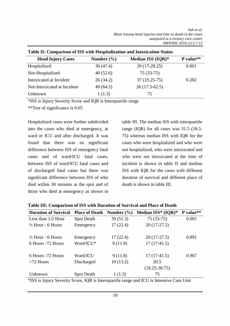


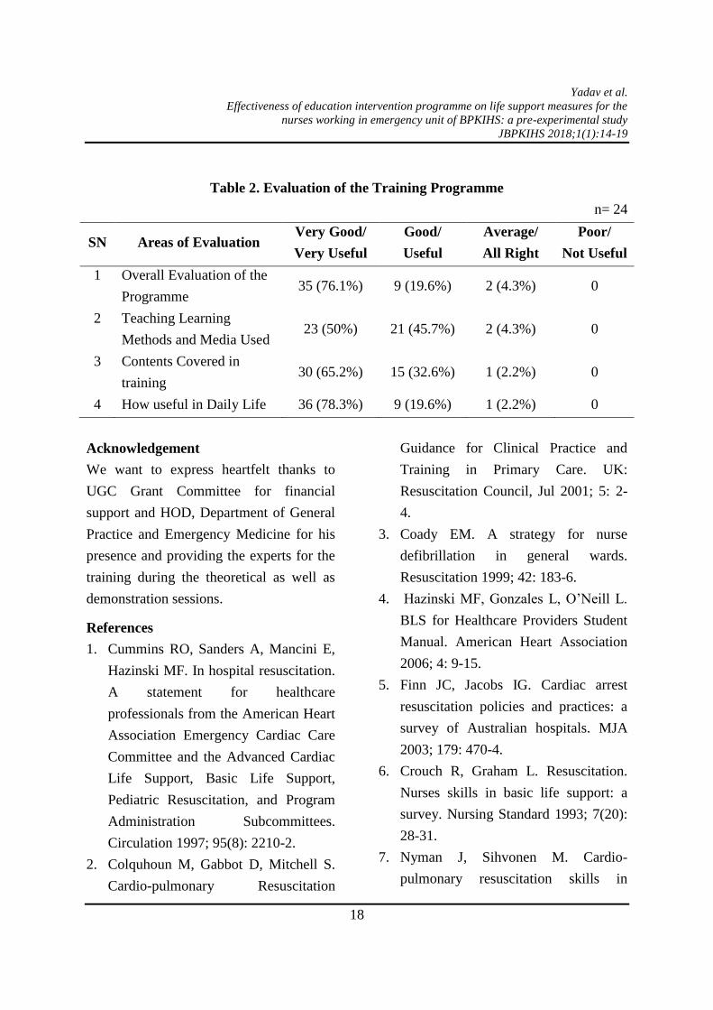


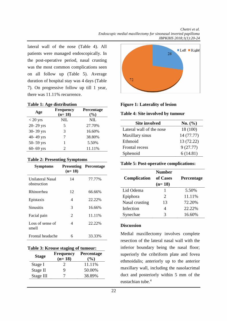

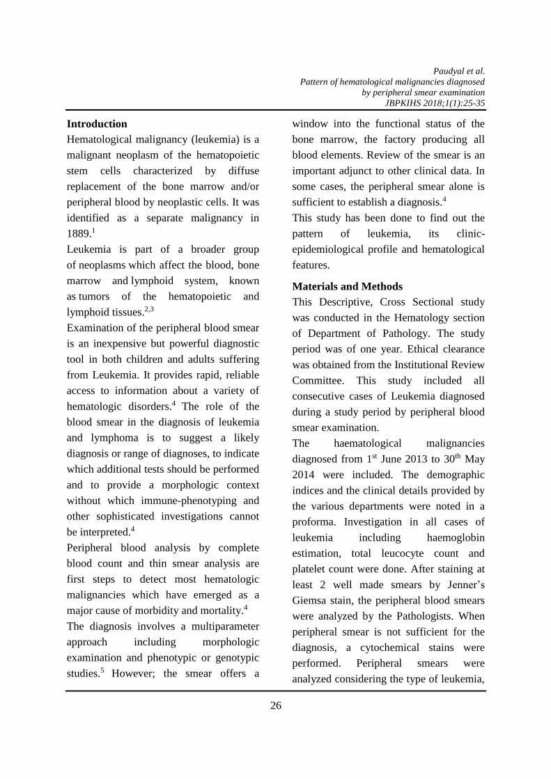
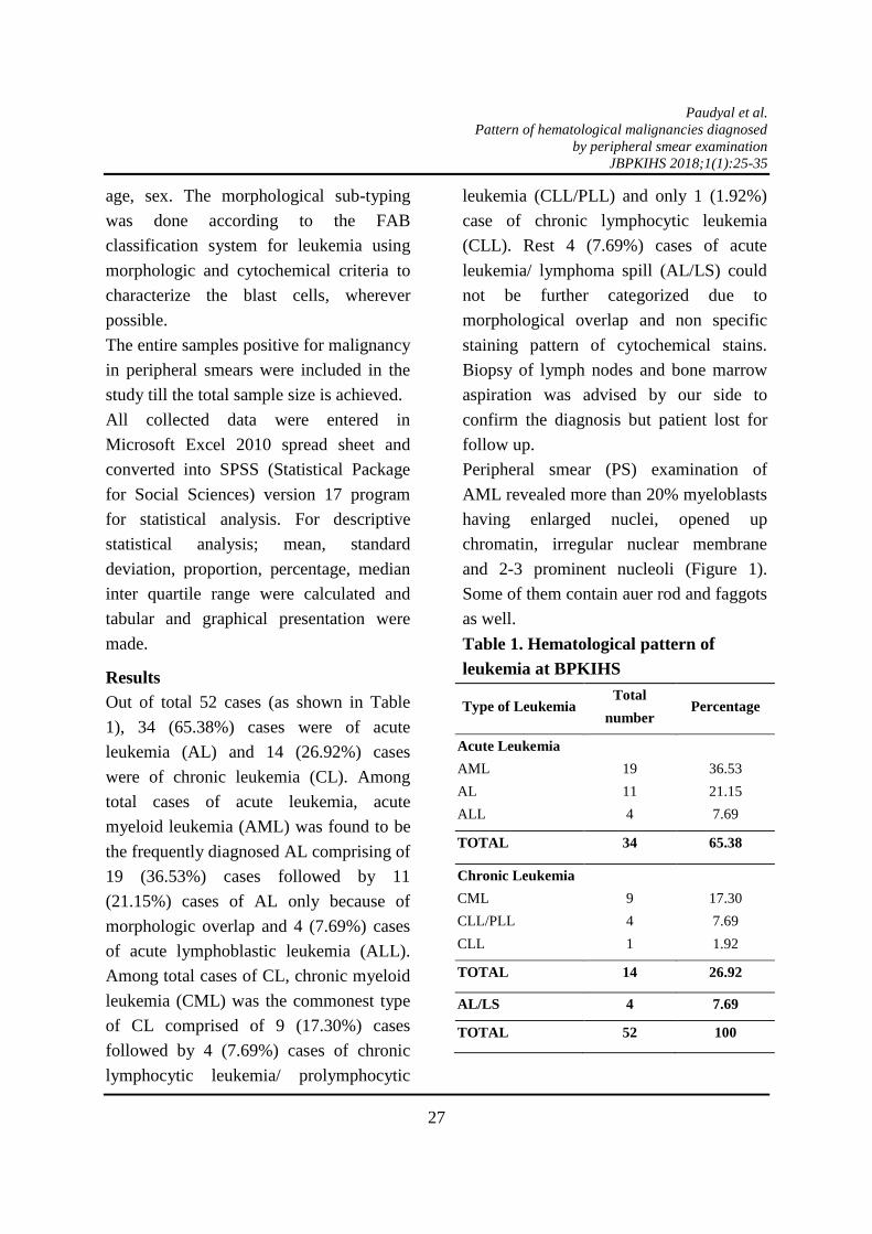
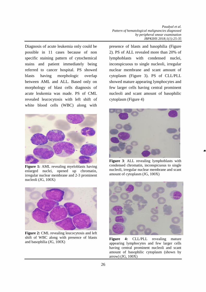

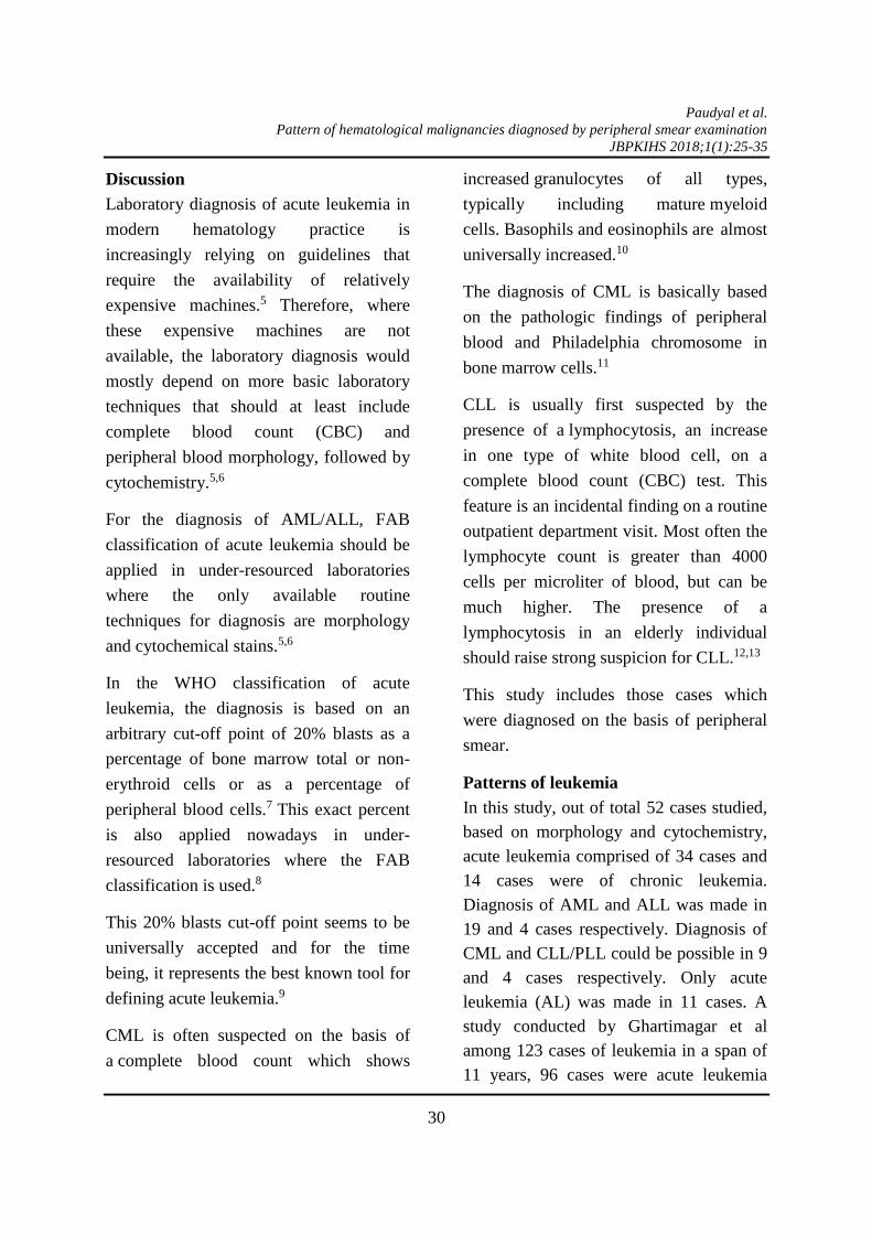


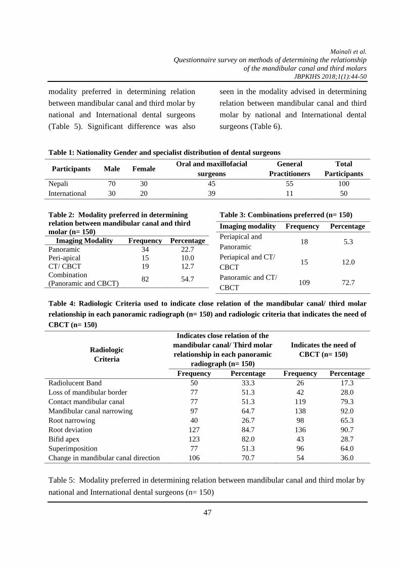
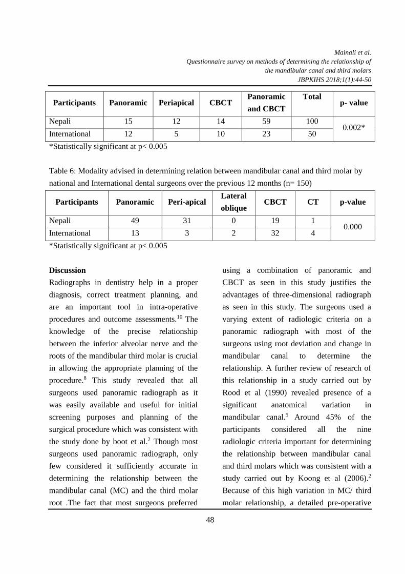


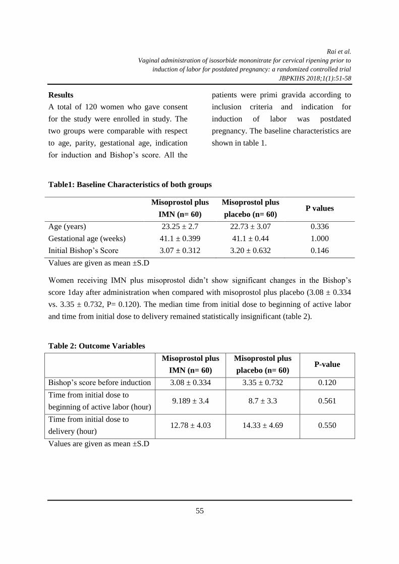


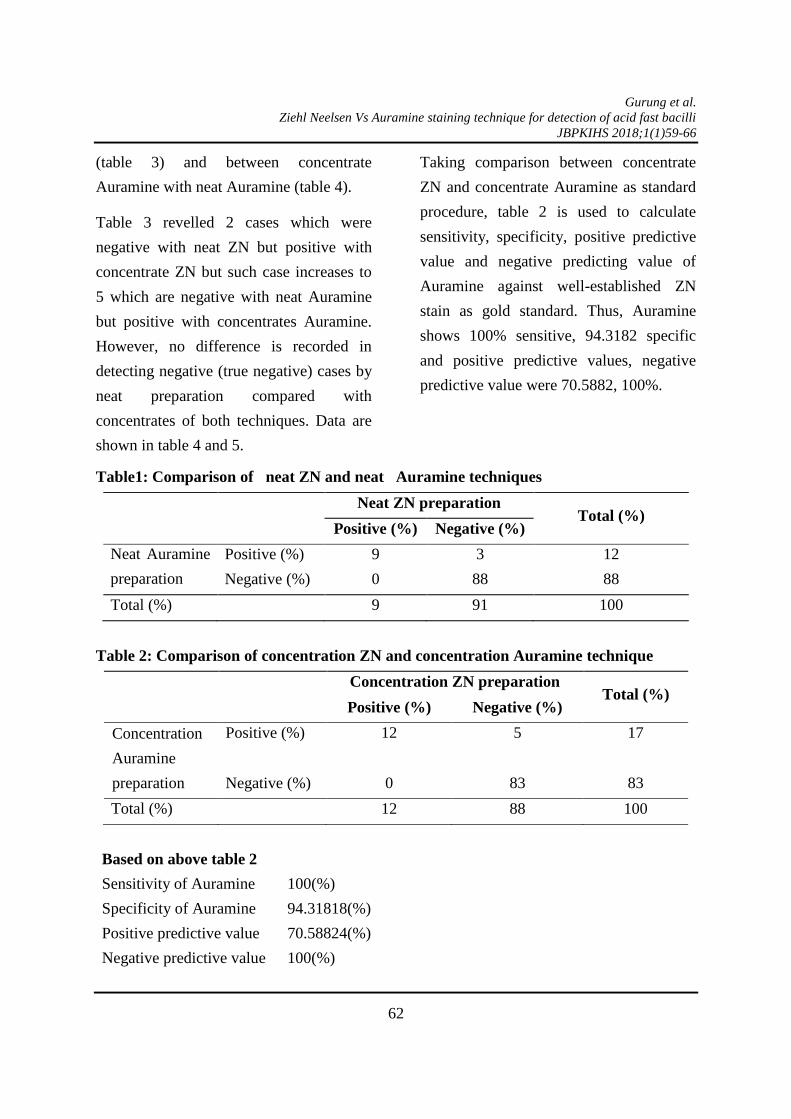
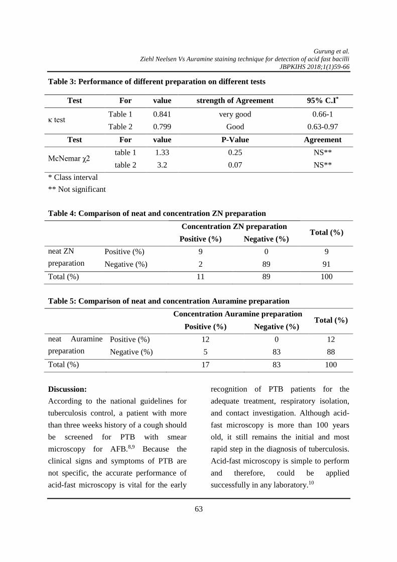


![Ling 403/603 Introduction to Phonology - udel.eduudel.edu/~koirala/phonology/day15.pdf · Introduction to Phonology. Morphophonemic Analysis ... changes to [u] in accusative forms.](https://static.documents.pub/doc/80x56/5aa788897f8b9a294b8c3994/ling-403603-introduction-to-phonology-udel-koiralaphonologyday15pdfintroduction.jpg)


