48
24.04.2017 confidential - for internal use only 1

24.04.2017 confidential - for internal use only 1

224.04.2017
Corneal Topography & Tomography
Introduction to measurement parameters and basic map interpretation,
and key factors for obtaining good quality measurements
Gregor Schmid, PhD
Senior Expert Clinical Applications (R&D)
Ziemer Ophthalmic Systems AG, Switzerland

324.04.2017
Topography & Tomography

Key Principles
Dual-Scheimpflug
Tomography
Placido Disk
Topography
24.04.2017 4
+

Scheimpflug Tomography
24.04.2017 5
Scheimpflug Principle
Object Plane
Image Plane
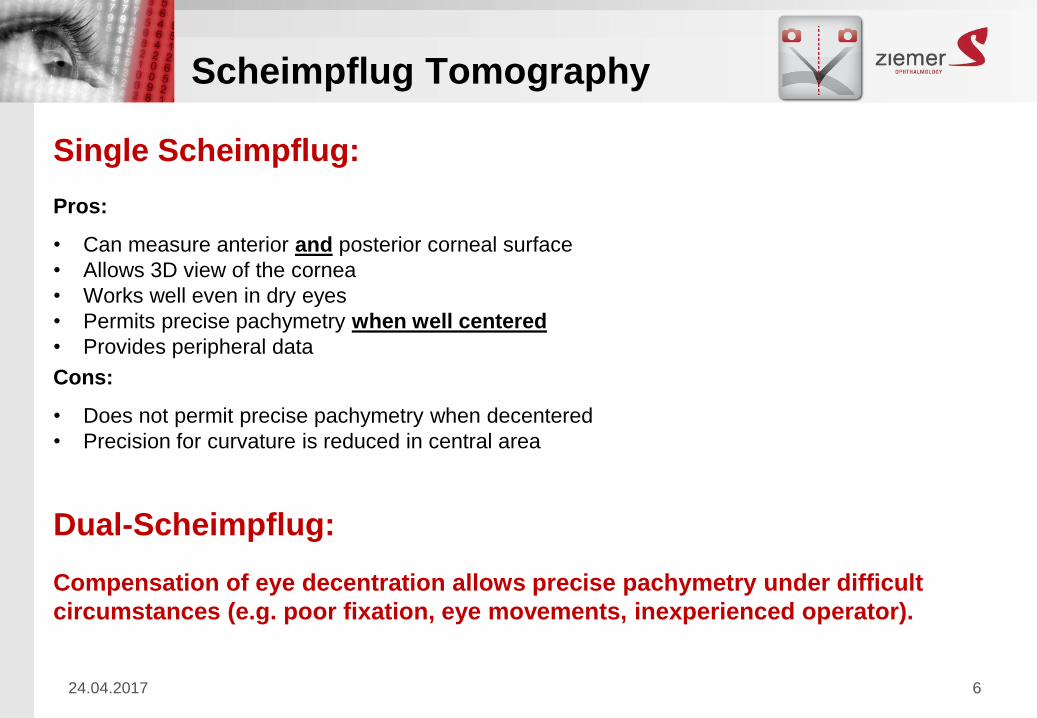
Single Scheimpflug:
Pros:
• Can measure anterior and posterior corneal surface
• Allows 3D view of the cornea
• Works well even in dry eyes
• Permits precise pachymetry when well centered
• Provides peripheral data
Cons:
• Does not permit precise pachymetry when decentered
• Precision for curvature is reduced in central area
Dual-Scheimpflug:
Compensation of eye decentration allows precise pachymetry under difficult
circumstances (e.g. poor fixation, eye movements, inexperienced operator).
24.04.2017 6
Scheimpflug Tomography

• When centered, the slit light is perpendicular to the surface
• Apparent thickness with the right and left SF camera are equal
Apparent Corneal Thickness when centered
Slit Light
Projected
Apparent Thickness
Right SF cameraleft SF camera
24.04.2017
Dual-Scheimpflug Tomography

Right SF camera:
Thinner
• The slit light is not perpendicular to the surface
• Apparent thickness is thinner/thicker than at center
• Averaging automatically corrects de-centration
Apparent Corneal Thickness when de-centered
Left SF camera:
Thicker
Slit Light
Projected
24.04.2017
Dual-Scheimpflug Tomography
24.04.2017 9
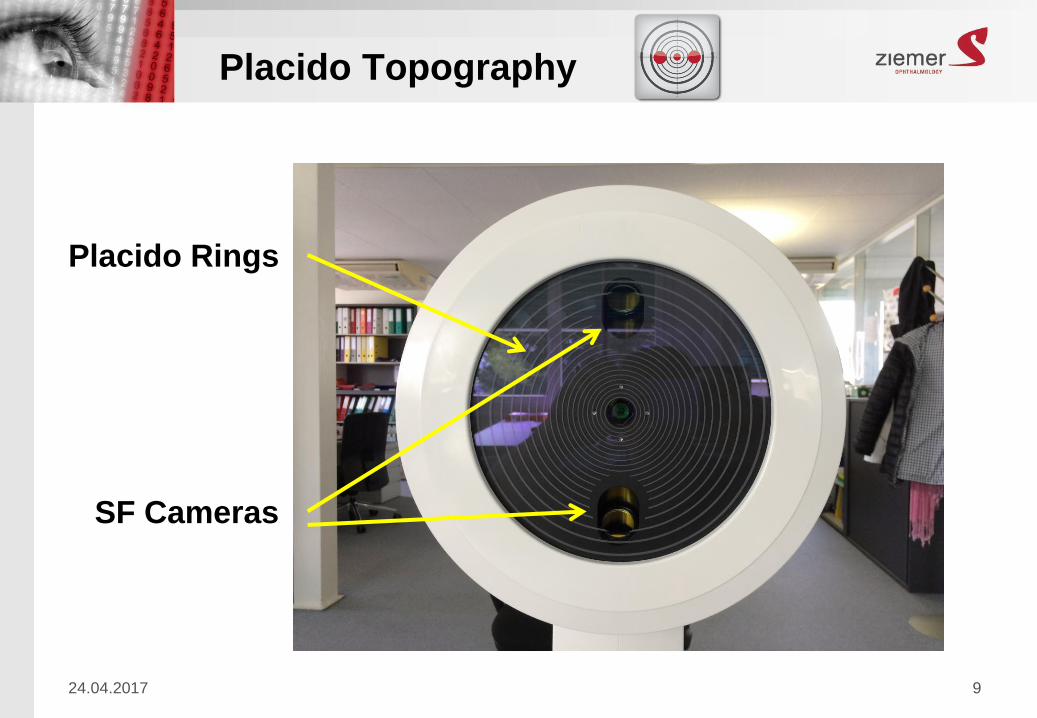
Placido Rings
SF Cameras
Placido Topography

24.04.2017 10
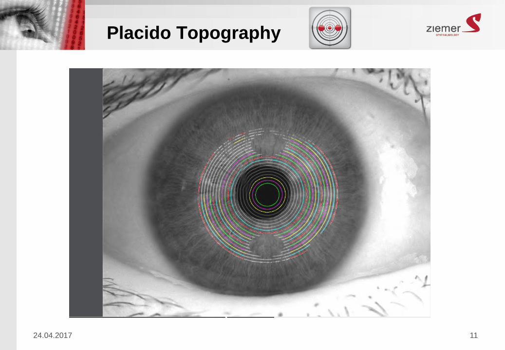
Placido Topography
Placido based topographers work on the principle of assessing the reflection of
a concentric set of white rings from the convex anterior surface of the cornea
24.04.2017 11
Placido Topography
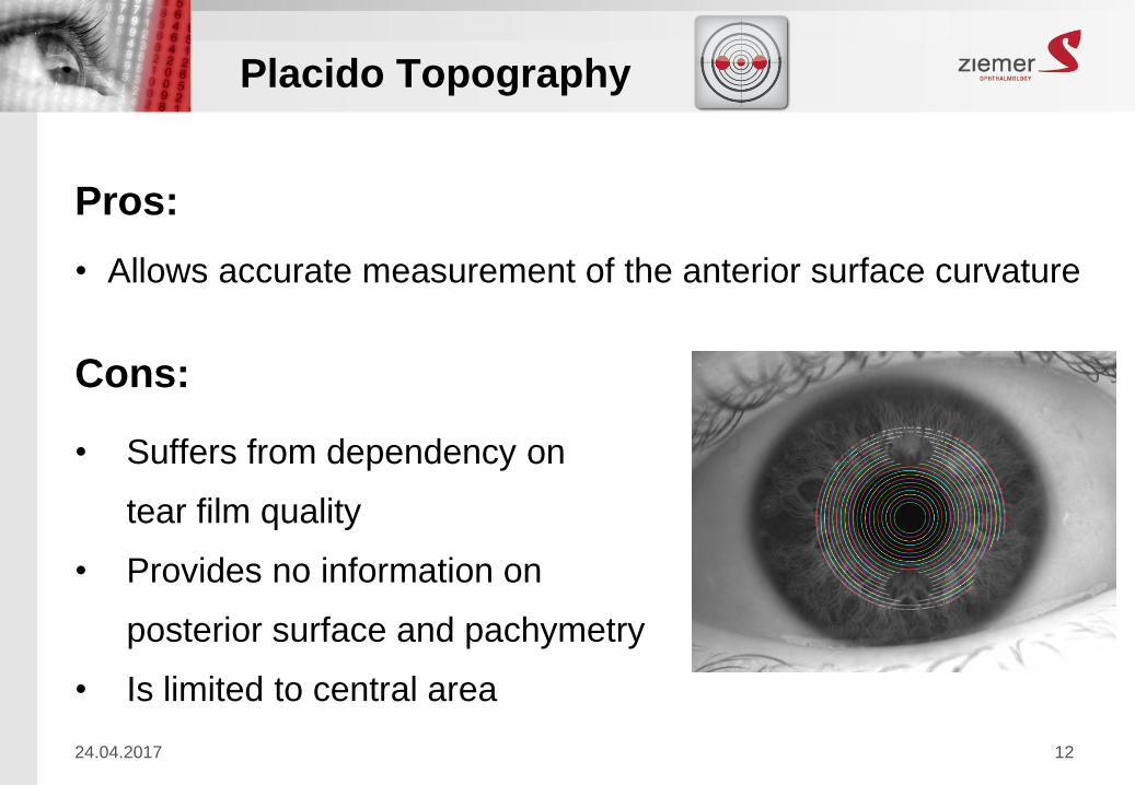
Pros:
• Allows accurate measurement of the anterior surface curvature
Cons:
• Suffers from dependency on
tear film quality
• Provides no information on
posterior surface and pachymetry
• Is limited to central area
24.04.2017 12
Placido Topography

Dual Scheimpflug + Placido
→ Combines the best of both worlds to produce
the most complete data set of the anterior
segment
24.04.2017 13

Maps and measurements aligned to apex (1st Purkinje
images)
Motion Compensation using iris pattern
a) Lateral motion correction (x/y-directions)
b) Rotational correction (around z-axis)
a) b)
a
Alignment & Motion Compensation
24.04.2017 14

24.04.2017 confidential - for internal use only 15
HOW TO GET
GOOD MEASUREMENTS

1. Alignment
24.04.2017 16
Left-Right/Up-Down In-Out

24.04.2017 17
Alignment: Purkinje Image 1
Visual Axis
P1
Visual Axis
Alignment to P1 ↔ Visual Axis Alignment to Apex ↔ ??
Apex

2. Tear Film
24.04.2017 18

3. Partial Lid Closure
24.04.2017 19

4. Eye Movement
24.04.2017 20
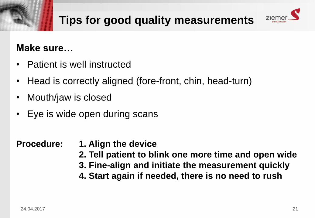
24.04.2017 21
Make sure…
• Patient is well instructed
• Head is correctly aligned (fore-front, chin, head-turn)
• Mouth/jaw is closed
• Eye is wide open during scans
Procedure: 1. Align the device
2. Tell patient to blink one more time and open wide
3. Fine-align and initiate the measurement quickly
4. Start again if needed, there is no need to rush
Tips for good quality measurements

Measurement Quality Values
24.04.2017 22

23
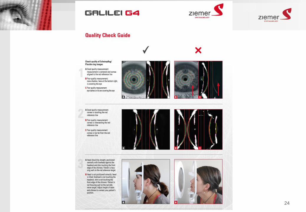
24

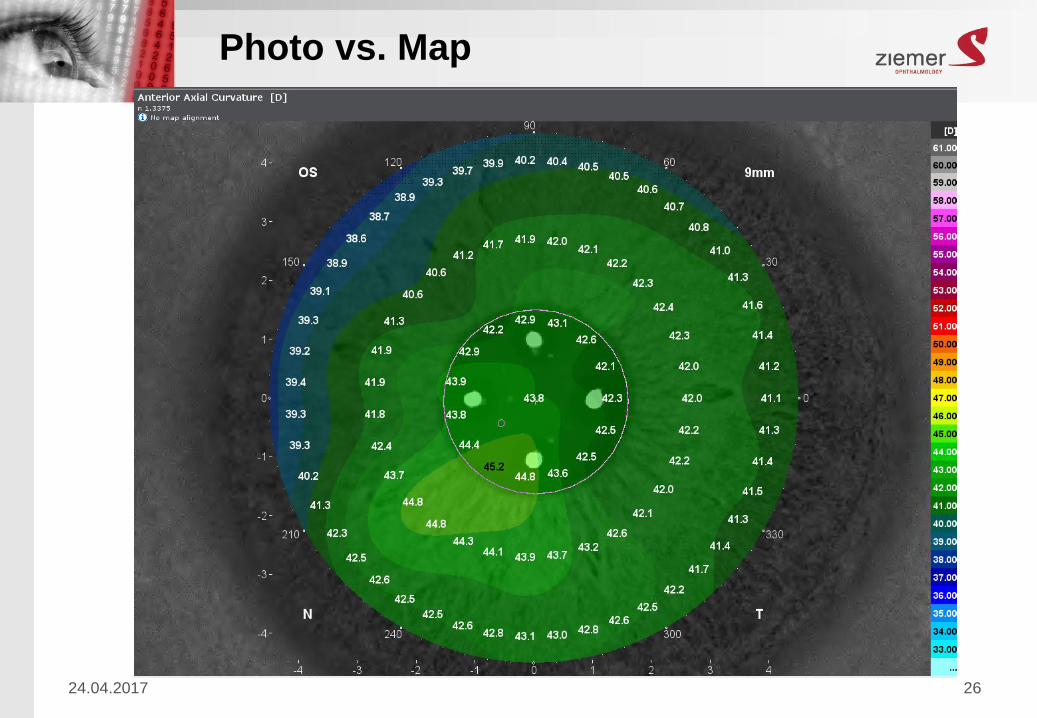
Photo vs. Map
24.04.2017 25
24.04.2017 26
Photo vs. Map

Anterior Axial Curvature
24.04.2017 27

24.04.2017 28
WTR Astigmatism

24.04.2017 29
ATR Astigmatism

24.04.2017 30
Oblique Astigmatism
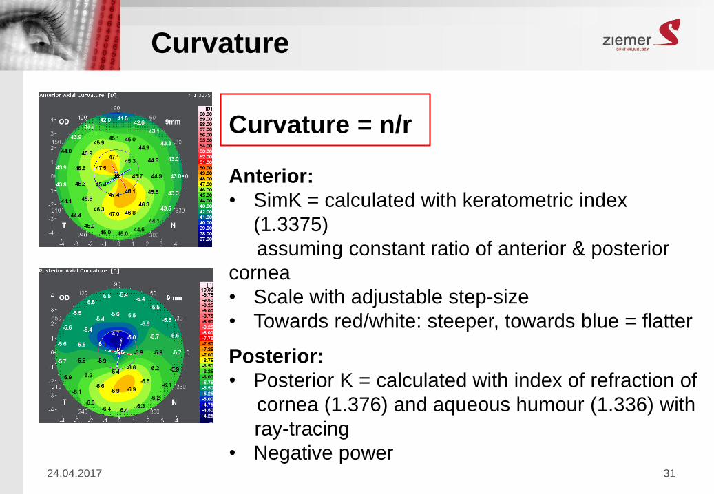
Curvature
24.04.2017 31
Curvature = n/r
Anterior:
• SimK = calculated with keratometric index
(1.3375)
assuming constant ratio of anterior & posterior
cornea
• Scale with adjustable step-size
• Towards red/white: steeper, towards blue = flatter
Posterior:
• Posterior K = calculated with index of refraction of
cornea (1.376) and aqueous humour (1.336) with
ray-tracing
• Negative power

Curvature
24.04.2017 32
• Instantaneous Curvature = local representation of curvature (C1, C2)
• Axial Curvature = smoothened curvature (radius extended to
reference axis: A1, A2), making steep areas flatter, and flat areas steeper

Axial Curvature
24.04.2017 33

Instantaneous Curvature
24.04.2017 34

24.04.2017 35
Instantaneous Curvature: mm

24.04.2017 36
Instantaneous Curvature: mm

Refractive Power
24.04.2017 37
• Calculated by ray-tracing through the anterior
corneal surface
Power = n/f
• f = focal length, n = refractive index
• Focal length is determined as the distance from the
reference plane to the intersection of the ray with
the central axis
• To determine the focal length the reference plane is
the anterior corneal suface in this case
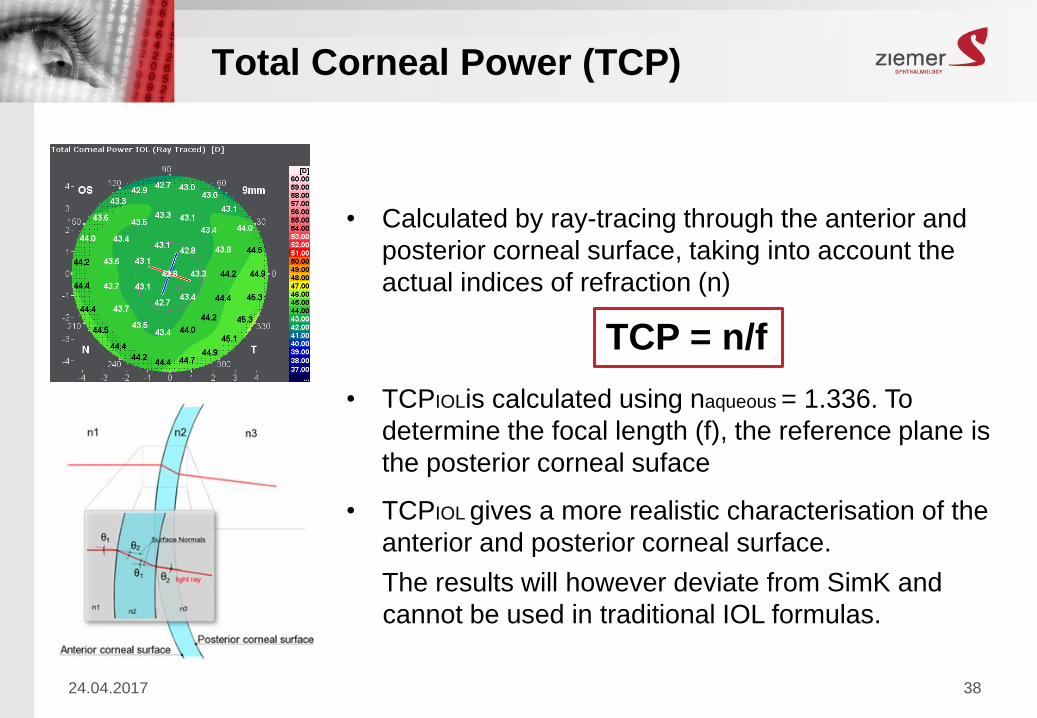
Total Corneal Power (TCP)
24.04.2017 38
• Calculated by ray-tracing through the anterior and
posterior corneal surface, taking into account the
actual indices of refraction (n)
TCP = n/f
• TCPIOLis calculated using naqueous = 1.336. To
determine the focal length (f), the reference plane is
the posterior corneal suface
• TCPIOL gives a more realistic characterisation of the
anterior and posterior corneal surface.
The results will however deviate from SimK and
cannot be used in traditional IOL formulas.

Pachymetry
24.04.2017 39
• Shows corneal thickness profiles in
20 µm steps
• Towards red/white: thinning, towards
blue: thickening
• Thinnest point = indicated by a small
circle
• CCT = central corneal thickness;
corresponds to central value of the
map

Elevation
24.04.2017 40
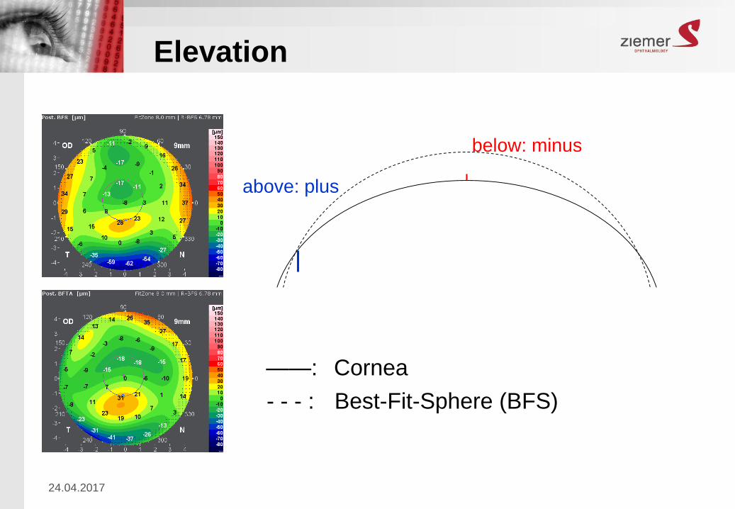
• Elevation requires a reference (plane, sphere,
asphere,…). GALILEI: BFS, BFA, BFTA
• Example: Elevation of a mountain
Mountain Top: 2128m
Sea Level: 0m
Elevation
24.04.201741
- - - : Best-Fit-Sphere (BFS)
——: Cornea
below: minus
above: plus

Corneal Shape Asymmetries
24.04.2017 42

Total Corneal Wavefront
24.04.2017 43
Corneal Wavefront Aberrations:
• Path length differences between the actual
wavefront and a plane wavefront at the entrance
pupil, normally expressed in µm
• Most common aberrations:
spherical aberration, astigmatism, coma, defocus
• Spherical Aberration: occurs when light experience
stronger refractive power at the periphery of the
cornea, resulting in a region of defocused light and
decreased image quality.

44 /13
Comparison

Eye Metrics
24.04.2017 45

Densitometry
24.04.2017 46

Clinical Benefits GALILEI
• Reliable and fast topography and
tomography screening
• Highly accurate anterior and
posterior curvature assessment for
sensitive keratoconus screening
• Maps and data aligned to the same
reference – the visual axis
• Spherical and aspherical
aberrations for wave front guided
treatments and toric IOL selection24.04.2017 47

24.04.2017 48
Dual Scheimpflug Analyzer


