25th Anniversary Article: Interfacing Nanoparticles and Biology: New Strategies for Biomedicine
Gulen Yesilbag Tonga , Krishnendu Saha , and Vincent M. Rotello*
G. Y. Tonga, K. Saha, Prof. V. M. RotelloDepartment of Chemistry University of Massachusetts Amherst 710 North Pleasant Street , Amherst Massachusetts , 01003 , USAE-mail: [email protected]
DOI: 10.1002/adma.201303001
1 . Introduction
Nanoparticles (NPs) provide platforms for crucial biomedical applications including delivery [ 1 ] and imaging. [ 2 ] Advances in these biomedical applications require a fundamental under-standing of the complex interactions between NPs, biomol-ecules, and biosystems. [ 3 ] Using this insight, the tools of chemical synthesis can be used to create nanomaterials that interact effi ciently and predictably with biosystems including proteins, [ 4 ] nucleic acids, [ 5 ] cells, [ 6 ] and tissues. [ 7 ]
In this review, we highlight selected systems that illustrate how the chemical structure of the NP surface functionality can be used to tune the interactions of particles with proteins and regulate cell uptake and distribution in tissues. [ 8 ] We also show how the surface can contribute functional attributes to the NPs, including targeting modalities [ 9 ] and the ability to respond to stimuli. [ 10 ]
2 . Interactions of Proteins with Surface-Engineered Nanoparticles
2.1 . Control of Protein Structure and Function Using Nanoparticle Receptors
Selective binding of artifi cial receptors to proteins provides a means to modulate a variety of biomedically important cel-lular processes including protein-protein interactions, protein-nucleic acid interactions, and enzyme activity. [ 11 ] The nature of the interaction of NPs and proteins is an inherently biophysical
question that builds upon the fundamen-tals of supramolecular chemistry. [ 12 ] In early studies, Rotello and coworkers dem-onstrated that 11-mercaptoundecanoic acid (MUA) functionalized gold NPs irre-versibly inhibited the activity of chymot-rypsin (ChT) with an apparent K i of 10 nM ( Figure 1 a). [ 13 ] The mechanism of this interaction was attributed to a two-step process featuring a fast reversible asso-ciation followed by a slower irreversible
denaturation, and could be readily tuned by the ionic strength of the aqueous media [ 14 ] and addition of surfactants. [ 15 ]
One inherent limitation of the use of simple ligands such as MUA is that protein structure and function were lost during the binding process. Preservation of the structure of NP sur-face-bound proteins is an essential prerequisite for many prag-matic applications including in vivo protein delivery and in vitro enzyme stabilization. [ 16 ] Structural integrity of proteins upon NP binding was achieved by introducing oligo(ethylene glycol) (OEG) tethers onto NP surfaces to provide TEG-OH and TCOOH (Figure 1 a). [ 17 ] The incorporation of a short (at least four repeat units) [ 18 ] OEG segment on the exterior of MUA pro-vided a non interacting ‘tabula rasa’ of NPs that could be further functionalized with particular headgroups, allowing tailoring of the NP surface (Figure 1 a). The stability of NP-ChT complexes was further investigated using gold NP featuring amino-acid side chains. [ 19 ] Binding studies using these NPs demonstrated that both electrostatic and hydrophobic interactions play a major role in the NP-ChT complex stability. Interestingly, the hydrophobic amino acid functionalized NPs did not affect the protein structure; however, hydrophilic amino acid functional-ized NPs destabilized the native structure of ChT. This fi nding contradicts the general belief that hydrophobic surfaces lead to denaturation of proteins. [ 20 ] In a later study, detailed thermody-namic investigations of the interaction of protein and amino acid functionalized NPs demonstrated that the enthalpy and entropy changes for these interactions strongly mimic protein-protein interactions. [ 21 ]
Protein unfolding on NP surfaces can cause unwanted bio-logical responses. For example, cryptic epitopes of proteins can be exposed that can further interact with cell membrane recep-tors, leading to aberrant infl ammatory responses. Minchin et al. have shown that poly(acrylic acid)-coated gold NPs can selec-tively interact with fi brinogen in human plasma and expose an amino acid sequence of fi brinogen γ -chain, promoting the spe-cifi c interaction with Mac-1 receptors on the cell surface. This interaction further leads to release of infl ammatory cytokines from human monocyte cells via NF- κ B signaling pathway. [ 22 ] However, appending appropriate functionality can improve
The exterior surface of nanoparticles (NPs) dictates the behavior of these systems with the outside world. Understanding the interactions of the NP surface functionality with biosystems enables the design and fabrication of effective platforms for therapeutics, diagnostics, and imaging agents. In this review, we highlight the role of chemistry in the engineering of nanomaterials, focusing on the fundamental role played by surface chemistry in controlling the interaction of NPs with proteins and cells.
the protein stability (vide supra), and even help in refolding the denatured proteins. Rotello et al. have demonstrated that 2-(10-mercaptodecyl) malonic acid functionalized gold NPs (AuDA) can refold thermally denatured cationic proteins, acting as artifi cial chaperones. The partially refolded proteins can be released from NP surfaces by changing the ionic strength of the solution (Figure 1 b). [ 23 ]
2.2 . NP Surface Chemistry Regulates the Affi nity and Identity of the ‘Protein Corona’
When surface-engineered NPs are exposed to complex protein biofl uids (e.g., blood, plasma) a protein “corona” forms on the particle surface, masking its original chemical nature. [ 24 ] This non-specifi c adsorption of biomolecules on NP surfaces is important for delivery applications, increasing the clearance rate of NPs via the reticulo-endothelial system and decreasing the pharmacokinetic half-life of NPs in vivo. Blood plasma con-tains thousands of proteins that can bind to NPs with the inter-action profi le strongly depending on the NP surface chemistry.
Gulen Yesilbag Tonga received her B.Sc. and M.Sc. in Chemistry from Bogazici University, Turkey in 2008 and in 2010. Presently, she is a Ph.D. candidate at the Department of Chemistry, University of Massachusetts at Amherst, USA under the guidance of Professor Vincent M. Rotello. Her cur-rent research is focused on
the use of supramolecular chemistry for delivery applica-tions of nanoparticles.
Krishnendu Saha received his B.Sc. in Chemistry from Jadavpur University, India in 2006 and M.Sc. in Chemistry from Indian Institute of Technology-Madras, India in 2008. Currently, he is a Ph.D. candidate at the Department of Chemistry, University of Massachusetts at Amherst, USA under the guidance of Professor Vincent M. Rotello.
His current research interest involves understanding the effect of nanoparticle surface functionality for cellular rec-ognition and delivery applications.
Vincent Rotello is the Charles A. Goessmann Professor of Chemistry at the University of Massachusetts at Amherst. He is a Fellow of the American Association for the Advancement of Science (AAAS) and of the Royal Society of Chemistry (UK). His research program focuses on using synthetic organic chemistry to engineer the interface between hard and
soft materials, and spans the areas of devices, polymers, and nanotechnology/bionanotechnology, with over 380 papers published to date. He is actively involved in the development of new nanomanufacturing methods. In the area of bionanotechnology, his research includes programs in delivery, imaging, diagnostics and nanotoxicology.
For example, positively charged polystyrene particles were shown to interact with proteins with isoelectric points less than 5.5 (e.g., serum albumin) while negatively charged particles
Figure 1. (a) Schematic representation of the effect of MUA, TEG-OH and TCOOH functionalized NPs on the structure and function of a cati-onic protein, chymotrypsin. (b) Schematic representation of the structure of AuDA, and AuDA-mediated refolding of thermally denatured proteins. This image is based on information obtained in the literature. Ref. [ 23 ] .
2.3 . Engineering NP Surface to Prevent Protein Adsorption
Protein corona formation masks functionality and decreases circulatory lifetime of NPs. Grafting hydrophilic polymers on NP surfaces is a widely employed strategy to reduce non-specifi c protein adsorption. For example, polyethylene glycol (PEG), a charge-neutral highly hydrophilic polymer, is often incorporated on NP surfaces for in vitro and in vivo applica-tions. [ 32 ] The advantage of NP surface modifi cation with PEG is two-fold: 1) PEG delays the opsonization process, thereby reducing NP uptake in macrophage cells; 2) the improved circulation allows NPs to preferentially accumulate in tumor microenvironment exploiting enhanced permeability and reten-tion effect. [ 33 ] Even though widely used for in vivo applications, PEG has several drawbacks. Chan et al. have shown that gold NPs with increasing surface PEG densities adsorb less serum proteins; however, even at high PEG grafting density the pro-teins were not fully removed. [ 34 ] Moreover, the different PEG densities infl uence the identity of the protein corona formed and subsequent macrophage uptake. Kiwada et al. have dem-onstrated siRNA encapsulated PEG-modifi ed lipid nanocarriers can produce antibodies against them; therefore, subsequent doses of the same PEGylated NPs were rapidly cleared in vivo. [ 35 ] PEG can also create unwanted immune responses. Moghimi et al. have demonstrated activation of complement system by NPs functionalized with PEG, [ 36 ] harnessing adverse immune responses and facilitating clearance by macrophages.
Recently, zwitterionic ligands including carboxybetaines and sulfobetaines have been introduced as ‘anti-fouling' coat-ings on NP surfaces. [ 37 ] These functionalities provide much lower adsorption of proteins compared to PEG and very low non-specifi c cellular uptake, providing a potential alternative to PEG ( Figure 3 a). [ 38 ] Jiang et al. have demonstrated that poly (carboxybetaine)- functionalized gold NPs were highly stable in undiluted blood plasma and serum while PEG-coated NPs aggregated. [ 39 ] Likewise, Mattoussi and coworkers have synthe-sized zwitterionic quantum dots (QDs) that showed superior colloidal stability compared to simple lipoic acid functionalized
preferentially bind to proteins with isoelectric points higher than 5.5 (e.g., IgG). [ 25 ] Müller et al. have further shown that negatively charged polymeric NPs of similar size and hydropho-bicity adsorb higher amount of proteins on their surfaces with increasing surface charge density; however, no apparent differ-ence was observed in the identity of the adsorbed proteins. [ 26 ]
The hydrophobicity of the NP surface is also an impor-tant parameter in NP-protein interactions. [ 27 ] For example, hydrophobic N -isopropylacrylamide-co- N -tert-butylacrylamide (NIPAM/BAM) copolymer NPs adsorb a greater amount of pro-tein on their surfaces than their hydrophilic counterparts, [ 28 ] due to higher protein affi nity and/or increased number of binding sites on hydrophobic NP surfaces. [ 29 ] NP surface hydrophobicity also regulates the nature of the protein corona formed on the surface. For example, hydrophobic NPs adsorb apolipoproteins on their surface; however, hydrophilic NPs tend to adsorb more abundant hydrophilic proteins in plasma including albumin and IgG. [ 30 ]
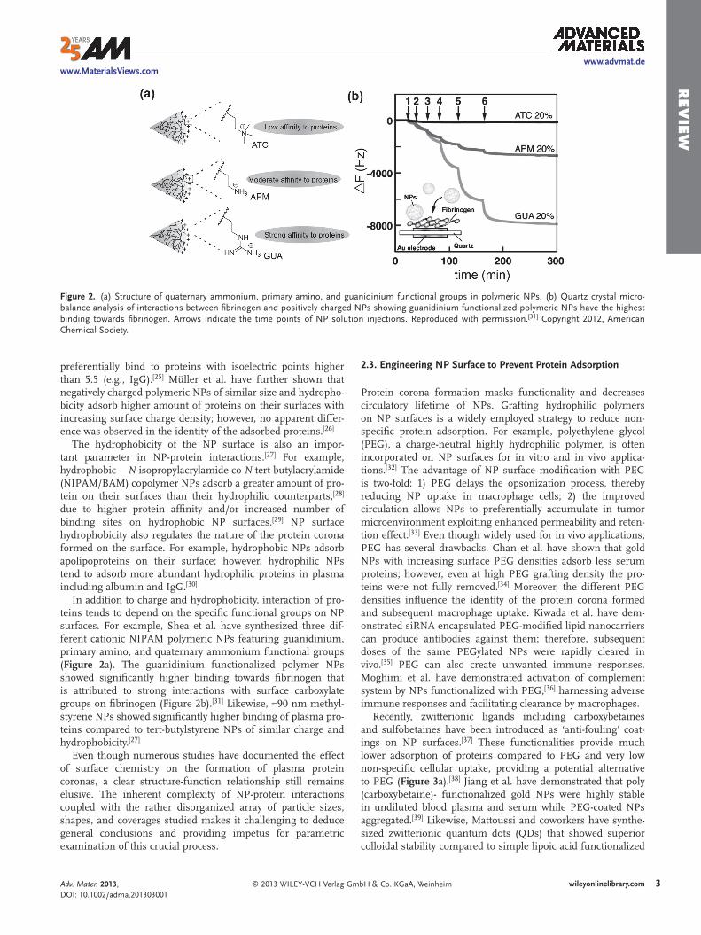
In addition to charge and hydrophobicity, interaction of pro-teins tends to depend on the specifi c functional groups on NP surfaces. For example, Shea et al. have synthesized three dif-ferent cationic NIPAM polymeric NPs featuring guanidinium, primary amino, and quaternary ammonium functional groups ( Figure 2 a). The guanidinium functionalized polymer NPs showed signifi cantly higher binding towards fi brinogen that is attributed to strong interactions with surface carboxylate groups on fi brinogen (Figure 2 b). [ 31 ] Likewise, ≈90 nm methyl-styrene NPs showed signifi cantly higher binding of plasma pro-teins compared to tert-butylstyrene NPs of similar charge and hydrophobicity. [ 27 ]
Even though numerous studies have documented the effect of surface chemistry on the formation of plasma protein coronas, a clear structure-function relationship still remains elusive. The inherent complexity of NP-protein interactions coupled with the rather disorganized array of particle sizes, shapes, and coverages studied makes it challenging to deduce general conclusions and providing impetus for parametric examination of this crucial process.
Figure 2. (a) Structure of quaternary ammonium, primary amino, and guanidinium functional groups in polymeric NPs. (b) Quartz crystal micro-balance analysis of interactions between fi brinogen and positively charged NPs showing guanidinium functionalized polymeric NPs have the highest binding towards fi brinogen. Arrows indicate the time points of NP solution injections. Reproduced with permission. [ 31 ] Copyright 2012, American Chemical Society.
coated with peptide motifs that penetrate the cell membrane and can facilitate endosomal escape into the cytosol. Cell penetrating pep-tides (CPPs) including RGD, [ 44 ] allatostatin 1, [ 45 ] PLL, [ 46 ] and arginine-rich peptides [ 47 ] have been conjugated onto NP surfaces for effi cient cellular delivery. Brust et al. have demonstrated cytosolic delivery of CPP con-jugated gold NPs; addition of a nuclear local-izing sequence (NLS) peptide resulted in localization to the nucleus. [ 48 ] In a similar study, gold NPs featuring NLS can penetrate to the nucleus after endosomal escape and induce DNA damage in cancer cells, demon-strating potential therapeutic strategy using surface-engineered NPs. [ 49 ] Gillies et al. have shown that superparamagnetic iron oxide NPs featuring dendritic guanidine moieties on their surface have similar cell-penetration effi ciency with immunodefi ciency virus-1 transactivator (HIV-TAT) peptide and showed better penetration effi ciency than amine functionalized NPs. [ 50 ]
3.2 . Effect of NP Surface Charge and Hydrophobicity on Cellular Uptake
Engineering the NP surface provides a versatile alternative to CPPs for cellular delivery. The surface charge of the NP is a key parameter for NP-cell membrane inter-action and subsequent intracellular inter-nalization. In general, cationic NPs interact
more strongly with the cell membrane due to the presence of negatively charged groups (e.g., sialic acid) onto cellular mem-branes, and hence show higher uptake effi ciency compared to their anionic and neutral counterparts ( Figure 4 a–c). [ 51 ] However, effi cient intracellular uptake of negatively charged NPs is known, presumably through pinocytosis or membrane diffusion. [ 52 ] Moreover, negatively charged QDs were shown to penetrate mouse skin, with higher degree of penetration observed upon exposure to UV light. [ 53 ] Likewise, DNA func-tionalized anionic gold NPs were shown to penetrate in epi-dermis layer of mouse skin with no apparent infl ammation or toxicity. [ 54 ]
Two-dimensional cell culture models present a vastly dif-ferent environment than tissues, complicating translation into in vivo systems. Rotello and Forbes et al. have investigated the role of NP surface charge in delivering covalently attached therapeutic drugs using a three-dimensional cell culture model. Signifi cantly, cationic particles were better at delivering drugs on the proliferating peripheral cells due to their higher uptake; however, anionic particles had higher diffusion rates, delivering drug- and fl uorophore-tagged NPs more rapidly to the center of the spheroid model ( Figure 5 ). [ 55 ]
In addition to surface charge, NP hydrophobicity plays an important role in the cellular uptake process. In one
QDs over a broad range of conditions including pH, salt, and undiluted serum. [ 40 ] Using zwitterionic mixed monolayer gold NPs, Rotello et al. have shown intracellular delivery of thera-peutics while maintaining low uptake and minimal cytotoxicity from the NP itself (vide infra). [ 41 ] Mukherjee et al. have further demonstrated that zwitterionic gold NPs had higher blood cir-culation lifetime and enhanced tumor accumulation, while positively and negatively charged NPs were rapidly cleared. [ 42 ] However, the surface charge distribution of zwitterionic NPs can infl uence the uptake and biodistribution. Bawendi et al. have reported that zwitterionic QDs exposing positive charges in their outermost layer show non-specifi c adsorption in vitro and in vivo, whereas zwitterionic QDs exposing negative charges in their outermost layer are far less susceptible to inter-actions with proteins (Figure 3 b). [ 43 ]
3 . Interaction of Cells with Surface-Engineered Nanoparticles
3.1 . Use of Cell-Penetrating Peptides on NP Surfaces
Penetration of the cell membrane and subsequent access of NPs to cytosol are two major hurdles for delivery of therapeutics.
Figure 3. (a) Illustration of protein adsorption on the surface of zwitterionic and PEGylated NPs. (b) Top: illustration of expected charge distribution on QDs. Bottom: non-specifi c binding of QDs with different charge distribution to HeLa cells. Reproduced with permission. [ 43 ] Copy-right 2013, WILEY-VCH Verlag GmbH & Co. KGaA, Weinheim.
pH, enzyme) or external stimulus (e.g., magnetic fi eld, light) can be used to enhance the therapeutic effi ciency of delivery vehicles. [ 58 ]
4 . 1. External Stimuli
4 . 1. 1. Light-Induced Release of Cargo
Light provides an excellent orthogonal stimulus for nanocar-riers, providing spatio-temporal control of delivery. For example, Tamanoi and Zink et al. have reported the intracellular delivery and release of the anticancer drug camptothecin using nano-impeller-controlled mesostructured silica nanoparticles that are functionalized with photo-switchable azobenzene moieties positioned in the pore interiors. [ 59 ] Lin et al. have presented a supramolecular assembly for visible light sensitive release of cargo from mesoporous silica nanoparticles (MS NPs) using Ru(bpy) 2 (PPh 3 ) moieties coordinated to mercaptopropyl func-tional groups as gatekeepers. [ 60 ] Upon irradiation with visible light, the Ru–S coordination bond is cleaved and encapsulated
study, Mailänder et al. have showed that increasing surface hydrophobicity increases intracellular uptake of polymeric NPs in a variety of cell lines. [ 56 ] Rotello et al. have demonstrated a linear correlation of surface hydrophobicity and intracellular uptake of gold NPs (2 nm core), stemming from a stronger interaction of hydrophobic NPs with serum albumin. [ 57 ] How-ever, in the absence of serum, no apparent trend between sur-face hydrophobicity and cellular uptake was observed, demon-strating the importance of serum proteins on the NP uptake process (vide supra).
4 . Design of Stimuli-Responsive Nanocarriers by Tailoring Surface Functionality
In addition to regulating interactions with the environment, nanoparticle surfaces can be used to impart functionality to nanoparticles. “Smart” surface functionality provides a strategy for creating delivery systems that can release drugs at target site, minimizing potential off-target issues. Stimuli-respon-sive nanocarriers that respond to either a physiological (e.g.,
Figure 4. Schematic representation of the interaction between gold NPs bearing different surface charge and SK-BR-3 breast cancer cells. (a) Citrate-coated (negative), (b) polyvinylalcohol-coated (neutral), and (c) poly(allyamine hydrochloride)–coated (positive) NPs. This image is based on informa-tion obtained in the literature. [ 51a ]
containing hydrogels to release biomacro-molecules on demand. [ 62 ]
Recently, Xing et al. have demonstrated the use of UCNPs to uncage D-luciferin molecules caged with 1-(2-nitrophenyl)ethyl group in vitro and in vivo ( Figure 8 a). [ 63 ] The absorption band of the photocaged D-lucif-erin overlaps with the emission band of UCNPs in the UV region; thus disassociation of D-luciferin from the surface of the nano-particle is triggered by excitation of UCNPs with NIR light. Released D-luciferin can rec-ognize fi refl y luciferase (fLuc) reporter genes and generate a bioluminescence signal which is an indication of successful release of the cargo from the carrier (Figure 8 b,c). [ 64 ] Their photocaged system has potential to selec-tively deliver payload in vivo with deep tissue penetration ability by NIR irradiation.
4.2 . Endogenous Stimulus for Triggering the Release of Cargo
4.2.1 . Non-Covalent Drug Delivery through Changes in Environmental Hydrophobicity
Endogenous release mechanisms use environmental changes in living systems to dictate their behavior. Non-covalent drug delivery systems using NPs can use either an encapsulation mechanism or a stabilizing pocket, [ 65 ] using electrostatic [ 66 ] and hydrophobic [ 67 ] interactions to reversibly bind the drug of interest. [ 68 ]
Burda et al. have demonstrated that encapsulating hydro-phobic drug Pc4 inside the monolayer of PEG functionalized gold NPs increases the drug accumulation in the tumor. Sig-nifi cantly, little to no accumulation was observed when Pc4 was covalently attached to the gold NP surface, demonstrating that reversible association is required for therapeutic activity. [ 69 ]
molecules are released. Furthermore, Rotello et al. have shown the UV light-triggered release of fl uorouracil from gold NP sur-face through a photoresponsive o -nitrobenzyl (ONB) linkage ( Figure 6 ). [ 41 ]
UV light has a very short tissue penetration depth (≈1 mm), making UV-activated systems unsuitable for deep tissue pen-etration. Branda et al. have addressed this issue by synthesizing NaYF 4 upconverting NPs (UCNPs) doped with the lanthanides, Tm 3+ and Yb 3+ [NaYF 4 :TmYb] with a UV light activated caging group (3 ′ ,5 ′ -di(carboxymethoxy)benzoin) ( Figure 7 a). [ 61 ] These UCNPs were used as antennae for harvesting the NIR (980 nm) light and converting it into UV light (290 nm) (Figure 7 a,b).
Figure 5. (a) Structure of mixed monolayer-protected cationic and anionic gold NPs loaded with thioalkylated fl uorescein isothiocyanate (FITC). Green fl uorescence images of tumor cylindroids treated with (b) cationic and (c) anionic particles. Reproduced with permission. [ 55 ] Copyright 2010, Nature Publishing Group.
Figure 6. (a) Photochemical reaction of Au_PCFU and delivery of fl uorouracil to cells. Bright-fi eld and fl uorescence-microscopy images of the cells exposed to UV before treated with Au_PCFU (b and c) and after treated with Au_PCFU (d and e). Reproduced with permission. [ 41 ] Copyright 2009, American Chemical Society.
for release at tumor sites. For example, Wang et al. have used carbamate linkage on a gold NP surface to release doxoru-bicin in response to the pH of acidic organelles after endo-cytosis. [ 75 ] They have further demonstrated that this delivery system can effectively inhibit the growth of multidrug-resistant MCF-7/ADR cancer cells, owing to the high endo-cytic uptake and subsequent pH responsive release in cells. Mirkin and Lippard et al. have combined the properties of DNA functionalized gold NPs and Pt(IV) prodrugs into a single agent for drug delivery. [ 76 ] They have engineered the surface of gold NPs to attach the prodrugs. They have demon-strated that the acidic environment in cancer cells facilitates reduction of the Pt(IV) and hence yields of the cytotoxic Pt(II) species ( Figure 10 ).
The acidic environment found in the endocytic compart-ments of cancer cells can also be used to release the drug molecules that are non-covalently attached to the nanoparticle
Rotello et al. have used hydrophobic pockets of gold NP mon-olayers to encapsulate highly hydrophobic dyes/therapeu-tics and release them upon interacting with cell membrane ( Figure 9 ). [ 70 ] The zwitterionic surface of NPs further provided biocompatibility to the NP carriers. Hydrophobic payloads (tamoxifen and lapachone) were shown to be released into the cell by membrane-mediated diffusion without the uptake of the carrier NPs, demonstrating the desired biocompatibility of the carrier.
4.2.2 . pH as an Internal Trigger to Release the Cargo
The average extracellular pH of solid tumors is 6 -7, lower than the pH of normal tissues and blood (7.4). [ 71 ] Sur-face functional groups that respond to acidic pH including acetal, [ 72 ] derivatized maleamate, [ 73 ] and hydrazone [ 74 ] have been used to attach the therapeutics to a variety of carriers
Figure 7. (a) Generation of UV light from NIR light using benzoin cage decorated UCNPs. (b) Changes in the UV/Vis absorption spectra of a solution of 1a[NaYF4:TmYb] when it is irradiated with 980 nm light. Reproduced with permission. [ 61 ] Copyright 2010, WILEY-VCH Verlag GmbH & Co. KGaA, Weinheim.
Figure 8. (a) Schematic illustration of photocaged UCNPs synthesis and release of D-luciferin upon NIR irradiation and bioluminescence through the use of photocaged core-shell upconversion NPs. (b-c) Bioluminescent images of fLuc activity in living mice (b) Left: injection with D-luciferin and right: injection with photocaged nanoparticles without NIR light irradiation. (c) Left: injection with photocaged nanoparticles and irradiation with UV light and right: injection with photocaged nanoparticles and irradiation with NIR light. Reproduced with permission. [ 63 ] Copyright 2012, WILEY-VCH Verlag GmbH & Co. KGaA, Weinheim.
IEW surface. For example, Haam et al. have reported
a pH-responsive drug delivering magnetic NPs utilizing non-covalent interactions ( Figure 11 ). [ 77 ] They have used a nanoemulsion method to syn-thesize MnFe 2 O 4 nanocrystals that are coated with α -pyrenyl- ω -carboxyl poly(ethylene glycol). Doxorubicin (DOX) molecules are loaded to the magnetic carrier via a strong π – π interac-tion between pyrene and DOX molecules. Upon intracellular uptake, protonation of DOX can decrease the π – π interaction, resulting in the release of DOX. [ 78 ] Receptor-mediated endo-cytosis was achieved by modifying surface of magnetic NPs with anti HER2/neu antibody, a tumor-targeting marker of the human HER2/ neu receptor of metastatic breast cancer.
In addition to cleaving the bond between drug and carrier, pH triggers can also be used to open molecular valves or lids, releasing encapsulated drugs. Zink et al. have loaded MS NPs with hydrophobic cargo and grafted pH-sensitive nanovalves to keep the cargo inside and release into the cells upon changes in pH ( Figure 12 ). [ 79 ] In another study, Feng et al. have reported the controlled release of guest molecules from MS NPs that are capped by acid-labile acetal group linked gold NPs. [ 80 ] At neutral pH, no diffusion from pores is observed as pores are strongly blocked by gold NPs. Hovewer, at acidic pHs, the gold capping agent can be removed due to hydrolysis of the pH-responsive acetal group, releasing the trapped molecules. Feng et al. have
Figure 9. (a) Delivery of payload to cell due to change in hydrophobicity through mono-layer-membrane interactions. (b) Schematic illustration of guest molecules entrapped in the hydrophobic pocket of zwitterionic NPs and structure of guest molecules: Bodipy, tamoxifen and lapachone. Confocal laser scanning microscopy images of MCF-7 cell treated with bodipy encapsulated zwitterionic NPs (c) green channel, (d) overlapped with bright fi eld. Reproduced with permission. [ 70 ] Copyright 2009, American Chemical Society.
Figure 10. (a) Schematic illustration of Pt-DNA-Au NP. (b-d) Live cell imaging of HeLa cells after incubation with platinum-tethered Cy5-DNA-Au NPs for (b) 6 h, (c) 12 h, and (d) colocalization of the particles with the cytoplasmic microtubules. Scale bars: 20 μ m. Reproduced with permission. [ 76 ] Copyright 2009, American Chemical Society.
the polymer becomes swollen and permeable to the encapsu-lated molecules after protonation.
4.2.3 . Enzyme-Responsive Nanoparticles for Delivery Applications
Enzyme-responsive NPs can be used to deliver cargos via enzymatic action at targeted locations. [ 82 ] Tuning the surface chemistry of nanoparticles by introducing bioactive moieties or enzyme cleavable linkers is the most common way followed to fabricate enzyme-responsive delivery systems. Stoddart et al. have designed a delivery vehicle with an enzyme-responsive snap-top motif, activated by porcine liver esterase (PLE). [ 83 ] In their system, MS NPs loaded with luminescent cargo mol-ecules (rhodamine B) were capped with the ester-linked ada-mantyl stopper. Hydrolysis of the adamantyl ester stopper by PLE resulted in the dethreading of the α -cyclodextrin and triggered the release of the rhodamine B from the pores. In another study, Akashi et al. have used hollow capsules pre-pared by MS NPs coated with chitosan and dextran sulfate for the delivery of the cargo. [ 84 ] Sustained release of the cargo was achieved through degradation of chitosan component by chitosanase.
Recently, Fukumura et al. have reported a multistage nano-particle-based delivery system with a deep tumor-penetration feature. [ 85 ] In this work, enzyme-degradable type A gelatin was crosslinked with glutaraldehyde, and PEG-stabilized quantum dots (QDs, ≈10 nm) were used to generate enzyme-degradable nanoparticles (≈100 nm). Due to the enhanced permeability and retention (EPR) effect, the enzyme-responsive nanoparticles preferentially extravasate from leaky regions of tumor vascula-ture. However, after extravasation into the tumor tissue, these nanoparticles can be degraded by MMP-2 and smaller 10 nm
also decorated MS NPs with poly(4-vinyl pyridine) (PVP) as a pH-sensitive capping nanoshell. [ 81 ] In their system, at low pH,
Figure 11. (a) Schematic illustration of anti HER2/neu antibody-modifi ed pH-sensitive drug-releasing magnetic NPs. (b) Magnetic resonance images of tumor-bearing mice after the intravenous injection of human epidermal growth factor receptor (HER) and irrelevant antibody (IRR) functionalized pH sensitive magnetic NPs at various time intervals, demonstrating HER modifi ed NPs accumulated in the tumor more than IRR modifi ed NPs. Reproduced with permission. [ 77 ] Copyright 2011, WILEY-VCH Verlag GmbH & Co. KGaA, Weinheim.
Figure 12. Top illustration: Synthesis of pH-sensitive nanovalve. Bottom illustration: Binding of the β -cyclodextrin ( β -CD) cap to neutral benzi-midazole stalk and releasing of β -CD and cargo from the carrier after protonation of the stalk. Reproduced with permission. [ 79 ] Copyright 2011, American Chemical Society.
IEW R. Hu , W. C. Law , H. Ding , C. W. Chang , P. N. Prasad , M. T. Swihart ,
ACS Nano , 2010 , 9 , 5131 . [3] a) W. J. Stark , Angew. Chem. Int. Ed. 2011 , 50 , 1242 ; b) D. F. Moyano ,
V. M. Rotello , Langmuir 2011 , 27 , 10376 . [4] a) K. Saha , A. Bajaj , B. Duncan , V. M. Rotello , Small 2011 , 7 , 1903 ;
b) V. Mirshafi ee , M. Mahmoudi , K. Lou , J. J. Cheng , M. L. Kraft , Chem. Commun. 2013 , 49 , 2557 .
[5] T. Pellegrino , S. Kudera , T. Liedl , A. M. Javier , L. Manna , W. J. Parak , Small 2005 , 1 , 48 .
[6] a) K. Saha , S. T. Kim , B. Yan , O. R. Miranda , F. S. Alfonso , D. Shlosman , V. M. Rotello , Small 2013 , 9 , 300 ; b) A. Verma , F. Stellacci , Small 2010 , 6 , 12 ; c) Y. K. Gong , F. M. Winnik , Nanoscale 2012 , 4 , 360 .
[7] a) S. T. Kim , K. Saha , C. Kim , V. M. Rotello , Acc. Chem. Res. 2013 , 46 , 681 ; b) A. E. Nel , L. Madler , D. Velegol , T. Xia , E. M. V. Hoek , P. Somasundaran , F. Klaessig , V. Castranova , M. Thompson , Nature Mater. 2009 , 8 , 543 ; c) A. Albanese , P. S. Tang , W. C. W. Chan , Annu. Rev. Biomed. Eng. 2012 , 14 , 1 .
[8] a) T. L. Doane , C. Burda , Chem. Soc. Rev. 2012 , 41 , 2885 ; b) B. Fadeel , A. E. Garcia-Bennett , Adv. Drug Deliv. Rev. 2010 , 62 , 362 .
[9] J. Xie , G. Liu , H. S. Eden , H. Ai , X. Chen , Acc. Chem. Res. 2011 , 44 , 883 .
[10] a) S. Ganta , H. Devalapally , A. Shahiwala , M. Amiji , J. Control. Release 2008 , 126 , 187 ; b) O. J. Cayre , N. Chagneux , S. Biggs , Soft Matter 2011 , 7 , 2211 .
[11] H. Yin , A. D. Hamilton , Angew. Chem. Int. Ed. 2005 , 44 , 4130 . [12] a) M. De , O. R. Miranda , S. Rana , V. M. Rotello , Chem. Commun.
2009 , 16 , 2157 ; b) L. Calzolai , F. Franchini , D. Gilliland , F. Rossi , Nano Lett. 2010 , 10 , 3101 .
[13] N. O. Fischer , C. M. McIntosh , J. M. Simard , V. M. Rotello , Proc. Natl. Acad. Sci. U.S.A. 2002 , 99 , 5018 .
[14] A. Verma , J. M. Simard , V. M. Rotello , Langmuir 2004 , 20 , 4178 . [15] N. O. Fischer , A. Verma , C. M. Goodman , J. M. Simard , V. M. Rotello ,
J. Am. Chem. Soc. 2003 , 125 , 13387 . [16] a) S. S. Bale , S. J. Kwon , D. A. Shah , A. Banerjee , J. S. Dordick ,
R. S. Kane , ACS Nano 2010 , 4 , 1493 ; b) D. A. Shah , S. J. Kwon , S. S. Bale , A. Banerjee , J. S. Dordick , R. S. Kane , Biomaterials 2011 , 32 , 3210 ; c) I. I. Slowing , B. G. Trewyn , V. S. Y. Lin , J. Am. Chem. Soc. 2007 , 129 , 8845 .
[17] R. Hong , N. O. Fischer , A. Verma , C. M. Goodman , T. Emrick , V. M. Rotello , J. Am. Chem. Soc. 2004 , 126 , 739 .
[18] C. C. You , M. De , V. M. Rotello , Org. Lett. 2005 , 7 , 5685 . [19] C. C. You , M. De , G. Han , V. M. Rotello , J. Am. Chem. Soc. 2005 ,
127 , 12873 . [20] a) H. J. Dong , A. Mukaiyama , T. Tadokoro , Y. C. Koga , K. Takano ,
S. Kanaya , J. Mol. Biol. 2008 , 378 , 264 ; b) A. Sethuraman , G. Belfort , Biophys. J. 2005 , 88 , 1322 .
[21] M. De , C. C. You , S. Srivastava , V. M. Rotello , J. Am. Chem. Soc. 2007 , 129 , 10747 .
[22] Z. J. Deng , M. T. Liang , M. Monteiro , I. Toth , R. F. Minchin , Nat. Nanotechnol. 2011 , 6 , 39 .
[23] M. De , V. M. Rotello , Chem. Commun. 2008 , 30 , 3504 . [24] a) C. D. Walkey , W. C. W. Chan , Chem. Soc. Rev. 2012 , 41 , 2780 ;
b) M. P. Monopoli , C. Aberg , A. Salvati , K. A. Dawson , Nat. Nano-technol. 2012 , 7 , 779 .
[25] A. Gessner , A. Lieske , B. R. Paulke , R. H. Müller , J. Biomed. Mater. Res. A 2003 , 65A , 319 .
[26] A. Gessner , A. Lieske , B. R. Paulke , R. H. Müller , Eur. J. Pharm. Biop-harm. 2002 , 54 , 165 .
[27] A. Gessner , R. Waicz , A. Lieske , B. R. Paulke , K. Mader , R. H. Müller , Int. J. Pharm. 2000 , 196 , 245 .
[28] T. Cedervall , I. Lynch , S. Lindman , T. Berggard , E. Thulin , H. Nilsson , K. A. Dawson , S. Linse , Proc. Natl. Acad. Sci. U.S.A. 2007 , 104 , 2050 .
[29] S. Lindman , I. Lynch , E. Thulin , H. Nilsson , K. A. Dawson , S. Linse , Nano Lett. 2007 , 7 , 914 .
QDs can be released from the surface, resulting a deeper pen-etration into the tumor parenchyma ( Figure 13 ).
5 . Conclusion
Surface functionalization of NPs controls the interface between nanomaterials and biosystems. In this review, we have high-lighted several examples that demonstrate the fundamental interaction of NP surface functional groups with proteins and cells, and showed how this understanding can be applied to delivery and imaging strategies.
This review focuses on innovative uses of surface function-alization to impart functional properties to NPs. In addition to these novel applications, surface functionalization dictates tox-icity, immune response, and bioavailability. Integrated study of nanoparticle function and behavior in vitro and in vivo will be essential to establish the structure-activity correlation required to move these promising systems towards the clinical use.
Acknowledgements This article is part of an ongoing series celebrating the 25th anniversary of Advanced Materials. This research was supported by the NIH (R01 GM077173 and R01 EB014277–01).
Received: July 1, 2013 Revised: August 2, 2013
Published online:
[1] a) R. A. Petros , J. M. DeSimone , Nat. Rev. Drug Discov. 2010 , 9 , 615 ; b) B. Duncan , C. Kim , V. M. Rotello , J. Control. Release 2010 , 148 , 122 .
[2] a) Z. Popovic , W. H. Liu , V. P. Chauhan , J. Lee , C. Wong , A. B. Greytak , N. Insin , D. G. Nocera , D. Fukumura , R. K. Jain , M. G. Bawendi , Angew. Chem. Int. Ed. 2010 , 49 , 8649 ; b) F. Erogbogbo , K. T. Yong ,
Figure 13. Top: Schematic of 100-nm gelatin-QD changing size to 10 nm QD NPs in response to MMP-2. Bottom: in vivo images of gelatin-QD NPs (a) 1h, (b) 3 h, and (c) 6 h after intratumoral injection into the HT-1080 tumor. Reproduced with permission. [ 85 ] Copyright 2011, National Academy of Sciences.
[56] S. Lorenz , C. P. Hauser , B. Autenrieth , C. K. Weiss , K. Landfester , V. Mailander , Macromol. Biosci. 2010 , 10 , 1034 .
[57] Z. J. Zhu , T. Posati , D. F. Moyano , R. Tang , B. Yan , R. W. Vachet , V. M. Rotello , Small 2012 , 8 , 2659 .
[58] B. P. Timko , T. Dvir , D. S. Kohane , Adv. Mater. 2010 , 22 , 4925 . [59] J. Lu , E. Choi , F. Tamanoi , J. I. Zink , Small 2008 , 4 , 421 . [60] N. Z. Knezevic , B. G. Trewyn , V. S.-Y. Lin , Chem. Commun. 2011 , 47 ,
2817 . [61] C. J. Carling , F. Nourmohammadian , J. C. Boyer , N. R. Branda ,
Angew. Chem. Int. Ed. 2010 , 49 , 3782 . [62] B. Yan , J. C. Boyer , D. Habault , N. R. Branda , Y. Zhao , J. Am. Chem.
Soc. 2012 , 134 , 16558 . [63] Y. Yang , Q. Shao , R. Deng , C. Wang , X. Teng , K. Cheng , Z. Cheng ,
L. Huang , Z. Liu , X. Liu , B. Xing , Angew. Chem. Int. Ed. 2012 , 51 , 3125 .
[64] a) W. Zhou , M. P. Valley , J. Shultz , E. M. Hawkins , L. Bernad , T. Good , D. Good , T. L. Riss , D. H. Klaubert , K. V. Wood , J. Am. Chem. Soc. 2006 , 128 , 3122 ; b) G. Niu , Z. Xiong , Z. Cheng , W. Cai , S. Gambhir , X. Chen , Mol. Imaging Biol. 2007 , 9 , 126 .
[65] J. M. Rosenholm , E. Peuhu , J. E. Eriksson , C. Sahlgren , M. Linden , Nano Lett. 2009 , 9 , 3308 .
[66] S. Manju , K. Sreenivasan , Langmuir 2011 , 27 , 14489 . [67] J. Lu , M. Liong , J. I. Zink , F. Tamanoi , Small 2007 , 3 , 1341 . [68] a) K. Müller-Dethlefs , P. Hobza , Chem. Rev. 2000 , 100 , 143 ;
b) T. T. Morgan , H. S. Muddana , E. I. Altinoglu , S. M. Rouse , A. Tabakovic , T. Tabouillot , T. J. Russin , S. S. Shanmugavelandy , P. J. Butler , P. C. Eklund , J. K. Yun , M. Kester , J. H. Adair , Nano Lett. 2008 , 8 , 4108 .
[69] Y. Cheng , A. C. Samia , J. Li , M. E. Kenney , A. Resnick , C. Burda , Langmuir 2010 , 26 , 2248 .
[70] C. K. Kim , P. Ghosh , C. Pagliuca , Z. J. Zhu , S. Menichetti , V. M. Rotello , J. Am. Chem. Soc. 2009 , 131 , 1360 .
[71] a) R. A. Cardone , V. Casavola , S. J. Reshkin , Nat. Rev. Cancer 2005 , 5 , 786 ; b) E. S. Lee , Z. G. Gao , Y. H. Bae , J. Control. Release 2008 , 132 , 164 .
[72] V. Knorr , V. Russ , L. Allmendinger , M. Ogris , E. Wagner , Bioconjugate Chem. 2008 , 19 , 1625 .
[73] Y. Lee , K. Miyata , M. Oba , T. Ishii , S. Fukushima , M. Han , H. Koyama , N. Nishiyama , K. Kataoka , Angew. Chem. Int. Ed. 2008 , 47 , 5163 .
[74] H. S. Yoo , E. A. Lee , T. G. Park , J. Control. Release 2002 , 82 , 17 . [75] F. Wang , Y.-C. Wang , S. Dou , M.-H. Xiong , T.-M. Sun , J. Wang , ACS
Nano 2011 , 5 , 3679 . [76] S. Dhar , W. L. Daniel , D. A. Giljohann , C. A. Mirkin , S. J. Lippard ,
J. Am. Chem. Soc. 2009 , 131 , 14652 . [77] E.-K. Lim , Y.-M. Huh , J. Yang , K. Lee , J.-S. Suh , S. Haam , Adv. Mater.
2011 , 23 , 2436 . [78] J. Kim , J. E. Lee , S. H. Lee , J. H. Yu , J. H. Lee , T. G. Park , T. Hyeon ,
Adv. Mater. 2008 , 20 , 478 . [79] M. Xue , X. Zhong , Z. Shaposhnik , Y. Q. Qu , F. Tamanoi , X. F. Duan ,
J. I. Zink , J. Am. Chem. Soc. 2011 , 133 , 8798 . [80] R. Liu , Y. Zhang , X. Zhao , A. Agarwal , L. J. Mueller , P. Feng , J. Am.
Chem. Soc. 2010 , 132 , 1500 . [81] R. Liu , P. Liao , J. Liu , P. Feng , Langmuir 2011 , 27 , 3095 . [82] R. de la Rica , D. Aili , M. M. Stevens , Adv. Drug Deliv. Rev. 2012 , 64 ,
967 . [83] K. Patel , S. Angelos , W. R. Dichtel , A. Coskun , Y.-W. Yang , J. I. Zink ,
J. F. Stoddart , J. Am. Chem. Soc. 2008 , 130 , 2382 . [84] Y. Itoh , M. Matsusaki , T. Kida , M. Akashi , Biomacromolecules 2006 ,
7 , 2715 . [85] C. Wong , T. Stylianopoulos , J. Cui , J. Martin , V. P. Chauhan , W. Jiang ,
Z. Popovic , R. K. Jain , M. G. Bawendi , D. Fukumura , Proc. Natl. Acad. Sci. U. S. A. 2011 , 108 , 2426 .
[30] T. Cedervall , I. Lynch , M. Foy , T. Berggad , S. C. Donnelly , G. Cagney , S. Linse , K. A. Dawson , Angew. Chem. Int. Ed. 2007 , 46 , 5754 .
[31] Y. Yonamine , K. Yoshimatsu , S. H. Lee , Y. Hoshino , Y. Okahata , K. J. Shea , ACS Appl. Mater. Interfaces 2013 , 5 , 374 .
[32] K. Knop , R. Hoogenboom , D. Fischer , U. S. Schubert , Angew. Chem. Int. Ed. 2010 , 49 , 6288 .
[33] J. Fang , H. Nakamura , H. Maeda , Adv. Drug Deliv. Rev. 2011 , 63 , 136 . [34] C. D. Walkey , J. B. Olsen , H. B. Guo , A. Emili , W. C. W. Chan , J. Am.
Chem. Soc. 2012 , 134 , 2139 . [35] T. Tagami , Y. Uehara , N. Moriyoshi , T. Ishida , H. Kiwada , J. Control.
Release 2011 , 151 , 149 . [36] a) I. Hamad , A. C. Hunter , K. J. Rutt , Z. Liu , H. Dai , S. M. Moghimi ,
Mol. Immunol. 2008 , 45 , 3797 ; b) A. J. Andersen , J. T. Robinson , H. J. Dai , A. C. Hunter , T. L. Andresen , S. M. Moghimi , ACS Nano 2013 , 7 , 1108 ; c) I. Hamad , O. Al-Hanbali , A. C. Hunter , K. J. Rutt , T. L. Andresen , S. M. Moghimi , ACS Nano 2010 , 4 , 6629 .
[37] a) G. Cheng , Z. Zhang , S. F. Chen , J. D. Bryers , S. Y. Jiang , Bioma-terials 2007 , 28 , 4192 ; b) L. Zhang , H. Xue , Z. Q. Cao , A. Keefe , J. N. Wang , S. Y. Jiang , Biomaterials 2011 , 32 , 4604 .
[38] a) V. V. Breus , C. D. Heyes , K. Tron , G. U. Nienhaus , ACS Nano 2009 , 3 , 2573 ; b) E. Muro , T. Pons , N. Lequeux , A. Fragola , N. Sanson , Z. Lenkei , B. Dubertret , J. Am. Chem. Soc. 2010 , 132 , 4556 .
[39] W. Yang , L. Zhang , S. L. Wang , A. D. White , S. Y. Jiang , Biomaterials 2009 , 30 , 5617 .
[40] N. Q. Zhan , G. Palui , H. Grise , H. L. Tang , I. Alabugin , H. Mattoussi , ACS Appl. Mater. Interfaces 2013 , 5 , 2861 .
[41] S. S. Agasti , A. Chompoosor , C. C. You , P. Ghosh , C. K. Kim , V. M. Rotello , J. Am. Chem. Soc. 2009 , 131 , 5728 .
[42] R. R. Arvizo , O. R. Miranda , D. F. Moyano , C. A. Walden , K. Giri , R. Bhattacharya , J. D. Robertson , V. M. Rotello , J. M. Reid , P. Mukherjee , Plos One 2011 , 6 , e24374 .
[43] H.-S. Han , J. D. Martin , J. Lee , D. K. Harris , D. Fukumura , R. K. Jain , M. Bawendi , Angew. Chem. Int. Ed. 2013 , 52 , 1414 .
[44] J. J. Green , E. Chiu , E. S. Leshchiner , J. Shi , R. Langer , D. G. Anderson , Nano Lett. 2007 , 7 , 874 .
[45] V. Biju , D. Muraleedharan , K. Nakayama , Y. Shinohara , T. Itoh , Y. Baba , M. Ishikawa , Langmuir 2007 , 23 , 10254 .
[46] H. Mok , J. W. Park , T. G. Park , Bioconjugate Chem. 2008 , 19 , 797 . [47] L. L. Sun , D. J. Liu , Z. X. Wang , Langmuir 2008 , 24 , 10293 . [48] P. Nativo , I. A. Prior , M. Brust , ACS Nano 2008 , 2 , 1639 . [49] B. Kang , M. A. Mackey , M. A. El-Sayed , J. Am. Chem. Soc. 2010 , 132 ,
1517 . [50] A. L. Martin , L. M. Bernas , B. K. Rutt , P. J. Foster , E. R. Gillies , Bio-
conjugate Chem. 2008 , 19 , 2375 . [51] a) E. C. Cho , J. W. Xie , P. A. Wurm , Y. N. Xia , Nano Lett. 2009 , 9 ,
1080 ; b) J. Park , J. Nam , N. Won , H. Jin , S. Jung , S. Jung , S. H. Cho , S. Kim , Adv. Funct. Mater. 2011 , 21 , 1558 .
[52] a) X. Y. Shi , T. P. Thomas , L. A. Myc , A. Kotlyar , J. R. Baker , Phys. Chem. Chem. Phys. 2007 , 9 , 5712 ; b) C. Wilhelm , C. Billotey , J. Roger , J. N. Pons , J. C. Bacri , F. Gazeau , Biomaterials 2003 , 24 , 1001 ; c) A. Villanueva , M. Canete , A. G. Roca , M. Calero , S. Veintemillas-Verdaguer , C. J. Serna , M. D. Morales , R. Miranda , Nanotechnology 2009 , 20 , 115103 ; d) P. C. Patel , D. A. Giljohann , W. L. Daniel , D. Zheng , A. E. Prigodich , C. A. Mirkin , Bioconjugate Chem. 2010 , 21 , 2250 .
[53] L. J. Mortensen , G. Oberdorster , A. P. Pentland , L. A. DeLouise , Nano Lett. 2008 , 8 , 2779 .
[54] D. Zheng , D. A. Giljohann , D. L. Chen , M. D. Massich , X. Q. Wang , H. Iordanov , C. A. Mirkin , A. S. Paller , Proc. Natl. Acad. Sci. U.S.A. 2012 , 109 , 11975 .
[55] B. Kim , G. Han , B. J. Toley , C. K. Kim , V. M. Rotello , N. S. Forbes , Nat. Nanotechnol. 2010 , 5 , 465 .
Adv. Mater. 2013, DOI: 10.1002/adma.201303001
Journal of Biomedical Materials Research, Part BImpact Factor: 2.308
Published on behalf of the Society for Biomaterials.
Journal of the American Ceramic SocietyImpact Factor: 2.107
The journal continues to far outpace all other
Ceramic related journals with over 30,500
total cites!
Published on behalf of the The American
Ceramic Society. SmallImpact Factor: 7.823
With an Impact Factor of 7.823,
Small continues to be the premier
journal for research at the nano- and
mircoscale.
Advanced Energy Materials First Impact Factor: 10.043
Advanced Energy Materials received
its first Impact Factor of 10.043. It
confirms in numbers that Advanced
Energy Materials has joined Advanced
Materials, Advanced Functional
Materials and Small as top quality
journal, publishing in the field of
energy-related research.
Advanced MaterialsImpact Factor: 14.829 One key to the success of Advanced
Materials is its pronounced
interdisciplinary, manifested in its rare
listing in six different subject categories.
It is ranked #1 with 91,952 citations in
Nanoscience & Nanotechnology and
ranked #2 in Multidisciplinary Materials
Science.
Top Journals and their 2012 Impact Factors*
NEW JOURNALS
Mat
eria
ls S
cien
ce
Journal of Biomedical Materials Research, Part AImpact Factor: 2.834
It is ranked #2 with 12,128 citations in Biomaterials.
Published on behalf of the Society for Biomaterials.
Get complimentary online access in 2013: wileyonlinelibrary.com/newjournals-optin
Advanced Optical Materials
First Immediacy Index will be announced in 2014. This new journal was founded in 2013 as a member
of the Advanced journals family.
It is covering all aspects of light-matter interactions,
including topics like plasmonics, metamaterials,
photonics and more. www.advopticalmat.com
Get complimentary online access in 2013&2014: wileyonlinelibrary.com/newjournals-optin
*2013 Release of Journal Citation Reports®
Source: Thomson Reuters 2012 Citation Data
Particle Impact Factor: 0.857
Particle, a member of the Advanced
journals family, focuses on all aspects of
particle research.
It is one of the top 10 journals in
Characterization & Testing by Impact
Factor and by total citations, too.
13-5
7244
wileyonlinelibrary.com/subject/materials
www.afm-journal.de
a d
Volume 9 · No. 13 – July 8 2013
www.small-journal.com
13 / 2013
Vol. 30 . No. 6 . June 2013
MacromolecularBioscience
6/2013
Impact Factor:3.9
Journal of
the American Ceramic SocietyVolume 96 Number 5 May 2013
AN OFFICIAL JOURNAL OF The Society for Biomaterials | The Japanese Society for Biomaterials | The Australasian Society for Biomaterials | The Korean Society for Biomaterials
SEPTEMBER 2013 VOLUME 101A ISSUE 9
ISSN 1549-3296
Journal of Biomedical Materials ResearchPART A
D10488
International Journal of Applied Glass Science First Impact Factor: 1.548
The journal received its first Impact Factor of 1.548
and has established itself as an indispensable
source of knowledge on the application of glass
science and engineering across the entire materials
spectrum.
Published on behalf of The American
Ceramic Society.
AppliedGlass
SCIENCE
INTERNATIONAL JOURNAL OF
ISSUE THEMEAdvances in Glass Science and Engineering
VOL4
NO1
MARCH
2013
Journal of Biomedical Materials ResearchPART B APPLIED BIOMATERIALS
AN OFFICIAL JOURNAL OF The Society for Biomaterials | The Japanese Society for Biomaterials | The Australasian Society for Biomaterials | The Korean Society for Biomaterials