+ All Categories
Home > Documents > 298: Cesarean for first stage arrest: modern practice does not follow contemporary labor patterns

298: Cesarean for first stage arrest: modern practice does not follow contemporary labor patterns

Date post: 24-Nov-2016
Category:
Upload: alison
View: 213 times
Download: 0 times
Share this document with a friend
2
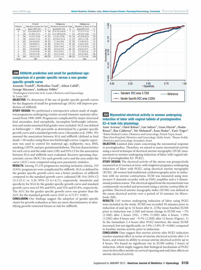
difference by BMI category (p0.5). Among nulliparas, there is in- creased risk of CD for OB vs. NW after controlling for age and race (aOR 2.0 [1.0-3.7], p0.04). This risk is not seen for OW vs. NW (OR 1.3 [0.8-2.4], p0.3). In nulliparas with CD, there was a difference in CD rate for failed IOL among groups (OB-27%, OW-32%, NW-6%, p0.001). In multiparas, BMI category was not associated with CD (p0.6). CONCLUSION: Our study demonstrates no difference in latent LT for women undergoing IOL, regardless of parity. However, there is an increased risk of CD and CD for failed IOL in OB nulliparas under- going IOL. Further research is needed to understand labor curves in OB women to optimize labor and delivery management and minimize unnecessary CD. 296 Ultrasonographic cervical length versus Bishop score for preinduction cervical assessment in parous women: a randomized clinical trial Aeli Ryu 1 , Kyo Hoon Park 1 , Sung Youn Lee 1 , Eun Ha Jeong 1 , Kyung Joon Oh 1 , Ahra Kim 1 1 Seoul National University College of Medicine, Seoul National University Bundang Hospital, Department of Obstetrics and Gynecology, Seongnam-si, Republic of Korea OBJECTIVE: To compare the ultrasonographic cervical length with the Bishop score in determining the administration of prostaglandin for preinduction cervical ripening in parous women at term. STUDY DESIGN: This trial enrolled 154 parous women at term present- ing for labor induction with a singleton, cephalic fetus and intact membranes. Patients were randomly assigned to receive prostaglan- din for preinduction cervical ripening based on the Bishop score or the sonographically-measured cervical length. An unfavorable cervix to be treated with prostaglandin for preinduction cervical ripening was defined as either a Bishop score 4 or a cervical length 28 mm. The primary outcome measures were induction success (defined as an ability to achieve the active phase of labor) and the percentage of patients treated with prostaglandin for preinduction cervical ripening. RESULTS: Baseline demographic characteristics, including gestational age, cervical length, and Bishop score were similar between the two groups. While 74% of parous women in the Bishop score group (n77) received prostaglandin, only 34% of those in the transvaginal ultrasound group (n77) received prostaglandin (P 0.0001). The rates of induction success and cesarean delivery, and the induction to delivery intervals were similar in the two groups. CONCLUSION: With the suggested cutoff values of a Bishop score 4 and a cervical length 28 mm for choosing candidates for pre-induc- tion cervical ripening, the use of sonographic cervical length, com- pared with the Bishop score for assessing the cervix prior to the in- duction of labor, can reduce the need for prostaglandin administration by approximately 50% without adversely affecting the outcome of induction in parous women at term. (ClinicalTrials.gov number, NCT01317823) 297 Birthweight difference from previous pregnancies is an independent risk factor for shoulder dystocia Aharon Tevet 1 , Shunit Armon 1 , Rachel Michaelson Cohen 1 , Rivka Farkash 1 , Sorina Grisaro Granovsky 1 , Arnon Samueloff 1 1 Shaare Zedek M.C. Hebrew University, Obstetrics & Gynecology, Jerusalem, Israel OBJECTIVE: Objective: To determine whether a birthweight difference from previous pregnancies is associated with the occurrence of shoul- der dystocia (SD) in the index pregnancy. STUDY DESIGN: A case control retrospective study. All cases of SD be- tween May 2005 and July 2011 were identified and stratified according to birthweight (100 gram intervals). Cases of Primiparity, Diabetes mellitus, intrauterine fetal death, preterm deliveries and multifetal gestation were excluded. Each Shoulder Dystocia case was matched with 4 cases of uncomplicated vaginal delivery according to birth- weight and use of instrumental delivery.Birthweight difference was defined as the difference between the birthweight in the index preg- nancy and the maximal birthweight in previous pregnancies. Cases of SD were compared to controls for birthweight difference (as defined), birthweight, parity, maternal age, epidural anesthesia and use of in- strumental delivery. Statistical analysis: descriptive, t test, chi-square, Pearson coefficient, Fisher’s Exact test as appropriate and logistic re- gression model. RESULTS: During the study period 73,871 births were attended. 133 cases of SD that met the entry criteria were identified, included in the study and matched to 514 controls similar in fetal birthweight (100 gram intervals) and use of instrumental delivery. The study and con- trol groups did not differ in fetal birthweight, parity, maternal age, epidural anesthesia and use of instrumental delivery. The mean birth- weight difference from previous deliveries in the SD group was 415 grams and 148 grams in the control group (p0.0001). An Increase in birthweight of more than 500 grams from the maximal PREVIOUS birthweight was positively associated with the risk of shoulder dysto- cia (OR 3.42 CI 2.28-5.15). This association is independent of birth- weight and other characteristics analyzed. CONCLUSION: A large fetal birthweight difference from previous preg- nancies is positively associated with and may be a risk factor for shoul- der dystocia. 298 Cesarean for first stage arrest: modern practice does not follow contemporary labor patterns Amanda Trudell 1 , Anthony Odibo 1 , Methodius Tuuli 1 , Kimberly Roehl 1 , George Macones 1 , Alison Cahill 1 1 Washington University in St. Louis, Obstetrics and Gynecology, St. Louis, MO OBJECTIVE: The rise in the cesarean delivery (CD) rate has become a public health concern. Recent evidence suggests a significant propor- tion of primary CDs are performed for arrest of dilation. We sought to investigate labor patterns preceding CD for arrest in the first stage of labor. STUDY DESIGN: We performed a retrospective cohort study of consec- utive births via CD over a four year period. We analyzed the labor patterns of women who underwent CD for arrest in the first stage of labor. Mean cervical dilation at arrest and mean duration of time spent at the arrested dilation with 95% CI were calculated. Duration of arrest was defined as time of no appreciable change ( 1cm) prior to delivery. A stratified analysis was performed based on parity. RESULTS: Of 549 women who underwent CD for arrest in the first stage, 320 (58%) were delivered prior to 6 cm of cervical dilation (active first stage). The average duration of arrest at 6 cm was 5.0 hours (95% CI 4.8-7.1). In the stratified analysis, a majority of both nullip- arous and multiparous women underwent CD at or before 6 cm (55% and 63%). The duration of arrest in primiparous and multiparous women at 6 cm was 5.0 hours (95% CI 4.7-6.4) and 4.2 hours (95% CI 3.4-10.1), respectively. The shortest duration of arrest prior to 6 cm in primiparous and multiparous women occurred at 2 cm and 3 cm (4.3 hours [95% CI 0.1-27.0] and 2.0 hours [95% CI 1.3-7.0]), respectively. CONCLUSION: According to contemporary labor patterns, active labor does not begin until after 6 cm dilation and women may take up to 10 hours to advance 1 cm of dilation before reaching 6 cm. We report a large proportion of laboring women undergo CD for arrest of dilation prior to active labor, and the median time at the dilation of arrest is much shorter than what most women require to advance in the latent phase. The potential to reduce the unyielding rise in the CD rate exists if obstetricians adopt contemporary labor curves and have the pa- tience to utilize these new standards. Poster Session II Clinical Obstetrics, Diabetes, Labor, Medical-Surgical-Disease, Physiology/Endocrinology, Prematurity www.AJOG.org S134 American Journal of Obstetrics & Gynecology Supplement to JANUARY 2013
Transcript

fw

BR

Bp

imdttdptw

ag(urd

i

F

I

fd

ttmgww

csgtewgbbcw

n

R

S

ptil

s((aaw3ph

Poster Session II Clinical Obstetrics, Diabetes, Labor, Medical-Surgical-Disease, Physiology/Endocrinology, Prematurity www.AJOG.org

difference by BMI category (p�0.5). Among nulliparas, there is in-creased risk of CD for OB vs. NW after controlling for age and race(aOR 2.0 [1.0-3.7], p�0.04). This risk is not seen for OW vs. NW (OR1.3 [0.8-2.4], p�0.3). In nulliparas with CD, there was a difference inCD rate for failed IOL among groups (OB-27%, OW-32%, NW-6%,p�0.001). In multiparas, BMI category was not associated with CD(p�0.6).CONCLUSION: Our study demonstrates no difference in latent LT forwomen undergoing IOL, regardless of parity. However, there is anincreased risk of CD and CD for failed IOL in OB nulliparas under-going IOL. Further research is needed to understand labor curves inOB women to optimize labor and delivery management and minimizeunnecessary CD.

296 Ultrasonographic cervical length versus Bishop scoreor preinduction cervical assessment in parousomen: a randomized clinical trial

Aeli Ryu1, Kyo Hoon Park1, Sung Youn Lee1, Eun Ha Jeong1,Kyung Joon Oh1, Ahra Kim1

1Seoul National University College of Medicine, Seoul National Universityundang Hospital, Department of Obstetrics and Gynecology, Seongnam-si,epublic of Korea

OBJECTIVE: To compare the ultrasonographic cervical length with theishop score in determining the administration of prostaglandin forreinduction cervical ripening in parous women at term.

STUDY DESIGN: This trial enrolled 154 parous women at term present-ng for labor induction with a singleton, cephalic fetus and intact

embranes. Patients were randomly assigned to receive prostaglan-in for preinduction cervical ripening based on the Bishop score orhe sonographically-measured cervical length. An unfavorable cervixo be treated with prostaglandin for preinduction cervical ripening wasefined as either a Bishop score �4 or a cervical length �28 mm. Therimary outcome measures were induction success (defined as an abilityo achieve the active phase of labor) and the percentage of patients treatedith prostaglandin for preinduction cervical ripening.

RESULTS: Baseline demographic characteristics, including gestationalge, cervical length, and Bishop score were similar between the tworoups. While 74% of parous women in the Bishop score groupn�77) received prostaglandin, only 34% of those in the transvaginalltrasound group (n�77) received prostaglandin (P �0.0001). Theates of induction success and cesarean delivery, and the induction toelivery intervals were similar in the two groups.

CONCLUSION: With the suggested cutoff values of a Bishop score �4and a cervical length �28 mm for choosing candidates for pre-induc-tion cervical ripening, the use of sonographic cervical length, com-pared with the Bishop score for assessing the cervix prior to the in-duction of labor, can reduce the need for prostaglandinadministration by approximately 50% without adversely affecting theoutcome of induction in parous women at term. (ClinicalTrials.govnumber, NCT01317823)

297 Birthweight difference from previous pregnancies is anndependent risk factor for shoulder dystocia

Aharon Tevet1, Shunit Armon1, Rachel Michaelson Cohen1, Rivkaarkash1, Sorina Grisaro Granovsky1, Arnon Samueloff1

1Shaare Zedek M.C. Hebrew University, Obstetrics & Gynecology, Jerusalem,srael

OBJECTIVE: Objective: To determine whether a birthweight differencerom previous pregnancies is associated with the occurrence of shoul-er dystocia (SD) in the index pregnancy.

STUDY DESIGN: A case control retrospective study. All cases of SD be-ween May 2005 and July 2011 were identified and stratified accordingo birthweight (100 gram intervals). Cases of Primiparity, Diabetes

ellitus, intrauterine fetal death, preterm deliveries and multifetalestation were excluded. Each Shoulder Dystocia case was matchedith 4 cases of uncomplicated vaginal delivery according to birth-

eight and use of instrumental delivery.Birthweight difference was

S134 American Journal of Obstetrics & Gynecology Supplement to JANUARY 2

defined as the difference between the birthweight in the index preg-nancy and the maximal birthweight in previous pregnancies. Cases ofSD were compared to controls for birthweight difference (as defined),birthweight, parity, maternal age, epidural anesthesia and use of in-strumental delivery. Statistical analysis: descriptive, t test, chi-square,Pearson coefficient, Fisher’s Exact test as appropriate and logistic re-gression model.RESULTS: During the study period 73,871 births were attended. 133ases of SD that met the entry criteria were identified, included in thetudy and matched to 514 controls similar in fetal birthweight (100ram intervals) and use of instrumental delivery. The study and con-rol groups did not differ in fetal birthweight, parity, maternal age,pidural anesthesia and use of instrumental delivery. The mean birth-eight difference from previous deliveries in the SD group was 415rams and 148 grams in the control group (p�0.0001). An Increase inirthweight of more than 500 grams from the maximal PREVIOUSirthweight was positively associated with the risk of shoulder dysto-ia (OR 3.42 CI 2.28-5.15). This association is independent of birth-eight and other characteristics analyzed.

CONCLUSION: A large fetal birthweight difference from previous preg-nancies is positively associated with and may be a risk factor for shoul-der dystocia.

298 Cesarean for first stage arrest: modern practice doesot follow contemporary labor patterns

Amanda Trudell1, Anthony Odibo1, Methodius Tuuli1, Kimberlyoehl1, George Macones1, Alison Cahill1

1Washington University in St. Louis, Obstetrics and Gynecology,t. Louis, MO

OBJECTIVE: The rise in the cesarean delivery (CD) rate has become aublic health concern. Recent evidence suggests a significant propor-ion of primary CDs are performed for arrest of dilation. We sought tonvestigate labor patterns preceding CD for arrest in the first stage ofabor.

STUDY DESIGN: We performed a retrospective cohort study of consec-utive births via CD over a four year period. We analyzed the laborpatterns of women who underwent CD for arrest in the first stage oflabor. Mean cervical dilation at arrest and mean duration of timespent at the arrested dilation with 95% CI were calculated. Duration ofarrest was defined as time of no appreciable change (� 1cm) prior todelivery. A stratified analysis was performed based on parity.RESULTS: Of 549 women who underwent CD for arrest in the firsttage, 320 (58%) were delivered prior to 6 cm of cervical dilationactive first stage). The average duration of arrest at 6 cm was 5.0 hours95% CI 4.8-7.1). In the stratified analysis, a majority of both nullip-rous and multiparous women underwent CD at or before 6 cm (55%nd 63%). The duration of arrest in primiparous and multiparousomen at 6 cm was 5.0 hours (95% CI 4.7-6.4) and 4.2 hours (95% CI.4-10.1), respectively. The shortest duration of arrest prior to 6 cm inrimiparous and multiparous women occurred at 2 cm and 3 cm (4.3ours [95% CI 0.1-27.0] and 2.0 hours [95% CI 1.3-7.0]), respectively.

CONCLUSION: According to contemporary labor patterns, active labordoes not begin until after 6 cm dilation and women may take up to 10hours to advance 1 cm of dilation before reaching 6 cm. We report alarge proportion of laboring women undergo CD for arrest of dilationprior to active labor, and the median time at the dilation of arrest ismuch shorter than what most women require to advance in the latentphase. The potential to reduce the unyielding rise in the CD rate existsif obstetricians adopt contemporary labor curves and have the pa-

tience to utilize these new standards.

013

cs

G

S

fd

(tc1sgTA

iE

R

O

tusl

wip((Iit

www.AJOG.org Clinical Obstetrics, Diabetes, Labor, Medical-Surgical-Disease, Physiology/Endocrinology, Prematurity Poster Session II

299 Stillbirth prediction and small for gestational age:omparison of a gender specific versus a non-genderpecific growth curve

Amanda Trudell1, Methodius Tuuli1, Ailson Cahill1,eorge Macones1, Anthony Odibo1

1Washington University in St. Louis, Obstetrics and Gynecology,t. Louis, MO

OBJECTIVE: To determine if the use of gender specific growth curvesor the diagnosis of small for gestational age (SGA) will improve pre-iction of stillbirth.

STUDY DESIGN: We performed a retrospective cohort study of single-ton pregnancies undergoing routine second trimester anatomy ultra-sound from 1990-2009. Pregnancies complicated by major structuralfetal anomalies, fetal aneuploidy, incomplete birthweight informa-tion and undocumented fetal gender were excluded. SGA was definedas birthweight � 10th percentile as determined by a gender-specificgrowth curve and a standard growth curve (Alexander et al. 1996). Weassessed the association between SGA and stillbirth (defined as fetaldeath �20 weeks) using these two birthweight curves. Logistic regres-sion was used to control for maternal age, nulliparity, race, BMI,smoking, CHTN, and pre-gestational diabetes. The test characteristicsfor each curve and the odds ratio (OR) and 95% CI for the associationbetween SGA and stillbirth were evaluated. Receiver operating char-acteristic curves (ROC) for each growth curve and the area under thecurve (AUC) were compared using non-parametric statistics.RESULTS: Among 57,170 pregnancies meeting inclusion criteria, 3190.6%) pregnancies were complicated by stillbirth. SGA as defined byhe gender specific growth curve was a better predictor of stillbirthompared to the standard growth curve (adjusted OR 19.6 [95% CI5.3-25.1] vs. 5.26 [95% CI 4.1-6.7]), respectively. Sensitivity andpecificity for SGA by the gender specific growth curve and standardrowth curve were 63.9% and 92%, and 32% and 92.8%, respectively.he AUC for the gender specific growth curve was greater than theUC for the standard growth curve (0.83 vs. 0.72 P� 0.001).

CONCLUSION: Our findings suggest the adoption of gender-specificcharts for growth evaluation as they are more discriminatory in iden-tifying growth-restricted fetuses at risk for stillbirth.

Supplem

300 Myometrial electrical activity in women undergoingnduction of labor with vaginal tablets of prostaglandins2–A look into physiology

Amir Aviram1, Oded Raban1, Liat Saltzer1, Liran Hiersh1, Hadarosen3, Ilan Calderon2, Nir Melamed1, Eran Hadar1, Yariv Yogev1

1Rabin Medical Center, Obstetrics and Gynecology, Petach Tiqva, Israel,2Bnei Zion Hospital, Obstetrics and Gynecology, Haifa, Israel, 3Shaare Zedek,

bstetrics and Gynecology, Jerusalem, IsraelOBJECTIVE: Limited data exists concerning the myometrial responseo prostaglandins. Therefore, we aimed to assess myometrial activitysing a novel technique of electrical uterine myography (EUM) mea-urement in women undergoing induction of labor with vaginal tab-ets of prostaglandins E2- PGE2).

STUDY DESIGN: The electrical activity of the uterus was prospectivelymeasured in 43 women at term, with singleton pregnancy undergoinginduction of labor with PGE2, using electrical uterine myography(EUM). All women had traditional cardiotocography prior to induc-tion with no uterine contractions. EUM was measured using non-invasive 9 channels recorder with an EMG amplifier and a 3-dimen-sional position sensor. The electrical signal from the myometrium wascontinuously recorded and processed using a uterine contractility al-gorithm. Electrical uterine myography index (EUMI) was defined asthe mean electrical activity over a period of 10 minutes (in Micro-Joule (MJ).RESULTS: 1)43 women undergoing induction of labor using PGE2

ere included in the study. EUMI was recorded 30 minutes prior tonduction of and up to 16 hours after it. 2)The mean baseline EUMIrior to induction was 2.5MJ and mean change in EUMI was �7%2.3MJ) after 2 hours (NS), �39% (3.5MJ) after 4 hours, �29%3.2MJ) after 8 hours and �9.7% (2.3MJ) after 12 hours (Figure). 3)n the immediate 2-4 hours after PGE2 insertion, the mean EUMIncreased, but not significantly, in 3.9%�5.8% (P�0.806) comparedo baseline uterine activity prior to induction.

CONCLUSION: Date suggest that uterine activity after PGE2 inductionreaches maximal effect in terms of uterine electrical activity after 2-4hours, and retains its ability to arouse uterine electrical activity up to8 hours. We found no significant rise in EUMI within 2 hours ofinduction, which might suggests that biological mechanism of PGE2exert its first effect mostly on cervical ripening and only later effects on

uterine electrical activity.

ent to JANUARY 2013 American Journal of Obstetrics & Gynecology S135


Recommended