Page 1
3 human papillomavirus species share
several biological properties with high risk
mucosal types
Dissertation
submitted to the Combined Faculties for
the Natural Sciences and for Mathematics of the
Ruperto-Carola University of Heidelberg, Germany
for the degree of
Doctor of Natural Sciences
presented by
Lucia Minoni, M.Sc. Born in Brescia, Italy
Page 2
Dissertation
submitted to the Combined Faculties for
the Natural Sciences and for Mathematics of the
Ruperto-Carola University of Heidelberg, Germany
for the degree of
Doctor of Natural Sciences
presented by
Lucia Minoni, M.Sc. Born in Brescia, Italy
Oral-examination: 8th May 2018
Page 3
3 human papillomavirus species share
several biological properties with high risk
mucosal types
Referees: Prof. Dr. Martin Müller Prof. Dr. Frank Rösl
Page 4
I
TABLE OF CONTENTS
Table of contents ............................................................................................................................ I
Summary ......................................................................................................................................... 1
Zusammenfassung ......................................................................................................................... 2
1 Introduction ........................................................................................................................... 3
1.1 Human papillomaviruses ............................................................................................... 3
1.1.1 Phylogenetic classification ..................................................................................... 3
1.1.2 Human papillomaviruses genome organization .................................................. 5
1.1.3 Human papillomavirus tropisms ........................................................................... 6
1.2 Human papillomaviruses and carcinogenesis ............................................................. 7
1.2.1 Cancer and HPV types ....................................................................................... 7
1.2.2 Cancer and HPV types ..................................................................................... 18
1.2.3 Cancer and types ............................................................................................. 21
2 Aim of the thesis ................................................................................................................. 23
3 Materials ............................................................................................................................... 24
3.1 Biological material ........................................................................................................ 24
3.1.1 Procaryotic cells .................................................................................................... 24
3.1.2 Eukaryotic cells ..................................................................................................... 25
3.2 Media and supplements ............................................................................................... 27
3.2.1 Procaryotic cells .................................................................................................... 27
3.2.2 Eukaryotic cells ..................................................................................................... 28
3.3 Human cells treatments and manipulation ................................................................ 29
3.4 Retroviral infection ....................................................................................................... 30
3.5 Molecular cloning ......................................................................................................... 30
3.5.1 Plasmids .................................................................................................................. 30
Page 5
II
3.5.2 Enzymes ................................................................................................................. 34
3.5.3 Oligonucleotides for siRNA knockdown .......................................................... 34
3.5.4 Oligonucleotides for cloning ............................................................................... 35
3.5.5 Oligonucleotides for Real Time PCR (RT-PCR) .............................................. 36
3.5.6 Oligonucleotides for site-directed mutagenesis ................................................ 37
3.5.7 Buffers and solutions ............................................................................................ 38
3.6 Reagents for protein analysis ...................................................................................... 38
3.6.1 Enzymes ................................................................................................................. 38
3.6.2 IP buffer ................................................................................................................. 38
3.6.3 Protein Buffer 10X ............................................................................................... 39
3.6.4 SDS-polyacrylamide electrophoresis .................................................................. 39
3.6.5 Western blot analysis ............................................................................................ 41
3.7 Immunological assays ................................................................................................... 42
3.7.1 Antibodies .............................................................................................................. 42
3.8 Maltose binding protein pulldown ............................................................................. 43
3.9 Illumina array ................................................................................................................ 43
3.9.1 Instruments and consumable .............................................................................. 43
3.9.2 Software .................................................................................................................. 44
3.10 Liquid chromatography and mass spectrometry .................................................. 44
3.10.1 Instruments and consumables ......................................................................... 44
3.10.2 Software .............................................................................................................. 44
3.11 Chemicals ................................................................................................................... 45
3.12 Kits .............................................................................................................................. 45
3.13 Laboratory equipment .............................................................................................. 46
3.13.1 Electrical equipment ......................................................................................... 46
3.13.2 Common use equipment .................................................................................. 48
Page 6
III
3.13.3 Software and websites ...................................................................................... 49
4 Methods ............................................................................................................................... 50
4.1 Cultivation and manipulation of prokaryotic cells ................................................... 50
4.1.1 Transformation of bacteria by heat shock ......................................................... 50
4.1.2 Cultivation and Storage of Bacteria .................................................................... 50
4.2 Cultivation and Manipulation of Eukaryotic Cell .................................................... 51
4.2.1 Cultivation of NIH/3T3 fibroblasts .................................................................. 51
4.2.2 Cultivation of Phoenix ......................................................................................... 51
4.2.3 Cultivation of human keratinocytes (primary and expressing E6/E7) .......... 52
4.2.4 Cultivation of HNC136 ........................................................................................ 54
4.2.5 Cells counting ........................................................................................................ 54
4.2.6 Cryopreservation and Thawing of Mammalian Cells ....................................... 54
4.2.7 Cell treatments ....................................................................................................... 55
4.2.8 siRNA knock-down .............................................................................................. 56
4.3 Retrovirus infection ...................................................................................................... 56
4.3.1 Transfection ........................................................................................................... 57
4.3.2 Infection ................................................................................................................. 58
4.3.3 Selection ................................................................................................................. 58
4.3.4 Test for the exit of the cells from the P3 ........................................................... 58
4.4 Molecular Methods ....................................................................................................... 59
4.4.1 Purification of plasmid DNA .............................................................................. 59
4.4.2 DNA visualization ................................................................................................ 59
4.4.3 Molecular Cloning ................................................................................................. 60
4.4.4 RNA manipulation ................................................................................................ 65
4.5 Protein analysis ............................................................................................................. 68
4.5.1 Protein extraction .................................................................................................. 68
Page 7
IV
4.5.2 Determination of Protein Concentration by Bradford Assay ......................... 68
4.5.3 Lambda Protein Phosphatase (PP) Treatment. ................................................ 68
4.5.4 Acrylamide gel ....................................................................................................... 69
4.5.5 Western Blot analysis ............................................................................................ 69
4.6 Maltose binding protein (MBP) pulldown ................................................................ 71
4.6.1 Preparation of beads ............................................................................................. 71
4.6.2 Cell extract preparation ........................................................................................ 72
4.6.3 Pulldown ................................................................................................................ 72
4.6.4 Western Blot .......................................................................................................... 73
4.7 Microarray-based whole genome expression profiling and data analysis .............. 73
4.7.1 RNA quality control ............................................................................................. 73
4.7.2 Micro Array ............................................................................................................ 73
4.7.3 Differential expression analysis ........................................................................... 74
4.7.4 Heatmap ................................................................................................................. 74
4.7.5 Pathway analysis .................................................................................................... 75
4.7.6 Comparative analysis ............................................................................................ 75
4.8 LC/MS supernatant analysis ....................................................................................... 75
4.8.1 Sample preparations ............................................................................................. 75
4.8.2 Analytical methods and instrumentation ........................................................... 75
4.8.3 Raw data preprocessing and filtration ................................................................ 76
5 Results .................................................................................................................................. 77
5.1 In vitro transforming abilities of 3 HPV E6 and E7 proteins ............................... 77
5.2 3 types 49, 75 and 76 E6/E7 efficiently alter cell cycle-related pathways .......... 82
5.2.1 pRb pathway is altered in 3 HPV E6/E7 HFKs ........................................... 82
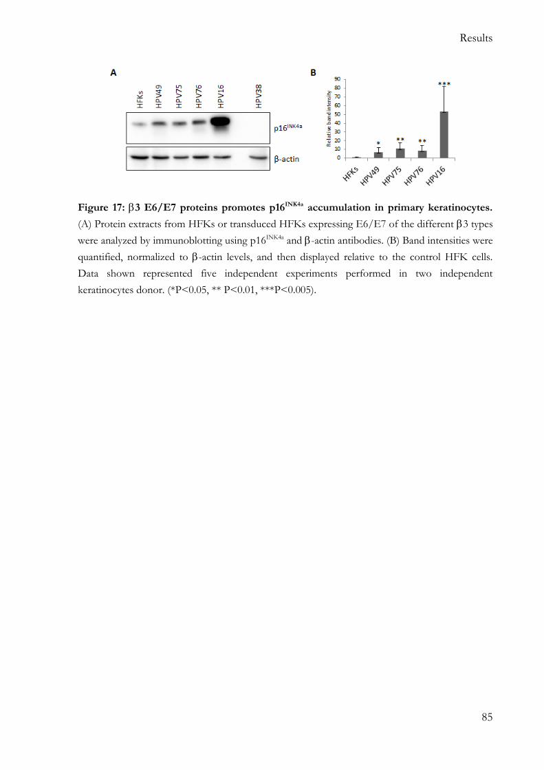
5.2.2 p16INK4a pathway is altered in 3 HPV E6/E7 HFKs ..................................... 84
5.3 p53 is degraded via proteasome pathway in 3 HPV E6/E7 HFKs .................... 86
Page 8
V
5.4 3 types 49, 75 and 76 E6/E7 efficiently up-regulate hTERT expression .......... 90
5.5 HPV76 E6 transforming properties are affected by mutations in the
corresponding regions of HPV16 E6 involved in p53 and E6AP binding ..................... 92
5.5.1 Mutants design ...................................................................................................... 92
5.5.2 HPV76 E6 mutants fail in the immortalization of primary keratinocytes .... 93
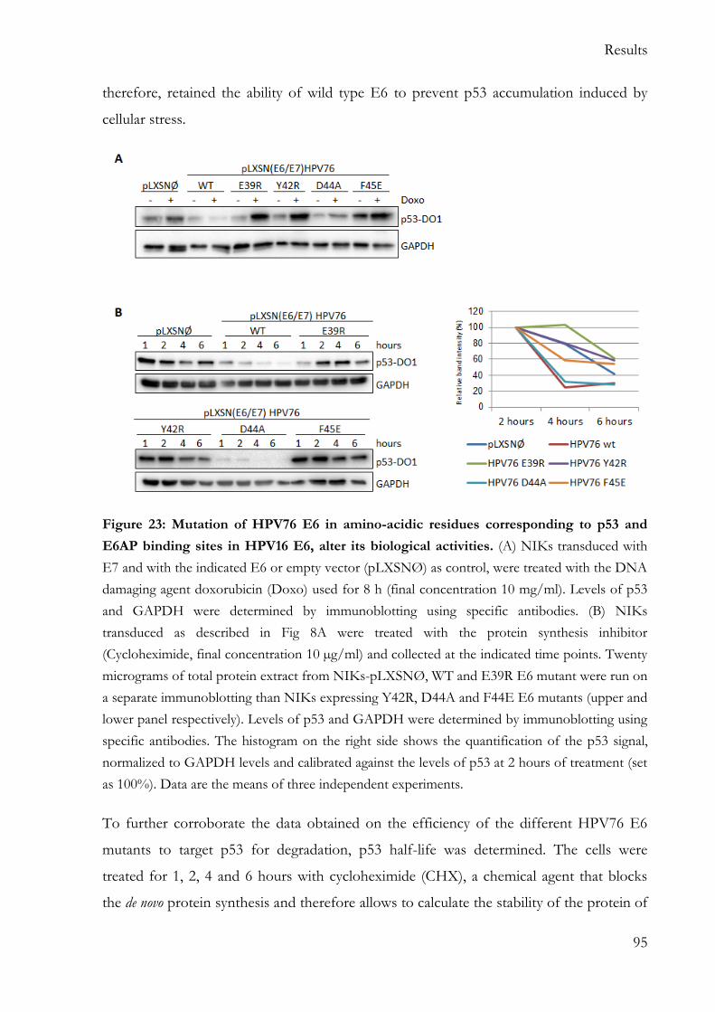
5.5.3 HPV76 E6 E38R, Y42R, F45E mutants fail to degrade p53 .......................... 94
5.5.4 E39R E6 mutant fail to up-regulate hTERT expression ................................. 96
5.6 3 HPV and HPV16 E6/E7 HFKs show some similarities in the alteration of
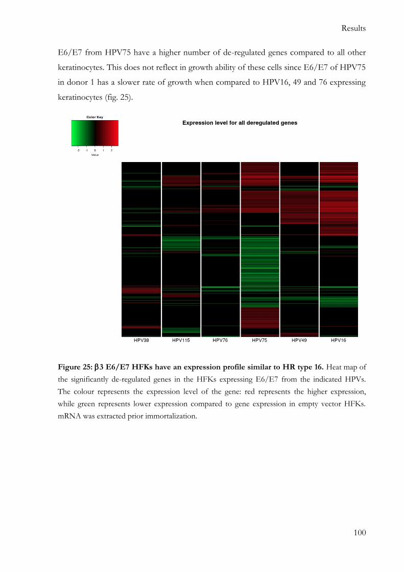
cellular gene expression .......................................................................................................... 99
5.6.1 Hierarchical clustering reveal higher similarity between 3 types and HPV16
E6/E7 keratinocytes .......................................................................................................... 99
5.6.2 Pathway analysis reveals an overall de-regulation of cell cycle, p53 and DNA
replication pathways in HFKs expressing E6/E7 from HPV49, 76 and HR HPV16
101
5.6.3 3 E6/E7 expressing keratinocytes share more de-regulated genes with HR
HPV16 than with 2 HPV38 .......................................................................................... 104
5.6.4 Array Validation .................................................................................................. 106
5.7 Metabolism and transformation ............................................................................... 108
6 Discussion and conclusions ............................................................................................. 114
6.1 Future prospectives .................................................................................................... 118
Acronyms and abbreviations .................................................................................................... 119
Amino acids............................................................................................................................ 121
References ................................................................................................................................... 122
Acknowledgments ..................................................................................................................... 143
Page 9
1
SUMMARY
human papillomaviruses (HPV) are subdivided into five species and are abundantly
detected in the skin, in particular the and 2 species. Therefore, HPV types are considered to have a cutaneous tropism. However, several recent studies have described
the presence of HPV also in the mucosal epithelia at different anatomical sites. In
particular, 3 HPV types are more prevalent in certain mucosal epithelia rather than in the
cutaneous tissues. Studies in different experimental models have also highlighted that 3 HPV49 share functional similarities with the mucosal high-risk (HR) HPV16. However, with the exclusion of HPV49, very little is known about the biology of the other known
3 HPV types (75, 76 and 115).
The aim of this thesis was the characterization of the biological properties of E6 and E7
of all known 3 HPV types, in relation to their interaction with key cellular pathways such as pRb, p53 and hTERT.
Similar to what was previously showed for HPV49 E6/E7, HPV75 and HPV76 E6 and E7, but not HPV115 E6 and E7, efficiently cooperate in the immortalization/extension of lifespan of human foreskin keratinocytes (HFKs). In detail, HPV49, 75 and 76 E6/E7 cause the accumulation of the phosphorylated form of pRb, leading to the release of the E2F factor and unscheduled S-phase entry. As observed for HR HPV16, cell cycle
deregulation mediated by 3 HPV onco-proteins leads to p16INK4a accumulation, while no
p16INK4a was detected in 2 HPV38 E6/E7 HFKs. Similarly to HPV49 E6, HPV75 and 76 E6s degrade p53 via an E6AP/proteasome-mediated mechanism. Mutation in HPV76 E6 amino-acids that correspond to HPV16 E6 amino-acids involved in the formation of the E6/E6AP/p53 ternary complex results in the failed immortalization of HFKs. All the
3 HPV types, with the exception of HPV115, induce the up-regulation of hTERT expression, another important step in cellular transformation. Comparative analysis of
cellular gene expression pattern of HFKs expressing E6 and E7 from HR HPV16, 3 HPV types and β2 HPV38 further highlights the functional similarities of HR HPV16 and
3 HPV49, 75, 76. The expression profiles of these four HPV HFKs show some
similarities and diverge substantially from 3 HPV115 E6/E7 and 2 HPV38 E6/E7 HFKs.
In conclusion, the data show that 3 HPV types share some similarities with HR HPV types and pave the way to additional studies aiming to evaluate their tissue tropism and their role in human pathologies.
Page 10
2
ZUSAMMENFASSUNG
Die humanen Papillomviren (HPV) des β-Genus sind in fünf Spezies unterteilt, wobei
insbesondere die β1- und β2-Spezies häufig in der Haut nachgewiesen werden können.
Aus diesem Grund wird davon ausgegangen, dass die β-HPV Typen einen vorwiegend
kutanen Tropismus haben. β3 HPV Typen werden dagegen hauptsächlich in bestimmten
Schleimhäuten nachgewiesen und nicht wie die anderen β-Typen in kutanem Gewebe.
Studien in verschiedenen experimentellen Modellen haben gezeigt, dass der β3 HPV Typ
49 funktionelle Eigenschaften mit dem mukosalen Hochrisikotyp (HR) HPV16 teilt. Mit
Ausnahme von HPV49 ist über die Biologie der restlichen, bekannten β3 HPV Typen
(HPV75, 76 und 115) bisher nur wenig bekannt.
Das Ziel dieser Arbeit war die Charakterisierung der biologischen Eigenschaften von E6
und E7 aller bekannten β3 HPV-Typen im Hinblick auf ihre Interaktion mit zellulären
Schlüssel-Signalwegen wie pRb, p53 und hTERT. Ähnlich wie HPV49 E6/E7 sind auch
HPV75 und HPV76 E6/E7 maßgeblich an der Immortalisierung/Verlängerung der
Lebensdauer primärer humaner Vorhautkeratinozyten (HFKs) beteiligt, ganz im
Gegensatz zu HPV115 E6/E7. Die Expression von HPV49, 75 und 76 E6/E7 resultierte
in der Akkumulation der phosphorylierten Form von pRB, welche zur Freisetzung des
E2F Transkriptionsfaktors und der außerplanmäßigen S-Phase führte. Wie bereits bei HR
HPV16 beobachtet, verursachte auch die Zellzyklusderegulation durch β3 HPV
Onkoproteine die Akkumulation von p16INK4a, wohingegen keine p16INK4a
Akkumulation bei β2 HPV 38 E6/E7 exprimierenden HFKs festgestellt wurde. Ebenfalls
vergleichbar zu HPV49 E6 führte auch die Expression von HPV75 und 76 E6 zur
E6AP/Proteasomen-vermittelten Degradation von p53. Basierend auf HPV16 E6 wurde
der putative E6AP/p53 Interaktionsbereich im HPV76 Protein durch Mutagenese
verändert. Alle vier untersuchten Mutanten von HPV76 E6 verloren die Fähigkeit HFK
zu immortalisieren. Alle β3 HPV Typen, mit Ausnahme von HPV115, induzierten die
Hochregulation der hTERT Expression, die einen weiteren wichtigen Schritt der
zellulären Transformation darstellt. Desweiteren demonstrierten Vergleichsanalysen
zellulärer Genexpressionsmuster von HFKs , die E6 und E7 des HR HPV16, der β3
HPV Typen und des β2 HPV38 E6 und E7 exprimierten die Ähnlichkeit der β3 HPV
Typen 49, 75 und 76 zum HR Typ HPV16. Das Expressionsprofil dieser vier HPV HFKs
wies einige Gemeinsamkeiten auf und unterscheiden sich damit deutlich von den β3
HPV115 und β2 HPV38 E6/E7 HFKs.
Diese Daten zeigen, dass β3 HPV Typen einige Übereinstimmungen in Bezug auf ihre
regulierenden Eigenschaften der zellulären Genexpression zu den HR HPV Typen
aufweisen und verdeutlichen somit die Notwendigkeit weiterer Studien, zur Erforschung
ihres Gewebetropismus und ihrer Rolle in der humanen Pathologie.
Page 11
Introduction
3
1 INTRODUCTION
1.1 HUMAN PAPILLOMAVIRUSES
The human papillomavirus (HPV) belong to the taxonomic family of Papillomaviridae and
represents a group of non-enveloped double-stranded (ds) DNA viruses. The viruses that
make up Papillomaviridae are highly diverse, and are present in most mammals and birds.
1.1.1 PHYLOGENETIC CLASSIFICATION
Over 240 papillomaviruses (PV) have been discovered so far, including more than 150
HPVs, and they are grouped by species and classified into 16 genera (indicated with
Greek letters), as shown in figure 1. This classification is based on the nucleotide
sequence of L1, with a genotype (or type) considered distinct when the sequence is at
least 10% divergent from other known PVs [1]. Currently, the alpha, beta and gamma (,
, ) genera include the majority of the HPV types.
The phylogeny classification of the PV based on the L1 nucleotide sequence does not
necessary correlate with the biological properties of the different HPV types. As an
example, the genus includes HPV types with different tissue tropism and/or oncogenic
properties.
Page 12
Introduction
4
Figure 1: Papillomaviruses phylogenetic tree. Classification of papillomaviruses based on L1
gene sequences alignment. Most of the HPVs belong to the , (blue) and (green) genera. the
genus HPV types are further sub-divided in low-risk cutaneous (light brown), low-risk
mucosal (yellow), or high-risk mucosal (pink) according to their association with the
development of cancer. The high-risk types highlighted with red text are confirmed as “human
carcinogens” by IARC. Mucosal and cutaneous tropism classification is indicated under each
genus. Figure from [2]
Page 13
Introduction
5
1.1.2 HUMAN PAPILLOMAVIRUSES GENOME ORGANIZATION
The HPV double stranded genome is approximately 8000 base pairs in size and, despite
the variety of HPVs, its organization is highly conserved. The genome consists of 3
regions: (i) a non-coding long control region (LCR), (ii) an early gene region, and (iii) a
late gene region, as shown in Figure 2.
Figure 2: Schematic overview of the double-stranded DNA genome of and human
papillomaviruses. In the left panel the HPVs genome organization, in the right panel the
HPVs genome organization. The late genes L1 and L2 are depicted in green; the early genes E1,
E2, E4, E5, E6 and E7 are depict in blue. Notably, E5 ORF is absent in the HPV genomes.
The URR, also called LCR, is depict in yellow. Figure modified from [3].
The LCR, otherwise known as the upstream regulatory region (URR), is located between
the late and early gene regions. It contains many of the responsive elements for
transcription factors involved in viral gene expression as well as elements essential for
viral DNA replication. Considerable heterogeneity exists among the LCR of the different
PVs and, notably, the LCR of the HPVs is shorter compared to the one of the HPVs.
The early gene region encodes for genes that are expressed in the early stage of the viral
life cycle and are mainly involved in viral replication (E1, E2), cell cycle entry, and
immune evasion (E6, E7, E5). This region also contains the E4 gene, which is expressed
at the late stage of the viral life cycle and thought to be involved in the virus release. E4 is
also thought to be important in the induction of the productive life cycle.
The early protein 1 (E1), with the aid of E2, binds to the viral origin of replication and it
assembles into a hexameric helicase. This complex exerts its function to unwind the viral
genome, providing the template for the replication of the viral genome [4, 5]. In addition,
Page 14
Introduction
6
the E2 protein also acts as viral transcription regulator by interacting with various host
proteins, modifying their function to regulate the viral life cycle. The roles of E4 and E5
are not yet fully understood, however, E4 seems to be involved in the destabilization of
the cytokeratin network through the formation of amyloid fibers and in the escape of the
virions from the epithelial surface [6, 7]. E5 is a short transmembrane protein that
mediates mitogenic signals of growth factors, such as the epidermal growth factor
receptor or the plated derived growth factor (PDGF) receptor [8, 9]. Remarkably, the
E5 gene is not present in the HPV genomes, indicating that the HPVs have probably
evolved to exert the same functions by different mechanisms.
E6 and E7 are the major onco-proteins involved in the HPV-induced carcinogenesis and
their biological properties are described in detail in paragraph 1.2. An additional gene, E8,
is located within the E1 open reading frame (ORF) and generates a spliced transcript
called E8^2C which has been implicated in the regulation of the viral replication [10].The
late region comprises the L1 and L2 genes that encode the major and minor capsid
proteins, respectively. The viral DNA is encased by a non-enveloped icosahedral capsid
of about 50-60 nm, composed of 72 capsomers. Each capsomer is formed by five L1 and
one L2 molecule.
1.1.3 HUMAN PAPILLOMAVIRUS TROPISMS
HPVs are also classified as mucosal or cutaneous types according to their ability to infect
the mucosal epithelia or the skin, respectively. So far, well-established mucosal HPV types
are included in genus and they can be sub-divided into low-risk (LR) or high-risk (HR),
accordingly to their ability to induce benign or malignant lesions respectively. The LR
HPV types are normally associated with genital warts while HR HPV types are the
etiological cause of cervical cancer as well as a subset of anogenital and head and neck
cancers [11]. The classification of HPVs in HR and LR types parallels their
transforming abilities in in vitro (cell culture) and in vivo model (transgenic mouse).
Many of the cutaneous HPV types belong to the and genera. Several findings have
indicated that HPV types, together with ultra-violet (UV) irradiation, promote the
development of Non-Melanoma-Skin Cancer (NMSC) [12].
Page 15
Introduction
7
It is still unclear what biological properties of the different HPV types determine their
tissue tropism. Based on available data, one possible hypothesis is that HPV tissue
tropism is not controlled at the entry level but it is primarily controlled by the LCR region
that controls viral gene expression [13, 14, 15]. Since E5, E6 and E7 present more
variability in sequence, it is possible that the tropism depends on the function and
regulation of these genes. Another hypothesis suggests that E4, given the considerable
heterogeneity among the sequences in the different types, may play a role in the tropism
and in the different transmission routes [16].
1.2 HUMAN PAPILLOMAVIRUSES AND CARCINOGENESIS
1.2.1 CANCER AND HPV TYPES
All HR HPV-associated cancers correspond to approximately 5% of all cancer cases
worldwide [17]. Thirteen HPV types (16, 18, 31, 33, 35, 39, 45, 51, 52, 56, 58, 59 and 66)
are classified as carcinogenic in humans (Group 1) for their role in the development of
cervical cancer [18]. HPV16 and HPV18 are responsible for approximately 50% and 20%
of cervical cancer cases respectively [19, 20]
The preferential infection site for the mucosal HPV is the junction between the
endocervix columnar cells and the ectocervix stratified squamous epithelial cells, known
as the cervical transformation zone. The majority of the HPV infections are cleared by the
immune system in a relatively short time, usually between 6 and 12 months, and therefore
do not lead to cytological abnormalities. Persistent infections that are not naturally cleared
by the immune system are associated with the development of cervical intraepithelial
neoplasia (CIN), which may regress or progress to invasive cervical carcinoma after a
relatively long period of latency [21]. Figure 3 schematically shows the evolution of an
HPV infection from CIN to invasive cancer.
Beside HPV infection, there are various risk factors associated with the development of
cervical cancer. These risk factors likely influence the ability of the host immune system
to clear the virus, or they may be carcinogenic themselves. Epidemiological studies have
Page 16
Introduction
8
identified certain sexual habits, cigarette smoking, oral contraceptive use and host genetic
predisposition as additional risk factors [22, 23, 24, 25, 26].
HR HPV types are also responsible for a set of anogenital cancer (anal, vaginal, vulvar
and penile cancers) [27] as well as oropharyngeal cancer (base of the tongue, tonsils, and
throat) [28, 29]. However, these cancers are predominately associated with HPV16, while
the other HR HPV types appear to play a minor role.
Due to their association with human carcinogenesis, the mucosal HR HPV types have
been extensively studied in the last 3 decades. Many studies have demonstrated the
transforming properties of the viral onco-proteins in in vitro and in vivo experimental
models.
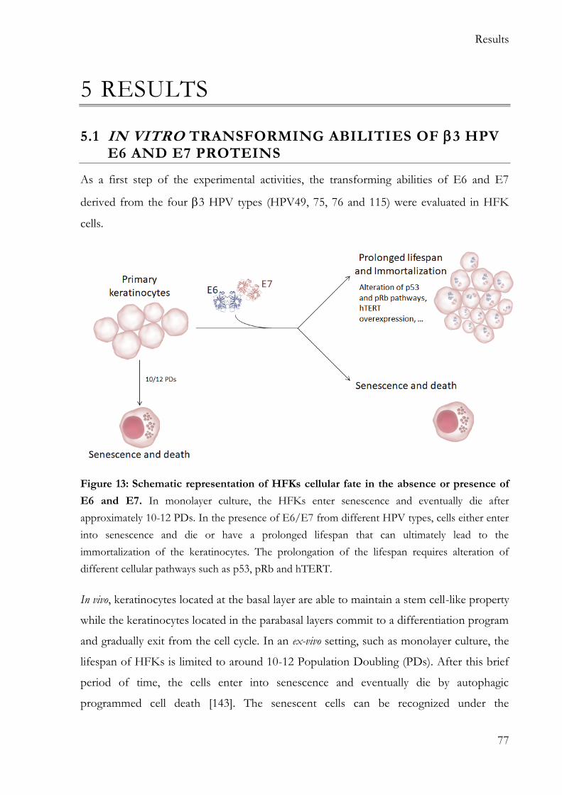
Figure 3: Stage of HPV infection, from CIN to invasive cancer. The basal layer of the
cervical epithelium is supported by the basement membrane and by the dermis. Infectious HPV
particles can access the basal layer, the primary site of infection, through micro abrasion.
Following the infection, the early HPV genes are expressed in the basal layer while the late gene
and E4 are expressed in the midzone and in superficial zone. In CIN1 and CIN2 lesions, the viral
genome remains episomal. The progression to CIN3 is characterized by viral genome integration
with the disruption of E2 gene and therefore up-regulation of E6 and E7. Figure modified from
[30].
Page 17
Introduction
9
HR HPV AND CELLULAR TRANSFORMATION
HR HPV genome encodes three transforming proteins: E5, E6, and E7.The E5 onco-
protein appears to have a role in the early steps of tumour initiation [31], and its
expression is sufficient to promote neoplasia in a transgenic mouse model [32, 33].
Interestingly, the E5 gene is often disrupted in cancer. This is because when the HPV
genome integrates into the host cell, the viral genome is subjected to rearrangement.
Almost in all the integrations, the E2-region of HPV genome is deleted or separated from
the LCR [34, 35, 36]. As consequence, the expression of several viral genes is lost,
including E5. The loss of E5 expression during integration provides an explanation why
E5 is thought to be an onco-protein in the early stage of carcinogenesis when the genome
remains mainly episomal [37]. This is likely an important step in the carcinogenic process
as 100% of HPV18-, 80% of HPV16- and 81% of HPV31-driven cancers contain
integrated viral genomes [38, 39]
E6 and E7 are essential for the maintenance of the transformed phenotype and are
actively transcribed in all HPV-positive cancer cells after integration.
E6 AND E7 TRANSFORMATION IN EXPERIMENTAL MODELS.
The first indication that E6 and E7 are onco-proteins and that they play an important role
in the carcinogenesis came from the analysis of cervical cancer-derived cells such as SiHa
and CaSki, which express both E6 and E7 [40]. The silencing of E6 and E7 in these cells
lines resulted in the rapid cell death, proving the essential role of these proteins for the
cell phenotype [41].
Multiple studies have shown that E6 and E7 display transforming abilities both in in vitro
and in vivo experimental models. The expression of E6 and E7 of HPV16 in immortalized
fibroblasts (NIH3T3) leads to their full transformation, with the acquisition of anchorage-
independent growth ability and ability to form tumours when injected into nude mice
[42]. In addition, HR E6 and E7 are able to immortalize human primary keratinocytes, the
natural host of the virus [43, 44, 45, 46, 47].
In accordance with the in vitro assay described above, transgenic mice expressing both
the viral genes under the basal cell keratin 14 (K14) promoter are susceptible to the
Page 18
Introduction
10
development of cancer, promoted by different means such as chemical carcinogens or
estrogen treatment [48, 49]. However, the main limitation of the transgenic mouse
model is that viral gene expression is regulated by a host promoter and not by the
endogenous LCR. The analysis of external factors, such as hormones, on the viral
transcription is compromised in this model.
A break-through in the modelling of the HPV life cycle came from the development of
organotypic raft culture, in which the HR HPV genome can be episomally maintained in
primary keratinocytes. In this system, keratinocytes containing HR HPV genomes are
differentiated in vitro, leading to recapitulation of the full differentiation program of the
host keratinocyte, and allowing for completion of the viral life cycle [50, 51, 52]. The
main limitation of this approach is the fact that the viral genome is intact, while in
cancer progression, the viral genome is integrated and the expression of E6 and E7 is
therefore up-regulated.
MAJOR STEPS INVOLVED IN CELLULAR TRANSFORMATION
During the keratinocyte life cycle, cells exit the basal layer and migrate to the superbasal
layer, committing to a program of terminal differentiation. [53].
The papillomaviruses lack most of the proteins necessary for viral DNA synthesis and
consequently, they use host DNA synthesis machinery for their own genome
amplification, which occurs primarily during the G2-like phase [54, 55]. Therefore, it is
vital for the HPV life cycle to uncouple differentiation, essential for the production of
viral progeny, from the proliferation, essential for viral DNA synthesis. Thanks to the
presence of E7, the infected cells, after they have left the basal layer, are pushed back into
the cell cycle, which ensures that these cells maintain their proliferative potential.
However, E7 protein is not sufficient to induce cellular immortalization. For example,
expression of HPV16 E7 in human keratinocytes triggers an autophagy-like cell death
[56]. This cell intrinsic tumour suppressive protection mechanism is often referred to as
“trophic sentinel response” and is triggered when there is an oncogenic proliferative
signal in conflict with the growth inhibitory signal generated by a lack of mitogenic
stimulation. Usually, this results in cell death, differentiation or senescence [57]. To avoid
Page 19
Introduction
11
cell death by the trophic sentinel response, HPV16 E6 targets p53 for degradation via the
proteasome pathway [58].
Figure 4: schematic outline of the major steps necessary for cellular transformation
induced by HR HPVs. Normal keratinocytes have a limited lifespan and they exit the cell cycle
as soon as they leave the basal layer of the epithelium. The expression of HPV16 E7 causes the
degradation of pRb and a consequent aberrant proliferation. In normal condition, the aberrant
proliferation causes the activation of the trophic sentinel response. The expression of HPV16 E6
cause the degradation of p53, major responsible for the trophic sentinel response, therefore the
cells continue to proliferate. As a last mechanism of protection from unlimited proliferation, the
telomeres shorten at every DNA replication; the presence of E6 and E7, with still not well
characterized mechanisms, cause the re-activation of the human telomerase, resulting in the
immortalization of the keratinocytes.
The expression of E6 and E7 causes extended proliferation, but this is not sufficient for
cellular immortalization. In fact, the somatic cells have another mechanism of protection
to limit the capacity of proliferation: the shortening of telomeres after cell division.
Page 20
Introduction
12
Therefore HR HPV E6 and E7 evolved to re-activate the human telomerase (hTERT)
in somatic cells to subvert this telomerase erosion [59, 60].
The major step involved in the immortalization of human primary keratinocytes are
shown in figure 4.
MAJOR CELLULAR TARGETS OF E7 ONCO-PROTEIN
HPV E7 proteins are relatively small polypeptides (approximately 100 amino-acids) and
notably, they lack any enzymatic activity. For this reason, E7 needs to hijack cellular
protein complexes and modify their functions to promote proliferation. In human cells,
E7 proteins are mainly located in the nucleus; interestingly the E7 protein lacks any
recognizable nuclear targeting sequence, although it is actively transported in the nucleus
through a non-classical Ran-dependent pathway [61, 62].
The main known target of E7 is the retinoblastoma tumour suppressor protein pRb (and
the associated pocket proteins p107 and p130 [63]. pRb is a nuclear protein that regulates
the activity of the transcription factor family E2F, which is mainly involved in the control
of the cell cycle progression. E2F transcription factors (1-3) form a heterodimer complex
with DP1. When the E2F1/DP1 complex is associated with pRb, it acts as a
transcriptional repressor of genes involved not only in cell cycle progression but also
genomic instability and apoptosis [64]. HR HPV E7 proteins can induce the degradation
of pRb through the proteasome pathway with a mechanism that involves the
reprogramming of the cullin 2 ubiquitin ligase complex. The destabilization of pRb results
in the release of E2F1-DP complex, which can act as a transcriptional activator for the
genes necessary for the entry and the progression of the S-phase [65, 66]. A schematic
representation of the effect of the interaction betweenHPV16 E7 and pRb is shown in
figure 5.
As previously mentioned, PV need to uncouple the cellular differentiation program from
proliferation. For this purpose, E7 has evolved to interact with p21CIP1, an important
cyclin-dependent kinase (CDK) inhibitor. p21CIP1 steady-state levels increase with the
differentiation of the cells where it inactivates cdk2 activity and therefore induces cell
cycle arrest. In cells expressing E7 from HR HPVs, p21 CIP1 levels are increased via
Page 21
Introduction
13
protein stabilization, however, cdk2 remains highly active, allowing the proliferation of
infected cells [67, 68, 69, 70].
E6 of HPVs subverts p53 functions via the proteasomal degradation. It is therefore
interesting that E7 is also able to interfere with the p53 pathway, suggesting a redundancy
of mechanisms for the inactivation of p53-mediated apoptosis. In normal cells, p53 half-
life is relatively short, due to degradation mediated by the ubiquitin ligase mdm2 [71].
However, in cells expressing E7 of HPV16, p53 is accumulated and its half-life is
increased [72, 73]. It has also been observed that mdm2 binds p53 with a lower efficiency
when E7 is expressed in the cells compared to normal cells. It is important to note that,
despite the increased levels, p53 is transcriptionally inactive [74].
Additional functions of E7 have also been identified such as alteration of cell metabolism
and chromosomal instability, both of which are involved in the transformation process
[75, 76]. For other E7-interacting partners, such as histone deacetylases and histone acetyl
transferase, their relevance for cellular immortalization is still unclear [77].
Figure 5: schematic representation of HPV16 E7 effect on pRb pathway. In normal cells,
pRb recognizes and binds E2F/DP transcription factors and repress the expression of genes
involved cell cycle progression, genomic instability and apoptosis. HPV16 E7 binds to pRb
causing its degradation via proteasome pathway. E2/DP complexes are released, and they
activate the transcription of genes involved in the cell cycle progression, causing an unscheduled
S phase.
Page 22
Introduction
14
E7 AND p16INK4a
The degradation of pRb mediated by E7 is an oncogenic stress event that is sensed by the
cells and leads to the up-regulation of the gene that encodes the CDK inhibitor p16 INK4a
[78, 79].
In uninfected cells p16 INK4a inhibits the activity of CDK4 and CDK6 that phosphorylate
pRb, therefore it causes the sequestration of E2F by the un-phosphorylated pRb and the
cell cycle arrest [80].
Figure 6: schematic representation of HPV16 E7 effect on p16INK4a. The degradation of pRb
is an oncogenic stress that causes an accumulation of p16 INK4a. In uninfected cells, p16 INK4a
inhibit the phosphorylation of pRb via CDK4 and CDK6. In presence of HPV16 E7, pRb is
degraded therefore the negative feedback is lost and p16 INK4a continues to accumulate.
The accumulation of p16 INK4a protein, in the absence of pRb (through pRb degradation
by HPV16 E7), causes a paradoxical increase in the levels of this protein and a surprising
addiction to p16INK4a expression [81, 82]. In fact, p16INK4a inhibits the CDKs, however,
the natural target pRb is degraded and p16INK4a continues to accumulate to inhibit
uncontrolled cellular replication. A simplified representation of p16INK4a accumulation is
shown in figure 6.
Page 23
Introduction
15
MAJOR CELLULAR TARGET OF E6 ONCO-PROTEIN
HPV E6 proteins are relatively small polypeptides (approximately 150 amino-acids) and
the main characteristic is the presence of four Cys-X-X-Cys motifs that allow the
formation of two zinc fingers [83].
The major effect of the E6 onco-protein is the elimination of the trophic sentinel
response caused by the expression of E7 through the inactivation of p53 [84].
It was shown several years ago that p53 is degraded via the proteasome pathway via
hijacking of the cellular enzyme E6AP (E6 Associated Protein), as schematically
represented in figure 7 [58, 85].
E6AP, also known as UBE3A, is a 100 kDa protein that acts as an E3 ubiquitin-protein
ligase and therefore transfers ubiquitin molecules to the target protein. In an uninfected
cell, p53 is not a natural target for E6AP, however, in HPV-infected cells, E6 diverts
E6AP in order to induce the degradation of p53 [86, 85]. The interaction of E6 with
E6AP causes the dimerization and ubiquitination of E6AP and subsequentially a
conformation change that allow the binding of the E6-E6AP complex to p53 [87, 88].
Once p53 is ubiquitinated, it becomes available for proteasome degradation where it is a
target for cytoplasmic proteasomes.
The consequence of the degradation of p53 mediated by E6/E6AP is the inhibition of
the growth arrest and apoptotic function of p53, allowing the cells to aberrantly grow
under the stimulus of E7 expression.
Different studies have shown that E6 can interfere with p53 function via mechanisms
other than degradation, suggesting that there are redundant mechanisms to target the
same pathway. HR E6 proteins can bind the histone acetyl transferase (HAT) p300,
inhibiting its enzymatic activity. Besides its chromosomal remodeling function, p300
acetylates p53, causing its activation as a transcription factor [89]. The interaction of E6
with p300 causes the conversion of the p53 complexes from activators to repressors [90].
Page 24
Introduction
16
Figure 7: schematic representation of HPV 16 E7 effect on p53 pathway. The interaction
between E6 and E6AP causes a conformation change that allows the recruitment of p53 to E6-
E6AP complex. E6AP exert its function and ubiquitinates p53, targeting it for proteasome
degradation.
Interestingly, E6 can also interfere with the apoptosis with a mechanism that is p53-
independent. In fact, E6 binds and causes the degradation of the pro-apoptotic protein
Bak [91]. Bak is generally sequestered and released only upon DNA damage, causing the
release of cytochrome C and the activation of the apoptotic caspase cascade. In the
presence of E6 Bak is degraded, therefore, its pro-apoptotic function is inhibited [92].
A considerable number of other interactors of E6 have been discovered, though the
biological significance of many of these is not yet clear. Among them E6 is able to
interact with a set of protein containing the PDZ motif; these proteins are important in
many cellular signal transduction pathways and the interaction of E6 with these proteins
seems to be relevant for the transforming ability of HPV [93, 94].
Page 25
Introduction
17
E6 AND hTERT
The activation of the telomerase enzyme, which adds telomere repeats to the end of
chromosomes, is an important step in the immortalization mediated by HR HPVs [60].
Different studies showed that E6 can activate the telomerase at the transcriptional level,
causing an up-regulation of it at the mRNA level [95, 96, 97]. The mechanism by which
hTERT is up-regulated has not been elucidated yet, however, there are suggestions that
E6AP binding is involved [95, 96]. One model proposes that the E6-E6AP complex
binds to the NFX1-91 (a TERT transcription repressor), leads to its ubiquitination and
degradation and eventually it causes the E6AP-dependent de-acetylation of histones [98,
99]. A different model indicates c-myc as a target of E6-E6AP complex binding,
somehow causing c-myc to be a better transcriptional activator for hTERT [97]. The two
proposed model are schematically depicted in figure 8.
Figure 8: Schematic representation of the proposed model for hTERT up-regulation in
presence of HPV16 E6. In the first model, E6-E6AP complex binds NFX1-91 and cause its
ubiquitination and degradation. This event cause at the same time an E6AP-dependent de-
acetylation of histones. In the second model, c-myc is targeted by E6-E6AP complex leading to a
transcriptional activation of hTERT.
Moreover, a study [100] showed that E7 is also partially contributing to hTERT up-
regulation. In this study, they showed that in the hTERT promoter there is an E2F site
that in normal condition act as an inhibitory element. The binding of E7 to pRb causes an
enhancement of hTERT promoter as well as an increase in the telomerase activity. It is
important to note that the role of E7 is marginal compared to E6 since E7 alone is
Page 26
Introduction
18
insufficient to initiate the transcription of the endogenous hTERT in primary
keratinocytes.
1.2.2 CANCER AND HPV TYPES
HPVs AND EPIDERMODYSPLASIA VERRUCIFORMIS
The first HPV types 5 and 8 have been isolated in the skin of individuals suffering from
a genetic disorder, known as epidermodysplasia verruciformis (EV). EV is a rare genetic
disorder characterized by the extensive skin warts mainly located in sun-exposed areas,
that often evolve into squamous cell carcinoma (SCC) [101, 102]. HPV 5 and 8 have
been detected in 90% of squamous cell carcinomas found in EV affected patients, leading
to their classification as “possibly carcinogenic” viruses [103, 104].
Patients with this rare disease are unable to clear HPV infections, while they are able to
clear HPV infections as well as infections caused by bacteria and other viruses [105,
106].
The genetic background of the disease has been identified for 75% of the cases in
mutation in the genes that encode EVER1 or EVER2 [107]. Although the exact role of
these two genes in the restriction of HPV infection is not completely understood, it is
known that EVER1 and EVER2 are involved in the immune response and skin
homeostasis [108, 109].
HPVs AND NON-MELANOMA SKIN CANCER
IMMUNOSUPPRESSED INDIVIDUALS
Immunosuppressed organ transplant recipients (OTR) are often subject to development
of HPV-induced warts as well as actinic keratosis (AK) and cutaneous SCC [110, 111].
Interestingly, HPV-induced warts associate and co-localize with SCC in OTR, suggesting
that persistent warts may progress into skin cancer [112]. Moreover, different studies have
shown that the prevalence of HPV DNA in the skin of OTR is higher than in the
general population, supporting the hypothesis that HPVs are the etiologic agent of
NMSC in immunosuppressed individuals [113, 114].
Page 27
Introduction
19
IMMUNOCOMPETENT INDIVIDUALS
HPV genomes are detected in NMSC but they are often also detected in the skin of
healthy immunocompetent individuals [115, 116]. However, a meta-analysis of case
control studies suggested that β HPV antibody positivity, in particular for the types
5/8/17/20/38, is associated with an increased risk of development of SCC [117].
The prevalence of HPV genomes is higher in the initial stage of the lesion, the actinic
keratosis, compared to the prevalence in the SCC supporting the hypothesis of a role of
the HPVs in the initial stages of the carcinogenesis [118]. HPV may play a role in the
initial stage facilitating the accumulation of UV-induced mutation (a well-established risk
factor for skin carcinogenesis [119]) in the host genome. After the establishment of the
cancer cell phenotype, the presence of the viral genome may not be necessary for the
maintenance of the phenotype and therefore it could be lost.
HUMAN PAPILLOMAVIRUS TYPE 38
HPV38 E6 and E7 were the first onco-proteins from types to show immortalization
ability in human foreskin keratinocytes (HFKs) [120]. Different to the HR HPV types,
HPV38 E6/E7 expressing keratinocyte immortalization is preceded by a lag-phase in
which the cells remain growth arrested [120].
Reflecting the different tropisms, the mechanisms, shown in Figure 9, by which E6 and
E7 of HPV38 interact with p53 and pRb pathway are different from those used by high
risk mucosal viruses (described in paragraph 1.2.1). HPV38 E7 induces accumulation of a
specific form of p53, that is phosphorylated at serine residues 15 and 392 [121]. This
particular form of p53 is efficiently recruited to an internal promoter of p73 causing the
expression of the truncated form Np73, which acts as an antagonist of the p53-
regulated pathway [121]. In addition, HPV38 E6 binds p53 with high affinity, however,
the consequence of this interaction remains to be fully characterized [122].
Similar to HR types, HPV38 E7 can associate with pRb, however, its expression in
human primary keratinocytes leads to a stabilization of pRb in the hyperphosphorylated
form [120]. In this form, pRb loses the ability to bind E2F transcription factors and
Page 28
Introduction
20
therefore E2Fs can induce the expression of the genes involved in the G1/S transition
[64].
Figure 9: Schematic representation of HPV38 E6/E7 effects on p53 and pRb pathways.
HPV38 E7 induces an accumulation of p53 phosphorylated at serines 15 and 392 and
consequently a nuclear accumulation of Np73, that act as an antagonist of the p53-regulated
pathway. The consequence of the interaction between HPV38 E6 and p53 remains to be fully
characterized. HPV38 E6 bind MAML1 and that causes the repression of Notch transcriptional
activation. An additional interaction partner of E6 is E6AP, that target NFX1-91 for degradation,
causing the transcriptional activation of hTERT. Finally, HPV38 E7 leads to the stabilization of
pRb in the hyperphosphorylated form.
As discussed for HPV16 in paragraph 1.2.1, HPV38 E6 and E7 have additional
interaction partners. It is important to note that, similar to all the HPVs, E6 binds
MAML1 (Mastermind-like 1) and in doing so causes the repression of Notch
transcriptional activation [123, 124, 125]. The Notch signaling pathway plays an important
role in cell-cycle exit and keratinocyte differentiation [126, 127]. Therefore, it is possible
Page 29
Introduction
21
that this interaction would benefit the viral life cycle since HPV needs to uncouple
differentiation and proliferation to complete its life cycle.
Moreover, HPV38 can up-regulate the expression of hTERT with two distinct
mechanisms. E6 is able to activate the transcription via E6AP and NFX1-91 binding
[128] while E7 promotes the accumulation of Np73 which in turn positively regulates
hTERT [129].
Transgenic (Tg) mouse models expressing E6 and E7 of HPV38 in the basal layer of the
epidermis under the control of the K14 promoter (cytokeratin promoter) have a higher
susceptibility to skin cancer compared to wild-type animals only when exposed to UV-
radiations [130]. When exposed to UV-irradiation for few weeks, the mice first develop
lesions similar to the human AK and later they develop SCC [130]. Interestingly, the Cre-
LoxP mediated deletion of E6 and E7 after the development of UV-induced skin lesions
did not affect tumour growth [131]. This recent data support the hypothesis that
cutaneous types have a “hit and run” mechanism, accentuating the deleterious effects of
the UV radiation.
1.2.3 CANCER AND TYPES
In addition to their ability to target the skin, recent findings have indicated that HPV
types can also infect other anatomical sites such as the oral mucosal epithelium, eyebrow
hairs, penile and external genital lesions [132, 133, 134, 135]. Although no findings have
supported the direct involvement of HPV types in pathological conditions at any of the
anatomical sites described above, a prospective study showed that DNA positivity for
some HPV types in the oral cavity was associated with the risk of head neck and cancer
[136].
Of particular interest are the types (HPV49, 75, 76 and 115), that are often found more
frequently in mucosal epithelia rather than in the skin [137, 135].
Interestingly biological studies on HPV49 have shown that E6 and E7 of this type
share some functional similarities with HR HPV16 onco-proteins [138].
Page 30
Introduction
22
HUMAN PAPILLOMAVIRUS TYPE 49
Like HPV16 and HPV38, expression of HPV49 E6 and E7 leads to the immortalization
of human primary keratinocytes. Interestingly, the expression of E6/E7 from HPV49
determines the continuous growth of the cells without the lagging phase that characterizes
the other types, such as HPV38 [138].
Another interesting similarity with HPV16 is the ability of HPV49 E6 to bind the
ubiquitin E3 ligase enzyme, E6AP, and to promote p53 degradation via the proteasome
pathway [138]. The mechanism of interaction with p53 is clearly different from the one
described for other HPV types, such as HPV38 [120, 121].
By contrast, the mechanism of interaction with pRb pathway is similar to what has been
observed for HPV38 with the accumulation of the phosphorylated form of pRb and the
subsequent release of E2F [138].
Other additional interaction partners have been identified also for HPV49 E6 and E7;
among these MAML1 interacts with E6, similar to what has been shown for many other
HPVs [122]. It is interesting to note that E6 of HPV49 is the only E6 protein of types
proven to be able to interact with both E6AP and MAML1 [122], supporting the
hypothesis of intermediate characteristics of this papillomavirus.
Transgenic mouse models, expressing E6 and E7 under the control of K 14 promoter,
provide further evidence for the functional similarities between HPV types 16 and 49.
K14 HPV type 49 or HPV type 16 E6/E7-Tg animals were found to be highly
susceptible to upper digestive tract carcinogenesis upon initiation with 4-nitroquinoline 1-
oxide (4NQO), while K14 2 HPV type 38 E6/E7-Tg mice were not affected much by
4NQO treatment [139].
Page 31
Aim of the thesis
23
2 AIM OF THE THESIS
Although more than 200 human papillomavirus types have been isolated so far, only a
small number of these have been extensively studied with respect to their biological
properties and association with human diseases. Within the beta genus, species 3 types
(HPV49, 75, 76 and 115) are of particular interest for their dual tropism for cutaneous
and mucosal epithelia, and for the similarities that E6 and E7 of HPV49 share with the
high risk type 16 [137, 135, 138]. The epidemiological and molecular findings indicate
that the 3 species may represent a subgroup of beta types with shared properties with
HR HPV types.
However, with the exclusion of HPV49, very little is known about the biology of the
remaining known 3 HPV types (75, 76 and 115).
Therefore, this thesis aimed to compare the immortalization properties of E6 and E7 of
all four 3 HPV types and their ability to interfere with major events related to cellular
transformation, such as those controlled by pRb and p53.
Page 32
Materials
24
3 MATERIALS
3.1 BIOLOGICAL MATERIAL
3.1.1 PROCARYOTIC CELLS
SUBCLONING EFFICIENCY™ DH5™ COMPETENT CELLS
(INVITROGEN)
Subcloning Efficiency™ DH5™ Competent Cells are an E.coli strain used for the
cloning of the gene of interest into plasmid vectors. This strain has been designed to have
high transformation efficiency: >1*106 transformed bacteria/µg DNA.
The cells grow at 37 °C.
RESISTANCE: None.
GENOTYPE: F- ɸ80lacZ∆M15 ∆(lacZYA-argF)U169 recA1 endA1 hsdR17(rk-,mk-)
phoA supE44 thi-1 gyrA96 relA1λ
ROSETTA
The Rosetta™ host strain derives from the BL21 strain and it’s designed to enhance the
expression of eukaryotic proteins. For this purpose, this strain is engineered to supply
tRNAs that are common in the eukaryotic codon usage but rare in E. coli. The Rosetta
strain carries the pRARE plasmid (with the chloramphenicol resistance gene), suppling
tRNAs for the codons AUA, AGG, AGA, CUA, CCC, and GGA. Moreover, this strain
carries a chromosomal copy of the T7 RNA polymerase under the control of the lacUV5
promoter. Therefore, the Rosetta strain can be used to produce recombinant protein
from genes cloned in pET system, after the induction with IPTG.
RESISTANCE: Chloramphenicol.
GENOTYPE: F- ompT hsdSB(rB- mB-) gal dcm (DE3) pRARE (CamR)
Page 33
Materials
25
3.1.2 EUKARYOTIC CELLS
NIH/3T3
NIH 3T3 mouse embryonic fibroblast cells come from a cell line isolated and initiated at
the New York University School of Medicine, Department of Pathology. The line has
been obtained from desegregated NIH Swiss mouse embryo fibroblasts and now is
recognized as the standard fibroblast cell line.
MEDIUM: DMEM +++
PHOENIX
The Phoenix cell line has been developed by the Nolan lab in Stanford University from
the 293T cell line (a human embryonic kidney line transformed with adenovirus E1a and
carrying a temperature sensitive T antigen co-selected with neomycin). The Phoenix cells
can be used as a packaging line since they carry a construct capable of producing gag, pol
and env for ecotropic and amphotropic viruses. The unique feature of this cell line is that
it is highly transfectable with either calcium phosphate mediated transfection or lipid-
based transfection protocols-- up to 50% or higher of cells can be transiently transfected.
MEDIUM: DMEM +++
PRIMARY KERATINOCYTES
Human primary keratinocytes cells are found in the basal layer of the stratified epithelium
and they have different roles. The structural role is to form tight junctions with the nerves
of the skin and keep Langerhans cells and lymphocytes of the dermis, in place. Since the
skin is the first line of defence, keratinocytes play also a role in immune system. The
keratinocytes, in fact, serve as a barrier between the organism and its environment. They
prevent the entering of pathogens and toxin into the body, but they also prevent the loss
of moisture and heat. These cells are also immune-modulators: they secrete inhibitory
cytokines in the absence of injury meanwhile they stimulate inflammation in response to
injury.
Page 34
Materials
26
The HPK used for this project were isolated from the foreskin of 3 different donors and
donate to the group by the following groups:
- Donor 1: cells donated by Dr. Hans-Jürgen Stark - Genetics of Skin
Carcinogenesis (DKFZ, Heidelberg)
- Donor 2 and 3: cells donated by the Laboratoire des substituts cutanes, Hopital E.
Herriot (Lyon, France).
MEDIUM: FAD
NATURALLY IMMORTALIZED KERATINOCYTES (NIKS)
The NIKs cell line was isolated and characterized by Allen-Hoffmann et al. in 2000. This cell
lined arose from the BC-1-Ep strain of normal foreskin keratinocytes and maintained
steady-state levels of transforming growth factor (TGF-), transforming growth factor-
1, epidermal growth factor receptor, c-myc, and keratin 14 mRNAs, similarly to the
parental cell line. NIKs are non-tumourigenic and produces a fully stratified squamous
epithelium in organotypic culture [140].
MEDIUM: DMEM +++
HNC136
HNC136 cell line is derived from head and neck tumour patient.
MEDIUM: DMEM +++
Page 35
Materials
27
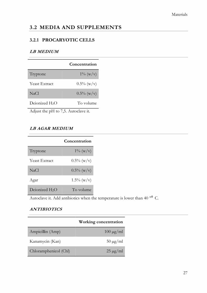
3.2 MEDIA AND SUPPLEMENTS
3.2.1 PROCARYOTIC CELLS
LB MEDIUM
Concentration
Tryptone 1% (w/v)
Yeast Extract 0.5% (w/v)
NaCl 0.5% (w/v)
Deionized H2O To volume
Adjust the pH to 7,5. Autoclave it.
LB AGAR MEDIUM
Concentration
Tryptone 1% (w/v)
Yeast Extract 0.5% (w/v)
NaCl 0.5% (w/v)
Agar 1.5% (w/v)
Deionized H2O To volume
Autoclave it. Add antibiotics when the temperature is lower than 40 °⁰ C.
ANTIBIOTICS
Working concentration
Ampicillin (Amp) 100 µg/ml
Kanamycin (Kan) 50 µg/ml
Chloramphenicol (Chl) 25 µg/ml
Page 36
Materials
28
3.2.2 EUKARYOTIC CELLS
FAD
Working
concentration Company
Ham’s F-12, with L-Glutamine 73 % (v/v) GIBCO, Invitrogen
DMEM high glucose with glutamine
(= Dulbecco’s Modified Eagle
Medium)
23% (v/v) GIBCO, Invitrogen
Fetal Bovine Serum (FBS) 4 % (v/v) GIBCO, Invitrogen
Pen Strep (Penicillin/Streptomycin) 100 U/ml GIBCO, Invitrogen
Adenine, (6-aminopurine) 24 µg/ml SIGMA
Recombinant Human EGF
(= Epidermal Growth Factor) 10 ng/ml R&D company
Insulin solution human 10 µg/ml Sigma Aldrich
Hydrocortisone 400 ng/ml Sigma Aldrich
Ciprofloxacin hydrochloride 10 µg/ml EUROMEDEX
Cholera Toxin 8,3 ng/ml List Biological laboratories, INC
DMEM + + +
Working
concentration Company
DMEM high glucose with glutamine 90% (v/v) GIBCO, Invitrogen
Fetal Bovine Serum (FBS) 10 % (v/v) GIBCO, Invitrogen
Pen Strep (Penicillin/Streptomycin) 100 U/ml GIBCO, Invitrogen
Ciprofloxacin hydrochloride 10 µg/ml EUROMEDEX
Page 37
Materials
29
OPTI-MEM
Opti-MEM™ Reduced Serum Medium, GIBCO, Invitrogen
DPBS
“Dulbecco's phosphate-buffered saline” by Gibco.
TRYPSIN
Trypsin-EDTA (0,25%), phenol red, Life Technologies
MITOMYCIN C
Mitomycin C from Streptomyces caes 2mg, Sigma Aldrich
CRYOMEDIUM
90% FBS + 10% DMSO
3.3 HUMAN CELLS TREATMENTS AND MANIPULATION
Designation Company
MG132 Sigma Aldrich #C2211
Cycloheximide Ozyme #2112S
Doxorubicin Sigma Aldrich #D1515
Lipofectamine 2000 Invitrogen #11668027
PolyFect Qiagen #301105
Effectene Qiagen #301425
Page 38
Materials
30
3.4 RETROVIRAL INFECTION
Designation Company
CalPhos Mammalian transfection kit BD-Biosciences
Chloroquine 25 mM Sigma Aldrich
Polybrene 5mg/ml Sigma Aldrich
G418 100 mg/ml Sigma Aldrich
3.5 MOLECULAR CLONING
3.5.1 PLASMIDS
pLXSN
pLXSN is a retroviral vector composed of elements from Moloney murine leukemia virus
(MoMuLV) and Moloney murine sarcoma virus (MoMuSV). The 5’ LTR comprises
promoter/enhancer sequences that control the transcription of + extended viral
packaging signal and of the gene of interest, which is cloned into the multiple cloning site
(MCS). The MCS has four unique cloning sites that are EcoRI, HpaI, XhoI, and BamHI.
The SV40 early promoter PSV40e regulates transcription of the neomycin resistance gene
for eukaryotic selection. The ColE1 origin of replication serves for replication of pLXSN
in bacteria as well as the ampicillin resistance gene allows selection of pLXSN-
transformed bacteria. After transfection of pLXSN into a packaging cell line, pLXSN
expresses the + packaging signal generating infectious but replication-incompetent
retroviral particles. The pLXSN features are shown in the schematic representation of
figure 10.
Page 39
Materials
31
Figure 10: pLXSN vector features. pLXSN is a 5874 bp retro-viral vector containing the
MoMuLV 5’ LTR that control the transcription of Psi packaging signal and of the gene of
interest. The neomycin resistance (in red), allow the selection of successfully infected cells. The
ampicillin resistance gene (in orange) with its promoter allows for selection in bacteria.
Description Reference
pLXSN(E6/E7)HPV16 M. Tommasino
pLXSN(E6/E7)HPV49 M. Tommasino
pLXSN(E6/E7)HPV75 This thesis
pLXSN(E6/E7)HPV76 This thesis
pLXSN(E6/E7)HPV115 This thesis
pLXSN(E6/E7)HPV76 mut E39R This thesis
pLXSN(E6/E7)HPV76 mut Y42R This thesis
pLXSN(E6/E7)HPV76 mut D44A This thesis
pLXSN(E6/E7)HPV76 mut F45E This thesis
Page 40
Materials
32
pET MBP 1C
The pET system is used for the expression of recombinant proteins in E. coli, under the
control of a strong bacteriophage T7 transcription. The expression of the recombinant
protein can be induced only when the system is provided with a source of T7 DNA
polymerase, usually using engineered E. coli strain (such as the Rosetta strain). The use of
the T7 induction system ensures the target gene expression is silent in the un-induced
state as well as the high quantity of the protein of interest after the induction. Target
genes are firstly cloned using non-expressing hosts (such as DH5 strain) and afterword
transferred to expression host, where the T7 DNA polymerase gene is under the control
of the lac promoter and therefore can be induced by the addition of IPTG.
The pET-MBP_1c plasmid is a pET vector modified by Gunter Stier (Universität
Heidelberg, Heidelberg, Center for Biochemistry) to carry the ORF of the Maltose
Binding Protein (MBP). This modified version of the pET vector maintains all the
characteristics of the pET system and, in addition, the recombinant protein is expressed
as a fusion protein with the maltose binding protein. The MBP is a part of the
maltose/maltodextrin system of Escherichia coli and can be used to increase the solubility
of recombinant proteins. The mechanism underlying the increased solubility of the fusion
proteins is still not fully understood but the MBP is able to prevent the aggregation of the
protein of interest. Moreover, the MBP can be used as an affinity tag for the purification
of the recombinant proteins using amylose coupled-beads.
The pET MBP 1c features are shown in the schematic representation of figure 11.
Page 41
Materials
33
Figure 11: pET MBP 1c vector features. The main feature of this plasmid is the maltose
binding protein gene (in pink). This gene can be expressed only in presence of T7 DNA
polymerase that recognizes the T7 binding site. The kanamycin resistance gene (in orange) allows
the selection of successfully transformed bacteria.
Description Reference
pET MBP 1c (E6) HPV16 This thesis
pET MBP 1c (E6) HPV49 This thesis
pET MBP 1c (E6) HPV75 This thesis
pET MBP 1c (E6) HPV76 This thesis
pET MBP 1c (E6) HPV115 This thesis
pET MBP 1c (E6) HPV76 mut E39R This thesis
pET MBP 1c (E6) HPV76 mut Y42R This thesis
pET MBP 1c (E6) HPV76 mut D44A This thesis
pET MBP 1c (E6) HPV76 mut F45E This thesis
Page 42
Materials
34
3.5.2 ENZYMES
Designation Company
Restriction enzymes New England Biolabs
HotStarTaq DNA Polymerase Qiagen
T4 DNA ligase Roche
Mesa Green qPCR Master Mix Plus for SYBR Assay (Eurogentec) Eurogentec
3.5.3 OLIGONUCLEOTIDES FOR siRNA KNOCKDOWN
Designation Sequence Company
ON-TARGETplus Human UBE3A siRNA
(E6AP) Not provided
Dharmacon
(L005137000005)
ON-TARGETplus Human CDKN2A siRNA
(p16) Not provided
Dharmacon
(L-011007-00-0005)
Scramble Not provided Eurofins
Page 43
Materials
35
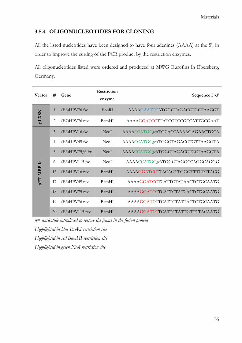
3.5.4 OLIGONUCLEOTIDES FOR CLONING
All the listed nucleotides have been designed to have four adenines (AAAA) at the 5’, in
order to improve the cutting of the PCR product by the restriction enzymes.
All oligonucleotides listed were ordered and produced at MWG Eurofins in Ebersberg,
Germany.
n= nucleotide introduced to restore the frame in the fusion protein
Highlighted in blue EcoRI restriction site
Highlighted in red BamHI restriction site
Highlighted in green NcoI restriction site
Vector # Gene Restriction
enzyme Sequence 5’-3’
pL
XS
N 1 (E6)HPV76 fw EcoRI AAAAGAATTCATGGCTAGACCTGCTAAGGT
2 (E7)HPV76 rev BamHI AAAAGGATCCTTATCGTCCGCCATTGCGAAT
pE
T M
BP
1c
3 (E6)HPV16 fw NcoI AAAACCATGGgtATGCACCAAAAGAGAACTGCA
4 (E6)HPV49 fw NcoI AAAACCATGGgtATGGCTAGACCTGTTAAGGTA
5 (E6)HPV75/6 fw NcoI AAAACCATGGgtATGGCTAGACCTGCTAAGGTA
6 (E6)HPV115 fw NcoI AAAACCATGGgtATGGCTAGGCCAGGCAGGG
16 (E6)HPV16 rev BamHI AAAAGGATCCTTACAGCTGGGTTTCTCTACG
17 (E6)HPV49 rev BamHI AAAAGGATCCTCATTCTATAACTCTGCAATG
18 (E6)HPV75 rev BamHI AAAAGGATCCTCATTCTATCACTCTGCAATG
19 (E6)HPV76 rev BamHI AAAAGGATCCTCATTCTATTACTCTGCAATG
20 (E6)HPV115 rev BamHI AAAAGGATCCTCATTCTATTGTTCTACAATG
Page 44
Materials
36
3.5.5 OLIGONUCLEOTIDES FOR REAL TIME PCR (RT-PCR)
All oligonucleotides listed were ordered and produced at MWG Eurofins in Ebersberg,
Germany.
Gene Sequence 5’-3’
Cdc2 fw: AATCTATGATCCAGCCAAACGAA
rev: TTCTTAATCTGATTGTCCAAATCATTAAA
Cdk2 fw: GGCTGCATCTTTGCTGAAAT
rev: CCCAGAGTCCGAAAGATCCG
p21 fw: GACACCACTGGAGGGTGACT
rev: CCACATGGTCTTCCTCTGCT
PUMA fw: GGATGAAATTTGGCATGGGGTCT
rev: GGACAAGTCAGGACTTGCAG
hTERT fw: TTC AAG GCT GGG AGG AAC AT
rev: ACA TGC GTG AAA CCT GTA CG
SERPINE fw: ATCGAGGTGAACGAGAGTGG
rev:ACTGTTCCTGTGGGGTTGTG
MT1X fw: AACTCCTGCTTCTCCTTGCC
rev: GCTCTATTTACATCTGAGAGCACAG
GADD45a fw: TGCGAGAACGACATCAACAT
rev: GCAGGATCCTTCCATTGAGA
GAPDH fw: AAGGTGGTGAAGCAGGCGT
rev: GAGGAGTGGGTGTCGCTGTT
Page 45
Materials
37
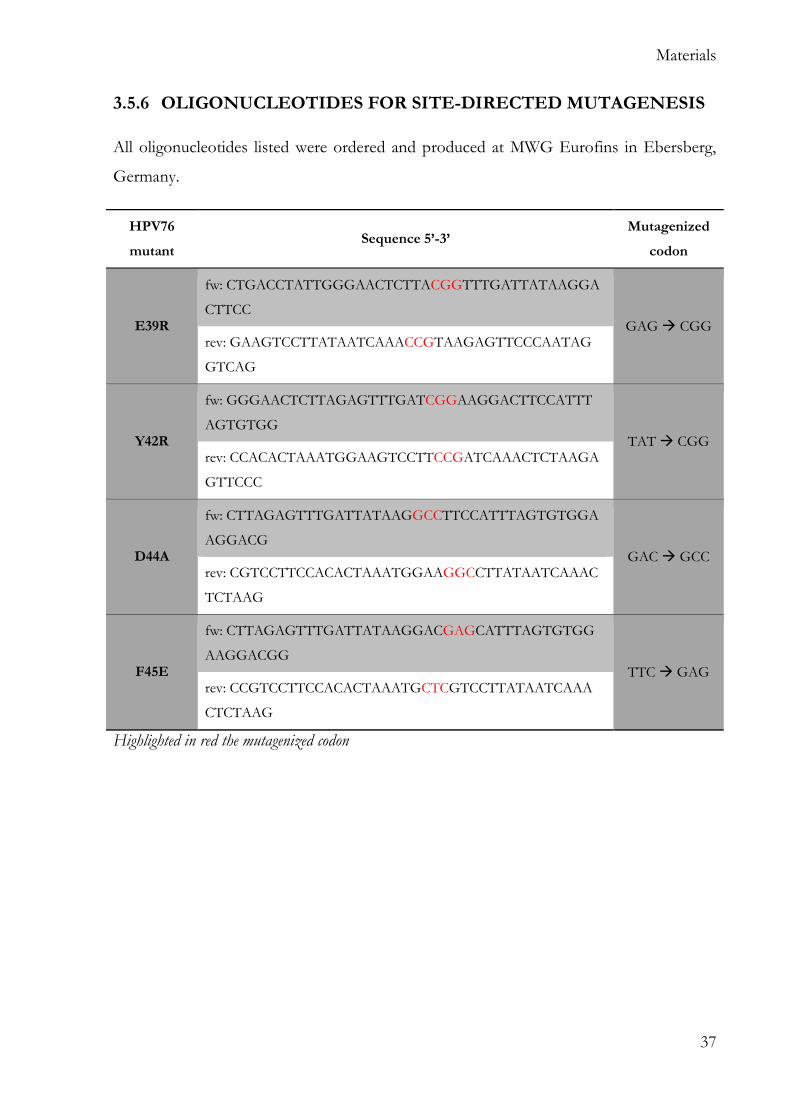
3.5.6 OLIGONUCLEOTIDES FOR SITE-DIRECTED MUTAGENESIS
All oligonucleotides listed were ordered and produced at MWG Eurofins in Ebersberg,
Germany.
HPV76
mutant Sequence 5’-3’
Mutagenized
codon
E39R
fw: CTGACCTATTGGGAACTCTTACGGTTTGATTATAAGGA
CTTCC GAG CGG
rev: GAAGTCCTTATAATCAAACCGTAAGAGTTCCCAATAG
GTCAG
Y42R
fw: GGGAACTCTTAGAGTTTGATCGGAAGGACTTCCATTT
AGTGTGG TAT CGG
rev: CCACACTAAATGGAAGTCCTTCCGATCAAACTCTAAGA
GTTCCC
D44A
fw: CTTAGAGTTTGATTATAAGGCCTTCCATTTAGTGTGGA
AGGACG GAC GCC
rev: CGTCCTTCCACACTAAATGGAAGGCCTTATAATCAAAC
TCTAAG
F45E
fw: CTTAGAGTTTGATTATAAGGACGAGCATTTAGTGTGG
AAGGACGG TTC GAG
rev: CCGTCCTTCCACACTAAATGCTCGTCCTTATAATCAAA
CTCTAAG
Highlighted in red the mutagenized codon
Page 46
Materials
38
3.5.7 BUFFERS AND SOLUTIONS
AGAROSE GEL ELECTROPHORESIS
Designation Composition / Company
50X TAE buffer ABCYS Eurobio
Massruler Loading Dye Life Technologies
MassRuler DNA Ladder, Mix, ready-to-use Life Technologies
GelRed staining Biotium
1% agarose gel
1% agarose (w/v)
1X TAE buffer
0.004 % GelRed (v/v)
3.6 REAGENTS FOR PROTEIN ANALYSIS
3.6.1 ENZYMES
Lambda Protein Phosphatase, New England Biolabs.
3.6.2 IP BUFFER
Ingredient Working concentration
Tris HCl pH 7.5 20 mM
NaCl 200 mM
EDTA 1 mM
NP40 0.5%
H2O Up to 10 ml
cOmplete™, Mini, EDTA-free Protease Inhibitor Cocktail
(Roche) 1 tablet
Page 47
Materials
39
3.6.3 PROTEIN BUFFER 10X
Ingredient
Glycin 144.1g
TRIS 30.3 g
H2O To volume of 1 l
3.6.4 SDS-POLYACRYLAMIDE ELECTROPHORESIS
The following recipes are intended for one gel (1.5 mm) using the “Mini-PROTEAN”
cast system from Biorad.
RUNNING GEL
Ingredient Volume
Gel 10% Gel 12%
Acrylamide 30% 3.3 ml 4.0 ml
Tris 1 M pH 8.8 2.5 ml 2.5 ml
SDS 10% 0.1 ml 0.1 ml
APS 10% 0.1 ml 0.1 ml
TEMED 0.004 ml 0.004 ml
H2O 4.0 ml 3.3 ml
Page 48
Materials
40
STACKING GEL
Ingredient Volume
Acrylamide 30% 0,33 ml
Tris 0,5 M pH 6.8 0,25 ml
SDS 10% 0,02 ml
APS 10% 0,02 ml
TEMED 0,002 ml
H2O 1,4 ml
RUNNING BUFFER
Ingredient Volume
Protein buffer 10x 100 ml
SDS 10 ml
H2O 890 ml
PROTEIN MARKER
PageRuler™ Prestained Protein Ladder, 10 to 180 kDa; Thermo Fisher (Germany)
LAEMMLI BUFFER 6X
Ingredient
SDS 1.2 g
Bromophenol blue 6 mg
Glycerol 4.7 ml
Tris HCl 0.5 M pH 6.8 1.2 ml
Beta-mercaptoethanol 0.5 ml
H2O 2.1 ml
Page 49
Materials
41
3.6.5 WESTERN BLOT ANALYSIS
TRANSFER BUFFER
Ingredient Volume
Protein buffer 10x 100 ml
Methanol 200 ml
H2O 700 ml
OTHER SOLUTIONS
Designation Composition/Company
Blocking solution 10 % (w/v) skim milk
PBS Tween
Antibody buffer 5 % (w/v) skim milk
PBS Tween
PBS Tween 1X PBS
0.064 % (v/v)
Stripping solution 15 g Glycine
10 ml Tween 20
1 g SDS
1 l H2O, pH 2.2
Clarity™ Western ECL Blotting Substrates BioRad, Munich, Germany
Page 50
Materials
42
3.7 IMMUNOLOGICAL ASSAYS
3.7.1 ANTIBODIES
All the following antibodies have been used with a 1:1000 dilution if not differently
stated.
Designation Description Reference
β-actin (C4) Mouse monoclonal antibody detecting human actin. MP Biomedicals,
#0869100
Phospho pRb
(Ser795)
Polyclonal rabbit antibody that detects endogenous levels
of Rb only when phosphorylated at serine 795.
Cell signaling,
#9301
Total pRb
Mouse monoclonal antibody that recognizes an epitope
between amino acids 332-344 of the human
retinoblastoma protein.
BD Pharma,
#554136
Cdc2 (ab-2) Mouse monoclonal antibody detecting Cdc2/Cdk1 Calbiochem,
#CC01
Cyclin A (H-432) Rabbit polyclonal antibody raised against full length cyclin
A of human origin.
Santa Cruz,
#sc751
p16INK4a Mouse monoclonal to CDKN2A/p16INK4a NovoCastra,
DCS-50
p53 DO1 Mouse monoclonal antibody raised against amino acids
11-25 of p53 of human origin.
Santa Cruz,
#sc126
E6AP-330 Mouse monoclonal Anti-E6AP antibody purified from
hybridoma cell culture.
Sigma Aldrich,
#E8655
GAPDH (6C5) Mouse monoclonal antibody raised against GAPDH. Santa Cruz,
#sc-32233
Secondary mouse (dil
1:2000) Anti-Mouse IgG (H+L), HRP Conjugate
Promega,
W4021
Secondary Rabbit (dil
1:2000) Anti-Rabbit IgG (H+L), HRP Conjugate
Promega,
W4011
Page 51
Materials
43
3.8 MALTOSE BINDING PROTEIN PULLDOWN
BUFFER AND SOLUTIONS
Designation Composition
IPTG 100 mM 0,238 g IPTG
10 ml H2O
IP buffer See 6.6.2
MBP-buffer
10 mM Hepes pH 7.4
100 mM NaCl
1 mM EDTA
10% Glycerol
0,1% triton X-100
BEADS
Anti-MBP Magnetic Beads. E8037S, New England Biolabs.
3.9 ILLUMINA ARRAY
3.9.1 INSTRUMENTS AND CONSUMABLE
Designation Company
Nanodrop® Thermo Scientific
2100 bioanalyzer Agilent
RNA 6000 Nano kit Agilent
HumanHT-12 v4 Expression BeadChips Illumina
TotalPrep RNA Amplification Kit (Ambion®) Illumina
BeadArray Reader Illumina
Page 52
Materials
44
3.9.2 SOFTWARE
Designation Company
Genome Studio V2010.2 Illumina
BRB-ArrayTools software v4.2 https://brb.nci.nih.gov/BRB-ArrayTools/
R software https://www.r-project.org/
Venny http://bioinfogp.cnb.csic.es/tools/venny/
3.10 LIQUID CHROMATOGRAPHY AND MASS SPECTROMETRY
3.10.1 INSTRUMENTS AND CONSUMABLES
Designation Company
0.2 m Captiva ND plates Agilent Technologies
a Rapid EPS well plate sealing tape BioChromato
6550 quadrupole time-of-flight mass spectrometer coupled to
1290 Infinity UHPLC system Agilent Technologies
Acquity UPLC HSS T3 column (1.8 m, 2.1 × 100 mm) Waters
3.10.2 SOFTWARE
Designation Company
MassHunter Acquisition B.05.01 Agilent Technologies
MassHunter Qualitative Analysis B.06.00 Agilent Technologies
DA Reprocessor, Mass Profiler Professional 12.1 Agilent Technologies
Profinder B 06.00 software Agilent Technologies
Page 53
Materials
45
3.11 CHEMICALS
All chemicals were of analytical grade or better and purchased from one of the following
companies:
Company Location
Gibco Eggenstein, Germany
Life Technologies Karlsruhe, Germany
Fisher-Scientific Schwerte, Germany
Merck Darmstadt, Germany
Roth Karlsruhe, Germany
Sigma-Aldrich Munich, Germany
3.12 KITS
Designation Company
QIAquick PCR Purification Kit Qiagen
QIAquick Gel Extraction Kit Qiagen
QuikChange Lightning Site-Directed Mutagenesis Kit Agilent technologies
NucleoSpin® RNA extraction kit MACHEREY-NAGEL GmbH
RevertAid First Strand cDNA Synthesis Kit Thermo Fisher
NucleoSpin® Plasmid mini prep kit MACHEREY-NAGEL GmbH
PureLink™ HiPure Plasmid Filter Maxiprep Kit Invitrogen
Senescence -Galactosidase Staining Kit Cell Signalling
Page 54
Materials
46
3.13 LABORATORY EQUIPMENT
3.13.1 ELECTRICAL EQUIPMENT
Designation Company
800 W microwave Bosch, Gerlingen-Schillerhohe, Germany
Bacterial culture shaker Informs AG, Bottmingen, Switzerland
Microbio Safe 12 hood Thermo Fisher, Germany
Incubateur CO2 jaquette eau series 3 Thermo Fisher, Germany
Developing Machine AgfaCurix60 Agfa, Munich, Germany
Computers and monitors Dell, Round Rock, United States
ChemiDoc™ XRS+ System BioRad, Munich, Germany
Mini-PROTEAN® Tetra Handcast
Systems BioRad, Munich, Germany
Sub-Cell® GT Cell BioRad, Munich, Germany
Mini Trans-Blot® Cell BioRad, Munich, Germany
PowerPac™ Basic Power Supply BioRad, Munich, Germany
GFC water bath Grant Instruments, Cambridge, UK
Ice maker Hoshizaki, Willich-Munchheide, Germany
Impulse Sealer RNS Corp, Taipei, Taiwan
Integra pipetboy Integra Bioscience GmbH, Fernwald,
Germany
Cell counter Biorad TC20 ™
microscope Statif inverse TS100 NIKON
MilliQ ultra-pure water unit Millipore Merck, Darmstadt, Germany
Nanodrop spectrophotometer PegLab, Erlangen, Germany
Page 55
Materials
47
Designation Company
Nitrogen tank Chrono Messer, Krefeld, Germany
pH meter Sartorius, Göttingen, Germany
Refrigerators and freezers Liebherr, Ochsenhausen, Germany
Sartorius Scale Sartorius, Göttingen, Germany
Thermomixer 5436 Eppendorf, Hamburg, Germany
Ultra-low freezer Eppendorf Inc., Enfield, USA
Vortex Genie 2™ Bender and Hobein, Ismaning, Germany
Western Blot Exposition Cassette Kodak, Stuttgart, Germany
Centrifuge 5810 Eppendorf, Germany
Centrifuge 5430 Eppendorf, Germany
Centrifuge 5424R (cold) Eppendorf, Germany
Centrifuge 5415D Eppendorf, Germany
Centrifuge 5417C Eppendorf, Germany
Centrifuge 5415R Eppendorf, Germany
Page 56
Materials
48
3.13.2 COMMON USE EQUIPMENT
Designation Company
1,5 ml and 2 ml reaction tubes Eppendorf, Hamburg, Germany
0,2 ml reaction tubes Eppendorf, Hamburg, Germany
15 ml reaction tubes Fisher Scientific, Waltham, United States
50 ml reaction tubes Fisher Scientific, Waltham, United States
25 cm2, 75 cm2 and 150 cm2 tissue culture
flasks Fisher Scientific, Waltham, United States
6, 10 and 15 cm cell culture plates Corning, New York, United States
6-, 12-, 24-, 48 and 96-well tissue culture
plates Corning, New York, United States
Cryotubes, 2 ml Carl Roth GmbH, Karlsruhe, Germany
Examination gloves XCEEDTM Nitril Starlab, Ahrensburg, Germany
One-time use filter, 0.2/0.4 m Renner, Dannstadt, Germany
Parafilm “M” American National Can, Chicago, USA
qPCR 96 well plate non skirted white Eurogentec, France
Petri dishes Greiner, Frickenhausen, Germany
Pipettes (1000, 200, 20, 10 and 2l) Gilson Middleton, USA
Pipette Tipps (1000, 200l, 20l, 10l) STARLAB, Hamburg, Germany
Syringes and needles BD Franklin Lakes, USA
Whatman filter paper 3MM Schleicher & Schuell, Dassel, Germany
Magnetic rack DynaMag™-2 Magnet Fisher Scientific, Waltham, United States
PVDF membrane EMD Millipore, Burlington, United
States
Page 57
Materials
49
3.13.3 SOFTWARE AND WEBSITES
Designation Company
Adobe CS4/CS6 Adobe, San Jose, USA
Microsoft Office 2003, 2010 Microsoft, Redmond, USA
Image Lab™ Software BioRad, Munich, Germany
Webcutter http://rna.lundberg.gu.se/cutter2
PaVE https://pave.niaid.nih.gov/
BRB-ArrayTools software v4.2 NIH
Serial Cloner http://serialbasics.free.fr/Serial_Cloner.html
Human Metabolome Database http://www.hmdb.ca/
METLIN database https://metlin.scripps.edu
Page 58
Methods
50
4 METHODS
4.1 CULTIVATION AND MANIPULATION OF PROKARYOTIC CELLS
4.1.1 TRANSFORMATION OF BACTERIA BY HEAT SHOCK
- Put 1 ng of pure DNA or 5 µl of ligation product in 50 l of bacteria
(DH5)DH5α) or 80 µl (Rosetta)
- Put the solution for 30 minutes on ice
- Then 30 seconds at 42°C
- And again in ice for 2 min
- Put the bacteria in 300 l of LB without antibiotics and incubate with agitation for
1 h and 30 min at 37°C
- Take a rate of 50 l and put in a Petri dish with LB + antibiotic; incubate at 37°C
overnight
- Centrifuge the rest of the bacteria, re-suspend in 100 l of LB and put in a Petri
dish with LB + antibiotic; incubate at 37°C overnight
Each colony formed originates from a single transformed bacterial cell. Therefore, each
cell within each colony contains identical plasmid DNA.
4.1.2 CULTIVATION AND STORAGE OF BACTERIA
BACTERIAL CULTURE
- Take a colony with a sterile tip and dissolve it in 6 ml of LB medium with
antibiotic.
- Incubate in agitation at 37°C overnight
- Process the bacteria to purify the plasmid DNA (mini-prep) or
- Take the pre-inoculum and put it in 200 ml of LB with antibiotic (maxi-prep)
- Put the culture at 37°C in agitation overnight.
Page 59
Methods
51
LONG TERM BACTERIAL STORAGE
For long-term storage of verified (sequenced) bacterial clones, 500 µl of a liquid culture
was transferred to a 2ml cryotube and 500 l of glycerol (100%) were added. The glycerol
stocks were stored at -80⁰C.
4.2 CULTIVATION AND MANIPULATION OF EUKARYOTIC CELL
All cell lines were cultivated in an incubator at 37⁰C, 5% CO2 and 90% humidity.
Passaging of the cells was performed when cells reached around 80% confluency.
4.2.1 CULTIVATION OF NIH/3T3 FIBROBLASTS
The fibroblast cells are fast growing cells and every 3/4 days is necessary to split them
because they became confluent.
The NIH/3T3 are kept in culture in T75 flasks.
SPLIT OF NIH/3T3
- Remove the medium
- Add 8 ml of DPBS and wash the cells
- Add 1 ml of trypsin and incubate 1 minute at 37°C
- Add 9 ml of DMEM +++ to inactivate the trypsin
- Centrifuge for 3 minutes at 900 rpm
- Discard the supernatant
- Re-suspend the pellet into 10 ml of DMEM +++
- Put an amount of the re-suspended cells in the flask (the amount depends on the
dilution).
4.2.2 CULTIVATION OF PHOENIX
The Phoenix cells are adherent cells but, unlike the NIH/3T3, they attach to the surface
poorly. It’ necessary to pay attention while washing the cells and changing the media to
Page 60
Methods
52
avoid the detachment of the cells from the surface of the dish. The protocol of the split
of the cells is the same used for the NIH/3T3.
The Phoenix are kept in culture in 10 cm dishes.
4.2.3 CULTIVATION OF HUMAN KERATINOCYTES (PRIMARY
AND EXPRESSING E6/E7)
The human keratinocytes were cultivated with the support of feeder layer (NIH/3T3
fibroblasts) with a protocol modified from Rheinwald and Green [141]. The cells of the
feeder layer not only secrete soluble factors into the culture medium but deposit ECM
molecules on the culturing surface that facilitate attachment and growth of co-cultured
cells [142].
The NIH/3T3 cells added to the human primary keratinocyte are treated with the
mitomycin to block their proliferation. The feeder layers need to be changed every time
the keratinocytes are split or every 2/3 days since the NIH/3T3 cells treated with the
mitomycin die.
The keratinocytes are kept in culture in T25 flasks.
PREPARATION OF THE FEEDER (NIH/3T3)
- Take a flask of NIH/3T3 (around the 80% of confluence), add 250 µl mitomycin
C and incubate at 37°C for 2 hours
- Remove the medium
- Add 3 ml of DPBS and wash the cells
- Add 1 ml of trypsin and incubate at 37°C for 1 minutes
- Add 9 ml of DMEM +++ to inactivate the trypsin
- Centrifuge for 3 minutes at 900 rpm
- Remove the solution
- Add 10 ml of FAD
- Counts the cells
Page 61
Methods
53
CHANGE OF FEEDER LAYER
- Remove the medium
- Add 3 ml of DPBS to wash the cells
- Add 3 ml of DPBS + EDTA and let it acts for 3 minutes. Only the fibroblasts tear
away from the flask meanwhile the keratinocyte remain adherent to the flask.
- Remove the solution
- Add 3 ml of DPBS to wash the cells
- Remove the solution
- Add 5 ml of FAD into the flask
- Add “x” µl of counted NIH/3T3 (for the amount of the NIH/3T3 see the table 1)
- Mix with a cross movement
SPLIT OF KERATINOCYTES
- Remove the medium
- Add 3 ml of DPBS to wash the cells
- Add 3 ml of DPBS + EDTA and let it acts for 3 minutes.
- Remove the solution
- Add 3 ml of DPBS to wash the cells
- Remove the solution
- Add 1 ml of trypsin and incubate at 37°C for 5-7 minutes
- Add 9 ml of DMEM +++ to inactivate the trypsin
- Centrifuge for 3 minutes at 900 rpm
- Remove the supernatant
- Re-suspend the pellet into the appropriate amount of FAD
- Add “x” µl of counted NIH/3T3 (for the amount of the NIH/3T3 see the table 1)
- Mix with a cross movement
For the maintenance of the culture always split the keratinocytes 1:2.
Page 62
Methods
54
Table 1: Amount of feeder for each type of support.
Type of support Number of NIH/3T3 cells
Small flask 2.5 x 10⁵ cells
Big flask 7 x 10⁵ cells
6 cm2 dish 1.5 x 10⁵ cells
10 cm2 dish 5 x 10⁵ cells
6 wells dish 1 x 10⁵ cells
4.2.4 CULTIVATION OF HNC136
The protocol of the split of the cells is the same used for the NIH/3T3 except for the
incubation time at 37 ⁰C with trypsin, which varies accordingly to the cell type.
This cells line can be maintained in culture in T75 or T150 flasks.
HNC136 are usually split 1:2 for maintenance.
4.2.5 CELLS COUNTING
- Put 10 µl of re-suspended cells in an Eppendorf
- Add 10 µl of trypan blue (allows to distinguish the vital from the not vital cells)
and mix
- Take 10 µl of the mix and add them to the counting plate
- Read the concentration of cells in the counting machine (Biorad TC20 ™).
4.2.6 CRYOPRESERVATION AND THAWING OF MAMMALIAN
CELLS
CRYOPRESERVATION
For cryopreservation, the cells were harvested by trypsinization and centrifuged. After
centrifugation, the cell pellet was resuspended in 1ml cryomedium and the cell solution
stored in cryotubes. Freezing of the cells was performed in a slow freeze chamber with
Page 63
Methods
55
isopropanol at -80°C for at least 24h before the cells were transferred to liquid nitrogen.
Using a container filled with isopropanol allows freezing the cells slowly by reducing the
temperature at approximately 1°C per minute. When the cells are re-suspended in
cryomedium put them immediately in the freezer to avoid the death of the cells (DMSO
is toxic).
THAWING
- Take the cryotubes out of liquid nitrogen and thaw the cell solution by keeping in
the hands (attention: once the cells are thaw, do the next steps immediately)
- Transfer 1 ml of cells in a falcon and add 10 ml of DMEM +++
- Centrifuge at 900 rpm for 3 minutes
- Discard the supernatant
- Gently resuspend the pellet in the appropriate medium.
When thawing the human keratinocytes, prepare in advance the inactivated feeder cells as
described in paragraph 4.2.3.
4.2.7 CELL TREATMENTS
DOXORUBICIN
- Plate 3.5x10⁵ HFKs cells or HFKs cells expressing E6/E7 of the different HPVs
in a well of a 6 well plate. Add the appropriate amount of feeder cells.
- After 24 hours remove the feeder as described in paragraph 4.2.3 and
- treat the cells with doxorubicin or DMSO
- collect the cells after 8 hours of treatment; preserve the 2/3 of the pellet in one 1.5
ml tube for future protein extraction and the remaining 1/3 of the pellet in
another 1.5 ml tube for future RNA extraction.
Page 64
Methods
56
MG132
- Plate 3.5x10⁵ HFKs cells or HFKs cells expressing E6/E7 of the different HPVs
in a well of a 6 well plate. Add the appropriate amount of feeder cells.
- After 24 hours remove the feeder as described in paragraph 4.2.3 and
- treat the cells with MG132 or DMSO
- collect the cells after 4 hours of treatment; preserve the 2/3 of the pellet in one 1.5
ml tube for future protein extraction and the remaining 1/3 of the pellet in
another 1.5 ml tube for future RNA extraction.
CYCLOHEXIMIDE
- Plate 3.5x10⁵ NIKs pLXSNØ cells or NIKs cells expressing E6/E7 of the
different HPVs in a well of a 6 well plate.
- After 24 hours, treat the cells with cycloheximide or DMSO
- collect the cells after 1, 2, 4 and 6 hours of treatment.
4.2.8 siRNA KNOCK-DOWN
- Seed 4x10⁵ HFKs or HFKs expressing E6/E7 in a well of a 6 wells-plate; add the
appropriate amount of feeder cells.
- After 24 hours from the seeding, transfect the cells with the same 10 nM of
scramble or siRNA using Lipo2000.
- After 6 hours replace the medium with fresh FAD without cholera toxin,
Penicillin/streptomycin and Ciprofloxacin.
- After 24 hours replace the medium with complete FAD.
- After 48 hours from the transfection remove the feeder and collect the cells.
4.3 RETROVIRUS INFECTION
The retrovirus infection donor 1 has been performed in Heidelberg in a P2 plus
laboratory by myself while the retrovirus infection donor 2 and 3 has been performed in
Lyon in a P3 laboratory by Cecilia Sirand and Gessica Tore.
Page 65
Methods
57
4.3.1 TRANSFECTION
Two days before transfection seed the Phoenix cells in 10 cm dishes, in this way they will
be 50/70% confluent the day of the transfection.
Infect the Phoenix cells with the construct, following the protocol:
PREPARE THE DNA SOLUTION
- in a 15 ml Falcon mix the DNA (10 µg) with H2O (to a volume of 440 µl) and 62
µl of CaCl2 (2M)
- mix gently
- add 500 µl of HBS (2X) drop by drop
- shake with energy the solution
PREPARE THE PHOENIX CELLS
- remove the old medium from the Phoenix cell cultures
- add 5 ml of fresh medium
- add 5 µl of Chloroquine and gently mix
ADD THE DNA SOLUTION
- add the DNA solution drop by drop evenly distributing the solution in the dish
- transport the cells into the P3 laboratory
CHANGE MEDIUM
- after 6/8 hours wash twice the phoenix cells with DPBS
- add 5 ml of fresh medium.
CHANGE MEDIUM
- after 24 hours change the medium without the wash with DPBS
Page 66
Methods
58
4.3.2 INFECTION
PREPARE RETROVIRAL SUSPENSION
- Collect medium of the transfected Phoenix cells and pass twice the medium
through the 0.22 µm filter.
- Add 5 µl of Polybrene into each retroviral suspension
INFECT THE PRIMARY KERATINOCYTE
- Add the retrovirus suspension to the keratinocytes
- After 3 hours remove the retrovirus suspension
- Add fresh FAD medium
4.3.3 SELECTION
- Split the cells 1:1 and start the selection 24 hours after the infection using the
neomycin.
Split 1:1 a flask of HPK not infected and add neomycin; when these cells die, the
selection is over.
4.3.4 TEST FOR THE EXIT OF THE CELLS FROM THE P3
To exit the cells from the P3 it is necessary to prove that the transformed HPK cells do
not produce viruses and so they are not dangerous anymore.
- Plate 0.5*105 NIH/3T3 cells in a 6 wells plate for each transformed HFK line
- add the medium from the transformed HFK on the NIH/3T3 cells
- start the selection with neomycin
- When all the NIH cells die the transformed cells can go out of P3.
Page 67
Methods
59
4.4 MOLECULAR METHODS
4.4.1 PURIFICATION OF PLASMID DNA
MINI-PREP
Isolation of plasmid DNA with the NucleoSpin® Plasmid mini prep kit was performed
from a single bacterial colony previously cultured in 6 ml liquid LB medium containing
the corresponding antibiotics. The preparation of plasmid DNA was performed
according to manufacturer´s instructions.
MAXI-PREP
Isolation of plasmid DNA with the PureLink™ HiPure Plasmid Filter Maxiprep Kit was
performed using 200ml LB medium with corresponding antibiotics which were inoculated
with either 1 ml from a liquid bacterial culture or from a bacterial glycerol stock. The
preparation of the plasmid DNA was performed according to manufacturer´s instructions
4.4.2 DNA VISUALIZATION
AGAROSE GEL ELECTROPHORESIS
To visualize the DNA or to purify the DNA after digestion, prepare the gel as follow.
The percentage of agarose depends on the size of the DNA to visualize.
- In a jar prepare 0.5 g (for a 1% gel) or 0.4 g (for a 0.8% gel) of agarose and add 50
ml of TAE 1X.
- Cover the jar with his cap without closing it and put it in the microwave to the
maximum heating until the agarose is completely melted.
- Under chemical hood add 1 µl of red gel and mix gently.
- Assembly the tray with the combs for the wanted number of wells.
- Pour the prepared solution into the tray and wait until the complete solidification.
- Remove the combs and put the tray into the electrophoretic system. The TAE 1X
buffer must submerge completely the agarose gel.
- Load the samples in the wells.
- Close the electrophoretic system and set the voltage (85V for 40 minutes).
Page 68
Methods
60
Before application of the DNA samples to the gel, the samples were mixed with 6x DNA
loading dye in a ratio 1:6. To determine the size of the corresponding DNA fragments, a
marker was loaded on the gel.
The DNA fragments were visualized with the ChemiDoc machine.
4.4.3 MOLECULAR CLONING
POLYMERASE CHAIN REACTION (PCR)
The Polymerase Chain Reaction (PCR) was performed to amplify the DNA sequences of
interest for cloning into selected destination vectors. The DNA sequences were amplified
using corresponding template DNAs encoding for the sequence of interest. Primers
needed for the amplification process were generated as described in paragraph 3.5.3 and
ordered as well as produced at MWG Eurofins, Ebersberg, Germany. Each Primer was
generated for a specific cloning process, containing different restriction sites. The sites to
be included in the primer sequence were selected depending on the specific destination
vector used for later cloning steps.
All PCRs were performed using the HotStarTaq DNA Polymerase from Qiagen,
according to the manufacturer´s instructions (table 2 and table 3). The annealing
temperature (Tm) strongly depends on the primers used for the PCR reaction.
Page 69
Methods
61
Table 2: reaction mix
Ci Cf V
Buffer 10X 1X 2.5 l
dNTP 10 M 200 M 1 l
Primer forward 10 M 0,3 M 1.5 l
Primer reverse 10 M 0,3 M 1.5 l
HotStartTaq pol 5 U/l 2,5 U/reaction 1 l
DNA // // 1 l
H2O // // To volume
25 l
Table 3: Standard PCR program
Temperature Time
95 °C 15 min
94 °C 1 min
35 cycles Tm 1 min
72 °C 1 min/kb of DNA to
extend
72 °C 10 min
4 °C ∞
PCR PURIFICATION
For the purification of the fragment of DNA obtained from PCR, the QIAquick PCR
Purification Kit from Qiagen was used. The purification procedure was carried out
according to the manufacturer´s instructions. The DNA was eluted in 10 µl.
Page 70
Methods
62
ENZYMATIC RESTRICTION OF PCR PRODUCTS
To prepare the PCR product for ligation into the target vector, the PCR products were
doubled digested with the appropriate restriction enzymes. NEB restriction enzymes were
used with CutSmart buffer after the compatibility for double digestion was checked on
the NEB website. A mix was prepared as in table 4 and the samples were incubated at
37°C for 1 hour.
Table 4: standard PCR product digestion mix
Ci Cf Vi
PCR amplified fragments // // 10 µl
Cut Smart Buffer 10X 1X 2 µl
Restriction enzyme 1 10 U/µl 0.27 U/µl 0.8 µl
Restriction enzyme 2 10 U/µl 0.27 U/µl 0.8 µl
H2O // // To volume
20 µl
ENZYMATIC RESTRICTION OF VECTOR
To prepare the vector for ligation with the insert, the vector was doubled digested with
the appropriate restriction enzymes. NEB restriction enzymes were used with CutSmart
buffer after the compatibility for double digestion was checked on the NEB website. A
mix was prepared as in table 5 and the samples were incubated at 37°C for 1 hour.
Page 71
Methods
63
Table 5: standard vector digestion mix
Ci Cf Volume per
reaction
Vector // 0,1 µg/µl //
Cut Smart Buffer 10X 1X 3 µl
Restriction enzyme 1 10 U/µl 0.4 U/µl 0.8 µl
Restriction enzyme 2 10 U/µl 0.4 U/µl 0.8 µl
H2O // // To volume
20 µl
GEL EXTRACTION
For purification of specific DNA fragments after enzymatic restriction, the QIAquick Gel
Extraction Kit from Qiagen was used. After the DNA fragments were separated by
agarose gel electrophoresis (paragraph 4.4.2), DNA was visualized with the help of a
254nm UV-light to excise the corresponding part of the gel for purification. The
following purification procedure was carried out according to the manufacturer´s
instructions.
LIGATION
The ligation mix was according to manufacturer´s instruction from Roche (see table 6).
Vector DNA and insert were used in a molar ratio of 1:3. The ligation mix was either
incubated at 16°C overnight.
Page 72
Methods
64
Table 6: ligation standard mix.
Volume per reaction
Vector DNA X l
Insert DNA Y l
T4 DNA ligase 1 l
Buffer 10X 2 l
H2O To volume
20 l
BACTERIAL TRANSFORMATION AND PLASMID DNA EXTRACTION
After the ligation, 5 l of the ligation mix was used to transform E.coli bacteria as
described in paragraph 4.1.1. The following day 5 colony of each transformation were
incubated in 6 ml of LB with the appropriate antibiotics. The plasmid DNA was extracted
using a NucleoSpin® Plasmid mini prep kit to be analyzed via analytic digestion.
ANALYTIC DIGESTION
To test isolated plasmid DNA for the absence or presence of a specific insert, 5l of the
DNA sample were used. For enzymatic restriction a master mix was prepared as in table 7
and the samples were incubated at 37°C for 1 hour. To analyze the restricted plasmid
DNA, the digestion products were loaded on a agarose gel and visualized with the
ChemiDoc machine.
Page 73
Methods
65
Table 7: analytic digestion mix.
Ci Cf V
PCR amplified fragments // // 5 µl
Cut Smart Buffer 10X 1X 2 µl
Restriction enzyme 1 10 U/µl 0.1 U/µl 0.2 µl
Restriction enzyme 2 10 U/µl 0.1 U/µl 0.2 µl
H2O // // To volume
20 µl
SEQUENCING
Among the clones analyzed through mini prep and sub-sequential analytical digestion,
only the ones with the insert were sent to sequencing to confirm the identity of the insert
and the correctness of the sequence. All the sequences have been sent to and analyzed by
GATC Biotech AG.
SITE DIRECTED MUTAGENESIS
For the site directed mutagenesis the “QuikChange Lightning Site-Directed Mutagenesis
Kit” by Agilent Technologies was used. The manufacturer protocol was followed and for
each mutant 100 ng of dsDNA template was used.
4.4.4 RNA MANIPULATION
RNA EXTRACTION
For the RNA extraction, the kit “NucleoSpin® RNA” by Macherey-Nagel has been used,
following the manufacturer’s instruction. To avoid degradation of RNA, keep the samples
in ice until retro-transcription. The RNA concentration has been measured using the
Nano-Drop machine.
Page 74
Methods
66
RETRO-TRANSCRIPTION
For the retro-transcription, the kit “RevertAid H Minus Reverse Transcriptase” by
Thermo Scientific has been used. After the extraction of the RNA the following protocol
has been followed:
- Prepare the mix as table 8:
- Mix gently and centrifuge briefly
- Incubate at 70 °C for 5 minutes, then put in ice and centrifuge briefly
- With the samples in ice add the reagents as table 9:
- Mix gently and centrifuge briefly
- Incubates 5 minutes at room temperature
- Add the 1 µl of retro-transcriptase enzyme
- Incubate 10 minutes at 25°C, 1 hour at 42°C and 10 minutes at 70°C.
Table 8: RNA-primer mix.
RNA (800 ng) X µl
Primer random 1 µl
H2O To volume
12 µl
Table 8: dNTPs mix.
Buffer 5X 4 µl
Riboblock inhibitor 1 µl
dNTP 2 µl
REAL TIME RETRO-TRANSCRIPTASE PCR (REAL TIME RT- PCR)
- Prepare the mesa green mix as in table 9 (for each sample of each couple of
primers):
- Vortex the mix and briefly centrifuge it
Page 75
Methods
67
- Prepare the cDNA mix as in table 10 (for each sample of each different DNA
sample):
- Vortex the mix and briefly centrifuge it
- Add the mesa green mix to the plate wells
- Add 9.2 of the cDNA mix to each well
- Close with the proper plastic foil
- Briefly centrifuge the plate
- Put the plate in the qPCR machine and set the program as in table 11:
Table 9: Mesa green mix.
Mesa green 10 µl
primer forward (10 µM) 0.4 µl
primer reverse (10 µM) 0.4 µl
Table 10: cDNA mix.
H2O 6.7 µl
cDNA 2.5 µl
Table 11: Standard Real time RT-PCR program.
Temperature Time
95 °C 10 min
95 °C 15 s
45 cycles 60 °C 45 s
72 °C 45 s
95 °C 1 min
55 °C 30 s
95 °C 30 s
Page 76
Methods
68
4.5 PROTEIN ANALYSIS
4.5.1 PROTEIN EXTRACTION
During the protein extraction the pellet, the IP solution and the proteins were always kept
on ice to avoid degradation. An adequate amount of IP buffer (paragraph 3.6.2) was used
to re-suspend the pellet. After incubation on ice for 30 minutes, the lysate has been
centrifuged at 13000 rpm and the supernatant collected and used for further analysis or
stored at -20 °C.
4.5.2 DETERMINATION OF PROTEIN CONCENTRATION BY
BRADFORD ASSAY
To determine the concentration of protein of a specific sample, a BSA calibration curve
was generated by titration. Therefore a 10 g/l BSA stock solution was diluted in H2O
to get a final concentration of 2 g/l BSA. From this BSA dilution a 1:2 dilution series
was prepared up to 0.125 g/l . The BSA dilutions were analyzed with the Bradford
Assay and a curve of concentrations and absorbance values was generated.
Following the preparation of the calibration curve, the samples were analyzed:
- For each sample prepare 2 ml of reagent A + 40 µl of reagent B
- Prepare also a “blank” sample without protein
- Mix and add 1 ml in a cuvette for protein
- Add 5 µl of protein extract
- Add another 1 ml of reagents mix to the cuvette
- Incubate at 37 °C for 30 minutes
- Read the OD at the spectrophotometer at 562 nm wavelength
4.5.3 LAMBDA PROTEIN PHOSPHATASE (PP) TREATMENT.
The lambda protein phosphatase enzyme detaches the phosphoryl group from the
phosphorylated serine, threonine and tyrosine residues of a protein. To 20 µg of protein
Page 77
Methods
69
extract, 1 µl of NEB “Lambda Protein Phosphatase (Lambda PP)” and the appropriate
amount of buffer were added. The reaction mix was incubated for 30 minutes at 30°C.
4.5.4 ACRYLAMIDE GEL
SDS-polyacrylamide gels were prepared by combining two kinds of gels with different
pore size and pH, the stacking gel (3% polyacrylamide, pH 6.8) and the running gel
(percentage of polyacrylamide depending on the experiment, pH 8.8). After the
polymerization of the SDS polyacrylamide, the ready-to-use gel was placed into the
destined running system and the 1X running buffer was added.
Twenty µg of protein samples were prepared by adding 6X loading buffer (containing
SDS and -mercaptoethanol) and water to a final volume of 20 µl. The samples were
denaturized at 95°C for 10min.
The samples were loaded into the wells of the SDS polyacrylamide gel and 5 l of a
protein ladder was loaded into a well for use as size standard. Until the proteins reached
the interface of stacking gel to separation gel the power was set to 80V and increased
afterwards to 120V for separation of the proteins according to their molecular weight.
4.5.5 WESTERN BLOT ANALYSIS
TRANSFER
For western blot analysis, the proteins had to be transferred from the SDS-polyacrylamide
gel to the PVDF (Polyvinylidene fluoride) membrane, where the proteins could be
analyzed by specific antibody detection. The hydrophobicity of PVDF makes it an ideal
support for binding proteins in electrophoretic blotting applications. Because of the
hydrophobic nature of PVDF, it does require a pre-wetting step in methanol of 15
seconds. PVDF is resistant to solvents and, therefore, these membranes can be easily
stripped and reused to look at other proteins.
After the activation of the PVDF membrane in methanol, the transfer “sandwich” has
been prepared as in figure 12 The transfer method is “wet”, therefore the preparation of
the “sandwich” has been performed in a tray with 1X transfer buffer.
Page 78
Methods
70
To increase the efficiency of the transfer, the transfer buffer was maintained cold with a
cooling unit and through continuous stir. The transfer was then performed at 120mA/per
gel for 90 min to ensure an adequate transfer of all proteins to the membrane.
Figure 12: Layers of the transfer sandwich. The proteins in the SDS-page are negatively
charged and transfer from the negative to the positive pole. The gel is position close to the
negative pole and the activated PVDF membrane is laid above the gel, close to the positive pole.
Three layers of Whatman paper are positioned at the extremities of the sandwich to protect the
PVDF membrane and the gel.
MEMBRANE BLOCKING
After the transfer of the protein from the gel to the membrane, to block remaining
protein binding sites on the PVDF membrane, the membrane was incubated in 10% skim
milk dissolved in PBS-tween at room temperature for 1h incubate.
PRIMARY ANTIBODY
The primary antibody solution, directed against the protein of interest, was prepared with
5% skim milk in PBS-tween. The antibody dilution was decided following the
manufacturer’s instruction (usually 1:1000 dilutions).
After a brief wash of the membrane with PBS-tween, the primary antibody solution has
been incubated overnight at 4 °C on the rocking platform shaker.
SECONDARY ANTIBODY
After incubation with the primary antibody, excess antibody was removed by washing the
membrane three times with PBS-tween for 10min. The corresponding secondary antibody
conjugated with horse reddish peroxidase (HRP) was diluted 1:2000 in 5% skim milk in
Page 79
Methods
71
PBS-tween and added to the membrane at room temperature for 1h. The previously
described washing steps were repeated after incubation of the secondary antibody to
remove unbound antibody from the membrane.
DEVELOP
For detection of the proteins solution A, containing luminol plus enhancer and solution
B, a peroxide solution of the chemiluminescence kit (paragraph 3.6.5) were mixed in a 1:1
ratio and the membrane was incubated in the substrate solution for 1min. The position of
the bound secondary antibody and therefore indirectly the protein of interest were
visualized using a Chemidoc machine.
STRIPPING
The removal of primary and secondary antibodies from a western blot membrane is
useful when one wants to investigate more than one protein on the same blot. After the
wash of the membrane at room temperature for 15 minutes in PBS-tween, the membrane
was incubated with the stripping solution (Paragraph 3.6.5) at room temperature for 15
minutes. The membrane was then washed with PBS-tween and re-activated with a brief
passage in methanol. After a wash with PBS-tween, the membrane was blocked with milk
10% for 1h at room temperature.
4.6 MALTOSE BINDING PROTEIN (MBP) PULLDOWN
4.6.1 PREPARATION OF BEADS
PRODUCTION OF FUSION PROTEINS
The fusion proteins have been produced in E. coli Rosetta, a strain engineered to produce
recombinant proteins. The following protocol has been used:
- Transform E.coli Rosetta cells and plate on agarose LB+ kan dishes
- Grow the bacteria in LB+KAN till the OD600 of 0.6
- Collect 1 ml of “Not Induced” bacteria
- Add 0.5 mM of IPTG and grow the cells for 2 hours at 37°C
- Collect 1 ml of “Induced” bacteria
Page 80
Methods
72
- Harvest the cells by centrifugation
- Centrifuge the “induced” and “not induced” bacteria, remove the supernatant and
resuspend in 100 µl of Laemmli loading buffer. Load on a SDS-page to see the
production of the fusion proteins
PRODUCTION OF MAGNETIC BEADS WITH FUSION PROTEINS
To obtain magnetic beads with the fusion proteins that can be used later for the
pulldown, the lysate from the induced bacteria was incubated with amylose magnetic
beads. The following protocol has been used:
- Re-suspend the pellet in 1 ml MBP buffer
- Disrupt the cells by sonication (in ice)
- Centrifuge for 30 min at 4°C at 5000 rpm
- incubate the supernatants with 50 l of amylose magnetic beads for 1 h at 4°C
- wash 8 times with MBP buffer
- store the beads in 10 µl aliquots
4.6.2 CELL EXTRACT PREPARATION
The cells of interest were harvest and lysate with an appropriate amount of IP lysis buffer
and the protein concentration was measured with the Bradford Assay.
4.6.3 PULLDOWN
PRE-CLEARING
A pre-clearing step was performed to decrease the non-specific binding of the cell
proteins to the maltose binding protein. The cell protein extract was incubated at 4°C for
1 hour on a rotating wheel with 10 µl of MBP-GFP beads.
INCUBATION
10 µl immobilized maltose binding protein (MBP) fusion proteins were incubated with
600 µg of pre-cleared protein for 2h at 4 °C. After the incubation 8 washes with MBP
buffer were performed to remove all the not-bound proteins.
Page 81
Methods
73
4.6.4 WESTERN BLOT
After the washes, the beads were re-suspended in 15 µl of Laemmli buffer and denatured
for 10 min at 95°C. The beads, as well as 5 µl of input cell lysate, have been loaded on
SDS-polyacrylamide gel and a western blot has been performed as described in paragraph
4.5.5. In addition, the membrane has been stained with Red Ponceau for the identification
of the fusion proteins.
4.7 MICROARRAY-BASED WHOLE GENOME EXPRESSION PROFILING AND DATA ANALYSIS
The micro array experiment was performed by Genetic Cancer Susceptibility Group at
IARC (Lyon, FR). The analysis of the micro array data was performed in collaboration
with the bio-informatician of Infection and Cancer Biology group (Lyon, FR).
4.7.1 RNA QUALITY CONTROL
RNA concentration and purity were evaluated with the Nanodrop® (Thermo Scientific).
RNA integrity and quantification were characterized by measuring the 28s/18s rRNA
ratio and RIN (RNA Integrity Number) using the Agilent 2100 bioanalyzer instrument
and the RNA 6000 Nano kit. The RIN software classifies the integrity of eukaryotic total
RNAs on a scale of 1 to 10, from most to least degraded.
4.7.2 MICRO ARRAY
Genome-wide gene expression profiling analysis was performed on Illumina HumanHT-
12 v4 Expression BeadChips (24,000 annotated genes covered). Candidate probe
sequences included on the HumanHT-12 v4 Expression BeadChip derived from the
NCBI RefSeq (Build 36.2, Rel22) and the UniGene (Build 199) databases. Using the
Illumina TotalPrep RNA Amplification Kit (Ambion®), 500 ng of extracted RNAs were
converted to cDNAs and subsequent biotin labelled single-stranded cRNAs. The
distribution of homogeneous in vitro transcription products (cRNAs) was checked with
the Agilent 2100 bioanalyzer instrument and the RNA 6000 Nano kit. 750 ng of biotin
labelled cRNAs of the 7 samples (including 2 controls) were hybridized overnight to 4
Page 82
Methods
74
HumanHT-12 Expression BeadChips. Subsequent steps included washing, streptavadin-
Cy3 staining and scanning of the arrays on an Illumina BeadArray Reader. The Illumina
Genome Studio V2010.2 was used to obtain the signal values (AVG-Signal), with no
normalization and no background subtraction. Data quality controls were performed
using internal controls present on the HumanHT-12 beadchip and were visualized as a
control summary plot and for each sample as noise-to-signal ratios calculated by P95/P05
signal intensities. All samples had P95/P05 >10, defined as sample quality threshold
The microarray experiments are MIAME compliant and have been deposited at the
NCBI Gene Expression Omnibus (GEO) database (http://www.ncbi.nlm.nih.gov/geo)
under accession GSE100681.
4.7.3 DIFFERENTIAL EXPRESSION ANALYSIS
Differential expression analysis was performed using BRB-ArrayTools software v4.2. The
raw signal intensities of all samples were log-transformed and quantile normalized without
background subtraction with the exclusion of any probe showing excess dispersion
(defined by more than 85% of individual probe values differing from the median by more
than 1.5-fold). Class comparison for Microarray Analysis using the t-test method was
performed for identification of differentially expressed probes. Probes with a p-value of <
0.001, with a minimum of 1.5-fold change and a False Discovery rate (FDR) of <0.01
were considered significantly differentially expressed. The different condition was then
hierarchically clustered (complete-linkage clustering) based on the Pearson distance of the
log2 fold change value for each gene
4.7.4 HEATMAP
The heatmap was generated with the use of "heatmap.2" function available under "gplots"
package version 3.0.1 with R version 3.4.3.
The different conditions were hierarchically clustered (complete-linkage clustering) based
on the Pearson distance of the log2 fold change value for each gene.
Page 83
Methods
75
4.7.5 PATHWAY ANALYSIS
The genes de-regulated in each condition were analyzed for pathway enrichment using the
Erinchr software and the KEGG database (2016 version) was used as annotated gene set.
The results were ranked based on the combined score and represented in histograms.
4.7.6 COMPARATIVE ANALYSIS
The genes de-regulated in each 3 expressing HFKs were compared to the genes de-
regulated in HPV16 and HPV38 expressing HFKs using Venny software
(http://bioinfogp.cnb.csic.es/tools/venny/). From the number of genes shared between
each 3 type and HPV16 or HPV38 a percentage of shared genes was calculated.
4.8 LC/MS SUPERNATANT ANALYSIS
4.8.1 SAMPLE PREPARATIONS
The samples were filtering the precipitate with 0.2 m Captiva ND plates (Agilent
Technologies) into a polypropylene well plate. The plate was then sealed with a Rapid
EPS well plate sealing tape (BioChromato) and kept at 4°C until analysis. Quality control
(QC) samples were prepared from a pool created from small aliquots of all study samples.
4.8.2 ANALYTICAL METHODS AND INSTRUMENTATION
All of the samples were analyzed by mass spectrometry (MS) by using a 6550 quadrupole
time-of-flight mass spectrometer coupled to a 1290 Infinity UHPLC system (Agilent
Technologies).
Analysis of the sample batch was initiated with 10 priming injections of the QC sample to
achieve a stable instrument response. Study samples were injected in a randomized order,
and the same QC sample was injected every 10 samples to monitor instrument
performance and sample stability over the course of the entire data acquisition. The same
batch of samples was successively analyzed by reversed-phase (RP) chromatography. The
RP analysis was performed on a Waters Acquity UPLC HSS T3 column (1.8 m, 2.1 ×
Page 84
Methods
76
100 mm) at 45°C. A flow rate of 0.4 mL/min was used with a linear gradient of 0.05%
formic acid in water (eluent A) and 0.05% formic acid in methanol (eluent B). The
gradient profile was as follows: 5–100% eluent B from 0 to 6 min and 100% eluent B
from 6 to 10.5 min, followed by a 2-min equilibration with 5% eluent B.
The mass spectrometer was operated in positive electrospray ionization (Pos) mode with
the use of the following conditions: drying gas (nitrogen) temperature, 175°C; drying gas
flow rate, 12 L/min; sheath gas temperature, 350°C; sheath gas flow rate, 11 L/min;
nebulizer pressure, 45 pounds/square inch; capillary voltage, 3500 V; nozzle voltage, 300
V; and fragmentor voltage, 175 V. Data acquisition was performed by using a 2-GHz
extended dynamic range mode across a mass range of 50–1050 m/z. The scan rate was
1.67 Hz, and data acquisition was in centroid mode. Continuous mass axis calibration was
performed by monitoring 2 reference ions from an infusion solution throughout the runs
(m/z 121.050873 and m/z 922.009798). Data were acquired by using MassHunter
Acquisition B.05.01 (Agilent Technologies).
4.8.3 RAW DATA PREPROCESSING AND FILTRATION
Preprocessing of the acquired data was performed by using MassHunter Qualitative
Analysis B.06.00, DA Reprocessor, Mass Profiler Professional 12.1 and Profinder B 06.00
software (Agilent Technologies). Recursive feature extraction was used to find
compounds as singly charged proton adducts [M+H]+. Initial data processing was
performed by using MassHunter Qualitative Analysis with a Molecular Feature Extraction
algorithm set to small molecules. Threshold values for mass and chromatographic peak
heights were set to 1500 and 10,000 counts, respectively. Two mass peaks were required
for a molecular feature, and the peak spacing tolerance for grouping of isotope peaks was
0.0025 m/z + 7 parts per million (ppm), with the isotope model set to common organic
molecules. Data were filtered to keep MS features, which were matched to metabolites in
either the Human Metabolome Database or the METLIN database on the basis of their
accurate mass (±10 ppm mass error), and only retain features that may be more easily
identified in subsequent analyses.
Page 85
Results
77
5 RESULTS
5.1 IN VITRO TRANSFORMING ABILITIES OF 3 HPV E6 AND E7 PROTEINS
As a first step of the experimental activities, the transforming abilities of E6 and E7
derived from the four 3 HPV types (HPV49, 75, 76 and 115) were evaluated in HFK
cells.
Figure 13: Schematic representation of HFKs cellular fate in the absence or presence of
E6 and E7. In monolayer culture, the HFKs enter senescence and eventually die after
approximately 10-12 PDs. In the presence of E6/E7 from different HPV types, cells either enter
into senescence and die or have a prolonged lifespan that can ultimately lead to the
immortalization of the keratinocytes. The prolongation of the lifespan requires alteration of
different cellular pathways such as p53, pRb and hTERT.
In vivo, keratinocytes located at the basal layer are able to maintain a stem cell-like property
while the keratinocytes located in the parabasal layers commit to a differentiation program
and gradually exit from the cell cycle. In an ex-vivo setting, such as monolayer culture, the
lifespan of HFKs is limited to around 10-12 Population Doubling (PDs). After this brief
period of time, the cells enter into senescence and eventually die by autophagic
programmed cell death [143]. The senescent cells can be recognized under the
Page 86
Results
78
microscope by their characteristic appearance: 5- to 100-fold larger size than young cells,
numerous dense particles that are probably protein aggregates and several vacuole-like
structures of various sizes [143, 144].
Many studies have shown that stable expression of E6 and E7 from certain HPV types
such as HPV16 leads to an extension of HFK lifespan and immortalization. This behavior
is schematically shown in Figure 13.
In the context of a natural infection, the E6 and E7 ORFs are transcribed as a single
polycistronic mRNA. To accurately mimic this in the tissue culture setting, the 3
genome fragment covering the two entire ORFs was cloned into the retroviral vector
pLXSN. In addition to the four 3 HPV types, the E6 and E7 proteins from HPV16,
which are known to immortalize cells in vitro, were included as a positive control. The
retroviral expression system was chosen to minimize possible artifacts due to ectopic
expression of viral proteins. Indeed, in the retroviral vector pLXSN, the two potential
onco-proteins are expressed under the control of a weak promoter. In addition, retroviral
transduction results in a single-copy integrant into the host cell, avoiding high expression
of E6 and E7. To produce recombinant retroviruses, the different pLXSN constructs
were transfected into the packaging cell line Phoenix, as described in the chapter
“Methods”. The pLXSN vector expresses a neomycin-resistance marker under the
control of an independent promoter. Therefore, the retro-transduced HFKs were selected
using media supplemented with neomycin. The expression of the E6/E7 polycistronic
transcript was determined by RT-PCR. As shown in Fig. 14, the HPV76 E7 PCR primers
also amplify HPV49 and 75 mRNA, possibly due to genome similarities within the 3
species. On the contrary, primers for HPV49, 75 and HPV115 showed high specificity
and only amplified the intended target sequence (Fig. 14). To confirm that the
immortalized HFKs expressed the specific 3 HPV genes and no cross-contamination of
the different HFK lines occurred during long term culture, additional analysis was
performed at various stages during the long term experiment. DNA from the different
cell lines was extracted and the E6/E7 region amplified using universal primers (designed
on the plasmid). The resulting PCR product was then sent for sequencing. The results
Page 87
Results
79
obtained from the sequencing showed that all the cell lines contained the correct plasmid
at all the time points (data not shown).
Figure 14: Transduced HFKs express the correct polycistronic E6/E7 mRNA. Total RNA
was extracted from HFKs transduced with the indicated retroviruses and subject to retro-
transcription to form cDNA. PCR was performed using the indicated cDNA template with
HPV-specific primers or the corresponding vector preparation as a positive control (C+).
The immortalization assay was performed on HFKs from three different donors, in order
to assess the eventual effect of the genetic background of the donor on the
immortalization abilities and the biological properties of E6 and E7.
In the first and second donor, expression of HPV49 and 76 E6/E7 proteins induced the
continuous growth of the HFKs, which have now reached more than 100 PDs, as shown
in figure 15A and Table 12. By contrast, HPV115 E6/E7-transduced HFKs were only
able to proliferate for a few PDs and died approximately at the same time of the HFKs
transduced with empty pLXSN (donor 1), or at the end of the drug selection process
(donors 2 and 3) (Fig. 15A and Table 12). Interestingly, HPV75 E6/E7 HFKs from
donor one appeared to proliferate less than HPV49 and 76 E6/E7 HFKs derived from
the same donor background and only reached approximately 60 PDs, despite being in
culture for the same amount of time (Fig. 15A and Table 12). However, no difference was
observed in the proliferation rate of HPV49, 75 and 76 E6/E7 HFKs from donor 2
(Table 12), suggesting that, at least for HPV75 E6/E7, the efficiency in promoting cellular
proliferation may be influenced by the genetic background of the donor. HFKs from the
third donor was cultured for a number of days not sufficient to draw a conclusion about
Page 88
Results
80
the immortalization abilities. Moreover, HFKs from a fourth donor expressing E6 and E7
from HPV76 was generated and kept in culture for 60 PDs (data not shown). As shown
in figure 15A, HFKs expressing HPV16 E6/E7 grow continuously and reach
immortalization with a higher proliferation rate compared to HPV49 or HPV76 E6/E7
expressing keratinocytes.
Figure 15: Stable expression of 3 E6/E7 proteins have different outcomes on the
lifespan of primary keratinocytes. (A) Growth curve of human foreskin keratinocytes (donor
1) expressing E6/E7 from the indicated HPVs. (B) Morphology of human foreskin keratinocytes
transduced with the indicated recombinant retroviruses at the indicated PDs. The elongated cells
visible in the microphotographs are NIH/3T3 fibroblasts. The same magnification was used for
all the microphotographs (20X).
In line with the low proliferative rate, the microphotographs in figure 15B show that
HPV115 E6/E7 HFKs displayed features of arrested and senescent cells, such as irregular
shape, intercellular bridges and large and multi-nucleated cells. By contrast, HFKs
expressing the E6/E7 proteins of the other 3 HPV types, as well as of HPV16, showed
the typical morphology of highly proliferative cells characterized by small size, high
brightness and regular shape (Fig. 15B).
As HFKs expressing HPV115 E6/E7 had a reduced lifespan in all donor backgrounds, it
was not possible to include them in the downstream analysis, shown in the following
paragraphs.
Page 89
Results
81
Table 12: Immortalization abilities of 3 types E6/E7 in three different donors. E6 and E7
from 3 HPV differentially affect the cell growth ability when retro-transduced in primary
keratinocytes. Population doublings (PD) reached to date by the indicated keratinocytes (from 3
different donors) stably expressing E6 and E7 of 3 HPVs or carrying the empty retroviral
vector (pLXSNØ). (*Not Done)
Retrovirus Donor 1 Donor 2 Donor 3
pLXSNØ Dead at 1 PD Dead at 1 PD Dead at 1 PD
pLXSN(E6/E7)HPV49 108 PD
(immortalized)
102 PD
(immortalized) 5 PD
pLXSN(E6/E7)HPV75 63.5 PD 101 PD
(immortalized) ND*
pLXSN(E6/E7)HPV76 101 PD
(immortalized)
101 PD
(immortalized) 6 PD
pLXSN(E6/E7)HPV115 Dead at 7.5 PD Dead at 1 PD Dead at 1 PD
Page 90
Results
82
5.2 3 TYPES 49, 75 AND 76 E6/E7 EFFICIENTLY ALTER CELL CYCLE-RELATED PATHWAYS
5.2.1 pRb PATHWAY IS ALTERED IN 3 HPV E6/E7 HFKs
The extension of lifespan and immortalization of primary cells are intimately linked to the
deregulation of the cell cycle. Many previous studies showed that E7 from different and
types are able to inhibit the pRb pathway, promoting unscheduled S-phase entry (see
chapter 1).
A previous study focusing on 3 type 49 showed that this E7 can inactivate pRb via
hyper-phosphorylation (see paragraph 1.2.3) [138]. This mechanism is different from the
one used by HPV16 E7, which binds pRb and leads to its degradation by proteasome
pathway (see paragraph 1.2.1).
As there appears to be a difference in the mechanism used by HPVs to inactivate the pRb
pathway, the status of pRb in HFKs expressing E6/E7 from 3 HPV types 49, 75 and 76
was determined. As shown in figure 16A, HPV49, 75 and 76 E6/E7 HFKs have high
levels of total pRb compared to both control cells (HFKs) and HPV16 E6/E7 HFKs.
Immunoblotting using a specific antibody against pRb phosphorylated at Serine795 ( pRb
Ser795) showed a band in all the analyzed 3 types but not in control HFKs or HPV16
E6/E7 expressing HFKs (fig. 16A). To further prove the phosphorylation status of pRb,
protein, extracts of control HFKs or E6/E7 expressing HFKs were treated with lambda
protein phosphatase (-PP) for 30 minutes. -PP is a Mn2+-dependent protein
phosphatase that releases the phosphate group from phosphorylated serine, threonine and
tyrosine residues. After -PP treatment, the band corresponding to the phosphorylated-
pRb was no longer visible for all the 3 types (fig. 16A). Thus, all these three 3 HPV
types are able to promote pRb phosphorylation, while in HPV16 E6/E7 HFKs, as
expected, pRb was not detectable in both the total or phosphorylated (Ser795) forms. The
experiment was repeated three times in cells derived from two different donors (data not
shown), and slight differences in the efficiency in promoting pRb phosphorylation were
detected; these differences are most likely due to the different proliferative status of the
cell cultures. However, in all experiments, the band detected with the antibody against
Page 91
Results
83
phosphorylated pRb (Ser795) was considerably higher in abundance than the one detected
in the control HFKs.
Figure 16: 3 E6/E7 proteins alter pRb function in cell cycle control. (A) Protein extracts
from HFKs or from those expressing E6/E7 of the indicated HPV types were incubated for 30
minutes at 30°C in presence or absence of lambda phosphatase (-pp). Samples were analyzed by
immunoblotting using total pRb, phosphorylated pRb (Ser795), and -actin antibodies. (B) Retro-
transcribed total RNA was used as a template for real-time PCR with primers specific for cdc2,
cdk2 gene or GAPDH. Cdc2 and cdk2 expression levels were normalized to GAPDH levels. The
result shown in the histogram is the mean of a total of six independent experiments performed in
keratinocytes from two independent donors. (C) Protein extract from HFKs or transduced HFKs
expressing E6/E7 of the different 3 types were analyzed by immunoblotting using cdc2, cyclin
A and -actin antibodies.
As mentioned in chapter 1, the destabilization of pRb, either through degradation or
phosphorylation, results in the release of E2F1-DP complex, which in turn induces the
expression of genes encoding positive regulators of cell cycle, such as cdc2 and cyclin A
[65, 66]. Accordingly, an increase in the mRNA levels of cdc2 and cdk2 was observed in
HFKs expressing E6/E7 from 3 types as well as HPV16, compared to control HFKs
Page 92
Results
84
(pLXSNØ) (fig. 16B). Reflecting the higher rate of growth of HFKs expressing E6/E7
from HPV16 (fig. 15A) compared to 3 E6/E7 HFKs, higher level of cdc2 and cdk2
mRNA levels were observed in HPV16 E6/E7 HFKs in comparison to the 3 HFKs
(fig. 16B). Moreover, the protein levels of the two cell cycle effector proteins cdc2 and
cyclin A (CycA) were assessed by immunoblotting (fig. 16C). In line with the mRNA level
shown in figure 16B, the protein level of cdc2 was detectable at higher level with a
specific antibody in the cells expressing E6/E7 from HPV16 and all the 3 types when
compared to the HKFs expressing the empty vector (pLXSNØ) (fig. 16C). Another gene
regulated predominantly by the E2F complex is cyclin A, a cyclin associated with the
passage of the transition point between G1 and S phase. As seen in figure 4C, CycA
protein levels were higher in HKFs expressing E6/E7 when compared to control HFKs.
This data indicates that the cells are indeed in active proliferation.
5.2.2 p16INK4a PATHWAY IS ALTERED IN 3 HPV E6/E7 HFKs
Previous studies showed that the cell cycle deregulation mediated by HR HPV16 onco-
proteins is associated with an accumulation of the cell cycle inhibitor p16 INK4a [81]. This
event appears to be specific for the alpha HR HPV types since HFKs immortalized by 2
HPV38 E6 and E7 do not express p16 INK4a (unpublished data).
Therefore, the levels of p16 INK4a were determined in HFKs expressing E6/E7 from
HPV16 as a positive control, HPV38 as a negative control alongside the 3 HFKs (fig.
17A and 17B). As shown in the immunoblot in figure 17A, accumulation of p16 INK4a was
detected in HFKs expressing HPV49, 75 or 76 E6/E7 (fig. 17A) compared to control
HFKs. However, p16 INK4a accumulation was much higher in HPV16 E6/E7 expressing
HFKs than in those expressing the 3 E6/E7 proteins (fig. 17A and 17B). The
differences between HPV16 and the 3 types in p16 INK4a accumulation reflect the
differences in efficiency in altering the expression of cell cycle regulators (such as cycA
and cdc2), shown in figure 16B and 16C. In addition, p16INK4a signal was not detected in
the HPV38 E6/E7 expressing cells, confirming the previous observation and indicating
that 2 and 3 HPV types interfere with the cell cycle by distinct mechanisms.
Page 93
Results
85
Figure 17: 3 E6/E7 proteins promotes p16INK4a accumulation in primary keratinocytes.
(A) Protein extracts from HFKs or transduced HFKs expressing E6/E7 of the different 3 types
were analyzed by immunoblotting using p16INK4a and -actin antibodies. (B) Band intensities were
quantified, normalized to -actin levels, and then displayed relative to the control HFK cells.
Data shown represented five independent experiments performed in two independent
keratinocytes donor. (*P<0.05, ** P<0.01, ***P<0.005).
Page 94
Results
86
5.3 p53 IS DEGRADED VIA PROTEASOME PATHWAY
IN 3 HPV E6/E7 HFKs
As described in paragraph 1.21, the second key step towards the immortalization of the
keratinocytes is the alteration of p53 functions, with a bearing on cell cycle arrest and
apoptosis.
Several studies have shown that different HPV types use distinct mechanisms to alter the
function of the tumour suppressor p53 [12]. To date, HPV49 E6 is the only known
HPV onco-protein that shares the same mechanism of HR HPV16 E6 in promoting p53
degradation, via the interaction with E6AP and the proteasome pathway [138]. Therefore,
the ability of the other 3 types to degrade p53 was tested.
Immunoblotting analysis using a p53 specific antibody showed that p53 basal levels (-
Doxo) were lower in 3 E6/E7 expressing HFKs when compared to control HFKs (fig.
18A). In the same condition, p53 was not detectable in HFKs expressing HPV16 E6/E7.
In uninfected cells, p53 levels accumulate following cellular stress whereby p53 exerts its
function as a cell cycle arrest effector, eventually promoting apoptosis. By contrast in
HPV16 infected cells, p53 is not only degraded in normal conditions but also in the
condition of cellular stress, allowing continuous proliferation [58]. To establish whether
the 3 E6/E7 proteins could counteract p53 function in both stressed and non-stressed
conditions, HFKs expressing E6/E7 were treated with doxorubicin (fig. 18A);
doxorubicin is an intercalating agent that causes apoptosis of the cells via p53-dependent
mechanisms. [145]
As expected, high accumulation of p53 was detected after the treatment with doxorubicin
in control HFKs, but no band was seen either before or after doxorubicin treatment in
cells expressing E6/E7 from HR types 16 (fig. 18A). In 3 HPV E6/E7 HFKs, p53
accumulation is attenuated compared to mock HFKs but still detectable by
immunoblotting (fig. 18A).
The accumulation and activation of p53 after cellular stress causes the up-regulation of a
number of genes involved in the cell cycle and apoptosis, such as p53 up-regulated
modulator of apoptosis (PUMA), as well as the primary mediator of p53-dependent cell
Page 95
Results
87
cycle arrest in response to DNA damage p21Waf1, a CDK inhibitor associated with the
inhibition of Cdk2 [146, 147]. In keeping with previous work that showed p53 mediates
cell cycle arrest and apoptosis, an increase in both PUMA and p21Waf1 mRNA levels was
observed after doxorubicin treatment in mock HFKs (fig. 18B). On the contrary, despite
the slight accumulation of p53 after doxo treatment (fig. 18A), no transcriptional
activation of either p21Waf1 or PUMA was observed in HPV49 and HPV76 E6/E7 HFKs
(fig. 18B). Instead, doxorubicin treatment of HPV75 E6/E7 keratinocytes resulted in a
considerable increase of both p21Waf1 and PUMA mRNA levels (fig. 18B), suggesting that
this HPV type is less efficient than HPV49 and HPV76 in targeting p53. HPV75 cells
appear to retain part of this function. As expected, p21Waf1 and PUMA mRNA levels were
not increased after doxorubicin treatment in cells expressing E6/E7 from HPV16 (fig.
18B).
Figure 18: p53 is degraded in HFKs expressing E6/E7 from 3 types. (A) HFKs cells as
indicated were treated for 8 hours with a DNA damaging agent doxorubicin (Doxo) used at the
final concentration of 2 µg/ml or with DMSO as a control. Protein levels of p53 and β-actin
were determined by immunoblotting using specific antibodies. (B) Total RNA extracted from
cells treated as in figure 3A was retro-transcribed and used as a template for RT-Real-Time PCR
analysis of PUMA and p21 gene expression, normalized to GAPDH. The result shown in the
histogram is the mean of three independent experiments performed in one donor. (C) The
indicated HFKs were treated for 4 hours with the proteasome inhibitor (MG132, final
concentration 10µM), and protein levels of p53 and β-actin determined by immunoblotting using
specific antibodies.
Page 96
Results
88
The low levels of p53 observed before and after doxorubicin treatment (fig. 18A) in
HFKs expressing E6 and E7 from the 3 types indicate that p53 is targeted by
degradation through the mediation of the viral onco-proteins, as previously demonstrated
for HPV16 (see paragraph 1.2.1). Cells were therefore treated with proteasome inhibitor
MG132. MG132 treatment caused an increase in p53 protein levels in HPV16 E6/E7
HFKs, detectable with immunoblotting (fig. 18C). Similarly, there was the rescue of p53
protein levels in HFKs expressing E6/E7 from HPV49, 75 and 76 (fig. 18C). On the
contrary, no difference in p53 protein level was observed when MG132 treatment was
applied on mock HFKs (fig. 18C). These results show that p53 is targeted for degradation
by proteasome pathway also in cells expressing E6 and E7 from the 3 types.
As described in paragraph 1.2.1, the ubiquitin ligase E6AP is involved in the HPV16 E6-
mediated degradation of p53. To examine its involvement in p53 degradation in 3
E6/E7 HFKs, E6AP expression was silenced by small interfering RNA (siRNA) and p53
protein levels were evaluated by immunoblotting (fig. 19A and 19B). In 3 E6/E7 HFKs
the silencing of E6AP expression led to an increase of p53 protein levels (fig. 19A),
similar to HPV16. The quantification of the immunoblots (fig. 19B) showed that the
rescue of p53 was significantly higher in HPV16 E6/E7 HFKs when compared to 3
HFKs, suggesting that a ubiquitin ligase different from E6AP might be also involved in
the degradation of p53.
In addition, maltose-binding protein (MBP) pull-down were performed with MBP-GFP
or MBP-E6 fusion proteins and the interaction with p53 and E6AP proteins was analyzed
with immunoblotting (fig. 19C and 19D). As shown in figure 19C, a light band
corresponding to p53 was detected when cell lysate was incubated with HPV16 and 3
E6-MBP fusion proteins but not when the cell lysate was incubated with the MBP-GFP
fusion protein. Similarly, a band corresponding to E6AP was detected in all the E6-MBP
fusion proteins, but a slight signal was detected using the control MBP-GFP fusion
protein (fig. 19C). For this reason, quantification of the bands from different pull-downs
was performed and the background signal from MBP-GFP fusion protein subtracted
from the signal obtained with the E6-MBP fusion protein (fig. 19D). The pull-down
experiments showed that E6 from 3 types form a complex with p53 and E6AP, even
Page 97
Results
89
though the binding detect via immunoblotting is lower when compared to the binding
detected for HPV16 E6.
Figure 19: p53 is degraded in HFKs expressing E6/E7 from 3 types with a mechanism
similar to HR HPV16. (A) HPV16, 49, 75 and 76 E6/E7 transduced HFKs were transiently
transfected with siRNA directed against E6AP (siRNA) or a scramble control (Scr). Levels of
p53, E6AP and β-actin were determined by immunoblotting using specific antibodies. (B) Band
intensities were quantified, normalized to β-actin levels, and then expressed relative to those
treated with the scramble siRNA. Data are the means of three independent experiments
performed in primary keratinocytes from two donors. (C) The indicated E6-MBP or MBP-GFP
fusion proteins were incubated with 600 g HNC136 total protein extract. Levels of p53 and
E6AP were determined by immunoblotting using specific antibodies. Fusion protein levels were
determined with Ponceau staining. (D) Band intensities were quantified and normalized to the
fusion protein levels and expressed as a percentage of the input. The histogram on the right panel
shows the relative amounts of binding of p53 and E6AP to the different E6, compared to
HPV16 E6, set as calibrator. The results are the mean of 5 independent experiments. (*P<0.05,
** P<0.01, ***P<0.005).
Page 98
Results
90
5.4 3 TYPES 49, 75 AND 76 E6/E7 EFFICIENTLY UP-REGULATE hTERT EXPRESSION
A major step in the immortalization of HFKs by HPV16 E6 and E7 is the up-regulation
of human telomerase (hTERT) transcription (see paragraph 1.2.1). Therefore, the mRNA
levels of hTERT were assessed in control HFKs, HFKs carrying the empty plasmid and
HFKs expressing E6/E7 from HPV16 and the 3 types.
As shown in figure 20A, hTERT mRNA levels were significantly higher in HPV49, 75
and 76 E6/E7 HFKs, when compared to both control HFKs and HFKs carrying the
empty vector (pLXSNØ). On the contrary, the cells expressing E6 and E7 from HPV115
show low levels of hTERT, comparable to HFKs and pLXSNØ HFKs (fig. 20A). As
expected, hTERT transcription is highly up regulated in HPV16 E6/E7 expressing
keratinocytes (fig. 20A).
Figure 20: 3 E6/E7 HFKs with prolonged lifespan efficiently up-regulate hTERT
expression. (A) total RNA extracted from cells at the same passage was retro-transcribed and
used as a template for RT-Real-Time PCR analysis of hTERT gene expression, normalized to
GAPDH. The result shown in the histogram is the mean of three independent experiments
performed in two donors. (B) RT RT-PCR products of figure 20A (hTERT and GAPDH) were
run on a 1.5% agarose gel.
Given the very low level of hTERT mRNA in HFKs and pLXSNØ HFKs, this data set
was then confirmed by loading the PCR product on an agarose gel (fig. 20B). The gel
confirmed low levels of hTERT in HFKs and pLXSNØ HFKs. The presence of multiple
Page 99
Results
91
bands indicates the existence of different mRNA species derived from alternative splicing
or non-specific amplification (fig. 20B). On the contrary, a very clear band of
amplification was obtained for HFKs expressing E6 and E7 from HPV16 and 3 types.
Page 100
Results
92
5.5 HPV76 E6 TRANSFORMING PROPERTIES ARE AFFECTED BY MUTATIONS IN THE CORRESPONDING REGIONS OF HPV16 E6 INVOLVED IN p53 AND E6AP BINDING
Given the ability of 3 E6s to target p53 for degradation (fig. 18), the link between the
interaction of E6 with p53 and the immortalization activity was evaluated. For this
additional experiment, HPV76 has been selected as an example of the 3 immortalizing
type.
5.5.1 MUTANTS DESIGN
Recent studies of the Zanier group in Strasbourg on the structure of E6-E6AP-p53
complex have shown that four amino-acids of E6 are essential for the formation of the
complex and for the subsequent degradation of p53 [88, 148]. The two aspartic acids in
position 44 and 49 (D44, D49) are involved in the formation of a polar bridge with p53
and mutations in these positions (D44R, D49R) result in the impaired formation of the
complex and p53 degradation. The phenylalanine in position 47 is part of a hydrophobic
core crucial for the interaction with p53 and its mutation (F47R) results in an impaired
ability to degrade p53. Instead, the leucine in position 50 mediates the interaction between
E6 and the LxxLL motif of E6AP and other cellular proteins and mutation in this
position (L50E) results in a loss of E6AP binding and p53 degradation [88, 148].
Considering that E6 of 3 types 49, 75 and 76 have similar abilities to HPV16 in the
degradation of p53, their sequences were aligned and compared to alpha HPV types (HR
and LR) (fig. 21). The alignment showed that despite the E6s of 3 are shorter both at the
N- and C-terminal, the overall sequence is conserved when compared to the alpha types
(fig. 21). In particular, the region of interaction with p53 and E6AP described above is
highly conserved, with the exception of the amino-acid of HPV115 corresponding to D49
of HPV16 (fig. 21, close up).
Based on these information, a set of four mutants was created via site direct mutagenesis
on the sequence of E6 of HPV76. The four mutagenized amino-acids are E39, Y42, D44
Page 101
Results
93
and F45 and they correspond, respectively, to D44, F47, D49 and L50 of HPV16 E6 (fig.
21).
Figure 21: Alignment of the E6 protein sequences from different and HPV types.
Dark arrows indicate HPV16 E6 amino-acids directly involved in p53 binding, while light blue
arrow indicates the amino-acid involved in binding the LxxLL motif of E6AP. Indicated in the
figure the mutations of HPV16 E6 and their phenotype and the corresponding mutations
designed for HPV76 E6.
5.5.2 HPV76 E6 MUTANTS FAIL IN THE IMMORTALIZATION OF
PRIMARY KERATINOCYTES
To assess the ability of E6 mutants to immortalize HFKs, HFKs were transduced with
HPV76 E6/E7 constructs carrying the wild type E7 and the wild type or the mutated
E6s. As expected, and as previously shown, HFKs expressing WT E6/E7 showed an
increased lifespan (fig. 22A) and the typical morphology of highly proliferative cells (fig.
22B). On the contrary, the mutant F45E rapidly died after the end of the selection (fig.
22A). HPV76 E6 E39R, Y42R and D44A were also unable to cooperate with wild type
E7 in increasing the lifespan of the keratinocytes and subvert the senescence program
even though, different to the F45E mutant, the cells remained in a senescent state for
approximately 80-100 days before death (fig. 22A). The morphology of the cells reflected
their inability to subvert the senescence, as shown in figure 22B.
Page 102
Results
94
Figure 22: HFKs expressing mutated E6 and wild type E7 do not become immortalized.
(A) Growth curve of human primary keratinocytes expressing E6/E7 with wild type E6 or
mutated E6, as indicated. Population doublings (PD) are shown on the y axis. (B) Morphology of
HFKs transduced with the indicated recombinant retroviruses at PD2. The same magnification
was used for all the microphotographs (20X).
5.5.3 HPV76 E6 E38R, Y42R, F45E MUTANTS FAIL TO DEGRADE p53
Given the short lifespan of the primary keratinocytes carrying the different mutant E6,
naturally immortalized keratinocytes (NIKs) stably expressing WT E7 and WT or mutated
E6 were generated. With this new experimental model, the ability of HPV76 E6 mutants
to target p53 for degradation was analysed in comparison to the WT E6.
The cells were treated with doxorubicin for 8 hours and then analysed via
immunoblotting for p53 protein levels. As expected, p53 protein levels increased upon
doxorubicin treatment while NIKs carrying HPV76 WT E6 and WT E7 showed low
levels of p53 both in basal condition and after treatment (fig. 23A). These results also
indicated that the doxorubicin challenge results obtained in HFKs are reproducible in
NIKs and therefore that the new model is suitable for the study of the functionality of the
E6 mutants. The immunoblotting shown in figure 11A shows that p53 levels were
accumulated after doxorubicin treatment in NIKs expressing HPV76 E6 E39R, Y42R
and F45E mutants when compared to their untreated counterparts (fig. 23A).
Interestingly, among these mutants, only F45E shows increased level of p53 compared to
control NIKs (pLXSNØ) in the untreated condition (fig. 23A). By contrast, p53 protein
levels in the mutant D44A did not change with treatment (fig. 23A). The D44A mutant,
Page 103
Results
95
therefore, retained the ability of wild type E6 to prevent p53 accumulation induced by
cellular stress.
Figure 23: Mutation of HPV76 E6 in amino-acidic residues corresponding to p53 and
E6AP binding sites in HPV16 E6, alter its biological activities. (A) NIKs transduced with
E7 and with the indicated E6 or empty vector (pLXSNØ) as control, were treated with the DNA
damaging agent doxorubicin (Doxo) used for 8 h (final concentration 10 mg/ml). Levels of p53
and GAPDH were determined by immunoblotting using specific antibodies. (B) NIKs
transduced as described in Fig 8A were treated with the protein synthesis inhibitor
(Cycloheximide, final concentration 10 µg/ml) and collected at the indicated time points. Twenty
micrograms of total protein extract from NIKs-pLXSNØ, WT and E39R E6 mutant were run on
a separate immunoblotting than NIKs expressing Y42R, D44A and F44E E6 mutants (upper and
lower panel respectively). Levels of p53 and GAPDH were determined by immunoblotting using
specific antibodies. The histogram on the right side shows the quantification of the p53 signal,
normalized to GAPDH levels and calibrated against the levels of p53 at 2 hours of treatment (set
as 100%). Data are the means of three independent experiments.
To further corroborate the data obtained on the efficiency of the different HPV76 E6
mutants to target p53 for degradation, p53 half-life was determined. The cells were
treated for 1, 2, 4 and 6 hours with cycloheximide (CHX), a chemical agent that blocks
the de novo protein synthesis and therefore allows to calculate the stability of the protein of
Page 104
Results
96
interest. The protein extracts were analysed via immunoblotting using a p53 specific
antibody. As shown in figure 23B, in pLXSNØ NIKs p53 levels decrease with time and
after 6 hours approximately 60% of p53 protein is degraded (fig. 23B, right panel).
Similarly to the pLXSNØ NIKs, p53 has a half-life of approximately 6 hours in NIKs
expressing HPV76 E6 E39R, Y42R and F45E mutants (fig. 23B). Corresponding to
results obtained with the doxorubicin challenge, HPV76 E6 WT and D44A mutant
showed a decreased p53 half-life, with approximately only 20% of the protein remaining
in the cells after 6 hours of cycloheximide treatment (fig. 23B).
5.5.4 E39R E6 MUTANT FAIL TO UP-REGULATE hTERT
EXPRESSION
As described in paragraph 5.4, HPV76 E6 can up-regulate the transcription of hTERT, in
a manner similar to HPV16. In HPV 16, an important role in the up-regulation of hTERT
is played by E6 and some studies have shown that the interaction between E6 and E6AP
might be involved [95, 96]. One of the mutations carried out in HPV76 E6 (F45E) is an
amino-acid suspected to play a role in the binding with E6AP, possibly with a bearing on
hTERT up-regulation.
Therefore, the mRNA levels of hTERT were assessed in NIKs carrying the empty
plasmid and NIKs expressing HPV76 wild type E7 and wild type or mutant E6. As
shown in figure 24, NIKs carrying the wild type HPV76 E6/E7 have a higher level of
hTERT mRNA when compared to control NIKs, a result that is mirrored in the primary
keratinocytes (fig. 20A). Interestingly, the F45E E6 mutant induces high hTERT
expression levels, similarly to the levels observed for the HPV76 E6 wild type (fig. 24).
The levels of hTERT mRNA were also significantly higher in the Y42R and D44A
mutants compared to mock NIKs (fig. 24). hTERT mRNA is considerably lower in the
E39R mutant compared to the other E6 containing NIKS and resembles the NIKs cells
expressing the empty vector (fig. 24).
Page 105
Results
97
Figure 24: HPV76 E6 mutants retain hTERT up-regulation ability with the exception of
E39R mutant. Total RNA extracted from NIKs carrying the indicated constructs was retro-
transcribed and used as a template for were RT-Real-Time PCR analysis of hTERT gene
expression, normalized to GAPDH. The result shown in the histogram is the mean of three
independent experiments.
The summary of the data obtained on the E6 mutants is shown in Table 13. The
mutagenesis in all the amino-acids corresponding to the regions of HPV16 E6 involved in
p53 and E6AP interactions results in an impaired ability to immortalize human primary
keratinocytes. Interestingly, the D44A mutant retained the ability to degrade p53 and up-
regulate hTERT, indicating that a mutation of this particular amino-acid affects other E6
properties that are essential for the extension of the life-span. Moreover, mutation of
amino-acid 39 (E39R) led to an impairment in both the degradation of p53 and hTERT
up-regulation, indicating that this amino-acid plays and important role in both pathways.
On the contrary, the mutation of amino-acids 42 and 45 led only to the loss of p53
degradation, confirming the role of these two amino-acids of HPV76 E6 dependent
degradation of p53, as shown for the corresponding amino-acids in HPV16 E6.
Page 106
Results
98
Table 13: summary of the E6 wild type and E6 mutants properties. The immortalization
ability has been tested in primary keratinocytes, while p53 degradation and hTERT up-regulation
abilities have been tested in NIKs. The symbol indicates a retained ability while the symbol
indicates an abolished ability. The phenotype of HPV16 E6 corresponding mutants is briefly
described.
HFKs
immortalization p53
degradation
hTERT up-
regulation In HPV16
Wild type
Mutant 1: E39R
impair p53 ternary complex assembly and
p53 degradation
Mutant 2: Y42R
defective for p53
degradation
Mutant 3: D44A
impair p53 ternary complex assembly and
p53 degradation
Mutant 4: F45E
decrease E6AP binding
and p53 degradation
Page 107
Results
99
5.6 3 HPV AND HPV16 E6/E7 HFKs SHOW SOME SIMILARITIES IN THE ALTERATION OF CELLULAR GENE EXPRESSION
To gain more insight into the biological properties of E6 and E7 from the 3 types, the
impact of 3 HPV E6 and E7 proteins on cellular gene expression was determined. Total
RNA from HFKs (donor 1) transduced with recombinant retroviruses expressing E6/E7
of HPV49, 75, 76 and 115 was extracted and converted to cDNA, and subsequently to
biotin-labelled single-stranded cRNA. The same procedure was carried out on HFKs
carrying E6/E7 of HR type 16 and β2 type 38 as well as HFKs from two different donors
transduced with an empty retrovirus. The samples were hybridized on HumanHT-12
Expression BeadChips (Illumina) and the signals were read with the Illumina BeadArray
Reader. Afterwards, the raw data were analyzed using BRB-ArrayTools software as
described in material and methods by the “Genetic Cancer Susceptibility Group”, IARC
(France). The two pLXSNØ HFKs were used as control and probes with a p-value of <
0.001, with a minimum of 1.5-fold change compared to pLXSNØ HFKs and a False
Discovery rate (FDR) of <0.01 were considered significantly differentially expressed.
5.6.1 HIERARCHICAL CLUSTERING REVEAL HIGHER
SIMILARITY BETWEEN 3 TYPES AND HPV16 E6/E7
KERATINOCYTES
First, the heat map of figure 25 was generated with the genes that are specifically de-
regulated by the expression of E6/E7 from the different 3 HPV types. This
representation of the data is useful to have an overall overview of the de-regulated genes
and it allows to visualize and compare the different conditions in a simple and effective
method.
As expected, the hierarchical clustering analysis revealed that among the 3 HPV types,
the non-immortalizing HKFs expressing E6 and E7 from HPV115 have the most
divergent profile from the mRNA profile of HPV16 (fig. 25). Interestingly, HPV38
E6/E7 HKFs are more divergent from HPV16 than HPV115, despite the ability of
HPV38 onco-proteins to immortalize primary keratinocytes (fig. 25). HFKs expressing
Page 108
Results
100
E6/E7 from HPV75 have a higher number of de-regulated genes compared to all other
keratinocytes. This does not reflect in growth ability of these cells since E6/E7 of HPV75
in donor 1 has a slower rate of growth when compared to HPV16, 49 and 76 expressing
keratinocytes (fig. 25).
Figure 25: 3 E6/E7 HFKs have an expression profile similar to HR type 16. Heat map of
the significantly de-regulated genes in the HFKs expressing E6/E7 from the indicated HPVs.
The colour represents the expression level of the gene: red represents the higher expression,
while green represents lower expression compared to gene expression in empty vector HFKs.
mRNA was extracted prior immortalization.
Page 109
Results
101
5.6.2 PATHWAY ANALYSIS REVEALS AN OVERALL DE-
REGULATION OF CELL CYCLE, p53 AND DNA REPLICATION
PATHWAYS IN HFKs EXPRESSING E6/E7 FROM HPV49, 76
AND HR HPV16
Many studies have shown that viral onco-proteins have developed redundant mechanisms
in deregulating cellular pathways. Thus, it is possible that viral proteins are able to
deregulate the expression of genes encoding proteins involved in the same pathways. To
evaluate this hypothesis an in silico pathway enrichment analysis was performed.
Figure 26: Schematic representation of the in silico pathway analysis. The genes de-
regulated in E6/E7 from a specific HPV type (“gene set”) are compared with the KEGG
database, using Enirchr algorithms. Enirchr software applies then statistical analysis and returns
the results in the form of a histogram and a table with the enriched pathway and their statistical
significance.
Page 110
Results
102
The enrichment analysis is a computational method for inferring knowledge on an input
gene set, using annotated gene sets representing prior biological knowledge. For this
experiment, the input gene set is represented by the genes de-regulated in E6/E7
expressing keratinocytes while the annotated gene set is represented by the KEGG
database, 2016 version (fig. 26). The enrichment analysis analyses whether there is an
overlap between the input set and the annotated gene set, it then applies statistical analysis
and calculates p-value, q-value, z-score and combined score in order to determine if the
overlap is significant (fig. 26). The output obtained from the Enrichr software is a bar
graph where the length of the bar represents the significance of that specific term. The
brighter the color, the more significant that term is (fig. 26). For this experiment, the
terms are ranked based on the combined score, from the more significant to the less
significant. Moreover, a table with the statistical values can be obtained.
Figure 27 shows the results of the pathway enrichment performed on the genes de-
regulated in HFKs expressing E6/E7 from HPV16, 38, 49, 75, 76 and 115 in comparison
to mock HFKs. Of note, the size of the gene sets is different, with HPV75 E6/E7 HFKs
having the biggest gene set (1085 de-regulated genes) and HPV76 E6/E7 HFKs having
the smallest gene set (172 de-regulated genes) (fig. 27). Interestingly, the pathway analysis
of HFKs expressing E6/E7 of HPV38 did not show a significant enrichment in any of
the pathways in the KEGG database, with a combined score ranging from 1.27 to 1.76
for the pathway showed in the bar graph of figure 27.
The pathway analysis revealed that the genes de-regulated in the 3 HPV E6/E7 HFKs
were enriched in cell cycle related genes, an enrichment shared also by HPV16 E6/E7
HFKs but not by 2 type 38 E6/E7 HFKs (fig. 27 and table 14). Moreover, the analysis
showed that only the 3 types E6/E7 that are able to prolong the HFKs lifespan have de-
regulated genes enriched in the p53 pathway and DNA repair, an enrichment obtained
also for E6/E7 HPV16 HFKs but not for E6/E7 HPV38 HFKs (fig. 27 and table 14).
Finally, only the immortalizing type 16, 49 and 76 share an enrichment in de-regulated
genes involved in DNA replication (fig. 27 and table 14).
Page 111
Results
103
Figure 27: 3 E6/E7 HFKs share pathway enrichment with HPV16 E6/E7 HFKs.
Pathway analysis (Enrichr) of the significantly de-regulated genes in cells expressing E6/E7 of
the indicated HPVs. The length of the bar and the brightness of the colour represent the
significance of the specific pathway (combined score range: HPV16 8.25-72.34; HPV38 1.27-
1.76; HPV49 6.88-72.91; HPV75 5.06-14.60; HPV76 2.57-6.99; HPV115 3.27-5.94). Gene set
sizes are indicated by the number on top of each histogram.
Page 112
Results
104
Table 14: Summary of the enrichment pathway analysis. The HFKs expressing E6/E7 of all
the β 3 types are enriched in the cell cycle pathway, while only the HFKs with prolonged lifespan
(HPV49, 75 and 76) share enrichment in the p53/DNA repair pathway. Only the immortalizing β
3 types share enrichment in the DNA replication pathway. The symbol indicates that the
pathway is found enriched in the HFKs expressing E6/E7 of that particular HPV type. The
symbol indicates that the genes de-regulated in HFKs expressing E6/E7 of that particular
HPV type are not enriched in the indicated pathway.
Cell cycle
(gene set size: 1706)
DNA replication
(gene set size: 296)
p53 and DNA repair
(gene set size: 594)
HPV16
HPV49
HPV75
HPV76
HPV115
HPV38
5.6.3 3 E6/E7 EXPRESSING KERATINOCYTES SHARE MORE DE-
REGULATED GENES WITH HR HPV16 THAN WITH 2 HPV38
The above experiments on E6/E7 properties from 3 types showed that E6 and E7 from
HPV49, 75 and 76 shares some similarities with E6 and E7 of the HR type 16
(paragraphs 5.2.2, 5.3 and 5.4). Moreover, the pathway analysis showed common
enrichment of genes involved in cell cycle, DNA replication and DNA repair (paragraph
5.6.2). Therefore, the similarity between the expression profiles of 3 HPV types and
HPV16 E6/E7 HFKs was examined. For this, a comparative analysis between each of the
3 HPV types, HPV16 and HPV38 expressing keratinocytes was performed. The Venn
Page 113
Results
105
diagram in figure 28A shows the type of analysis performed using HPV76 as an example
and the results of all the comparative analysis are summarized in the histogram of figure
28B. The genes de-regulated in HPV76 E6/E7 HFKs were compared to the genes de-
regulated in HPV16 E6/E7 HFKs and to the genes de-regulated in HPV38 E6/E7
HFKs. The Venn diagram shows the number of genes similarly de-regulated in HPV16
and HPV76, the genes similarly de-regulated in HPV38 and HPV76, the genes similarly
de-regulated in HPV16, HPV38 and HPV76, and the genes exclusively de-regulated in
HPV76 (fig. 28A). Of particular interest is the number of genes that HPV76 share
exclusively with HPV16 or with HPV38. As shown by the percentages in the histogram
of figure 28B, all the HFKs expressing E6/E7 from the 3 HPV types share more de-
regulated genes with HFKs expressing E6/E7 of the type 16 than with HFKs
expressing 2 HPV38 E6/E7.
Figure 28: 3 HFKs shares more de-regulated genes with HPV16 than with HPV38
HFKs. (A) Venn diagram of the genes de-regulated in the cells expressing E6/E7 from HPV16,
HPV38 and HPV76. (B) Percentage of genes of the indicated 3 type that are also deregulated in
HPV16 or HPV38. Common genes with opposite deregulation trends have been excluded from
the analysis.
Page 114
Results
106
5.6.4 ARRAY VALIDATION
As a final step, the gene expression data detected with Illumina platform was validated by
RT RT-PCR. Both mRNA for the microarray and for RT RT PCR validation were
extracted at the same cell passages, in the pre-immortalization status.
Four genes were selected for the validation, based on their pattern of de-regulation in the
different HPV types as well as their possible role in the immortalization process.
The gene cdk2, which is involved in the regulation of the cell cycle, was found up-
regulated in cells expressing E6/E7 from HPV16, HPV49, HPV75 and HPV76 but not in
HPV115 when compared to HFKs. The RT RT-PCR analysis confirmed the data
obtained from the array (fig. 16B).
SERPINE1 encodes for the plasminogen activator inhibitor 1 (PAI1), a serine protease
inhibitor that functions as the principal inhibitor of tissue plasminogen activator (tPA)
and urokinase (uPA). Interestingly, elevated levels of PAI1 in oral squamous carcinoma
(OSC) are a prognostic marker of poor outcome while a low level of PAI1 is detected in
HPV-positive OSC [149]. Confirming the data of the array, down-regulation of
SERPINE1 was found in HFKs expressing E6/E7 from HPV16, 49 and 76 while up-
regulation was found in HFKs expressing E6/E7 from HPV75 and 115 when compared
to mock HFKs (fig. 29A).
GADD45a is a gene that is highly transcribed following cellular stress-dependent growth
arrest or treatment with the DNA-damaging agent, and its transcription is mediated by
both p53-dependent and p53-independent mechanisms. Interestingly, GADD45a is up-
regulated in the HFKs expressing E6/E7 of HPV75 and HPV115 but not in the other 3
HPV types or in HPV16 E6/E7 expressing keratinocytes (fig. 29B).
The last gene tested for the array validation is MT1X, a Metallothionein whose function is
still not fully understood. However, a recent study showed that a significant loss of MT1X
expression is observed in Oral Squamous Cell Carcinoma [150]. The RT RT-PCR on this
gene showed a significant loss of MT1X in in HFKs expressing E6/E7 from HPV16, 49
and 76 but not in HPV75 and HPV115 E6/E7 expressing keratinocytes (fig. 29C).
Page 115
Results
107
Figure 29: total RNA extracted from the indicated E6/E7 expressing HFKs (donor 1) was retro-
transcribed and used as a template for were RT-Real-Time PCR analysis of SERPINE1,
GADD45a and MT1X gene expression, normalized to GAPDH. The result shown in the
histogram is the mean of three independent experiments.
Page 116
Results
108
5.7 METABOLISM AND TRANSFORMATION
In cancer development, the rewiring of the cellular metabolism is a key process for the
cancer cells to support the new biosynthetic needs, in particular, cellular metabolism must
adapt to support the increased cell proliferation seen in cancer cells. The first evidence
showed that different to normal non-cancerous cells, the metabolism of cancer cells relies
mostly on glycolysis, even in the presence of sufficient oxygen [151]. More recent studies
have shown that the glucose metabolism is not the only altered metabolic pathway, but
also nucleotides and protein synthesis are altered to support the transformation of the
cells [152].
Additionally, the presence of HPV16 onco-proteins favors metabolic changes that might
be important for the cellular transformation. As an example, a study showed that the
expression of E7 from HPV16 causes a significant increase in the glycolytic rate and a
significant increase in the conversion of glucose to lactate [153]. This study showed that
E7 is also able to physically interact with M2-pyruvate kinase, an enzyme that plays a
crucial role in metabolism reprogramming and cell cycle progression [154], stabilizing the
dimeric form in presence of metabolites that favor the tetrameric form [153, 75].
The alteration of metabolism in HPKs expressing E6/E7 from different HPV type can be
detected not only inside the cells but also in the alteration of the intake or secretion of
metabolites. Of particular interest could be the secretion of new metabolites or an
increase in a metabolite secretion as a marker of infection. Therefore, in collaboration
with the Nutrition and Metabolism - Biomarkers Group of IARC, metabolites produced
by the HFKs expressing E6/E7 from the 3 HPV types and from HPV16 were analyzed
by liquid chromatography coupled with the mass spectrometry (LC/MS). The LC/MS
and the subsequent processing of the data were performed by William Cheung.
To better understand the results obtained on the metabolites produced by the HFKs
expressing E6 and E7, a brief overview of the experimental model is given below and it’s
schematically represented in figure 30.
Page 117
Results
109
Figure 30: Schematic representation of the metabolites analysis experimental design.
Human primary keratinocytes were stably transduced with E6 and E7 of 3 HPV types or E6
and E7 from HPV16 and kept in culture on a feeder layer. At passage 10 the supernatant was
collected, divided into 5 aliquots and the cells counted; the supernatant was collected, and the
cells counted at PD3 for HPV115 E6/E7 expressing keratinocytes and at PD4 for HFKs. The
supernatant was also collected from 3 cultures of a feeder layer. The supernatants were diluted in
order to normalize against the number of cells and analyzed via liquid chromatography coupled
with mass spectrometry. Three aliquots of FAD media were also analyzed via LC coupled MS.
After the stable expression of E6 and E7 from the different HPV types, the cells have
been kept in culture and at PD10 the supernatants were collected, and the cells were
counted. For the HFKs expressing E6/E7 of HPV115, the supernatant was collected at
PD3, given their short lifespan. Also for the donor HFKs, the supernatant was collected
at earlier PD (PD4), a passage in which the cells are still actively proliferating and do not
show any sign of senescence. As a control, NIH/3T3 fibroblasts inactivated with
mitomycin C (only feeders) were cultured with FAD media and after 2 days the
supernatant was collected and the cells were counted. Afterwards, the supernatant
Page 118
Results
110
samples were cell-count normalized by dilution in the FAD media and analyzed with
liquid chromatography (LC) in tandem with mass spectrometry (MS). Additionally, a
sample of FAD media was analyzed via LC coupled with MS (fig. 30).
The MS features obtained from the LC/MS were processed as schematically represented
in figure 31: the features found in the FAD media or in the feeder cells were not retained
for further analysis as they are not the product of the HFKs metabolism. All the features
that were not present in all the 5 replicates of each HPV type were discarded as
considered unreliable data. Ultimately, only MS features that could be matched to
metabolites in either the Human Metabolome Database or the METLIN database based
on their accurate mass were retained. All the MS features that were retained were tested
with Kruskal-Wallis ANOVA and manually checked and corrected for integration error
(fig. 31).
Figure 31: Schematic representation of the raw data preprocessing and filtration. The MS
features obtained from the LC/MS were filtered and features present also in FAD media and in
the feeder at were discarded. A quality control filter was applied, and all the features not present
in the 5 replicates of the same sample were discarded. The remaining features were tested with
Kruskal-Wallis ANOVA and manually checked.
Page 119
Results
111
The ion abundance of each feature in E6/E7 HFKs was compared to the corresponding
ion abundance in primary keratinocytes and only the features with a significant difference
were used for the heat map and the hierarchical clustering map generation.
After processing, 54 significant MS features were found in the positive mode and 16
features were found in the negative mode. Heat map and hierarchical cluster analysis
(HCA) were generated using the online graphic user interface platform “Metaboanalyst”
on the retained features and the results are shown in figures 32 and 33, respectively for
the positive and negative mode. In the heat map, the red represents metabolites that are
more concentrated in the E6/E7 keratinocytes than in the mock HFKs, therefore, they
are metabolites that are produced by the E6/E7 HFKs. By contrast, the blue represents
metabolites that are less concentrated in the E6/E7 keratinocytes than in the mock
HFKs, therefore, they are metabolites that are not produced or produced less by the
E6/E7 HFKs.
The heat map on the positive mode MS feature (fig. 32, left panel) shows that HPV76 and
HPV49 E6/E7 expressing HFKs have a very similar pattern of metabolite de-regulation,
correlating with the similarities observed in the growth abilities and in the interference
with the major cellular pathways. HPV16 and HPV115 E6/E7 expressing HFKs have
very different metabolites de-regulation patterns, results expected given the opposite
ability of E6 and E7 of these types to immortalize the keratinocytes (fig. 32, left panel).
The hierarchical cluster analysis (fig. 32, right panel) revealed that the 5 replicates of each
HPV type in the positive mode cluster together, indicating that the results obtained are
reliable. Moreover, as shown also in the heat map, HPV115 (in red) is clustering alone
and it is the more distant from both HPV16 and the other 3 E6/E7 expressing HFKs
(fig. 32, right panel). As observed in the heat map, the HCA showed that HPV49 and
HPV76 E6/E7 expressing HFKs cluster together while HPV75 E6/E7 expressing HFKs
cluster with HFKs expressing E6/E7 from HPV16 (fig. 32 right panel).
Page 120
Results
112
Figure 32: Immortalizing 3 E6/E7 HFKs cluster together with HPV16 E6/E7 HFKs in
positive mode metabolism LC/MS analysis. The heat map of the positive mode was
generated using the 54 retained MS features obtained after the processing. In blue the molecules
that are less present in the samples than in the HFKs and in red the molecules more present in
the samples than in the HFKs (left panel). The 54 retained MS features were used to generate a
hierarchical clustering dendrogram, using Euclidean distance and ward clustering algorithm (right
panel). The length of the arms is a measure of similarity between the sample.
The heat map on the negative mode MS feature (fig. 33, left panel) shows that HPV76
and HPV49 E6/E7 expressing HFKs have a very similar pattern of metabolite de-
regulation, similar to what was shown on the positive mode. For the negative mode,
HPV16 and HPV115 E6/E7 expressing HFKs have almost opposite metabolite de-
regulation (fig. 33, left panel). The hierarchical cluster analysis (fig. 33, right panel)
revealed that the 5 replicates of HFKs expressing E6 and E7 from HPV49 and 76 do not
perfectly cluster within the types, indicating a less accurate analysis compared to the
positive mode. Moreover, as shown also in the heat map, HPV75 and HPV16 E6/E7
expressing HFKs cluster together and the remaining 3 E6/E7 expressing HFKs cluster
together (fig. 33, right panel). The negative mode pole results are not in concordance with
Page 121
Results
113
the positive mode results and they are overall less accurate when compared to the positive
mode. It is important to note that the number of feature of negative mode before the
processing is significantly lower compared to the positive mode.
Figure 33: HPV16 and HPV75 E6/E7 HFKs cluster together in negative mode
metabolism LC/MS analysis. The heat map of the positive mode was generated using the 16
retained MS features obtained after the processing. In blue the molecules that are less present in
the samples than in the HFKs and in red the molecules more present in the samples than in the
HFKs (left panel). The 16 retained MS features were used to generate a hierarchical clustering
dendrogram, using Euclidean distance and ward clustering algorithm (right panel). The length of
the arms is a measure of similarity
Page 122
Discussion and conclusions
114
6 DISCUSSION AND CONCLUSIONS
The first beta HPV types (HPV5 and 8) were isolated from the skin of patients suffering
from the genetic disease epidermodysplasia verruciformis (EV). HPV genomes have been
detected in 90% of the squamous cell carcinoma in patients with EV [103, 104].
Moreover, HPV genomes have been detected in skin warts and in SCC of organ
transplant recipient patients, supporting the hypothesis that HPVs are the etiologic
agent of NMSC in immunosuppressed individuals [112, 113, 114]. However, HPV
genomes are also detected in the skin of healthy immune-competent individuals [115,
116]. As a consequence, all the beta HPV types belonging to the 5 species (beta-1-5) are
classified, by assumption, as cutaneous types.
Epidemiological studies on the association between HPVs and cutaneous SCC have
shown that 1 and 2 genomes are often found on the skin of immune-competent
individuals [155, 156, 157]. In particular, the findings of a recent meta-analysis suggested
an association between the 1 and 2 HPV types 5, 8, 15, 17, 20, 24, 36, and 38 and
development of SCC [155]. Epidemiological serological studies have also suggested an
association between SCC and HPV types, in particular with the 1 HPVs [156]. On the
contrary, in all the studies mentioned above, 3 HPV types were rarely found on the skin
of healthy individuals and they were never associated with an increased risk of SCC
development [157].
Interestingly, recent epidemiological studies have shown that 3 HPV types are found,
with different prevalence, in mucosal epithelia [137, 135]. Hampras et al. showed that the
3 HPV types are differently distributed at different anatomical sites. HPV49 appears to
be more abundant in the mucosal tissues (anal and oral mucosa) than in the keratinized
tissues (genital and forearm skin, eyebrow hairs). Instead, HPV76 is equally distributed in
eyebrow hair and anal mucosa but is present with low prevalence in the oral mucosa.
HPV75 has been detected in the anal mucosa, with low prevalence in eyebrow hair and
forearm skin [135]. Another recent epidemiological study showed that HPV115 is rarely
found in the mucosal anatomical sites (genital, anal canal and oral cavity), an expected
Page 123
Discussion and conclusions
115
result given the non-transforming abilities of E6 and E7 of this type [158]. In the same
study, HPV76 is the 3 type with the highest prevalence in genital and anal canal tissue,
13.1% and 6% respectively, followed by HPV49 with 7.9% prevalence in genital tissue
and 3.5% in the anal canal. HPV75, paralleling the lower transforming abilities of its
E6/E7, is found in the genital tissue and in the anal canal with only 3.3% and 1.1%
prevalence [158].
The data contained in this thesis demonstrates that the 3 types are a sub-group of HPVs
that share some biological similarities with both 2 HPV types (e.g. HPV38) and HR
HPV types (e.g.HPV16). The mechanism of interaction of 3 E6 and E7 with the pRb
pathway is similar to what is known for the 2 type 38 [120]. The expression of HPV
types E6 and E7 in in vitro models leads to an accumulation of the phosphorylated form
of pRb, which loses its ability to bind E2F, while mucosal HR HPV types induce the
degradation of hypo-phosphorylated pRb with the consequent release of E2F
transcription factors [65]. Contrarily, 3 HPV E6 and E7 can induce an accumulation of
p16INK4a in a manner similar to E7 derived from the HR types [81]. Moreover, 3 types
shares with HR type 16 the ability to degrade p53 via the proteasome pathway,
mediated by the interaction with the ubiquitin ligase E6AP, while 2 HPV38 E7
promotes the accumulation of p53 and p73 antagonist ΔNp73 [86, 85, 84, 58, 121]. The
expression profile analysis showed that 3 immortalizing types share with HR HPV16 an
enrichment of de-regulated genes involved in p53, DNA repair pathway and DNA
replication. Interestingly, both type 16 and 2 type 38 induce the up-regulation of
hTERT expression, a property also shared by 3 immortalizing types.
The phylogenetic classification of papillomaviruses is based on the gene sequence of the
major capsid protein L1, and the DNA sequence of the L1 ORF must differ by more than
10% from the closest known PV type to be considered a new type [1]. To date, only four
3 HPVs have been isolated, but the existence of additional types cannot be ruled out. It
is likely that with the introduction of next-generation sequencing technology new PV
types will be found. The presence of the known 3 HPVs in mucosal tissue and their
biological properties suggest that the research of new 3 should be carried out in an
Page 124
Discussion and conclusions
116
anatomical region other than skin. Moreover, the phylogenetic assembly does not
necessary reflect the biological properties or the tissue tropism of the viruses.
Corroborating this statement, the alpha genus includes mucosal as well as cutaneous
types. Among the mucosal types, there are low-risk and high-risk HPV types that are
associated, respectively, with benign and malignant genital lesions [159]. Hence, it is
plausible to hypothesize that not all the HPVs share similar tissue tropism and
biological properties.
The biological similarities between HPV49 and HPV16 onco-proteins are further
corroborated by in vivo studies on mouse models. Massimo Tommasino’s group have
developed two different transgenic mice (Tg) models that express 2 HPV38 or 3
HPV49 E6 and E7 proteins in epithelial cells under the control of the keratin 14
promoter. These animal models have highlighted the ability of the HPV types to
synergize with different environmental risk factors in promoting cancer development.
Viarisio et al. showed that Tg mice expressing E6/E7 of HPV16 and HPV49 in the basal
layer of the epithelia are highly susceptible to the development of upper digestive tract
cancer upon treatment with the tobacco-equivalent chemical 4-nitroquinoline 1-oxide
(4NQO) [139]. Whole exome sequencing of DNA extracted form upper-digestive tract
cancers revealed that 4NQO treatment promoted accumulation of DNA mutation
signature of tobacco use (unpublished data). By contrast, wild type mice and Tg mice
expressing E6/E7 from HPV38 treated with 4NQO developed a significantly lower
number of lesions, mostly histologically benign in origin [139]. Vice versa, 2 HPV38 E6
and E7 Tg mice are highly susceptible to UV-driven skin carcinogenesis [131, 130].
Exposure to chronic UV irradiation resulted in the development of AK-like lesions and
subsequently of SCC [130]. Whole exome sequencing showed that chronic UV irradiation
of 2 HPV38 E6/E7 transgenic mice resulted in the accumulation of a large number of
UV-induced DNA mutations, which resembles the mutation pattern detected in human
NMSC [131]. Using the same model of UV-exposure, the wild type and the Tg mice
expressing E6/E7 HPV49 did not develop skin cancer and did not accumulate UV-
induced mutations [139, 130].
Page 125
Discussion and conclusions
117
It is not yet clear whether 3 HPV types are involved in specific human pathological
conditions, such as cancer. To date, there are no available studies on the presence of 3
HPVs in tumour tissue. A well-recognized hypothesis is that 1 and 2 HPVs act with a
“hit and run” mechanism, favoring the initial stage of tumour development and the
accumulation of UV-induced mutations. Supporting this hypothesis, a recent in vivo study
on Tg expressing 2 HPV38 E6/E7 in the basal layer of the epithelia showed that the
knock down of E6/E7 expression after the development of the cancerous lesions do not
cause a regression of the tumour [131]. Moreover, 2 HPV genomes are usually found in
high copy number/cell (>50 copies/cell) in the pre-cancerous AK tissue while the
genomes are almost completely lost with the development of SCC (less than 1 copy/cell),
indicating a loss of HPV genome during the cancer development [118].
It is plausible to hypothesize that also the 3 HPV types act with a “hit and run”
mechanism, with E6 and E7 favoring the accumulation of mutations caused by additional
environmental factors. 3 HPVs might play an important role in the early stage of the
tumour development and in the establishment of a transformed phenotype of the cells.
To better understand the role of 3 HPVs in the development of cancer, it would be
interesting to characterize the changes in the methylome induced by the expression of 3
E6/E7 and compare it with the signature of fully characterized human cancers. Changes
in the DNA methylation are an irreversible epigenetic signature driven by environmental
factors. Several studies have reported that HPVs are able to interfere with the DNA
methylation machinery, creating methylome signatures [160]. Supporting this notion is
evidence that the progression from normal tissue to HPV-driven cervical cancer is
associated with a progressive cellular genome hypomethylation [161]. However, certain
genomic DNA regions become hypermethylated, causing the silencing of the tumour
suppressor genes (reviewed in [162]).
Page 126
Discussion and conclusions
118
6.1 FUTURE PROSPECTIVES
The data presented in this thesis characterized some of the 3 E6/E7 functions, however,
some questions require further clarification.
The data presented here show that E6 is able to interact with E6AP, however, it has been
previously shown that HPV E6 proteins preferentially interact with MAML1 over E6AP,
with the exception of the HPV types [123]. Moreover, White et al. showed that HPV76
is able to degrade p53 but preferentially binds MAML1 over E6AP [122]. Both MAML1
and E6AP contain the same LXXLL motif that is recognized by the different E6 proteins
and, given the similarities between 3 and the HPV types, it is plausible to hypothesize
that 3 E6s are able to physically interact with both MAML1 and E6AP. The analysis of
mutation on the HPV76 E6 region responsible for the interaction with p53 and E6AP
showed that D44A mutant retain both the ability to degrade p53 and up-regulate hTERT,
however it is unable to collaborate with E7 in the immortalization assay. A possible
explanation could be that this particular mutant is still able to interact with p53 and E6AP
but the interaction with MAML1 is impaired, causing the inability to immortalize the
HFKs. Further molecular analysis is required to understand the interaction of 3 E6s with
both MAML1 and E6AP.
Another interesting question is the biological significance of p16INK4a overexpression. In
fact, it has been previously shown that in HPV16 E6/E7 expressing HFKs, p16INK4a
favors proliferation and survival through more than one molecular mechanism
=[82]=[82]. It remains unclear if 3 E6/E7 HFKs are also “addicted” to the up-
regulation of p16INK4a and how this protein, which is normally classified as a tumour
suppressor, plays a role in the transformation process of 3 HFKs.
Finally, the association between 3 HPVs and human diseases could be facilitated by the
identification of markers of infection. For this purpose, the analysis of the metabolome
was performed. The initial data highlighted differences in metabolite secretion between
the immortalizing and non-immortalizing types. Additional studies are required to
characterize these differences with the view to find potential disease or pre-malignant
lesion biomarkers to aid disease identification.
Page 127
Acronyms and abbreviations
119
ACRONYMS AND ABBREVIATIONS
⁰ C: Degree Celsius
4NQO: 4-nitroquinoline 1-oxide
AK: actinic keratosis
Amp: Ampicillin
APS: Ammonium persulfate
bp: base pairs
CDK: cyclin dependent kinase
Chl: Chloramphenicol
CIN: Cervical intraepithelial neoplasia
DMEM: Dulbecco´s modified Eagles
medium
DMSO: Dimethyl sulfoxide
Ds: Double stranded
E.coli: Escherichia coli
E6: Human papillomavirus early protein 6
E6AP: E6 Associated protein
E7: Human papillomavirus early protein 7
EDTA: Ethylen-di-amino-tetra-acetate
EV: epidermodysplasia verruciformis
Fw: primer forward
H: Hours
HAT: histone acetyl transferase
HFK: human foreskin keratinocyte
HPV: Human Papillomavirus
HR: High risk
hTERT: human telomerase
K14: keratin 14
Kan: Kanamycin
kb: Kilo base pair
kDa: kilo Dalton
l: Liter
L1: Human papillomavirus major capsid
protein
L2: Human papillomavirus minor capsid
protein
LB: Luria Broth
LR: low risk
M: Molar
mA: milli Ampère
MAML1: mastermind-like 1
MBP: maltose binding protein
min: minutes
Page 128
Acronyms and abbreviations
120
ml: Milliliter
mM: Millimolar
mRNA: Messenger RNA
MS: Mass-spectrometry
nM: Nanomolar
NMSC: Non melanoma skin cancer
ORF: open reading frame
Ori: Origin of replication
OTR: organ transplant recipients
PBS: Phosphate buffered saline
PBS-Tween: Phosphate buffered saline-
tween
PCR: Polymerase chain reaction
PDGF: Plated derived growth factor
PV: Papillomaviruses
Rb: Retinoblastoma protein
Rev: primer reverse
RNA: Ribonucleic acid
rpm: Revolutions per minute
RT: Room temperature
SCC: Squamous cell carcinoma
SDS: Sodium dodecyl sulfate
siRNA: Short interference RNA
TEMED: N,N,N´,N´-tetra-
methylethylendiamin
Tg: Transgenic
URR: Upstream regulatory region
UV: ultra-violet
wt: Wild type
g: Microgram
l: Microliter
M: Micromolar
Page 129
Acronyms and abbreviations
121
AMINO ACIDS
One letter symbol Amino-acid
A Alanine
C Cysteine
D Aspartate
E Glutamate
F Phenylalanine
G Glycine
H Histidine
I Isoleucine
K Lysine
L Leucine
M Methionine
N Asparagine
P Proline
Q Glutamine
R Arginine
S Serine
T Threonine
V Valine
W Tryptophan
Y Tyrosine
Page 130
References
122
REFERENCES
[1] Ethel-Michele de Villiers, Claude Fauquet, Thomas R Broker, Hans-Ulrich
Bernard, and Harald zur Hausen. Classification of papillomaviruses. Virology, 324(1):17 –
27, 2004.
[2] Nagayasu Egawa, Kiyofumi Egawa, Heather Griffin, and John Doorbar. Human
papillomaviruses; epithelial tropisms, and the development of neoplasia. Viruses,
7(7):3863–3890, 2015.
[3] Margaret A. Stanley. Epithelial cell responses to infection with human
papillomavirus. Clinical Microbiology Reviews, 25(2):215–222, April 2012.
[4] Stenlund A. Sedman, J. Co-operative interaction between the initiator e1 and the
transcriptional activator e2 is required for replicator specific dna replication of bovine
papillomavirus in vivo and in vitro. The EMBO Journal, 14(24):6218–6228, 1995.
[5] Van G. Wilson, Michael West, Kelly Woytek, and Dandapani Rangasamy.
Papillomavirus e1 proteins: Form, function, and features. Virus Genes, 24(3):275–290,
2002.
[6] Pauline B. McIntosh, Peter Laskey, Kate Sullivan, Clare Davy, Qian Wang,
Deborah J. Jackson, Heather M. Griffin, and John Doorbar. E1^e4-mediated keratin
phosphorylation and ubiquitylation: a mechanism for keratin depletion in hpv16-infected
epithelium. Journal of Cell Science, 123:2810–22, Aug 2010.
[7] Pauline B. McIntosh, Stephen R. Martin, Deborah J. Jackson, Jameela Khan,
Erin R. Isaacson, Lesley Calder, Kenneth Raj, Heather M. Griffin, Qian Wang, Peter
Laskey, John F. Eccleston, and John Doorbar. Structural analysis reveals an amyloid form
of the human papillomavirus type 16 e1(∧) e4 protein and provides a molecular basis for
its accumulation. Journal of Virology, 82:8196–203, Aug 2008.
[8] Banks L. Pim D, Collins M. Human papillomavirus type 16 e5 gene stimulates the
transforming activity of the epidermal growth factor receptor. Oncogene, 7(1)::27–32, 1992.
Page 131
References
123
[9] Daniel DiMaio and Lisa M. Petti. The {E5} proteins. Virology, 445(1–2):99 –
114, 2013. Special Issue: The Papillomavirus Episteme.
[10] Marcel Dreer, Saskia van de Poel, and Frank Stubenrauch. Control of viral
replication and transcription by the papillomavirus e8^e2 protein. Virus Research, pages –,
2016.
[11] John Doorbar, Wim Quint, Lawrence Banks, Ignacio G. Bravo, Mark Stoler,
Tom R. Broker, and Margaret A. Stanley. The biology and life-cycle of human
papillomaviruses. Vaccine, 30, Supplement 5:F55 – F70, 2012. Comprehensive Control of
HPV Infections and Related Diseases.
[12] Massimo Tommasino. The biology of beta human papillomaviruses. Virus Research,
pages –, 2016.
[13] Patricia M. Day and Mario Schelhaas. Concepts of papillomavirus entry into host
cells. Current Opinion in Virology, 4:24 – 31, 2014. Virus entry / Environmental virology.
[14] B M Steinberg, K J Auborn, J L Brandsma, and L B Taichman. Tissue site-specific
enhancer function of the upstream regulatory region of human papillomavirus type 11 in
cultured keratinocytes. Journal of Virology, 63(2):957–960, 1989.
[15] Matthias Ottinger, Jennifer A. Smith, Michal-Ruth Schweiger, Dana Robbins,
Maria L.C. Powell, Jianxin You, and Peter M. Howley. Cell-type specific transcriptional
activities among different papillomavirus long control regions and their regulation by E2.
Virology, 395(2):161 – 171, 2009.
[16] John Doorbar. The E4 protein; structure, function and patterns of expression.
Virology, 445(1-2):80 – 98, 2013. Special Issue: The Papillomavirus Episteme.
[17] Catherine de Martel, Jacques Ferlay, Silvia Franceschi, Jerome Vignat, Freddie
Bray, David Forman, and Martyn Plummer. Global burden of cancers attributable to
infections in 2008: a review and synthetic analysis. The Lancet. Oncology, 13:607–15, Jun
2012.
[18] Iarc monographs volume 90.
Page 132
References
124
[19] Vincent Cogliano, Robert Baan, Kurt Straif, Yann Grosse, Beatrice Secretan, and
Fatiha El Ghissassi. Carcinogenicity of human papillomaviruses. The Lancet Oncology,
6(4):204.
[20] Harald zur Hausen. Papillomaviruses and cancer: from basic studies to clinical
application. Nature reviews. Cancer, 2:342–50, May 2002.
[21] A. G. Ostor. Natural history of cervical intraepithelial neoplasia: a critical review.
International journal of gynecological pathology : official journal of the International Society of
Gynecological Pathologists, 12:186–92, Apr 1993.
[22] R. W. Jones, J. S. Park, M. R. McLean, and K. V. Shah. Human papillomavirus in
women with vulvar intraepithelial neoplasia iii. The Journal of reproductive medicine, 35:1124–6,
Dec 1990.
[23] P. K. Magnusson, P. Sparen, and U. B. Gyllensten. Genetic link to cervical
tumours., Jul 1999.
[24] V. Moreno, N. Munoz, F. X. Bosch, S. de Sanjose, L. C. Gonzalez, L. Tafur,
M. Gili, I. Izarzugaza, C. Navarro, and A. Vergara. Risk factors for progression of cervical
intraepithelial neoplasm grade iii to invasive cervical cancer. Cancer epidemiology, biomarkers
& prevention : a publication of the American Association for Cancer Research, cosponsored by the
American Society of Preventive Oncology, 4:459–67, Jul-Aug 1995.
[25] Victor Moreno, F. Xavier Bosch, Nubia Munoz, Chris J. L. M. Meijer, Keerti V.
Shah, Jan M. M. Walboomers, Rolando Herrero, and Silvia Franceschi. Effect of oral
contraceptives on risk of cervical cancer in women with human papillomavirus infection:
the iarc multicentric case-control study. Lancet (London, England), 359:1085–92, Mar 2002.
[26] M. H. Schiffman, N. J. Haley, J. S. Felton, A. W. Andrews, R. A. Kaslow, W. D.
Lancaster, R. J. Kurman, L. A. Brinton, L. B. Lannom, and D. Hoffmann. Biochemical
epidemiology of cervical neoplasia: measuring cigarette smoke constituents in the cervix.
Cancer research, 47:3886–8, Jul 1987.
Page 133
References
125
[27] Maura L. Gillison, Anil K. Chaturvedi, and Douglas R. Lowy. Hpv prophylactic
vaccines and the potential prevention of noncervical cancers in both men and women.
Cancer, 113:3036–46, Nov 2008.
[28] Anil K. Chaturvedi, Eric A. Engels, Ruth M. Pfeiffer, Brenda Y. Hernandez,
Weihong Xiao, Esther Kim, Bo Jiang, Marc T. Goodman, Maria Sibug-Saber, Wendy
Cozen, Lihua Liu, Charles F. Lynch, Nicolas Wentzensen, Richard C. Jordan, Sean
Altekruse, William F. Anderson, Philip S. Rosenberg, and Maura L. Gillison. Human
papillomavirus and rising oropharyngeal cancer incidence in the united states. Journal of
clinical oncology : official journal of the American Society of Clinical Oncology, 29:4294–301, Nov
2011.
[29] M. L. Gillison, W. M. Koch, R. B. Capone, M. Spafford, W. H. Westra, L. Wu,
M. L. Zahurak, R. W. Daniel, M. Viglione, D. E. Symer, K. V. Shah, and D. Sidransky.
Evidence for a causal association between human papillomavirus and a subset of head
and neck cancers. Journal of the National Cancer Institute, 92:709–20, May 2000.
[30] Ciaran B. J. Woodman, Stuart I. Collins, and Lawrence S. Young. The natural
history of cervical hpv infection: unresolved issues. Nature Reviews Cancer, 7:11, January
2007.
[31] V. Bouvard, G. Matlashewski, Z. M. Gu, A. Storey, and L. Banks. The human
papillomavirus type 16 e5 gene cooperates with the e7 gene to stimulate proliferation of
primary cells and increases viral gene expression. Virology, 203:73–80, Aug 1994.
[32] Sybil M. Genther Williams, Gary L. Disbrow, Richard Schlegel, Daekee Lee,
David W. Threadgill, and Paul F. Lambert. Requirement of epidermal growth factor
receptor for hyperplasia induced by e5, a high-risk human papillomavirus oncogene.
Cancer research, 65:6534–42, Aug 2005.
[33] J. M. Arbeit, K. Munger, P. M. Howley, and D. Hanahan. Progressive squamous
epithelial neoplasia in k14-human papillomavirus type 16 transgenic mice. Journal of virology,
68:4358–68, Jul 1994.
Page 134
References
126
[34] C. C. Baker, W. C. Phelps, V. Lindgren, M. J. Braun, M. A. Gonda, and P. M.
Howley. Structural and transcriptional analysis of human papillomavirus type 16
sequences in cervical carcinoma cell lines. Journal of virology, 61:962–71, Apr 1987.
[35] Stuart I. Collins, Christothea Constandinou-Williams, Kaisheng Wen, Lawrence S.
Young, Sally Roberts, Paul G. Murray, and Ciaran B. J. Woodman. Disruption of the e2
gene is a common and early event in the natural history of cervical human papillomavirus
infection: a longitudinal cohort study. Cancer research, 69:3828–32, May 2009.
[36] S. A. Corden, L. J. Sant-Cassia, A. J. Easton, and A. G. Morris. The integration of
hpv-18 dna in cervical carcinoma. Molecular pathology : MP, 52:275–82, Oct 1999.
[37] H. Shirasawa, Y. Tomita, S. Sekiya, H. Takamizawa, and B. Simizu. Integration and
transcription of human papillomavirus type 16 and 18 sequences in cell lines derived from
cervical carcinomas. The Journal of general virology, 68 ( Pt 2):583–91, Feb 1987.
[38] L. Pirami, V. Giache, and A. Becciolini. Analysis of hpv16, 18, 31, and 35 dna in
pre-invasive and invasive lesions of the uterine cervix. Journal of clinical pathology, 50:600–4,
Jul 1997.
[39] A. P. Cullen, R. Reid, M. Campion, and A. T. Lorincz. Analysis of the physical
state of different human papillomavirus dnas in intraepithelial and invasive cervical
neoplasm. Journal of virology, 65:606–12, Feb 1991.
[40] E. Schwarz, U. K. Freese, L. Gissmann, W. Mayer, B. Roggenbuck, A. Stremlau,
and H. zur Hausen. Structure and transcription of human papillomavirus sequences in
cervical carcinoma cells. Nature, 314:111–4, Mar 1985.
[41] Luis M. Alvarez-Salas and Joseph A. DiPaolo. Molecular approaches to cervical
cancer therapy. Current drug discovery technologies, 4:208–19, Oct 2007.
[42] S. Yasumoto, A. L. Burkhardt, J. Doniger, and J. A. DiPaolo. Human
papillomavirus type 16 dna-induced malignant transformation of nih 3t3 cells. Journal of
virology, 57:572–7, Feb 1986.
Page 135
References
127
[43] M. Durst, R. T. Dzarlieva-Petrusevska, P. Boukamp, N. E. Fusenig, and
L. Gissmann. Molecular and cytogenetic analysis of immortalized human primary
keratinocytes obtained after transfection with human papillomavirus type 16 dna. Oncogene,
1:251–6, 1987.
[44] L. Pirisi, S. Yasumoto, M. Feller, J. Doniger, and J. A. DiPaolo. Transformation of
human fibroblasts and keratinocytes with human papillomavirus type 16 dna. Journal of
virology, 61:1061–6, Apr 1987.
[45] A. Storey, D. Pim, A. Murray, K. Osborn, L. Banks, and L. Crawford. Comparison
of the in vitro transforming activities of human papillomavirus types. The EMBO journal,
7:1815–20, Jun 1988.
[46] M. A. Bedell, K. H. Jones, S. R. Grossman, and L. A. Laimins. Identification of
human papillomavirus type 18 transforming genes in immortalized and primary cells.
Journal of virology, 63:1247–55, Mar 1989.
[47] S. A. Sedman, M. S. Barbosa, W. C. Vass, N. L. Hubbert, J. A. Haas, D. R. Lowy,
and J. T. Schiller. The full-length e6 protein of human papillomavirus type 16 has
transforming and trans-activating activities and cooperates with e7 to immortalize
keratinocytes in culture. Journal of virology, 65:4860–6, Sep 1991.
[48] Anthony J. Schaeffer, Marie Nguyen, Amy Liem, Denis Lee, Cristina Montagna,
Paul F. Lambert, Thomas Ried, and Michael J. Difilippantonio. E6 and e7 oncoproteins
induce distinct patterns of chromosomal aneuploidy in skin tumors from transgenic mice.
Cancer research, 64:538–46, Jan 2004.
[49] Rebeccah R. Riley, Stefan Duensing, Tiffany Brake, Karl Munger, Paul F. Lambert,
and Jeffrey M. Arbeit. Dissection of human papillomavirus e6 and e7 function in
transgenic mouse models of cervical carcinogenesis. Cancer research, 63:4862–71, Aug 2003.
[50] C. Meyers, M. G. Frattini, J. B. Hudson, and L. A. Laimins. Biosynthesis of human
papillomavirus from a continuous cell line upon epithelial differentiation. Science (New
York, N.Y.), 257:971–3, Aug 1992.
Page 136
References
128
[51] S. C. Dollard, J. L. Wilson, L. M. Demeter, W. Bonnez, R. C. Reichman, T. R.
Broker, and L. T. Chow. Production of human papillomavirus and modulation of the
infectious program in epithelial raft cultures. off. Genes & development, 6:1131–42, Jul 1992.
[52] C. Meyers and L. A. Laimins. In vitro systems for the study and propagation of
human papillomaviruses. Current topics in microbiology and immunology, 186:199–215, 1994.
[53] Levana Sherman, Anna Jackman, Hagar Itzhaki, Melissa Conrad Stöppler, Debbie
Koval, and Richard Schlegel. Inhibition of serum- and calcium-induced differentiation of
human keratinocytes by hpv16 e6 oncoprotein: Role of p53 inactivation. Virology,
237(2):296 – 306, 1997.
[54] F. Stubenrauch and L. A. Laimins. Human papillomavirus life cycle: active and
latent phases. Seminars in cancer biology, 9:379–86, Dec 1999.
[55] N. Sanjib Banerjee, Hsu-Kun Wang, Thomas R. Broker, and Louise T. Chow.
Human papillomavirus (hpv) e7 induces prolonged g2 following s phase reentry in
differentiated human keratinocytes. Journal of Biological Chemistry, 286(17):15473–15482,
2011.
[56] Xiaobo Zhou and Karl Munger. Expression of the human papillomavirus type 16
e7 oncoprotein induces an autophagy-related process and sensitizes normal human
keratinocytes to cell death in response to growth factor deprivation. Virology, 385:192–7,
Mar 2009.
[57] G. I. Evan and K. H. Vousden. Proliferation, cell cycle and apoptosis in cancer.
Nature, 411:342–8, May 2001.
[58] M. Scheffner, B. A. Werness, J. M. Huibregtse, A. J. Levine, and P. M. Howley.
The e6 oncoprotein encoded by human papillomavirus types 16 and 18 promotes the
degradation of p53. Cell, 63:1129–36, Dec 1990.
[59] T. Kiyono, S. A. Foster, J. I. Koop, J. K. McDougall, D. A. Galloway, and A. J.
Klingelhutz. Both rb/p16ink4a inactivation and telomerase activity are required to
immortalize human epithelial cells. Nature, 396:84–8, Nov 1998.
Page 137
References
129
[60] A. J. Klingelhutz, S. A. Foster, and J. K. McDougall. Telomerase activation by the
e6 gene product of human papillomavirus type 16. Nature, 380:79–82, Mar 1996.
[61] H. Sato, S. Watanabe, A. Furuno, and K. Yoshiike. Human papillomavirus type 16
e7 protein expressed in escherichia coli and monkey cos-1 cells: immunofluorescence
detection of the nuclear e7 protein. Virology, 170:311–5, May 1989.
[62] Michael Angeline, Eric Merle, and Junona Moroianu. The e7 oncoprotein of high-
risk human papillomavirus type 16 enters the nucleus via a nonclassical ran-dependent
pathway. Virology, 317:13–23, Dec 2003.
[63] N. Dyson, P. M. Howley, K. Munger, and E. Harlow. The human papilloma virus-
16 e7 oncoprotein is able to bind to the retinoblastoma gene product. Science (New York,
N.Y.), 243:934–7, Feb 1989.
[64] Hugh Cam and Brian David Dynlacht. Emerging roles for e2f: beyond the g1/s
transition and dna replication. Cancer cell, 3:311–6, Apr 2003.
[65] E. Berezutskaya, B. Yu, A. Morozov, P. Raychaudhuri, and S. Bagchi. Differential
regulation of the pocket domains of the retinoblastoma family proteins by the hpv16 e7
oncoprotein. Cell growth & differentiation : the molecular biology journal of the American Association
for Cancer Research, 8:1277–86, Dec 1997.
[66] S. N. Boyer, D. E. Wazer, and V. Band. E7 protein of human papilloma virus-16
induces degradation of retinoblastoma protein through the ubiquitin-proteasome
pathway. Cancer research, 56:4620–4, Oct 1996.
[67] J. O. Funk, S. Waga, J. B. Harry, E. Espling, B. Stillman, and D. A. Galloway.
Inhibition of cdk activity and pcna-dependent dna replication by p21 is blocked by
interaction with the hpv-16 e7 oncoprotein. Genes & development, 11:2090–100, Aug 1997.
[68] D. L. Jones, R. M. Alani, and K. Munger. The human papillomavirus e7
oncoprotein can uncouple cellular differentiation and proliferation in human
keratinocytes by abrogating p21cip1-mediated inhibition of cdk2. Genes & development,
11:2101–11, Aug 1997.
Page 138
References
130
[69] R. M. Alani, J. Hasskarl, and K. Munger. Alterations in cyclin-dependent kinase 2
function during differentiation of primary human keratinocytes. Molecular carcinogenesis,
23:226–33, Dec 1998.
[70] F. Noya, W. M. Chien, T. R. Broker, and L. T. Chow. p21cip1 degradation in
differentiated keratinocytes is abrogated by costabilization with cyclin e induced by
human papillomavirus e7. Journal of virology, 75:6121–34, Jul 2001.
[71] R. Honda, H. Tanaka, and H. Yasuda. Oncoprotein mdm2 is a ubiquitin ligase e3
for tumor suppressor p53. FEBS letters, 420:25–7, Dec 1997.
[72] D. L. Jones and K. Munger. Analysis of the p53-mediated g1 growth arrest
pathway in cells expressing the human papillomavirus type 16 e7 oncoprotein. Journal of
virology, 71:2905–12, Apr 1997.
[73] G. W. Demers, C. L. Halbert, and D. A. Galloway. Elevated wild-type p53 protein
levels in human epithelial cell lines immortalized by the human papillomavirus type 16 e7
gene. Virology, 198:169–74, Jan 1994.
[74] S. E. Seavey, M. Holubar, L. J. Saucedo, and M. E. Perry. The e7 oncoprotein of
human papillomavirus type 16 stabilizes p53 through a mechanism independent of
p19(arf). Journal of virology, 73:7590–8, Sep 1999.
[75] S. Mazurek, W. Zwerschke, P. Jansen-Durr, and E. Eigenbrodt. Effects of the
human papilloma virus hpv-16 e7 oncoprotein on glycolysis and glutaminolysis: role of
pyruvate kinase type m2 and the glycolytic-enzyme complex. The Biochemical journal,
356:247–56, May 2001.
[76] S. Duensing and K. Munger. Centrosome abnormalities, genomic instability and
carcinogenic progression. Biochimica et biophysica acta, 1471:M81–8, 2001.
[77] A. Brehm, S. J. Nielsen, E. A. Miska, D. J. McCance, J. L. Reid, A. J. Bannister,
and T. Kouzarides. The e7 oncoprotein associates with mi2 and histone deacetylase
activity to promote cell growth. The EMBO journal, 18:2449–58, May 1999.
Page 139
References
131
[78] Agnieszka K. Witkiewicz, Karen E. Knudsen, Adam P. Dicker, and Erik S.
Knudsen. The meaning of p16(ink4a) expression in tumors: Functional significance,
clinical associations and future developments, Aug 2011.
[79] S. N. Khleif, J. DeGregori, C. L. Yee, G. A. Otterson, F. J. Kaye, J. R. Nevins, and
P. M. Howley. Inhibition of cyclin d-cdk4/cdk6 activity is associated with an e2f-
mediated induction of cyclin kinase inhibitor activity. Proceedings of the National Academy of
Sciences of the United States of America, 93:4350–4, Apr 1996.
[80] R H Medema, R E Herrera, F Lam, and R A Weinberg. Growth suppression by
p16ink4 requires functional retinoblastoma protein. Proceedings of the National Academy of
Sciences, 92(14):6289–6293, 1995.
[81] Margaret E. McLaughlin-Drubin, Donglim Park, and Karl Munger. Tumor
suppressor p16ink4a is necessary for survival of cervical carcinoma cell lines. Proceedings of
the National Academy of Sciences of the United States of America, 110:16175–80, Oct 2013.
[82] Alexander Pauck, Barbara Lener, Monika Hoell, Andreas Kaiser, Andreas M.
Kaufmann, Werner Zwerschke, and Pidder Jansen-Durr. Depletion of the cdk inhibitor
p16ink4a differentially affects proliferation of established cervical carcinoma cells. Journal
of virology, 88:5256–62, May 2014.
[83] M. S. Barbosa, D. R. Lowy, and J. T. Schiller. Papillomavirus polypeptides e6 and
e7 are zinc-binding proteins. Journal of virology, 63:1404–7, Mar 1989.
[84] D. L. Jones, D. A. Thompson, and K. Munger. Destabilization of the rb tumor
suppressor protein and stabilization of p53 contribute to hpv type 16 e7-induced
apoptosis. Virology, 239:97–107, Dec 1997.
[85] M. Scheffner, J. M. Huibregtse, R. D. Vierstra, and P. M. Howley. The hpv-16 e6
and e6-ap complex functions as a ubiquitin-protein ligase in the ubiquitination of p53.
Cell, 75:495–505, Nov 1993.
[86] P. Beer-Romero, S. Glass, and M. Rolfe. Antisense targeting of e6ap elevates p53
in hpv-infected cells but not in normal cells. Oncogene, 14:595–602, Feb 1997.
Page 140
References
132
[87] U. Nuber, S. E. Schwarz, and M. Scheffner. The ubiquitin-protein ligase e6-
associated protein (e6-ap) serves as its own substrate. European journal of biochemistry,
254:643–9, Jun 1998.
[88] Denise Martinez-Zapien, Francesc Xavier Ruiz, Juline Poirson, Andre Mitschler,
Juan Ramirez, Anne Forster, Alexandra Cousido-Siah, Murielle Masson, Scott Vande Pol,
Alberto Podjarny, Gilles Trave, and Katia Zanier. Structure of the e6/e6ap/p53 complex
required for hpv-mediated degradation of p53. Nature, 529:541–5, Jan 2016.
[89] Sara M. Reed and Dawn E. Quelle. p53 acetylation: Regulation and consequences,
Mar 2015.
[90] Mary C. Thomas and Cheng-Ming Chiang. E6 oncoprotein represses p53-
dependent gene activation via inhibition of protein acetylation independently of inducing
p53 degradation. Molecular cell, 17:251–64, Jan 2005.
[91] A. E. Griep, R. Herber, S. Jeon, J. K. Lohse, R. R. Dubielzig, and P. F. Lambert.
Tumorigenicity by human papillomavirus type 16 e6 and e7 in transgenic mice correlates
with alterations in epithelial cell growth and differentiation. Journal of virology, 67:1373–84,
Mar 1993.
[92] H. Pan and A. E. Griep. Temporally distinct patterns of p53-dependent and p53-
independent apoptosis during mouse lens development. Genes & development, 9:2157–69,
Sep 1995.
[93] Marco van Ham and Wiljan Hendriks. Pdz domains-glue and guide. Molecular
biology reports, 30:69–82, Jun 2003.
[94] Marie L. Nguyen, Minh M. Nguyen, Denis Lee, Anne E. Griep, and Paul F.
Lambert. The pdz ligand domain of the human papillomavirus type 16 e6 protein is
required for e6’s induction of epithelial hyperplasia in vivo. Journal of virology, 77:6957–64,
Jun 2003.
[95] L. Gewin and D. A. Galloway. E box-dependent activation of telomerase by
human papillomavirus type 16 e6 does not require induction of c-myc. Journal of virology,
75:7198–201, Aug 2001.
Page 141
References
133
[96] S. T. Oh, S. Kyo, and L. A. Laimins. Telomerase activation by human
papillomavirus type 16 e6 protein: induction of human telomerase reverse transcriptase
expression through myc and gc-rich sp1 binding sites. Journal of virology, 75:5559–66, Jun
2001.
[97] T. Veldman, I. Horikawa, J. C. Barrett, and R. Schlegel. Transcriptional activation
of the telomerase htert gene by human papillomavirus type 16 e6 oncoprotein. Journal of
virology, 75:4467–72, May 2001.
[98] Lindy Gewin, Hadley Myers, Tohru Kiyono, and Denise A. Galloway.
Identification of a novel telomerase repressor that interacts with the human
papillomavirus type-16 e6/e6-ap complex. Genes & development, 18:2269–82, Sep 2004.
[99] Rachel A. Katzenellenbogen, Portia Vliet-Gregg, Mei Xu, and Denise A. Galloway.
Nfx1-123 increases htert expression and telomerase activity posttranscriptionally in
human papillomavirus type 16 e6 keratinocytes. Journal of virology, 83:6446–56, Jul 2009.
[100] Xuefeng Liu, Jeffrey Roberts, Aleksandra Dakic, Yiyu Zhang, and Richard
Schlegel. Hpv e7 contributes to the telomerase activity of immortalized and tumorigenic
cells and augments e6-induced htert promoter function. Virology, 375:611–23, Jun 2008.
[101] S. Jablonska, J. Dabrowski, and K. Jakubowicz. Epidermodysplasia verruciformis
as a model in studies on the role of papovaviruses in oncogenesis. Cancer research, 32:583–
9, Mar 1972.
[102] G. Orth, S. Jablonska, M. Favre, O. Croissant, M. Jarzabek-Chorzelska, and
G. Rzesa. Characterization of two types of human papillomaviruses in lesions of
epidermodysplasia verruciformis. Proceedings of the National Academy of Sciences of the United
States of America, 75:1537–41, Mar 1978.
[103] S. Majewski and S. Jablonska. Human papillomavirus-associated tumors of the skin
and mucosa. Journal of the American Academy of Dermatology, 36:659–85; quiz 686–8, May
1997.
Page 142
References
134
[104] Veronique Bouvard, Robert Baan, Kurt Straif, Yann Grosse, Beatrice Secretan,
Fatiha El Ghissassi, Lamia Benbrahim-Tallaa, Neela Guha, Crystal Freeman, Laurent
Galichet, and Vincent Cogliano. A review of human carcinogens–part b: biological
agents., Apr 2009.
[105] Aron Gewirtzman, Brenda Bartlett, and Stephen Tyring. Epidermodysplasia
verruciformis and human papilloma virus. Current opinion in infectious diseases, 21:141–6, Apr
2008.
[106] S. Jablonska and G. Orth. Epidermodysplasia verruciformis. Clinics in dermatology,
3:83–96, Oct-Dec 1985.
[107] Nicolas Ramoz, Luis-Alfredo Rueda, Bakar Bouadjar, Luz-Stella Montoya, Gerard
Orth, and Michel Favre. Mutations in two adjacent novel genes are associated with
epidermodysplasia verruciformis. Nature genetics, 32:579–81, Dec 2002.
[108] Maciej Lazarczyk, Christian Pons, Jose-Andres Mendoza, Patricia Cassonnet, Yves
Jacob, and Michel Favre. Regulation of cellular zinc balance as a potential mechanism of
ever-mediated protection against pathogenesis by cutaneous oncogenic human
papillomaviruses. The Journal of experimental medicine, 205:35–42, Jan 2008.
[109] Agnieszka Kalinska-Bienias, Cezary Kowalewski, and Slawomir Majewski. The
ever genes - the genetic etiology of carcinogenesis in epidermodysplasia verruciformis and
a possible role in non-epidermodysplasia verruciformis patients. Postepy dermatologii i
alergologii, 33:75–80, Apr 2016.
[110] J. N. Bouwes Bavinck, B. J. Vermeer, F. J. van der Woude, J. P. Vandenbroucke,
G. M. Schreuder, J. Thorogood, G. G. Persijn, and F. H. Claas. Relation between skin
cancer and hla antigens in renal-transplant recipients. The New England journal of medicine,
325:843–8, Sep 1991.
[111] Sylvie Euvrard, Jean Kanitakis, and Alain Claudy. Skin cancers after organ
transplantation. The New England journal of medicine, 348:1681–91, Apr 2003.
Page 143
References
135
[112] S. Euvrard, Y. Chardonnet, C. Pouteil-Noble, J. Kanitakis, M. C. Chignol,
J. Thivolet, and J. L. Touraine. Association of skin malignancies with various and multiple
carcinogenic and noncarcinogenic human papillomaviruses in renal transplant recipients.
Cancer, 72:2198–206, Oct 1993.
[113] V. Shamanin, M. Glover, C. Rausch, C. Proby, I. M. Leigh, H. zur Hausen, and
E. M. de Villiers. Specific types of human papillomavirus found in benign proliferations
and carcinomas of the skin in immunosuppressed patients. Cancer research, 54:4610–3, Sep
1994.
[114] V. Shamanin, H. zur Hausen, D. Lavergne, C. M. Proby, I. M. Leigh, C. Neumann,
H. Hamm, M. Goos, U. F. Haustein, E. G. Jung, G. Plewig, H. Wolff, and E. M.
de Villiers. Human papillomavirus infections in nonmelanoma skin cancers from renal
transplant recipients and nonimmunosuppressed patients. Journal of the National Cancer
Institute, 88:802–11, Jun 1996.
[115] G. Astori, D. Lavergne, C. Benton, B. Hockmayr, K. Egawa, C. Garbe, and E. M.
de Villiers. Human papillomaviruses are commonly found in normal skin of
immunocompetent hosts. The Journal of investigative dermatology, 110:752–5, May 1998.
[116] Bishr Aldabagh, Jorge Gil C. Angeles, Adela R. Cardones, and Sarah T. Arron.
Cutaneous squamous cell carcinoma and human papillomavirus: is there an association?
Dermatologic surgery : official publication for American Society for Dermatologic Surgery [et al.], 39:1–
23, Jan 2013.
[117] Jad Chahoud, Adele Semaan, Yong Chen, Ming Cao, Alyssa G. Rieber, Peter Rady,
and Stephen K. Tyring. Association between beta-genus human papillomavirus and
cutaneous squamous cell carcinoma in immunocompetent individuals-a meta-analysis.
JAMA dermatology, 152:1354–1364, Dec 2016.
[118] Soenke Jan Weissenborn, Ingo Nindl, Karin Purdie, Catherine Harwood, Charlotte
Proby, Judy Breuer, Slawomir Majewski, Herbert Pfister, and Ulrike Wieland. Human
papillomavirus-dna loads in actinic keratoses exceed those in non-melanoma skin cancers.
The Journal of investigative dermatology, 125:93–7, Jul 2005.
Page 144
References
136
[119] D. S. Preston and R. S. Stern. Nonmelanoma cancers of the skin. The New England
journal of medicine, 327:1649–62, Dec 1992.
[120] Sandra Caldeira, Ingeborg Zehbe, Rosita Accardi, Ilaria Malanchi, Wen Dong,
Marianna Giarre, Ethel-Michele de Villiers, Raffaele Filotico, Petra Boukamp, and
Massimo Tommasino. The e6 and e7 proteins of the cutaneous human papillomavirus
type 38 display transforming properties. Journal of virology, 77:2195–206, Feb 2003.
[121] Rosita Accardi, Wen Dong, Anouk Smet, Rutao Cui, Agnes Hautefeuille, Anne-
Sophie Gabet, Bakary S. Sylla, Lutz Gissmann, Pierre Hainaut, and Massimo Tommasino.
Skin human papillomavirus type 38 alters p53 functions by accumulation of Δnp73, Mar
2006.
[122] Elizabeth A. White, Rebecca E. Kramer, Min Jie Alvin Tan, Sebastian D. Hayes,
J. Wade Harper, and Peter M. Howley. Comprehensive analysis of host cellular
interactions with human papillomavirus e6 proteins identifies new e6 binding partners and
reflects viral diversity, Dec 2012.
[123] N. Brimer, C. Lyons, A. E. Wallberg, and S. B. Vande Pol. Cutaneous
papillomavirus e6 oncoproteins associate with maml1 to repress transactivation and notch
signaling. Oncogene, 31:4639–46, Oct 2012.
[124] Jordan M. Meyers, Jennifer M. Spangle, and Karl Munger. The human
papillomavirus type 8 e6 protein interferes with notch activation during keratinocyte
differentiation. Journal of virology, 87:4762–7, Apr 2013.
[125] Min Jie Alvin Tan, Elizabeth A. White, Mathew E. Sowa, J. Wade Harper, Jon C.
Aster, and Peter M. Howley. Cutaneous beta-human papillomavirus e6 proteins bind
mastermind-like coactivators and repress notch signaling. Proceedings of the National Academy
of Sciences of the United States of America, 109:E1473–80, Jun 2012.
[126] L. Wu, J. C. Aster, S. C. Blacklow, R. Lake, S. Artavanis-Tsakonas, and J. D.
Griffin. Maml1, a human homologue of drosophila mastermind, is a transcriptional co-
activator for notch receptors. Nature genetics, 26:484–9, Dec 2000.
Page 145
References
137
[127] A. Rangarajan, C. Talora, R. Okuyama, M. Nicolas, C. Mammucari, H. Oh, J. C.
Aster, S. Krishna, D. Metzger, P. Chambon, L. Miele, M. Aguet, F. Radtke, and G. P.
Dotto. Notch signaling is a direct determinant of keratinocyte growth arrest and entry
into differentiation. The EMBO journal, 20:3427–36, Jul 2001.
[128] Kristin M. Bedard, Michael P. Underbrink, Heather L. Howie, and Denise A.
Galloway. The e6 oncoproteins from human betapapillomaviruses differentially activate
telomerase through an e6ap-dependent mechanism and prolong the lifespan of primary
keratinocytes. Journal of virology, 82:3894–902, Apr 2008.
[129] Anne-Sophie Gabet, Rosita Accardi, Angelica Bellopede, Susanne Popp, Petra
Boukamp, Bakary S. Sylla, J. Arturo Londono-Vallejo, and Massimo Tommasino.
Impairment of the telomere/telomerase system and genomic instability are associated
with keratinocyte immortalization induced by the skin human papillomavirus type 38.
FASEB journal : official publication of the Federation of American Societies for Experimental Biology,
22:622–32, Feb 2008.
[130] Daniele Viarisio, Karin Mueller-Decker, Ulrich Kloz, Birgit Aengeneyndt, Annette
Kopp-Schneider, Hermann-Josef Grone, Tarik Gheit, Christa Flechtenmacher, Lutz
Gissmann, and Massimo Tommasino. E6 and e7 from beta hpv38 cooperate with
ultraviolet light in the development of actinic keratosis-like lesions and squamous cell
carcinoma in mice. PLoS pathogens, 7:e1002125, Jul 2011.
[131] Daniele Viarisio, Karin Müller-Decker, Rosita Accardi, Alexis Robitaille,
Matthias Dürst, Katrin Beer, Lars Jansen, Christa Flechtenmacher, Matthias Bozza,
Richard Harbottle, Catherine Voegele, Maude Ardin, Jiri Zavadil, Sandra Caldeira, Lutz
Gissmann, and Massimo Tommasino. Beta hpv38 oncoproteins act with a hit-and-run
mechanism in ultraviolet radiation-induced skin carcinogenesis in mice. PLOS Pathogens,
14(1):e1006783, January 2018.
Page 146
References
138
[132] Danielle Bottalico, Zigui Chen, Anne Dunne, Janae Ostoloza, Sharod McKinney,
Chang Sun, Nicolas F. Schlecht, Mahnaz Fatahzadeh, Rolando Herrero, Mark Schiffman,
and Robert D. Burk. The oral cavity contains abundant known and novel human
papillomaviruses from the betapapillomavirus and gammapapillomavirus genera. The
Journal of infectious diseases, 204:787–92, Sep 2011.
[133] Christine M. Pierce Campbell, Jane L. Messina, Mark H. Stoler, Drazen M. Jukic,
Massimo Tommasino, Tarik Gheit, Dana E. Rollison, Laura Sichero, Bradley A. Sirak,
Donna J. Ingles, Martha Abrahamsen, Beibei Lu, Luisa L. Villa, Eduardo Lazcano-Ponce,
and Anna R. Giuliano. Cutaneous human papillomavirus types detected on the surface of
male external genital lesions: a case series within the hpv infection in men study. Journal of
clinical virology : the official publication of the Pan American Society for Clinical Virology, 58:652–9,
Dec 2013.
[134] Maria Gabriella Dona, Tarik Gheit, Maria Fenicia Vescio, Alessandra Latini,
Domenico Moretto, Maria Benevolo, Antonio Cristaudo, Massimo Tommasino, and
Massimo Giuliani. Incidence, clearance and duration of cutaneous beta and gamma
human papillomavirus anal infection., Oct 2016.
[135] Shalaka S. Hampras, Dana E. Rollison, Anna R. Giuliano, Sandrine McKay-
Chopin, Lucia Minoni, Karen Sereday, Tarik Gheit, and Massimo Tommasino. Prevalence
and concordance of cutaneous beta human papillomavirus infection at mucosal and
cutaneous sites. The Journal of infectious diseases, 216:92–96, Jul 2017.
[136] Ilir Agalliu, Susan Gapstur, Zigui Chen, Tao Wang, Rebecca L. Anderson, Lauren
Teras, Aimee R. Kreimer, Richard B. Hayes, Neal D. Freedman, and Robert D. Burk.
Associations of oral alpha-, beta-, and gamma-human papillomavirus types with risk of
incident head and neck cancer. JAMA oncology, Jan 2016.
[137] Ola Forslund, Hanna Johansson, Klaus Gregaard Madsen, and Kristian Kofoed.
The nasal mucosa contains a large spectrum of human papillomavirus types from the
betapapillomavirus and gammapapillomavirus genera. The Journal of infectious diseases,
208:1335–41, Oct 2013.
Page 147
References
139
[138] Iris Cornet, Veronique Bouvard, Maria Saveria Campo, Miranda Thomas,
Lawrence Banks, Lutz Gissmann, Jerome Lamartine, Bakary S. Sylla, Rosita Accardi, and
Massimo Tommasino. Comparative analysis of transforming properties of e6 and e7 from
different beta human papillomavirus types. Journal of virology, 86:2366–70, Feb 2012.
[139] Daniele Viarisio, Karin Muller-Decker, Paola Zanna, Ulrich Kloz, Birgit
Aengeneyndt, Rosita Accardi, Christa Flechtenmacher, Lutz Gissmann, and Massimo
Tommasino. Novel ss-hpv49 transgenic mouse model of upper digestive tract cancer.
Cancer research, 76:4216–25, Jul 2016.
[140] B. Lynn Allen-Hoffmann, Sandra J. Schlosser, Cathy A. R. Ivarie, Lorraine F.
Meisner, Sean L. O’Connor, and Carol A. Sattler. Normal growth and differentiation
in a spontaneously immortalized near-diploid human keratinocyte cell line, niks. Journal of
Investigative Dermatology, 114(3):444–455, March 2000.
[141] J. G. Rheinwald and H. Green. Serial cultivation of strains of human epidermal
keratinocytes: the formation of keratinizing colonies from single cells. Cell, 6:331–43, Nov
1975.
[142] K. Alitalo, E. Kuismanen, R. Myllyla, U. Kiistala, S. Asko-Seljavaara, and
A. Vaheri. Extracellular matrix proteins of human epidermal keratinocytes and feeder 3t3
cells. The Journal of cell biology, 94:497–505, Sep 1982.
[143] Karo Gosselin, Emeric Deruy, Sébastien Martien, Chantal Vercamer, Fatima
Bouali, Thibault Dujardin, Christian Slomianny, Ludivine Houel-Renault, Fazia Chelli,
Yvan De Launoit, and Corinne Abbadie. Senescent keratinocytes die by autophagic
programmed cell death, Feb 2009.
[144] Noboru Mizushima. Autophagy: process and function. Genes & development,
21:2861–73, Nov 2007.
[145] Terence Kin-Wah Lee, Tracy Ching-Man Lau, and Irene Oi-Lin Ng. Doxorubicin-
induced apoptosis and chemosensitivity in hepatoma cell lines. Cancer chemotherapy and
pharmacology, 49:78–86, Jan 2002.
Page 148
References
140
[146] Hikisz Paweł and Kiliańska Zofia. Puma, a critical mediator of cell death – one
decade on from its discovery, 2012.
[147] Wafik S. El-Deiry. p21(waf1) mediates cell-cycle inhibition, relevant to cancer
suppression and therapy. Cancer research, 76:5189–91, Sep 2016.
[148] Katia Zanier, Sebastian Charbonnier, Abdellahi Ould M’hamed Ould Sidi,
Alastair G. McEwen, Maria Giovanna Ferrario, Pierre Poussin-Courmontagne, Vincent
Cura, Nicole Brimer, Khaled Ould Babah, Tina Ansari, Isabelle Muller, Roland H. Stote,
Jean Cavarelli, Scott Vande Pol, and Gilles Trave. Structural basis for hijacking of cellular
lxxll motifs by papillomavirus e6 oncoproteins. Science (New York, N.Y.), 339:694–8, Feb
2013.
[149] Marzena Wyganowska-Swiatkowska and Jerzy Jankun. Plasminogen activation
system in oral cancer: Relevance in prognosis and therapy (review). International journal of
oncology, 47:16–24, Jul 2015.
[150] Marco T. Brazao-Silva, Maria Fernandes S. Rodrigues, Ana Lucia A. Eisenberg,
Fernando L. Dias, Luciana M. de Castro, Fabio D. Nunes, Paulo R. Faria, Sergio V.
Cardoso, Adriano M. Loyola, and Suzana C. O. M. de Sousa. Metallothionein gene
expression is altered in oral cancer and may predict metastasis and patient outcomes.
Histopathology, 67:358–67, Sep 2015.
[151] Warburg O. Uber den stoffwechsel der carcinomzelle. Naturwissenschaften 12, 1131-
1137, 1924.
[152] Jie Hu, Jason W. Locasale, Jason H. Bielas, Jacintha O’Sullivan, Kieran Sheahan,
Lewis C. Cantley, Matthew G. Vander Heiden, and Dennis Vitkup. Heterogeneity of
tumor-induced gene expression changes in the human metabolic network. Nature
biotechnology, 31:522–9, Jun 2013.
[153] W. Zwerschke, S. Mazurek, P. Massimi, L. Banks, E. Eigenbrodt, and P. Jansen-
Durr. Modulation of type m2 pyruvate kinase activity by the human papillomavirus type
16 e7 oncoprotein. Proceedings of the National Academy of Sciences of the United States of America,
96:1291–6, Feb 1999.
Page 149
References
141
[154] Weiwei Yang and Zhimin Lu. Pyruvate kinase m2 at a glance. Journal of Cell Science,
128(9):1655–1660, May 2015.
[155] Chahoud J, Semaan A, Chen Y, and et al. Association between β-genus human
papillomavirus and cutaneous squamous cell carcinoma in immunocompetent individuals:
a meta-analysis. JAMA Dermatology, 152(12):1354–1364, 2016.
[156] Michelle R. Iannacone, Tarik Gheit, Tim Waterboer, Anna R. Giuliano, Jane L.
Messina, Neil A. Fenske, Basil S. Cherpelis, Vernon K. Sondak, Richard G. Roetzheim,
Kristina M. Michael, Massimo Tommasino, Michael Pawlita, and Dana E. Rollison. Case-
control study of cutaneous human papillomaviruses in squamous cell carcinoma of the
skin. Cancer epidemiology, biomarkers & prevention : a publication of the American Association for
Cancer Research, cosponsored by the American Society of Preventive Oncology, 21:1303–13, Aug 2012.
[157] Ola Forslund, Thomas Iftner, Kristin Andersson, Bernt Lindelof, Eva Hradil,
Peter Nordin, Bo Stenquist, Reinhard Kirnbauer, Joakim Dillner, and Ethel-Michele
de Villiers. Cutaneous human papillomaviruses found in sun-exposed skin: Beta-
papillomavirus species 2 predominates in squamous cell carcinoma. The Journal of infectious
diseases, 196:876–83, Sep 2007.
[158] Emily Montosa Nunes, Staci L. Sudenga, Tarik Gheit, Massimo Tommasino,
Maria Luiza Baggio, Silvaneide Ferreira, Lenice Galan, Roberto C. Silva, Christine M.
Pierce Campbell, Eduardo Lazcano-Ponce, Anna R. Giuliano, Luisa L. Villa, and Laura
Sichero. Diversity of beta-papillomavirus at anogenital and oral anatomic sites of men:
The him study. Virology, 495:33–41, Aug 2016.
[159] Davit Bzhalava, Peng Guan, Silvia Franceschi, Joakim Dillner, and Gary Clifford.
A systematic review of the prevalence of mucosal and cutaneous human papillomavirus
types. Special Issue: The Papillomavirus Episteme, 445(1):224–231, October 2013.
[160] Prakriti Sen, Pooja Ganguly, and Niladri Ganguly. Modulation of dna methylation
by human papillomavirus e6 and e7 oncoproteins in cervical cancer. Oncology letters, 15:11–
22, Jan 2018.
Page 150
References
142
[161] Fu-Fen Yin, Ning Wang, Xiao-Ning Bi, Xiao Yu, Xiao-Hui Xu, You-Lin Wang,
Cheng-Quan Zhao, Bing Luo, and Yan-Kui Wang. Serine/threonine kinases 31(stk31)
may be a novel cellular target gene for the hpv16 oncogene e7 with potential as a dna
hypomethylation biomarker in cervical cancer. Virology journal, 13:60, Apr 2016.
[162] Alfonso Dueñas-González, Marcela Lizano, Myrna Candelaria, Lucely Cetina,
Claudia Arce, and Eduardo Cervera. Epigenetics of cervical cancer. an overview and
therapeutic perspectives. Molecular Cancer, 4:38–38, October 2005.
Page 151
143
ACKNOWLEDGMENTS
I want to express my gratitude to all the people who have been fundamental in the
successful completion of my PhD.
I had the opportunity to complete my PhD under the supervision of three great scientists:
Dr. Massimo Tommasino, Dr. Martin Müller and Dr. Michael Pawlita. I am grateful to
each of them for giving me the opportunity to learn something new every day. I would
like to thank Dr. Tommasino for the opportunity he gave me and for the constant
interaction we had in the past three years. A necessary thank you also goes to Dr. Pawlita
for “adopting” me in his lab for the year I have spent at DKFZ. I’m also grateful to Dr.
Müller for the help he and his lab gave me during the year at DKFZ.
I express my gratitude also to Dr. Rosita Accardi-Gheit, who gave me a constant support
during the PhD, and to Dr. Tarik Gheit.
The shared PhD between DKFZ and IARC allowed me to meet people from all over the
world and I want to thank all of them for enriching me in the past three years. A special
thanks goes to my friends Almira, Cecilia, Daniele, Maria, Xin, Fan, Robin, Xueer, Caro,
Nicole, Alicia, Dwain, Matthias, Alberto, Roberta and the rest of the Italian crew.
I would like to thank Antonin for his unconditional love and for his patience, especially in
last few months.
Last but not least, a special thanks to my family. Words cannot express how grateful I am.
My parents and my brother gave me constant support, standing by my side in the good
and in the bad moments. They help me reaching this important milestone and they always
believed in me, even when I wasn’t. This thesis is also yours.