3 Results and Discussion 3.1 Evolutionary conservation of the Tat targeting information Cryptophytes are an unusual group of flagellate algae common in marine and fresh water. In contrast to plant plastids derived from endosymbiosis of a cyanobacterium, cryptophytes acquire their plastid by engulfing and stably integrating a red algal cell, leading to an eukaryote-eukaryote chimera. Another peculiarity of cryptophytes is a dif- ferential arrangement of the light-harvesting apparatus in comparison with that found in the thylakoids of cyanobacteria and red algae. In cryptophytes, the photosynthetic pigments like phycobilin and the respective phycobiliproteins are located on the lume- nal rather than the stromal side of the thylakoid membrane. However, it is not clear how these phycobiliproteins like phycoerythrin are sorted. Sequence analysis of the phycobiliprotein–phycoerythrin alpha (PEα)–revealed that all genes encode preproteins containing a bi- or even tripartite topogenic signal, which is composed of an N-terminal signal peptide for co-translational import into the ER lumen via the Sec61 complex, followed by a transit peptide-like region mediating transport across the remaining three membranes into the plastid stroma (Gould et al., 2006). Additionally, more than half of them carry an additional, third topogenic signal com- prising a twin arginine motif which is indicative of Tat-specific targeting signals. To analyze the transport property as well as the organelle localization of PEα, in organello as well as in thylakoido import assays have been performed. The results show that PEα is transported into the thylakoid lumen and that the Tat-dependent pathway catalyzes the transport process which indicates that the Tat targeting information is conserved between cryptophytes and higher plant. 3.1.1 Localization of PEα protein within chloroplast The PEαC (the full-length PEα) as well as its derivatives, PEαTP (a deletion deriva- tive lacking the ER-targeting signal), and PEαRR (a deletion derivative lacking both the N-terminal ER-targeting signal and the chloroplast transit peptide) were analy- zed by incubation of their in vitro translation products with isolated pea chloroplasts 29
Transcript
3 Results and Discussion
3.1 Evolutionary conservation of the Tat targeting
information
Cryptophytes are an unusual group of flagellate algae common in marine and fresh
water. In contrast to plant plastids derived from endosymbiosis of a cyanobacterium,
cryptophytes acquire their plastid by engulfing and stably integrating a red algal cell,
leading to an eukaryote-eukaryote chimera. Another peculiarity of cryptophytes is a dif-
ferential arrangement of the light-harvesting apparatus in comparison with that found
in the thylakoids of cyanobacteria and red algae. In cryptophytes, the photosynthetic
pigments like phycobilin and the respective phycobiliproteins are located on the lume-
nal rather than the stromal side of the thylakoid membrane. However, it is not clear
how these phycobiliproteins like phycoerythrin are sorted.
Sequence analysis of the phycobiliprotein–phycoerythrin alpha (PEα)–revealed that all
genes encode preproteins containing a bi- or even tripartite topogenic signal, which is
composed of an N-terminal signal peptide for co-translational import into the ER lumen
via the Sec61 complex, followed by a transit peptide-like region mediating transport
across the remaining three membranes into the plastid stroma (Gould et al., 2006).
Additionally, more than half of them carry an additional, third topogenic signal com-
prising a twin arginine motif which is indicative of Tat-specific targeting signals. To
analyze the transport property as well as the organelle localization of PEα, in organello
as well as in thylakoido import assays have been performed. The results show that PEα
is transported into the thylakoid lumen and that the Tat-dependent pathway catalyzes
the transport process which indicates that the Tat targeting information is conserved
between cryptophytes and higher plant.
3.1.1 Localization of PEα protein within chloroplast
The PEαC (the full-length PEα) as well as its derivatives, PEαTP (a deletion deriva-
tive lacking the ER-targeting signal), and PEαRR (a deletion derivative lacking both
the N-terminal ER-targeting signal and the chloroplast transit peptide) were analy-
zed by incubation of their in vitro translation products with isolated pea chloroplasts
29
3 Results and Discussion
under standard import conditions (see Materials and Methods). As shown in Fig 3.1,
Fig 3.1: In organello import of the phycoerythrin derivatives. A, The full size pro-tein sequence of PEαC. The predicted targeting signals are marked with rectangles andindicated on the top of the sequence. B, In organello import assay. The in vitro translationproducts of PEαC, PEαTP and PEαRR were incubated with isolated pea chloroplasts for20 min at 25 ◦C in the light. After the import reaction, the chloroplasts were either treatedwith thermolysin (150 µg/ml) for 30 min on ice (lanes C+) or mock-treated (lanes C−),and then re-isolated by centrifugation through a Percoll cushion. Aliquots of the protease-treated chloroplasts were additionally fractionated into stroma (lanes S) and thylakoids.The thylakoid fractions were treated with either thermolysin (200 µg/ml, 30 min on ice,lanes T+), or mock-treated (lanes T−). Stoichiometric amounts of each chloroplast fracti-on, corresponding to 12.5 µg chlorophyll, were separated on 10-17.5% SDS-polyacrylamidegradient gels and visualized by phosphor-imaging. In lanes t, 1 µl of the respective in vitrotranslation products were loaded. Positions of the precursor (p) and mature proteins (m) areindicated by closed and open arrowheads, respectively. Putative degradation bands in thestroma fraction are marked with stars. C, Control of protease sensitivity of PEαC, PEαTPand PEαRR. The respective in vitro translation products were subjected to import bufferlacking chloroplasts and treated with thermolysin (150 µg/ml, 30 min on ice, lanes 1, or 200µg/ml, 30 min on ice, lanes 2).
both PEαC and PEαTP, regardless of the presence of ER targeting signal, were suc-
cessfully transported into the organelles and processed to their predicted mature form
which migrates with an apparent molecular weight of 12 kDa upon SDS-PAGE. These
mature proteins are resistent to externally added protease indicating their chloroplast
internal localization (Fig 3.1, B, lanes C+). In contrast, the absence of a chloroplast
transit peptide (i.e. PEαRR) leads to the failure of the accumulation of mature PEα
proteins, even though some of the precursors tightly associated with the isolated chlo-
roplasts which to some degree were also resistent to the protease treatment (Fig 3.1,
B). It is not clear at present what the reasons for this association as well as for protease
treatment resistance are, since in the absence of chloroplasts, the in vitro translation
products were completely degraded (Fig 3.1, C).
To examine the localization of PEα within the chloroplast, the chloroplasts were further
fractionated, after import of PEαC and PEαTP, into stroma and thylakoids. The results
(Fig 3.1, B) show that in both instances the presumed mature protein of approximately
12 kDa is found exclusively in the thylakoid fraction where it is resistant to protease
added from the stromal side to the vesicles (Fig 3.1, B, lanes T+). It should be pointed
30
3 Results and Discussion
out that inside the stroma fraction, a product around 13 kDa has been found (Fig
3.1, B, marked with stars). At first glance it might represent a stromal intermediate in
which the chloroplast-targeting transit peptide has been removed by stromal processing
peptidase (SPP), while the thylakoid targeting signal peptide is still present. However,
the size difference of only ∼1 kDa compared to the mature protein argues against such
a transport intermediate, because the thylakoid targeting Tat-signal comprises at least
24 residues (Fig. 3.1, A). Furthermore, a polypeptide of similar size is sometimes found
also in the stromal fraction of the import assay when analyzing PEαRR (Fig. 3.1, B),
which is not imported into chloroplasts. While the reason for the formation of this
band is not clear, it likely represents a degradation product formed in the presence of
organelles upon thermolysin treatment.
3.1.2 Transport of PEα protein across the thylakoid
membrane is mediated by Tat-dependent pathway
The above result indicates that PEα protein can be transported into the thylakoid sys-
tem of higher plant chloroplasts. For further examination, in thylakoido experiments
were performed. As shown in Fig 3.2 A, all three derivatives were successfully imported
into the thylakoid lumen including the PEαRR which is not imported in the in or-
Fig 3.2: Import of phycoerythrin derivatives into thylakoid vesicles isolated frompea chloroplasts. A, Isolated thylakoids were incubated with radiolabelled precursor pro-teins for 15 min at 25 ◦C in the light. After the import reaction, thylakoids were treated witheither thermolysin (200 µg/ml, 30 min on ice, lanes +), or mock-treated (lanes -). In lanest, 1 µl of the respective in vitro translation assays were loaded. The asterisks indicate theputative N-terminally truncated translation product, which presumably derive from trans-lation initiation at an internal start codon. B, Saturation of the Tat-dependent pathwayinhibits transport of PEα across the thylakoid membrane. Thylakoid transport experimentswere performed in the presence and absence of increasing amounts of precursor of the 23 kDasubunit of the oxygen-evolving system (OEC23 kDa) that were obtained by overexpressionin E. coli. The concentration of competitor protein (in µM) present in each assay is indicatedabove the lanes. After the import reaction, the assays were loaded without further treatmentto 10-17.5% SDS-polyacrylamide gradient gels. Mature PEα accumulating in the thylakoidlumen was quantified for each protein, and the relative amounts (in terms of percentage ofmature PEα accumulating in the absence of competitor protein) are given below the lanes.For further details, see the legend to Fig. 3.1
31
3 Results and Discussion
ganello assay since the putative thylakoid targeting transport signal of PEαRR is not
sufficient for organelle import. Additionally, this result further confirms the observation
that the N-terminal extension of the Tat signal peptide has no effect on the ability of
mediating transport by the signal peptide (Mould et al., 1991; Klosgen et al., 1992;
Fincher et al., 1998).
As the signal peptide of PEα contains a RR-motif, which is indicative of Tat-mediated
transport, competition assays were performed using the authentic Tat-specific transport
substrate OEC23 kDa protein (Fig 3.2, B). The accumulation of the mature PEα
protein was impaired substantially in a dose-dependent manner when the concentration
of the competitor was increased which unequivocally proves that they are translocated
by the Tat pathway across the thylakoid membrane.
3.1.3 Discussion of the transport of PEα protein
The unusual localization of the photosynthetic pigments in cryptophytes makes its
light-harvesting system different from others like red algae, glaucophytes and cyano-
bacteria (Gould et al., 2007). In the present work, the import analysis into the pea
chloroplast provides additional experimental proof for the thylakoid lumen localization
of the PEα protein, one subunit of the light-harvesting system of cryptophytes.
In contrast to red alga, glaucophytes and cyanobacteria, whose phycobiliproteins are
associated to the thylakoid membrane from the stromal or cytosolic side, the thylakoid
lumen localization of PEα in cryptophytes suggest that a new targeting information
mediating their transport across the intraorganellar membranes had been developed
upon evolution. As indicated by the presence of the RR-motif in its signal peptide,
the PEα could be a Tat-pathway substrate. By analyzing this prediction using isolated
thylakoid vesicles from pea chloroplast, it could be concluded that indeed the Tat
translocase mediates the transport of PEα. From the evolutionary point of view, this
result strongly suggests that the targeting information of PEα protein was evolutionary
conserved between cryptophytes and higher plant and probably a protein transport
pathway corresponding to the Tat pathway of higher plant chloroplasts exists also in
cryptophyte plastids.
3.2 Analysis of the Tat transport mechanism across
the thylakoid membrane.
Transporting folded proteins across the membranes makes Tat pathway so unique (Mori
and Cline, 2001; Robinson and Bolhuis, 2004; Muller and Klosgen, 2005; Gutensohn et
32
3 Results and Discussion
al., 2006). However, the big challenge for Tat transport machinery is how to transport
substrates with divergent size (from 10-100 kDa), shape and surface features (Berks et
al., 2000; Muller and Klosgen, 2005), meanwhile avoiding the leakage of components
like ions. Diverse transport mechanisms have been suggested (Musser and Theg, 2000;
Bruser and Sanders, 2003; Muller and Klosgen, 2005), and now it is generally accepted
that Tat translocase is formed only transiently, probably by oligomerization of TatA
monomers (Muller and Klosgen, 2005; Dabney-Smith et al., 2006). Furthermore, it was
suggested that these oligomerized TatA form a transient pore to perform the transport
of Tat substrates (Mori and Cline, 2002; Alami et al., 2003). It is, however, still not
clear how TatA oligomerizes to form a functional translocase and if the same translo-
cation pore is used for different substrates or if an appropriate pore is selected based
on the substrate?
Substrates which are blocked at specific transport stages giving rise to the so-called
transport intermediates (Ti) are valuable tools for analyzing transport mechanism,
since their characterization is always helpful for obtaining a greater understanding of
the protein transport process. For this purpose, construction of chimeric proteins which
contain protein domains from different transport substrates has been used successfully
to obtain such transport intermediates (Fincher et al., 1998; Berghofer and Klosgen,
1999; Marques et al., 2003, 2004; Muller and Klosgen, 2005; Hou et al., 2006). For
example, attaching a tightly folded protein behind the precursor which is transported
in an unfolded manner allows often to block the transport and to get such Ti for
subsequent analyzes (e.g. Vestweber and Schatz, 1988; Pfanner et al., 1992; Schulke et
Fig 3.3: In thylakoido transport of the 16/23 chimera. Protease treatment of the im-port reaction mixture of in vitro translated 16/23 chimera with thylakoid vesicles generatestwo transport intermediates Ti-1 and Ti-2 (lane +). The formation of the two transportintemediates was cartooned beside the SDS-gel as indicated with arrows: The signal peptideof OEC16 kDa protein is in red and the mature OEC23 kDa is in blue; the predicted ther-molysin cleavage sites for the formation of Ti-1 and Ti-2 were given with closed arrowheadsin the cartoon. For further figure legend details, see the legends to Fig. 3.1 and Fig. 3.2.For further details about Ti-1 and Ti-2 see Berghofer and Klosgen (1999) and Hou et al.,(2006).
33
3 Results and Discussion
al., 1997). In the case of Tat protein transport research, however, it is more difficult
to block the transport and to find translocation intermediates because Tat translocase
can transport folded proteins (Muller and Klosgen, 2005). Interestingly, a chimeric
protein, 16/23, which is composed of the transit peptide from OEC16 kDa protein
and the mature OEC23 kDa protein, both from oxygen-evolving complex (OEC) that
are targeted by Tat pathway, turned out to be a particular suitable candidate for
Tat research, since this protein is not blocked but rather retarded during membrane
transport (Fig 3.3; Berghofer and Klosgen, 1999; Hou et al., 2006; Frielingsdorf and
Klosgen, 2007). However, because of the capability of Tat translocase to translocate
folded protein domains, it is still not possible using 16/23 chimera to get an idea how
the Tat transport machinery arranges different substrates. To this end, we wondered
if it is possible to construct a “train-like” protein as substrate in which two different
proteins with different size and shape are combined in a sequential order. If there is a
successful translocation event, it should be possible by analyzing how this substrate is
transported to get an idea about the dynamic properties of the Tat translocase. For this
purpose, based on the unique transport performance property of the 16/23 chimera,
a new “train-like” 16/23-EGFP chimera, in which EGFP (enhanced green fluorescent
protein) was attached to the C-terminus of the 16/23 protein by use of a small peptide
linker, was generated (Fig 3.4). The construction of this new chimera was based on the
following considerations: (i) both mature OEC23 kDa and EGFP have been shown to
be transported successfully into the thylakoid lumen when fused to Tat specific signal
Fig 3.4: Schematic representation of the “train-like” chimera 16/23-EGFP. A,Based on the 16/23 chimera, the “train-like” chimera was constructed by attaching themature EGFP behind the 16/23 chimera using a linker which was shown on the top of thecartoon. The 3D structure as well as the molecular weight of OEC23 kDa and EGFP werealso shown. Accordingly, amino acid sequence of 16/23-EGFP is given in B. The signalpeptide of OEC 16 kDa protein is given in red, the mature OEC23 kDa in blue, the linker inbold and black, and the mature EGFP in green. Similarly, EGFP was fused to the C-terminusof 23/23 and PC/PC to form 23/23-EGFP and PC/PC-EGFP chimera.
34
3 Results and Discussion
peptides (Berghofer and Klosgen, 1999; Marques et al., 2003, 2004; Hou et al., 2006);
(ii) most importantly, the 3D crystal structures of OEC23 kDa and EGFP are known
(Fig 3.4) (Yang et al., 1996; Ifuku et al., 2004). OEC23 kDa is a globular protein with
a diameter of ∼25 A in average (minimum 25 A and maximum 45 A) (Martin Caffrey,
personal communication), which is composed of two distinct domains, in which six
antiparallel β-sheets form the central part and one α-helix covers the β-sheets from
both sides (Ifuku et al., 2004). In contrast, EGFP shows a cylindrical property with a
diameter of 30 A and 40 A in length. The crystal structure of EGFP shows that it is
comprised of two quite regular β-barrels with 11 strands on the outside of the cylinder
(Yang et al., 1996). Both 23 kDa and EGFP have a tightly folded core fragment which
is largely protease-resistant (Miyao et al., 1988; Creighton et al., 1995). (iii) EGFP is
not of plant origin and displays no significant homology to any known plastid protein.
Thus, it can be considered as a ”neutral” passenger protein that is unaffected by any
internal plant regulatory circuits (Marques et al., 2003, 2004).
3.2.1 Two mature proteins can be transported by a single Tat
signal peptide
Incubation of in vitro translated 16/23-EGFP protein with isolated thylakoid vesicles
Fig 3.5: In thylakoido transport of “train-like” chimeric proteins. A, in vitro trans-lated precursors of 16/23-EGFP and 23/23-EGFP were incubated with isolated thylakoidvesicles under import conditions. After protease treatment of the import reaction of 16/23-EGFP, three protease-resistant degradation products indicative of putative transport inter-mediates (Ti) are indicated by open arrowheads (d14, d32 and d54). As a control, the importresult of 23/23-EGFP as well as 16/23 chimera are shown. The two transport intermediates,Ti-1 and Ti-2, from 16/23 chimera are indicated on the left. B, Import assay of PC/PC-EGFP shows no transport of this “train-like” chimera. C, Control of protease sensitivity of16/23-EGFP. The respective in vitro translation products were subjected to import bufferlacking thylakoids and treated with thermolysin at the concentration indicated above thefigure. The bands generated by thermolysin treatment in A and C are indicated by a star.For further details, see the legends to Fig. 3.1 and Fig. 3.2.
35
3 Results and Discussion
from pea under standard import conditions (Hou et al., 2006) showed that the “train-
like” precursor protein is efficiently imported into the thylakoid lumen and processed
by thylakoid processing peptidase (TPP) to a protein of approximately 51 kDa (Fig
3.5, A, lanes -), which corresponds well to the size expected for the “mature” 23-EGFP
chimera after removal of the transport signal. This putative mature 23-EGFP protein
is resistant against externally added protease which is an indication of its internal
localization (Fig 3.5, A, lanes +). The successful transport of this “train-like” precursor
protein is not limited to the signal peptide of 16 kDa protein as the same result was
found also for 23/23-EGFP which contains the transit peptide from 23 kDa protein (Fig
3.5, A). In contrast, if instead of a Tat signal peptide, a Sec-signal peptide, e.g. from
PC (plastocyanin), is used in the train-like construct forming PC/PC-EGFP, no such
transport can be observed (Fig 3.5, B). The reason for the transport incompentence
of PC/PC-EGFP is not known, but might be due to the folding of EGFP in the
import assays which is not suitable for Sec transport, as only unfolded proteins can be
transported by Sec pathway. Taken together, these results demonstrate that a single
Tat specific signal peptide is sufficient to transport two passenger proteins.
3.2.2 Three transport intermediates can be distinguished
during the transport of the “train-like” protein
During the transport of the 16/23 chimera, protease treatment generates two conse-
cutive transport intermediates (Fig 3.3, 3.5). Among them, Ti-1 represents the stage
of membrane insertion with both N- and C-termini located outside of the thylakoid
membrane, while Ti-2 represents the stage of successful transport of the passenger but
before TPP cleavage, i.e. with the N-terminus located outside of thylakoid membrane
while the C-terminus is located inside the thylakoid lumen (Fig 3.3; for details see
Berghofer and Klosgen, 1999; Hou et al., 2006). Interestingly, protease treatment of
transport reaction of the “train-like” precursors gave rise to three potential transport
intermediates, named d14, d32 and d54, respectively (Fig 3.5 A). Similarly, like with
Fig 3.6: A simplified schematic representation of d14, d32 and d54. The shadowrepresents the stroma exposed part is degraded after protease treatment, while the membraneprotected part forms d14, d32 and d54 respectively.
36
3 Results and Discussion
the 16/23 chimera, d14 of the “train-like” precursor stands for the loop insertion stage
with both N- and C-termini protruding into the stromal side of thylakoid membrane,
because its size is identical to that of Ti-1 of 16/23 (Fig 3.5, compare lanes 3 and 6; Fig
3.6). Potentially, d54 of the train-like protein corresponds to the stage when the whole
23-EGFP was transported into the thylakoid lumen, while TPP has not yet performed
its function to release the mature 23-EGFP (Fig 3.6). This situation is identical to the
Ti-2 stage of 16/23 chimera. Concerning d32, it possibly represents a stage between d14
and d54 during Tat transport (Fig 3.6), as its size is smaller than the mature protein
but larger than d14 as well as Ti-2 of 16/23 chimera (Fig 3.5 A). This needs further
characterization (see next). Beside the three potential transport intermediates, there is
another degradation fragment formed (Fig 3.5, marked with star). However, this band
is not a transport intermediate. Instead, it is a degradation band directly resulting from
the protease treatment of the precursor (Fig 3.5, C).
To confirm that the observed respective degradation fragments are real transport in-
termediates from the 16/23-EGFP transport, three independent approaches have been
used. Fig 3.7 shows the time course experiments which clearly demonstrate that with
Fig 3.7: Time course experiment of the 16/23-EGFP shows the kinetics of thethree potential transport intermediates. A, Import reactions were conducted for theincubation time as indicated above the figure, then treated as described in Fig 3.2 and ana-lyzed by SDS-PAGE. B, The band intensities of the three potential transport intermediatesas well as the mature protein shown in A were quantified to show the kinetics of the process.For further details, see the legends for Fig 3.1 and 3.2
increasing incubation time, the amount of d14 decreases gradually, while both d54 and
mature 23-EGFP accumulate with time. In contrast, the amount of d32 first increases
but after 10 minutes decreases substantially. These kinetic patterns indicate that d14
appears first even at 0 min, which is only shortly incubated on ice, then comes d32
and following d54. All of these transport intermediates finally decrease leading to the
accumulation of mature 23-EGFP.
37
3 Results and Discussion
As the second strategy, the TPP cleavage site was mutated at position -1 from Ala to
Leu, since it has been shown that TPP cleavage occurs at the site of Ala-X-Ala, and
cleavage is abolished if -1 Ala was mutated to Leu (Shackleton and Robinson, 1991).
This mutation should thus block the transport at the stage of d54, and as a result, the
transport might be “frozen” at the stage of d32 as well. This effect could even saturate
the Tat translocase. In contrast, because the formation of d14 is independent of Tat
translocase, thus it should still be inserted substantially which should form even more
d14. As shown in Fig 3.8, this Ala to Leu mutation indeed abolished the appearance
of the mature 23-EGFP proving that the TPP cleavage was not anymore occurring. In
contrast, the transport intermediates d32 and d54 are slightly increased in the TPP
mutation (Fig 3.8, B for d32 and d54). This strongly suggests that a “traffic-jam” effect
exists, because otherwise one can speculate that there should be no differences after
Fig 3.8: Abolishment of TPP cleavage enhances the formation of transport in-termediates. A, In vitro translated 16/23-EGFP and 16/23-EGFP(A83L) were incubatedat import conditions with isolated thylakoid vesicles for the time periods indicated abovethe lanes. B, The band intensity of the three potential transport intermediates as well as ofthe mature protein shown in A were quantified. For comparison between 16/23-EGFP and16/23-EGFP(A83L), each transport intermediate was put in the same figure (d14, d32, d54in Fig. B, respectively). The dash lines represent the 16/23-EGFP(A83L) and the solid linesrepresent the 16/23-EGFP. For details see text and legends of Fig 3.1 and 3.2.
TPP mutation if each transport step is independent (i.e. the three transport inter-
mediates form independently). Interestingly, in the mutant d14 has a higher intensity
when compared with the original “train-like” precursor under the same import reaction
time (Fig 3.8, compare lanes “+” for d14 at each time point in A and in B for d14)
which is in line with the proposed saturation effect. This observation further indicates
that these three transport intermediates are formed in a consecutive manner, since the
38
3 Results and Discussion
final TPP cleavage was abolished and because of the “traffic jam” effect, thus leading
to the accumulation of d14 at earlier steps. Another conclusion of this TPP cleavage
site mutation result is obviously that TPP functions only after the Tat transport has
successfully been finished. This correlates with the results of Frielingsdorf and Klosgen
(2007). Furthermore, these authors provide also the evidence that the bitopic transloca-
tion intermediate Ti-2 of 16/23 is not necessarily associated with the Tat translocation
pore, i.e. laterally released into the lipid bilayer, before TPP cleavage (Frielingsdorf and
Klosgen, 2007). In line with this result, probably the biotopic translocation intermedia-
te d54 of 16/23-EGFP is also laterally released into the lipid bilayer. As shown in Fig
3.8 B (quantification of d54 and d32), the amount of d54 of the 16/23-EGFP(A83L)
is not significantly accumulated compared with 16/23-EGFP. If d54 is still associated
with the active translocase until cleavage occurs, one would expect that the accumula-
tion of d54 of 16/23(A83L) should be similar as of 16/23-EGFP. In contrast, if d54 is
completely laterally released from translocase, significant amount accumulation of d54
of 16/23(A83L) will be expected. Slight accumulation of d54 of 16/23(A83L) indicates
maybe only a small amount of the bitopic translocation intermediate d54 is laterally
released into the lipid bilayer before TPP cleavage.
The third approach was a competition experiment. In such assays, in vitro translated
“train-like” substrates were mixed with thylakoids in the presence of excess amounts
of the over-expressed, non-radiolabled authentic OEC23 kDa precursor which was fre-
quently used in such competition experiments (Berghofer and Klosgen, 1999; Molik et
al., 2001; Hou et al., 2006). The Tat translocase could be saturated by this authentic
precursor, which should lead to a competition effect between the radiolabeled “train-
Fig 3.9: Saturation of the Tat-dependent pathway affects the formation of thepotential transport intermediates Ti-2 and Ti-3 as well as the accumulationof mature 23-EGFP. Import experiments were performed in the presence of increasingamounts of E. coli overexpressed OEC23kDa at the concentration (µM) indicated above thelanes in each assay.
39
3 Results and Discussion
like” precursors and the overexpressed OEC23 kDa precursors. As a consequence, it can
be expected that the amount of imported “train-like” substrate will be substantially
decreased. Indeed it turned out that under such competition conditions, appearance of
the transport intermediates as well as the mature 23-EGFP were impaired with incre-
asing amount of overexpressed authentic Tat pathway protein (Fig 3.9). This further
confirms that the observed bands are indeed transport intermediates resulting from the
transport of the “train-like” precursor by Tat translocase.
3.2.3 d32 represents the “train-like” protein spanning the
thylakoid membrane with mature EGFP located outside
but mature 23 kDa located inside the thylakoid lumen
As discussed above, d14 and d54 of the “train-like” chimera represent the early and late
steps of transport process, respectively. In case of d32, it is assumed that this transport
intermediate represents an intermediate step between d14 and d54. Since its size (32
kDa) is even larger than the 16/23 precursor (31 kDa),it must contain some part from
the mature EGFP. This consideration strongly indicates that part of mature EGFP
Fig 3.10: Predicted thermolysin cleavage sites of 16/23-EGFP. A, Amino acid se-quence of 16/23-EGFP. The signal peptide of OEC 16 kDa protein is given in red, the matureOEC23 kDa in blue, the linker is in bold and black, and the mature EGFP in green. Thepredicted thermolysin cleavage sites important for the formation of three potential trans-port intermediates are shown with closed arrowheads. B, Hydropathicity of 16/23-EGFPwas calculated by use of the program ProtScale (http://www.expasy.org/cgibin/ protscale.pl). The predicted thermolysin cleavage site for the formation of three potential transportintermediates are indicated by closed arrowheads and indicated at the bottom of the figure.
40
3 Results and Discussion
is protected by the thylakoid membrane. In other words, EGFP is very likely present
in a membrane-spanning conformation. To get an idea about d32, first a detailed cal-
culation of its size, based on the molecular marker as standard, using the computer
program (Compute pI/Mw tool, http:/ca.expasy.orgtoolspi tool.html) was carried out.
The molecular weights of d14, d32, d54 were approximately 14 kDa, 32 kDa, 54 kDa
and the predicted thermolysin cleavage sites based on these calculations are shown in
Fig 3.10. Obviously the thermolysin cleavage site for the formation of d32 (from the
C-terminus) is located inside the mature EGFP domain (Fig 3.10 A). This part is loca-
ted in a hydrophilic valley of EGFP according to the hydrophobicity examination (Fig
3.10 B).
To examine the above results experimentally, immunoprecipitation experiments using
anti-OEC23 kDa and EGFP specific antibodies was conducted (Fig 3.11). When anti-
OEC23 kDa antibody was used, all three transport intermediates could be immuno-
precipitated indicating that all of them contain the whole or at least part of the ma-
ture OEC23 kDa protein (Fig 3.11). In contrast, when anti-EGFP antibody was used,
Fig 3.11: Immunoprecipitation of three transport intermediates from 16/23-EGFP. Radiolabelled 16/23-EGFP was incubated with pea thylakoids at 25 ◦C for 5 min.After import, thylakoids were reisolated and washed twice to remove unbound 16/23-EGFPproteins, and finally solubilized in buffer containing 1% Triton-X 100 and a protease inhibitorcocktail. After sedimentation of the nonsolubilized material (N), the solubilized thylakoidswere incubated with 20 µg of purified IgGs. After incubation over night under 4 ◦C withagitation, protein A-Sepharose was added to a final concentration of 1% (w/v) for additional1 hour agitation. After centrifugation, the supernatants (U, unbound material) and pellets(B, bound material) were recovered. The pellets were once washed with binding buffer andsubsequently boiled in the presence of SDS-loading buffer. After centrifugation, the super-natant was loaded as bound material (B). Finally the samples were analyzed by SDS-PAGEand autoradiography.
as expected, d14 could not be immunoprecipitated, while both d54 and mature 23-
EGFP could be immunoprecipitated. Most importantly, d32 was also precipitated by
the EGFP antibody indicating that this transport intermediate contains at least part
41
3 Results and Discussion
from EGFP, which correlates with the prediction results discussed above. As a control,
the immunoprecipitation experiments were performed using a PC-specific antibody. The
results showed that neither of the transport intermediates nor the mature 23-EGFP
could be immunoprecipitated which further confirms the specificity of immunoprecipi-
tation results using OEC23 kDa and EGFP antibodies (Fig 3.11 and data not shown).
Taken together, both calculation and experimental results suggest that d32 represents
a membrane-spanning transport stage of the “train-like” chimera, in which the mature
OEC23 kDa protein as well as the N-terminal part of EGFP are protected by the thy-
lakoid membrane, while the C-terminal part of mature EGFP is still located outside of
the thylakoid membrane that is still accessible to the externally added thermolysin.
3.2.4 Two high molecular weight Tat complexes can be
identified by BN-PAGE.
Previous work has shown that two putative complexes of approximately 560 and 620
kDa are found by blue-native gel electrophoresis (BN-PAGE) containing the 16/23 chi-
mera during thylakoid transport (Fig 3.12; Berghofer and Klosgen, 1999; Frielingsdorf
and Klosgen, 2007). This result is an indication of interaction between the transport
substrates and the Tat transport machinery, presumably thylakoidal TatB/C comple-
Fig 3.12: Two high molecular weight complexes could be identified on BN-PAGE.In vitro synthesised 16/23 and 16/23-EGFP chimera, as well as the corresponding TPPcleavage site mutatant 16/23-EGFP(A83L) proteins, were incubated for 15 min with isolatedthylakoids under standard import conditions. After the import reaction, the membranes werewashed twice and separated, after mild solubilisation with 1% digitonin, on a 5-13.5% bluenative polyacrylamide gel as described in materials and methods. The arrows point to twoputative Tat complexes to which the radiolabelled translocation intermediates of the 16/23and 16/23-EGFP chimeras have bound.
42
3 Results and Discussion
xes (Cline and Mori, 2001). Since in 16/23-EGFP, an additional protein was attached
to the C-terminus of the 16/23 chimera, it can be speculated that there must be a band
shift on BN-PAGE when comparing the two complexes from 16/23-EGFP and 16/23,
respectively. This, indeed, holds true, as shown in Fig 3.12 (compare lanes 1 and 3).
Even though it is difficult to get a precise view of the exact molecular weight for these
complexes from BN-PAGE, the molecular weights of the two complexes from the im-
port reaction of 16/23-EGFP were deduced from the molecular masses of the molecular
markers. The calculated molecular weights of the two complexes from the 16/23-EGFP
chimera were roughly 610 and 644 kDa. Comparison of the complexes recovered from
16/23 (560 and 620 kDa) and 16/23-EGFP (610 and 644 kDa), the band shift for the
upper complex corresponds approximately to the size of mature EGFP (27 kDa), while
the lower band shift (∼50 kDa) corresponds approximately to two copies of EGFP (27
kDa). This suggests 1-2 copies of precursor per complex.
Furthermore, as shown in Fig 3.8, probably part of the biotopic translocation interme-
diate d54 of 16/23-EGFP and Ti-2 of 16/23 is laterally released into the lipid bilayer.
The released d54 transport intermediates will migrate in the BN-PAGE faster, while
only those intermediates which are still bound to the translocation pore will contribute
to the intensity of the detectable signal on BN-PAGE. As a consequence, no differences
for the formation of the two high molecular weight complexes have been observed bet-
ween the authentic chimera and the mutated derivatives (3.12, compare lanes 1 and 2,
3 and 4).
3.2.5 Discussion of the Tat transport mechanism across the
thylakoid membrane.
It is a general approach to attach a folded protein domain behind the transport sub-
strate to generate a chimera which could block the transport process in order to iden-
tify transport intermediates for the characterization of the translocase (Vestweber and
Schatz, 1988; Joly and Wickner, 1993; Schulke et al., 1997; Muller and Klosgen, 2005).
Because of the ability of the Tat pathway to transport folded proteins across mem-
branes, however, this approach was not successful in the case of Tat protein transport
analysis (Clark and Theg, 1997; Hynds et al., 1998; Musser and Theg, 2000). Fortuna-
tely, due to the unique hydrophobicity and/or polarity pattern of the 16/23 chimera
within the TPP cleavage site region (Frielingsdorf and Klosgen, 2007), the transport
velocity of 16/23 was significantly reduced, thus allowing to identify transport inter-
mediates (Berghofer and Klosgen, 1999; Hou et al., 2006). On the basis of this unique
chimera, a new “train-like” chimera has been constructed in the present work. With
this new chimera, a new transport intermediate (d32) which spans the membrane could
43
3 Results and Discussion
be identified for the first time. It is tempting to speculate that this special membrane-
spanning transport intermediate can provide a useful tool for the isolation of the Tat
translocase as well as for further characterization of this unique transport machinery.
The time course experiment indicated that the three transport intermediates are se-
quentially formed. The competition experiment further confirmed that they are really
formed during the Tat transport process. Furthermore, sequential formation of the re-
spective transport intermediates is further evidenced from the analysis of the TPP
cleavage site mutatant 16/23-EGFP(A83L). The situation of stopped transport at the
stage of the mature protein located on the lumenal side of the thylakoid membrane
but before the TPP cleavage is not surprising since some of the membrane anchored
proteins like the Rieske protein is positioned in the membrane in such a configuration
(Karnauchov et al., 1997; Molik et al., 2001).
Because some spontaneously inserting thylakoid proteins, like CFo-II or PsbW, also
carry cleavable signal peptides but do not depend on either Sec or Tat translocase
for membrane transport, it seems that the thylakoidal processing peptidase (TPP) is
not associated with protein transport machinery but instead floating freely within the
membrane bilayer, i.e. probably TPP is not a component of the Tat transport machi-
nery (Frielingsdorf and Klosgen, 2007). In other words, it indicates that the bitopic
protein (i.e. d54) is not necessarily associated with the Tat translocase for cleavage by
the thylakoidal processing peptidase. Instead, they might be released from the mem-
brane complex prior to terminal processing. This data are in line with the finding of
Frielingsdorf and Klosgen (2007) who also demonstrates that TPP cleavage can take
place independently of Tat transport. Together, it is very likely that most, if not all,
Tat substrates, after being successfully transported into the thylakoid lumen, are re-
leased laterally into the lipid bilayer. If the protein functions in the membrane, it will
remain anchored in the thylakoid membrane without further TPP cleavage, while for
those proteins that have to function in the thylakoid lumen, a TPP cleavage event is
required to release the mature proteins into the thylakoid lumen (Molik, 2005).
The phenomenon of transporting two mature proteins by use of a single transit peptide
as described in the present work seems not be restricted to the thylakoidal Tat trans-
locase, because a similar finding has been reported recently in bacteria (Fisher et al.,
2006). Even though the aim of these authors was different from the work presented he-
re, the strategy used by Fisher et al. was very similar. In their study, series of tripartite
fusion proteins were constructed in which one ssTorA Tat signal peptide was used for
directing the transport of different target proteins followed by the TEM1-βlactamase
protein (Fig 3.13). In each case, the chimeric protein was successfully exported across
44
3 Results and Discussion
the bacterial plasma membrane and obtained its native structure as proven by the
functional activity of TEM1-βlactamase. Considering the evolutionary conservation of
the Tat system in bacteria and thylakoids, it is reasonable to assume that all of these
Fig 3.13: The schematic representation of the tripartite proteins constructed byFisher et al. (2006). According to Fisher et al., 2006.
constructs are transported by bacterial Tat translocase also in a step-by-step manner
as described in the present work (Fig 3.14). Taken together, these results suggest that
it should be possible to fuse a biomedically interesting protein behind the Tat trans-
port substrate and take advantage of translocating folded proteins by Tat system to
get fusion proteins for biotechnology purposes.
Fig 3.14: Working model for the transport of the “train-like” chimeric protein.The Tat-dependent translocation process of 16/23-EGFP can be dissected into the followingsteps: 1, Unassisted or spontaneous loop insertion of precursor protein into the thylakoidmembrane (d14). 2, Recognition by the TatBC receptor complex (d14). 3, Oligomerizationof TatA to form the translocon and translocation of mature 23-EGFP in a step-by-stepmanner (d32). 4, Translocon disassembly and diffusion of the precursor into the lipid phase(d54). 5, TPP cleavage and release of the mature protein. For further details see text.
Results from many labs have suggested that TatB and TatC form the receptor complex,
while TatA forms a transient translocase only on requirement (Muller and Klosgen,
2005). All the previous work to characterize the Tat translocase led to the speculation
that Tat translocase is a dynamic transport machinery. This speculation is further sup-
ported in the present work by the identification of the membrane-spanning transport
intermediate (d32), which suggests that the two mature 23 kDa and EGFP proteins are
45
3 Results and Discussion
transported in a “step-by-step” manner (Fig 3.14). Even though the size of these two
mature proteins is not significantly different, it is obvious that their 3D structure and
shape are divergent. Remarkably, the size differences are more obvious in the tripartite
chimeras constructed by Fisher et al. (2006) (Fig 3.13): the sizes of the target proteins
(in total 12 different proteins) that followed the Tat signal peptide varied from 11 kDa
to 49 kDa, while the C-terminal protein TEM1-βlactamase had always the molecular
weight of 30 kDa. Considering that the two differently shaped mature proteins of the
“train-like” precursor must pass through the same Tat translocase (Fig 3.14), it appears
reasonable to assume that the Tat translocase could dynamically change its pore size
to accommodate transport substrates with variable sizes.
Many working models regarding the membrane transport of the Tat pathway have
postulated that the Tat translocation pores are composed predominantly or even ex-
clusively of TatA (Mori and Cline, 2002; Gohlke et al., 2005; Dabney-Smith et al., 2006;
Sargent et al., 2006). This suggests that the number of TatA should be present in the
membranes in excess amounts. However, this is not always the case when compared in
Arabidopsis thaliana, Pisum sativum, as well as in E. coli and significant discrepancies
have been observed (M. Jacob et al., submitted). This observation argues against the
TatA function as main constituent of Tat translocation pores. Just recently, another
suggestion that TatA facilitates transport by ”weakening”the membrane in a yet un-
known manner has been postulated (Natale et al., 2007). Such weakening might enable
the substrates to pass the lipid phase either directly or assisted by a conformational
change of the TatB/C receptor complex. At present it is hard to say which hypothesis
is more suitable to explain how the “train-like” protein is transported. Furthermore,
the band shift of the upper complex on BN-PAGE of 16/23-EGFP in comparison to the
complex of 16/23 indicates that this shift could be attributed to another component
except the mature EGFP. However what is this unknown component and what is its
function are not clear at this moment.
3.3 Analysis of the fate of the Tat signal peptides
Transit peptides play a key role in mediating membrane insertion, receptor recognition
and interaction of the precursors with translocation apparatus during protein trans-
port. However, after transport and release from the precursors, these small amino acid
sequences have been shown can be severely harmful for the structural and functional
integrity of the membranes as tested with both natural and artificial lipid bilayers (Zar-
deneta and Horowitz, 1992; Maduke and Roise, 1993; Wieprecht et al., 2000). Several
groups have reported that these peptides can result in membrane lysis, uncoupling
of respiration, and dissipation of the mitochondrial membrane potential, possibly by
46
3 Results and Discussion
forming pores or inducing channels (Glaser and Cumsky, 1990; Nicolay et al., 1994;
Matsuzaki et al., 1996; Lu and Beavis, 1997). Thus, to avoid potentially harmful effects
of these free peptides, either fast degradation processes or export mechanism are ur-
gently required for removing these signal peptides after release from precursor proteins.
In human ER membranes and E. coli inner membranes, the released signal peptides are
cleaved rapidly into subfragments for further degradation (Novak and Dev, 1988; Lyko
et al., 1995; Weihofen et al., 2002) to avoid their harmful effects. The fate of the Tat
signal peptides, after release by TPP, however, is totally unknown to date. To analyze
the fate of Tat signal peptides, a series of chimeric proteins were constructed. The
results show for the first time, that these small peptides are indeed further cleaved into
subfragments after transport. TPP cleavage is even a prerequisite for the subsequent
cleavage events. Furthermore, by testing different types of protease inhibitors, it could
be concluded that a metalloprotease is involved. Several preliminary analyzes about
the determinants affecting the cleavage event were additionally performed.
3.3.1 Construction of a “tandem-substrate” for analyzing the
fate of the Tat signal peptide
Generally, the analysis of the fate of the released signal peptides and of the putative
cleavage products are complicated by the following facts: (i) there are few methionine
or cysteine residues located within the signal peptides which complicates the efficient
radio-labelling for their detection; (ii) the signal peptides are too small to be distinguis-
hed from those small peptides that are produced upon in vitro translation as a result
of premature chain termination; (iii) small peptides can be only separated in specific
modified gel systems; (iv) probably the most important reason is that the signal pepti-
des are only transiently stable and possibly undergo a fast turnover process that makes
it not easy to detect the fragments derived from cleavage reactions.
To overcome the above mentioned difficulties for the analysis of what happens to the Tat
signal peptide after TPP cleavage, a “tandem-substrate”, 16/23-16LTD/EGFP (Fig 3.15
A), was constructed. In this chimera, the two chimeric proteins 16/23 and 16LTD/EGFP
(both chimera have been thoroughly used in the lab for Tat analysis) were ligated in a
sequential order. Because 1O the transit peptide of OEC16 kDa protein contains two
parts: the stroma targeting domain (STD) which is responsible for the translocation
across the Toc and Tic complexes, and the thylakoid lumenal targeting domain (LTD)
which is responsible for the translocation across the thylakoid membrane; 2O during
the transport process, the STD will be removed in the stroma and only LTD remains
for mediating further Tat transport (see Fig 1.2), thus in the “tandem-substrate”, the
47
3 Results and Discussion
first transport signal is the full length transit peptide of OEC16 kDa protein, while the
second signal contains solely the LTD, (hereafter named 16LTD). This mimics the real
situation in Tat protein transport.
Fig 3.15: In thylakoido transport of the “tandem-substrate”. A, Schematic repre-sentation of the “tandem-substrate”. The STD of the transit peptide of OEC16 kDa proteinis given in black and LTD in open square, respectively. 23 means the mature part of OEC23kDa protein and E means mature EGFP. B, Due to the presence of two transport signalswithin the “tandem-substrate”, two potential transport possibilities exist: (a), the internalsignal peptide (16LTD) mediates the transport of mature EGFP. After TPP cleavage, EGFPis released while the 16/23-16LTD part is probably hooked into the membrane (α); β re-presents a hypothesized event related to the 16LTD. (b) first signal peptide (16) mediatesthe transport of the whole protein, 23-16LTD/EGFP, as illustrated like the train-like chi-mera. C, Incubation of 16/23-16LTD/EGFP with isolated thylakoid vesicles generates twomature proteins: 1Omature EGFP, which is identical to the result from the import reactionof 16/EGFP (compare lanes 2, 3 and 5, 6 for mEGFP). “Ti-2 like” represents the trans-port intermediate of 16/EGFP. This Ti corresponds to the Ti-2 of 16/23 (for 16/EGFP, seeMarques et al., 2003, 2004); 2O mature 23-16LTD/EGFP, which is identical to the in vitrotranslated 23-16LTD/EGFP . “d54 like” and “d32 like” represent two transport interme-diates corresponding to d54 and d32 of 16/23-EGFP (see Fig 3.5). α and β represent twohypothesized products as illustrate in B, (a) panel marked with dash line. For further detailssee text the legends to Fig 3.5.
As illustrated in Fig 3.15, a panel, the internal signal peptide (16LTD) within the
“tandem-substrate” should be able to mediate the transport of EGFP because it has
been shown that extension at the N-terminus of Tat signal peptide has no effect on its
function (Fincher et al., 1998; Berghofer et al., 1999; Hou et al., 2006; Gould et al.,
2007). After TPP cleavage, 16LTD should still bind to the C-terminus of 16/23 thus
forming 16/23-16LTD (α). As a result of the loop insertion mechanism of Tat signal
peptides (Fincher et al., 1998), 16/23-16LTD must be located outside of the membrane.
Furthermore, because Tat signal peptides can spontaneously insert into the thylakoid
membrane (Hou et al., 2006), probably 16/23-16LTD is still bound to the the thylakoid
membrane by use of the first signal peptide. In this way, the events occurring to the
16LTD part might be easily detectable using 16/23 as a reporter protein (Fig 3.15, B,
a panel).
48
3 Results and Discussion
As shown in Fig 3.15 C, incubation of the in vitro translated chimeric “tandem-
substrate” of 16/23-16LTD/EGFP with isolated thylakoids under standard import con-
ditions generates a band corresponding to mature EGFP as its size is identical to the
mature EGFP resulting from the import assay of 16/EGFP (Fig 3.15 C, lanes 2, 3
and 5, 6). This indicates that in this case the internal signal peptide is functional and
mediates the transport of EGFP, as is the case for 16/EGFP. In addition, a band (Fig
3.15 C, Ti-2-like, lanes 3 and 6) which corresponds to the stage of Ti-2 of the 16/23 chi-
mera is observed with both, 16/EGFP and 16/23-16LTD/EGFP (Marques et al., 2003,
2004). The appearance of this transport intermediate further indicates the identity for
the transport of both chimera regardless of the N-terminal extension located in front of
the internal 16LTD signal peptide within the “tandem-substrate”. More interestingly,
two additional bands (Fig 3.15 C, lane 5) appear, named band α and β. Among them,
the band α corresponds well in size (∼33 kDa) with the expected band 16/23-16LTD
hypothesized in Fig 3.15 B, a panel. This indicates that band α probably represents the
situation that after TPP cleavage, the 16LTD part remains bound to the C-terminus of
the 16/23 chimera as suggested in Fig 3.15 B a panel (see also next). Band β probably
correlates with an event as illustrated in Fig 3.15 B (a) panel and needs further cha-
racterization. Furthermore, externally added proteases remove both bands suggesting
that band α and β must be accessible from the outside of the thylakoid membranes as
suggested above.
The above results are based on the internal signal peptide mediated protein trans-
port. However, because of the existence of the first signal peptide within the “tandem-
substrate”, it is also reasonable that the N-terminal signal peptide mediates the trans-
port process thus leading to the whole substrate, 23-16LTD/EGFP, being transported.
This is indeed the case as shown in Fig 3.15, C, as the mature 23-16LTD/EGFP shows
the expected electrophoretic mobility on SDS-PAGE as was confirmed by comparison
of the in vitro translated 23-16LTD/EGFP (Fig 3.15 C, lanes 5, 6 and 7). This indi-
cates that also the first transit peptide is functional and can mediate the transport
of 23-16LTD/EGFP as an entity. In this situation the internal signal peptide has no
targeting function but operates only as a linker peptide, corresponding to the linker
in the “train-like” chimera 16/23-EGFP. After protease treatment, the presence of the
d54- and d32-like transport intermediates further confirm this scenario of transport like
the “train-like” chimera (Fig 3.15 B, b panel and C).
In both cases, the mature proteins are resistant to the externally added protease which
is a characteristic of thylakoid lumen localization. The existence of the transport inter-
mediates further confirms successful transport taking place by both transport signals
(Fig 3.15 B). However, it is not clear how Tat translocase deals with the “tandem-
49
3 Results and Discussion
substrate” in two ways and further investigations are required.
3.3.2 Band α and β contain the mature 23 part.
If the formation of band α and β indeed works as hypothesized in Fig 3.15 B, a pa-
nel, they must both contain the mature OEC23 kDa protein. As a consequence, these
two bands should be detectable by OEC23 kDa specific antibodies. To test this idea,
immunoprecipitation experiments were performed using OEC23 kDa protein specific
antibodies. As shown in Fig 3.16 C, both bands can be recognized and precipitated by
Fig 3.16: Time course and immunoprecipitation experiments. In vitro synthesized“tandem-substrate” 16/23-16LTD/EGFP was incubated with thylakoid vesicles under stan-dard import conditions for the time period indicated above the lanes. After washing twicewith HM buffer, aliqots of the sample were subjected directly to SDS-PAGE without furthertreatment (A). The band intensities of α and β in A were quantified to show the kinetics ofthe two band formation (B). The other aliquot was subjected to the immunoprecipitationexperiment using anti-23 kDa antibodies (C). U, unbound material; B, bound material. Forfurther details see the legend to Fig 3.11
OEC23 kDa protein specific antibodies, while mature EGFP cannot be precipitated as
expected. Additionally, in the control assay, both bands can not be recognized by PC
specific antibody which further confirms the specificity of the precipitation experiment.
Furthermore, this result strongly indicates that these two bands are formed as supposed
in Fig 3.15 B, a panel.
The time course experiment shown in Fig 3.16 A demonstrates that the formation of
bands α and β occurs in a time-dependent manner. With increasing time, band α first
slightly increases but finally substantially decreases (Fig 3.16 B). In contrast, band β
constantly accumulates with time. Together, these results suggest that band α forms
at earlier time points and is then processed to band β.
50
3 Results and Discussion
3.3.3 Formation of band α and β depends on the internal
signal peptide mediated transport
The results shown above are in line with the transport model depicted in Fig 3.15 B,
a panel. However, because of the two transport possibilities, further analysis for the
formation of band α and β are required. Since Tat transport depends strictly on the
presence of the RR-motif in its signal peptide (Chaddock et al., 1995; Stanley et al.,
2000; DeLisa et al., 2002; Ize et al., 2002), the RR-motifs of either the first signal pep-
tide or the internal signal peptide (16LTD) were mutated to KK (Fig 3.17 A). Analyzes
Fig 3.17: Formation of band α and β depends on transport mediated by theinternal signal peptide. A, Schematic representation of 16/23-16LTD/EGFP and itsderivatives. The mutated amino acids of RR to KK are in bold. B, In vitro synthesized16/23-16LTD/EGFP as well as the derivatives were incubated with thylakoid vesicles un-der standard import conditions for 15 min, then were treated as described in Fig 3.1 andanalyzed by SDS-PAGE. For further details, see text.
of these mutants by in thylakoido assays showed that if RR was mutated in the first
signal peptide, the appearance of mature 23-16LTD/EGFP as well as of the d54 like
transport intermediate were abolished (Fig 3.17 B, lane 6). However, this mutation
has no effect on the transport mediated by the internal signal peptide because mature
EGFP still accumulates inside the thylakoid lumen and the corresponding Ti-2 like
transport intermediate is still present too (Fig 3.17 B, lane 6). Most interestingly, the
RR to KK mutation in the first signal peptide has no effect on the appearance of band
α and β suggesting that transport by the first signal peptide is not responsible for their
formation.
In contrast, RR to KK mutation of the internal signal peptide (Fig 3.17 A) results
in a block of EGFP transport as well as in the formation of its corresponding Ti-2
like transport intermediate, while the appearance of mature 23-16LTD/EGFP and its
51
3 Results and Discussion
corresponding d54 like transport intermediate remains unchanged (Fig 3.17 B, lane 9).
Furthermore, the appearance of bands α and β is completely abolished (Fig 3.17 B, lane
8), demonstrating that formation of bands α and β is correlated with Tat transport
mediated by the internal signal peptide.
3.3.4 Formation of band α and β depends on the TPP
cleavage of the internal signal peptide
In case of band β, there are at least two possibilities (Fig 3.18 A): (i), after transport,
the 16/23-16LTD is further processed by TPP which could have access to the cleavage
site of the first signal peptide (Michl et al., 1994) thus generating a fragment “23-16LTD”
which might represent band β (possibility I); (ii), after transport of EGFP and TPP
cleavage at the internal signal peptide (16LTD), this internal signal peptide is further
cleaved into subfragments. This cleavage generates band β (possibility II).
Fig 3.18: Formation of band α and β depends on TPP cleavage of the internalsignal peptide. A, After TPP cleavage of the internal signal peptide, there are two pos-sibilities for the formation of band β: either from TPP cleavage of the first signal peptide(possibility I) or from a further cleavage of the internal signal peptide (possibility II). B, The“tandem-substrate” derivatives of the TPP cleavage site mutation. The mutated cleavagesites are shown in bold. C, In vitro synthesized 16/23-16LTD/EGFP as well as the TPPcleavage site mutated derivatives were incubated with thylakoid vesicles under standard im-port conditions at 15 min and analyzed by SDS-PAGE (marked on the left). For furtherdetails see text.
To distinguish between these two possibilities, a series of TPP cleavage site mutations
within the parent “tandem-substrate” was performed. These mutations were either in
the first or in the internal signal peptide or in both (Fig 3.18 B) subsequently analyzed
by import experiments. It turned out that when the TPP cleavage site of the first signal
peptide was mutated, no mature 23-16LTD/EGFP could accumulate in the thylakoid
52
3 Results and Discussion
lumen. In contrast, the d54-like transport intermediate is still visible (Fig 3.18 C, lane
6). This indicates that transport mediated by the first signal peptide still works but
TPP cleavage is abolished. Interestingly, in this case the formation of band α as well as
band β is not affected (Fig 3.18 C, lane 5) which demonstrates that the formation of
these two bands is independent from the TPP cleavage of the first signal peptide which
excludes possibility I in Fig 3.18 A. This conclusion is further confirmed by the result of
double mutation experiments, in which RR-motif was mutated to KK-version in order
to abolish the first signal peptide mediated thylakoid import and TPP cleavage. In this
case, no transport takes place by the first signal peptide but still bands α and β are
present (Fig 3.18 C, lanes 8 and 9).
In contrast, TPP cleavage site mutation within the internal signal peptide from Ala to
Leu at -1 position abolishes as expected the appearance of mature EGFP (Fig 3.18 C,
lanes 11 and 12). The existence of Ti-2 like transport intermediate (Fig 3.18 C, lane 12)
after protease treatment demonstrates, however, that the transport still occurs. While
this mutation has no effect on the first signal peptide mediated Tat transport (Fig 3.18
C, lanes 11 and 12), the formation of band α and β are abolished as a consequence of
this mutation (Fig 3.18 C, lane 11). This proves that the formation of band α and β
depends on the TPP cleavage of the internal signal peptide. This conclusion was further
confirmed by using an in vitro translated 23-16LTD as a reference protein. It turned out
that band β and 23-16LTD have different electrophoretic mobilities upon SDS-PAGE
(see Fig 3.24 A, compare lanes 2 and 4). Furthermore, this result suggested that TPP
cleavage of the internal signal peptide is a prerequisite for the formation of band α and
β. Taken together, the hypothesis shown in Fig 3.18 A, possibility II is responsible for
the formation of band β. All these data point to the conclusion that band β is very
likely a subfragment from the cleavage of the 16LTD signal peptide as hypothesized in
Fig 3.15 B, a panel.
3.3.5 Tat signal peptides are cleaved into subfragments
As shown above, band α probably represents the internal signal peptide bound to the
C-terminal end of 16/23 chimera after TPP cleavage. To provide direct evidence, a
reference protein, 16/23-16LTD (Fig 3.19 A) was produced. Consequent experiments on
SDS-PAGE showed that band α and the reference protein 16/23-16LTD are identical
in size (Fig 3.19 B, compare band α in lane 2 with lane 3) therefore confirming the
hypothesis shown in Fig 3.15 B, a panel.
The formation of band α results from the TPP cleavage at the internal signal peptide,
which is a prerequisite for the formation of band β. The finding that a single site
53
3 Results and Discussion
mutation in the internal 16LTD-signal peptide prevents the formation of both band
α and β and the fact that only a single band of the mEGFP is detected (Fig 3.15
C) excludes that the later part, 16LTD/EGFP, of the tandem-substrate contains a
second TPP-cleavage site that becomes recognized by the protease. Since the size of
band α corresponds to the expected 16/23-16LTD intermediate, it can be concluded
that band α is the original TPP cleavage product and that band β results from further
cleavage of band α. Thus, formation of band β from α should be a separate process that
occurs independently from the TPP-catalyzed release of band α. In order to examine
this possibility, the reference protein 16/23-16LTD was directly used as a substrate for
cleavage (Fig 3.19 B).
Fig 3.19: Band α and β result from the cleavage events related to the 16LTD
signal peptide. A, Schematic representation of 16/23-16LTD and its derivative in whichthe the last amino acid Ala was mutated to Leu inside the second signal peptide. B, In vitrosynthesized 16/23-16LTD/EGFP and 16/23-16LTD are incubated with thylakoid vesiclesunder standard import conditions for 15 min, then analyzed by SDS-PAGE without furthertreatment. And this process is explained in C.
After incubation of in vitro translated 16/23-16LTD with isolated thylakoids, a band
appears which corresponds in size to band β on SDS-PAGE (Fig 3.19 B, compare lanes
2 and 4). This identical cleavage pattern is expected because if the 16/23-16LTD also
spontaneously inserts into the membrane and the 16LTD-part adopts a transmembrane
orientation similar (or identical) to that formed in the course of EGFP-transport and
TPP catalyzed cleavage of the “tandem-substrate”. Furthermore, the signal intensity of
band β generated by 16/23-16LTD cleavage is even higher than that produced with the
“tandem-substrate”. This is reasonable if a sequential mode of processing, catalyzed by
TPP-cleavage as a first step and a second cleavage step by another protease, is assumed.
Using the “tandem-substrate”, formation of band β depends on successful transport of
EGFP, which might be the rate-limiting step, and TPP cleavage of the internal signal
peptide. Afterwards, the free internal signal peptide is available for further cleavage.
In contrast, reference protein 16/23-16LTD directly can be used as a substrate for the
54
3 Results and Discussion
cleavage event after spontaneous insertion of 16LTD into the membrane (see next).
In order to further confirm that band β results from cleavage of the 16LTD signal
peptide, a derivative of the 16/23-16LTD was generated carrying a carboxy-terminal
hexahistidine tag. As expected, the mobility of the in vitro translated precursor of
16/23-16LTD-His6 on SDS-PAGE is higher than that from 16/23-16LTD (Fig 3.19 B).
The difference corresponds well to the size of the His6-tag. If cleavage occurs at the C-
terminal tagged site of the precursor, i.e. inside the 16LTD signal peptide, the cleavage
product should be identical to band β (Fig 3.19 C), if the cleavage occurs at the first
signal peptide, a size shift of the cleavage product, similar to that of the substrates,
will be expected. After incubation of 16/23-16LTD and 16/23-16LTD-His6 with isolated
thylakoids, cleavage bands appeared which are identical in size (Fig 3.19 B, compare
lanes 4 and 6). This demonstrates that band β indeed represents a cleavage fragment
resulting from removal of the C-terminal part of the 16LTD signal peptide.
Additionally, the His-tag experiment also shows that possibly the length (and/or the
amino acid composition) of the attached sequence influences the efficiency of Tat signal
peptide cleavage (see also Fig 3.27). Only 10% of the His-tagged precursor was cleaved
after 10 min incubation with isolated thylakoids, compared to about 48% cleavage of
the 16/23-16LTD substrate (Fig 3.19 B, compare lanes 4 and 6).
Cleavage of the internal signal peptide normally depends on the preceding transport of
the passenger protein and its cleavage by TPP. Mutation of the TPP-cleavage site still
allows transport to take place but prevents EGFP release and thus also the following
signal peptide cleavage. The latter was assumed to be an indirect consequence of the
prevented TPP-cleavage, because a single site mutation several amino acid residues
apart from the mutagenesis site should not influence a proteolytic step taking place.
In order to examine this hypothesis, the same single site mutation was introduced
into the 16/23-16LTD. In this new derivative, the terminal Ala residue was mutated to
Leu forming 16/23-16LTD(L) (Fig 3.19 A). After incubation with isolated thylakoids
the same cleavage products as described for 16/23-16LTD and the “tandem-substrate”
(band β) were found (Fig 3.19 B, compare lanes 2, 4 and 8) although the efficiency
decreased slightly. This observation demonstrates that the amino acid residue occupying
the TPP cleavage site is not important for further cleavage of Tat signal peptide.
3.3.6 The first signal peptide is important for the analysis
Because of its uniquely retarded transport behavior (Berghofer and Klosgen, 1999; Hou
et al., 2006; Frielingstof and Klosgen, 2007), the 16/23 chimera contributes significantly
55
3 Results and Discussion
to the convenience for the analysis of the fate of Tat signal peptide by use of the
“tandem-substrate” as shown above. If the 16/23 part was replaced by the authentic
OEC 23 kDa protein, most of the precursor (87%) will be transported as an entity (i.e.
23-16LTD/EGFP), as confirmed by in vitro translated 23-16LTD/EGFP as a reference
protein (Fig 3.20, compare lanes 7 and 8), while only 13% percent are transported by
the internal signal peptide (Fig 3.20). This makes it difficult to detect the subfragments
described for the 16/23-16LTD/EGFP, although a faint band corresponding to band β
does exist (Fig 3.20, lane 7).
Fig 3.20: In thylakoido transport of the 23/23-16LTD/EGFP chimera. In vitrotranslated 23/23-16LTD/EGFP was incubated with isolated thylakoid vesicles for 10 minand treated as described in Fig 3.1. The in vitro translated 23-16LTD/EGFP was used as acontrol. For details see text.
On the other hand, without the first signal peptide (i.e. like in the reference protein
23-16LTD/EGFP used in Fig 3.15 C and Fig 3.21 A), the results showed that expected
Fig 3.21: In thylakoido transport of the 23-16LTD/EGFP chimera. A, Schematicrepresentation of the events during the transport of 23-16LTD/EGFP. B, After incubationof in vitro translated 23-16LTD/EGFP with isolated thylakoid vesicles for 15 min understandard import conditions, the reaction mixture was centrifuged for 4 min at 10,000 rpm,then the supernatant (Sn) was loaded on SDS-gel. After washing with import buffer twotimes, the sample was treated as described in Fig 3.1 and analyzed by SDS-PAGE as well asphosphor-imaging visualization. The unknown band in Sn (star) and the two putative bandscorrelating with band α and β are indicated with a question mark. For further details, seelegend to Fig 3.2
56
3 Results and Discussion
bands do appear in the supernatant (Fig 3.21 B, lane 2, marked with ?). After trans-
port, the 16LTD remains attached to the 23 part and undergoes the cleavage process
(Fig 3.21 A). In this situation the cleaved subfragments will probably be released into
the supernatant and do not keep bound to the thylakoid membrane. However, in the
in vitro translated products two bands (Fig 3.21 B, lane 1, marked with ?) also appear
because of the internal initiation of the translation, which makes it difficult to analyze
the results. Furthermore, in the supernatant resulting from the import assay of this
chimera, one unexpected band which is running at the range of 50 kDa (Fig 3.21 B, la-
ne 2, marked with a star) appears in the experiments performed. At present there is no
reasonable explanation about the formation of this band which further indicates that
the 23-16LTD/EGFP is not suitable for the analysis of the fate of Tat signal peptides.
Obviously, the 16/23-16LTD is more convenient for the analysis of the fate of Tat si-
gnal peptide, as transport and TPP cleavage are not required. However, two kinds of
considerations could be argued: First, does the first signal peptide really insert into
the thylakoid membrane (i.e. Fig 3.22 A, upper panel)? In other words, if the first
Fig 3.22: The first signal peptide of the 16/23-16LTD inserts into the thylakoidmembrane. A, Two alternative localization of the “16/23 containing part” after cleavageof the signal peptide. B, in vitro synthesized 16/23-16LTD was incubated with isolatedthylakoid vesicles for 10 min under standard import conditions, the reaction mixture wascentrifuged for 4 min at 10,000 rpm. Then the supernatant (Sn) was loaded on SDS-gel andthe pellet was treated as described in Fig 3.1 (marked as “-”). Both samples were analyzedby SDS-gel and phosphor-imaging visualization. For further details, see legend to Fig 3.2
signal peptide does not insert into the thylakoid membrane, it could be speculated
that the cleaved subfragments will be released into the supernatant (Fig 3.22 A, lower
panel). However, such bands could not be identified in the supernatant (Fig 3.22 B).
Furthermore, one can speculate that without the first signal peptide, i.e. 23-16LTD, the
cleaved subfragments will be released into the supernatant (Fig 3.23 A). After checking
the in thylakoido assay of 23-16LTD, this is indeed the case as expected subfragments
accumulate in the supernatant (Fig 3.23 B). Taken together, these data strongly sug-
gest that without the first signal peptide, the cleaved subfragments are released into
the supernatant. It can therefore be concluded that most, if not all, of the first signal
peptide inserts into the thylakoid membrane.
57
3 Results and Discussion
Fig 3.23: Without the first signal peptide (i.e. 23-16LTD), the subfragments arenot hooked into the thylakoid membrane. A, Without the first signal peptide, i.e.23-16LTD, it is speculated that the cleaved subfragments will be released into the superna-tant. B, in vitro synthesized 23-16LTD was incubated with isolated thylakoid vesicles for 10min under standard import conditions, the reaction mixture was centrifuged for 4 min at10,000 rpm. Then the supernatant (Sn) was loaded on SDS-gel and the pellet was treatedas described in Fig 3.1 (marked as “-”). Both samples were analyzed by SDS-gel as well asphosphor-imaging visualization. For further details, see legend to Fig 3.2
The second consideration could be: since the first signal peptide is still functional, why
could not the 23-16LTD be transported as the mature protein? As shown in Fig 3.24 A,
import experiments showed that one mature protein, which is protease resistant, does
exist (Fig 3.24 A, lanes 2 and 3). However, its size is smaller than the expected mature
protein 23-16LTD but larger than mature 23 kDa (Fig 3.24 A, compare lanes 3, 4 and
7 for mature proteins). Interestingly, its size (∼25 kDa) corresponds well to the size of
Fig 3.24: The N-terminal subfragment is cotransported with 23 kDa by the firstsignal peptide after cleavage of 16LTD. A, in vitro synthesized 16/23-16LTD was in-cubated with isolated thylakoid vesicles for 15 min under standard import conditions, thentreated as described in Fig 3.2. B, Schematic representation of the transport process of16/23-16LTD.
23-16LTD−N , i.e. the N-terminal subfragment from the cleavage of 16LTD together with
mature 23 kDa. Furthermore, one transport intermediate, Ti-2-like (Fig 3.24 A, lane
3), corresponds well in size to the Ti-2 of 16/23 plus the N-terminal subfragment after
cleavage of 16LTD (Fig 3.24 A, compare lanes 3 and 7 for transport intermediates).
Taken together, this demonstrates that most, if not all, of 16/23-16LTD first inserts
into the thylakoid membrane with both signal peptides (Fig 3.24 B, step I), then the
58
3 Results and Discussion
N-terminal subfragment together with 23 kDa is further transported as one entity by
the first signal peptide (step II-V).
3.3.7 The cleavage site is localized in the hydrophobic domain
of Tat signal peptide
During the transport process, Tat transit peptide first inserts into the thylakoid mem-
brane in an unassisted manner by forming a loop structure, i.e. both N- and C-terminus
located on the outside of thylakoid membrane. After the mature protein was success-
fully transported across the thylakoid membrane, the transit peptides obtain a trans-
Fig 3.25: The cleavage site is located in the hydrophobic domain of the Tat signalpeptide. A, Schematic representation of 16/23-16LTD and its deletion derivatives. Thedeletion started from the N-terminus of the second signal peptide (16LTD). The sequenceof 16LTD is numbered from the N-terminus and the N-, H-, C-domains are underlined. Thedetermined cleavage site is indicated by an arrow. B, In vitro synthesized 16/23-16LTD
and its deletion derivatives were incubated with isolated thylakoid vesicles under standardimport conditions for 10 minutes, washed two times, and loaded onto SDS-PAA-gels foranalysis without further treatment, except for the 16/23-16LTD∆(1−20) which was treatedfollowing the protocol described in Fig 3.2. The transport of 16/23-16LTD∆(1−20) followingthe process as 16/23 chimera and its transport intermediate related to Ti-2 stage for 16/23as well as the mature protein are indicated. For further details see the legends to Fig 3.2,3.3 and 1.4. C, Based on the prediction results, the 16/23-16LTD∆(17−34) derivative wasconstructed and after in vitro synthesis, loaded onto the SDS-gel for comparison with bandβ generated from the assay of 16/23-16LTD. The subfragment (band β) from the cleavageevent is marked as sub in this figure.
membrane topology, i.e. the N-terminus is located on the stroma-side of thylakoid
membrane, while the C-terminus is located on the lumenal side of thylakoid membra-
ne. To obtain an idea about the position of the cleavage site within the signal peptide,
59
3 Results and Discussion
a series of deletion constructs from the N-terminus of the 16LTD signal peptide within
the 16/23-LTD chimera was generated (Fig 3.25 A).
As shown in Fig 3.25 B, the deletions within the N-domain (16/23-16LTD∆(1−5), 16/23-
16LTD∆(1−11) and 16/23-16LTD∆(1−13)), have no effect on the cleavage of 16LTD transit
peptide (Fig 3.25 B, lanes 1-8), which indicates that the cleavage site is not located
within the N-domain. However, when the deletion covers part of the H-domain (16/23-
16LTD∆(1−20)), the cleavage event was abolished. Instead, most of the substrate is then
transported and accumulates as 23-16LTD∆(1−20) protein within the thylakoid lumen
(Fig 3.25 B, lanes 10 and 11). This result indicates that the truncated second signal
peptide cannot anymore insert into the thylakoid membrane and/or the cleavage site
is lost thus leading to the abolishment of the cleavage event. In contrast, most of the
precursor is transported by the first signal peptide. Together, the data strongly suggest
that the cleavage site must be located in the hydrophobic domain of the signal peptide.
To further address this question, theoretical calculations have been performed based
on these derivatives using the standard protein marker as molecular weight standard
combined with the computer program “Compute pI/Mw Tool” from ExPASy (Gastei-
ger et al., 2003). The results of the molecular weight of each derivative on SDS-PAGE
as well as their relative theoretical molecular weight from Compute pI/Mw Tool are
shown below.
The derivatives Size of precursors Size of relative subfragmentsSDS-PAGE Computer SDS-PAGE calculated size
From the above table, it is obvious that there is a variability between the theoreti-
cal size obtained from computer program and running behavior upon SDS-PAGE of
each derivative. Thus the relationship between the size on SDS-PAGE and computer
program were expressed as Y=1.02328X (Y=computer program calculated molecular
weight, X=calculated molecular weight from SDS-PAGE) based on the above table.
After calculating the size of each derivative and its corresponding subfragment from
SDS-PAGE, the size differences were calculated between each derivative and its corre-
sponding subfragment. Finally the average size difference from all the derivatives on
SDS-PAGE was 1572.5 Da, which corresponds to the theoretical size of 1609.11 Da
60
3 Results and Discussion
according to Y=1.02328X. This theoretical size (1609.11 Da) finally was used with
“Compute pI/Mw Tool program” for prediction of the cleavage site. After these preci-
se calculations, it could finally be concluded that the cleavage site is located between
Gly-17 and Phe-18 of the 16LTD, which is indeed located inside the H-domain, in line
with the deletion result shown above (Fig 3.25). As a direct proof for this conclusion,
a reference derivative (16/23-16LTD∆(17−34)) was constructed (Fig 3.25 A) in which the
amino acids from 18 Phe to the C-terminal end of the signal peptide were deleted. The
electrophoretic mobility of the subfragment (i.e. band β) from the cleavage of 16LTD
with reference peptide were compared on SDS-PAGE. As shown in Fig 3.25 C, the size
of the in vitro translated 16/23-16LTD∆(17−34) is identical to the size of the subfrage-
ment (band β) which further confirms that the cleavage site located inside the H-region
and very likely between Gly-17 and Phe-18.
3.3.8 The RR-motif is not required for the cleavage of Tat
signal peptide
A conserved RR-motif located at the boundary of N-domain and H-domain is a specific
character of Tat signal peptides (Muller and Klosgen, 2005). However, deletion experi-
ments shown above indicate that this RR-motif is not required, or at least its deletion
has no strong effect on the cleavage of Tat signal peptides. To further examine this
result, the conserved RR-motif within the 16LTD was mutated to twin lysines (Fig 3.26
A), as it has been shown that this mutation completely abolishes Tat transport (e.g.
Fig 3.26: The RR-motif is not required for the cleavage event. A, Schematic repre-sentation of 16/23-16LTD derivative in which the RR motif was mutated to KK inside the16LTD signal peptide to form 16/23-16LTD(R11K,R12K). The sequence of 16LTD is numberedfrom the N-terminus and the mutated amino acids are in bold and numbered. B, In vitrosynthesized 16/23-16LTD and 16/23-16LTD(R11K,R12K) were incubated with isolated thyla-koid vesicles for 10 min under standard import conditions and washed twice then loadedonto the SDS-gel without further treatment. The subfragment from cleavage event is markedas sub (corresponds to band β) in this figure. For further details see the legend to Fig 3.2.
Chaddock et al., 1995; Stanley et al., 2000; DeLisa et al., 2002). The results shown in
Fig 3.26 B indicate that this mutation has no effect on the cleavage event of the 16LTD
signal peptide. This result strongly suggests that the cleavage event is probably is a
more general phenomenon occurring at the thylakoid membranes for signal peptides
61
3 Results and Discussion
cleaved off by signal peptidase (TPP), rather than restricted to the Tat signal peptides
only (see next).
3.3.9 The distance from the cleavage site to the C-terminal
end of the Tat signal peptide is a determinant of
cleavage efficiency
Previous results (Fig 3.18) have shown that TPP cleavage is a prerequisite for the clea-
vage of the 16LTD signal peptide. In other words, only those signal peptides without
a C-terminal extension can be substrates for the cleavage event. This indicates that
probably the length (or size) from the cleavage site to the C-terminus of the cleavage
substrate is important for occurring of this cleavage event. Furthermore, depending on
the functional place and the presence of TPP cleavage site, precursors handled by Tat
pathway have different final localizations, i.e. located inside the thylakoid lumen or
within thylakoid membrane. For example, the signal peptide of OEC 16 kDa protein
is cleaved off by TPP, whereas the transport signal of the Rieske protein functions as
a membrane anchor after transport rather than being cleaved off (Molik et al., 2001).
Obviously in the latter case, this signal anchor domain must not be destroyed or clea-
ved into subfragments, in order to perform its function. These considerations make it
Fig 3.27: The distance from the cleavage site to the C-terminal end of the signalpeptide has an effect on the cleavage efficiency. A, Schematic representation of 16/23-16LTD extension derivatives in which different numbers of amino acids were added behind the16LTD signal peptide as indicated in their name. B, In vitro synthesized 16/23-16LTD andthe extension derivatives were incubated with isolated thylakoid vesicles for 10 min understandard import conditions, washed twice and then loaded onto the SDS-gel without furthertreatment. The subfragment from cleavage event is marked as sub (corresponds to band β)in this figure. For further details see the legend to Fig 3.2. C, Quantification result basedon the B and the cleavage percentage were calculated as the band intensity of subfragmentdivided by sum of band intensity of subfragment and the remaining precursors.
62
3 Results and Discussion
reasonable to assume that the distance from the cleavage site inside the H-domain to
the C-terminus of the signal peptide probably has an impact on the cleavage event of
the signal peptide as indicated by the His-tag result from Fig 3.19.
To test this hypothesis, the distance from the cleavage site to the C-terminus of the
signal peptide was gradually increased by adding between 3∼45 amino acids to the
16/23-16LTD C-terminus (Fig 3.27 A). The molecular weight of these derivatives in-
creased correspondingly as seen by their electrophoretic mobilities upon SDS-PAGE
(Fig 3.27 B). In contrast the subfragments appearing for each of these derivatives all at
the same position on SDS-PAGE which indicates that cleavage occurs at the same site
within the signal peptide regardless of the difference on its C-terminus (Fig 3.27 B).
This result is in line with the observation of His6-tag result (Fig 3.19 B). However, the
efficiency for this cleavage decreased significantly with increasing length of the exten-
sion on the C-terminus of the signal peptide, from 27% down to 2.5% (Fig 3.27 B and
C). This result suggests that the distance from the cleavage site to the C-terminus of
the transit peptide is an important determinant for the cleavage of Tat signal peptide
into its subfragments.
3.3.10 The position of helix forming residues within the Tat
signal peptide has an effect on the cleavage event
As the above results (Fig 3.25) have shown, the cleavage site within the Tat signal pep-
tide is located inside the hydrophobic domain, i.e. in the plane of thylakoid membrane.
This is an unusual phenomenon, because the proteolysis occurs in the membrane where
is a hydrophobic environment. Recently, a so-called regulated intramembrane proteo-
lysis (RIP) has been described, in which the proteolysis occurs inside the membrane
(Wolfe and Kopan, 2004). One specific requirement for successful cleavage of RIP is
the presence of helix-breaking or bending residues located inside the transmembrane
domain of the substrates (Wolfe and Kopan, 2004). This specific requirement has be-
en explained in such a way that the helix-breaking residues must unwind the helical
structure of the transmembrane segment, thus making it more easier to be attacked by
RIP protease (Ye et al., 2000; Lemberg and Martoglio, 2002; Akiyama and Maegawa,
2007).
Structural studies of signal peptides by use of nuclear magnetic resonance (NMR) in
membrane-mimicking environments with anionic detergents have shown that a func-
tional signal peptide adopts a dynamical “helix-break-helix” conformation (Rizo et al.,
1993; Chupin et al., 1995). The helix propensities of amino acids are: Ile>Leu>Val>Met>
Phe>Ala>Gln>Tyr>Thr>Ser>Asn>Gly>Pro. It has been suggested that these struc-
63
3 Results and Discussion
tural helix-breaking motifs probably play an important role for the efficiency of trans-
location initiation, the membrane integration of the signal sequence, and the substrate
cleavability by RIP proteases (van Klompenburg and de Kruijff, 1998; Lemberg and
Martoglio, 2004).
For successful cleavage into subfragments, Tat signal peptides might also have to adopt
a helix-breaking structure. In other words, if helix-forming amino acids are introduced,
an effect on the cleavage event will be expected. To test this idea, double Leucine motifs
were introduced into the 16LTD signal peptide within the 16/23-16LTD chimera at three
different positions starting from the predicted cleavage site to the C-terminus of 16LTD
(Fig 3.28 A). Although leucine and alanine residues are often predicted to have similar
Fig 3.28: The position of helix forming residues within the Tat signal peptidehas an effect on the cleavage event. A, Localization of double LL-motifs inside the16/23-16LTD. The sequence of 16LTD is numbered from the N-terminus and the mutatedamino acids are in bold and numbered. The predicted cleavage site is also indicated by anarrow and a dashed line. B, In vitro synthesized 16/23-16LTD and mutation derivatives16/23-16LTD(A20L,A21L), 16/23-16LTD(S25L,G26L) and 16/23-16LTD(A34L) were incu-bated with isolated thylakoid vesicles for 10 min under standard import conditions, washedtwice, and then loaded onto the SDS-gel without further treatment. The subfragment fromcleavage event is marked as sub (corresponds to band β) in this figure. For further details seethe legend to Fig 3.2. C, The introduction of LL-motif into 16LTD has no strong effect onthe Tat-dependent transport except a 16/EGFP(A34L). For further details, see the legendto Fig 3.2. and 3.3
propensities for forming an α-helix, a considerably higher α-helical content is observed
in signal peptides which contain predominantly polyleucine core regions (Izard et al.,
1995).
(1) As shown in Fig 3.29, most of the Tat signal peptides have a surprisingly high
amount of alanine residues in their H-domains. Thus, the first LL-motif was introduced
64
3 Results and Discussion
by mutagenesis of the double AA-motif inside 16LTD, which is near the predicted clea-
Fig 3.29: The sequences of thylakoid lumenal targeting domains of some Tatsubstrates. The N-, H-, C-region are marked on the top of the sequences. The Ala aminoacid inside the H-region is in bold. Sp, spinach; Ma, maize; Wh, wheat; Cot, cotton; Ara,Arabidopsis; Bar, barley.
vage site (Fig 3.28 A, 16/23-16LTD(A20L, A21L)). Interestingly, the mutation of A20L,
A21L completely abolished the cleavage of the Tat signal peptide. This indicates that
either the AA-motif contributes to the recognition by the protease or the two leucines
tend to form an α-helix which is not suitable for substrate recognition by the protease.
This point needs, however, further confirmation. (2) The second mutation is located
near the end of the H-domain of 16LTD, since both amino acids, Ser25 and Gly26, are
helix-breaking residues. The results shown in Fig 3.28 B (16/23-16LTD(S25L,G26L) in-
dicate that the 16LTD signal peptide can still be cleaved into its subfragments. However,
the efficiency decreases from 48% to 20% which suggests that the helix-breaking struc-
ture might play a role in the cleavage event. (3) The third LL-motif is located at the
very end of 16LTD (Fig 3.28 A) which is inside the C-domain and out of the H-domain
of the signal peptide. For the formation of this LL-motif, the last amino acid Ala was
mutated to Leu, together with the Leu amino acid in front to form a LL-motif. The
results shown in Fig 3.28 B (16/23-16LTD(A34L)) suggest that the LL-motif located
at the very end of the signal peptide has no significant effect since the efficiency only
slightly decreases from 48% to 37%. Taken together, the effect of LL-motif introduction
is position-dependent which indicates that probably the structure of the signal peptide
is more critical for the cleavage event. However, at present it still can not be excluded
that the observed effect is more dependent on the amino acid composition rather than
on the structure.
Finally, to rule out that the observed effects are only due to the fact that the mutated
65
3 Results and Discussion
signal peptides cannot anymore insert into the thylakoid membrane thus leading to the
inhibition of the cleavage event, the 16/EGFP mutants bearing respective mutations in
the signal peptide were tested for import and the results were shown in Fig 3.28 C. Even
though the transport is not as efficient as that of the original 16/EGFP, transport still
takes place, since mature EGFP still accumulates in the thylakoid lumen, except for the
16/EGFP(A34L) precursor. Because TPP cleavage in this later derivative is abolished,
no mature EGFP protein accumulates. But the transport intermediate corresponding
to the Ti-2 of 16/23 still exists in 16/EGFP(A34L) and its size is identical to the
other mutated substrates which indicates that in this case transport still takes place.
In summary, the mutations apparently have no or only minor effects on the membrane
insertion of the signal peptide, since otherwise transport could not have taken place.
Furthermore, the LL-motif theoretically should enhance the membrane insertion of the
signal peptide according to the high hydrophobicity of leucine (Hessa et al., 2005)
which provides further evidence that the introduction of LL-motif should not affect the
membrane insertion of the Tat signal peptide.
3.3.11 Cleavage of signal peptides in the thylakoid membrane
is not restricted to Tat signal peptides
The fact that RR-motif is not required for the cleavage event (Fig 3.25, 3.26) indicates
that the cleavage process is probably not only restricted to Tat signal peptides but
is instead a more general event for thylakoid targeting signal peptides. Furthermore,
Fig 3.30: The signal peptide of PC is also cleaved into subfragments. A, Schematicrepresentation of 16/23-PCLTD. B, In vitro synthesized 16/23-PCLTD was incubated withisolated thylakoid vesicles in the presence of stroma for 10 min under standard importconditions, washed twice, and then loaded onto the SDS-gel without further treatment. Thesubfragment from cleavage event is marked as sub. After cleavage, the N-terminal part of thePC signal peptide still binds to the C-terminus of OEC 23 kDa protein and as a consequenceof the first signal peptide (i.e. 16 kDa protein signal peptide), the mature 23 kDa proteinplus the N-terminal part of PC signal peptide is transported and released by TPP cleavage(marked as m23-PCLTD−N ). Correspondingly, the transport intermediate relative to Ti-2of 16/23 was also indicated as Ti-2-like. For further details, see the legend to Fig 3.2 andFig 3.19
66
3 Results and Discussion
because Sec-signal peptides insert into the membrane also in a loop-mechanism which
should adopt a similar topology as Tat signal peptide (Kuhn et al., 1994), it is reasona-
ble to assume that probably similar cleavage event also occurs to Sec-signal peptides.
To test this possibility, the signal peptide of plastocyanin (PC), a transport substra-
te of the Sec pathway, was attached to the 16/23 chimera to form 16/23-PCLTD (Fig
3.30 A). After incubation of in vitro translated 16/23-PCLTD with isolated thylakoid
vesicles, the signal peptide of PC was successfully cleaved into subfragments as for the
signal peptide of OEC16 kDa protein (Fig 3.30 B, marked as sub). However, at this
point the efficiency is extremely low. The reason is not further analyzed yet. Still, this
result strongly suggests that the cleavage of signal peptide is probably a more general
event within thylakoid membranes to remove signal peptides after transport.
3.3.12 A metalloprotease is involved in the cleavage of Tat
signal peptides
To determine which class of enzymes is involved in the proteolysis of the signal peptides,
experiments in the presence of different protease inhibitors were performed with 16/23-
16LTD. The results show that serine-, aspartyl- and cysteine-protease inhibitors PMSF,
Fig 3.31: A metalloprotease is responsible for the cleavage of Tat signal peptides.A, Inhibitors from different protease families (serine-, aspartyl- and cysteine-proteases) werepre-incubated with isolated thylakoid vesicles on ice for 10 minutes. The in vitro translated16/23-16LTD was added, transferred to the standard import conditions for another 10 minu-tes and further treated as described in Fig 3.26. The concentrations of the respective proteaseinhibitors are indicated above the lanes and 1% DMSO is the solvent for PMSF. B, Treat-ment of the reaction for the metalloprotease inhibitors EDTA, EGTA and phosphoramidonwas as described in A. 5% methanol (Meth) was the solvent for phosphoramidon experi-ment. C, Treatment of the reaction for the metalloprotease inhibitors 1,10-phenanthrolineand its control analogue 1,7-phenanthroline was as described in A. The band intensity ineach lane was quantified to show the cleavage efficiency (in terms of the band intensity ofthe subfragments divided by the sum of subfragments and the remaining precursors). Thequantification result is shown in D.
67
3 Results and Discussion
pepstain and E-64, respectively, had no detectable effect on 16/23-16LTD cleavage (Fig
3.31 A). Instead, the activity was inhibited by several metalloprotease inhibitors (Fig
3.31 B and C).
Among the metalloprotease inhibitors, EDTA has a significant effect indicating that
the activity of the protease is dependent upon divalent cations. However, since this
compound is an excellent chelator of calcium, which is required for many enzymes in
biological systems, an indirect effect could not be excluded. In order to investigate whe-
ther the observed inhibition resides on calcium binding or not, EDTA was replaced by
the calcium-specific chelator EGTA. The result shows that EGTA has no severe effect
(Fig 3.31 B) demonstrating that calcium is probably not involved in this signal peptide
cleavage process.
Another specific metalloprotease inhibitor, 1,10-phenanthroline, also severely inhibits
the cleavage of 16LTD. Since its analog, 1,7-phenanthroline, does not bind bivalent ca-
tions, it was used as a control (Fig 3.31 C). In the concentration range up to 3 mM,
specific inhibition of protease by 1,10-phenanthroline was observed. When the concen-
tration was increased to 5 mM, however, unspecific inhibition occurs, since in the assay
containing 1,7-phenanthroline, an inhibition has been observed too (Fig 3.31 C).
Phosphoramidon, an inhibitor specific for thermolysin-like metalloproteases, only has a
weak effect on the 16LTD cleavage (Fig 3.31 B), indicating that the protease responsible
for the signal peptide cleavage does not belong to this sub-group of metalloproteases.
Taken together, it can be concluded that a divalent cation dependent metalloprotease
is responsible for the cleavage of thylakoid targeting signal peptides after transport.
3.3.13 Discussion of the fate of the Tat signal peptides
In this chapter, an analysis of the fate of a Tat-pathway signal peptide after release by
TPP has been performed. The experiments demonstrate that Tat signal peptides are
cleaved by a metalloprotease.
The “tandem-substrate” allows the detection of subfragment cleaved from
the Tat signal peptide
Normally, it is difficult to monitor a fate of the signal peptide after its release by signal
peptidase due to its small size. Previously, Lyko et al. (1995) successfully analyzed
signal peptide processing in rough microsomes after introducing methionine into the
signal peptide for detection. However, this method maybe is not ideal for analyzing the
fate of Tat signal peptides, because features like hydrophobicity or polarity of the signal
68
3 Results and Discussion
peptides probably will be affected by amino acid substitution, and this may eventually
affect membrane transport. For example, membrane insertion of the transported pro-
tein depends not only on the total hydrophobicity but also on amino acid composition
(Hessa et al., 2005). It has also been shown that the Tat passenger protein can be
completely rerouted to the Sec pathway by as few as two amino acid substitutions wi-
thin the Tat signal peptide (Cristobal et al., 1999; Blaudeck et al., 2003). Additionally,
amino acid substitution might affect also the signal peptide cleavage process.
To analyze Tat signal peptide cleavage, an alternative strategy has therefore been used
in the present work. Two chimeric proteins, 16/23 and 16LTD/EGFP, which both can
be successfully transported by Tat pathway were ligated in tandem. Due to the pro-
perty of Tat signal peptides to spontaneously insert into thylakoid membranes (Hou et
al., 2006), the internal signal peptide should remain at the first precursor, which should
allow to monitor the cleavage events. Previously, Fincher et al. (1998) used a similar
stragety to show that the Tat signal peptide inserts into the thylakoid membrane in a
loop mechanism. The key difference between their chimeric protein and the “tamdem-
substrate” used in this work is the composition of the internal signal peptide. Their
substrates contain the full length transit peptide composed of both STD and LTD from
the OEC 16 kDa protein. However, under in vivo conditions, STD is already cleaved
off by stroma processing peptidase (SPP) before targeting to the thylakoid membrane.
These authors claimed that no further cleavage or processing of the Tat signal peptide
was found (Fincher et al., 1998). The reasons that make the difference are not clear at
present, but probably it is due to the existence of the STD. It has been observed that
the presence of STD at least has an effect on the membrane interaction of the LTD with
artificial membranes (P. Hanner, M. Jacob, R. B. Klosgen, personal communication).
Furthermore, it has also been reported that sequential signal peptides could compete
with each other for membrane insertion (Monne et al., 2005). However it is not clear at
present how the presence of STD affects the subfragments cleavage event of Tat signal
peptide.
Although the “tandem-substrate” constructed in this work is complicated, it provides
the possibility to get a rather easy detection of the processed signal peptide. Even
though one part of the “tandem-substrate” is translocated across the thylakoid mem-
brane using the first transit peptide and all the rest is considered as a mature protein, it
is not known how the Tat translocase deals with the “tandem-substrate” in two ways.
Whatsoever, this strategy could overcome the problems that the small signal peptide
cannot be distinguished from small peptides produced in in vitro translation systems
as well as the problem of low radiolabeling efficiency. The first transit peptide within
the “tandem-substrate” is also critically important for this analysis. First, by taking
69
3 Results and Discussion
advantage of its spontaneous membrane insertion property, the fragments can stick to
the membrane after cleavage of the Tat signal peptide thus avoiding the difficulty to
distinguish them from the in vitro translation bands within the supernatant of the im-
port reaction mixture. Second, if the first signal peptide within the “tandem-substrate”
is not 16LTD but 23LTD, i.e. the reporter protein as authentic OEC 23 kDa protein, the
results show that most of the “tandem-substrate” is transported by using the first signal
peptide and 23-16LTD/EGFP as a complete passenger protein (Fig 3.20). Obviously,
this transport behavior is not helpful for analyzing the fate of Tat signal peptide. This
observation is not surprising if the different transport behavior of authentic OEC 23
kDa protein and the chimeric 16/23 are considered: the former one is transported too
fast to give transport intermediates, while the latter one is significantly retarded during
Tat transport thus giving rise of the transport intermediates (Berghofer and Klosgen,
1999; Hou et al., 2006; Frielingsdorf and Klosgen, 2007).
Requirements for the subfragments cleavage of the Tat signal peptides
The cleavage process of Tat signal peptide occurs in a time-dependent manner, as the
cleaved subfragments gradually accumulate over time as soon as TPP cleavage relea-
ses the free signal peptide (Fig 3.16). TPP activity is a prerequisite for signal peptide
cleavage (Fig 3.18). This is critically important for the thylakoid system to avoid the
erroneous cleavage of transport signals that function as membrane anchors. For exam-
ple, Rieske protein inserts into the thylakoid membrane using Tat pathway (Molik et
al., 2001). After Tat transport, the signal peptide is not cleaved off by TPP. Instead, it
is used as a membrane anchor. Of course, this kind of Tat signal should not be cleaved.
On the other hand, Tat signal peptides released by TPP cleavage into the thylakoid
membrane now become potentially harmful for thylakoid membrane, and thus, should
undergo a clearance process. In this regard, the cleavage of free Tat signal peptides is
not only important to maintain the integrity of the thylakoid membrane, but also for
the subsequent polypeptide translocation. Without clearance, they will possibly cause
a ”traffic jam”, because it has been shown that inhibition of E. coli signal peptida-
se results in inhibition of translocation (Chen and Tai, 1989). Thus, the cleavage of
Tat signal peptides possibly contributes to maintain a high efficiency of protein trans-
location across thylakoid membranes. As soon as the signal peptides are released by
TPP cleavage from Tat precursors, possibly the subfragments cleavage of Tat signal
peptides will start automatically as the reference 16/23-16LTD which contains a free
Tat signal peptide could be used as a substrate for cleavage directly. Additionally, the
length of the cleavage site to the C-terminal end of the signal peptide has also an ef-
fect on the subfragments cleavage process, since the extension of the C-terminus of the
Tat signal peptide significantly reduced the efficiency of the cleavage process (Fig 3.27).
70
3 Results and Discussion
As determined in Fig 3.25, the cleavage site is located in the hydrophobic domain of
the signal peptide and probably embedded in the membrane which is an indication of
regulated intramembrane proteolysis (Wolfe and Kopan, 2004). For successful cleavage,
the signal peptide must be to some degree unstructured as to be accessible for the
proteolysis. Interestingly, most signal peptides have a “helix-breaking-helix” structure
(van Klompenburg and de Kruijff, 1998). In other words, α-helix formation can be ex-
pected to potentially affect cleavage. By mutation of AA to LL near the cleavage site,
the α-helix formation was enforced and interestingly, the cleavage of Tat signal peptide
was significantly affected (Fig 3.28). In contrast, replacement of helix-breaking residues
far away from the cleavage site, e.g. SG mutated to LL, only decrease the cleavage
efficiency. Furthermore, if the helix-forming motif LL locates outside of the H-domain,
its effect is not anymore predominant (Fig 3.28). These preliminary data suggest that
unwinding of the signal peptide can probably be another determinant as supposed for
RIP proteolysis (Wolfe and Kopan, 2004). But at this point more efforts are required
to elucidate the amino acid effect on the cleavage event.
Even though the analysis was based mostly on Tat specific signal peptides, it may
well represent a common phenomenon for clearance of thylakoid membrane targeting
signal peptides, since the RR-motif is not required (Fig 3.26). This hypothesis could be
experimentally confirmed by use of PC-signal peptide which is a Sec-pathway specific
transport substrate (Fig 3.30).
The protease involved in the cleavage of the Tat signal peptides
After being transported across the envelope membrane of chloroplast, the STD is clea-
ved off as a result of the function of stroma processing peptidase (SPP). After proces-
sing, SPP remains bound to STDs and cleaves them into subfragments for further turno-
ver by transit peptide subfragment-degrading enzyme (TP-DE) (Richter and Lamppa,
1999; Richter and Lamppa, 2002). Obviously, this is not the case for Tat signal pep-
tide cleavage, since it has been shown that the function of TPP is even stimulated by
chelating agents such as EDTA and EGTA (Kirwin et al., 1987), while the protease
responsible for cleavage of Tat signal peptide is inhibited by EDTA. This suggests that
the tasks of signal peptide cleavage from its precursor and cleavage of free Tat signal
peptide are fulfilled by different proteases.
The inhibitor experiments (Fig 3.31) showed that a calcium-independent metallopro-
tease is responsible for cleavage of the Tat signal peptides. It is too early at present
to draw a conclusion what kind of metalloprotease is involved in this process, since we
71
3 Results and Discussion
know nothing about its characteristics and diverse proteases exist in chloroplast (Adam
and Clarke, 2002; Rawlings et al., 2006). However, there are some potential candida-
tes: (i) First, considering TPP cleavage is a critical prerequisite for occurring of Tat
signal peptide cleavage, this correlates with the identification that FtsH, a thylakoid
membrane bound, ATP-dependent metalloprotease, which in some cases requires also
an initial cleavage for recognition and complete degradation of its substrate (Lindahl
et al., 2000; Preiss et al., 2001). This indicates that possibly FtsH is involved in the
cleavage of Tat signal peptide, too. FtsH belongs to a larger family of AAA proteins
(ATPases associated with diverse cellular activities) (Langer, 2000). FtsH is an integral
thylakoid membrane protein and its catalytic as well as ATP-binding domains are lo-
cated at the stroma-exposed regions of thylakoid (Lindahl et al., 1996). This protease
is involved in both the degradation of unassembled subunits of membrane complexes,
such as the Rieske Fe-S protein of the cytochrome complex, and the degradation of
oxidatively damaged protein such as the D1 protein of the photosystem II (PS II) re-
action centre (Adam and Ostersetzer, 2001). In plant chloroplasts, six potential FtsH
proteases have been identified (Adam and Ostersetzer, 2001). Recently, involvement
of FtsH in Tat protein transport has been hypothesized (Bruser and Sanders, 2003).
However, no findings of FtsH digestion of signal peptides have been reported yet and
even the hypothesis of FtsH involvement in Tat pathway needs experimental support.
(ii) The second possible candidate is a group of putative plant metalloproteases be-
long to the recently identified “regulated intramembrane-cleaving proteases” (iCLiP)
(Kinch et al., 2006). Implications for this speculation are based on the observation
that the cleavage site for subfragments cleavage of Tat signal peptide is located within
the hydrophobic domain. This indicates that possibly the event of Tat signal peptide
cleavage falls into the phenomenon of RIP story (Wolfe and Kopan, 2004). In addition,
a preliminary cleavage is critically required for sequential occurring of intramembrane
proteolysis which gives another clue that the proteolysis described here can be grouped
into the RIP, as TPP is critically required. Furthermore, α-helix formation affects the
cleavage event which gives further hints that it is probably a RIP-like phenomenon.
Whatsoever, the questions about the protease involved and its characteristics as well
as its physiological role in chloroplast biogenesis now are open for answering.


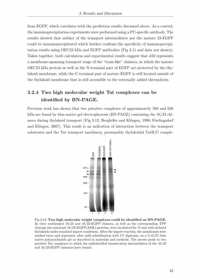


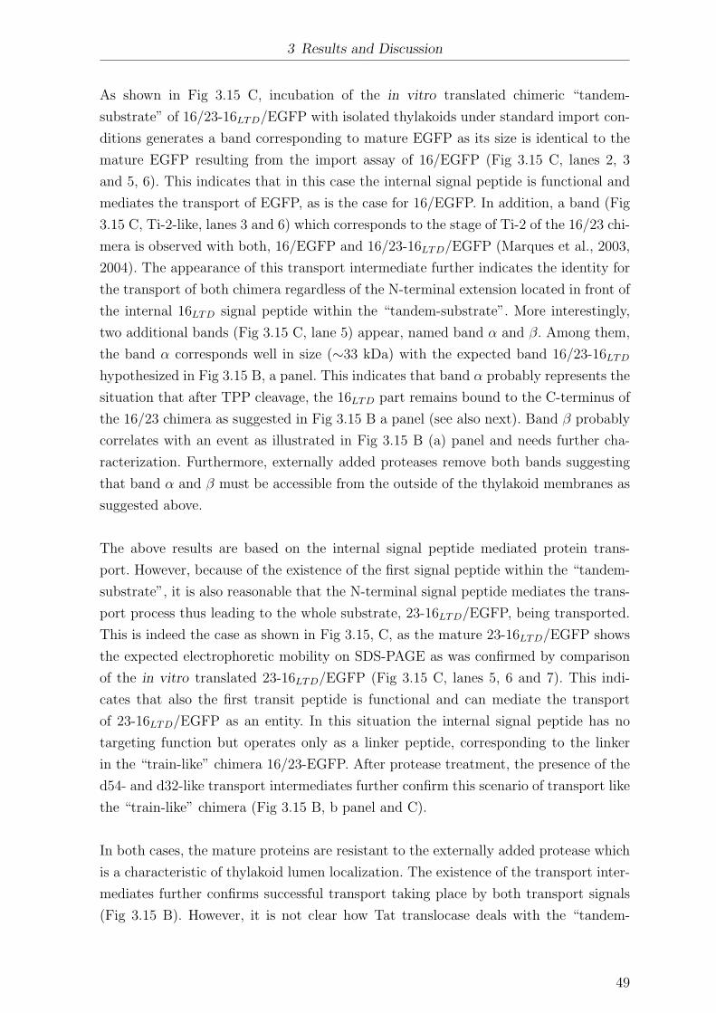


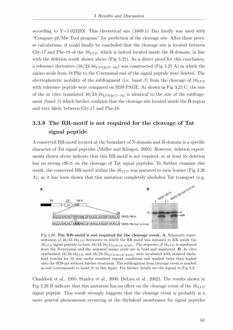


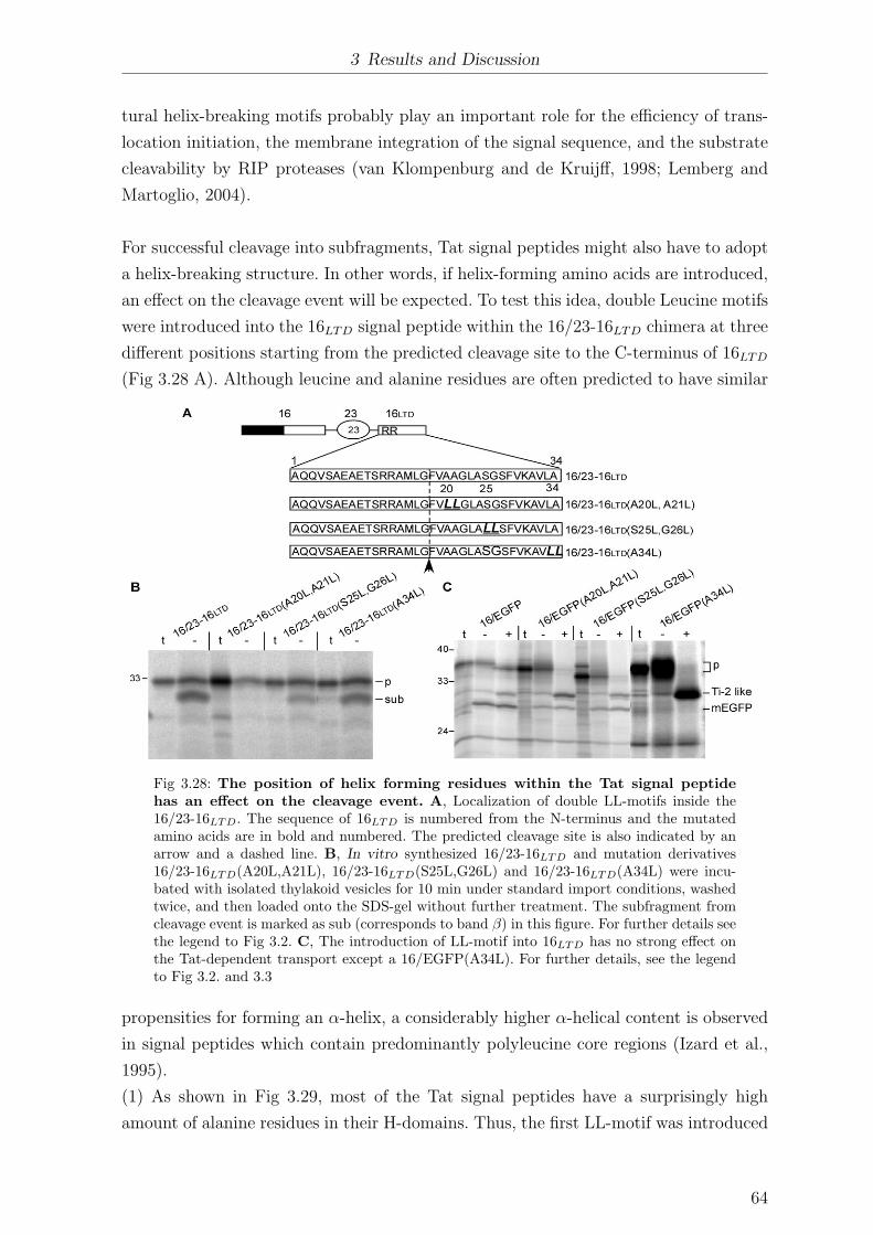


![T. Rogosch *, P. Imming , M. Kathmann - uni-halle.depharmchem.pharmazie.uni-halle.de/pdf/rogosch_vortrag_london.pdf · diseases like multiple sklerosis. Ki [nM] of endogenous ligands](https://static.documents.pub/doc/80x56/5c80c91209d3f2c0278b7d50/t-rogosch-p-imming-m-kathmann-uni-halle-diseases-like-multiple-sklerosis.jpg)


