3,350+ OPEN ACCESS BOOKS 108,000+ INTERNATIONAL AUTHORS AND EDITORS 115+ MILLION DOWNLOADS BOOKS DELIVERED TO 151 COUNTRIES AUTHORS AMONG TOP 1% MOST CITED SCIENTIST 12.2% AUTHORS AND EDITORS FROM TOP 500 UNIVERSITIES Selection of our books indexed in the Book Citation Index in Web of Science™ Core Collection (BKCI) Chapter from the book Type 1 Diabetes - Complications, Pathogenesis, and Alternative Treatments Downloaded from: http://www.intechopen.com/books/type-1-diabetes-complications- pathogenesis-and-alternative-treatments PUBLISHED BY World's largest Science, Technology & Medicine Open Access book publisher Interested in publishing with IntechOpen? Contact us at [email protected]
Transcript
3,350+OPEN ACCESS BOOKS
108,000+INTERNATIONAL
AUTHORS AND EDITORS115+ MILLION
DOWNLOADS
BOOKSDELIVERED TO
151 COUNTRIES
AUTHORS AMONG
TOP 1%MOST CITED SCIENTIST
12.2%AUTHORS AND EDITORS
FROM TOP 500 UNIVERSITIES
Selection of our books indexed in theBook Citation Index in Web of Science™
Core Collection (BKCI)
Chapter from the book Type 1 Diabetes - Complications, Pathogenesis , and AlternativeTreatmentsDownloaded from: http://www.intechopen.com/books/type-1-diabetes-complications-pathogenesis-and-alternative-treatments
PUBLISHED BY
World's largest Science,Technology & Medicine
Open Access book publisher
Interested in publishing with IntechOpen?Contact us at [email protected]
Bruno Vergès Service Endocrinologie, Diabétologie et Maladies Métaboliques
Dijon University Hospital France
1. Introduction
Cardiovascular disease is the major cause of death in persons with type 1 diabetes (Libby et al., 2005). Dyslipidemia has been shown to be a significant coronary heart disease risk factor in type 1 diabetes (Soedamah-Muthu et al., 2004; Grauslund et al., 2010). Thus, it seems important to pay attention to lipid abnormalities, in patients with type 1 diabetes, in order to reduce cardiovascular disease in this population. Patients with type 1 diabetes show lipid disorders, mostly qualitative abnormalities of lipoproteins, which may promote atherogenesis. The pathophysiology of these lipid abnormalities is not totally explained, but hyperglycemia and peripheral hyperinsulinemia, due to the subcutaneous route of insulin administration, are likely to play a role. After a brief review of lipoprotein metabolism and some information on the role of insulin on lipid metabolism, quantitative abnormalities then qualitative abnormalities of lipoproteins, in type 1 diabetes, will be discussed.
2. Brief review of lipoprotein metabolism
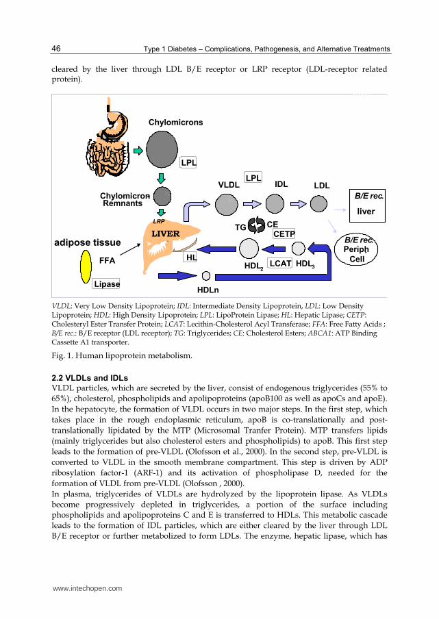
Lipoproteins, which transport non-water soluble cholesterol and triglycerides in plasma, are spherical particles composed of a central core of non-polar lipids (cholesterol esters, triglycerides) and a surface monolayer of phospholipids, free cholesterol and apolipoproteins. Lipoproteins are generally classified according to their density as chylomicron, Very Low Density Lipoprotein (VLDL), Intermediate Density Lipoprotein (IDL), Low Density Lipoprotein (LDL) and High Density Lipoprotein (HDL). An overview of lipoprotein metabolism is shown in Figure 1.
2.1 Chylomicrons
Chylomicrons, the largest lipoprotein particles, are responsible for the transport of dietary triglycerides and cholesterol. Chylomicrons are composed of triglycerides (85-90%), cholesterol esters, phospholipids and apolipoproteins (mainly apoB48 but also apoA-I and apoA-IV). The formation of chylomicrons takes place in the enterocytes, and the process associating the lipid components (triglycerides, cholesterol esters, phospholipids) and the apoB48 is performed by the MTP (Microsomal Tranfer Protein). Chylomicrons are secreted into the lymphatic circulation before entering the bloodstream. In plasma, triglycerides of chylomicrons are hydrolyzed by the lipoprotein lipase leading to the formation of smaller, triglyceride-poorer particles known as chylomicron-remnants. Chylomicron-remnants are
www.intechopen.com
Type 1 Diabetes – Complications, Pathogenesis, and Alternative Treatments
46
cleared by the liver through LDL B/E receptor or LRP receptor (LDL-receptor related protein).
CHU
Dijon
adipose tissue
VLDL LDL
HDL3HDL2
LIVER
Chylomicrons
CETPCETG
LPL
HL
LPL
RemnantsChylomicron-
IDL
LRP
Cell.Periph
liver
B/E rec.
B/E rec.
HDLn
LCATFFA
Lipase
VLDL: Very Low Density Lipoprotein; IDL: Intermediate Density Lipoprotein, LDL: Low Density Lipoprotein; HDL: High Density Lipoprotein; LPL: LipoProtein Lipase; HL: Hepatic Lipase; CETP: Cholesteryl Ester Transfer Protein; LCAT: Lecithin-Cholesterol Acyl Transferase; FFA: Free Fatty Acids ; B/E rec.: B/E receptor (LDL receptor); TG: Triglycerides; CE: Cholesterol Esters; ABCA1: ATP Binding Cassette A1 transporter.
Fig. 1. Human lipoprotein metabolism.
2.2 VLDLs and IDLs
VLDL particles, which are secreted by the liver, consist of endogenous triglycerides (55% to
65%), cholesterol, phospholipids and apolipoproteins (apoB100 as well as apoCs and apoE).
In the hepatocyte, the formation of VLDL occurs in two major steps. In the first step, which
takes place in the rough endoplasmic reticulum, apoB is co-translationally and post-
translationally lipidated by the MTP (Microsomal Tranfer Protein). MTP transfers lipids
(mainly triglycerides but also cholesterol esters and phospholipids) to apoB. This first step
leads to the formation of pre-VLDL (Olofsson et al., 2000). In the second step, pre-VLDL is
converted to VLDL in the smooth membrane compartment. This step is driven by ADP
ribosylation factor-1 (ARF-1) and its activation of phospholipase D, needed for the
formation of VLDL from pre-VLDL (Olofsson , 2000).
In plasma, triglycerides of VLDLs are hydrolyzed by the lipoprotein lipase. As VLDLs
become progressively depleted in triglycerides, a portion of the surface including
phospholipids and apolipoproteins C and E is transferred to HDLs. This metabolic cascade
leads to the formation of IDL particles, which are either cleared by the liver through LDL
B/E receptor or further metabolized to form LDLs. The enzyme, hepatic lipase, which has
www.intechopen.com
Lipid Disorders in Type 1 Diabetes
47
both triglyceride lipase and phospholipase activities, is involved in this metabolic process
generating LDL particles from IDLs.
2.3 LDLs
LDL is the final product of the VLDL-IDL-LDL cascade. LDL is the main cholesterol-bearing lipoprotein in plasma. Each LDL particle contains one molecule of apoB100, which plays an important role in LDL metabolism, particularly recognition of its dedicated LDL B/E receptor. Clearance of LDL is mediated by the LDL B/E receptor. Seventy percent of LDL B/E receptors are located on hepatic cells and 30% on the other cells of the body.
2.4 HDLs
HDL particles are secreted by the hepatocytes as small lipid-poor lipoproteins, containing mostly apoA-I, which receive, in the circulation, phospholipids, apoCs and apoE from chylomicrons and VLDLs. Nascent or lipid-poor HDLs get from peripheral cells free cholesterol and phospholipids through ABCA1 transporter (ATP Binding Cassette A1 transporter), allowing the transport of free cholesterol and phospholipids from the cell cytoplasm into the HDL particles (Oram & Lawn, 2001). Within HDL particles, free cholesterol is esterified by LCAT (Lecithin Cholesterol AcylTransferase) leading to the formation of HDL3 particles. The fusion of 2 HDL3 particles, which is promoted by PLTP (PhosphoLipid Transfer Protein), leads to the formation of one larger size HDL2 particle. HDL2 lipoproteins, rich in cholesterol ester, are degraded by the hepatic lipase and the endothelial lipase, leading to the formation of HDL remnant particles that are cleared by the liver after recognition by SR-B1 receptor (Scavenger Receptor class B type 1) (Jian et al., 1998).
2.5 Lipid transfer proteins
Lipoprotein metabolism is largely influenced by lipid transfer proteins. Among these, two
play an important role: CETP (Cholesteryl Ester Transfer Protein) and PLTP (PhosphoLipid
Transfer Protein). CETP facilitates the transfer of triglycerides from triglyceride-rich
lipoproteins (mainly VLDLs) toward HDLs and LDLs and the reciprocal transfer of
cholesteryl esters from HDLs and LDLs toward VLDLs (Lagrost, 1994). PLTP facilitates the
transfer of phospholipids and -tocopherol between lipoproteins. PLTP is also involved in
the formation of HDL2 lipoproteins from HDL3 particles (Lagrost et al., 1998). Any
modification of CETP or PLTP activities is likely to promote significant qualitative
abnormalities of lipoproteins.
3. Insulin and lipoprotein metabolism
Insulin plays a central role in the regulation of lipid metabolism (Vergès, 2001). The main sites of action of insulin on lipoprotein metabolism are shown in Figure 2. In adipose tissue, insulin inhibits the hormone-sensitive lipase. Thus, insulin has an anti-lipolytic action, promoting storage of triglycerides in the adipocytes and reducing release of free fatty acids from adipose tissue in the circulation. Insulin inhibits VLDL production from the liver. In normal subjects, it has been shown that
insulin induces a 67% decrease of VLDL-triglyceride production and a 52% decrease of
VLDL-apoB production (Lewis et al., 1993; Malmström et al., 1998). Insulin reduces VLDL
production by diminishing circulating free fatty acids (due to its antilipolytic effect), which
www.intechopen.com
Type 1 Diabetes – Complications, Pathogenesis, and Alternative Treatments
48
are substrates for VLDL, but also by a direct inhibitory effect in the hepatocyte (Malmström
et al., 1998). Insulin is a potent activator of lipoprotein lipase (LPL), promoting the
catabolism of triglyceride-rich lipoproteins and reducing, as a consequence, plasma
triglyceride level. Insulin not only enhances LPL activity (Brunzell et al., 1998), but has also
a direct positive effect on LPL gene, promoting LPL synthesis (Fried etal., 1993). Insulin
promotes the clearance of LDL, by increasing LDL B/E receptor expression and activity
(Chait et al., 1979, Mazzone et al., 1984).
Insulin acts also on HDL metabolism by activating LCAT and hepatic lipase activities
Fig. 2. Main effects of insulin on lipoprotein metabolism.
4. Quantitative lipid abnormalities in type 1 diabetes
4.1 Untreated (diabetic ketoacidosis) type 1 diabetes
In type 1 diabetic patients with diabetic ketoacidosis, quantitative lipid abnormalities are
observed, due to insulin deficiency.
www.intechopen.com
Lipid Disorders in Type 1 Diabetes
49
Triglyceride-rich lipoproteins (chylomicrons, VLDLs) are increased leading to hypertriglyceridemia. This is mainly due to decreased lipoprotein lipase activity (Vergès, 2001; Dullaart, 1995). Diabetic ketoacidosis is a situation of severe insulin deficiency with reduced lipoprotein lipase activity as a consequence, because insulin usually stimulates its activity. Decreased lipoprotein lipase activity leads to profound reduction of triglyceride-rich lipoprotein catabolism (Taskinen, 1987). In this condition of severe insulin deficiency, reduced catabolism of triglyceride-rich lipoproteins is, by far, the main factor involved in hypertriglyceridemia. This hypertriglyceridemia resolves rapidly after well titrated insulin therapy (Weidman et al., 1982). LDL-cholesterol is decreased during diabetic ketoacidosis (Weidman et al., 1982). This fall in plasma LDL-cholesterol level is the direct consequence of the reduction of triglyceride-rich lipoprotein catabolism, due to decreased lipoprotein lipase activity (see above). In diabetic ketoacidosis, HDL-cholesterol level is significantly decreased (Weidman et al., 1982). This is a consequence of hypertrigliceridemia observed in this condition. Indeed, the augmented level of plasma triglyceride-rich lipoproteins drives, through CETP, the transfer of triglycerides from triglyceride-rich lipoproteins to HDLs leading to the formation of triglyceride-rich HDL particles. HDLs enriched in triglycerides become very good substrate for hepatic lipase, leading to increase their catabolism and, thus, to decrease plasma HDL-cholesterol level. This low HDL-cholesterol condition resolves rapidly after well titrated insulin therapy (Weidman et al., 1982).
4.2 Treated type 1 diabetes
Patients with treated type 1 diabetes may show quantitative lipid disorders. In a prospective study performed in 895 young subjects with type 1 diabetes, 20.1% had plasma triglycerides above 1.7 mmol/l, 9.6% had LDL-cholesterol above 3.4 mmol/l and 25.9% had non-HDL cholesterol above 3.4 mmol/l (Marcovecchio et al., 2009). It has been shown that abnormal lipid levels, in type 1 diabetes, predict worse cardiovascular outcomes (Soedamah-Muthu et al., 2004). HbA1c has been shown to be independently correlated with LDL-cholesterol, non-HDL cholesterol and triglyceride levels, indicating that these disorders were mostly observed in patients with poor glycemic control (Marcovecchio et al., 2009). In a British follow-up study of 229 children with type 1 diabetes, LDL cholesterol and non-HDL cholesterol values increased with duration of diabetes (Edge et al., 2008). In that study, total cholesterol, triglycerides and non-HDL cholesterol were positively correlated with HbA1c and around 10% of the patients had lipid values outside recommendations (Edge et al., 2008). In a large study performed in 29 979 patients with type 1 diabetes, multivariate analyses showed a significant positive association between HbA1c and total cholesterol (p<0.0001), LDL cholesterol (p<0.0001) and a significant negative association between HbA1c and HDL cholesterol (p<0.0001) (Schwab et al., 2009). In the Diabetes Control and Complications Trial (DCCT), HbA1c correlated positively with total cholesterol, LDL-cholesterol and triglycerides at baseline (The DCCT Research Group, 1992). Data from the Coronary Artery Calcification in type 1 diabetes (CACTI) study, which examined 652 patients with type 1 diabetes, have shown, in patients not using hypolipidemic agents, that a higher HbA1c was associated with significantly higher levels of total cholesterol, triglycerides, LDL cholesterol and non-HDL cholesterol (Maahs et al., 2010). In that study, 1% change in HbA1c was associated with an increase of 0.101 mmol/l (4 mg/dl) for total cholesterol, of 0.052 mmol/l (4.5 mg/dl) for triglycerides, of 0.103 mmol/l (4 mg/dl) for LDL cholesterol and of 0.129 mmol/l (5 mg/dl) for non-HDL cholesterol (Maahs et al.,
www.intechopen.com
Type 1 Diabetes – Complications, Pathogenesis, and Alternative Treatments
50
2010). In a recent study, performed in 512 young patients with type 1 diabetes and in 188
healthy age-matched controls, patients with suboptimal control (HbA1c 7.5%) had much more lipid quantitative disorders than patients with optimal control (HbA1c<7.5%) (Guy et al., 2009). All these data suggest that quantitative lipid abnormalities are more frequent, when type 1 diabetes is not well controlled. In addition, some patients with type 1 diabetes may have insulin resistance, in situation of abdominal obesity and/or family history of type 2 diabetes. Such patients have been shown to have greater dyslipidemia (Purnell et al., 2003). In a recent study performed in 60 young type 1 diabetic patients and 40 adults with type 1 diabetes, it has been shown, using hyperinsulinemic clamp studies, that lower glucose infusion (more insulin resistance) was associated with lower levels of HDL cholesterol in youths with type 1 diabetes and with higher levels of triglycerides and higher triglyceride/HDL ratio in both youths and adults (Maahs et al., 2011). These data indicate that insulin resistance may be an additional factor that could induce quantitative lipid abnormities in some type 1 diabetic patients with a background of insulin resistance (abdominal obesity, family history of type 2 diabetes). In this chapter we will consider only the typical situation of type 1 diabetes without insulin resistance.
4.2.1 Treated type 1 diabetes with poor or suboptimal glycemic control
In case of poor or suboptimal control, patients with type 1 diabetes may show increased
plasma triglyceride levels (Dullaart,1995). This hypertriglyceridemia is due to increased
production of VLDL, promoted by elevated circulating free fatty acids secondary to the
Type 1 diabetic patients with poor or suboptimal glycemic control show increased LDL-
cholesterol levels as compared to non–diabetic individuals and type 1 diabetic patients with
optimal glycemic control (Dullaart, 1995; Guy et al., 2009). Indeed, in this condition, VLDL
production is increased (see above), when catabolism of triglyceride-rich lipoproteins is not
importantly decreased, which leads to increase LDL production (Dullaart, 1995).
4.2.2 Treated type 1 diabetes with optimal glycemic control
In well controlled type 1 diabetes, the lipid profile is totally different than in poorly
controlled type 1 diabetes (Dullaart, 1995; Nikkilä & Kekki, 1973).
Plasma triglycerides are normal or slightly decreased (Dullaart, 1995; Nikkilä & Kekki,
1973). This slight decrease in plasma triglycerides may be observed with intense insulin
therapy because of increased down control of VLDL production by augmented plasma
insulin levels as a consequence of the subcutaneous route of insulin delivery (Dashti &
Wolfbauer, 1987; Taskinen, 1992). Furthermore, in patients with well controlled type 1
diabetes, peripheral hyperinsulinemia has been shown to be associated with increased
lipoprotein lipase activity that could be an additional factor responsible for decreased
plasma triglycerides (Nikkilä et al., 1977).
Plasma LDL-cholesterol level is normal or slightly decreased (Winocour et al., 1986). This
slight decrease in plasma LDL-cholesterol may be observed with intense insulin therapy as a
consequence of decreased VLDL production by peripheral hyperinsulinemia (see above).
Plasma HDL-cholesterol level is normal or slightly increased in well controlled type 1 diabetic patients (Dullaart, 1995). Some studies have shown an increase in HDL subfraction 2 (Eckel et al., 1981; Kahri et al., 1993), when others have found an increase in HDL
www.intechopen.com
Lipid Disorders in Type 1 Diabetes
51
subfraction 3 (Winocour et al., 1986). It has also been reported that elevation of HDL in type 1 diabetic patients with good glycemic control was caused by an increase of HDL particles containing only apoA-I (LpA-I) (Kahri et al., 1993). This increase in plasma HDL-cholesterol could be the consequence of the elevated Lipoprotein Lipase/Hepatic Lipase ratio that is observed in patients with well controlled type 1 diabetes (increased Lipoprotein Lipase activity and normal Hepatic Lipase activity) (Kahri et al., 1993). The increased Lipoprotein Lipase activity observed in these patients is likely to be due to peripheral hyperinsulinemia as a consequence of the subcutaneous route of insulin administration (Kahri et al., 1993).
4.2.3 Subcutaneous insulin therapy versus intraperitoneal insulin therapy
Intensive subcutaneous insulin therapy results in normalization of plasma glucose, but at the expense of peripheral hyperinsulinemia, which is likely to modify lipoprotein metabolism (as discussed above). Implantable insulin pumps with intraperitoneal insulin administration mimic the physiologic route of insulin delivery and are likely to restore the normal portal-peripheral insulin gradient. For this reason, several studies have been performed to analyze the modification of lipoprotein metabolism after replacement of subcutaneous insulin therapy by intraperitoneal insulin therapy. Plasma triglycerides have been found increased in one study (Selam et al., 1989) and unchanged in three other studies (Bagdade & Dunn, 1996; Ruotolo et al., 1994; Duvillard et al., 2005). Total cholesterol and apoB were found unchanged (Bagdade & Dunn, 1996; Ruotolo et al., 1994; Duvillard et al., 2005). HDL-cholesterol has been found decreased (Selam et al., 1989) or not modified (Bagdade & Dunn, 1996; Ruotolo et al., 1994; Duvillard et al., 2007). The discrepancies of these studies that may be due to confounding factors such as degree of glycemic control and peripheral insulin levels during subcutaneous insulin therapy. Further studies are needed to clearly evaluate the effect of intraperitoneal insulin administration on lipoprotein metabolism.
4.2.4 Type 1 diabetes with nephropathy
In type 1 diabetic patients with nephropathy and overt albuminuria, elevated plasma levels of total cholesterol, triglycerides and LDL-cholesterol are observed whereas HDL-cholesterol is decreased due to a fall in HDL2 (Dullaart ,1995; Taskinen, 1992; Jensen et al., 1987). In the EURODIAB IDDM Complications study, macroalbuminuria was associated with significantly increased plasma triglycerides, cholesterol, LDL-cholesterol and LDL/HDL ratio in both sexes and decreased HDL-cholesterol in women (Mattock et al., 2001). Some quantitative lipid modifications are also observed in type 1 diabetic patients with microalbuminuria. Microalbuminuric patients compared with normoalbuminuric patients show increased plasma apoB (Jones et al., 1989, Dullaart et al, 1989a; Jay et al., 1991), LDL cholesterol (Jones et al., 1989, Dullaart et al, 1989a) and apoB/apoA1 ratio (Dullaart et al, 1989a; Jay et al., 1991). A positive correlation has been found between urinary albumin excretion rate and plasma apoB and apoB/apoA1 ratio (Dullaart et al, 1989a). In the EURODIAB IDDM Complications study, microalbuminuria was associated with increased plasma triglycerides (Mattock et al., 2001). In a prospective study performed in 895 young subjects with type 1 diabetes, total cholesterol and non-HDL cholesterol were independently related to longitudinal changes in albumin-to-creatinine ratio (Marcovecchio et al., 2009). The mechanisms responsible for these lipoprotein abnormalities in type 1 diabetic patients with microalbuminuria remain unclear.
www.intechopen.com
Type 1 Diabetes – Complications, Pathogenesis, and Alternative Treatments
52
Moreover, serum lipids have been shown to be associated with the progression of nephropathy in type 1 diabetes. In a prospective study performed in 152 patients with type 1 diabetes followed for 8-9 years, LDL-cholesterol was an independent factor associated with progression of nephropathy (Thomas et al., 2006).
5. Qualitative lipid abnormalities in type 1 diabetes
Several qualitative abnormalities of lipoproteins are observed in patients with type 1 diabetes, even in those with good metabolic control, who do not have significant quantitative lipid changes. These qualitative lipid abnormalities are not totally reversed by optimal glycemic control and are likely to be atherogenic.
5.1 VLDLs
VLDLs from patients with type 1 diabetes are frequently enriched in esterified cholesterol at the expense of triglycerides leading to an increased VLDL cholesterol/triglyceride ratio (Rivellese et al., 1988; Bagdade et al., 1991a). It has been suggested that this compositional changes may be due to increased cholesteryl ester transfer between lipoproteins (Bagdade et al., 1991a). It has been shown that the VLDL cholesterol/triglyceride ratio was significantly reduced with intraperitoneal insulin therapy (Dunn, 1992). Furthermore, the free cholesterol /lecithin ratio within the peripheral layer of VLDL particles is increased (Dullaart, 1995; Bagdade et al., 1991a). Such increase in the free cholesterol /lecithin ratio within the peripheral layer of lipoproteins has been shown to raise the risk for cardiovascular events possibly by reducing fluidity and stability of lipoproteins (Kuksis, 1982). Moreover, VLDLs from patients with type 1 diabetes have been shown, in vitro, to induce abnormal response of cellular cholesterol metabolism in human macrophages (Klein et al., 1989).
5.2 LDLs
In patients with type 1 diabetes, LDLs are often enriched in triglycerides and increased number of small dense LDL particles is observed (Guy et al., 2009; Lahdenperä et al., 1994; James & Pometta, 1990; Skyrme-Jones et al., 2000). In a study performed in 2657 patients with type 1 diabetes, it has been shown that dense LDL increased with HbA1c with buoyant LDL shifting toward dense LDL for HbA1c values above 8% (Albers et al., 2008). It has been shown that the presence of small dense LDL particles is associated with increased cardiovascular risk (Austin et al., 1990). Many data indicate that small dense LDL particles have atherogenic properties. Indeed, small dense LDL particles have reduced affinity for the LDL B/E receptor and are preferentially taken up by macrophages, through the scavenger receptor, leading to the formation of foam cells. Small dense LDL particles have higher affinity for intimal proteoglycans than large LDL particles which may favor the penetration of LDL particles into the arterial wall (Chapman et al., 1998). It has been shown that subjects with small dense LDL particles show an impaired response to endothelium dependent vasodilator acetylcholine (Vakkilainen et al., 2000). Moreover, small dense LDL particles show an increased susceptibility to oxidation (Tribble et al., 1992). A reduction of the proportion of small dense LDL particles has been reported after optimization of glycemic control in patients with type 1 diabetes (Caixàs et al., 1997). The free cholesterol /lecithin ratio within the peripheral layer of LDL particles is increased (Dullaart, 1995; Bagdade et al., 1991a). In patients with type 1 diabetes, glycation of ApoB
www.intechopen.com
Lipid Disorders in Type 1 Diabetes
53
occurs within LDL in parallel with plasma hyperglycemia. It has been shown that apoB glycation reduces significantly LDL binding to the B/E receptor even when apoB glycation is moderate (Witztum et al., 1982; Steinbrecher et al, 1984). Furthermore, glycated LDLs are preferentialy taken up by macrophages through the scavenger receptor, leading to the formation of foam cells in the arterial wall. In patients with type 1 diabetes, advanced glycation end products-modified LDL have been shown to be positively associated with increased intima media thickness (IMT) (Lopes-Virella et al., 2011). Moreover, patients with type 1 diabetes may show an increased oxidation of LDL which is promoted by glycemic excursions (de Castro et al., 2005). Increased urinary excretion of malondialdehyde, reflecting enhanced lipid peroxidation, has been reported in patients with type 1 diabetes (Hoeldtke et al., 2009). Oxidative modification of LDL results in rapid uptake by macrophages, leading to foam cell formation. Oxidized LDLs produce chemotactic effects on monocytes by increasing the synthesis of adhesion molecules, such as ICAM-1 (intercellular adhesion Molecule 1) by endothelial cells. Oxidized LDLs stimulate the
formation by macrophages of cytokines, such as TNF or IL1, which amplify the inflammatory atherosclerotic process. It has recently been shown that oxidized LDL particles were significantly associated with progression and increased levels of IMT in type 1 diabetes (de Castro et al., 2005).
5.3 HDLs HDL particles from patients with type 1 diabetes are often enriched in triglycerides (Dullaart, 1995; Bagdade et al., 1991a). This modification has been attributed to increased cholesteryl ester transfer between lipoproteins (Bagdade et al., 1991a). In HDL particles from patients with type 1 diabetes, sphingomyelin/lecithin ratio within the peripheral layer is augmented, which may increase HDL rigidity (Bagdade & Subbaiah, 1989). These alterations are not totally reversed after achievement of optimal glycemic control (Bagdade et al., 1991b). ApoA-I within HDL is glycated in patients with type 1 diabetes, which may impair the HDL-mediated reverse cholesterol pathway. Indeed, it has been shown that HDL particles containing glycated apoA-I were less effective to promote cholesterol efflux from the cells (Fievet et al., 1992). In addition to their role in the reverse cholesterol pathway, HDLs have anti-oxidative, anti-inflammatory, anti-thrombotic and vasorelaxant properties, potentially anti-atherogenic (Link et al., 2007). Some of these properties have been shown to be reduced in patients with type 1 diabetes. Indeed, a significant reduction of the activity of paraoxonase, an anti-oxidative enzyme associated with HDLs, is observed in patients with type 1 diabetes (Boemi et al., 2001; Ferretti et al., 2004). As a consequence, HDLs from patients with type 1 diabetes protect less efficiently erythrocyte membranes and LDL particles against oxidative damage than HDLs from normal individuals (Boemi et al., 2001; Ferretti et al., 2004). Furthermore, using rabbit aorta rings, it has been shown that HDL from patients with type 1 diabetes are no more able to prevent the endothelium dependent vasoconstriction induced by oxidized LDL, whereas HDL from normal individuals can prevent it (Perségol et al., 2007).
5.4 Lipid transfer proteins
In some studies, an increased cholesteryl ester transfer between lipoproteins (Bagdade et al., 1991a; Bagdade et al., 1994) or an augmented activity of CETP (Colhoun et al., 2001) have been found in normolipidemic patients with type 1 diabetes. In some other studies, increased CETP activity has been reported only in type 1 diabetic patients that smoke or
www.intechopen.com
Type 1 Diabetes – Complications, Pathogenesis, and Alternative Treatments
54
those having microalbuminuria (Dullaart et al., 1989b; Dullaart et al., 1991). This augmented CETP activity may explain the increase in free cholesterol/ triglycerides ratio within VLDL and its decrease within HDL. Some studies have shown a positive correlation between CETP activity and hyperglycemia (Ritter & Bagdade, 1994; Chang et al., 2001). However, the main factor which is likely to be responsible for increased CETP activity, in type 1 diabetes, could be peripheral hyperinsulinemia secondary to the subcutaneous route of insulin administration. Indeed, peripheral hyperinsulinemia has been shown to be responsible for increased lipoprotein lipase activity in patients with type 1 diabetes (Nikkilä et al., 1977) and it has been reported that lipoprotein lipase, in presence of VLDL, enhances CETP activity (Sammett & Tall,1985; Pruneta et al., 1999). Moreover, it has been shown, in patients with type 1 diabetes, that the increase in both lipoprotein lipase and CETP activities was abolished when insulin was administrated intraperitoneously with implantable insulin pumps, mimicking the physiologic portal route or after pancreatic graft (Bagdade et al., 1994; Bagdade et al., 1996). Increased PLTP activity has been reported in patients with type 1 diabetes (Colhoun et al., 2001). In this study, PLTP activity was positively correlated with CETP activity, LDL-cholesterol and HDL-cholesterol (Colhoun et al., 2001). The reasons and consequences of this increased PLTP activity are not clear.
6. Conclusion
In conclusion, quantitative lipid abnormalities are observed in patients with poorly controlled type 1 diabetes (increased triglyceride and LDL-cholesterol levels) or with micro- or macroalbuminuria (increased triglycerides and LDL-cholesterol, decreased HDL-cholesterol). Patient with optimally controlled type 1 diabetes show normal or slightly decreased triglycerides and LDL-cholesterol levels and sometimes increased HDL-cholesterol levels. Qualitative abnormalities of lipoproteins are observed in patients with type 1 diabetes, even in good glycemic control. These abnormalities are not fully explained by hyperglycemia and may partly be due to peripheral hyperinsulinemia associated with the subcutaneous route of insulin administration. The exact consequences of these qualitative lipid changes on the development of cardiovascular disease in type 1 diabetes are still unknown.
InTech ChinaUnit 405, Office Block, Hotel Equatorial Shanghai No.65, Yan An Road (West), Shanghai, 200040, China
Phone: +86-21-62489820 Fax: +86-21-62489821
This book is intended as an overview of recent progress in type 1 diabetes research worldwide, with a focus ondifferent research areas relevant to this disease. These include: diabetes mellitus and complications,psychological aspects of diabetes, perspectives of diabetes pathogenesis, identification and monitoring ofdiabetes mellitus, and alternative treatments for diabetes. In preparing this book, leading investigators fromseveral countries in these five different categories were invited to contribute a chapter to this book. We havestriven for a coherent presentation of concepts based on experiments and observation from the authors ownresearch and from existing published reports. Therefore, the materials presented in this book are expected tobe up to date in each research area. While there is no doubt that this book may have omitted some importantfindings in diabetes field, we hope the information included in this book will be useful for both basic scienceand clinical investigators. We also hope that diabetes patients and their family will benefit from reading thechapters in this book.
How to referenceIn order to correctly reference this scholarly work, feel free to copy and paste the following:
Bruno Verge ̀s (2011). Lipid Disorders in Type 1 Diabetes, Type 1 Diabetes - Complications, Pathogenesis, andAlternative Treatments, Prof. Chih-Pin Liu (Ed.), ISBN: 978-953-307-756-7, InTech, Available from:http://www.intechopen.com/books/type-1-diabetes-complications-pathogenesis-and-alternative-treatments/lipid-disorders-in-type-1-diabetes