Page 1
9/11/2012
1
1
Chapter 32
Hematology
2
Learning Objectives
• Describe the physiology of blood and its components
• Discuss the pathophysiology and signs and symptoms of specific hematological disorders.
• Outline the general assessment and management of patients with hematological disorders.
3
Copyright © 2013 by Jones & Bartlett Learning, LLC, an Ascend Learning Company
Page 2
9/11/2012
2
Blood and Blood Components
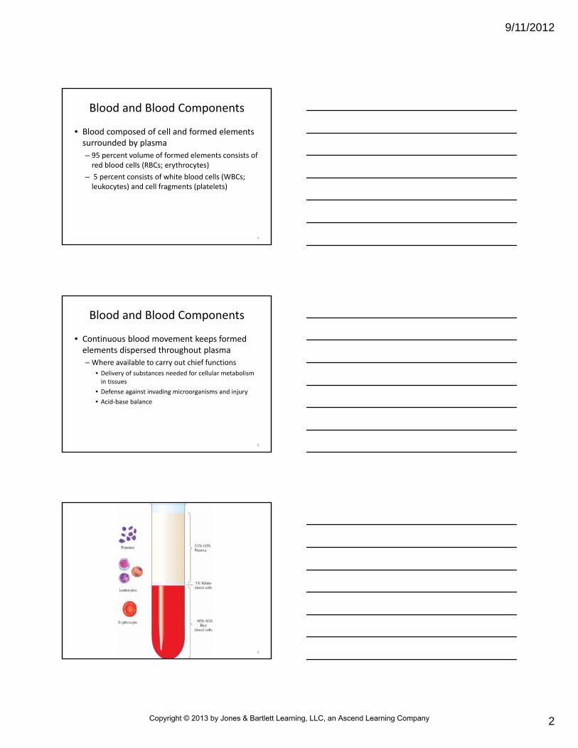
• Blood composed of cell and formed elements surrounded by plasma
– 95 percent volume of formed elements consists of red blood cells (RBCs; erythrocytes)
– 5 percent consists of white blood cells (WBCs; leukocytes) and cell fragments (platelets)
4
Blood and Blood Components
• Continuous blood movement keeps formed elements dispersed throughout plasma
– Where available to carry out chief functions
• Delivery of substances needed for cellular metabolism in tissues
• Defense against invading microorganisms and injury
• Acid‐base balance
5
6
Copyright © 2013 by Jones & Bartlett Learning, LLC, an Ascend Learning Company
Page 3
9/11/2012
3
Blood and Blood Components
• Blood cells formed within red bone marrow
– Present in all tissues at birth
• Adult red bone marrow primarily found in membranous bone
– Vertebrae, pelvis, sternum, ribs
• Yellow marrow produces some white cells
– Composed mainly of connective tissue and fat
7
Blood and Blood Components
• Other blood‐forming organs
– Lymph nodes
• Produce lymphocytes and antibodies
– Spleen
• Stores large quantities of blood
• Produces lymphocytes, plasma cells, antibodies
– Liver
• Blood‐forming organ only during intrauterine life
• Plays important role in coagulation process
8
Plasma
• Clear portion of blood, is about 92 percent water
• Contains three important proteins
– Albumin
• Most plentiful protein
• Similar to egg white
• Gives blood gummy texture
• Keeps water concentration low so water diffuses readily from tissues into blood
9
Copyright © 2013 by Jones & Bartlett Learning, LLC, an Ascend Learning Company
Page 4
9/11/2012
4
Plasma
• Contains three important proteins– Globulins (alpha, beta, and gamma)
• Transport other proteins
• Provide immunity to disease
– Fibrinogen• Responsible for blood clotting
• Maintaining blood pH (acting as either acid or base)
• Transporting fat‐soluble vitamins, hormones, carbohydrates
• Allowing body to digest them temporarily for food
10
Red Blood Cells
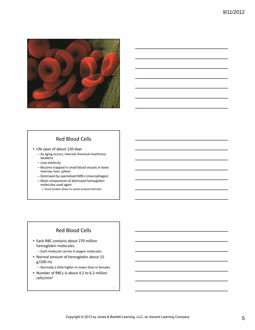
• Most abundant cells in body
– Primarily responsible for tissue oxygenation
– Appear as small rounded disks with nearly hollowed‐out centers
– Comprised mainly of water and red protein hemoglobin
11
Red Blood Cells
• Production continues throughout life– Replace blood cells that grow old and die, killed by disease or lost through bleeding
– After production occurs in marrow, new cell divides until there are 16 RBCs
– Cells produce hemoglobin protein until concentration of protein becomes 95 percent of dry weight of cell
– Cell expels nucleus, giving cell its characteristic pinched look
– New shape increases surface area of cell and oxygen‐carrying potential
12
Copyright © 2013 by Jones & Bartlett Learning, LLC, an Ascend Learning Company
Page 5
9/11/2012
5
13
Red Blood Cells
• Life span of about 120 days– As aging occurs, internal chemical machinery weakens
– Lose elasticity
– Become trapped in small blood vessels in bone marrow, liver, spleen
– Destroyed by specialized WBCs (macrophages)
– Most components of destroyed hemoglobin molecules used again
• Some broken down to waste product bilirubin
14
Red Blood Cells
• Each RBC contains about 270 million hemoglobin molecules
– Each molecule carries 4 oxygen molecules
• Normal amount of hemoglobin about 15 g/100 mL
– Normally a little higher in males than in females
• Number of RBCs is about 4.2 to 6.2 million cells/mm2
15
Copyright © 2013 by Jones & Bartlett Learning, LLC, an Ascend Learning Company
Page 6
9/11/2012
6
White Blood Cells
• Arise from bone marrow
– Released into bloodstream
– Destroy foreign substances (e.g., bacteria and viruses)
– Clear bloodstream of debris
16
White Blood Cells
• Leukocyte production increases in response to infection
– Causes elevated WBC count in blood
– Bone marrow and lymph glands continually produce and maintain reserve
– Not many WBCs in healthy bloodstream
17
White Blood Cells
• Normal WBC count is about 5,000 to 10,000 cells/mm2
– Monocytes make up about 5 percent of total WBC count
– Increase with chronic infections
– Lymphocytes account for about 27.5 percent
– Neutrophils about 65 percent
– Eosinophils and basophils together about 2.5 percent
18
Copyright © 2013 by Jones & Bartlett Learning, LLC, an Ascend Learning Company
Page 7
9/11/2012
7
White Blood Cells
• Increased WBC count is specific for various illnesses
– Bacterial infection
– Inflammation
– Leukemia
– Trauma
– Stress
19
White Blood Cells
• Differential count (also called diff)
– Identifies different types of leukocytes present in blood
– Test performed by
• Spreading drop of blood on microscope slide
• Staining slide
• Examining under microscope
20
White Blood Cells
• Differential count (also called diff)
– Identified by
• Shape and appearance of nucleus
• Color of cytoplasm
• Presence and color of granules
– Percentage of each cell type is reported
– Red cells and platelets are examined for abnormalities in appearance
21
Copyright © 2013 by Jones & Bartlett Learning, LLC, an Ascend Learning Company
Page 8
9/11/2012
8
What body functions are impaired if the WBC number or function
is diminished?
22
Platelets
• Platelets (thrombocytes) are small, sticky cell fragments
– Important role in blood clotting
– When blood vessel is cut
• Travel to site and swell into odd, irregular shapes
• Adhere to damaged wall
• Plug the leak
• Allow other cells to stick and form clot
23
Platelets
• Platelets (thrombocytes) are small, sticky cell fragments
– If damage is too great, platelets chemically signal complex clotting process or clotting cascade
• Repair millions of ruptured capillaries each day
• Often make rest of clotting cascade unnecessary
24
Copyright © 2013 by Jones & Bartlett Learning, LLC, an Ascend Learning Company
Page 9
9/11/2012
9
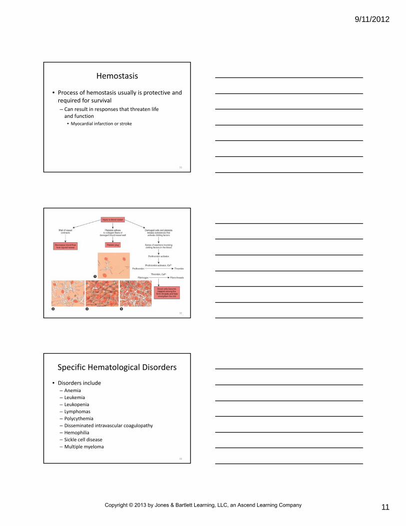
Hemostasis
• Initial physiological response to wounding, causes bleeding to cease
• Initiated when break in integrity of vascular endothelium
25
Hemostasis
• Vascular reaction or physiology of hemostasis involves– Vasoconstriction
• Resulting from injury is rapid but temporary
• In response to injury, severed blood vessels constrict and retract with aid of surrounding subcutaneous tissues
• Vessel spasm slows blood loss immediately
• Usually sustained as long as 10 minutes
• Blood coagulation mechanisms activated to produce clot
26
Hemostasis
• Vascular reaction or physiology of hemostasis involves– Formation of platelet plug
• Adhere to injured vessels and collagen in connective tissue that surrounds injured vessel
• Contact collagen, they swell, become sticky, and secrete chemicals that activate other surrounding platelets
• Process causes platelets to adhere to one another
• If opening in wall is small, plug may be sufficient to stop blood loss completely
• If opening is large, a blood clot is necessary to arrest blood flow
27
Copyright © 2013 by Jones & Bartlett Learning, LLC, an Ascend Learning Company
Page 10
9/11/2012
10
Hemostasis
• Vascular reaction or physiology of hemostasis involves
– Coagulation
– Growth of fibrous tissue into clot that permanently closes and seals injured vessel
28
Hemostasis
• Coagulation occurs as result of chemical process that begins within seconds of severe vessel injury
– Progresses rapidly; within 3 to 6 minutes after vessel rupture, entire end of vessel filled clot
– Within 30 minutes, clot retracts and vessel is sealed further
29
Hemostasis
• Coagulation occurs as result of chemical process that begins within seconds of severe vessel injury– Clotting mechanism is complex process and includes three mechanisms
• Prothrombin activator is formed in response to rupture or damage of blood vessel
• Prothrombin activator stimulates conversion of prothrombinto thrombin
• Thrombin in presence of calcium ions act as enzyme to convert fibrinogen into fibrin threads
• Threads entrap platelets, blood cells, and plasma to form clot
30
Copyright © 2013 by Jones & Bartlett Learning, LLC, an Ascend Learning Company
Page 11
9/11/2012
11
Hemostasis
• Process of hemostasis usually is protective and required for survival
– Can result in responses that threaten life and function
• Myocardial infarction or stroke
31
32
• Disorders include– Anemia
– Leukemia
– Leukopenia
– Lymphomas
– Polycythemia
– Disseminated intravascular coagulopathy
– Hemophilia
– Sickle cell disease
– Multiple myeloma
Specific Hematological Disorders
33
Copyright © 2013 by Jones & Bartlett Learning, LLC, an Ascend Learning Company
Page 12
9/11/2012
12
• Anemia
– Condition in which concentration of hemoglobin or erythrocytes is below normal
– Precipitating causes
• Chronic or acute blood loss
• Decreased production of erythrocytes
• Increased destruction of erythrocytes
– Symptom of disease
Specific Hematological Disorders
34
• Anemia– Persons at greatest risk are those with
• Chronic kidney disease
• Diabetes
• Heart disease
• Cancer
• Chronic inflammatory conditions
• Persistent infections
– Conditions interfere with production of oxygen‐carrying RBCs
Specific Hematological Disorders
35
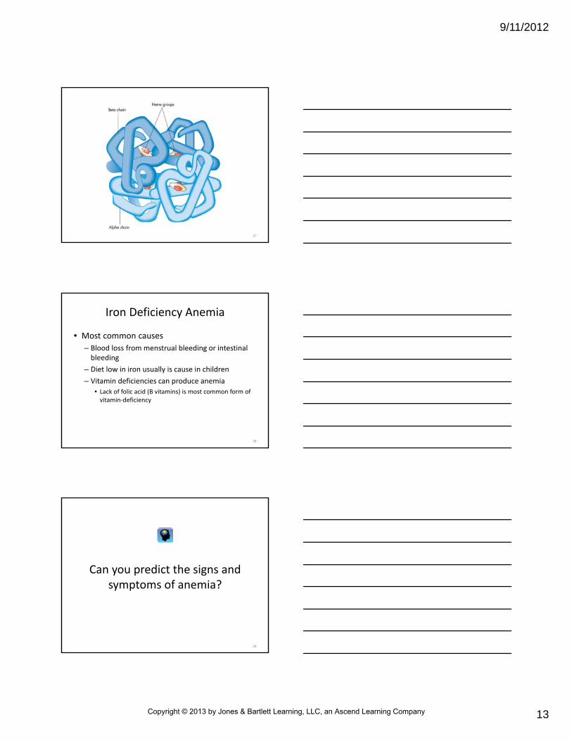
Iron Deficiency Anemia
• Iron is critical part of a hemoglobin molecule
– Gives ability to bind oxygen
– Lack of iron prevents bone marrow from making enough hemoglobin for RBCs
• RBCs produced are small and have pale center
• Reduced oxygen‐carrying capacity
36
Copyright © 2013 by Jones & Bartlett Learning, LLC, an Ascend Learning Company
Page 13
9/11/2012
13
37
Iron Deficiency Anemia
• Most common causes
– Blood loss from menstrual bleeding or intestinal bleeding
– Diet low in iron usually is cause in children
– Vitamin deficiencies can produce anemia
• Lack of folic acid (B vitamins) is most common form of vitamin‐deficiency
38
Can you predict the signs and symptoms of anemia?
39
Copyright © 2013 by Jones & Bartlett Learning, LLC, an Ascend Learning Company
Page 14
9/11/2012
14
Hemolytic Anemia
• Cause
– Premature destruction of RBCs in blood (hemolysis) causes hemolytic anemia
• Can result from inherited disorder inside RBC
• Can result from disorder outside cell
• Condition usually acquired later in life
40
Inherited Disorders
• Hemolysis
– Can occur as result of abnormal rigidity of cell membrane
• Causes cell to become trapped at an early stage of its life span in smaller blood vessels (usually of spleen)
• In these smaller blood vessels, RBC is destroyed by macrophages
41
Inherited Disorders
• Hemolysis
– Can occur from genetic defect in hemoglobin within cell (e.g., sickle cell anemia and thalassemia)
– Can occur from defect in one of the enzymes in cell that helps protect cell from chemical damage during infectious illness
• Deficiency glucose‐6‐phosphate dehydrogenase is common in African‐Americans
42
Copyright © 2013 by Jones & Bartlett Learning, LLC, an Ascend Learning Company
Page 15
9/11/2012
15
Acquired Disorders
• Acquired hemolytic anemia results from
– Disorders in which normal RBCs are disrupted as result of mechanical forces
• Abnormal blood vessel linings
• Blood clots
– Autoimmune disorders
• Can destroy RBCs with antibodies that are produced by immune system
• Drug‐induced hemolytic anemia
• Incompatible blood transfusion
– Conditions that can cause hemolytic anemia when RBCs are destroyed by microorganisms in blood (e.g., malaria)
43
Signs and Symptoms of Anemia
• All forms of anemia share signs and symptoms
– Fatigue and headaches
– Sore mouth or tongue
– Brittle nails
– Breathlessness and chest pain
44
Signs and Symptoms of Anemia
• Other patient complaints are related to abnormal decrease in number of WBCs (leukopenia) or reduction in platelets (thrombocytopenia) and may include– Bleeding from mucous membranes
– Cutaneous bleeding
– Fatigue
– Fever
– Lethargy
45
Copyright © 2013 by Jones & Bartlett Learning, LLC, an Ascend Learning Company
Page 16
9/11/2012
16
Diagnosis and Treatment
• Diagnostic tools– Signs and symptoms
– Patient history
– Examination of patient’s blood through blood tests and bone marrow biopsy
– Example• Iron deficiency anemia usually reveals RBCs that are smaller than normal
• Hemolytic anemia shows RBCs that are immature and abnormally shaped
46
Diagnosis and Treatment
• Treatment
– Indicated to correct, modify, or diminish mechanism or process leading to defective RBC production or reduced RBC survival
47
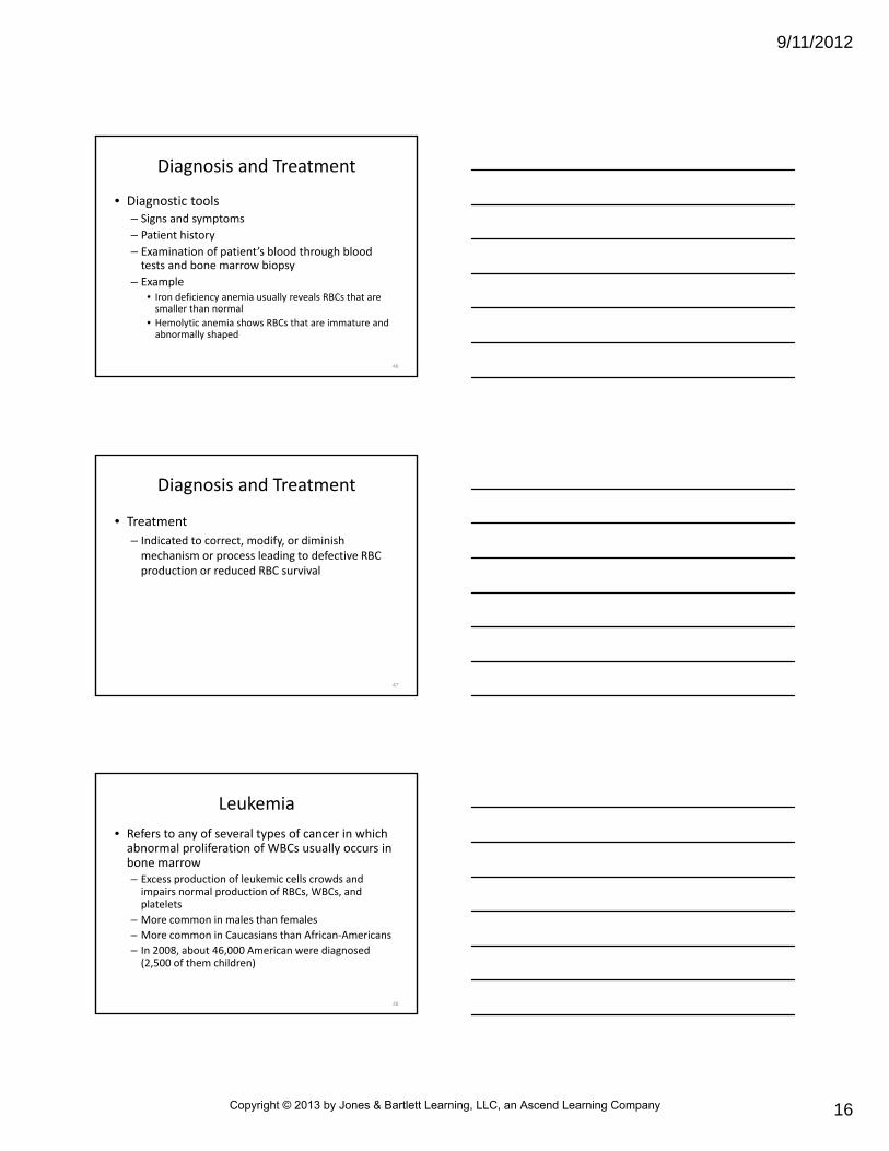
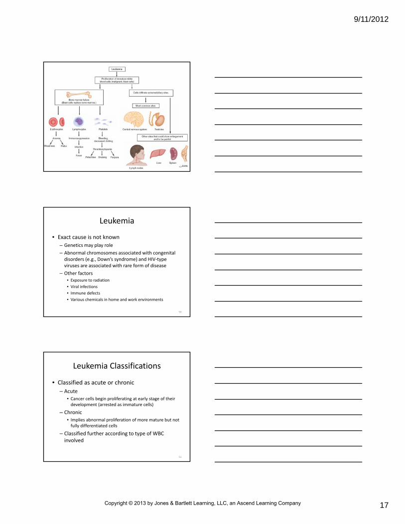
Leukemia
• Refers to any of several types of cancer in which abnormal proliferation of WBCs usually occurs in bone marrow– Excess production of leukemic cells crowds and impairs normal production of RBCs, WBCs, and platelets
– More common in males than females
– More common in Caucasians than African‐Americans
– In 2008, about 46,000 American were diagnosed (2,500 of them children)
48
Copyright © 2013 by Jones & Bartlett Learning, LLC, an Ascend Learning Company
Page 17
9/11/2012
17
49
Leukemia
• Exact cause is not known
– Genetics may play role
– Abnormal chromosomes associated with congenital disorders (e.g., Down’s syndrome) and HIV‐type viruses are associated with rare form of disease
– Other factors
• Exposure to radiation
• Viral infections
• Immune defects
• Various chemicals in home and work environments
50
Leukemia Classifications
• Classified as acute or chronic
– Acute
• Cancer cells begin proliferating at early stage of their development (arrested as immature cells)
– Chronic
• Implies abnormal proliferation of more mature but not fully differentiated cells
– Classified further according to type of WBC involved
51
Copyright © 2013 by Jones & Bartlett Learning, LLC, an Ascend Learning Company
Page 18
9/11/2012
18
Leukemia Classifications
• Two common forms of leukemia
– Acute lymphocytic leukemia
• Affects mostly children under age 15
• Sometimes called childhood leukemia
– Acute myelogenous leukemia
• Affects mostly middle‐aged adults
52
Leukemia Classifications
• Two common forms of leukemia– In both types, abnormal WBCs are produced in such large amounts that they eventually accumulate in vital organs (liver, spleen, lymph, brain)
• Impedes function of these organs and leads to death
– Chronic forms of leukemia can develop slowly, often over many years
• Often are discovered by chance during routine blood analysis
53
Leukemia Signs and Symptoms
• Proliferation of leukemic cells or resulting inadequate production of other normal blood cells makes patient highly susceptible to
– Serious infections
– Anemia
– Bleeding episodes
54
Copyright © 2013 by Jones & Bartlett Learning, LLC, an Ascend Learning Company
Page 19
9/11/2012
19
Leukemia Signs and Symptoms
• Signs and symptoms
– Abdominal fullness
– Bleeding
– Bone pain
– Elevated body temperature and diaphoresis
– Enlargement of lymph nodes
– Enlargement of the liver, spleen, and testes
– Fatigue
– Frequent bruising
– Headache
– Heat intolerance
– Night sweats
– Weight loss
55
If a child presents with a lot of unusual bruises, what would you
suspect if a diagnosis of leukemia is not known?
56
• Diagnosis
– Confirmed by bone marrow biopsy
• Severity assessed by
– Degree of liver and spleen enlargement
– Anemia
– Lack of platelets in blood
Leukemia Diagnosis and Treatment
57
Copyright © 2013 by Jones & Bartlett Learning, LLC, an Ascend Learning Company
Page 20
9/11/2012
20
• Treatment: acute
– Transfusion of blood and platelets
– Antibiotic therapy to manage anemia and infection
– Anticancer drugs
– Radiation
– Bone marrow transplant
• Treatment: chronic leukemia
– Managed effectively with medication
– Many patients require no treatment in its early stages
Leukemia Diagnosis and Treatment
58
Lymphomas
• General term applied to any neoplasticdisorder of lymphoid tissue
– Hodgkin’s disease is one type
• All others are called non‐Hodgkin’s lymphomas
• All lymphomas are malignant (cancerous tumors that tend to metastasize)
59
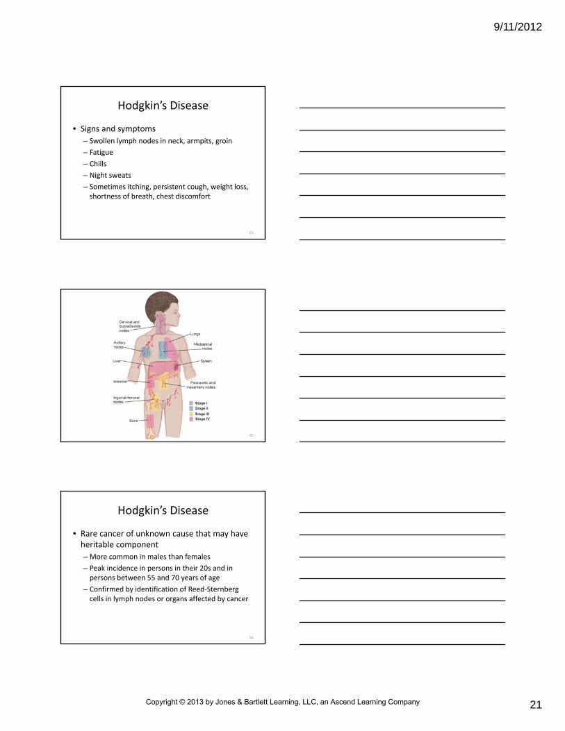
Hodgkin’s Disease
• Characterized by painless, progressive enlargement of lymphoid tissue found mainly in lymph nodes and spleen
– Left unchecked, cancer cells multiply and eventually displace healthy lymphocytes
• Suppresses immune system
60
Copyright © 2013 by Jones & Bartlett Learning, LLC, an Ascend Learning Company
Page 21
9/11/2012
21
Hodgkin’s Disease
• Signs and symptoms
– Swollen lymph nodes in neck, armpits, groin
– Fatigue
– Chills
– Night sweats
– Sometimes itching, persistent cough, weight loss, shortness of breath, chest discomfort
61
62
Hodgkin’s Disease
• Rare cancer of unknown cause that may have heritable component
– More common in males than females
– Peak incidence in persons in their 20s and in persons between 55 and 70 years of age
– Confirmed by identification of Reed‐Sternberg cells in lymph nodes or organs affected by cancer
63
Copyright © 2013 by Jones & Bartlett Learning, LLC, an Ascend Learning Company
Page 22
9/11/2012
22
Hodgkin’s Disease
• Treatment
– Depends on level of lymph node and organ system involvement (stage of disease)
– Can consist of radiation and chemotherapy with anticancer drugs
– Hodgkin’s disease is one of most curable cancers
64
Non‐Hodgkin’s Lymphomas
• Vary in their malignancy according to nature and activity of abnormal cells
– At least 10 types of non‐Hodgkin’s lymphoma identified
• Ranked as low, intermediate, high grade
– Ranking based on how aggressively disease behaves
– Low‐grade
• Progress slowly
• Tend not to spread beyond lymphatic system
– High‐grade
• Can spread to distant organs within few months
65
Non‐Hodgkin’s Lymphomas
• Signs and symptoms
– Painless swelling of one or more groups of lymph nodes
– Enlargement of liver and spleen
– Fever
– In rare cases, abdominal pain and GI bleeding
66
Copyright © 2013 by Jones & Bartlett Learning, LLC, an Ascend Learning Company
Page 23
9/11/2012
23
Non‐Hodgkin’s Lymphomas
• Cause largely unknown
– Burkitt’s lymphoma
• Childhood cancer
• In Africa, strongly associated with infection by Epstein‐Barr virus
– Other types worldwide have been linked to infection by HIV‐type viruses and other conditions that affect immune system
67
Non‐Hodgkin’s Lymphomas
• Treatment
– Radiation therapy
– Anticancer drugs
– Sometimes bone marrow transplantation
68
Polycythemia
• Increase in total RBC mass of blood
– May be natural response to chronic hypoxia (secondary polycythemia)
– May occur for unknown reasons (primary polycythemia)
– Can result from dehydration (apparent polycythemia)
• RBC production does not exceed upper limits of normal
69
Copyright © 2013 by Jones & Bartlett Learning, LLC, an Ascend Learning Company
Page 24
9/11/2012
24
Secondary Polycythemia
• Can be naturally present in persons who live in or visit areas of high altitude
– Due to reduced air pressure and low O2
– When O2 supply to blood is reduced, kidneys produce hormone erythropoietin
• Stimulates RBC production in bone marrow to make up for reduced O2 supply
• Result is increase in oxygen‐carrying efficiency of blood
70
Secondary Polycythemia
• RBC numbers return to normal when person returns to sea level
– Can be present in heavy smokers
– Can be caused by chronic bronchitis and conditions that increase erythropoietin production (e.g., liver cancer and some kidney disorders)
71
Primary Polycythemia
• Also known as polycythemia vera
– Rare disorder of bone marrow
– Increased production of RBCs causes blood to thicken
– Primarily develops in persons 50 or older
72
Copyright © 2013 by Jones & Bartlett Learning, LLC, an Ascend Learning Company
Page 25
9/11/2012
25
Primary Polycythemia
• Can lead to several physiological problems:
– Blurred vision
– Dizziness
– Generalized itching
– Headache
– Hypertension
– Red hands and feet; red‐purple complexion
– Splenomegaly
73
Primary Polycythemia
• Other complications
– Platelet disorders, which cause bleeding or clot formation
– Stroke
– Development of other bone marrow diseases (e.g., leukemias)
74
Primary Polycythemia
• Treatment
– Phlebotomy
• Slow removal of blood through vein
– Anticancer drug therapy
• Controls overproduction of RBCs in marrow
75
Copyright © 2013 by Jones & Bartlett Learning, LLC, an Ascend Learning Company
Page 26
9/11/2012
26
• Complication of severe injury, trauma, or disease
• Common abnormal clotting disorder
– Most often seen in critical care setting
– Disrupts balance among
• Procoagulants
• Inhibitors
• Thrombus formation
• Lysis
Disseminated Intravascular Coagulopathy
76
Disseminated Intravascular Coagulopathy
• Signs and symptoms
– Dyspnea
– Bleeding
– Those associated with hypotension and hypoperfusion
77
Disseminated Intravascular Coagulopathy
• Occurs in two phases
– First phase characterized by
• Free thrombin in blood
• Fibrin deposits
• Aggregation of platelets
– Second phase characterized by
• Hemorrhage caused by depletion of clotting factors
78
Copyright © 2013 by Jones & Bartlett Learning, LLC, an Ascend Learning Company
Page 27
9/11/2012
27
Disseminated Intravascular Coagulopathy
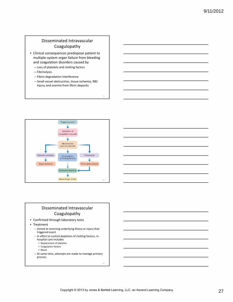
• Clinical consequences predispose patient to multiple‐system organ failure from bleeding and coagulation disorders caused by
– Loss of platelets and clotting factors
– Fibrinolysis
– Fibrin degradation interference
– Small vessel obstruction, tissue ischemia, RBC injury, and anemia from fibrin deposits
79
80
Disseminated Intravascular Coagulopathy
• Confirmed through laboratory tests
• Treatment– Aimed at reversing underlying illness or injury that triggered event
– In effort to control depletion of clotting factors, in‐hospital care includes
• Replacement of platelets
• Coagulation factors
• Blood
– At same time, attempts are made to manage primary process
81
Copyright © 2013 by Jones & Bartlett Learning, LLC, an Ascend Learning Company
Page 28
9/11/2012
28
Hemophilia
• Refers to a medical condition that causes uncontrolled bleeding and loss of bleeding control mechanisms– Group of inherited bleeding disorders– Hemophilia A is due to a deficiency in factor VIII
• Factor essential to process of blood clotting
– Hemophilia B is caused by a deficiency of factor IX• Also is known as Christmas disease
– All types present with similar problems• Specific factor involved determines severity of bleeding
– About 18,000 people in U.S. have hemophilia• About 400 are born with disorder each year
82
Hemophilia
• Bleeding from hemophilia can occur spontaneously, even after minor injury
– Can occur during some medical procedures (e.g., tooth extraction)
– Hemorrhage can occur anywhere in body
83
Hemophilia
• Bleeding from hemophilia can occur spontaneously, even after minor injury– Most common sites
• Joints
• Deep muscles
• Urinary tract
• Intracranial sites
– Head trauma is potentially life threatening
– CNS bleeding is major cause of death for patients in all age groups
84
Copyright © 2013 by Jones & Bartlett Learning, LLC, an Ascend Learning Company
Page 29
9/11/2012
29
Hemophilia
• Controlled by infusions of concentrates of factor VIII– Can be administered by patient
– Serious or unusual bleeding often requires hospitalization
– Patients are advised to avoid activities that may increase risk of injury
– Most patients are knowledgeable about their disease
– Most seek emergency care only when complicated problems and trauma‐related issues arise
85
Imagine that you are caring for a patient with hemophilia who has fallen 15 feet from a ladder. This patient refuses care and transportation. What should you
do?
86
Thrombocytopenia
• Low platelet count– In healthy people, blood normally contains 150,000 to 450,000 platelets/microliter of blood
– At levels of 20,000 to 30,000 platelets/microliter of blood, bleeding can occur with relatively minor trauma
– At levels less than 20,000 platelets/microliter of blood, spontaneous bleeding can occur, increasing risk for shock and death
• Especially true if bleeding occurs in brain
– Bleeding on skin is usually first sign of low platelet count
87
Copyright © 2013 by Jones & Bartlett Learning, LLC, an Ascend Learning Company
Page 30
9/11/2012
30
Thrombocytopenia
• Bleeding of skin may appear as
– Small red or purple spots on skin (petechiae), often on lower legs
– Purple, brown, red bruises (purpura) that happen easily and often
– Prolonged bleeding, even from minor cuts
– Bleeding or oozing from mouth or nose, especially nosebleeds or bleeding from brushing teeth
– Unusually heavy menstrual flow
88
Thrombocytopenia
• Can occur when body either doesn’t produce enough platelets
– If too many platelets are destroyed
– If spleen holds on to too many platelets
89
Thrombocytopenia
• Often associated with
– Leukemia or lymphoma
– Aplastic anemia
– Vitamin B12 or folic acid deficiency anemias
– Enlarged spleen
– Infectious diseases such as HIV/AIDS
– Massive blood transfusions
90
Copyright © 2013 by Jones & Bartlett Learning, LLC, an Ascend Learning Company
Page 31
9/11/2012
31
Thrombocytopenia
• Two diseases that occur because of increased destruction of platelets
– Idiopathic thrombocytopenic purpura (ITP)
• Occurs when antibodies attack and destroy body’s platelets for unknown reasons
• In children, can be acute condition that occurs after infection
• Acute ITP is rare in adults
• Chronic ITP most frequently affects women ages 20 to 40 years
91
Thrombocytopenia
• Two diseases that occur because of increased destruction of platelets – Thrombotic thrombocytopenic purpura (TTP)
• Life‐threatening disease that occurs when small blood clots form suddenly throughout body
• Can result in cardiac hemorrhage and death
• Occurs more often in women and is associated with pregnancy, metastatic cancer, chemotherapy, HIV/AIDS, some prescription drugs
• Patients experience kidney failure or decreased kidney function, fever, neurological complications
92
Thrombocytopenia
• Treatment depends on cause and severity
– Some only require careful monitoring of platelet counts
– More serious cases
• Corticosteroids (prednisone)
• Transfusion of platelets
• Rarely, surgical removal of spleen
93
Copyright © 2013 by Jones & Bartlett Learning, LLC, an Ascend Learning Company
Page 32
9/11/2012
32
Sickle Cell Disease
• Inherited blood disorder that affects red blood cells
• Several types, most common is sickle cell anemia
• Debilitating and unpredictable genetic illness
94
Sickle Cell Disease
• Affects persons of African descent, and less commonly, persons of Mediterranean origin
– 1 in 12 African‐Americans
– More than 70,000 Americans of different ethnic origins have disease
– In U.S., about 1,000 are born with disease each year
– 12.5 million Americans have sickle cell trait
95
Sickle Cell Disease
• Signs and symptoms
– Delayed growth, development, and sexual maturation in children
– Jaundice
– Priapism in adolescent and adult males
– Splenomegaly
– Stroke
96
Copyright © 2013 by Jones & Bartlett Learning, LLC, an Ascend Learning Company
Page 33
9/11/2012
33
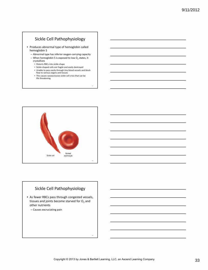
Sickle Cell Pathophysiology
• Produces abnormal type of hemoglobin called hemoglobin S– Abnormal type has inferior oxygen‐carrying capacity
– When hemoglobin S is exposed to low O2 states, it crystallizes
• Distorts RBCs into sickle shape
• Sickle‐shaped cells are fragile and easily destroyed
• Unable to pass easily through tiny blood vessels and block flow to various organs and tissues
• This causes vasoocclusive sickle cell crisis that can be life threatening
97
98
Sickle Cell Pathophysiology
• As fewer RBCs pass through congested vessels, tissues and joints become starved for O2 and other nutrients
– Causes excruciating pain
99
Copyright © 2013 by Jones & Bartlett Learning, LLC, an Ascend Learning Company
Page 34
9/11/2012
34
Sickle Cell Pathophysiology
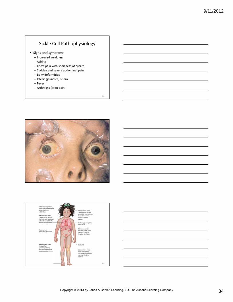
• Signs and symptoms– Increased weakness
– Aching
– Chest pain with shortness of breath
– Sudden and severe abdominal pain
– Bony deformities
– Icteric (jaundice) sclera
– Fever
– Arthralgia (joint pain)
100
101
102
Copyright © 2013 by Jones & Bartlett Learning, LLC, an Ascend Learning Company
Page 35
9/11/2012
35
How do you think a client with such chronic pain must feel at the beginning of a sickle cell crisis?
103
Sickle Cell Pathophysiology
• Sickle cell crisis
– Can occur in any part of body
– Can vary in intensity from one person to next and from one crisis to next
– Over time can destroy spleen, kidneys, gallbladder, other organs
– May occur for no apparent reason
104
Sickle Cell Pathophysiology
• It also may be triggered by
– Dehydration
– Exposure to extremes in temperature
– Infection
– Lack of O2
– Strenuous physical activity
– Stress
– Trauma
105
Copyright © 2013 by Jones & Bartlett Learning, LLC, an Ascend Learning Company
Page 36
9/11/2012
36
Sickle Cell Pathophysiology
• 3 less common types of sickle cell crisis
– Aplastic
• Bone marrow temporarily stops producing RBCs
– Hemolytic
• RBCs break down too rapidly to be replaced adequately
– Splenic sequestration
• Childhood difficulty that occurs when blood becomes trapped in spleen
• Causes organ to enlarge and may lead to death
106
Sickle Cell Management
• No cure exists
• Because of eventual damage to spleen
– Patients are at increased risk for septicemia if infected by certain types of bacteria
– Children with disease should be current with all immunizations
107
Sickle Cell Management
• When in crisis, require prompt treatment with
– O2 if hypoxic
– IV therapy to manage dehydration
– Antibiotics to manage infection
– Analgesics (e.g., morphine) to manage pain
108
Copyright © 2013 by Jones & Bartlett Learning, LLC, an Ascend Learning Company
Page 37
9/11/2012
37
Sickle Cell Management
• In severe cases, blood transfusion may be indicated
– Effect temporary replacement of hemoglobin S
– Can be advised during pregnancy to reduce risk of a crisis that can be fatal to mother and fetus
– May be advised before surgery because anesthesia can be hazardous to those with disease
109
Multiple Myeloma
• Malignant neoplasm of bone marrow
– Tumor, composed of plasma cells, destroys bone tissue (especially in flat bones)
– Causes
• Pain
• Fractures
• Hypercalcemia
• Skeletal deformities
110
Multiple Myeloma
• Malignant neoplasm of bone marrow
– Neoplastic cells produce large amounts of protein (M protein) that affect viscosity of blood
– Masses of coagulated protein can accumulate within tissues and impair function
111
Copyright © 2013 by Jones & Bartlett Learning, LLC, an Ascend Learning Company
Page 38
9/11/2012
38
Multiple Myeloma
• Some patients die of kidney failure
– Kidneys fail because of buildup of proteins that infiltrate kidneys and block renal tubules
– In many ways, resembles leukemia
– Plasma cell proliferation generally is confined to bone marrow
112
Multiple Myeloma
• Other associated disorders
– Proteinuria
– Anemia
– Weight loss
– Pulmonary complications from rib fracture
– Recurrent infections from suppression of immune system
113
Multiple Myeloma
• Patient complaints
– Weakness
– Skeletal pain
– Hemorrhage
– Hematuria
– Lethargy
– Weight loss
– Frequent fractures
114
Copyright © 2013 by Jones & Bartlett Learning, LLC, an Ascend Learning Company
Page 39
9/11/2012
39
Multiple Myeloma
• Occurs rarely before 40 years of age, then occurs increasingly with age
– Disease is more common in males than females and may have heritable component
• Diagnosed through
– X‐ray films
– Blood studies
– Tumor biopsy
115
Multiple Myeloma
• Treatment
– Chemotherapy with anticancer drugs
– Radiation
– Plasma exchange
– Bone marrow transplantation
116
General Assessment andManagement
• Most patients are knowledgeable about their disease– Often call EMS to help manage “change” in their condition
– May call to arrange for transportation to an emergency department for physician evaluation
– Situations that invoke call for emergency care vary by patient and disease
– Common chief complaints can be classified by body system
117
Copyright © 2013 by Jones & Bartlett Learning, LLC, an Ascend Learning Company
Page 40
9/11/2012
40
Prehospital Care
• Mainly supportive, must perform– General assessment
– Focused history
– Focused physical examination• Will guide patient care
• Will help determine appropriateness of emergency transport
• Some patients will have complex medical histories– When possible, should be transported to primary hospital where they usually receive medical care
118
Prehospital Care
• Patient may have variety of complaints and physical findings
– Some may be vague
• Can further complicate assessment
119
Prehospital Care
• After ensuring adequate airway, ventilatory, circulatory status
– Assess vital signs
– Perform physical examination
– Assess skin for color and turgor, noting any cyanosis or jaundice, warmth or coolness, bruising, edema, or ulcerations
120
Copyright © 2013 by Jones & Bartlett Learning, LLC, an Ascend Learning Company
Page 41
9/11/2012
41
Prehospital Care
• Ascertain any new onset of– Fever
– Weakness
– Cough
– Rash
– Spontaneous bleeding (e.g., bleeding gums, epistaxis)
– Vomiting
– Diarrhea
121
Prehospital Care
• Some hematological disorders can involve ability of blood to deliver enough oxygen to tissues
– Question all patients with hematological disorders specifically about
• Recent dizziness
• Syncope
• Difficulty breathing
• Heartbeat irregularities
122
Prehospital Care
• Other key elements
– Identify existing hematological disease
• Including any family history of hematological disease
– Significant medical history or recent injury
– Medication use
– Allergies
– Alcohol or illicit drug use
123
Copyright © 2013 by Jones & Bartlett Learning, LLC, an Ascend Learning Company
Page 42
9/11/2012
42
Prehospital Care
• Based on patient’s condition, prehospital care:
– O2 administration
– IV fluid replacement
– Antidysrhythmics
– Analgesics for pain management
– Some of these patients will be gravely ill
• Calming and comfort measures for patient and family
124
Summary
• Blood is composed of cells and formed elements surrounded by plasma
– About 95 percent of volume of formed elements consists of RBCs (erythrocytes)
• Remaining 5 percent consists of WBCs (leukocytes) and cell fragments (platelets)
125
Summary
• Anemia is condition in which amount of hemoglobin or erythrocytes in blood is below normal– Two common forms of anemia are iron deficiency anemia and hemolytic anemia
– All forms of anemia share signs and symptoms• Include fatigue and headaches, sometimes a sore mouth or tongue, brittle nails, and, in severe cases, breathlessness and chest pain
• Diagnosis is made by history and from blood tests and bone marrow biopsy
126
Copyright © 2013 by Jones & Bartlett Learning, LLC, an Ascend Learning Company
Page 43
9/11/2012
43
Summary
• Leukemia refers to any of several types of cancer in which abnormal proliferation of WBCs usually occurs in bone marrow– Proliferation of leukemic cells crowds and impairs normal production of RBCs, WBCs, and platelets
– Leukemia is classified as acute or chronic
– Proliferation of leukemic cells makes the patient highly susceptible to serious infections, anemia, and bleeding episodes
– Diagnosis is confirmed by bone marrow biopsy
127
Summary
• Lymphoma refers to a group of diseases that range from slowly growing chronic disorders to rapidly evolving acute conditions
– Hodgkin’s disease is one type; all others are called non‐Hodgkin’s lymphomas
128
Summary
• Polycythemia is characterized by an unusually large number of RBCs in blood as a result of their increased production by bone marrow
– Polycythemia may be natural response to hypoxia
• Known as secondary polycythemia
– Polycythemia also may occur for unknown reasons
• Known as primary polycythemia
129
Copyright © 2013 by Jones & Bartlett Learning, LLC, an Ascend Learning Company
Page 44
9/11/2012
44
Summary
• Disseminated intravascular coagulopathy is complication of severe injury, trauma, or disease– Disrupts balance among procoagulants, thrombin formation, inhibitors, and lysis
– Signs and symptoms of disseminated intravascular coagulation include dyspnea, bleeding, and those associated with hypotension and hypoperfusion
– Treatment aimed at reversing underlying illness or injury that triggered event
130
Summary
• Hemophilia A is caused by deficiency of blood protein called factor VIII
– Hemophilia B is caused by deficiency of factor IX
– Bleeding from hemophilia can occur spontaneously, after even minor injury, or during some medical procedures
131
Summary
• Thrombocytopenia is a low platelet count
– Can occur when body either doesn’t produce enough platelets; if too many platelets are destroyed; of if spleen holds on to too many platelets
– Bleeding is chief complication of thrombocytopenia
132
Copyright © 2013 by Jones & Bartlett Learning, LLC, an Ascend Learning Company
Page 45
9/11/2012
45
Summary
• Sickle cell disease is a debilitating and unpredictable recessive genetic illness– Affects persons of African descent
– Less often, affects persons of Mediterranean origin
– Sickle cell anemia produces abnormal type of hemoglobin
• Called hemoglobin S
• Has inferior oxygen‐carrying capacity
– Complications include episodes of severe pain, fatigue, pallor, jaundice, stroke, delayed growth, hematuria, priapism, and splenomegaly
133
Summary
• Multiple myeloma is a malignant neoplasm of the bone marrow
– Tumor destroys bone tissue (especially flat bones) and causes pain, fractures, hypercalcemia, and skeletal deformities
• In many cases of hematological disorders, prehospital treatment is supportive
– Treatment includes ensuring adequate airway, ventilatory, and circulatory support
134
Questions?
135
Copyright © 2013 by Jones & Bartlett Learning, LLC, an Ascend Learning Company