87
3/28 • Pass back tests • Do the analysis/scoring • Review most missed questions HW – Ch. 45 and 46 due Wed. – use guided notes
| Date post: | 25-Dec-2015 |
| Category: |
Documents |
| Upload: | beverly-cummings |
| View: | 217 times |
| Download: | 0 times |

3/28
• Pass back tests• Do the analysis/scoring• Review most missed questions
HW – Ch. 45 and 46 due Wed. – use guided notes

3/29
• Ch. 44 Excretory with practice• Check grades• I’ll check 43-46 tomorrow
HW – Ch. 45 and 46 due Wed. – use guided notes

Ch. 44 Goals
• Know the overall function of the Excretory system• Include a picture of the organs of the Excretory system• Know what osmoregulation and excretion are• Know the 3 categories of nitrogenous waste, which
animal groups produce each, and why• Recognize the variations of excretory systems of
Platyhelminthes, Annelida, Insects/Arthropods, and Vertebrates
• Know the 4 main steps in urine formation• Know the components of a nephron, and what occurs in
each region• Know how hormones affect water balance by acting on
the nephron

Excretory System
• Functions?
• Organs?
(Let’s label practice worksheet)
• Functional Unit?

Osmoregulation & Excretion
• Osmoregulation =
• Excretion =

3 types of nitrogenous waste
• Ammonia –
• Urea –
• Uric acid (Urate) -

Copyright © 2002 Pearson Education, Inc., publishing as Benjamin Cummings
Fig. 44.13
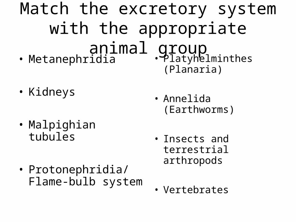
Match the excretory system with the appropriate animal group
• Metanephridia
• Kidneys
• Malpighian tubules
• Protonephridia/Flame-bulb system
• Platyhelminthes (Planaria)
• Annelida (Earthworms)
• Insects and terrestrial arthropods
• Vertebrates

• Most excretory systems produce a filtrate by pressure-filtering body fluids into tubules.– This filtrate is then
modified by the transport epithelium which reabsorbs valuable substances, secretes other substances, like toxins and excess ion, and then excretes the contents of the tubule.
Copyright © 2002 Pearson Education, Inc., publishing as Benjamin Cummings
Fig. 44.17

• Flatworms have an excretory system called protonephridia, consisting of a branching network of dead-end tubules.– These are capped by a
flame bulb with a tuft of cilia that draws water and solutes from the interstitial fluid, through the flame bulb, and into the tubule system.
Copyright © 2002 Pearson Education, Inc., publishing as Benjamin Cummings
Fig. 44.18

• Metanephridium, another tubular excretory system, consists of internal openings that collect body fluids from the coelom through a ciliated funnel, the nephrostome, and release the fluid through the nephridiopore.– Found in most annelids, each segment of a
worm has a pair of metanephridia.
Copyright © 2002 Pearson Education, Inc., publishing as Benjamin Cummings
Fig. 44.19

• Insects and other terrestrial arthropods have organs called Malpighian tubules that remove nitrogenous wastes and also function in osmoregulation.– These open into the
digestive system and dead-end at tips that are immersed in the hemolymph.
Copyright © 2002 Pearson Education, Inc., publishing as Benjamin Cummings
Fig. 44.20

Copyright © 2002 Pearson Education, Inc., publishing as Benjamin Cummings
Fig. 44.21

Let’s do the website activities to better learn and visualize how a
nephron functions
• www.campbellbiology.com
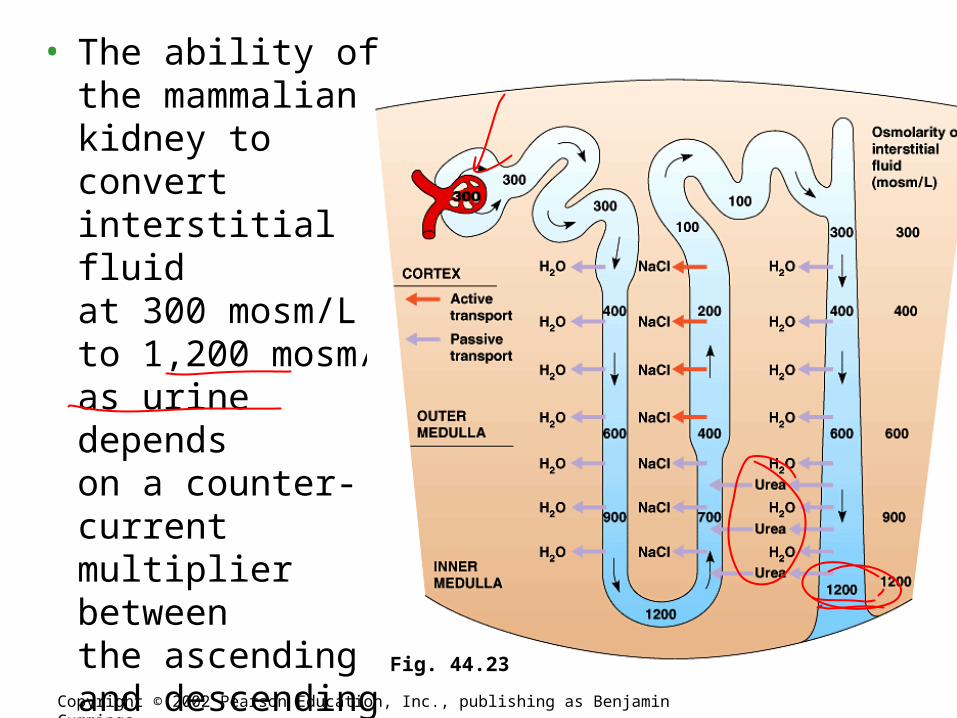
• The ability of the mammaliankidney to convertinterstitial fluidat 300 mosm/Lto 1,200 mosm/Las urine dependson a counter-current multiplier betweenthe ascending and descending limbsof the loop
of Henle.
Copyright © 2002 Pearson Education, Inc., publishing as Benjamin Cummings
Fig. 44.23

Copyright © 2002 Pearson Education, Inc., publishing as Benjamin Cummings
Fig. 44.24a
Copyright © 2002 Pearson Education, Inc., publishing as Benjamin Cummings
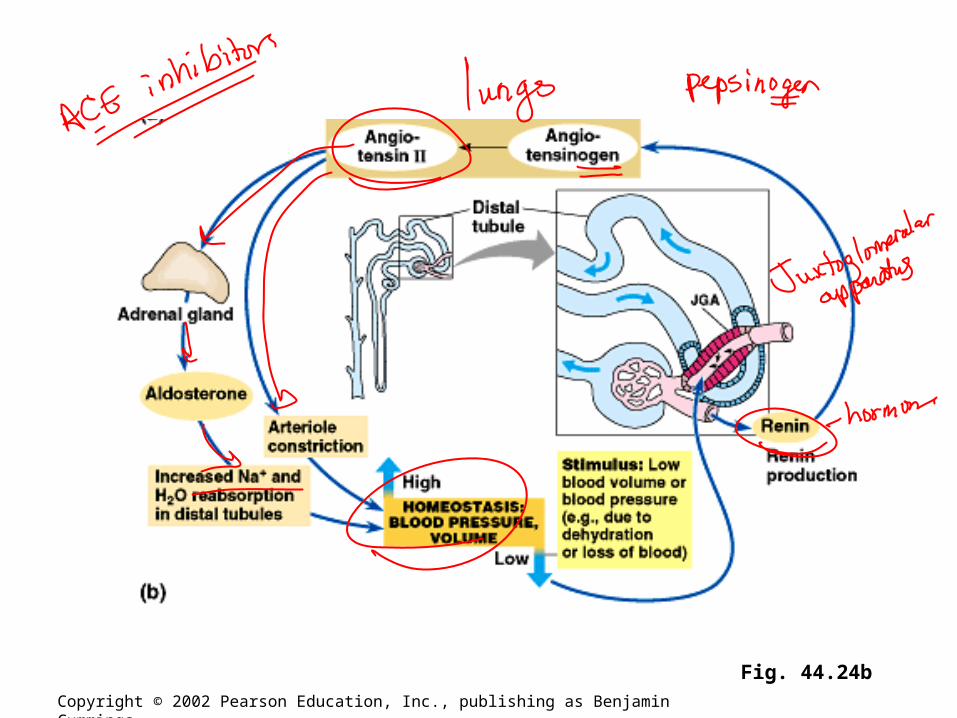
Fig. 44.24b

• Still another hormone, atrial natriuretic factor (ANF), opposes the RAAS.– The walls of the atria release ANF in response to an
increase in blood volume and pressure.– ANF inhibits the release of renin from the JGA, inhibits
NaCl reabsorption by the collecting ducts, and reduces aldosterone release from the adrenal glands.
– These actions lower blood pressure and volume.– Thus, the ADH, the RAAS, and ANF provide an
elaborate system of checks and balances that regulates the kidney’s ability to control the osmolarity, salt concentration, volume, and pressure of blood.
Copyright © 2002 Pearson Education, Inc., publishing as Benjamin Cummings

3/30/11
• Finish Excretory• Endocrine Intro• Quiz each other on table while I check
notes 43-46• Volunteers to come up and explain/draw
feedback loops
HW – Animal Development questions due Friday

Ch. 45 Goals
• Know the function of the endocrine system• Include a picture of the system and be able to name all
of the glands• Know 2 ways hormones affect target organs in general• Know the table of glands and their hormones and
general effects• Know in detail the secretion, target, action, and
regulation of at least three hormones• Be able to describe positive and negative feedback in the
regulation of homeostasis by hormones (practice drawing the loops from the textbook)

Endocrine System
• Function –
• What other system does it work closely with?
• What are endocrine vs. exocrine glands?
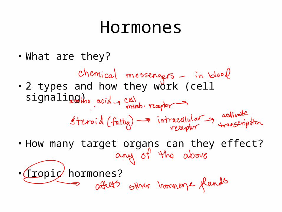
Hormones
• What are they?
• 2 types and how they work (cell signaling)
• How many target organs can they effect?
• Tropic hormones?

Fig. 45.2
Copyright © 2002 Pearson Education, Inc., publishing as Benjamin Cummings
Even insects use hormones to regulate molting

Tropic hormonestarget other endocrineglands and areimportant forunderstandingchemicalcoordination.
• Humans havenine endocrineglands.
Human Endocrine System
Copyright © 2002 Pearson Education, Inc., publishing as Benjamin CummingsFig. 45.5

The hypothalamus integrates endocrine and nervous function. Neurosecretory cells of the hypothalamus
produce hormones. Releasing hormones stimulate the anterior pituitary
(adenohypophysis) to secrete hormones. Inhibiting hormones prevent the anterior pituitary
from secreting hormones.
The hypothalamus and pituitary integrate many functions of the
vertebrate endocrine system
Copyright © 2002 Pearson Education, Inc., publishing as Benjamin Cummings

Fig. 45.6b
Copyright © 2002 Pearson Education, Inc., publishing as Benjamin Cummings

The posteriorpituitary(neurohypo-physis)stores andsecretes hormonesproducedby thehypothalamus.
Copyright © 2002 Pearson Education, Inc., publishing as Benjamin CummingsFig. 45.6a

Table 45.1Copyright © 2002 Pearson Education, Inc., publishing as Benjamin Cummings

Table 45.1 (continued)Copyright © 2002 Pearson Education, Inc., publishing as Benjamin Cummings

Quiz each other on table
• Silent study for 5-10 minutes
• Quiz each other on table
• When done, practice drawing feedback loops for blood calcium, blood glucose, thyroid metabolism, and adrenal gland (negative feedback – more gets you less) and uterine contractions (positive feedback – more gets you more)

The thyroid glandof mammals consistsof two lobes locatedon the ventral surfaceof the trachea. Triiodothyronine (T3)
and thyroxine (T4): amines.
Stimulates and maintainmetabolic processes.
Secretion regulated by TSHhormones.
Thyroid hormones function in development, bioenergetics, and homeostasis
Copyright © 2002 Pearson Education, Inc., publishing as Benjamin Cummings Fig. 45.8
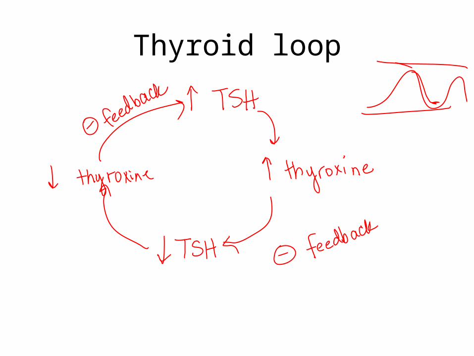
Thyroid loop

Copyright © 2002 Pearson Education, Inc., publishing as Benjamin Cummings Fig. 45.9

Blood Calcium

Copyright © 2002 Pearson Education, Inc., publishing as Benjamin Cummings Fig. 45.10


Copyright © 2002 Pearson Education, Inc., publishing as Benjamin Cummings
Fig. 45.14

Uterine contractions- positive feedback

Positive Feedback
3/31
• Finish endocrine
• Start reproductive
HW – Animal Repro and Development questions 1-16 due Friday

Ch. 46 Goals
• Know the overall function of the reproductive system• Include a picture of the male and female anatomy –
know all the parts• Know the different types of reproduction in the animal
kingdom• Know the different mechanisms for fertilization• Know the hormonal control of the menstrual and ovarian
cycle (spend some time on this)• Know how oogenesis and spermatogenesis differ• Know the function of human chorionic gonadotropin
(hCG)

Reproductive System
• Function –
• Asexual vs. Sexual?
• Advantages of each?
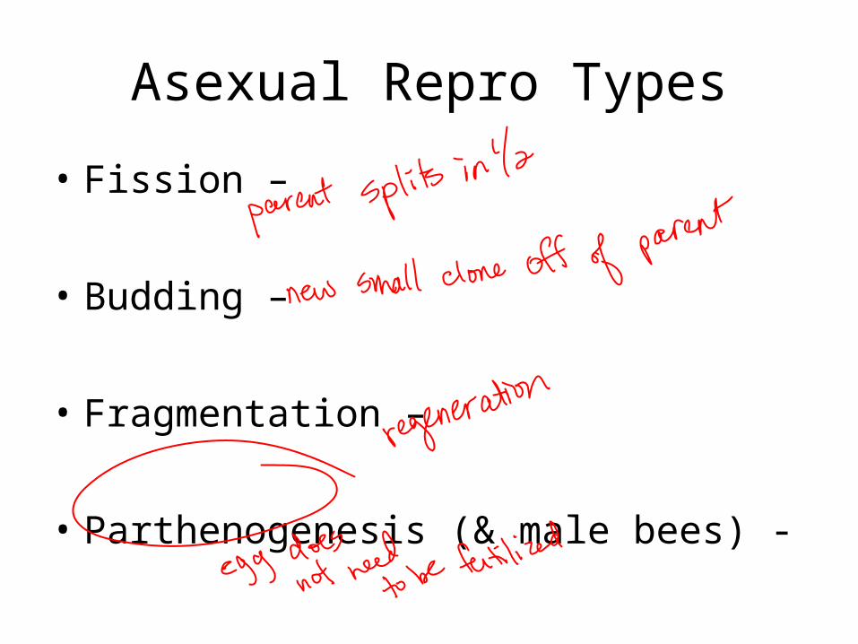
Asexual Repro Types
• Fission –
• Budding –
• Fragmentation –
• Parthenogenesis (& male bees) -

Triggers/Patterns of Reproduction
• Ovulation – may be cyclical, young produced at times likely to survive
• Hermaphroditism – common in stationary animals
• Sex reversal – example of wrasse fish – largest female becomes male if the male of the harem dies
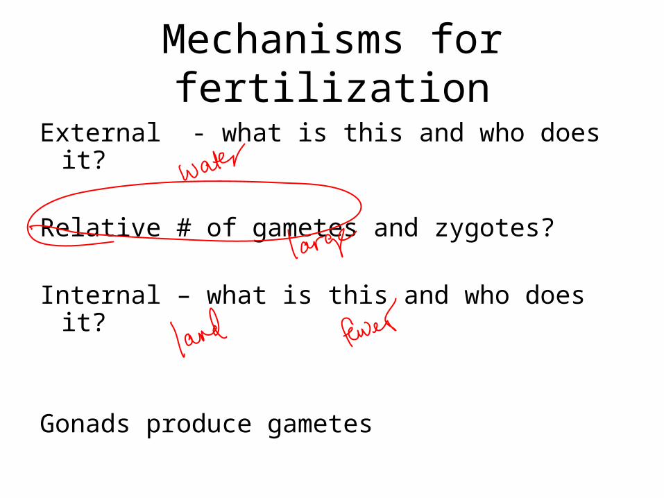
Mechanisms for fertilization
External - what is this and who does it?
Relative # of gametes and zygotes?
Internal – what is this and who does it?
Gonads produce gametes

Internal fertilization requires cooperative behavior that leads to copulation.
Internal and external fertilization both depend on mechanisms ensuring that
mature sperm encounter fertile eggs of the same species
Copyright © 2002 Pearson Education, Inc., publishing as Benjamin Cummings

External fertilization requires a moist habitat that will protect a developing egg from desiccation and heat stress.– Specific mating behaviors assure that sperm and
egg will be in the same place at the same time.
Copyright © 2002 Pearson Education, Inc., publishing as Benjamin Cummings
Fig. 46.4

Pheromones: chemical signals released by one organism that influence the behavior of other individuals of the same species. Many act as male attractants.
Copyright © 2002 Pearson Education, Inc., publishing as Benjamin Cummings

Internal fertilization usually results in the production of fewer zygotes than does internal fertilization. However, the survival rate is lower for external
fertilization than it is for internal fertilization.
Species with internal fertilization usually produce fewer zygotes but
provide more parental protection than species with external fertilization
Copyright © 2002 Pearson Education, Inc., publishing as Benjamin Cummings

The externally fertilized eggs of fishes and amphibians are surrounded by a gelatinous coat.
The internally fertilized amniote eggs of birds, reptiles, and monotremes are protected by calcium and protein shells. In mammals the embryo is retained within the
females reproductive tract.
Copyright © 2002 Pearson Education, Inc., publishing as Benjamin Cummings

– Parental care ofoffspring may occurregardless of whetherfertilization is externalor internal.
Copyright © 2002 Pearson Education, Inc., publishing as Benjamin Cummings
Fig. 46.5

The least complex reproductive systems lack gonads. Polychaete worms lack gonads.
Eggs and sperm develop from undifferentiated cells lining the coelom.
• Some reproductive systems, such as that seen in parasitic flatworms, can be very complex.
Complex reproductive systems have evolved in many animal phyla
Copyright © 2002 Pearson Education, Inc., publishing as Benjamin Cummings

Copyright © 2002 Pearson Education, Inc., publishing as Benjamin Cummings Fig. 46.6

Most insects have separate sexes with complex reproductive systems. In many species the female reproductive
system includes a spermatheca, a sac in which sperm may be stored for a year or more.
Copyright © 2002 Pearson Education, Inc., publishing as Benjamin CummingsFig. 46.7

The basic plan of all vertebrate reproductive systems are very similar. However, there are variations.
In many non-mammalian vertebrates the digestive, excretory, and reproductive systems share a common opening to the outside, the cloaca.
Mammals have separate opening for the digestive and reproductive systems. Female mammals also have separate openings for the
excretory and reproductive systems.
Copyright © 2002 Pearson Education, Inc., publishing as Benjamin Cummings
Copyright © 2002 Pearson Education, Inc., publishing as Benjamin Cummings
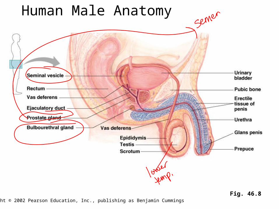
Fig. 46.8
Human Male Anatomy

Copyright © 2002 Pearson Education, Inc., publishing as Benjamin Cummings
Fig. 46.8

Copyright © 2002 Pearson Education, Inc., publishing as Benjamin Cummings
Let’s watch animation

Spermatogenesis is the production of mature sperm cells from spermatogonia.
A continuous and prolific process in the adult male. Each ejaculation contains 100 – 650 million sperm. Occurs in seminiferous tubules. As spermatogenesis progresses the developing sperm
cells move from the wall to the lumen of a seminiferous tubule.
Spermatogenesis
Copyright © 2002 Pearson Education, Inc., publishing as Benjamin Cummings

Sperm structure: Haploid nucleus. Tipped with an acrosome.
Contains enzymes that help the sperm penetrate to the egg.
A large numberof mitochondriaprovide ATP topower theflagellum.
Copyright © 2002 Pearson Education, Inc., publishing as Benjamin Cummings
Fig. 46.12

Fig. 46.9
Copyright © 2002 Pearson Education, Inc., publishing as Benjamin Cummings

Fig. 46.9
Copyright © 2002 Pearson Education, Inc., publishing as Benjamin Cummings

Each follicle consists of one egg cell surrounded by one or more layers of follicle cells. Follicles produce the primary female sex hormones:
estrogens. Follicle cells nourish and protect the developing egg cell. A woman is born with about 400,000 follicles.
Only several hundred of which will release eggs during a female’s reproductive years.
Copyright © 2002 Pearson Education, Inc., publishing as Benjamin Cummings

Usually one follicle matures and releases its egg during each menstrual cycle. After ovulation the remaining follicular tissue
develops into the corpus luteum. Secretes estrogens and progesterone. Maintain the uterine
lining during pregnancy. If pregnancy does not
occur the corpus luteumdisintegrates.
Copyright © 2002 Pearson Education, Inc., publishing as Benjamin Cummings
Fig. 46.10

Fig. 46.13
Copyright © 2002 Pearson Education, Inc., publishing as Benjamin Cummings
Let’s watch animation

Oogenesis is the production of ova from oogonia.
Differs from spermatogenesis in three major ways: At birth an ovary contains all of the primary oocytes
it will ever have and oogenesis ends at menopause. (vs. men make sperm their entire adult life)
Unequal cytokinesis during meiosis results in the formation of a single large secondary oocyte and three small polar bodies. (vs. 4 sperm cells)
The polar bodies degenerate. Oogenesis has long “resting” periods – eggs
arrested in Prophase I prior to female’s birth. Meiosis not completed until after fertilization.
Copyright © 2002 Pearson Education, Inc., publishing as Benjamin Cummings

4/1
• Finish reproductive• Check Animal Development questions
• HW – Ch. 48 & 49 due the Tuesday back (will email the goals along with this week’s Powerpoint), AP review manual - do the Body Systems review questions (will check this Thursday that we are back)
The Male Pattern. Androgens secreted by Leydig cells are
responsible for primary and secondary sex characteristics. Primary sex characteristics:
Development of the vasa deferentia and other ducts. Development of the external reproductive structures Sperm production.
A complex interplay of hormones regulates reproduction
Copyright © 2002 Pearson Education, Inc., publishing as Benjamin Cummings

Secondary sexcharacteristics: Deepening of
the voice. Distribution
pattern of facialand pubic hair.
Muscle growth. Androgens are also
responsible forsexual behaviorand generalaggressiveness.
Copyright © 2002 Pearson Education, Inc., publishing as Benjamin Cummings
Fig. 46.14

The Female Pattern. A cyclic pattern of hormone secretion and
reproductive events. Humans and many other primates have
menstrual cycles. If pregnancy does not occur the endometrium is
shed through the cervix and vagina: menstruation.
Copyright © 2002 Pearson Education, Inc., publishing as Benjamin Cummings

Other mammals have estrous cycles. If pregnancy does not occur the endometrium is
reabsorbed by the uterus. Associated with more pronounced behavioral cycles
than are menstrual cycles. More pronounced seasonal and climatic effects than
seen associated with menstrual cycles.
Humans females may be sexually receptive throughout their cycles. Most mammals will copulate only during the period
surrounding ovulation. This period of sexual activity is called estrus.
Copyright © 2002 Pearson Education, Inc., publishing as Benjamin Cummings

Spend some time with Figure 9.11 of Menstrual Cycle on p. 259 of my
manual
• Then you take turns describing it to your neighbor

Estrogens are also responsible for female
secondary sex characteristics. Deposition of fat in the breasts and hips. Increased water retention. Affects calcium metabolism. Stimulates of breast development. Mediates female sexual behavior.
Menopause: cessation of ovarian and menstrual cycles. Usually occurs between ages 46 and 54. Due to ovaries decreased responsiveness to
gonadotropins.
Copyright © 2002 Pearson Education, Inc., publishing as Benjamin Cummings

From Conception to Birth. In placental mammals, pregnancy or gestation
is the condition of carrying one or more embryos. Pregnancy is preceded by conception and continues
until birth. A human pregnancy averages 266 days.
Pregnancy
Copyright © 2002 Pearson Education, Inc., publishing as Benjamin Cummings

Human gestation is divided into three trimesters. First trimester.
Fertilization occurs in the oviduct. 24 hours latter the zygote begins cleavage. 3- 4 days after fertilization the zygote that reaches
the uterus the embryo is a ball of cells. It takes about 1 week past fertilization for the
blastocyst to form. After 5 more days it implants in the endometrium.
Copyright © 2002 Pearson Education, Inc., publishing as Benjamin Cummings

Fig. 46.16
Copyright © 2002 Pearson Education, Inc., publishing as Benjamin Cummings

For the first 2 – 4 weeks of development the embryo obtains nutrients from the endometrium.
Then the placenta provides for the diffusion of material between maternal and embryonic circulations.
Fig. 46.17
Copyright © 2002 Pearson Education, Inc., publishing as Benjamin Cummings

Organogenesis occurs during the first trimester. By week 4: the heart is beating. By the end of week 8: all of the major structures of the adult
are present in rudimentary form. The rapidity of development makes this a time when the
embryo is especially sensitive to environmental insult.
Copyright © 2002 Pearson Education, Inc., publishing as Benjamin Cummings

Maternal changes during the first trimester. The embryo secretes human chorionic
gonadotropin (HCG). Maintains the corpus luteum and thus maintains the
endometrium. High levels of progesterone cause.
Increased mucus in the cervix. Growth of the maternal part of the
placenta. Enlargement of the uterus. Cessation of ovarian and menstrual
cycling. Breasts enlarge rapidly and are often
very tender.Copyright © 2002 Pearson Education, Inc., publishing as Benjamin Cummings

Second trimester. Fetus grows rapidly and is very active. Hormonal levels stabilize as HCG declines. Corpus luteum deteriorates. Placenta secretes progesterone, which maintains the
pregnancy.
Copyright © 2002 Pearson Education, Inc., publishing as Benjamin Cummings

Third trimester. Fetus grows rapidly. Fetal activity may decrease as the fetus fills the space
available to it. Maternal abdominal organs become compressed and
displaced. Terminates with parturition.
Copyright © 2002 Pearson Education, Inc., publishing as Benjamin Cummings

Hormonal regulation of birth.
Fig. 46.19
Copyright © 2002 Pearson Education, Inc., publishing as Benjamin Cummings

Parturition occurs as a result of labor. First stage: opening
up and thinningof the cervix.
Ending in completedilation.
Second stage:Expulsion of thebaby as a resultof strong uterinecontractions.
Third stage:Expulsion of the placenta.
Fig. 46.20Copyright © 2002 Pearson Education, Inc., publishing as Benjamin Cummings

Fig. 46.21
Contraception

Prenatal diagnosis of genetic and congenital abnormalities. Invasive techniques.
Amniocentesis. Chorionic villus sampling.
Modern technology offers solutions for some reproductive problems
Copyright © 2002 Pearson Education, Inc., publishing as Benjamin Cummings

Noninvasive techniques. Ultrasound imaging. Maternal blood contains fetal blood cells that can be tested.
Copyright © 2002 Pearson Education, Inc., publishing as Benjamin Cummings

Infertility treatment. Sperm donors. In vitro fertilization.
Copyright © 2002 Pearson Education, Inc., publishing as Benjamin Cummings


