37
Chapter 367 Intracranial Internal Carotid Artery Aneurysms 22/12/2015
| Date post: | 10-Apr-2017 |
| Category: |
Health & Medicine |
| Upload: | neurosurgery-vajira |
| View: | 108 times |
| Download: | 0 times |

Chapter 367 Intracranial Internal Carotid Artery Aneurysms
22/12/2015
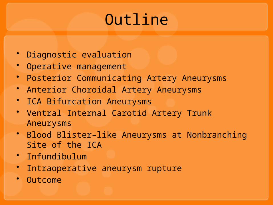
Outline
• Diagnostic evaluation• Operative management• Posterior Communicating Artery Aneurysms• Anterior Choroidal Artery Aneurysms• ICA Bifurcation Aneurysms• Ventral Internal Carotid Artery Trunk Aneurysms• Blood Blister–like Aneurysms at Nonbranching Site of the ICA• Infundibulum• Intraoperative aneurysm rupture• Outcome

Diagnostic evaluation
• CT– extremely sensitive for detecting SAH in the acute
phase– possible location of the aneurysm
• CTA,3D CTA,MRA– reliable results in detecting aneurysms equal to or
grater than 2 mm in diameter

Diagnostic evaluation
• Digital subtraction angiography (DSA)– gold standard– when the CTA findings are
negative or doubtful– demonstrates some of the
perforating arteries in and around the aneurysm andthe parent vessel
– location of the proximal neck of the aneurysm– projection of the angiographic pictures

Preoperative Care
• Airway, breathing, circulation• Rebleeding
– 6% in 48 hr– Devastating result
• Hydrocephalus– Detect on CT brain– EVD will help return most of these patients back to
a better grade

Preoperative Care
• Electrolyte abnormalities• Vasospasm• Grading• Nimodipine 60 mg oral q 4 hr• Euvolemic• Control hypertension• Anticonvulsants are used in patients who
develop seizure after SAH

Grade GCS
I 15
II 14
III 13
IV 12-7
V 6-3

Preoperative Care
• Time for treatment– Favorable-grade : within 24-48 hr– Poor grade(mWFNS gr IV,V)
• allowed to recover in the intensive care• only treated if they show improvement in SAH
grade• Broad-spectrum antibiotics before the operation

Intraoperative Management
• 20% mannitol 0.5 mg/kg after skin incision• Brain retractors : used only after wide splitting of
the sylvian fissure • Dexamethasone : no supporting evidence

Postoperative Care
• Vasospasm– euvolemic to slightly hypervolemic state– CVP 8-12 cmH2O– allowed to rise to the patient’s high normal
• Hydrocephalus– VP shunt insertion if there is persistent symptomatic
hydrocephalus• Electrolyte imbalance
– Na keep 135 to 148 mEq/L

Postoperative Care
• Seizures– SAH induced seizure– Phenytoin postoperatively for 6 months to 1 year
• Brain swelling• Postoperative stroke• Rebleeding from a residual portion of the
aneurysm• Routine DSA
– on postoperative day 7 to 10 to ensure complete obliteration of the aneurysm
Posterior Communicating Artery Aneurysms

Anatomy
• C1 segment• Arises from the posteromedial surface of the ICA• Courses medially and inferiorly, through the
membrane of Liliequist, above and medial to the oculomotor nerve
• Join the PCA at the junction of the P1-P2 segment
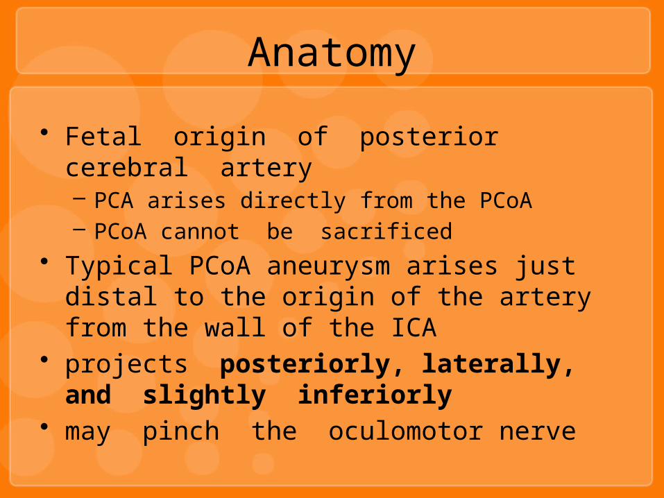
• Fetal origin of posterior cerebral artery– PCA arises directly from the PCoA– PCoA cannot be sacrificed
• Typical PCoA aneurysm arises just distal to the origin of the artery from the wall of the ICA
• projects posteriorly, laterally, and slightly inferiorly
• may pinch the oculomotor nerve
Anatomy

Presentation
• Usually cause symptoms when smaller than 10 mm• Compress the third cranial nerve : painful
non–pupil-sparing oculomotor nerve palsy• Retro-orbital pain• CT
– SAH at lateral suprasellar,ambient cistern pattern– intraparenchymal hemorrhage into the uncus of the
temporal lobe– IVH into the temporal horn– hemorrhage into the subdural space
Operative Technique
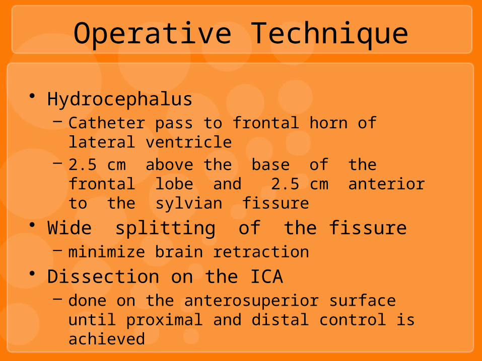
• Hydrocephalus– Catheter pass to frontal horn of lateral ventricle– 2.5 cm above the base of the frontal lobe and
2.5 cm anterior to the sylvian fissure• Wide splitting of the fissure
– minimize brain retraction• Dissection on the ICA
– done on the anterosuperior surface until proximal and distal control is achieved

Operative Technique
• The clot on the base of the aneurysm is swiped away from the neck to visualize it better
• Identified : PCoA, anterior thalamic perforator, anterior choroidal artery
• Anteromedial retraction on the ICA is dangerous– may pull on the dome of the aneurysm and tear It– may cause permanent damage to 3rd nerve

Operative Technique
• Temporary clipping– used in large aneurysms to reduce the flow– this will help to avoid tearing the aneurysm neck– no longer than 3 minutes at a time, while allowing at
least 5 minutes• After Clipping
– the tips are inspected to ensure complete closure around the aneurysm
– patency of the posterior communicating artery, thalamoperforator, and most important, anterior choroidal artery

Operative Technique
• Punctured with a 25-gauge needle to ensure obliteration
• Patency of the carotid is confirmed with intraoperative doppler

Selective Intradural Anterior Clinoidectomy
• PCoA aneurysms (or proximal carotid aneurysms) with a very proximal neck
• The intraoperative exposure is not satisfactory unless the anterior clinoid process can be removed
• Direction of drill– medial to lateral, from the optic nerve – toward the superior orbital fissure
• If the proximal neck of the aneurysm is found to be under the distal dural ring of the carotid artery, the dura ring and the falciform ligaments are opened
• Other,proximal control should be obtained at the cervical carotid
Anterior Choroidal Artery Aneurysms
Anatomy
• C1 segment• Distal and lateral to the PCoA• Swinging initially laterally and then posteriorly,
following the optic tract• Supplying a branch to the mesial temporal
structures

• May be difficult to differentiate radiologically from those arising from the PCoA segment
• CT– SAH : lateral suprasellar, ambient cisterns– rarely causing intraparenchymal or subdural
hematomas– IVH usually involve the temporal horn– cranial nerve deficits are unlikely– The aneurysm may be buried within the uncus of the
temporal lobe
Presentation
• Wide splitting of the sylvian fissure• Excessive temporal lobe retraction is avoided
– it may rip the dome of the aneurysm, which frequently adheres to the mesial temporal lobe
• In 70% of the cases, the anterior choroidal artery arises as a single trunk from the inferior aspect of the neck of the aneurysm
Operative Technique
Operative Technique
• Occlusion of this artery– Contralateral hemiparesis– Homonymous hemianopia– Hemisensory deficit
• It is usually easier to start the dissection on the inferior aspect of the neck
• Define the anterior choroidal artery and the plane between it and the aneurysm and to have proximal control

Operative Technique
• Next define superior border• The recurrent artery of Heubner may be on the
medial aspect of the aneurysm and must be preserved
• Straight, clip– lower blade above the anterior choroidal artery and
the upper blade against the superior aspect of the neck of the aneurysm
Operative Technique
• No reliable technique to confirm the patency of the anterior choroidal artery except for the direct visualization and inspection of flow inside the artery
• Punctured with a 25-gauge needle

ICA Bifurcation Aneurysms

Anatomy
• The bifurcation of the ICA into an anterior and middle cerebral artery
• ACA– forward and medially over the optic nerve to meet its counterpart
in the midline through the ACoA– perforating branches : the recurrent artery of Heubner,
lenticulostriate perforators– Supply : basal ganglia, the optic apparatus, hypothalamus, and
mesial temporal lobe.• MCA
– laterally and posteriorly• Aneurysms of the ICA bifurcation
– tend to point up : anterosuperiorly, straight superiorly, or posterosuperiorly
Presentation
• Most commonly present with SAH• Intraparenchymal hemorrhage into the basal
ganglia• May enlarge to a giant size and compress the
optic apparatus
Operative Technique
• Split sylvian fissure• Proximal control• ACA and MCA identified• Aneurysm neck and the perforating vessels
identified• Dome of the aneurysm, which is usually buried
into the substance of the basal forebrain• Small frontal corticotomy for visualization of the
lenticulostriate and the recurrent artery of Heubner

Operative Technique
• lamina terminalis is opened – for CSF drainage– better visualization of the anterior communicating
complex and its perforators – decrease the rate of shunt dependent hydrocephalus
• Aneurysm– straight clip or laterally angled straight clip– checked with Doppler– punctured with a 25-gauge needle

Ventral Internal Carotid Artery Trunk Aneurysms
• Not common• Atherosclerotic changes in the wall of the
carotid artery• Dome may project anteromedially, displacing the
anterior perforators or the pituitary stalk• Proximal control
Blood Blister–like Aneurysms at Nonbranching Site of the ICA
• Thin-walled, broad-based aneurysms that lack an identifiable neck
• Fragile and Postoperative bleeding• Diagnosis : 3D CTA• Not recommend endovascular• Proximal control• If clipping is not successful
– carotid sacrifice by trapping with or without revascularization should be done
• Most Important point– preoperative diagnosis– planning for every possible scenario
Infundibulum
• Dilation of the take-off of a branch of the ICA • 3 mm or less • Usually the PCoA

Intraoperative aneurysm rupture
• Proximal control• Ability to apply a temporary clip on the parent vessel• Rupture before temporary clip apply
– Two large-bore suctions should be in the wound immediately– Temporary clip apply– Dissection is then done under high blood pressure to reduce the
ischemic insult• Rupture occurs after completing the dissection of the
neck of the aneurysm– Apply clip

Outcome
• Surgical outcome is good• Poor result
– Poor aneurysm grade– Atherosclerotic intracranial carotid artery– Severe persistent vasospasm
• Third nerve palsy – usually resolves completely after 3 months 90% – resolves partially 10%


