Shoulder Pain: How to Make the Diagnosis Mary Lloyd Ireland, MD 40th Lexington Family Medicine Review, 5-14-09 1 Shoulder Pain: How to Make the Diagnosis By Mary Lloyd Ireland, M.D. 40 th Annual Family Medicine Review May 14 th , 2009 Lexington, Kentucky Objectives • Develop concepts of correlation anatomy, injury mechanism, PE and imaging to make correct diagnosis • Show case-based examples of shoulder disorders • Understand making the correct primary diagnosis will improve patient outcomes and management of shoulder pain patients Differential Diagnosis Think Joint Mechanism Joints (3) Glenohumeral One Event SC AC Spaces (2) Subacromial Repetitive Scapulothoracic Referred Neck Repetitive - No event Scapula Lung Ribs FUNCTIONAL ANATOMY: Joints Primary Diagnosis • Involved Structure • Age Group • Younger Instability (<30 yrs) • Older Rotator cuff (>40 yrs) • Diagnosis • Inflammation • Tear • Sprain • Instability Elevation/Depression of the Scapula
Transcript
Shoulder Pain: How to Make the Diagnosis
Mary Lloyd Ireland, MD
40th Lexington Family Medicine Review, 5-14-09 1
Shoulder Pain:How to Make the Diagnosis
By Mary Lloyd Ireland, M.D.
40th Annual Family Medicine ReviewMay 14th, 2009
Lexington, Kentucky
Objectives
• Develop concepts of correlation anatomy, injury mechanism, PE and imaging to make correct diagnosis
• Show case-based examples of shoulder disorders
• Understand making the correct primary diagnosis will improve patient outcomes and management of shoulder pain patients
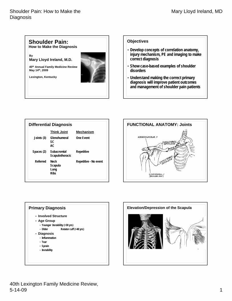
Differential DiagnosisThink Joint Mechanism
Joints (3) Glenohumeral One EventSCAC
Spaces (2) Subacromial RepetitiveScapulothoracic
Referred Neck Repetitive - No eventScapulaLungRibs
Palpate and ManualMuscle TestArm in varyingdegrees of abduction androtation
Internal and External Rotators
Shoulder Pain: How to Make the Diagnosis
Mary Lloyd Ireland, MD
40th Lexington Family Medicine Review, 5-14-09 4
Rotator Cuff Testing
• Empty can position• Weakness in external rotation
Be Specific:The diagnosis should define the structure that is injured and the condition
Diagnosis Rotator Cuff• Inflammation• Tear
• Partial vs. Complete• Articular side vs. Bursal side
Complete Tear
• Suspension bridge• Free side of tear (cable)• Attachments of tear
or (supports at each end)
MRI
• Full Thickness supraspinatus tear
Window shade to sill(cuff) (greater tuberosity)Use this comparison for patient education
SIZEof
TEAR
There are many clinical tests named after someone. Instead of description by name:
• Think of the motion of joint and forces you apply:• Is it labral?
• (Axial loading like McMurray’s)• Is it the rotator cuff?
• (compressing or impinging)• Is it instability?
• (distraction of joint capsule subluxing the humeral head)
Shoulder Pain: How to Make the Diagnosis
Mary Lloyd Ireland, MD
40th Lexington Family Medicine Review, 5-14-09 5
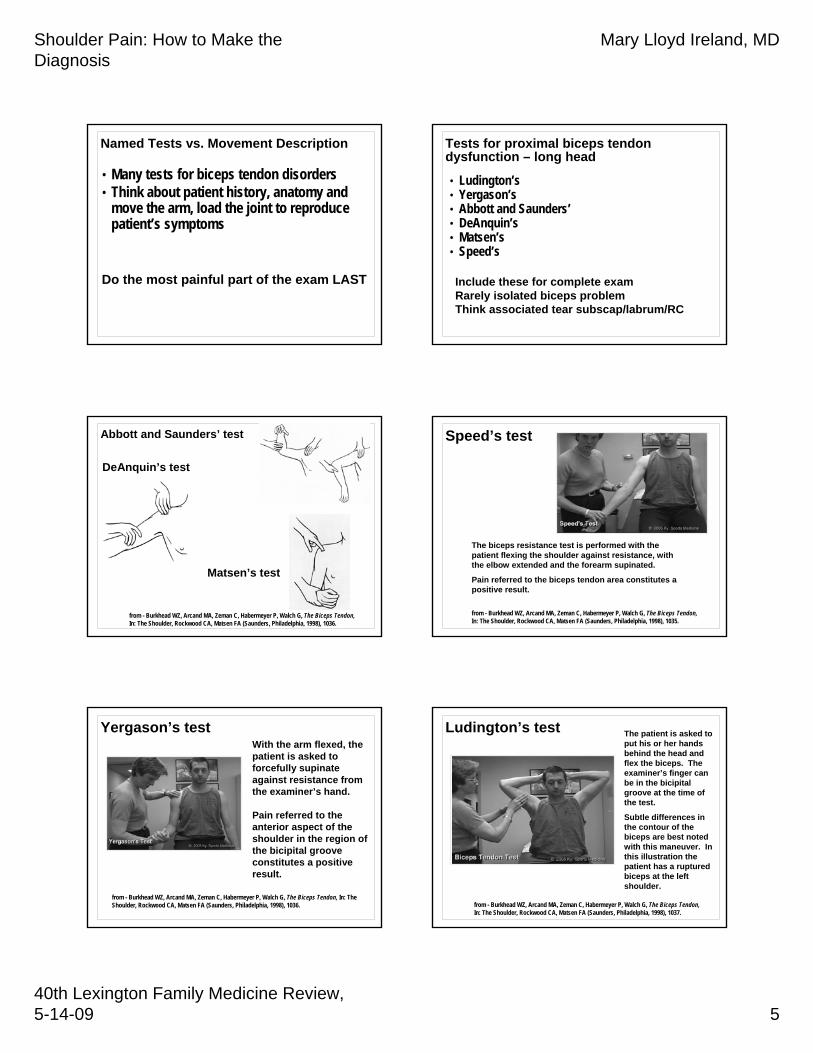
Named Tests vs. Movement Description
• Many tests for biceps tendon disorders• Think about patient history, anatomy and
move the arm, load the joint to reproduce patient’s symptoms
Do the most painful part of the exam LAST
Tests for proximal biceps tendon dysfunction – long head
• Ludington’s• Yergason’s• Abbott and Saunders’• DeAnquin’s• Matsen’s• Speed’s
Include these for complete examRarely isolated biceps problemThink associated tear subscap/labrum/RC
Abbott and Saunders’ test
from - Burkhead WZ, Arcand MA, Zeman C, Habermeyer P, Walch G, The Biceps Tendon, In: The Shoulder, Rockwood CA, Matsen FA (Saunders, Philadelphia, 1998), 1036.
DeAnquin’s test
Matsen’s test
Speed’s test
The biceps resistance test is performed with the patient flexing the shoulder against resistance, with the elbow extended and the forearm supinated.
Pain referred to the biceps tendon area constitutes a positive result.
from - Burkhead WZ, Arcand MA, Zeman C, Habermeyer P, Walch G, The Biceps Tendon, In: The Shoulder, Rockwood CA, Matsen FA (Saunders, Philadelphia, 1998), 1035.
Yergason’s testWith the arm flexed, the patient is asked to forcefully supinate against resistance from the examiner’s hand.
Pain referred to the anterior aspect of the shoulder in the region of the bicipital groove constitutes a positive result.
from - Burkhead WZ, Arcand MA, Zeman C, Habermeyer P, Walch G, The Biceps Tendon, In: The Shoulder, Rockwood CA, Matsen FA (Saunders, Philadelphia, 1998), 1036.
Ludington’s test The patient is asked to put his or her hands behind the head and flex the biceps. The examiner’s finger can be in the bicipital groove at the time of the test.
Subtle differences in the contour of the biceps are best noted with this maneuver. In this illustration the patient has a ruptured biceps at the left shoulder.
from - Burkhead WZ, Arcand MA, Zeman C, Habermeyer P, Walch G, The Biceps Tendon, In: The Shoulder, Rockwood CA, Matsen FA (Saunders, Philadelphia, 1998), 1037.
Guanche CA and Jones DC, “Clinical Testing for Tears of the GlenoidLabrum,” in Arthroscopy: The Journal of Arthroscopic and Related Surgery, vol 19, no 5 (May-June 2003), 517-523.
• Prospective study• 61 shoulders, 62 patients• Tests Used
• Only O’Brien and Jobe relocation test were statistically correlated with presence of labrum tear, including SLAP• Other five not found useful for labral tears• None of the tests or combinations statistically valid for
SLAP lesion only
O’Brien’s Test
Shoulder Palpation Crank Tests Shoulder Stability
Shoulder Pain: How to Make the Diagnosis
Mary Lloyd Ireland, MD
40th Lexington Family Medicine Review, 5-14-09 7
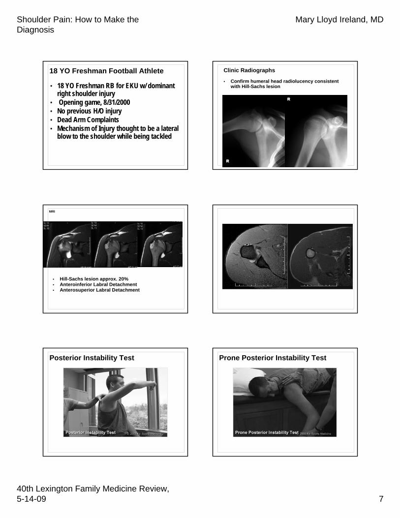
18 YO Freshman Football Athlete
• 18 YO Freshman RB for EKU w/ dominant right shoulder injury
• Opening game, 8/31/2000• No previous H/O injury• Dead Arm Complaints• Mechanism of Injury thought to be a lateral
blow to the shoulder while being tackled
Clinic Radiographs
• Confirm humeral head radiolucency consistent with Hill-Sachs lesion
Posterior Instability Test Prone Posterior Instability Test
Shoulder Pain: How to Make the Diagnosis
Mary Lloyd Ireland, MD
40th Lexington Family Medicine Review, 5-14-09 8
Vicious Cycle: Laxity to Instability Multi-Directional Instability
•Voluntary posterior direction - symptomatic
S/P Open anterior shoulder reconstructionMulti-Directional Instability, bilateral shoulders.
More symptomatic on operated right side.
18 YO Right-Hand-Dominant Discus Thrower
• Threw the discus• Felt pop, pain,inability to move her arm• Went to the emergency room
Posterior Dislocation• X-rays showed humeral head posteriorly
dislocated on axillary view• This direction of dislocation still is missed in
emergency rooms
ER view Axillary
PosteriorlyDislocated
Posteriorly dislocated
Stryker view
Shoulder Pain: How to Make the Diagnosis
Mary Lloyd Ireland, MD
40th Lexington Family Medicine Review, 5-14-09 9
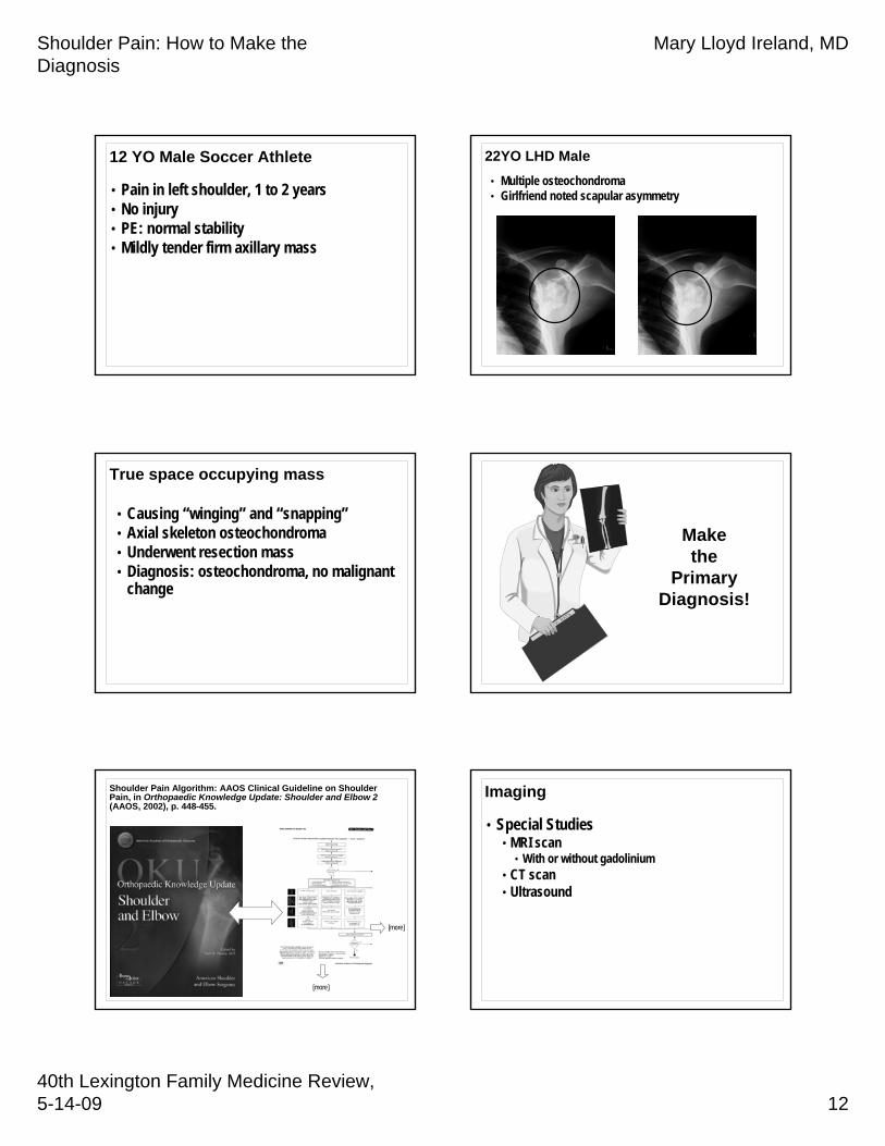
Shoulder Pain Algorithm: AAOS Clinical Guideline on Shoulder Pain, in Orthopaedic Knowledge Update: Shoulder and Elbow 2(AAOS, 2002), p. 448-455.
[more]
[more]
Imaging
• Plain films• Make the diagnosis by history and physical
and plain films• Institute treatment • Re-examine• Then special Imaging Studies
Shoulder Pain Algorithm: AAOS Clinical Guideline on Shoulder Pain, in Orthopaedic Knowledge Update: Shoulder and Elbow 2(AAOS, 2002), p. 448-455.
• Initial Imaging• True AP in 0º external rotation• Lateral in scapular plane• Axially view
• When imaging studies are indicated during the initial evaluation and treatment of a patient with shoulder pain, appropriate plain “x-rays” should be obtained. More sophisticated imaging studies (such as shoulder MRI, ultrasound, or arthrography) are not indicated.
IMAGING
AP Internal View Stryker Notch View
Shoulder Pain: How to Make the Diagnosis
Mary Lloyd Ireland, MD
40th Lexington Family Medicine Review, 5-14-09 10
Outlet View Outlet Upright View
Axillary Lateral View Modified Axillary View in Humeral External Rotation
Subscapularis Muscle Subscapularis Tears
• Lift Off (75% tear 5-30)• Hand or back Lspine• Maximum LR
• Napoleon (50% tear)• Press belly, flexes wrist
• Bear Hug (Upper tear, most sensitive)• Hand on opposite shoulder• Elbow forward• Examiner pulls hand off shoulder
Shoulder Pain: How to Make the Diagnosis
Mary Lloyd Ireland, MD
40th Lexington Family Medicine Review, 5-14-09 11
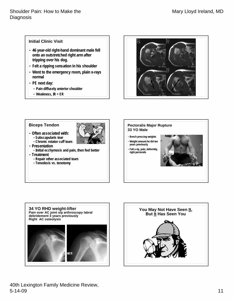
Initial Clinic Visit
• 46 year-old right-hand dominant male fell onto an outstretched right arm after tripping over his dog.
• Felt a ripping sensation in his shoulder• Went to the emergency room, plain x-rays
normal• PE next day:
• Pain diffusely anterior shoulder• Weakness, IR > ER
Ultrasound showing symptomatic progression of previously asymptomatic rotator cuff tear.
1991 1997
Yamaguchi K et. al., “Natural history of asymptomatic rotator cuff tears: A longitudinal analysis of asymptomatic tears detected sonographically,”J Shoulder Elbow Surg 2001;10:199-203.
Shoulder Pain Algorithm: AAOS Clinical Guideline on Shoulder Pain, in Orthopaedic Knowledge Update: Shoulder and Elbow 2(AAOS, 2002), p. 448-455.