Accepted by B. Mantovani: 5 Sept. 2014; published: 2 Oct. 2014
ZOOTAXA
ISSN 1175-5326 (print edition)
ISSN 1175-5334 (online edition)Copyright © 2014 Magnolia Press
Zootaxa 3869 (4): 397–408
www.mapress.com/zootaxa/
Article
397
http://dx.doi.org/10.11646/zootaxa.3869.4.4
http://zoobank.org/urn:lsid:zoobank.org:pub:1EE6BD5F-F661-44CC-8484-3C590B758037
A tiny new species of leaf insect (Phasmatodea, Phylliidae) from New Guinea
THOMAS VAN DE KAMP1,2
& FRANK H. HENNEMANN3
1
ANKA / Institute for Photon Science and Synchrotron Radiation (IPS), Karlsruhe Institute of Technology (KIT), Hermann-von-Helm-
holtz-Platz 1, D-76344 Eggenstein-Leopoldshafen, Germany. E-mail: [email protected]
2
State Museum of Natural History Karlsruhe, Erbprinzenstr. 13, D-76133 Karlsruhe, Germany
3
Reiboldstrasse 11, D-67251 Freinsheim, Germany. E-mail: [email protected]. Website: www.phasmatodea.com
Abstract
The female and egg of the new Papuan leaf insect Phyllium (Phyllium) riedeli n. sp. are described and illustrated. The
species belongs in the siccifolium species-group of the subgenus Phyllium and, with a body length of 56.3 mm, represents
the smallest leaf insect so far described for the genus. The type-specimens are stored in the State Museum of Natural His-
tory Karlsruhe, Germany (SMNK). A checklist and key is provided for the New Guinean representatives of Phyllium
(Phyllium) Illiger, 1798.
Key words: Phasmatodea, leaf insects, Phyllium (Phyllium) riedeli n. sp., West Papua
Introduction
The family Phylliidae is well-known for containing the so-called leaf insects, which have always fascinated
entomologists due to their remarkable camouflage. The Phylliidae comprises two tribes—the Nanophylliini with
the genus Nanophyllium Redtenbacher, 1906 and the Phylliini with the three genera Chitoniscus Stål, 1875,
Microphyllium Zompro, 2001 and Phyllium Illiger, 1798. Currently, the family contains 53 described extant
species, 40 of which are placed in the genus Phyllium. From these 40 known species 29 belong in the subgenus
Phyllium (Phyllium) Illiger, 1798 and 12 in the subgenus Phyllium (Pulchriphyllium) Griffini, 1898. Eophyllium
messelensis, known from a 47-million-year-old fossil male, already exhibits many of the morphological characters
found in modern leaf insects (Wedmann et al. 2007).
Comprehensive studies on the Phylliidae were done by Klante (1976), Größer (2001, 2008) and Zompro &
Größer (2003). Brock & Hasenpusch (2003) revised the Australian species. The head morphology of Phyllium
(Ph.) siccifolium (Linnaeus, 1758) was described in detail by Friedemann et al. (2011). The eggs of leaf insects
were examined e.g. by Clark (1978), Viscuso & Longo (1983) and Hausleithner (1984). Hennemann et al. (2009)
published an extensive survey of the genus with a revision of Philippine species, which questioned the validity of
the so far established systematic groups and provided a reclassification as well as clarification of several
identifications.
This paper describes and discusses a tiny new leaf insect found in West Papua (New Guinea), which is in
accordance to the classification presented by Hennemann et al. (2009) placed in the siccifolium species-group of
the subgenus Phyllium (Phyllium). The description is based on a single adult female and four eggs.
Methods
A single female of Ph. (Phyllium) riedeli n. sp. was collected alive in 2010 in the highlands of West New Guinea.
After collection, the specimen survived for 13 days in captivity and dropped four eggs before its death. The eggs
were incubated at room temperature and were regularly sprayed with water, but no hatchlings emerged. After one
VAN DE KAMP & HENNEMANN 398 · Zootaxa 3869 (4) © 2014 Magnolia Press
year, the eggs were placed next to the holotype and designated paratypes of Ph. (Phyllium) riedeli n. sp.. The left
hindleg of the holotype was removed immediately after its death and fixed in 100% ethanol to provide molecular
samples for possible future phylogenetic studies.
The holotype was photographed with a Canon EOS 50D camera equipped with a Tamron AF 90mm 2.8 Di
Macro 1:1 SP lens and a Canon Macro Twin Lite MT-24EX flash. Photographs of the eggs were taken with a JVC
KY70 camera attached to a Leica Z6 APO Macroscope. The in-focus components of 50 images were combined
automatically with the software Auto-Montage® (Synchroscopy). Scanning electron microscopy images of the
antennae were taken with a Zeiss EVO LS 15 at 15kV and a chamber pressure of 30–45 Pa. The background of
some images was corrected using Adobe® Photoshop® CS6. Measurements were taken using a digital caliper and
are given to 0.1 mm. The eggs examined and illustrated were fully developed and were laid by the holotype. The
terminology used to describe egg structures generally follows that of Clark-Sellick (1997). Fig. 6 is based on a
public domain map of New Guinea available at Wikimedia Commons (http://commons.wikimedia.org).
Abbreviations
HT Holotype
PT Paratype
SMNK State Museum of Natural History Karlsruhe, Germany
SMNS State Museum of Natural History Stuttgart, Germany
ZSMC Zoologische Staatssammlung München, Germany
Phyllium Illiger, 1798
Phyllium (Phyllium) Illiger, 1798
Type species: Gryllus (Mantis) siccifolius Linnaeus, 1758: 425, by monotypy.
Differing from the subgenus Ph. (Pulchriphyllium) by the lack of exterior lobes on the tibiae of both sexes, ± round
cross-section of antennomeres IV–VIII of ♀ as well as the longer tegmina, which at least reach to abdominal
segment III, and ventrally unarmed antennomeres of ♂. Eggs differ by the raised hairy, umbrella- or feather-like
appendages, lack of longitudinal lamellae of the capsule and flat operculum.
For full details, as well as diagnoses of the genus and subgenus and keys see Hennemann et al. (2009).
Phyllium (Phyllium) riedeli n. sp.
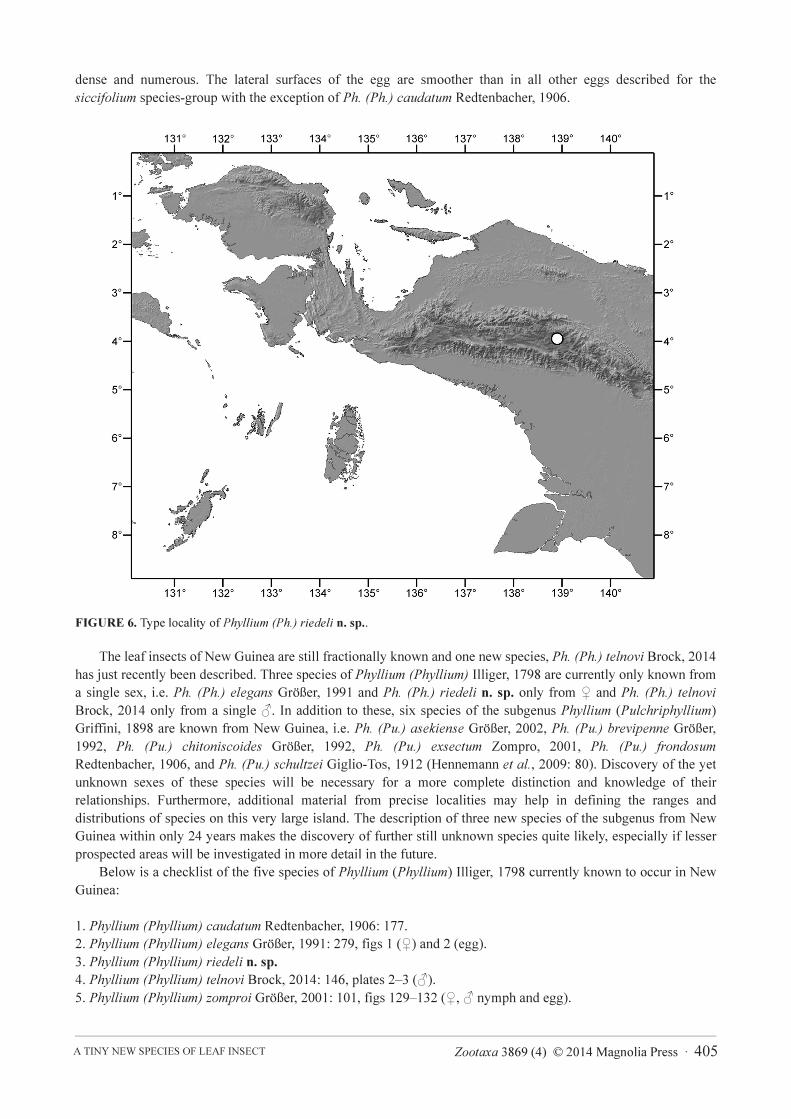
HT, ♀: Papua Province, Indonesia, S03° 57.161´ E138° 57.357´, 1875m, Nothofagus forest, 11.XII.2010, beaten
(SMNK).
PT, 4 eggs: laid by the HT in captivity (SMNK).
Differentiation. The ♀ of Ph. (Ph.) riedeli n. sp. is the smallest adult ♀ known in the genus so far, with a body
length of only 56.3 mm. The shape of the abdomen, which is gradually tapered from segment IV onwards, shape of
the profemora, lack of exterior lobes on the tibiae and ventrally unarmed antennomeres clearly place this new
species in the subgenus Phyllium (Phyllium).
Since Ph. (Ph.) riedeli n. sp. is from New Guinea, it is most certainly endemic to that island and hence unlikely
to occur elsewhere, especially throughout the Wallacea. This is also the case for the other four New Guinean
members of the subgenus i.e. Ph. (Ph.) caudatum Redtenbacher, 1906, Ph. (Ph.) elegans Größer 1991, Ph. (Ph.)
telnovi Brock, 2014 and Ph. (Ph.) zomproi Größer, 2001. An exception is Ph. (Ph.) caudatum, which also occurs in
New Britain and the Solomon Islands. Consequently, Ph. (Ph.) riedeli n. sp. is here only differentiated from the
species occurring in New Guinea.
The most closely related and similar species appears to be Ph. (Ph.) caudatum. From this species ♀ of Ph.
(Ph.) riedeli n. sp. can be distinguished by: the smaller size, less granulose head capsule, less spiny mesothorax,
Zootaxa 3869 (4) © 2014 Magnolia Press · 399A TINY NEW SPECIES OF LEAF INSECT
broader and apically rounded tegmina, shape of the subgenital plate, smaller teeth of the interior lobe of the
profemora, more slender and less expanded exterior lobe of the profemora and smaller interior lobes of the
protibiae.
From Ph. (Ph.) zomproi ♀ differ by: the much smaller size, gradually tapered abdominal segment VI–X,
presence of a distinct posteromedian tubercle on the head, lower number of teeth on the pars stridens of
antennomere III (25 in Ph. riedeli n. sp., 48–50 in Ph. zomproi), less spiny mesothorax, shape of the subgenital
plate, much more slender and less rounded interior and exterior lobes of the profemora and shorter but more
distinctly rounded interior lobe of the protibiae. From Ph. (Ph.) elegans ♀ differ by: the much smaller size, much
more slender abdomen, which is gradually tapered from segment VI onwards with segments VII and VIII not
lobed, shorter and broader apically rounded tegmina, shape of the profemora and shape of the subgenital plate.
Ph. (Ph.) telnovi seems to be closely related but is only known from the ♂. However, with a body length of 50
mm the ♂ of this species is by far too large to represent the opposite sex of Ph. (Ph.) riedeli n. sp.. Also, the
distinct armature of the interior lobe of the profemora and fairly large spines of the mesothorax clearly show it to be
distinct.
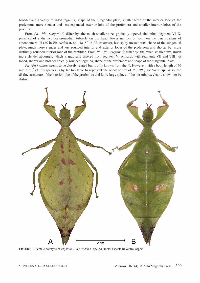
FIGURE 1. Female holotype of Phyllium (Ph.) riedeli n. sp.. A: Dorsal aspect; B: ventral aspect.
VAN DE KAMP & HENNEMANN 400 · Zootaxa 3869 (4) © 2014 Magnolia Press
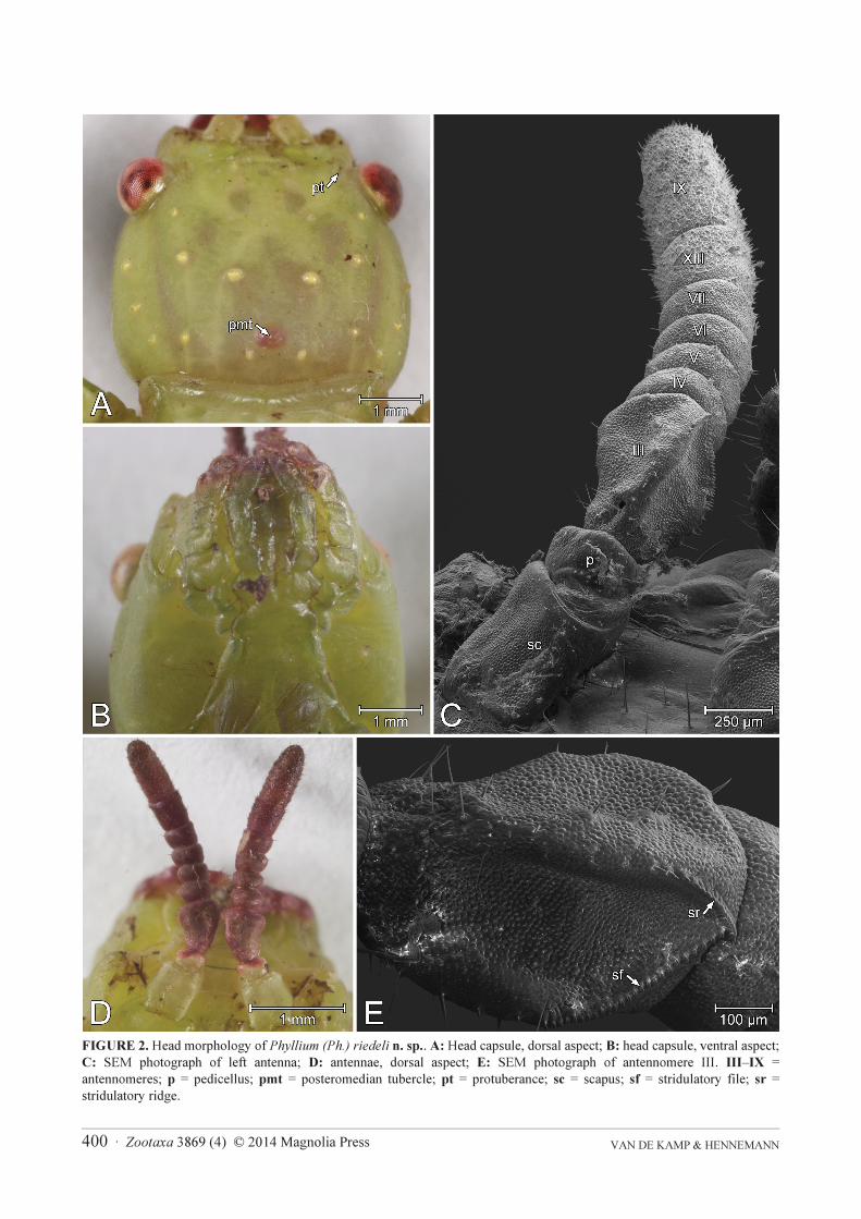
FIGURE 2. Head morphology of Phyllium (Ph.) riedeli n. sp.. A: Head capsule, dorsal aspect; B: head capsule, ventral aspect;
C: SEM photograph of left antenna; D: antennae, dorsal aspect; E: SEM photograph of antennomere III. III–IX =
antennomeres; p = pedicellus; pmt = posteromedian tubercle; pt = protuberance; sc = scapus; sf = stridulatory file; sr =
stridulatory ridge.
Zootaxa 3869 (4) © 2014 Magnolia Press · 401A TINY NEW SPECIES OF LEAF INSECT
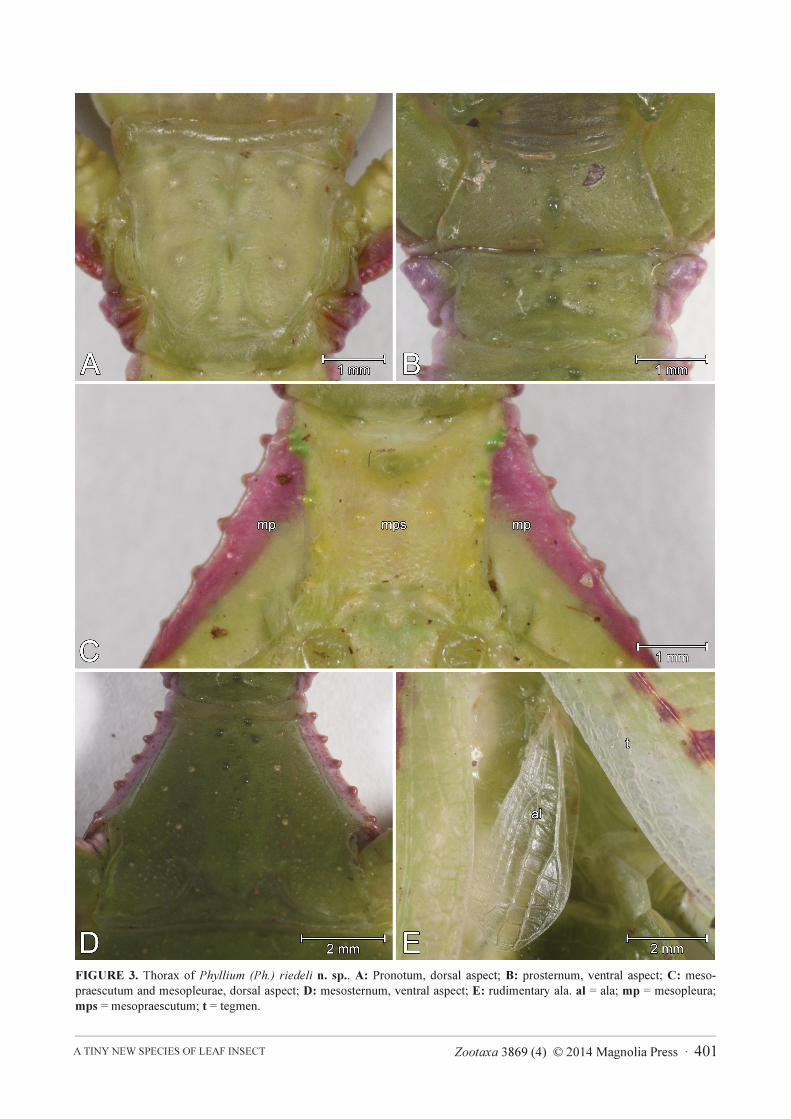
FIGURE 3. Thorax of Phyllium (Ph.) riedeli n. sp.. A: Pronotum, dorsal aspect; B: prosternum, ventral aspect; C: meso-
praescutum and mesopleurae, dorsal aspect; D: mesosternum, ventral aspect; E: rudimentary ala. al = ala; mp = mesopleura;
mps = mesopraescutum; t = tegmen.
VAN DE KAMP & HENNEMANN 402 · Zootaxa 3869 (4) © 2014 Magnolia Press
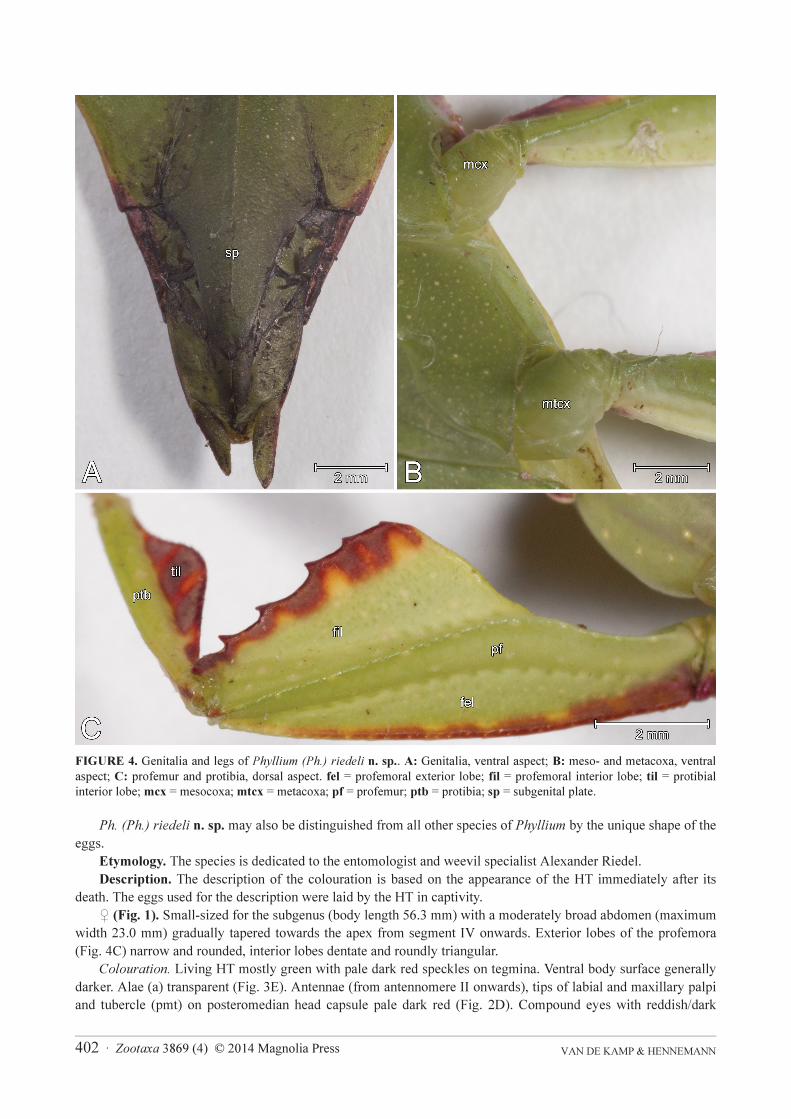
FIGURE 4. Genitalia and legs of Phyllium (Ph.) riedeli n. sp.. A: Genitalia, ventral aspect; B: meso- and metacoxa, ventral
aspect; C: profemur and protibia, dorsal aspect. fel = profemoral exterior lobe; fil = profemoral interior lobe; til = protibial
interior lobe; mcx = mesocoxa; mtcx = metacoxa; pf = profemur; ptb = protibia; sp = subgenital plate.
Ph. (Ph.) riedeli n. sp. may also be distinguished from all other species of Phyllium by the unique shape of the
eggs.
Etymology. The species is dedicated to the entomologist and weevil specialist Alexander Riedel.
Description. The description of the colouration is based on the appearance of the HT immediately after its
death. The eggs used for the description were laid by the HT in captivity.
♀ (Fig. 1). Small-sized for the subgenus (body length 56.3 mm) with a moderately broad abdomen (maximum
width 23.0 mm) gradually tapered towards the apex from segment IV onwards. Exterior lobes of the profemora
(Fig. 4C) narrow and rounded, interior lobes dentate and roundly triangular.
Colouration. Living HT mostly green with pale dark red speckles on tegmina. Ventral body surface generally
darker. Alae (a) transparent (Fig. 3E). Antennae (from antennomere II onwards), tips of labial and maxillary palpi
and tubercle (pmt) on posteromedian head capsule pale dark red (Fig. 2D). Compound eyes with reddish/dark
Zootaxa 3869 (4) © 2014 Magnolia Press · 403A TINY NEW SPECIES OF LEAF INSECT
brown pattern (Fig. 2A). Mesopleurae (mp) pale dark red, anterior-most spiniform tubercles of meso-praescutum
(mps) dark green (Fig. 3C). Fine light green median line extending from posterior abdominal segment II to VIII.
Distinct white spot with brown frame on the posteromedian ventral abdominal segment I. Similar, but smaller
triangular-shaped spots at segments V and VI. Large light spots in the center of each side of abdominal segment V,
best visible ventrally but also dorsally below the tegmina. Abdomen with a thin, brownish frame extending from
abdominal segment IV towards the apex. Segment IX dorsally with dark red spots extending to the lateral margins.
Ventral coxal surfaces green (Fig. 4B), dorsal surface of procoxae and protrochanters brown. Interior lobes (fil) of
profemora (Fig. 4C) with yellowish/brownish-red pattern at dentate side. Dorsal part of the exterior lobes (fel) of
profemora brownish-red (coxal margin) to green (tibial margin). Protibial interior lobes (til) brownish-red with
three yellowish lines perpendicular to the tibia (Fig. 4C). Propraetarsi pale dark red. Exterior lobes of mesofemora
with a brownish spot on the proximal margin and three pale dark red spots of different sizes distally (proximodistal
sequence: large, small, medium). Interior lobes of mesofemora exhibit a similar colour pattern as the profemora.
Mesotibiae with a pale dark red spot near the proximodorsal margin. Colour pattern of metafemora very similar to
mesofemora, but with a larger gap between the second and third distal spot of the exterior lobe. Metatibiae similar
coloured to mesotibiae, but with reddish distal margin. Metatarsomeres I to IV reddish.
Morphology. Head capsule (Figs 2A,B) broad with convex cheeks. Vertex smooth except for several granules,
a distinct posteromedian tubercle (pmt) and small protuberances (pt) between compound eyes and antennal bases.
Antennae (Figs 2C,D) moderately slender and elongate (3.4 mm), longer than postocular section of head capsule
(2.7 mm) and consisting of nine segments. Apical antennomere (IX) cylindrical with rounded apex, about 2x longer
than wide and ± 2x as long as VIII. Stridulatory file (pars stridens) on antennomere III with 25, stridulatory ridge
(plectron) with 19 teeth (Fig. 3E). Pronotum (Fig. 3A) roughly squarish with slightly concave anterior margin and
moderately convex lateral and posterior margins. Anterior margin of pronotum thickened, in particular around the
openings of the prothoracic defensive glands. Prosternum smooth, except for some minor granules (Fig. 3B).
Meso-praescutum (mps) almost as long as wide (length-width ratio 1.1 : 1), gently narrowing towards posterior
(Fig. 3C). Lateral margins armed with few spiniform tubercles of variable sizes, the larger ones located near the
anterior margin. Disc with 8 ± prominent tubercles. Mesopleurae (mp) strongly and gradually diverging; their
lateral margins with five distinct spiniform tubercles (Fig. 3C). Anterior mesosternum sparsely and irregularly
granulose (Fig. 3D). Tegmina (length 31.2 mm, max. width 12.3 mm) extending over 1/3 of abdominal segment
VII. Alae (al) rudimentary (length 6.7 mm, max. width 2.6 mm), roughly 1/5 the length of tegmina (Fig. 3E).
Abdominal segments II–IV gradually widened, IV widest segment, first 1/3 gradually diverging. V–X gradually
tapering towards the apex. Anal segment (X) ± as long as wide with rounded apex. Subgenital plate (sp) rather
short, slightly projecting over the posterior margin of abdominal segment IX (Fig. 4A). Profemora (Fig. 4C) with a
narrow and rounded exterior lobe (fel). Interior lobe (fil) dentate, roundly triangular and distinctly wider than
exterior lobe. Protibiae (Fig. 4C) without exterior lobe, interior lobe (til) roundly isosceles triangular (angle > 90°).
Exterior and interior lobe of meso- and metafemora gently rounded, with the exterior lobe narrower than the
interior lobe. Protarsus about 5/4 the length of the protibia, probasitarsus roughly 2x longer than wide.
Measurements [mm]: Length of body 56.3, length of head 5.0, length of pronotum 3.9, length of mesonotum
4.5, length of tegmina 31.2, greatest width of tegmina 12.3, length of alae 6.7, greatest width of abdomen 23.0,
length of profemora 8.4, length of mesofemora 7.8, length of metafemora 9.1, length of protibiae 5.2, length of
mesotibiae 5.9, length of metatibiae 6.6, length of antennae 3.4.
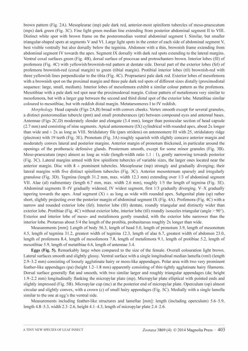
Eggs (Fig. 5). Remarkably large when compared to the size of the female. Overall colouration light brown.
Lateral surfaces smooth and slightly glossy. Ventral surface with a single longitudinal median lamella (vml) (length
2.9–3.2 mm) consisting of loosely agglutinate hairy or moss-like appendages. Polar area with two very prominent
feather-like appendages (pa) (height 1.2–1.8 mm) apparently consisting of thin tightly agglutinate hairy filaments.
Dorsal surface generally flat and smooth, with two similar larger and roughly triangular appendages (da; height
1.9–2.2 mm) longitudinally flanking the micropylar plate (mp). Micropylar plate elliptical with pointed ends and
slightly impressed (Fig. 5B). Micropylar cup (mc) at the posterior end of micropylar plate. Operculum (op) almost
circular and slightly convex, with a crown (c) of small hairy appendages (Fig. 5C). Medially with a single lamella
similar to the one at egg’s the ventral side.
Measurements including feather-like structures and lamellae [mm]: length (including operculum) 5.6–5.9,
length 4.8–5.3, width 2.3–2.6, height 4.1–4.3, length of micropylar plate 2.4–2.6.
VAN DE KAMP & HENNEMANN 404 · Zootaxa 3869 (4) © 2014 Magnolia Press
FIGURE 5. Egg of Phyllium (Ph.) riedeli n. sp.. A: Lateral aspect; B: dorsal aspect; C: ventral aspect. c = opercular crown; da
= dorsal appendage; mc = micropylar cup; mp = micropylar plate; op = operculum; pa = polar appendage; vml = ventromedian
lamella.
Distribution and ecology. So far only known from the type locality in the mountain ranges of central West
Papua, having been collected in mountainous Nothofagus forest (family Nothofagaceae) at almost 2000 m above
sea level.
Discussion
Small leaf insects are known mainly from the genera Nanophyllium, Microphyllium and Chitoniscus. Apart from
Ph. (Ph.) riedeli n. sp., the only other species of the genus with females of a reported body length below 60 mm is
Ph. (Pu.) suzukii with 57.3 mm (Größer 2008), while the females of most species achieve lengths between 70 and
100 mm. The fossil male of Eophyllium messelensis has a body length of 63.1 mm, which is comparable to the
males of most extant Phyllium species. Given that Eophyllium is the assumed sister group of the extant leaf insects
(Wedmann et al. 2007), it seems that the small body size has been acquired within the extant crown group of the
Phylliidae.
The eggs of Ph. riedeli n. sp. are of remarkable size when compared to size of the female and relatively larger
than e.g. those of Ph. (Ph.) siccifolium (Linnaeus, 1758) (length including operculum ca. 5 mm), who’s females
achieve a body length up to 98 mm (Größer 2008). The occurrence of a flat operculum and feather-like appendages
is only known from the siccifolium species-group of the subgenus Phyllium (Phyllium), which confirms the
placement of Ph. (Ph.) riedeli n. sp. in this species-group. Eggs of the celebicum species-group exhibit a conically
raised operculum and only short, moss-like appendages, while eggs of the subgenus Pulchriphyllium generally lack
hairy structures (see Table 10 in Hennemann et al. 2009). A longitudinal median lamella and feather-like
appendages on the dorsal surface and polar area are shared with other species of the siccifolium species-group, but
the splitting of the lateral appendages into a distinct dorsal and polar part and the triangular shape of the latter are
unique for Ph. (Ph.) riedeli n. sp.. While the ventral longitudinal median lamella and the appendages of the
operculum are similar to the ones observed in other species, the hairs of the lateral appendages appear to be more
Zootaxa 3869 (4) © 2014 Magnolia Press · 405A TINY NEW SPECIES OF LEAF INSECT
dense and numerous. The lateral surfaces of the egg are smoother than in all other eggs described for the
siccifolium species-group with the exception of Ph. (Ph.) caudatum Redtenbacher, 1906.
FIGURE 6. Type locality of Phyllium (Ph.) riedeli n. sp..
The leaf insects of New Guinea are still fractionally known and one new species, Ph. (Ph.) telnovi Brock, 2014
has just recently been described. Three species of Phyllium (Phyllium) Illiger, 1798 are currently only known from
a single sex, i.e. Ph. (Ph.) elegans Größer, 1991 and Ph. (Ph.) riedeli n. sp. only from ♀ and Ph. (Ph.) telnovi
Brock, 2014 only from a single ♂. In addition to these, six species of the subgenus Phyllium (Pulchriphyllium)
Griffini, 1898 are known from New Guinea, i.e. Ph. (Pu.) asekiense Größer, 2002, Ph. (Pu.) brevipenne Größer,
1992, Ph. (Pu.) chitoniscoides Größer, 1992, Ph. (Pu.) exsectum Zompro, 2001, Ph. (Pu.) frondosum
Redtenbacher, 1906, and Ph. (Pu.) schultzei Giglio-Tos, 1912 (Hennemann et al., 2009: 80). Discovery of the yet
unknown sexes of these species will be necessary for a more complete distinction and knowledge of their
relationships. Furthermore, additional material from precise localities may help in defining the ranges and
distributions of species on this very large island. The description of three new species of the subgenus from New
Guinea within only 24 years makes the discovery of further still unknown species quite likely, especially if lesser
prospected areas will be investigated in more detail in the future.
Below is a checklist of the five species of Phyllium (Phyllium) Illiger, 1798 currently known to occur in New
Guinea:
1. Phyllium (Phyllium) caudatum Redtenbacher, 1906: 177.
2. Phyllium (Phyllium) elegans Größer, 1991: 279, figs 1 (♀) and 2 (egg).
3. Phyllium (Phyllium) riedeli n. sp.
4. Phyllium (Phyllium) telnovi Brock, 2014: 146, plates 2–3 (♂).
5. Phyllium (Phyllium) zomproi Größer, 2001: 101, figs 129–132 (♀, ♂ nymph and egg).
VAN DE KAMP & HENNEMANN 406 · Zootaxa 3869 (4) © 2014 Magnolia Press
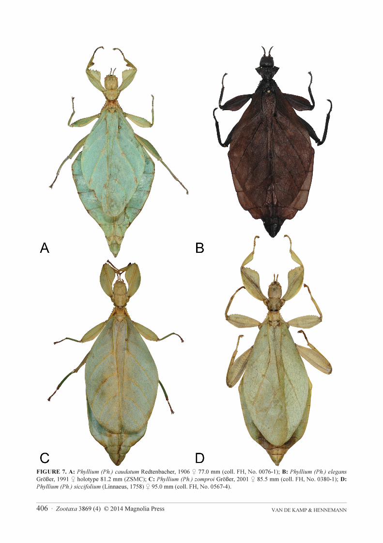
FIGURE 7. A: Phyllium (Ph.) caudatum Redtenbacher, 1906 ♀ 77.0 mm (coll. FH, No. 0076-1); B: Phyllium (Ph.) elegans
Größer, 1991 ♀ holotype 81.2 mm (ZSMC); C: Phyllium (Ph.) zomproi Größer, 2001 ♀ 85.5 mm (coll. FH, No. 0380-1); D:
Phyllium (Ph.) siccifolium (Linnaeus, 1758) ♀ 95.0 mm (coll. FH, No. 0567-4).
Zootaxa 3869 (4) © 2014 Magnolia Press · 407A TINY NEW SPECIES OF LEAF INSECT
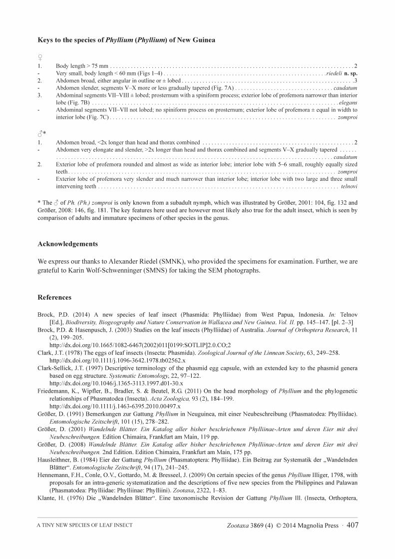
Keys to the species of Phyllium (Phyllium) of New Guinea
♀
1. Body length > 75 mm . . . . . . . . . . . . . . . . . . . . . . . . . . . . . . . . . . . . . . . . . . . . . . . . . . . . . . . . . . . . . . . . . . . . . . . . . . . . . . . . . . 2
- Very small, body length < 60 mm (Figs 1–4) . . . . . . . . . . . . . . . . . . . . . . . . . . . . . . . . . . . . . . . . . . . . . . . . . . . . . . .riedeli n. sp.
2. Abdomen broad, either angular in outline or ± lobed . . . . . . . . . . . . . . . . . . . . . . . . . . . . . . . . . . . . . . . . . . . . . . . . . . . . . . . . . .3
- Abdomen slender, segments V–X more or less gradually tapered (Fig. 7A) . . . . . . . . . . . . . . . . . . . . . . . . . . . . . . . . . caudatum
3. Abdominal segments VII–VIII ± lobed; prosternum with a spiniform process; exterior lobe of profemora narrower than interior
lobe (Fig. 7B) . . . . . . . . . . . . . . . . . . . . . . . . . . . . . . . . . . . . . . . . . . . . . . . . . . . . . . . . . . . . . . . . . . . . . . . . . . . . . . . . . . .elegans
- Abdominal segments VII–VII not lobed; no spiniform process on prosternum; exterior lobe of profemora ± equal in width to
interior lobe (Fig. 7C) . . . . . . . . . . . . . . . . . . . . . . . . . . . . . . . . . . . . . . . . . . . . . . . . . . . . . . . . . . . . . . . . . . . . . . . . . . . . zomproi
♂*
1. Abdomen broad, <2x longer than head and thorax combined . . . . . . . . . . . . . . . . . . . . . . . . . . . . . . . . . . . . . . . . . . . . . . . . . . . 2
- Abdomen very elongate and slender, >2x longer than head and thorax combined and segments V–X gradually tapered . . . . . .
. . . . . . . . . . . . . . . . . . . . . . . . . . . . . . . . . . . . . . . . . . . . . . . . . . . . . . . . . . . . . . . . . . . . . . . . . . . . . . . . . . . . . . . . . . . . . caudatum
2. Exterior lobe of profemora rounded and almost as wide as interior lobe; interior lobe with 5–6 small, roughly equally sized
teeth . . . . . . . . . . . . . . . . . . . . . . . . . . . . . . . . . . . . . . . . . . . . . . . . . . . . . . . . . . . . . . . . . . . . . . . . . . . . . . . . . . . . . . . . . . zomproi
- Exterior lobe of profemora very slender and much narrower than interior lobe; interior lobe with two large and three small
intervening teeth . . . . . . . . . . . . . . . . . . . . . . . . . . . . . . . . . . . . . . . . . . . . . . . . . . . . . . . . . . . . . . . . . . . . . . . . . . . . . . . . . telnovi
* The ♂ of Ph. (Ph.) zomproi is only known from a subadult nymph, which was illustrated by Größer, 2001: 104, fig. 132 and
Größer, 2008: 146, fig. 181. The key features here used are however most likely also true for the adult insect, which is seen by
comparison of adults and immature specimens of other species in the genus.
Acknowledgements
We express our thanks to Alexander Riedel (SMNK), who provided the specimens for examination. Further, we are
grateful to Karin Wolf-Schwenninger (SMNS) for taking the SEM photographs.
References
Brock, P.D. (2014) A new species of leaf insect (Phasmida: Phylliidae) from West Papua, Indonesia. In: Telnov
[Ed.], Biodiversity, Biogeography and Nature Conservation in Wallacea and New Guinea. Vol. II. pp. 145–147. [pl. 2–3]
Brock, P.D. & Hasenpusch, J. (2003) Studies on the leaf insects (Phylliidae) of Australia. Journal of Orthoptera Research, 11
(2), 199–205.
http://dx.doi.org/10.1665/1082-6467(2002)011[0199:SOTLIP]2.0.CO;2
Clark, J.T. (1978) The eggs of leaf insects (Insecta: Phasmida). Zoological Journal of the Linnean Society, 63, 249–258.
http://dx.doi.org/10.1111/j.1096-3642.1978.tb02562.x
Clark-Sellick, J.T. (1997) Descriptive terminology of the phasmid egg capsule, with an extended key to the phasmid genera
based on egg structure. Systematic Entomology, 22, 97–122.
http://dx.doi.org/10.1046/j.1365-3113.1997.d01-30.x
Friedemann, K., Wipfler, B., Bradler, S. & Beutel, R.G. (2011) On the head morphology of Phyllium and the phylogenetic
relationships of Phasmatodea (Insecta). Acta Zoologica, 93 (2), 184–199.
http://dx.doi.org/10.1111/j.1463-6395.2010.00497.x
Größer, D. (1991) Bemerkungen zur Gattung Phyllium in Neuguinea, mit einer Neubeschreibung (Phasmatodea: Phylliidae).
Entomologische Zeitschrift, 101 (15), 278–282.
Größer, D. (2001) Wandelnde Blätter. Ein Katalog aller bisher beschriebenen Phylliinae-Arten und deren Eier mit drei
Neubeschreibungen. Edition Chimaira, Frankfurt am Main, 119 pp.
Größer, D. (2008) Wandelnde Blätter. Ein Katalog aller bisher beschriebenen Phylliinae-Arten und deren Eier mit drei
Neubeschreibungen. 2nd Edition. Edition Chimaira, Frankfurt am Main, 175 pp.
Hausleithner, B. (1984) Eier der Gattung Phyllium (Phasmatoptera: Phylliidae). Ein Beitrag zur Systematik der „Wandelnden
Blätter“. Entomologische Zeitschrift, 94 (17), 241–245.
Hennemann, F.H., Conle, O.V., Gottardo, M. & Bresseel, J. (2009) On certain species of the genus Phyllium Illiger, 1798, with
proposals for an intra-generic systematization and the descriptions of five new species from the Philippines and Palawan
(Phasmatodea: Phylliidae: Phylliinae: Phylliini). Zootaxa, 2322, 1–83.
Klante, H. (1976) Die „Wandelnden Blätter“. Eine taxonomische Revision der Gattung Phyllium Ill. (Insecta, Orthoptera,
VAN DE KAMP & HENNEMANN 408 · Zootaxa 3869 (4) © 2014 Magnolia Press
Phasmatoptera). Zoologische Beiträge, 22, 49–76.
Redtenbacher, J. (1906) Die Insektenfamilie der Phasmiden. I. Phasmidae, Areolatae. Verlag W. Engelmann, Leipzig, pp. 180.
[pl. 1–6]
Viscuso, R. & Longo, G. (1983) The egg of leaf insect Phyllium pulchrifolium Serv. (Phasmatodea: Phyllidae): morphology and
ultrastructure. Archives de Biologie, 94 (4), 441–457.
Wedmann, S., Bradler, S. & Rus, J. (2007) The first fossil leaf insect: 47 million years of specialized cryptic morphology and
behavior. Proceedings of the National Academy of Sciences of the United States of America, 104 (2), 565–569.
http://dx.doi.org/10.1073/pnas.0606937104
Zompro, O. & Größer, D. (2003) A generic revision of the insect order Phasmatodea: The genera of the areolate stick insect
family Phylliidae (Walking Leaves) (Insecta, Orthoptera). Spixiana, 26 (2), 129–141.


![a 2012 [ver. 49] -77 090-3869-9204 · 2020. 10. 9. · a 2012 [ver. 49] -77 090-3869-9204 . Created Date: 10/9/2020 9:55:25 AM](https://static.documents.pub/doc/80x56/60cf7828eca43f0aaa393a5e/a-2012-ver-49-77-090-3869-9204-2020-10-9-a-2012-ver-49-77-090-3869-9204.jpg)


