16
Application note we specialize because you do | Anorectal Endosonography | 3D Anorectal Ultrasound
Applica
tion
not
e
we specialize because you do
| Anorectal Endosonography |
3D Anorectal Ultrasound

2
Ultrasound image credits
Figs. 3 and 9 courtesy of Professor C. Bartram, St. Mark’s Hospital, London.
Fig. 12 courtesy of Dr. I. P. Olsen, Hammerfest Hospital, Norway.
Figs. 16 and 17 courtesy of courtesy of Professor Ganansia, Hôpital Léopold Belan, Paris.
Figs. 10, 11, 14, 15, 18-23, 25-30, and 33 courtesy of Docent, Dr. Med. Marianne Starck, Malmö
University Hospital, Sweden.
Figs. 34 and 35 courtesy of Docent, Dr. Med. Tom Öresland, Gothenburg, Sweden.
33
Ultrasound image credits
3D Anorectal Ultrasound
Docent, Dr. Med. Marianne Starck, Consultant Colorectal Surgeon, Malmö University Hospital, Sweden
Gert Karlsson, Product Manager, BK Medical
2D and 3D anorectal endosonography has
proved its usefulness, and today medical
specialists within such areas as pelvic floor
imaging, coloproctology, colorectal surgery,
incontinence, and uro-gynecology are well
aware of its value.
Introduction
Although two-dimensional endosonography is
very valuable, it has some shortcomings1. Images
are normally produced only in the transaxial
scanning plane with the anterior of the patient at
the 12 o’clock position. The only way to extend
a scanning in the proximal-distal direction is to
move the probe farther in or out of the anal canal
or rectum. Precise positioning of the probe is
crucial to the examination.
3D anorectal endosonography extends the
usefulness of anal endosonography2,3,4,5,6. The data
from a series of closely-spaced two-dimensional
images is combined to create a 3D image that
can be freely rotated and sliced to allow the
operator to get the most information out of the
data – while not under the time pressure of the
examination itself. In some situations, if it is
difficult to pass the area occupied by a rectal
tumor, only one acquisition of images may be
possible. The data stored in a file originating from
an acquisition can then be reviewed at any time.
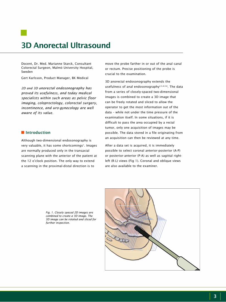
After a data set is acquired, it is immediately
possible to select coronal anterior-posterior (A-P)
or posterior-anterior (P-A) as well as sagittal right-
left (R-L) views (Fig 1). Coronal and oblique views
are also available to the examiner.
Fig. 1. Closely spaced 2D images are combined to create a 3D image. The 3D image can be rotated and sliced for further inspection.

4
3D Anorectal Ultrasound
In contrast to a conventional 2D image or indeed a
3D image, new techniques such as Volume Render
mode contains features for modifying the opacity
of a 3D data set7. This can be combined with filter
and thickness features for truly new presentations
of sphincter tears and for following fistulas-in-
ano in all anatomical planes. These views offer
an unsurpassed source of information to use
in evaluating the patient. The results of the 3D
anorectal scan can change the treatment of the
patient. It is possible to see the invasion of a
rectal tumor, the type of a fistula, and the extent
of anal sphincter damage. This knowledge can be
used to change the surgical management of the
patient. The diagnosis is less dependent on one
examiner, because the data can be reviewed by
several specialists.
a 3D image can be freely ro-tated and sliced, allowing the operator to get the most in-formation out of the data.
Previously the gold standard for imaging anal
fistula cases was magnetic resonance (MR)
using an endoanal coil. Using an MR machine
for this application, however, is delicate and
time-consuming. The introduction of very
high frequency ultrasound transducers and
3D anorectal ultrasound, with the improved
imaging possibilities that result, may change the
standard to using ultrasound for imaging anal
fistulas3,6,7,8,9,10.
Several studies have been done on morphology
of the anal canal seen on 2D and 3D endoanal
ultrasound5,11,12,13 and correlation between
endosonographic sphincter defects and anal
incontinence14,15,16,17,18. Today ultrasound is
the gold standard when investigating fecal
incontinence.
The value of endoanal ultrasound in anal cancer is
more controversial19,20,21. Some find it very useful
for preoperative staging as well as follow-up after
radiation treatment19,20.
MR, using an endocoil in staging of rectal tumors,
has not shown higher accuracy in early rectal
cancer than 2D rectal ultrasound3,22,23,24. To date,
no comparative studies have been done on 3D
endorectal ultrasound and MR in benign tumors
or early rectal cancer.
Using 2D and 3D endorectal ultrasound it is
possible to see the invasion of a rectal tumor22,25,
to distinguish between benign lesions and early
rectal cancer26,27, and it has shown to be useful
in follow up of rectal cancer28. When compared
to 2D, 3D endorectal ultrasound is advantageous
because the image can be saved and studied in
different planes after the examination.
5
Equipment
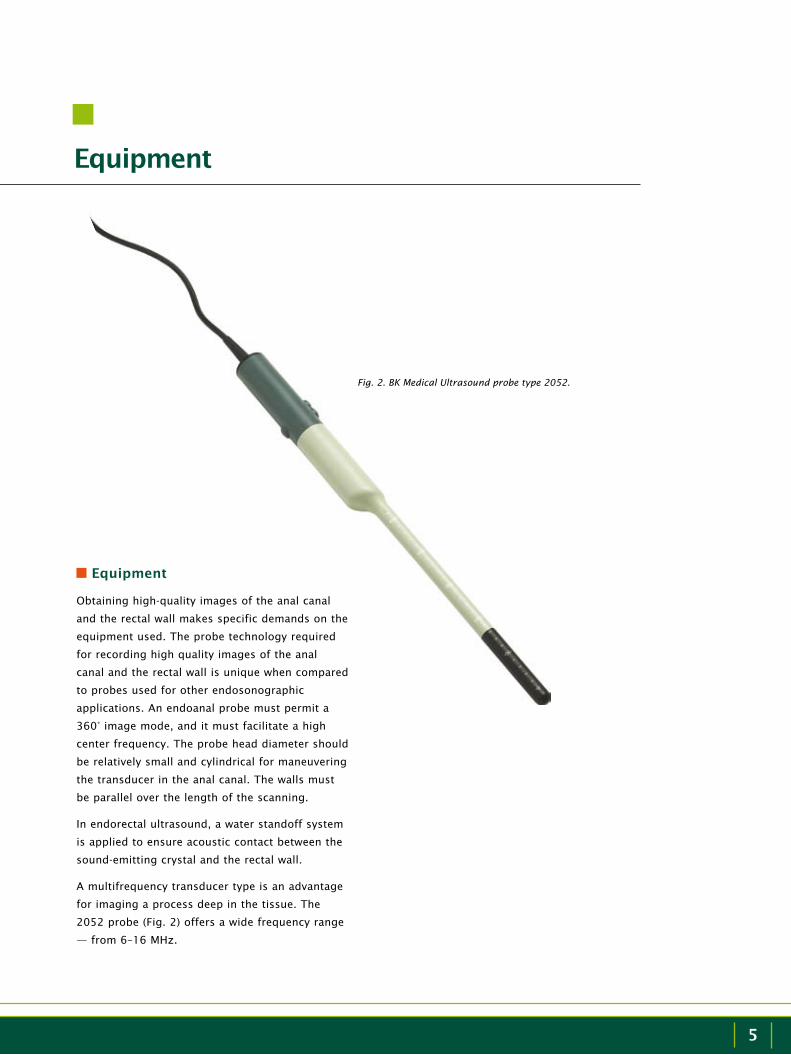
Fig. 2. BK Medical Ultrasound probe type 2052.
Equipment
Obtaining high-quality images of the anal canal
and the rectal wall makes specific demands on the
equipment used. The probe technology required
for recording high quality images of the anal
canal and the rectal wall is unique when compared
to probes used for other endosonographic
applications. An endoanal probe must permit a
360° image mode, and it must facilitate a high
center frequency. The probe head diameter should
be relatively small and cylindrical for maneuvering
the transducer in the anal canal. The walls must
be parallel over the length of the scanning.
In endorectal ultrasound, a water standoff system
is applied to ensure acoustic contact between the
sound-emitting crystal and the rectal wall.
A multifrequency transducer type is an advantage
for imaging a process deep in the tissue. The
2052 probe (Fig. 2) offers a wide frequency range
— from 6–16 MHz.
6
Anal Endosonography
High
Acquisition of 3D anorectal images
The same type of transducer facilitates 2D and 3D
imaging techniques.
A 3D reconstruction is based on a high number of
parallel transaxial images acquired by means of a
precision movement of the crystal assembly inside
the BK Medical ultrasound probe type 2052.
The built-in high-resolution 3D acquisition system
can be operated at different levels of definition.
For the anorectal application, the usual setting
is 0.25 mm between adjacent transaxial images.
Scanning the anal canal or the rectum wall with
these settings over an acquisition distance
between 30 and 60 mm will typically yield from
120 to 240 parallel image slices (Fig.1).
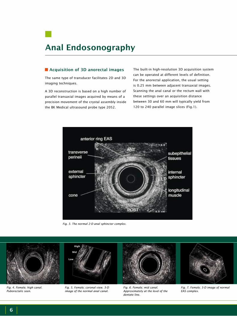
Fig. 3. The normal 2-D anal sphincter complex.
Fig. 4. Female; high canal. Puborectalis seen.
Fig. 6. Female; mid canal. Approximately at the level of the dentate line.
Fig. 5. Female; coronal view. 3-D image of the normal anal canal.
Fig. 7. Female; 3-D image of normal EAS complex.
High
Mid
Low

7
The labels in Fig. 5 indicate the High, Mid and Low
positions on a 3D coronal A-P image.
In Fig. 7, note the perfect circular EAS anteriorly
and left-lateral in this nulliparous female. The
perineal body, together with the pubo-analis, is
also very clearly seen.
In some women, you can see a descending pelvis
(Figs. 10 and 11).
Anal sphincter tears
Anal sphincter tears can either be isolated to
a defect in the EAS or the IAS alone or be a
combination of an internal and an external
sphincter defect. Scars can be either hypo- or
hyperechoic.
Anal Endosonography
Fig. 3 shows a 2D image of the anal canal. The
external sphincter is seen forming a 360° intact
circle. The image is recorded as a mid-canal image
(slightly higher than the level of the dentate line).
The perineal body is partially visible ventral to
the anterior EAS, and the transverse perineii are
imaged at 11 and 1 o’clock. Note the conjoining
longitudinal muscle and the complete IAS.
The first ultrasound image recorded is normally at
puborectalis level (high), where the perineal body
is also seen in females. This image is normally
documented and labeled HIGH (Fig. 4).
In a normal patient, moving the probe a few mm
in the distal direction will show an intact anterior
external anal sphincter (EAS) forming just below
the superficial transverse perineal muscles. This
image is a mid-canal projection where the internal
anal sphincter (IAS) conjoining the longitudinal
muscle (LM) and the superficial EAS all are
identified. This image will be labeled MID (Fig. 6).
When the probe is pulled farther out, the image of
the IAS will disappear, and only the subepithelium
and the subcutaneous segment of the LM+EAS will
be seen. This last image will be labeled LOW (Fig
8).
Fig. 8. Female; low canal. Only the subcutaneous EAS is seen.
Fig. 9. Male; 3-D image of normal EAS complex.
Fig. 10. 2D image does not show the IAS at the transverse perineal muscle level.
Fig. 11. 3D image shows the IAS exists but is located more proximally (arrow).

8
Anal Endosonography
Fig. 12 shows a transaxial image of an EAS tear,
while Fig. 13 shows a 3D image of an anterior
EAS tear using volume rendering techniques. The
2D image of a combined internal and external
sphincter defect is seen in Fig. 14. 3D images of
the same female illustrates the rubber band effect
of a ruptured internal sphincter and the missing
anterior part of the sphincters (Fig. 15).
Anal fistulas and abscesses
It is considered a delicate and problematic
task to image anal fistula cases using only 2D
transaxial ultrasound. The fistula tracts are almost
impossible to follow, and it is even harder to
identify any internal opening.
In these cases, 3D endoanal ultrasound offers
a significant advantage over conventional
2D ultrasound. If an external opening can
be identified, some doctors will introduce
hydrogen peroxide H2O2 (3–5%) into the opening
immediately before acquiring a 3D data set. Data
acquisition will take approximately 30 sec. for
a high-resolution scan. For this short period,
the H2O2 enhances the fistula tracts so that
they appear as bright white structures in the
ultrasound image. Applying Volume Render Mode
to a 3D data set will further enhance the image
of the branches of a fistula with or without the
presence of any enhancing medium.
Fig. 16 shows a 2D transaxial image of an
extrasphincteric fistula (verified on the 3D
Fig. 18. 2D image of internal fistula opening anteriorly.
Fig. 19. 3D image reveals the rest of the fistula tract.
Fig. 16. Extrasphincteric anal fistula.
Fig. 17. Same patient as in Fig. 16, with H²O² enhancement.
Fig. 12. A large anterior EAS tear is seen at 12-1 o’clock in this female (postpartum).
Fig. 13. Anterior EAS tear. Fig. 14. IAS from 9 to 3 o’clock. EAS from 10 to 2 o’clock.
Fig. 15. Rubber band effect visualized with 3D.

9
Fig. 22. Anal cancer before treatment.
Fig. 23. Anal cancer after treatment.
data set shown in Fig. 17 that was recorded in
connection with the acquisition of this image). A
fragment of the fistula in the 2D image and the
entire fistula in the 3D image are seen as bright
white echoic structures because of the image-
enhancing properties of H2O2. An example of a
low anal fistula is seen in Figs. 18 and 19.
An anovaginal fistula is seen as a fistula tract with
air on ultrasound. Fig. 20 shows a fistula tract at
12 o’clock in the proximal part of the anal canal
with hyperechoic air.
Abscesses are easily visualized as in this case,
where the abscess is seen as an echo-poor cavity
(Fig. 21).
Anal cancer
Figs. 22 and 23 show an example of anal cancer
before and after radiation therapy. The tumor is
located from 2 to 8 o’clock and penetrates the
external sphincter (T2b). After radiation treatment,
the tumor has disappeared.
Fig. 20. A fistula tract is seen at 12 o’clock.
Fig. 21. An intersphincteric abscess located between the mucosa and the external sphincter is seen at 6 o’clock.

10
Rectal Endosonography
Rectal Endosonography
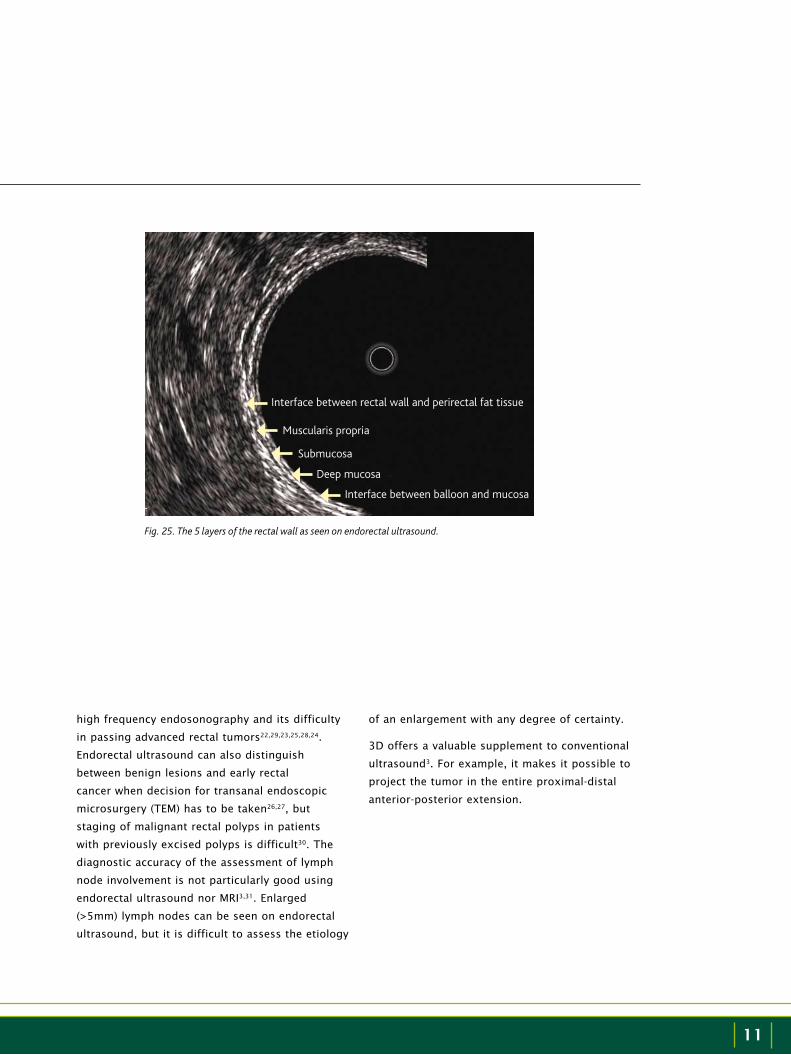
The rectal wall consists of 3 layers surrounded by
perirectal fat (Fig. 24). Ultrasound studies of the
rectum show 5 interfaces represented in the image
as 3 hyperechoic and 2 hypoechoic structures (Fig.
25).
The interfaces represent 1) the hyperechoic
interface between the water-filled balloon and
the mucosa, 2) the hypoechoic deep mucosa
(lamina propria and muscularis mucosae), 3)
the hyperechoic submucosa, 4) the hypoechoic
muscularis propria and 5) the hyperechoic
Fig. 24. The anatomy of the rectum.
Mucosa
Submucosa
Muscularis propria
Perirectal fat
interface between the rectal wall and the perirectal
fat tissue.
Before deciding whether management of the
patient should include preoperative radiation
therapy for rectal cancer, it is important to know
whether the tumor is confined to the rectal wall
(T1 or T2 tumor) or penetrates into the perirectal
fat (T3 tumor).
Studies have shown that endorectal ultrasound
is superior to MRI in staging early rectal cancer.
In advanced T3 or T4 tumors, MRI should be
preferred because of the lower image depth of
11
Fig. 25. The 5 layers of the rectal wall as seen on endorectal ultrasound.
Interface between rectal wall and perirectal fat tissue
Muscularis propria
Submucosa
Deep mucosa
Interface between balloon and mucosa
high frequency endosonography and its difficulty
in passing advanced rectal tumors22,29,23,25,28,24.
Endorectal ultrasound can also distinguish
between benign lesions and early rectal
cancer when decision for transanal endoscopic
microsurgery (TEM) has to be taken26,27, but
staging of malignant rectal polyps in patients
with previously excised polyps is difficult30. The
diagnostic accuracy of the assessment of lymph
node involvement is not particularly good using
endorectal ultrasound nor MRI3,31. Enlarged
(>5mm) lymph nodes can be seen on endorectal
ultrasound, but it is difficult to assess the etiology
of an enlargement with any degree of certainty.
3D offers a valuable supplement to conventional
ultrasound3. For example, it makes it possible to
project the tumor in the entire proximal-distal
anterior-posterior extension.
12
Rectal Endosonography
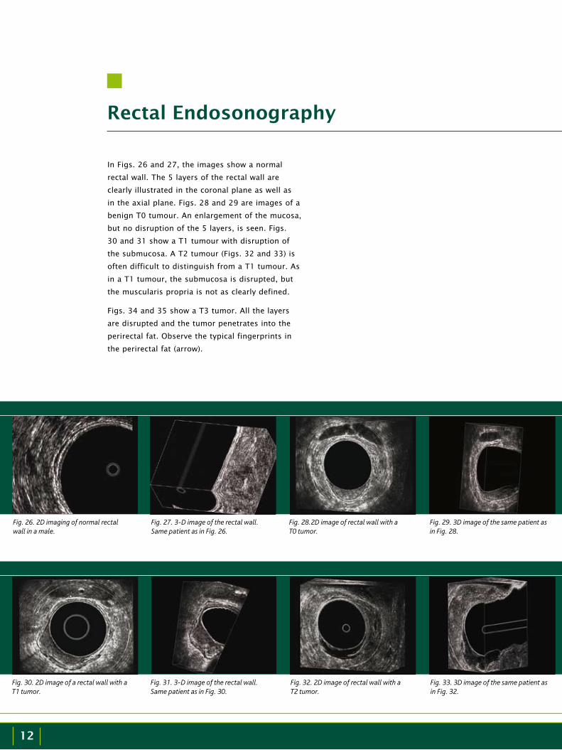
In Figs. 26 and 27, the images show a normal
rectal wall. The 5 layers of the rectal wall are
clearly illustrated in the coronal plane as well as
in the axial plane. Figs. 28 and 29 are images of a
benign T0 tumour. An enlargement of the mucosa,
but no disruption of the 5 layers, is seen. Figs.
30 and 31 show a T1 tumour with disruption of
the submucosa. A T2 tumour (Figs. 32 and 33) is
often difficult to distinguish from a T1 tumour. As
in a T1 tumour, the submucosa is disrupted, but
the muscularis propria is not as clearly defined.
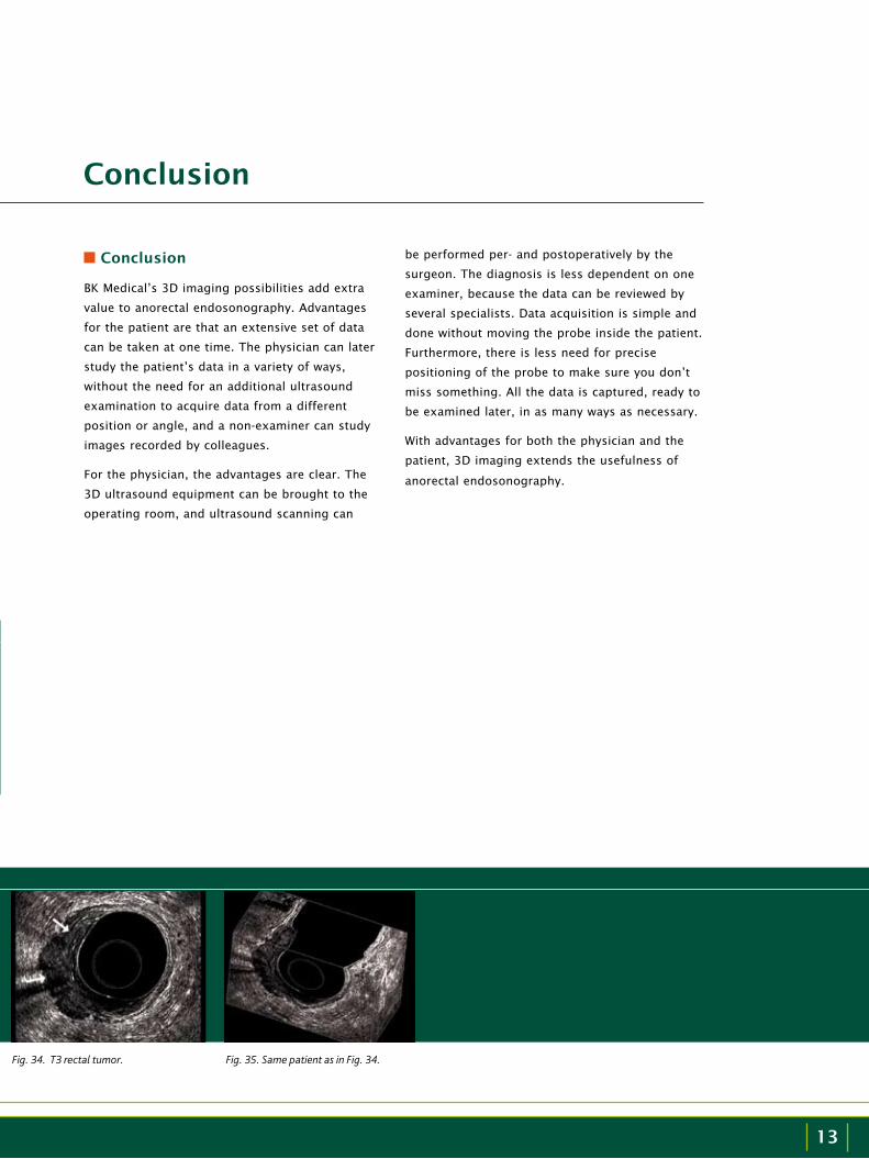
Figs. 34 and 35 show a T3 tumor. All the layers
are disrupted and the tumor penetrates into the
perirectal fat. Observe the typical fingerprints in
the perirectal fat (arrow).
Fig. 30. 2D image of a rectal wall with a T1 tumor.
Fig. 31. 3-D image of the rectal wall. Same patient as in Fig. 30.
Fig. 32. 2D image of rectal wall with a T2 tumor.
Fig. 33. 3D image of the same patient as in Fig. 32.
Fig. 26. 2D imaging of normal rectal wall in a male.
Fig. 27. 3-D image of the rectal wall. Same patient as in Fig. 26.
Fig. 28.2D image of rectal wall with a T0 tumor.
Fig. 29. 3D image of the same patient as in Fig. 28.
13
Conclusion
Conclusion
BK Medical’s 3D imaging possibilities add extra
value to anorectal endosonography. Advantages
for the patient are that an extensive set of data
can be taken at one time. The physician can later
study the patient’s data in a variety of ways,
without the need for an additional ultrasound
examination to acquire data from a different
position or angle, and a non-examiner can study
images recorded by colleagues.
For the physician, the advantages are clear. The
3D ultrasound equipment can be brought to the
operating room, and ultrasound scanning can
be performed per- and postoperatively by the
surgeon. The diagnosis is less dependent on one
examiner, because the data can be reviewed by
several specialists. Data acquisition is simple and
done without moving the probe inside the patient.
Furthermore, there is less need for precise
positioning of the probe to make sure you don’t
miss something. All the data is captured, ready to
be examined later, in as many ways as necessary.
With advantages for both the physician and the
patient, 3D imaging extends the usefulness of
anorectal endosonography.
Fig. 34. T3 rectal tumor. Fig. 35. Same patient as in Fig. 34.

14
3D Anorectal Ultrasound
References
1. Bartram CI, Frudinger A.Handbook of Anal Endosonography. Petersfield, UK: Wrightson Biomedical
Publishing Ltd; 1997.
2. Regadas FSP, Murad-Regadas SM, Lima DMR et al.Anal canal anatomy showed by three-dimensional anorectal ultrasonography. Surg. Endosc.
2007;21(12):2207-2211.
3. Gravante G, Giordano P.The role of three-dimensional endoluminal ultrasound imaging in the evaluation of anorectal diseases: a review. Surg Endosc.
2008;22(7):1570-1578.
4. Sun MRM, Smith MP, Kane RA. Current techniques in imaging of fistula in ano: three-dimensional endoanal ultrasound and magnetic resonance imaging. Semin Ultrasound CT MR.
2008;29(6):454-471.
5. Gregory WT, Boyles SH, Simmons K, Corcoran A, Clark AL.External anal sphincter volume measurements using 3-dimensional endoanal ultrasound. Am J Obstet Gynecol.
2006;194(5):1243-1248.
6. Buchanan GN, Bartram CI, Williams AB, Halligan S, Cohen CRG. Value of hydrogen peroxide enhancement of three-dimensional endoanal ultrasound in fistula-in-ano. Dis Colon Rectum.
2005;48(1):141-147.
7. Santoro GA, Fortling B. The advantages of volume rendering in three-dimensional endosonography of the anorectum. Dis Colon Rectum.
2007;50(3):359-368.
8. Lengyel AJ, Hurst NG, Williams JG. Pre-operative assessment of anal fistulas using endoanal ultrasound. Colorectal Dis.
2002;4(6):436-440.
9. Gustavsson UM, Kahvecioglu B, Aström G, Ahlström H, Graf W. Endoanal ultrasound or magnetic resonance imaging for preoperative assessment of anal fistula: a comparative study. Colorectal Dis.
2001;3(3):189-197.
10. Felt-Bersma RJF.
Endoanal ultrasound in perianal fistulas and abscesses. Dig Liver Dis.
2006;38(8):537-543.
11. Starck M, Bohe M, Fortling B, Valentin L. Endosonography of the anal sphincter in women of different ages and parity. Ultrasound Obstet Gynecol.
2005;25(2):169-176.
12. Olsen I, Augensen K, Wilsgaard T, Kiserud T.Three-dimensional endoanal ultrasound assessment of the anal sphincters during rest and squeeze. Acta Obstet Gynecol Scand.
2008;87(6):669-674.
13. Oberwalder M, Thaler K, Baig MK et al. Anal ultrasound and endosonographic measurement of perineal body thickness: a new evaluation for fecal incontinence in females. Surg Endosc.
2004;18(4):650-654.
14. Starck M, Bohe M, Valentin L. The extent of endosonographic anal sphincter defects after primary repair of obstetric sphincter tears increases over time and is related to anal incontinence. Ultrasound Obstet Gynecol.
2006; 27(2):188-197.
15. Norderval S, Markskog A, Røssaak K, Vonen B. Correlation between anal sphincter defects and anal incontinence following obstetric sphincter tears: assessment using scoring systems for sonographic classification of defects. Ultrasound Obstet Gynecol.
2008;31(1):78-84.

15
References
16. Vasanth A, Sultan AH, Thakar R, Jones PW. Occult anal sphincter injuries – myth or reality? BJOG.
2006;113(2):195-200.
17. Richter HE, Fielding JR, Bradley CS et al. Endoanal ultrasound findings and fecal incontinence symptoms in women with and without recognized anal sphincter tears. Obstet Gynecol.
2006;108(6):1394-1401.
18. Norderval S, Delhi T, Vonen B. Three-dimensional endoanal ultrasonography: intraobserver and interobserver agreement using scoring systems for classification of anal sphincter defects. Ultrasound Obstet Gynecol.
2009;33(3):337-343.
19. Martellucci J, Naldini G, Colosimo C, Cionini L, Rossi M. Accuracy of endoanal ultrasound in the follow-up assessment for squamous cell carcinoma of the anal canal treated with radiochemotherapy. Surg. Endosc.
2009;23(5):1054-1057.
20. Tarantino D, Bernstein MA. Endoanal ultrasound in the staging and management of squamous-cell carcinoma of the anal canal – potential implications of a new ultrasound staging system. Dis Colon Rectum.
2002;45(1):16-22.
21. Lund JA, Sundstrom SH, Haaverstad R, Wibe A, Svinsaas M, Myrvold HE.Endoanal ultrasound is of little value in follow-up of anal carcinomas. Dis Colon Rectum.
2004;47(6):839-842.
22. Engelen SME, Beets GL, Beets-Tan RGH.Role of preoperative local and distant staging in rectal cancer.Onkologie.
2007;30(3):141-145.
23. Goh V, Halligan S, Bartram CI. Local radiological staging of rectal cancer. Clin Radiol.
2004;59(3):215-226.
24. Kauer WKH, Prantl I, Dittler HJ, Siewert JR. The value of endosonographic rectal carcinoma staging in routine diagnostics. Surg.Endosc.
2004;18(7):1075-1078.
25. Worrell S, Horvath K,
Blakemore T, Flum D.Endorectal ultrasound detection of focal carcinoma within rectal adenomas. Am J Surg.
2004;187(5):625-629.
26. Starck M, Bohe M, Simanaitis M, Valentin L.Rectal endosonography can distinguish benign rectal lesions from invasive early rectal cancers. Colorectal Dis.
2003;5(3):246-250.
27. Zorcolo L, Fantola G, Cabras F, Marongiu L, D’Alia G, Casula G. Preoperative staging of patients with rectal tumors suitable for transanal endoscopic microsurgery (TEM): comparison of endorectal ultrasound and histopathologic findings. Surg Endosc.
2009;23(6):1384-1389.
28. de Anda EH, Lee S-H, Finne CO, Rothenberger DA, Madoff RD, García-Aguilar J. Endorectal ultrasound in the follow-up of rectal cancer patients treated by local excision or radical surgery. Dis Colon Rectum.
2004:47(6):818-824.
29. Harewood GC.Assessment of publication bias in the reporting of EUS performance in staging rectal cancer. Am J Gastroenterol.
2005;100(4):808-816.
30. García-Aguilar J, de Anda EH, Rothenberger DA, Finne CO, Madoff RO.Endorectal ultrasound in the management of patients with malignant rectal polyps. Dis Colon Rectum.
2005;48(5):910-917.
31. Rieger N, Tjandra J, Solomon M. Endoanal and endorectal ultrasound: Applications in colorectal surgery. ANZ J Surg.
2004;74(8):671-675.
| BK Medical |
World HeadquartersMileparken 34DK-2730 HerlevDenmark
Tel: +45 4452 8100 • Fax: +45 4452 8199 • www.bkmed.com
With more than 30 years of commitment to ultrasound innovation,
BK Medical specializes in the development, manufacture and distribution
of dedicated ultrasound solutions. BK Medical has its headquarters
in suburban Copenhagen, Denmark, and has offices and distributors
throughout the world.
A wholly owned subsidiary of Analogic Corporation
BO
0008-H
0
9/2
009


