HAL Id: hal-01254388 https://hal.inria.fr/hal-01254388 Submitted on 30 Aug 2016 HAL is a multi-disciplinary open access archive for the deposit and dissemination of sci- entific research documents, whether they are pub- lished or not. The documents may come from teaching and research institutions in France or abroad, or from public or private research centers. L’archive ouverte pluridisciplinaire HAL, est destinée au dépôt et à la diffusion de documents scientifiques de niveau recherche, publiés ou non, émanant des établissements d’enseignement et de recherche français ou étrangers, des laboratoires publics ou privés. 3D Physics-Based Registration of 2D Dynamic MRI Data Raffaella Trivisonne, Igor Peterlik, Stéphane Cotin, Hadrien Courtecuisse To cite this version: Raffaella Trivisonne, Igor Peterlik, Stéphane Cotin, Hadrien Courtecuisse. 3D Physics-Based Regis- tration of 2D Dynamic MRI Data. MMVR - Medicine Meets Virtual Reality, Apr 2016, Los Angeles, United States. hal-01254388
Transcript
HAL Id: hal-01254388https://hal.inria.fr/hal-01254388
Submitted on 30 Aug 2016
HAL is a multi-disciplinary open accessarchive for the deposit and dissemination of sci-entific research documents, whether they are pub-lished or not. The documents may come fromteaching and research institutions in France orabroad, or from public or private research centers.
L’archive ouverte pluridisciplinaire HAL, estdestinée au dépôt et à la diffusion de documentsscientifiques de niveau recherche, publiés ou non,émanant des établissements d’enseignement et derecherche français ou étrangers, des laboratoirespublics ou privés.
3D Physics-Based Registration of 2D Dynamic MRIData
Raffaella Trivisonne, Igor Peterlik, Stéphane Cotin, Hadrien Courtecuisse
To cite this version:Raffaella Trivisonne, Igor Peterlik, Stéphane Cotin, Hadrien Courtecuisse. 3D Physics-Based Regis-tration of 2D Dynamic MRI Data. MMVR - Medicine Meets Virtual Reality, Apr 2016, Los Angeles,United States. �hal-01254388�
3D Physics-Based Registration of 2DDynamic MRI Data
Raffaella TRIVISONNE a,b Igor PETERLIK b,c Stéphane COTIN b
Hadrien COURTECUISSE a,b
a CNRS Strasbourg and Strasbourg Universityb Inria Nancy and Strasbourg University, IHU Strasbourg
c Institute of Computer Science, Masaryk University, Brno, Czech Republic
Abstract.We present a method allowing for intra-operative targeting of a specific anatom-
ical feature. The method is based on a registration of 3D pre-operative data to 2Dintra-operative images. Such registration is performed using an elastic model re-constructed from the 3D images, in combination with sliding constraints imposedvia Lagrange multipliers. We register the pre-operative data, where the feature isclearly detectable, to intra-operative dynamic images where such feature is no morevisible. Despite the lack of visibility on the 2D MRI images, we are able both todetermine the location of the target as well as follow its displacement due to respi-ratory motion.
Medical imaging is by now one of the essential aspects to perform most of the ordinarydaily surgeries. It represents not only a support for diagnostics, but also an actual op-erative instrument during particular therapeutic procedures. Typically, tomography tech-niques (CT and MRI) guarantee better results due to their accuracy into tissues differen-tiation and the consequent amount of information provided. In order to exploit the pre-cision and the efficiency of these techniques, and to optimize as well the working envi-ronment for the operators, new procedures have been developed involving the combineduse of tomography and robotic systems. So far it has concerned mainly percutaneous in-terventions, spacing from cryoablation for prostate cancer to neurosurgical applications.
Each technique presents its benefits and drawbacks: CT scans are faster and can pro-vide particularly detailed anatomical representations of high contrasted organs; on theother hand they involve X-rays absorption. MRI images, being based on magnetism, donot have any absorbed dose, moreover they offer an excellent contrast of soft tissues;the main disadvantages are the length of the procedure as well as the necessity of to-tally non-ferromagnetic equipment. From a physiological point of view, organs are notstatic objects and even the simplest breathing motion can induce shape deformations.These modifications may invalidate the preoperative planning since the location of inter-nal structures may significantly vary. Modern MRI allow for dynamic scanning without
any artifacts due to movement, but it is for now restricted to only one plan of acquisition.Therefore, given the absence of any volumetric information, some anatomical structuresmight be excluded. In this paper we extend the method presented in [4]. We aim to targetan inflame porcine gallbladder as during as a robotic MRI-guided percutaneous proce-dure. We show that 3 orthogonal MRI slices are sufficient to perform an entire 3D regis-tration of a preoperative segmented CT scan. Lastly, as the dynamic motion of the livercan only be acquired along a single plan at time, we show how to combine static sliceswith a single dynamic acquisition in order to provide an entire volumetric interpolationof the breathing motion.
2. Literature Review
The advantages of surgical robots and manipulators are well recognized (from daily as-sistance to telesurgery) [5]. In particular, MRI compatible robots are being developedfor biopsies [13], prostate cancer [1] and even neurosurgical applications [10]. Typically,at the beginning of the intervention, clinicians select a couple of images and manuallydefine a trajectory and some landmarks, in order to fix an entry point, a needle orien-tation and an approximative path. The robotic system will then semi-automatically pro-ceed towards the target following the set trajectory. One remaining limitation is that ini-tially chosen images may be suitable to plan needle insertion but far from the target.A real-time system tracking is then necessary to specifically take into account all thedeformations caused by external forces or natural motions (such as breathing) [12].
Augmented Reality is an active research area which allows overlaying key informa-tions on the top of medical images, such as the location of a tumor. It is a promisingtechnique for mini invasive surgery assistance as well as for robotic-assisted procedures[7]. Most of augmented reality systems are still limited to rigid registration, but it hasbeen reported (for instance for the liver motion) that purely rigid transformation is notsufficient for most of the surgeries. Recently, biomechanical models have been used fortheir ability to regularize the ill-posed non rigid registration problem. [8] uses a FiniteElement model and the iterative closest point (ICP) algorithm for the registration of mus-cular structures. [14] proposed a physics-based shape matching (PBSM) method basedon an electrostatic formulation. An elastic body is electrically charged to slides towardan oppositely charged rigid shape. [2] formulated the non rigid registration problem as aninverse mechanical problem. The method provides a set of boundary conditions bringingthe FE model to the desired geometrical deformations.
The above methods require a complete segmentation of intraoperative surfaces but,as stated above, dynamic MRI acquisition is only possible along a single orientation ata time. In [9], the boundary of the liver surface is extracted and tracked from a laparo-scopic camera during the intervention, but the tracking method may occur into cameraocclusions. [6] use a linear elastic FE model of the brain that is driven by active surfacematching of one acquired image during brain shift, but no significant dynamic motionare taken into account. In [4] a 3D volume is registered with 3 orthogonal sequencesof dynamic MRI recorded one after an other and synchronized manually. We proposean extension of this method where a single dynamic slice is combined with static MRIslices. The main contributions of this work are: register a generic organ with missingcomplete volumetric information, target an anatomical feature not visible in MRI images,and perform all these operations with a combination of dynamic and static mode.
3. Methodology
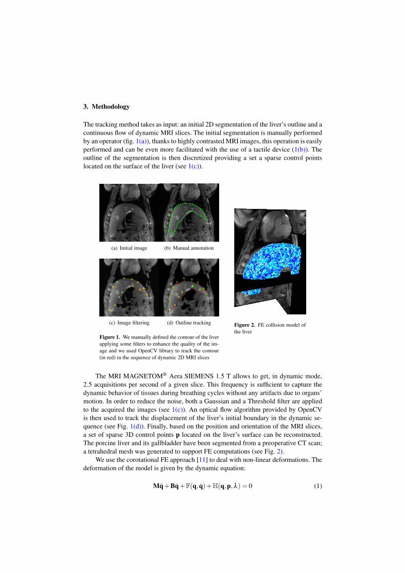
The tracking method takes as input: an initial 2D segmentation of the liver’s outline and acontinuous flow of dynamic MRI slices. The initial segmentation is manually performedby an operator (fig. 1(a)), thanks to highly contrasted MRI images, this operation is easilyperformed and can be even more facilitated with the use of a tactile device (1(b)). Theoutline of the segmentation is then discretized providing a set a sparse control pointslocated on the surface of the liver (see 1(c)).
(a) Initial image (b) Manual annotation
(c) Image filtering (d) Outline tracking
Figure 1. We manually defined the contour of the liverapplying some filters to enhance the quality of the im-age and we used OpenCV library to track the contour(in red) in the sequence of dynamic 2D MRI slices
Figure 2. FE collision model ofthe liver
The MRI MAGNETOM® Aera SIEMENS 1.5 T allows to get, in dynamic mode,2.5 acquisitions per second of a given slice. This frequency is sufficient to capture thedynamic behavior of tissues during breathing cycles without any artifacts due to organs’motion. In order to reduce the noise, both a Gaussian and a Threshold filter are appliedto the acquired the images (see 1(c)). An optical flow algorithm provided by OpenCVis then used to track the displacement of the liver’s initial boundary in the dynamic se-quence (see Fig. 1(d)). Finally, based on the position and orientation of the MRI slices,a set of sparse 3D control points p located on the liver’s surface can be reconstructed.The porcine liver and its gallbladder have been segmented from a preoperative CT scan;a tetrahedral mesh was generated to support FE computations (see Fig. 2).
We use the corotational FE approach [11] to deal with non-linear deformations. Thedeformation of the model is given by the dynamic equation:
Mq̈+Bq̇+F(q, q̇)+H(q,p,λ ) = 0 (1)
where F(q, q̇) are internal volumes forces for given positions q and velocities q̇. M andB are respectively the mass and the damping matrices. H(q,p,λ ) gathers the constraintsforces λ for given positions q and p.
The location of the corresponding control points p on the liver’s surface is notknown. This problem is solved employing the Iterative Closest Point (ICP) method asdescribed in [4]. At the beginning of each simulation step, each control point is associ-ated with its respective nearest triangle on the surface of the segmented liver. We assumethat this association stays constant within a simulation step (see [3] for details), allowingto define the Jacobian of Constraints J and the violation of constraint δ = p− q̄, definedas the distance between p and its respective closest projection on liver’s surface q̄:{
Mq̈+Bq̇+F(q, q̇)+J λ = 0JT q̄ = δ
(2)
Due to off-plan motions, tracked points may not be associated with the same po-sitions on the liver’s surface. We use then sliding constraints (see [4]) allowing the FEmodel to slide around the configuration, minimizing the mechanical energy needed toperform the registration. A normal direction n is associated to each constraint fromBezier subdivision of liver triangulation surface. After the resolution, λ is computed suchthat no violation remains in n-direction (δ = 0), with any tangential force.
A Backward Euler implicit time integration is used to perform the simulation. It in-volves a non-linear problem solved with a single iteration step of the Newton solver. Af-ter the linearization, the problem is written as an augmented linear system: Ax+Jλ = bwhere A = 1
h M + B + h ∂F∂q , x and b being respectively the increment and residual
in the Newton solver (see [3] for details). The Schur complement method is used tosolve the augmented linear system, involving the computation of the compliance matrixW = JA−1JT that relates the mechanical coupling between constraints.
The constrained problem has a solution if and only if all the constraints are exactlyverified at the end of each simulation step. However, the tracking method may generateoutliers resulting in a non physically plausible deformation. To avoid excessive deforma-tions of the biomechanical model, a soft compliance factor Wsoft is added to the compli-ance matrix: (W+Wsoft)λ −δ = 0, where Wsoft is a diagonal matrix whose coefficientsare directly related to the confidence of the tracking method. Instead of deforming the FEliver model, higher compliance allows moving outliers on liver’s surface if the necessaryenergy is be too high to satisfy the constraint. Thanks to variations in compliance it ispossible to combine dynamic constraints with static ones.
4. Results
In order to validate our method, we evaluate the error between the registered mesh andits actual position into MRI images; this error is calculated as the distance between thesurface of the registered mesh and some control points outlining the contour of eachanatomical feature on MRI images.
To have the information as global as possible, we acquired MRI dynamic data along9 different orientations. Given an average porcine respiratory rate of 15-20 breaths perminute and an MRI-acquisition frequency of 0.4 Hz for each dataset we selected the10 images corresponding to a single breathing cycle. Such slices have been then syn-
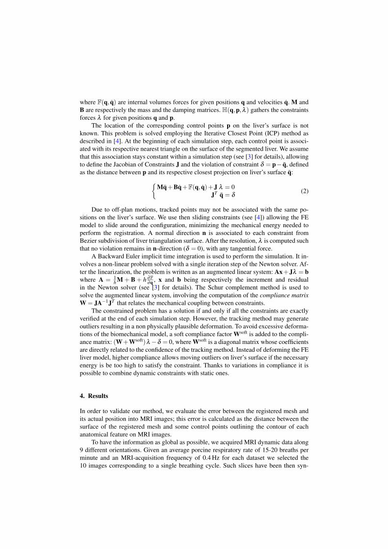
SlicesOutline Gallbladder
average [mm] average [mm]
C0 8.8 10.5
A0 15.8 10.6
S0 6.6 5.81
C0 A0 S0 2.0 3.9
C0 A0 Smax 1.7 1.5
C0 A0 Smin 1.9 1.7
C0 Amax S0 2.5 10.6
C0 Amin S0 7.4 9.7
Cmax A0 S0 3.2 5.9
Cmin A0 S0 2.0 1.4Table 1. Average distance between registered mesh and control points on MRI images
chronized setting as init the slice corresponding to the maximum inspiration and controlpoints have been selected onto starting image.
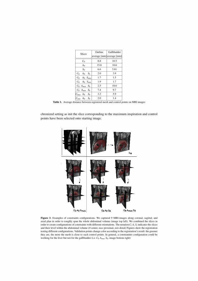
Figure 3. Examples of constraints configurations. We captured 9 MRI-images along coronal, sagittal, andaxial plan in order to roughly span the whole abdominal volume (image top left). We combined the slices inorder to create configurations of constraints with different orientations. The notation Ci Ai Si indicates the slicesand their level within the abdominal volume (0 center, max proximal, min distal) Figures show the registrationtesting different configurations. Validation points change color according to the registration’s result: the greenerthey are, the more the mesh is close to such control points. In general, a constranints configuration could beworking for the liver but not for the gallbladder (i.e. C0 Amax S0, image bottom right)
For the static part, each test was performed activating a set of 3 different constraintsat once, while the control points for the validation distance were considered in the wholearea. For each test, we registered the liver changing one constraint (i.e. MRI-slice) pertime; in particular, we chose the most external ones (min,max), defining the boundary ofour volume. Results of the average error of gallbladder and liver registration, as well asthe configuration of the slices, are shown in (Fig. 3).
Outcomes are rather satisfying: average deviations float around 2 mm, except forthe combination of constraints the distal axial slice. In order to achieve optimal results,two criteria must be fulfilled by a constraint: it must be set on a region with high volumedensity and it has to present a not generic contour.” In this case, such axial image, not onlybelongs to a restricted portion of the liver, but its outline doesn’t give enough informationabout the shape. The best result is achieved with the combination of central coronal,central axial and proximal sagittal. Furthermore, we show how a combination of threeslices is preferable in respect of a single constraint (Tab. 1).
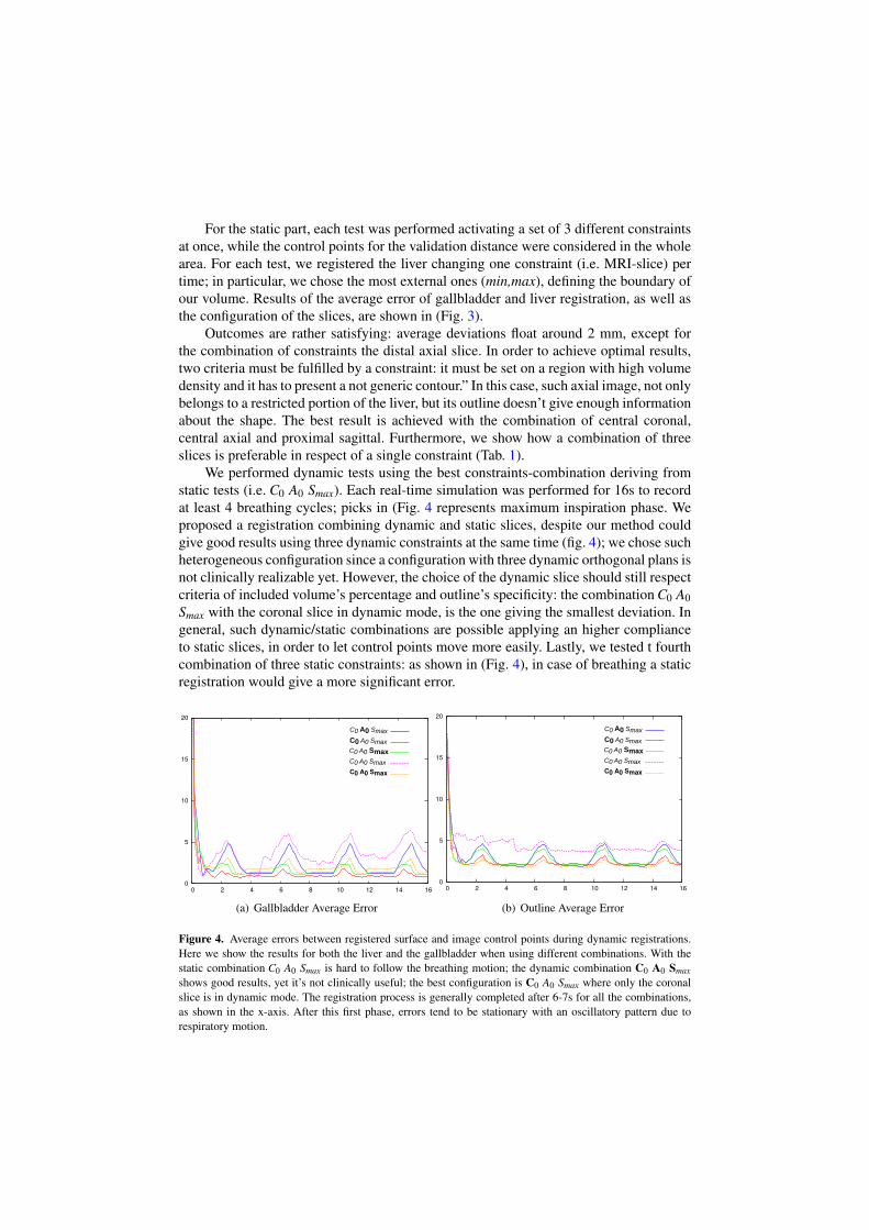
We performed dynamic tests using the best constraints-combination deriving fromstatic tests (i.e. C0 A0 Smax). Each real-time simulation was performed for 16s to recordat least 4 breathing cycles; picks in (Fig. 4 represents maximum inspiration phase. Weproposed a registration combining dynamic and static slices, despite our method couldgive good results using three dynamic constraints at the same time (fig. 4); we chose suchheterogeneous configuration since a configuration with three dynamic orthogonal plans isnot clinically realizable yet. However, the choice of the dynamic slice should still respectcriteria of included volume’s percentage and outline’s specificity: the combination C0 A0Smax with the coronal slice in dynamic mode, is the one giving the smallest deviation. Ingeneral, such dynamic/static combinations are possible applying an higher complianceto static slices, in order to let control points move more easily. Lastly, we tested t fourthcombination of three static constraints: as shown in (Fig. 4), in case of breathing a staticregistration would give a more significant error.
0
5
10
15
20
0 2 4 6 8 10 12 14 16
C0 A0 Smax
C0 A0 Smax C0 A0 Smax C0 A0 Smax
C0 A0 Smax
(a) Gallbladder Average Error
0
5
10
15
20
0 2 4 6 8 10 12 14 16
C0 A0 Smax
C0 A0 Smax C0 A0 Smax C0 A0 Smax
C0 A0 Smax
(b) Outline Average Error
Figure 4. Average errors between registered surface and image control points during dynamic registrations.Here we show the results for both the liver and the gallbladder when using different combinations. With thestatic combination C0 A0 Smax is hard to follow the breathing motion; the dynamic combination C0 A0 Smaxshows good results, yet it’s not clinically useful; the best configuration is C0 A0 Smax where only the coronalslice is in dynamic mode. The registration process is generally completed after 6-7s for all the combinations,as shown in the x-axis. After this first phase, errors tend to be stationary with an oscillatory pattern due torespiratory motion.
5. Conclusion
We proposed a method for non-rigid registration on dynamic data. We combined 2D MRIimages with physics-based simulation to provide a 3D representation of the liver duringbreathing motion. As future work we first plan to perform a quantitative evaluation of thecompliance and its influence on the simulation; the main objective would be to find theoptimal value to drive the simulation. In addition, we will implement the method to workwith a single dynamic constraint, so that registration could be performed on ultrasoundimages.
References
[1] S. Abdelaziz, L. Esteveny, P. Renaud, B. Bayle, L. Barbé, M. De Mathelin, and A. Gangi. Designconsiderations for a novel mri compatible manipulator for prostate cryoablation. International Journalof Computer Assisted Radiology and Surgery, 6(6):811–819, 2011.
[2] E. Coevoet, N. Reynaert, E. Lartigau, L. Schiappacasse, J. Dequidt, and C. Duriez. Introducing interac-tive inverse FEM simulation and its application for adaptive radiotherapy. In MICCAI - 17th Interna-tional Conference on Medical Image Computing and Computer-Assisted Intervention, Boston, UnitedStates, September 2014.
[3] H. Courtecuisse, P. Allard, J.and Kerfriden, S. Bordas, S. Cotin, and C. Duriez. Real-time simulation ofcontact and cutting of heterogeneous soft-tissues. Medical image analysis, 18(2):394–410, 2014.
[4] H. Courtecuisse, I. Peterlik, R. Trivisonne, C. Duriez, and S. Cotin. Constraint-Based Simulation forNon-Rigid Real-Time Registration. In Medicine Meets Virtual Reality, Manhattan Beach, California,United States, February 2014.
[5] I. Elgezua, Y. Kobayashi, and M. Fujie. Survey on current state-of-the-art in needle insertion robots:Open challenges for application in real surgery. Procedia {CIRP}, 5:94 – 99, 2013.
[6] M Ferrant, a Nabavi, B Macq, F a Jolesz, R Kikinis, and S K Warfield. Registration of 3-D intraoperativeMR images of the brain using a finite-element biomechanical model. IEEE transactions on medicalimaging, 20(12):1384–97, December 2001.
[8] B. Gilles and D. Pai. Fast musculoskeletal registration based on shape matching. In D. Metaxas, L. Axel,G. Fichtinger, and G. Székely, editors, Medical Image Computing and Computer-Assisted Intervention– MICCAI 2008, volume 5242 of Lecture Notes in Computer Science, pages 822–829. 2008.
[9] N. Haouchine, J. Dequidt, I.Peterlik, E. Kerrien, and M.-O. Berger. Image-guided simulation of het-erogeneous tissue deformation for augmented reality during hepatic surgery. In Proc. of ISMAR, Int.Symposium on Mixed and Augm. Reality, 2013.
[10] K Masamune, E Kobayashi, Y Masutani, M Suzuki, T Dohi, H Iseki, and K Takakura. Development ofan MRI-compatible needle insertion manipulator for stereotactic neurosurgery. Journal of image guidedsurgery, 1(4):242–248, 1995.
[11] M. Müller and M. Gross. Interactive virtual materials. In GI ’04: Proc. of Graphics Interface 2004,pages 239–246, School of Computer Science, University of Waterloo, Waterloo, Ontario,Canada, 2004.
[12] M Neumann, L Cuvillon, E Breton, and M de Matheli. Evaluation of an image-based tracking workflowwith Kalman filtering for automatic image plane alignment in interventional MRI. Conference proceed-ings : ... Annual International Conference of the IEEE Engineering in Medicine and Biology Society.IEEE Engineering in Medicine and Biology Society. Annual Conference, 2013:2968–2971, 2013.
[13] R. Seifabadi, S. Song, A. Krieger, N. Cho, J. Tokuda, G. Fichtinger, and I. Iordachita. Robotic systemfor mri-guided prostate biopsy: feasibility of teleoperated needle insertion and ex vivo phantom study.International Journal of Computer Assisted Radiology and Surgery, 7(2):181–190, 2012.
[14] S. Suwelack, S.and Röhl, S. Bodenstedt, D. Reichard, R. Dillmann, T. dos Santos, L. Maier-Hein,M. Wagner, J. Wünscher, H. Kenngott, B. Müller, and S. Speidel. Physics-based shape matching forintraoperative image guidance. Med. Phys., 41(11):111901, 2014.