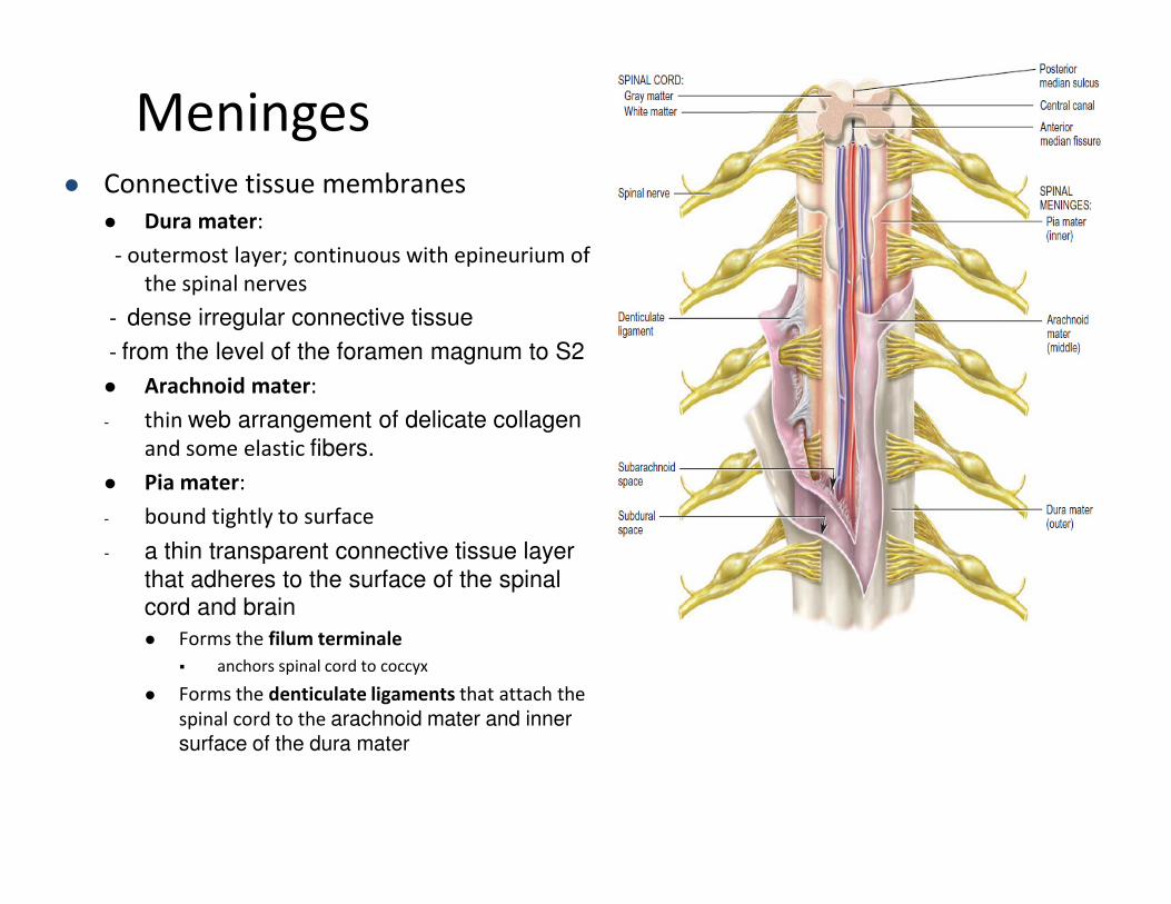
Meninges Connective tissue membranes Dura mater: - outermost layer; continuous with epineurium of the spinal nerves - dense irregular connective tissue - from the level of the foramen magnum to S2 Arachnoid mater: - thin web arrangement of delicate collagen and some elastic fibers. Pia mater: - bound tightly to surface - a thin transparent connective tissue layer that adheres to the surface of the spinal cord and brain Forms the filum terminale anchors spinal cord to coccyx Forms the denticulate ligaments that attach the spinal cord to the arachnoid mater and inner surface of the dura mater
Transcript
Meninges� Connective tissue membranes
� Dura mater:
- outermost layer; continuous with epineurium of
the spinal nerves
- dense irregular connective tissue
- from the level of the foramen magnum to S2
� Arachnoid mater:
- thin web arrangement of delicate collagen
and some elastic fibers.
� Pia mater:
- bound tightly to surface
- a thin transparent connective tissue layer
that adheres to the surface of the spinal cord and brain
� Forms the filum terminale
� anchors spinal cord to coccyx
� Forms the denticulate ligaments that attach the
spinal cord to the arachnoid mater and inner surface of the dura mater
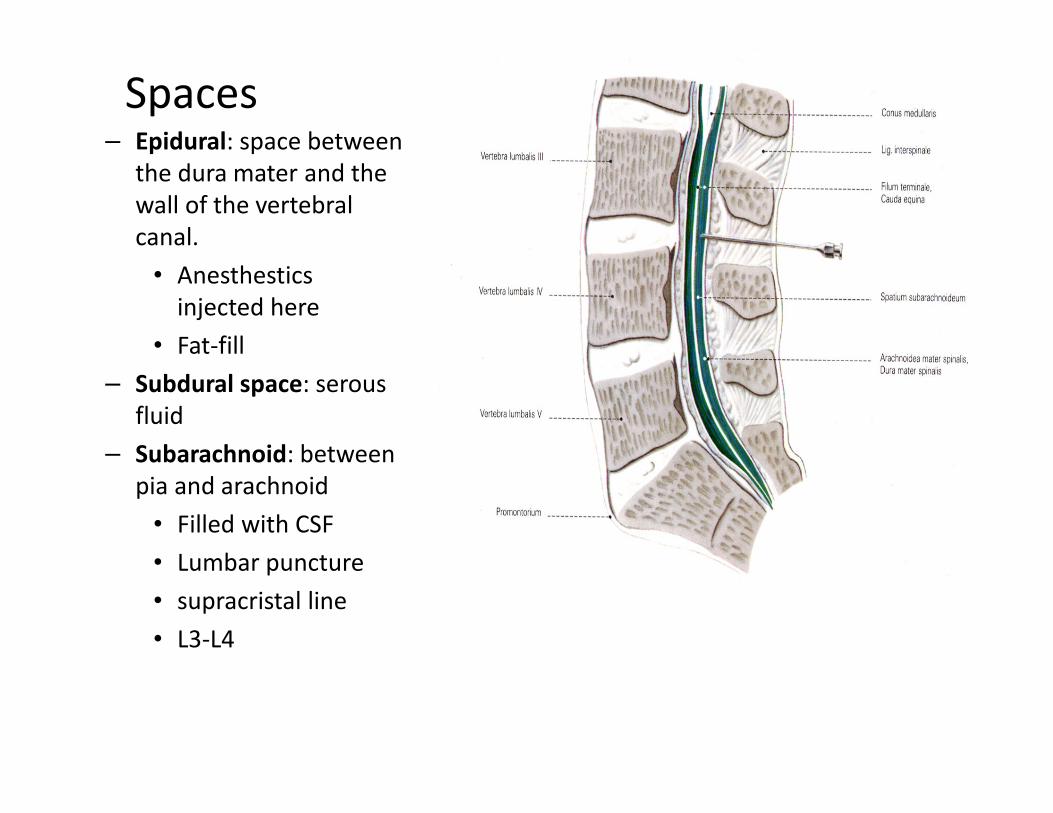
Spaces– Epidural: space between
the dura mater and the
wall of the vertebral
canal.
• Anesthestics
injected here
• Fat-fill
– Subdural space: serous
fluid
– Subarachnoid: between
pia and arachnoid
• Filled with CSF
• Lumbar puncture
• supracristal line
• L3-L4
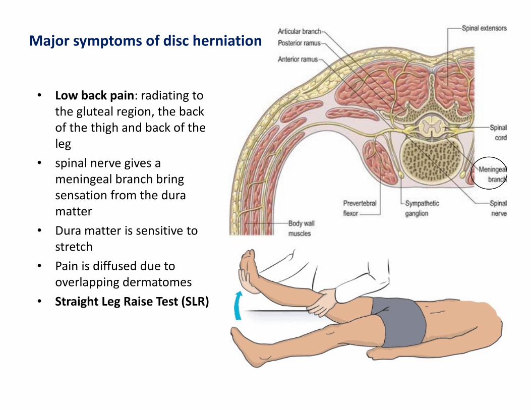
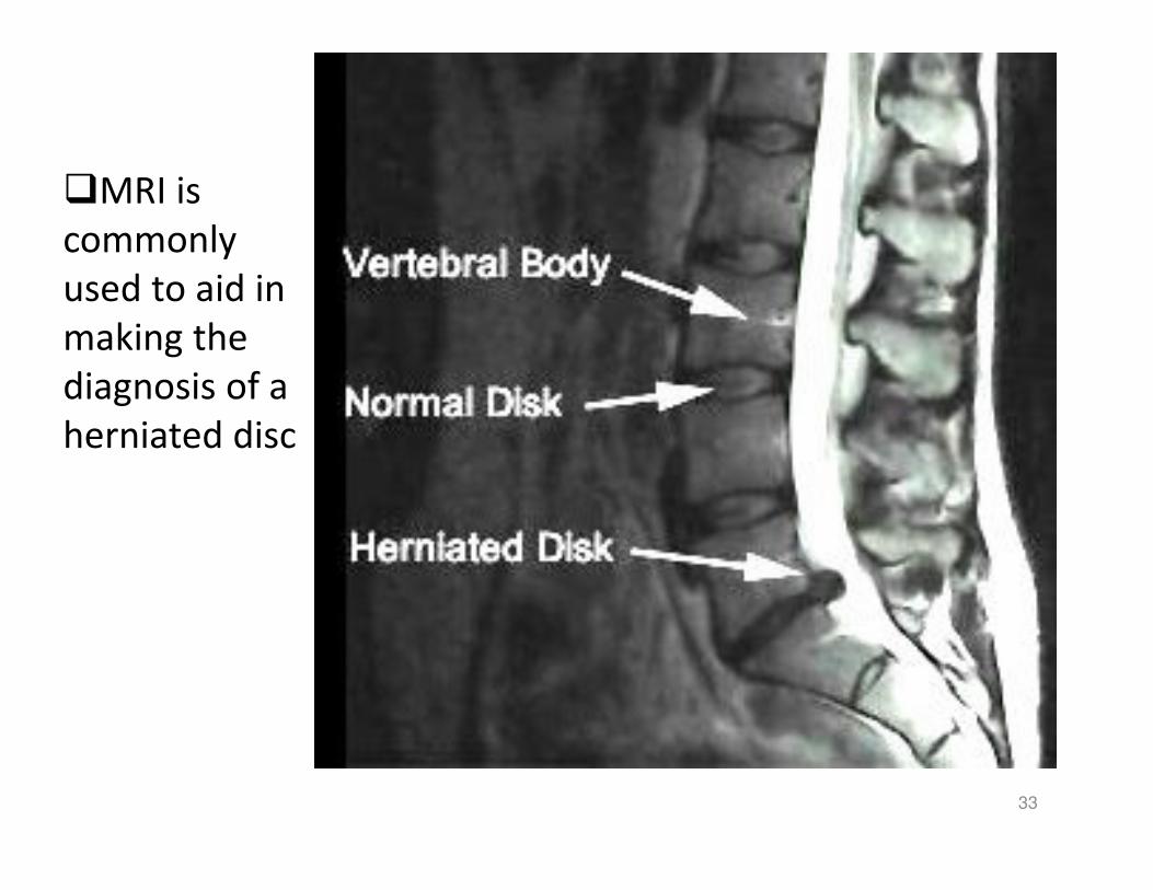
protrusion (leakage) of the gelatinous nucleus
pulposus through the anulus fibrosus of IV disc
Posterolateral direction:
Thinner annulus fibrosus
95% in L4/L5 or L5/S1
Herniated Disc/ ruptured disc/ slipped disc
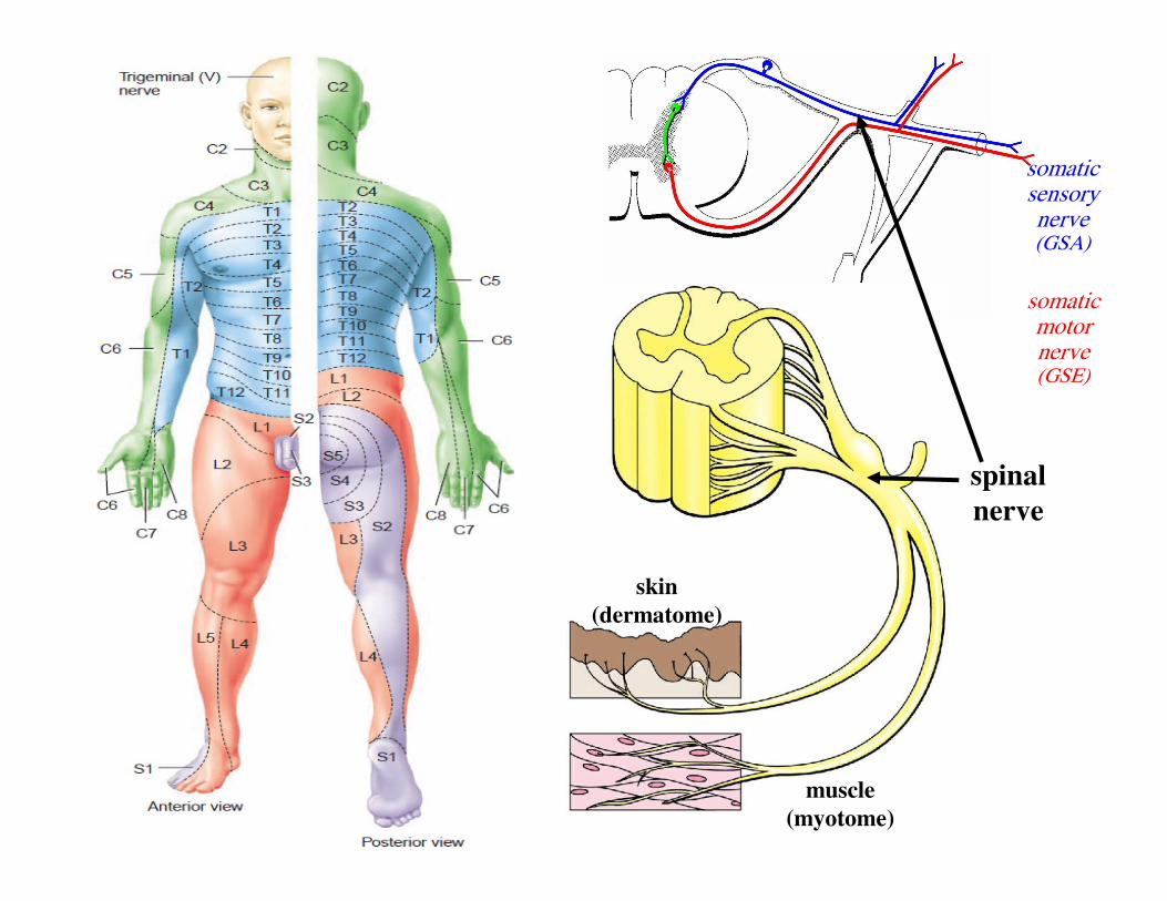
somaticsensorynerve(GSA)
somaticmotornerve(GSE)
spinal
nerve
skin
(dermatome)
muscle
(myotome)
Common lumbar disc problems
Important
myotomes
of lower
limb
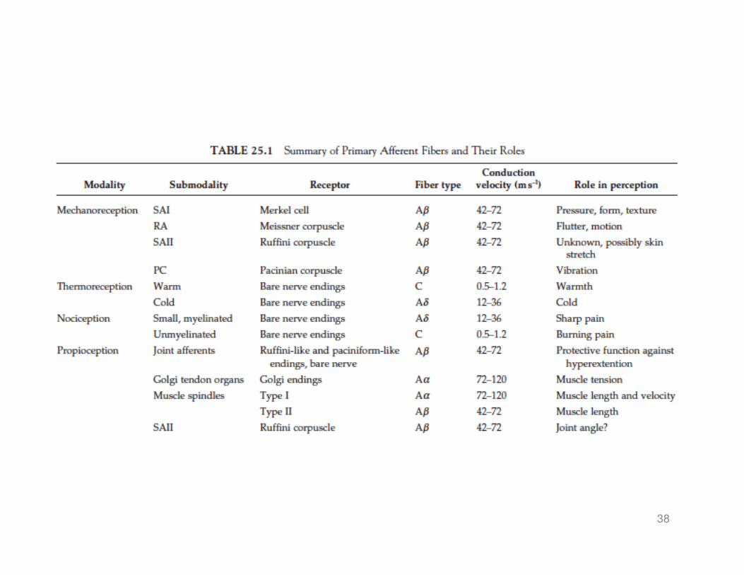
Disc Root Percentage Motor weakness Sensory
changes
Reflex affected
L3-L4 L4 3-10% Knee extension
(Quadriceps femoris
Anteriomedial
leg (saphenous)
Knee jerk
L4-L5 L5 40-45% Big toe dorsifelxion
(EHL) and TA
Big toe ,
anteriolateral
leg (Common P)
Hamstring jerk
L5-S1 S1 45-50% Foot planter flextion
(Gastrocnemius)
Lateral border
of foot (sural)
Ankle jerk
�Test L5: by asking the patient to stand on his heels
�Test S1: by asking the patient to stand on his tiptoes
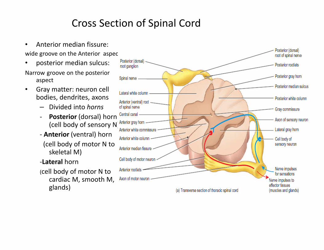
- Posterior (dorsal) horn (cell body of sensory N)
- Anterior (ventral) horn
(cell body of motor N to skeletal M)
-Lateral horn
(cell body of motor N to cardiac M, smooth M, glands)
Ascending tracts
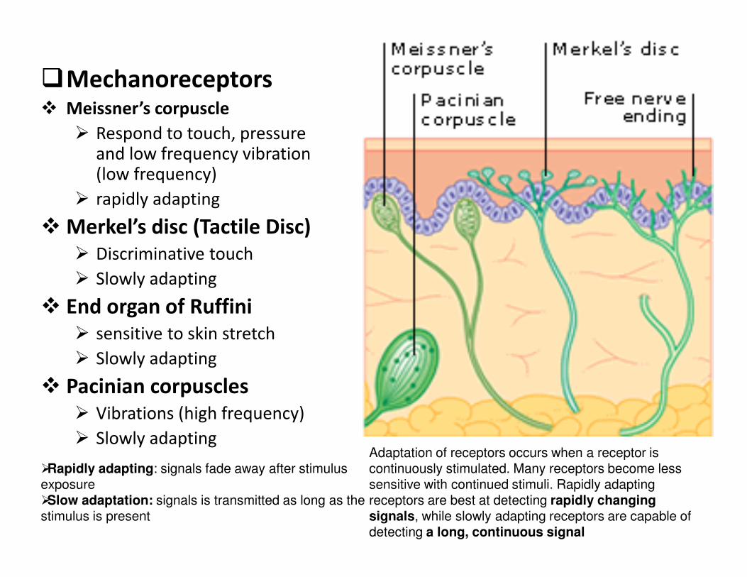
�Mechanoreceptors� Meissner’s corpuscle
� Respond to touch, pressure and low frequency vibration (low frequency)
� rapidly adapting
� Merkel’s disc (Tactile Disc)
� Discriminative touch
� Slowly adapting
� End organ of Ruffini
� sensitive to skin stretch
� Slowly adapting
� Pacinian corpuscles
� Vibrations (high frequency)
� Slowly adaptingAdaptation of receptors occurs when a receptor is continuously stimulated. Many receptors become less sensitive with continued stimuli. Rapidly adapting receptors are best at detecting rapidly changing signals, while slowly adapting receptors are capable of detecting a long, continuous signal
�Rapidly adapting: signals fade away after stimulus exposure�Slow adaptation: signals is transmitted as long as the stimulus is present
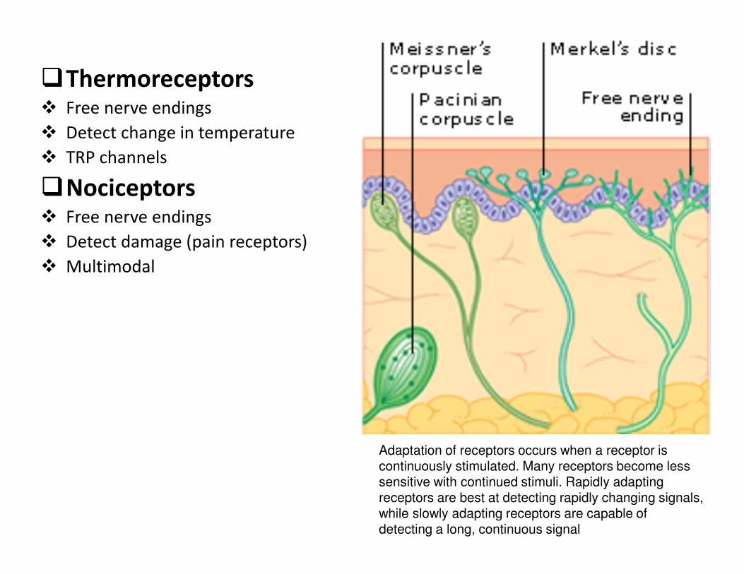
�Thermoreceptors� Free nerve endings
� Detect change in temperature
� TRP channels
�Nociceptors� Free nerve endings
� Detect damage (pain receptors)
� Multimodal
Adaptation of receptors occurs when a receptor is continuously stimulated. Many receptors become less sensitive with continued stimuli. Rapidly adapting receptors are best at detecting rapidly changing signals, while slowly adapting receptors are capable of detecting a long, continuous signal
38
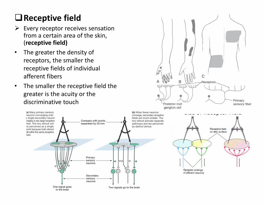
�Receptive field � Every receptor receives sensation
from a certain area of the skin, (receptive field)
• The greater the density of
receptors, the smaller the
receptive fields of individual
afferent fibers
• The smaller the receptive field the
greater is the acuity or the
discriminative touch
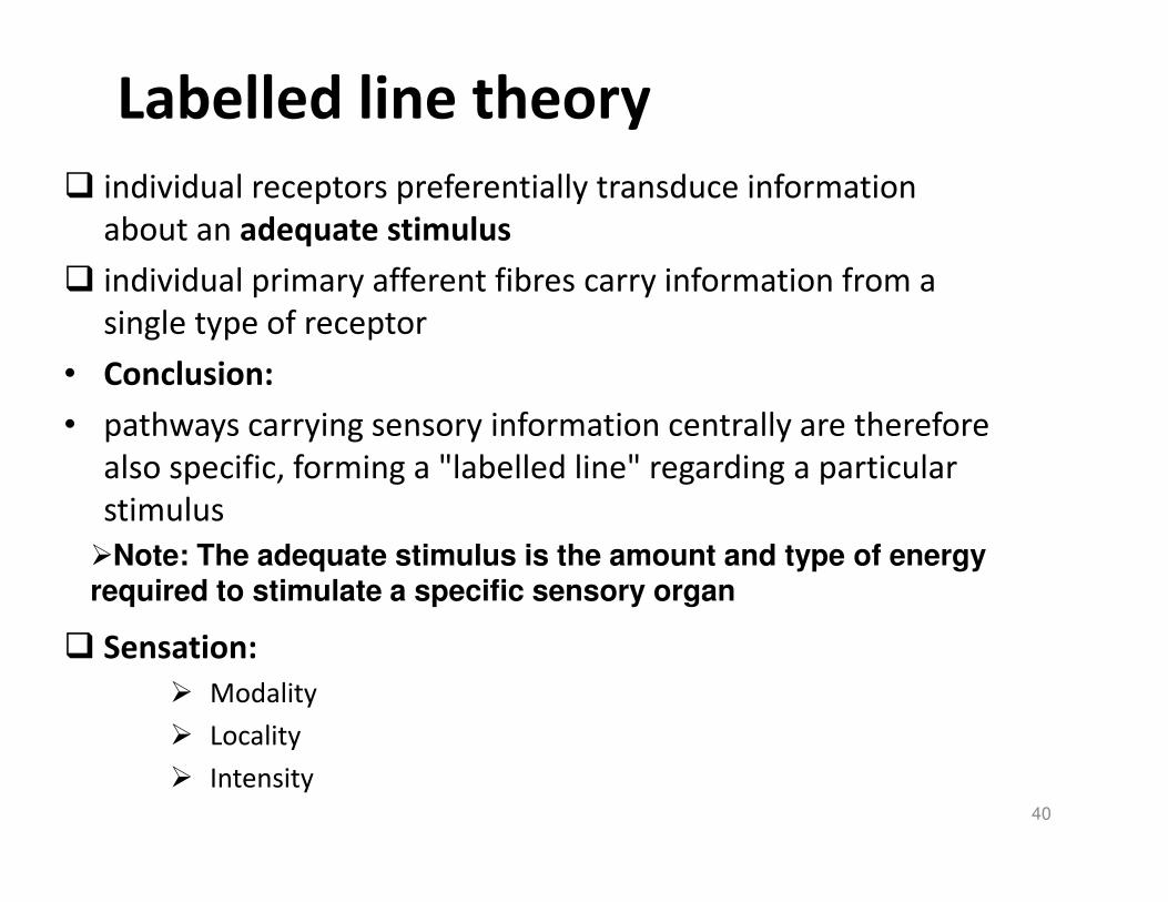
Labelled line theory
� individual receptors preferentially transduce information
about an adequate stimulus
� individual primary afferent fibres carry information from a
single type of receptor
• Conclusion:
• pathways carrying sensory information centrally are therefore
also specific, forming a "labelled line" regarding a particular
stimulus
40
�Note: The adequate stimulus is the amount and type of energy required to stimulate a specific sensory organ


![INDEX [doctor2015.jumedicine.com] · Lecture Notes & Rounds These summaries might not contain all the topics ….. but I tried to cover most of them I would like to thank my amazing](https://static.documents.pub/doc/80x56/5ec0bfa068336b7b5a6f1392/index-lecture-notes-rounds-these-summaries-might-not-contain-all-the-topics.jpg)


