74
Diagnosis and Management of Acute Stroke Briana Witherspoon DNP, ACNP-BC

Diagnosis and Management of Acute Stroke
Briana Witherspoon DNP, ACNP-BC

Stroke Objectives
• Review etiology of strokes• Identify likely location/type of stroke based of
physical exam• Acute management of ischemic stroke• Acute management of hemorrhagic stroke
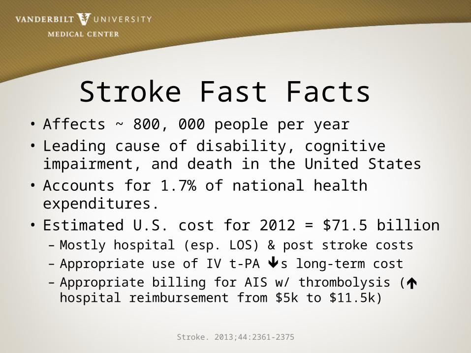
Stroke Fast Facts • Affects ~ 800, 000 people per year• Leading cause of disability, cognitive impairment,
and death in the United States• Accounts for 1.7% of national health expenditures.• Estimated U.S. cost for 2012 = $71.5 billion
– Mostly hospital (esp. LOS) & post stroke costs– Appropriate use of IV t-PA s long-term cost– Appropriate billing for AIS w/ thrombolysis ( hospital
reimbursement from $5k to $11.5k)Stroke. 2013;44:2361-2375

Where We’re Headed• By 2030 ~ 4% of the US population over the
age of 18 is projected to have had a stroke• Between 2012 and 2030, total direct stroke-
related medical costs are expected to increase from $71.55 billion to $183.13 billion
• Total annual costs of stroke are projected to increase to $240.67 billion by 2030, an increase of 129%
Stroke. 2013;44:2361-2375

Three Stroke TypesIschemicStroke
Clot occludingartery85%
Intracerebral Hemorrhage
Bleedinginto brain
10%
Subarachnoid Hemorrhage
Bleeding around brain5%
www.acponline.org/about_acp/chapters/ok/gordon.ppt
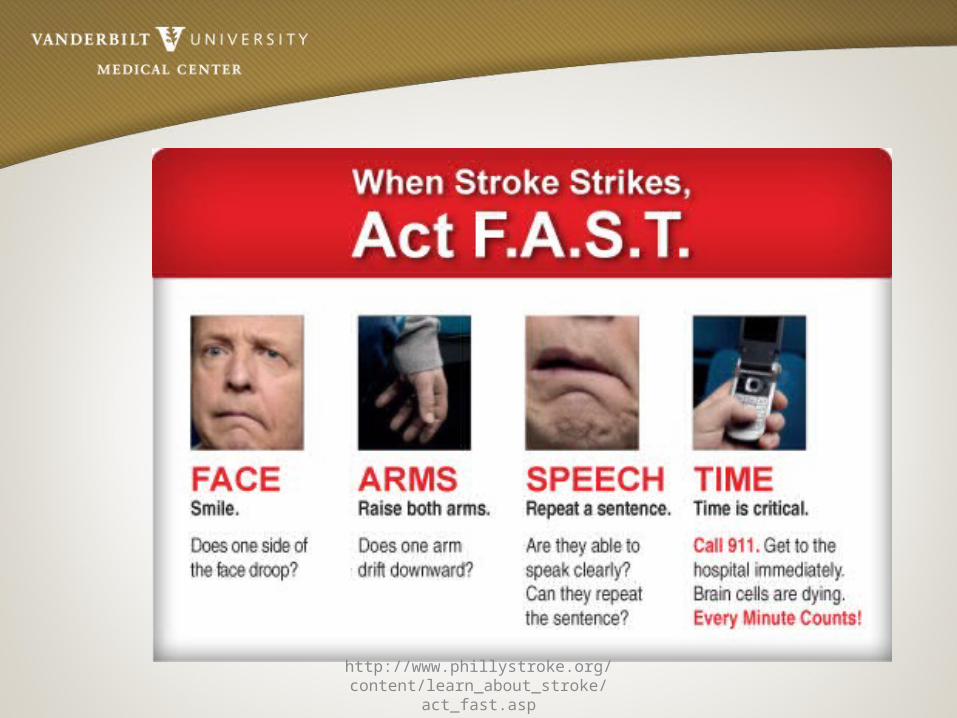
http://www.phillystroke.org/content/learn_about_stroke/act_fast.asp

NIHSS• NIHSS (National Institute of Health Stroke Scale)
– Standardized method used by health care professionals to measure the level of impairment caused by a stroke
– Purpose• Main use is as a clinical assessment tool to determine whether
the degree of disability is severe enough to warrant the use of tPA
• Another important use of the NIHSS is in research, where it allows for the objective comparison of efficacy across different stroke treatments and rehabilitation interventions
– Scores are totaled to determine level of severity– Can also serve as a tool to determine if a change in exam has
occurred
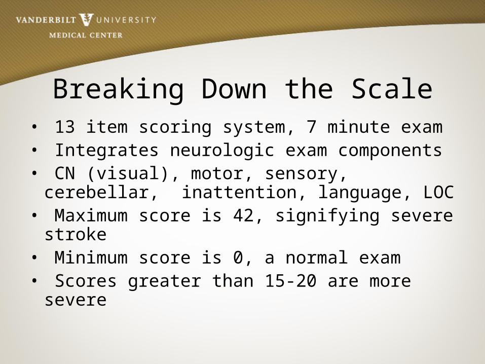
Breaking Down the Scale• 13 item scoring system, 7 minute exam • Integrates neurologic exam components• CN (visual), motor, sensory, cerebellar,
inattention, language, LOC• Maximum score is 42, signifying severe stroke• Minimum score is 0, a normal exam• Scores greater than 15-20 are more severe
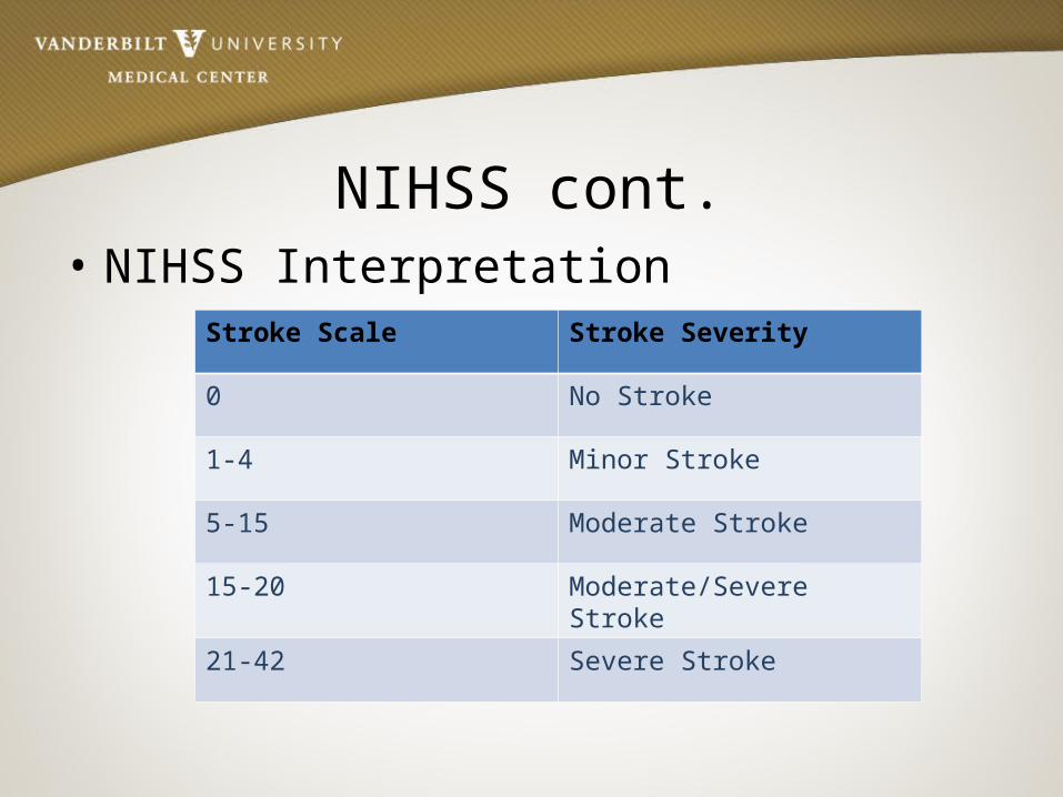
NIHSS cont.• NIHSS Interpretation
Stroke Scale Stroke Severity
0 No Stroke
1-4 Minor Stroke
5-15 Moderate Stroke
15-20 Moderate/Severe Stroke
21-42 Severe Stroke
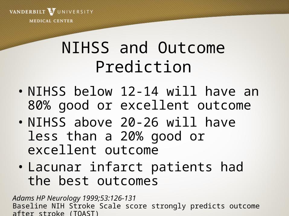
NIHSS and Outcome Prediction
• NIHSS below 12-14 will have an 80% good or excellent outcome
• NIHSS above 20-26 will have less than a 20% good or excellent outcome
• Lacunar infarct patients had the best outcomes
Adams HP Neurology 1999;53:126-131Baseline NIH Stroke Scale score strongly predicts outcome after stroke (TOAST)
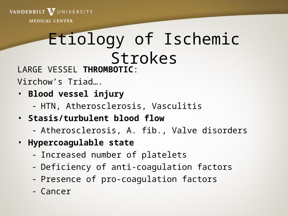
Etiology of Ischemic StrokesLARGE VESSEL THROMBOTIC:Virchow’s Triad….• Blood vessel injury
- HTN, Atherosclerosis, Vasculitis• Stasis/turbulent blood flow
- Atherosclerosis, A. fib., Valve disorders• Hypercoagulable state
- Increased number of platelets- Deficiency of anti-coagulation factors - Presence of pro-coagulation factors- Cancer

Etiology Of Ischemic Stroke:
LARGE VESSEL EMBOLIC:• The Heart
– Valve diseases, A. Fib, Dilated cardiomyopathy, Myxoma
• Arterial Circulation (artery to artery emboli)– Atherosclerosis of carotid, Arterial dissection, Vasculitis
• The Venous Circulation – PFO w/R to L shunt, Emboli
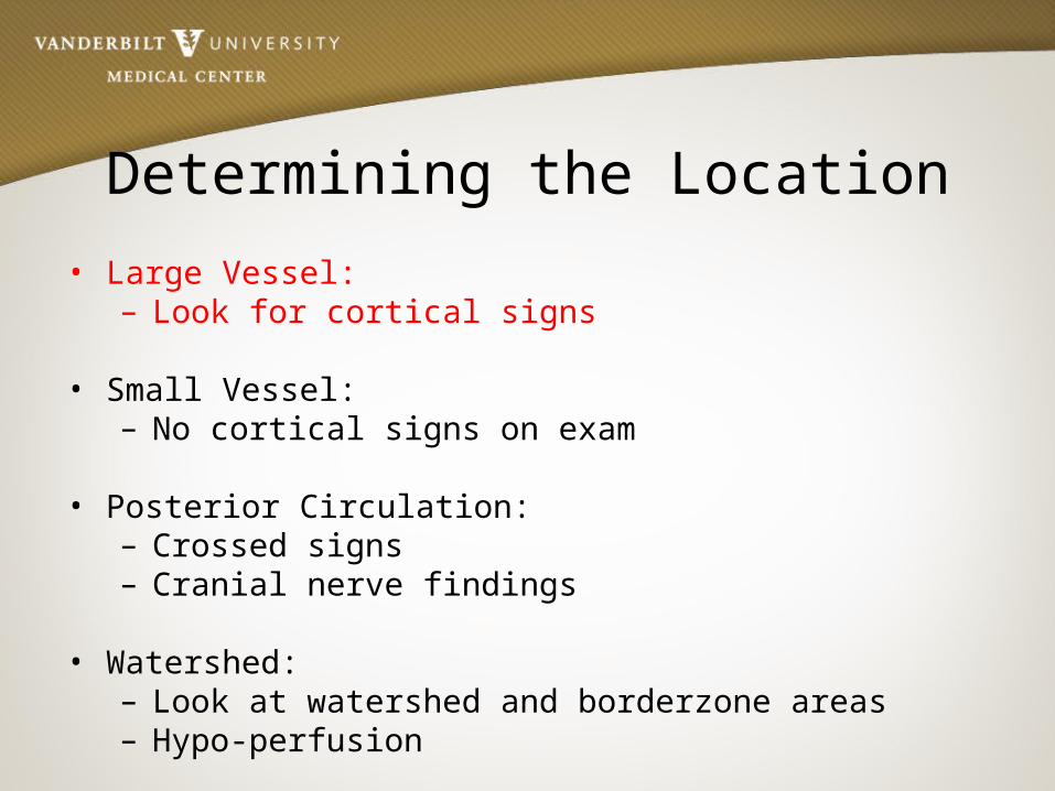
Determining the Location• Large Vessel:
– Look for cortical signs
• Small Vessel:– No cortical signs on exam
• Posterior Circulation:– Crossed signs– Cranial nerve findings
• Watershed:– Look at watershed and borderzone areas– Hypo-perfusion

Cortical SignsRIGHT BRAIN: LEFT BRAIN:
- Right gaze preference - Left gaze preference
- Neglect - Aphasia
• If present, think LARGE VESSEL stroke

Large Vessel Stroke Syndromes• MCA:
– Arm>leg weakness– LMCA cognitive: Aphasia– RMCA cognitive: Neglect,, topographical difficulty, apraxia,
constructional impairment
• ACA: – Leg>arm weakness, grasp– Cognitive: muteness, perseveration, abulia, disinhibition
• PCA: – Hemianopia– Cognitive: memory loss/confusion, alexia
• Cerebellum: – Ipsilateral ataxia

Aphasia• Broca’s
– Expressive aphasia– Left posterior inferior
frontal gyrus
• Wernicke’s– Receptive aphasia– Posterior part of the superior temporal gyrus– Located on the dominant side (left) of the brain

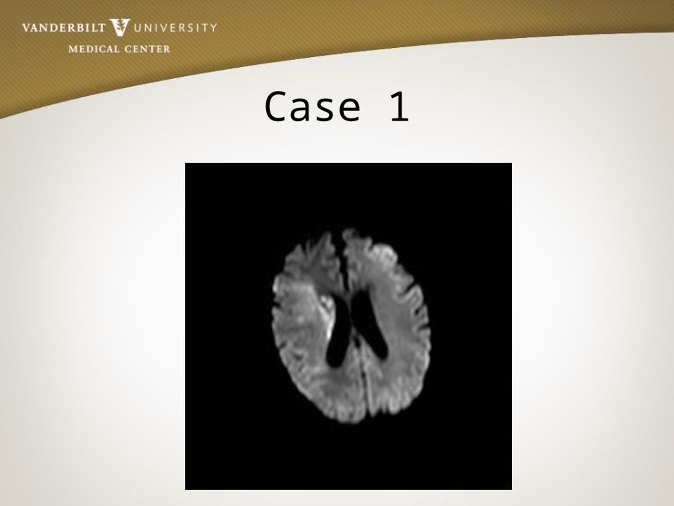
Case 1 • 74 year old African American female with sudden
onset of left-sided weakness
• She was at church when she noted left facial droop
• History of HTN and atrial fibrillation
• Meds: Losartan

Case 1• BP- 172/89, P– 104, T- 98.0, RR– 22, O2- 94%
• General exam: Unremarkable except irregular rate and rhythm
• NEURO EXAM:- Speech dysarthric but language intact- Right gaze preference- Left facial droop- Left- sided hemiplegia- Neglect
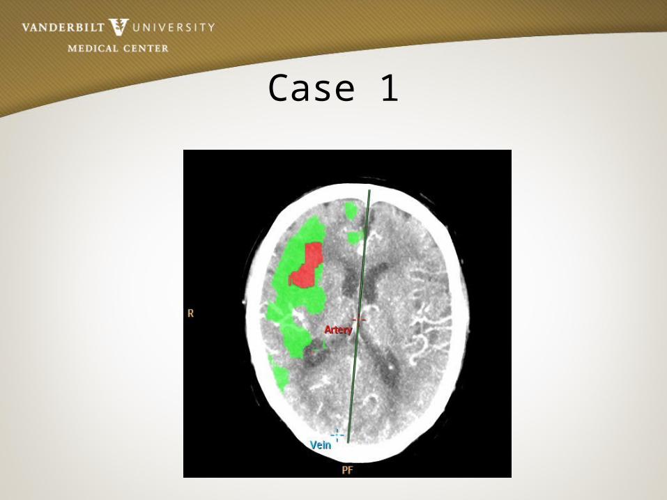
Case 1

Case 1

Case 1
Case 1
Case 1
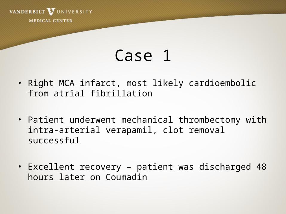
• Right MCA infarct, most likely cardioembolic from atrial fibrillation
• Patient underwent mechanical thrombectomy with intra-arterial verapamil, clot removal successful
• Excellent recovery – patient was discharged 48 hours later on Coumadin

Determining the Location• Large Vessel:
– Look for cortical signs
• Small Vessel:– No cortical signs on exam
• Posterior Circulation:– Crossed signs– Cranial nerve findings
• Watershed:– Look for watershed pattern – S/S of Hypo-perfusion

Etiology of Stroke
SMALL VESSEL (Lacunes <1.5cm)•Risk Factors
– HTN– HLD– DM– Tobacco Use– Sleep apnea

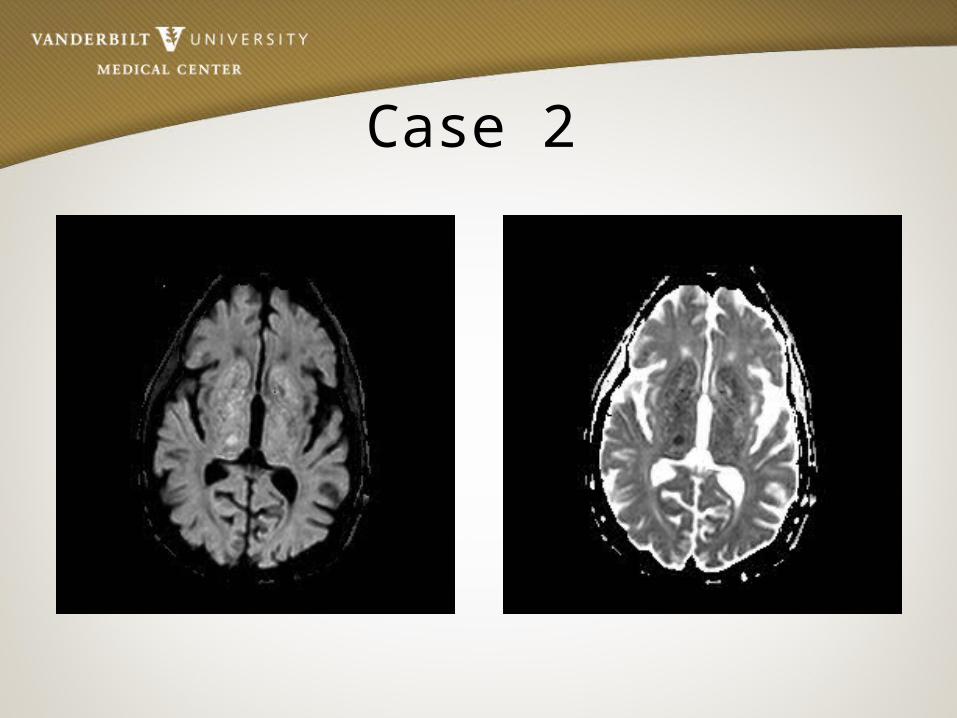
Case 2
• 85 year old male who woke up with left face, arm, and leg numbness
• History of HTN, DM, and tobacco use
• Meds: Insulin, aspirin

Case 2• BP- 168/96, P– 92
• General exam: Unremarkable, RRR
• NEURO EXAM:- Decreased sensation on left face, arm, and leg
Case 2
Case 2
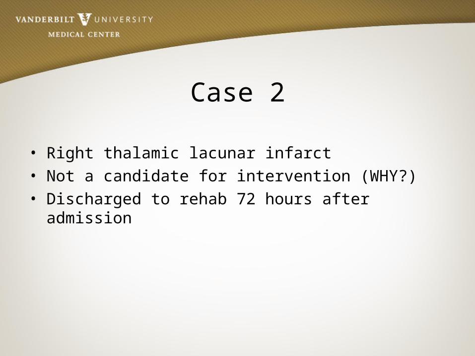
• Right thalamic lacunar infarct• Not a candidate for intervention (WHY?)• Discharged to rehab 72 hours after admission

Determining the Location• Large Vessel:
– Look for cortical signs
• Small Vessel:– No cortical signs on exam
• Posterior Circulation:– Crossed signs– Cranial nerve findings
• Watershed:– Look at watershed and borderzone areas– Hypo-perfusion

Brainstem Stroke Syndromes
• Rarely presents with an isolated symptom
• Usually a combination of cranial nerve abnormalities, and crossed motor/sensory findings such as:
– Double vision– Facial numbness and/or weakness– Slurred speech– Difficulty swallowing– Ataxia– Vertigo– Nausea and vomiting– Hoarseness
Case 3• 55 year old male with acute onset of right sided numbness
and tingling, left sided face pain and numbness, gait imbalance, nausea/vomiting, vertigo, swallowing difficulties, and hoarse speech
• History of CAD s/p CABG, DM2, HTN, HLD, OSA
• Meds: Aspirin, plavix, insulin, lipitor, metoprolol, lisinopril

Case 3• NEURO EXAM: BP- 194/102, P– 105
• General exam: Unremarkable, RRR
• NEURO EXAM:- Decreased sensation on left face- Decreased sensation on right body- Left ataxia on FNF, and unsteady gait- Voice hoarse- Nystagmus
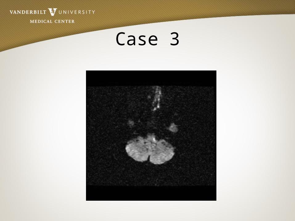
Case 3
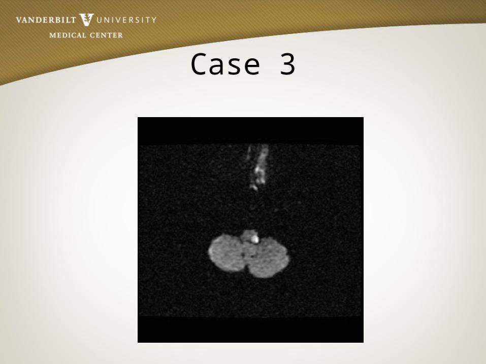
Case 3

Case 3
• Brainstem Stroke• Received IV tPa• Post-tPa symptoms greatly improved
regained sensation, ataxia resolved• Discharged home with out patient PT/OT
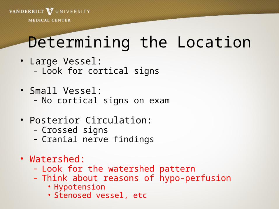
Determining the Location• Large Vessel:
– Look for cortical signs
• Small Vessel:– No cortical signs on exam
• Posterior Circulation:– Crossed signs– Cranial nerve findings
• Watershed:– Look for the watershed pattern– Think about reasons of hypo-perfusion
• Hypotension• Stenosed vessel, etc

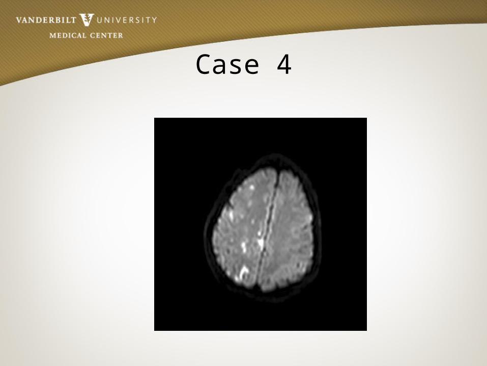
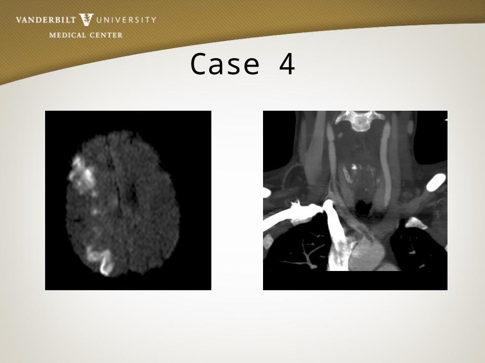
Case 4• 56 year old female who upon waking post-op after elective
surgery was found to have L sided weakness and neglect
• History of HTN
• Meds - Lisinopril

Case 4• BP- 132/74, P– 84
• General exam: Unremarkable, RRR
• NEURO EXAM:- Left face, arm, and leg weakness- Neglect- DTR’s brisk on the left, toe up on left

Case 4

Case 4

Case 4

Case 4

Case 4
Case 4
Case 4

Case 4
• Right hemisphere watershed infarct secondary to hypoperfusion in the setting of Right ICA stenosis
• On review of anesthesia records, blood pressure dropped to 82/54 during the procedure
• Patient was discharged to in-patient rehab
Intracranial Hemorrhages
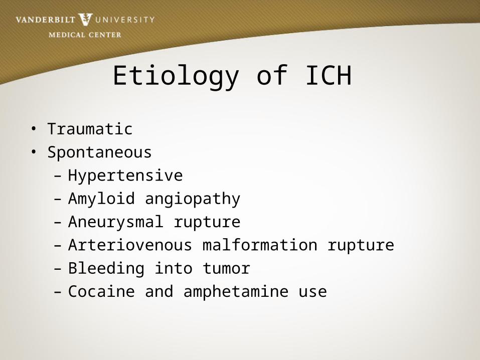
Etiology of ICH
• Traumatic• Spontaneous
– Hypertensive– Amyloid angiopathy– Aneurysmal rupture– Arteriovenous malformation rupture– Bleeding into tumor– Cocaine and amphetamine use

Causes of ICH
http://spinwarp.ucsd.edu/neuroweb/Text/non-trauma-ER.htm

Hypertensive ICH• Spontaneous rupture of a small artery deep in the brain• Typical sites
– Basal Ganglia– Cerebellum– Pons
• Typical clinical presentation– Patient typically awake and often stressed, then abrupt
onset of symptoms with acute decompensation
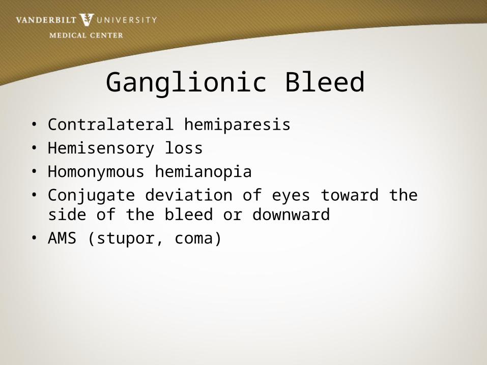
Ganglionic Bleed • Contralateral hemiparesis• Hemisensory loss• Homonymous hemianopia• Conjugate deviation of eyes toward the side of the bleed or
downward• AMS (stupor, coma)

Cerebral Hemorrhage
JPG
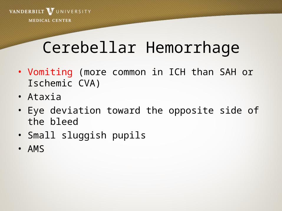
Cerebellar Hemorrhage• Vomiting (more common in ICH than SAH or Ischemic CVA)• Ataxia• Eye deviation toward the opposite side of the bleed • Small sluggish pupils• AMS

Cerebellar Hemorrhage

Pontine Hemorrhage• Pin-point but reactive pupils• Abrupt onset of coma• Decerebrate posturing or flaccidity • Ataxic breathing pattern
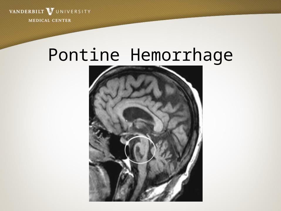
Pontine Hemorrhage

Subarachnoid Hemorrhage
• “Worst headache of my life”• AMS• Photophobia• Nuchal rigidity • Seizures• Nausea and vomiting
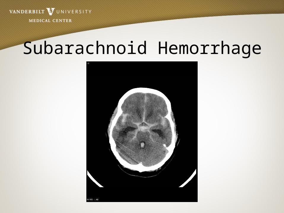
Subarachnoid Hemorrhage
Management

Airway • Most likely related to decreased level of consciousness (LOC),
dysarthria, dysphagia• GCS < 8 - INTUBATE• Avoid Hyperventilation or Hypoventilation• NPO until swallow assessment completed- high aspiration risk • Begin mobilization as soon as clinically safe• Keep HOB greater than 30 degrees

Stroke Algorithm

ImagingCT scan• Non- contrast CTH remains
the gold standard as it is superior for showing IVH and ICH
• CT with contrast may help identify aneurysms, AVMs, or tumors but is not required to determine whether or not the patient is a tPa candidate
MRI• Superior for showing
underlying structural lesions• Contraindications
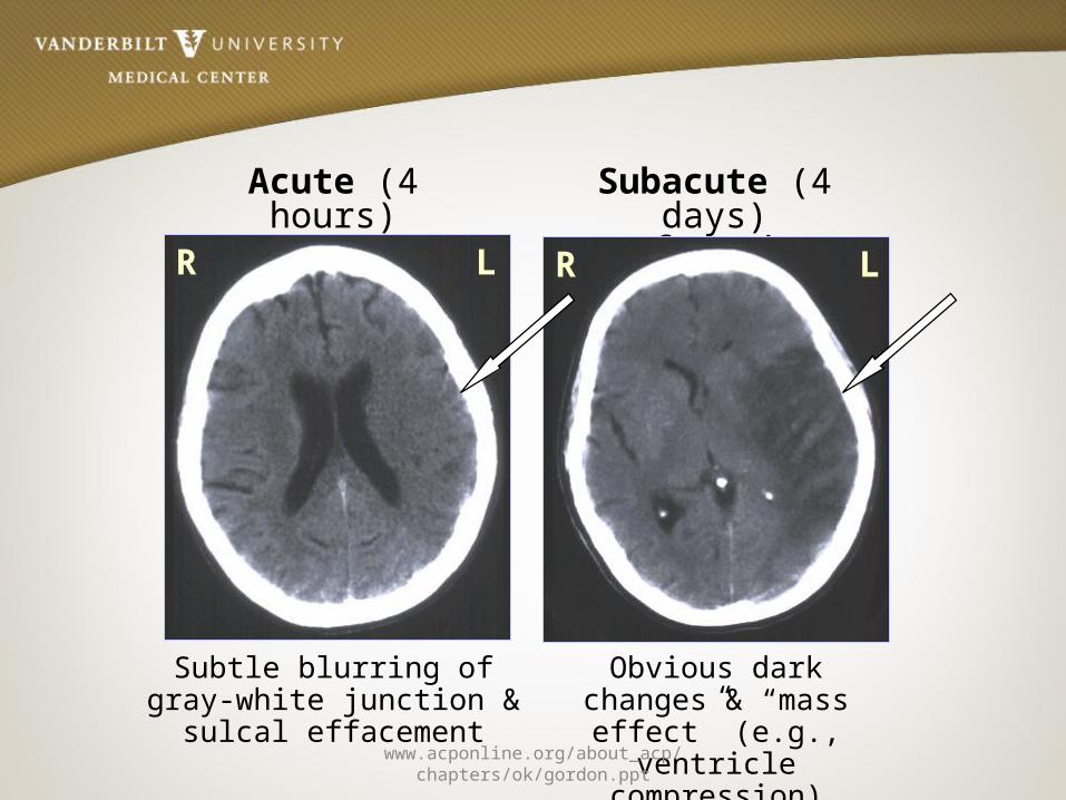
Acute (4 hours)Infarction
Subtle blurring of gray-white junction & sulcal effacement
Subacute (4 days) Infarction
Obvious dark changes & “mass effect” (e.g.,
ventricle compression)
RR L L
www.acponline.org/about_acp/chapters/ok/gordon.ppt

Multimodal ImagingMultimodal CT• Typically includes non-
contrast CT, perfusion CT, and CTA
• Two types of perfusion CT– Whole brain perfusion CT– Dynamic perfusion CT
Multimodal MRI• Standard MRI sequences
( T1 weighted, T2 weighted, and proton density) are relatively insensitive to changes in cerebral ischemia
• Multimodal adds diffuse-weighted imaging (DWI) and PWI (perfusion- weighted imaging)

tPaFast Facts• Tissue plasminogen
activator• “clot buster”• IV tpa window 3 hours• IA tpa window 4.5 hours • Disability risk 30% despite
~5% symptomatic ICH risk
Contraindications• Hemorrhage• SBP > 185 or DBP > 110• Recent surgery, trauma or
stroke • Coagulopathy• Seizure at onset of symptoms• NIHSS >21 • Age?• Glucose < 50

Mechanical Thrombolysis• Often used in adjunct with tPa• MERCI (Mechanical Embolus Removal in
Cerebral Ischemia) Retrieval System is a corkscrew-like apparatus designed to remove clots from vessels
• PENUMBRA system aspirates the clot

Blood Pressure Management•BP Management
– The goal is to maintain cerebral perfusion!!– CPP = MAP – ICP (needs to be at least 70)– Higher BP goals with Ischemic stroke– Lower BP goals with Hemorrhagic stroke (avoid hemorrhagic
expansion, especially in AVMs and aneurysms)

BP-AIS Relationship• BP increase is due to
arterial occlusion (i.e., an effort to perfuse penumbra)
• Failure to recanalize (w/ or w/o thrombolytic therapy) results in high BP and poor neuro outcomes
• Lowering BP starves penumbra, worsens outcomes
Penumbra
Core
Clot in Artery
www.acponline.org/about_acp/chapters/ok/gordon.ppt

Save the Penumbra!!
CEREBRALBLOODFLOW(ml/100g/min)
CBF< 8
CBF8-18
TIME (hours)
1 2 3
20
15
10
5
PENUMBRA
CORE
Neuronal dysfunctio
n
Neuronal death
Normal function
www.acponline.org/about_acp/chapters/ok/gordon.ppt

Supportive Therapy
• Glucose Management– Infarction size and edema increase with acute and chronic
hyperglycemia– Hyperglycemia is an independent risk factor for hemorrhage
when stroke is treated with t-PA• Antiepileptic Drugs
– Seizures are common after hemorrhagic CVAs– ICH related seizures are generally non-convulsive and are
associated to with higher NIHSS scores, a midline shift, and tend to predict poorer outcomes

Hyperthermia
• Treat fevers!– Evidence shows that fevers > 37.5 C that persists
for > 24 hrs correlates with ventricular extension and is found in 83% of patients with poor outcomes
References• Adams, H., del Zappo, G., Alberts, M., Bhatt, D., Brass, L., Furlan, A., Grubb, R., &
Higashida, R. (2007). Guidelines for the early management of adults with ischemic stroke. Stroke, 38, 1655-1711.
• Bradley G Walter, Daroff B Robert, Fenichel M Gerald, Jancovic, Joseph; Neurology in clinical practice, principles of diagnosis and
management. Philadelphia Elsevier, 2004.
• Castillo, J., Leira, R., Garcia, M., Serena, J., Blanco, M. Blood pressure decrease during the acute phase of ischemic stroke is associated with brain injury and poor
stroke outcome. Stroke. 2004: 35: 520-526.• Goals for Management of Patients With Suspected Stroke Algorithm.
http://circ.ahajournals.org/content/112/24_suppl/IV-111/F1.expansion.html. Accessed May 8, 2012
• Gordon, D. L. (n.d.). Update in stroke management . Retrieved from www.acponline.org/about_acp/chapters/ok/gordon.ppt
• Hesselink, J. Imaging of cerebral hemorrhages and AV malformations. http://spinwarp.ucsd.edu/neuroweb/Text/br-740.htm. accessed May 10, 2012.

Questions?
![Lecture 4_ Equalization [Compatibility Mode]](https://static.documents.pub/doc/80x56/577cd75d1a28ab9e789ec837/lecture-4-equalization-compatibility-mode.jpg)


