4/16/2014 1 Labral Repair and Preservation: My Approach April 16, 2014 Joseph U. Barker, M.D. Team Physician: Carolina Hurricanes and NC State Raleigh Orthopaedic Clinic Raleigh, NC P/N 57586 Disclaimer The information presented is solely for internal training purposes. It is not intended for distribution to customers, surgeons, or user facilities. Promotion of ArthroCare products is to be on-label and consistent with approval indications and intended uses. The information in this presentation is considered proprietary and is not to be copied or distributed. Disclosures • Consultant: Arthrocare Corporation • Editorial Board: Journal of Knee Surgery
Transcript
4/16/2014
1
Labral Repair and Preservation:
My Approach
April 16, 2014
Joseph U. Barker, M.D.
Team Physician: Carolina Hurricanes and NC State
Raleigh Orthopaedic Clinic
Raleigh, NC
P/N 57586
Disclaimer
The information presented is solely for internal
training purposes. It is not intended for distribution
to customers, surgeons, or user facilities.
Promotion of ArthroCare products is to be on-label
and consistent with approval indications and
intended uses.
The information in this presentation is considered
proprietary and is not to be copied or distributed.
Disclosures
• Consultant: Arthrocare Corporation
• Editorial Board: Journal of Knee Surgery
4/16/2014
2
Outline
• Labral Anatomy
• Labral Function
• Labral Repair vs debridement
• Surgical Technique
Hip Labrum
• Anatomy
– Triangular
fibrocartilage rim
that deepens the
acetabulum
– Attached to the
acetabular rim
– Continuous with the
transverse
acetabular ligament
• Arrow: labral-chondral junction is continuous
• Arrowhead: capsulolabral recess lies loose connective tissue that also provides blood supply to the labrum.
• Note the bony projection of the acetabulum into the labrum.
Labral Anatomy: Macroscopic
Safran MR. JAAOS 2010.
4/16/2014
3
• 3 Layers by SEM – First: 10 μm without
distinct orientation – Second: 40 μm with
lamellar orientation – Third: 200-300 μm with
circumferential orientation
Labral Anatomy: Microscopic
Petersen et al. Arch Orthop Trauma Surg 2003.
• Physiologic cleft at the chondro-labral junction – Chondral: hyaline
cartilage
• Histology – Articular side:
fibrocartilage with chondrocytes
– Capsular side: dense connective tissue with fibroblasts
Labral Anatomy: Microscopic
Petersen et al. Arch Orthop Trauma Surg 2003.
– Anastamosis between
Medial/lateral
circumflex, deep
branch of superior
gluteal, inferior gluteal
arteries to provide
branches to the
capsule and synovium
– Increased vascularity
at the capsular side
Labral Anatomy: Vasculature
Kalhour M et al. JBJS 2009; Kelly BT et al. Arthroscopy 2005.
4/16/2014
4
Labral Vascularity
• 12 Hip human cadavers
– Capsular side vascularity > articular side vascularity
– No difference in vascularity between intact and torn specimens
• 10 Skeletally mature female sheep
– Surgically induced labral tears/suture anchors
– All healed via fibrovascular repair tissue (capsule), direct bony attachment, or both
Kelly et al., Arthroscopy 2006, Phillippon et al., Arthroscopy 2007.
• Hip pain related to labral injury – Torn
– Impingement
• Originates from the nerve to the quadratus femoris and obturator nerve
• Multiple nerve
endings – Pain: unmyelinated
nerve endings in the
antero-superior labrum
– Pressure: corpuscles
(Vater-Pacini, Golgi-
Mazzoni, Ruffini,
Krause)
– Propioception:
mechanoreceptors
Labral Anatomy: Innervation
Kim YT et al. CORR 1995.
Labral Function
• Deepens the socket allowing for greater coverage of the femoral head (≈21%)
• Provides a fluid seal for the hip joint
• Joint stability
• Shock absorber
• Pressure distributor
**Most common area of injury is at the capsulolabral junction
Parvizi J, Leunig M, Ganz R. JAAOS 2007.
4/16/2014
5
Labral Tears/Cartilage Injuries
• Rarely occur in isolation; 90+% due to bone abnormality (over or under coverage)
• Determine the underlying diagnosis
– FAI (over coverage)
– Dysplasia (under coverage)
Labral Repair vs Debridement
• Level 3 evidence:
• Philippon et al JBJS Br 2009
– 58 labral repair, 54 labral debridement
– HHS 87 labral repair, 81 labral debridement
• Schilders et al JBJS Br 2009
– 69 labral repairs, 32 labral debridements
– HHS 94 labral repair, 89 labral debridement
Labral Repair vs Debridement
• Larson et al AJSM 2012
• 44 labral debridement vs 50 labral repair
• Mean 3.5 year f/u
• HHS, SF12, VAS scores significantly improved with labral repair
• Labral debridement: 68% G/E results
• Labral repair: 92% G/E results
• Level 3 evidence
4/16/2014
6
1. Diagnostic evaluation of cartilage and labral injury
2. Treat cartilage and labrum
3. Address underlying sources of impingement (acetabulum, femur, ileopsoas)
Treatment: Goals of Surgical
Intervention
Treatment: Equipment
•Landmarks
• - ASIS
•- Greater trochanter
•Standard Portals
• Anterolateral
• Anterior
• Mid-Anterior
• Distal AL Accessory
Robertson WR & Kelly BT. Arthroscopy 2008.
X
X
Arthroscopic Portals
4/16/2014
7
Arthroscopic Joint Access
• Start with spinal needle and
fluoroscopy to achieve access
• Modified anterior portal under
direct access
• Cannulas for both portals
• Switch camera to modifed
anterior portal to ensure no
labral penetration
•Use arthroscopic
blade (Curved,
Straight, or Blunt)
or RF probe
• AL portal
• Posterior: 10
o’clock
• Anterior: Ant
portal
• Anterior portal:
extend to 3
o’clock
Arthroscopic Capsulotomy
Arthroscopic Soft Tissue
Abalation
• Use RF probe
and shaver to
remove soft
tissue over
pincer lesion
• Important to
preserve
labrum (no
use of shaver
near labrum)
4/16/2014
8
• Ablate soft tissue superior to the acetabular labrum from 12 0’clock (superior) to 3 o’clock (anterior)
• Reflected head of rectus
• Rim trim w/ 5.5mm burr
• AL portal: superior
• Ant portal: anterior
• Extent
• May see area of pathologic rim
• Fluoroscopy
Arthroscopic Acetabular Rim
Trim
4/16/2014
9
• Place cannula in AL portal
• Place camera in MA portal
• First anchor placed through this portal (superior and superior posterior repair)
• Pass suture with lasso or tissue penetrator
Robertson WR & Kelly BT. Arthroscopy 2008.
X
X
Anchor Placement
Robertson WR & Kelly BT. Arthroscopy 2008.
X
X
X
Anchor Placement
• Switch camera to AL portal
• Switch cannula to MA portal
• Distal portal created through stab incision for placing anterior anchors
• Pass suture with lasso or tissue penetrator
4/16/2014
10
Fry R. Arthroscopy 2010.
Arthroscopic Labral Refixation
Can we create more anatomic repair of the hip labrum?
- Rim trim
- Anchor placement: Closer to the articular surface without penetration
- Stitch: Mattress is more anatomic than simple
4/16/2014
11
Thank You!
www.josephbarkermd.com
1
Labral Repair:
Why Knot?
Mark S. Muller, MD
W.B. Carrell Memorial Clinic
Team Physician, Dallas Cowboys
P/N 57607
Disclaimer
The information presented is solely for internal training
purposes. It is not intended for distribution to customers,
surgeons, or user facilities. Promotion of ArthroCare
products is to be on-label and consistent with approved
indications and intended uses.
The information in this presentation is considered
proprietary and is not to be copied or distributed.
University of Colorado School of Medicine, Department of Orthopedics
PN57645
Disclaimer
The information presented is solely for training
purposes. It is not intended for distribution to
customers, surgeons, or user facilities. Promotion of
ArthroCare products is to be on-label and consistent
with approval indications and intended uses. The
information in this presentation is considered
proprietary and is not to be copied or distributed.
Financial Disclosures
Consultant:
• ArthroCare®
Research support:
• ArthroCare • Stryker®
4/14/2014
2
Reconstruction of the labrum in patients with deficient or
resected labra provides the theoretical advantage of
sealing and stabilizing the hip joint and restoring the fluid
layer which could potentially prevent continued cartilage
degeneration
Labral Reconstruction
Arthroscopic Labral Reconstruction Is Superior to Segmental Resection for Irreparable Labral Tears in the Hip:
A Matched-Pair Controlled Study With Minimum 2-Year Follow-up. Am J Sports Med January 2014. Domb BG, El Bitar YF, Stake CE, Trenga AP, Jackson TJ, Lindner D
Reconstruction…
Main questions:
Why ?
When ?
Who ?
How ?
I & CI
4/14/2014
3
Over coverage w/ ossified labrum
4/14/2014
4
Large Os Acetabuli
Large Os Acetabuli
Dysplastic Patients • Prior to PAO
• Usually after previous failed scope
BL Dysplastic Patients • To increase functional coverage with
oversized labral graft
4/14/2014
5
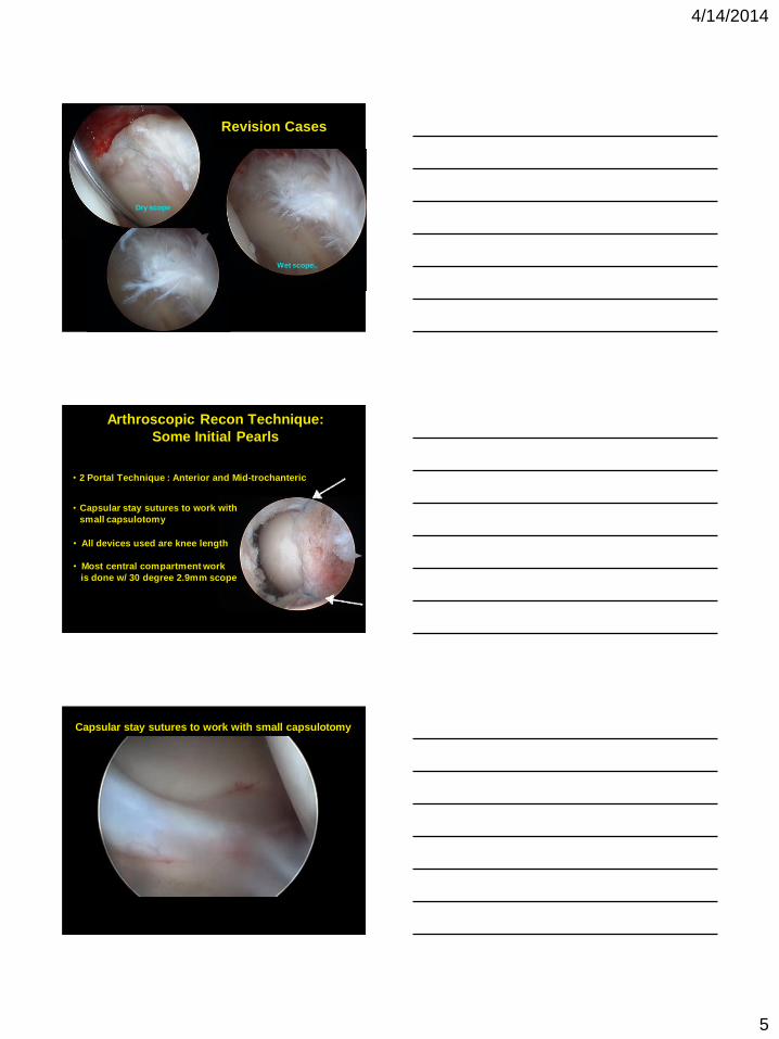
Revision Cases
Dry scope
Wet scope..
• Capsular stay sutures to work with
small capsulotomy
• 2 Portal Technique : Anterior and Mid-trochanteric
Arthroscopic Recon Technique:
Some Initial Pearls
• All devices used are knee length
• Most central compartment work
is done w/ 30 degree 2.9mm scope
Capsular stay sutures to work with small capsulotomy
4/14/2014
6
The knotless “base grab” and
“eversion-inversion” concepts
Controlled tensioning of sutures allows
anatomic refixation
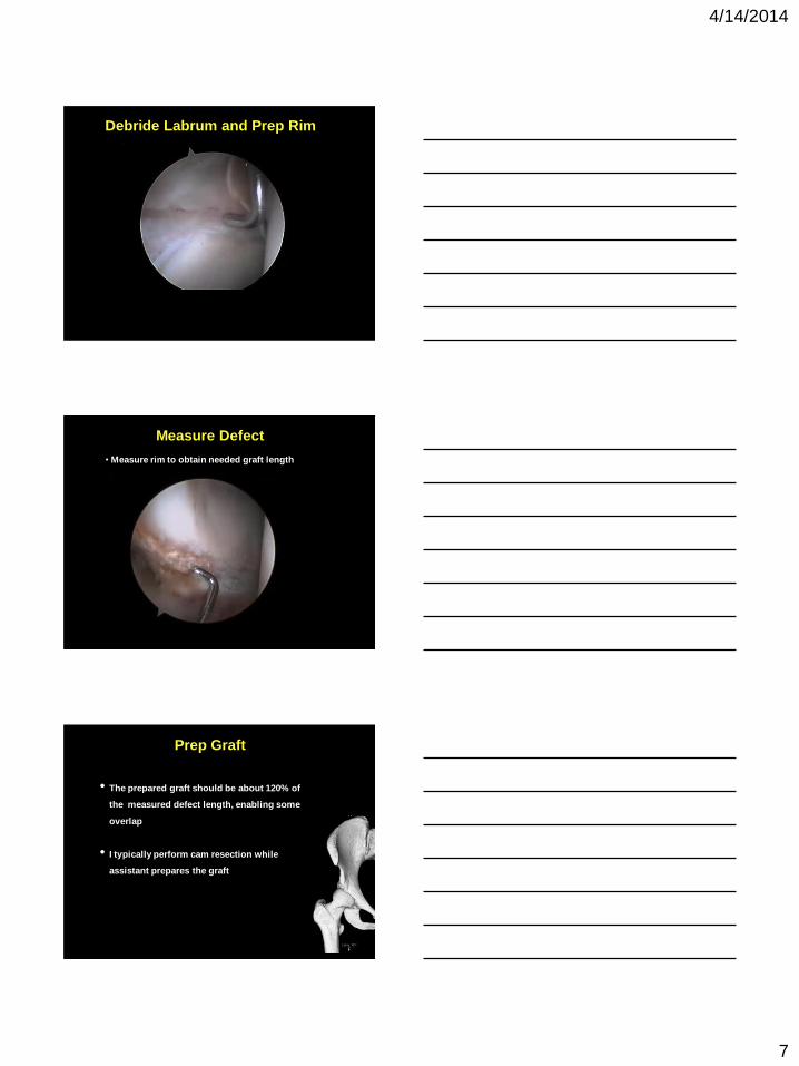
• During labral debridement, ensure you leave good
edges of native labrum at the ends of the defect
for graft-labrum anastomosis
• Make sure to reduce pincer bone and prepare the
rim for proper anchor hole drilling (check w/ and
w/o traction)
Labral Reconstruction Technique
4/14/2014
7
Debride Labrum and Prep Rim
Measure Defect
• Measure rim to obtain needed graft length
• The prepared graft should be about 120% of
the measured defect length, enabling some
overlap
• I typically perform cam resection while
assistant prepares the graft
Prep Graft
4/14/2014
8
TFL graft preparation
Use of fascia lata graft allows surgeon
to adjust length and width with ease
and, in my hands, is less apt to swell when compared to tendon grafts
Resorbable suture stitch is used to tighten and stiffen the rolled allograft
and is seen only on the 'capsular side' of the graft
Different colors for leading stitches (#2 MagnumWire®)
4/14/2014
9
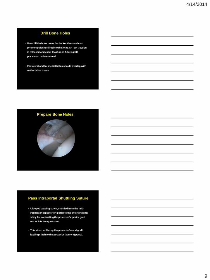
• Pre-drill the bone holes for the knotless anchors
prior to graft shuttling into the joint, AFTER traction
is released and exact location of future graft
placement is determined
• Far lateral and far medial holes should overlap with
native labral tissue
Drill Bone Holes
Prepare Bone Holes
PPass Intraportal Shuttling Suture
• A looped passing stitch, shuttled from the mid-
trochanteric (posterior) portal to the anterior portal
is key for controlling the posterior/superior graft
end as it is being secured.
• This stitch will bring the posterior/lateral graft
leading stitch to the posterior (camera) portal.
4/14/2014
10
PPass Shuttling Suture Between Portals
ShSShuttle posterior graft leading stitch
ImIntroduce Graft with Knotless Implant
• Leading suture ends of graft are passed into the
SpeedLock® HIP knotless implant, and this is led
into the joint over a slotted cannula.
4/14/2014
11
ImSecure and Orient Graft Anteriorly
Secure Graft Posteriorly
• Once the graft is secured anteriorly with 1-2
anchors, the camera is switched to the Ant. portal
and the Post. aspect of the graft is secured using a
SpeedLock HIP, forming a ‘bucket-handle’ labrum
and allowing easier control of the graft
Secure and Orient Posterior Graft with
SpeedLock HIP
4/14/2014
12
SuSuture Passing with SpeedStitch®
• The Speedstitch® suture passer allows for timely
and accurate passing of the sutures through the
base of the graft (or around it)
• Variable tension on each of the suture ends allows
control of the rotation of the graft to seat it on the
rim so that it reconstitutes the suction seal with the
femoral head
SKFixation and Orientation with
SpeedLock HIP Implant
SKFixation and Orientation with
SpeedLock HIP Implant
4/14/2014
13
PTechnique Pearl for 3 & 9 o’clock
• In cases where the reconstruction spans all the way
anterior or posterior where the rim is thin, I would
use small all-suture anchors (Q-FixTM) and use the
Speedstitch suture passer to grab onto the graft
and pass the suture through
PTechnique for 3 & 9 o’clock
AAnastomosis Stitches
• Side-to-side anastomosis of the graft to the native
labrum improves the stability of the graft at each end
and the continuous seal
• This can be done with the suture limb used with the
Speedlock HIP or via soft tissue resorbable suture
anastomosis
4/14/2014
14
AAnastomosis Stitches
Capsule Management
Post-Op Protocol Same as for labral repair
• PWB Crutches for 7-10 days
• Ride indoor trainer starting at 1-2 days post-op
• Designated rehab protocol
• Jogging and running allowed 10-12 weeks postop
4/14/2014
15
Advantages of this Technique
1. Speedlock HIP allows individual tensioning of suture ends which
controls the version of the graft and allows it to seat against the
femoral head
2. Knotless anchors decrease complexity of the case and speed up the
graft fixation
3. Two portal technique decreases tissue damage
4. Use of fascia lata graft allows surgeon to adjust length and width
with ease and, in my hands, is less apt to swell when compared to
tendon grafts
5. Speedstitch suture passer allows a quick and easy labral base grab
and fixation
Thank you!
Omer Mei-Dan, MD CU Sports Medicine
Division of Sports Medicine and Hip Preservation
University of Colorado School of Medicine, Department of Orthopedics