Page 1
This presentation is the intellectual property of the author.Contact them for permission to reprint and/or distribution.
42ND Annual Symposium on Sports Medicine UT Health Science Center San Antonio School of Medicine
January 22-24, 2015
PCL injuries- only 5 to 10% of all knee ligament injuries.
Incidence of PCL injury is 3%
At NFL Rookie Combines- 2% isolated PCL laxity - players usually unaware
Intra-articular / extra-synovial
38 mm length / 13 mm width
Fan-shaped structure narrowest at its midportion widest at its origin on the MFC (32mm in AP diameter)
Compact insertion @ posterior tibial shelf- 1 cm distal to the tibial plateau
Page 2
This presentation is the intellectual property of the author.Contact them for permission to reprint and/or distribution.
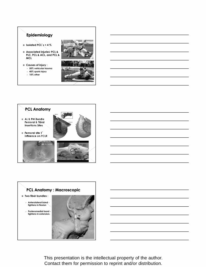
Isolated PCL’s < 4 %
Associated injuries: PCL & PLC, PCL & ACL, and PCL & MCL
Causes of injury :› 50% vehicular trauma› 40% sports injury› 10% other
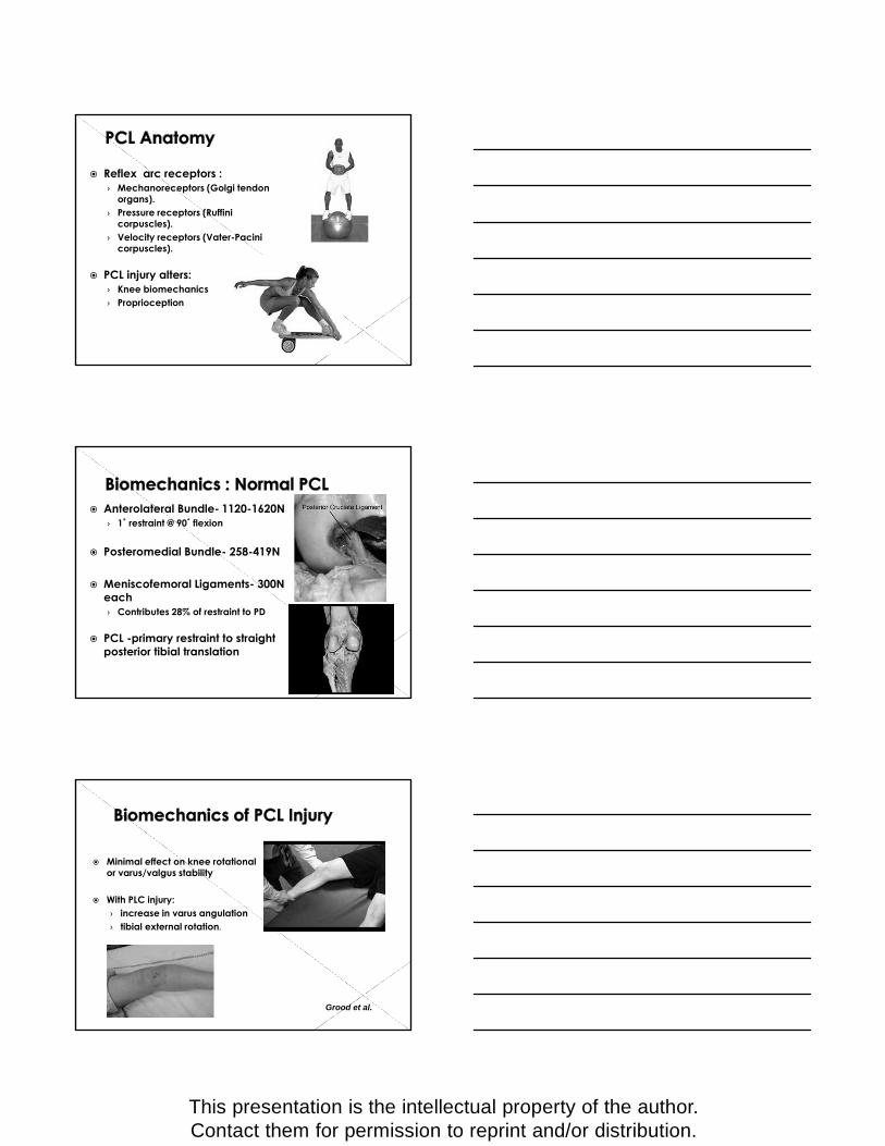
AL & PM Bundle Femoral & Tibial Insertions Sites
Femoral site 1˚ influence on PCLR
Two fiber bundles :
› Anterolateral band -tightens in flexion
› Posteromedial band -tightens in extension.
Page 3
This presentation is the intellectual property of the author.Contact them for permission to reprint and/or distribution.
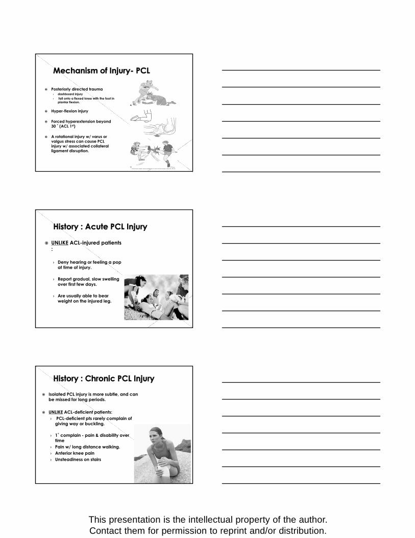
Reflex arc receptors :› Mechanoreceptors (Golgi tendon
organs).› Pressure receptors (Ruffini
corpuscles).› Velocity receptors (Vater-Pacini
corpuscles).
PCL injury alters:› Knee biomechanics› Proprioception
Anterolateral Bundle- 1120-1620N› 1˚ restraint @ 90˚ flexion
Posteromedial Bundle- 258-419N
Meniscofemoral Ligaments- 300N each › Contributes 28% of restraint to PD
PCL -primary restraint to straight posterior tibial translation
Minimal effect on knee rotational or varus/valgus stability
With PLC injury:› increase in varus angulation › tibial external rotation.
Grood et al.
Page 4
This presentation is the intellectual property of the author.Contact them for permission to reprint and/or distribution.
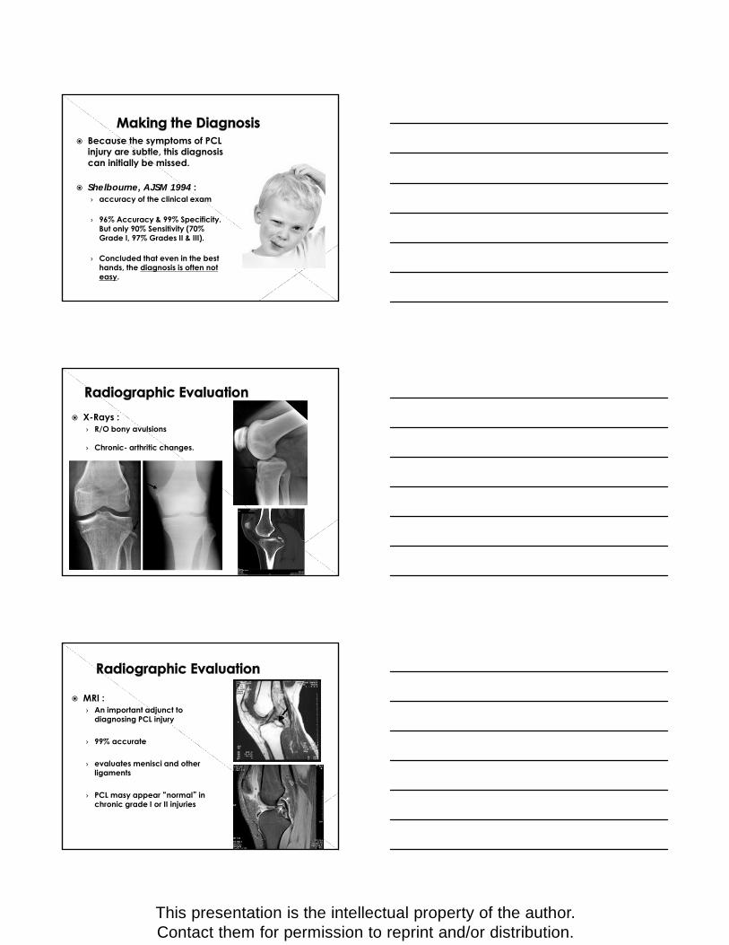
Posteriorly directed trauma › dashboard injury › fall onto a flexed knee with the foot in
plantar flexion.
Hyper-flexion injury
Forced hyperextension beyond 30 ˚ (ACL 1st)
A rotational injury w/ varus or valgus stress can cause PCL injury w/ associated collateral ligament disruption.
UNLIKE ACL-injured patients :
› Deny hearing or feeling a pop at time of injury.
› Report gradual, slow swelling over first few days.
› Are usually able to bear weight on the injured leg.
Isolated PCL injury is more subtle, and can be missed for long periods.
UNLIKE ACL-deficient patients:› PCL-deficient pts rarely complain of
giving way or buckling.
› 1˚ complain - pain & disability over time
› Pain w/ long distance walking.› Anterior knee pain› Unsteadiness on stairs
Page 5
This presentation is the intellectual property of the author.Contact them for permission to reprint and/or distribution.
Abrasions/ecchymosis @ tibial tubercle
suspect PCL injury
Mild-Moderate swelling
Posterior knee pain
Typically lack 10-20˚ of Knee flexion
Careful NV exam
Beware of subtle Multi-ligament Injuries
Suspect vascular injury angiogram.
Posterior Drawer :
› The most sensitive and specific test.
› Performed at 90 degrees of knee flexion.
› In normal knees, the anterior border of the tibial plateau is 1 cm anterior to the femoral condyles. Note direction of force
Page 6
This presentation is the intellectual property of the author.Contact them for permission to reprint and/or distribution.
Posterior Drawer
› Grade I : anterior tibial stepoff is only 5 mm.
› Grade II : there is no stepoff.› Grade III : tibial crest lies
posterior to the condyles.
A Grade III drawer usually combined ligamentous injury, most often PCL and PLC.
Posterior Sag Sign:
› Supine -knee flexed 90 degrees, the tibia sags posterior subluxation
› Acutely, can be limited by quads spasm, effusion and pretibial swelling.
Godfrey test :› A modification of the posterior sag test › hip and knee both flexed to 90 ˚› Gravity accentuates the posterior subluxation.
Page 7
This presentation is the intellectual property of the author.Contact them for permission to reprint and/or distribution.
Quadriceps Active Test :
› The quads contracted against resistance- knee flexed between 70 and 90 degrees.
› With PCL tear-isometric quads contraction reduces the tibia.
› This test is usually too painful to perform acutely, but is helpful with chronic cases.
Reverse pivot-shift test :› + if reduction sensation is appreciated as the
flexed, ext. rotated knee is extended with a valgus stress.
Assessing the PL Corner :› Dial Testing› Hughston ER/recurvatum
test
Assessing the ACL : › Lachman, Anterior drawer,
Pivot shift
Assessing the collateral ligaments :› Varus/valgus stress testing
at 30 and 0 degrees
**Occurs in 50-90% of PCL injuries
Page 8
This presentation is the intellectual property of the author.Contact them for permission to reprint and/or distribution.
Because the symptoms of PCL injury are subtle, this diagnosis can initially be missed.
Shelbourne, AJSM 1994 :› accuracy of the clinical exam
› 96% Accuracy & 99% Specificity.But only 90% Sensitivity (70% Grade I, 97% Grades II & III).
› Concluded that even in the best hands, the diagnosis is often not easy.
X-Rays :› R/O bony avulsions
› Chronic- arthritic changes.
MRI :› An important adjunct to
diagnosing PCL injury
› 99% accurate
› evaluates menisci and other ligaments
› PCL masy appear “normal” in chronic grade I or II injuries
Page 9
This presentation is the intellectual property of the author.Contact them for permission to reprint and/or distribution.
Originally thought benign course with neglect
Progressive disability and DJD› Primarily Medial & PF compartments
Shelbourne et al, 1999:› 88% of patients > 4 year- x-ray
evidence of DJD.› Return to Sport: 50% same level/ 33%
lower level/ 17 % changed sports› No correlation between grade of laxity
& DJD
Parolie & Bergfeld, 1986:› (+) correlation between improved
scores & quad strength› No correlation between laxity & RTS
› Return to Sport: av 6 wks post-injury 68% same level 16% lower level 16 % no sports
FACTORS:
› Acute vs. chronic.› Degree of laxity.› Associated injuries.› Symptoms and complaints.› Patient’s activity level and
demands.
Page 10
This presentation is the intellectual property of the author.Contact them for permission to reprint and/or distribution.
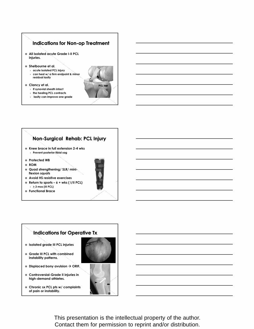
All isolated acute Grade I-II PCL injuries.
Shelbourne et al.› acute isolated PCL injury› can heal w/ a firm endpoint & minor
residual laxity
Clancy et al.› If synovial sheath intact› the healing PCL contracts› laxity can improve one grade
Knee brace in full extension 2-4 wks› Prevent posterior tibial sag
Protected WB ROM Quad strengthening/ SLR/ mini-
flexion squats Avoid HS resistive exercises Return to sports – 6 + wks ( I/II PCL)
› > 3 mos (III PCL) Functional Brace
Isolated grade III PCL injuries
Grade III PCL with combined instability patterns.
Displaced bony avulsion ORIF.
Controversial-Grade II injuries in high-demand athletes.
Chronic sx PCL pts w/ complaints of pain or instability.
Page 11
This presentation is the intellectual property of the author.Contact them for permission to reprint and/or distribution.
Acute reconstructions outcomes > chronic
No graft type superior› Achilles Allograft- most popular
Most PCLR have residual laxity› Improve 1+ grade
Single Bundle PCLR
Double Bundle PCLR
Trans-tibial Technique
Inlay Technique
“Killer turn”
› Difficult to effectively tension graft.
› predisposes graft to fraying and elongation.
Page 12
This presentation is the intellectual property of the author.Contact them for permission to reprint and/or distribution.
Avoid “Killer turn” Tibial inlay technique› Bergfeld et al: less posterior tibial
translation & graft degradation vs Trans-Tibial
› Biomechanical cadaver model› Clinically- no advantage
Trans-Tibial vs Inlay: › Clinical studies-No differences› Seon & Song, 2006› MacGillivray, 2006› Song et al., 2014
Double -bundle PCLR› More closely
reproduce native PCL› Biomechanical studies-
improved vs single bundle
› Bergfeld (AJSM 2005)- w/ inlay technique SB=DB
Comparative clinical studies (DB vs SB)› Houe & Jorgensen› Nyland et al› Wang et al› No signif. differences
Acute Isolated PCL Injury
Grade I or II
Non-operative TX-2-4 wk extension splint-Quads sets-Gradual return to activity
Grade III
Young/Athletic or Avulsion Injury
Non-operative Tx-4 wk extension splint-avoid post tibial sublux-Quad sets-limit activity
Operative Tx-ORIF Avulsion Fx-Single vs Double Bundle
NO YES
Page 13
This presentation is the intellectual property of the author.Contact them for permission to reprint and/or distribution.
Acute “Combined” PCL Injury
Operative Tx-Surgery < 2wks- acute repair collaterals-Single bundle PCLR- dislocated knee-Consider single bundle augment for gr II
PCL
“Combined”-PLC (+/- LCL)-MCL/PMCACL (+/- collaterals/knee dislocation)
Chronic PCL Injury
Grade I or II
Non-operative TX-Quads sets-activity modification
Grade III
Sx pain or instability
Operative Tx-Double Bundle PCLR Operative Tx
-Biplanar osteotomy-Staged PCLR
NO
YES
Malalignment?YES
NO
Trend toward poorer results with chronic injury› Sekiya et al:
75% N/NN acute/subacute grp 40% N/NN chronic group
Worse functional scores with chondrosis at time of injury› PCLR does not prevent progression› Hermans et al:
60% chondral injury› Strobel et al:
45% chondral injury with PCL 37% MFC/ 34% patella
Page 14
This presentation is the intellectual property of the author.Contact them for permission to reprint and/or distribution.
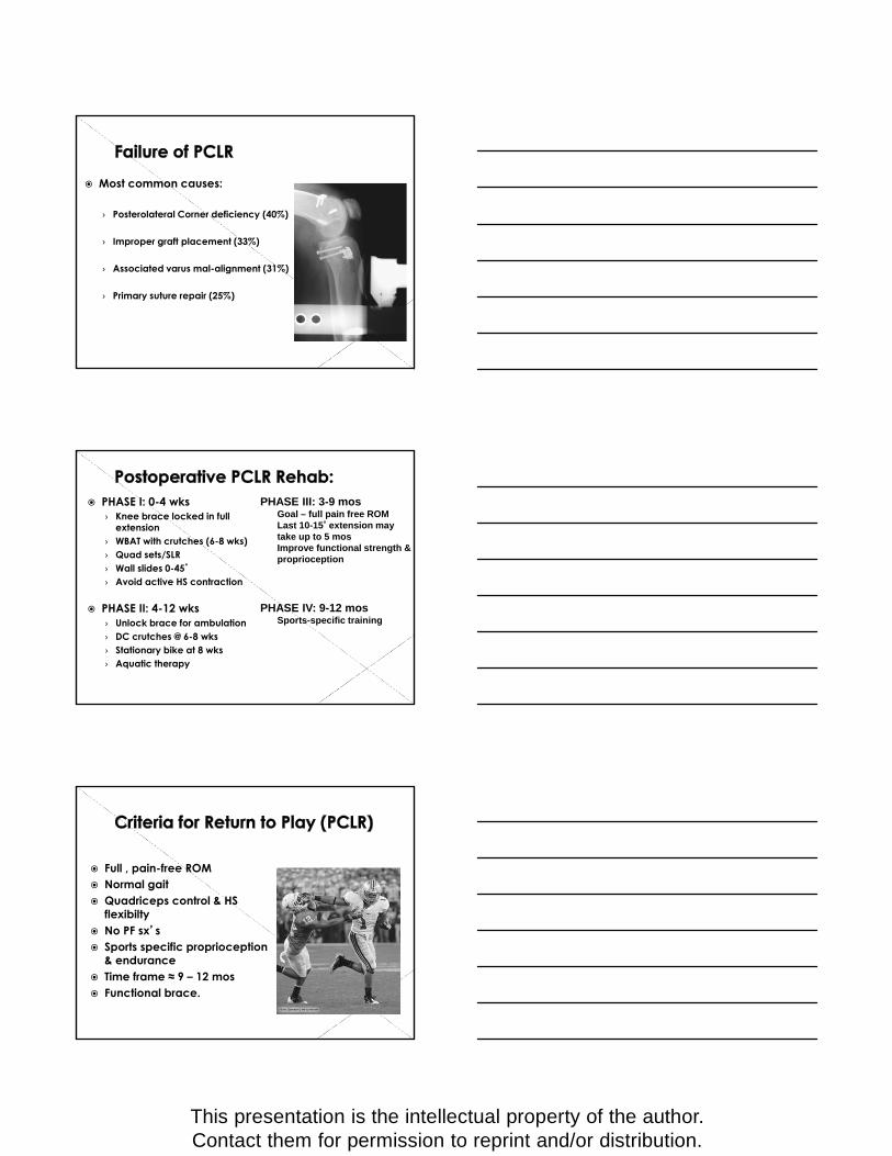
Most common causes:
› Posterolateral Corner deficiency (40%)
› Improper graft placement (33%)
› Associated varus mal-alignment (31%)
› Primary suture repair (25%)
PHASE I: 0-4 wks› Knee brace locked in full
extension › WBAT with crutches (6-8 wks)› Quad sets/SLR› Wall slides 0-45˚› Avoid active HS contraction
PHASE II: 4-12 wks› Unlock brace for ambulation› DC crutches @ 6-8 wks› Stationary bike at 8 wks› Aquatic therapy
PHASE III: 3-9 mosGoal – full pain free ROMLast 10-15˚ extension may take up to 5 mosImprove functional strength & proprioception
PHASE IV: 9-12 mosSports-specific training
Full , pain-free ROM Normal gait Quadriceps control & HS
flexibilty No PF sx’s Sports specific proprioception
& endurance Time frame ≈ 9 – 12 mos Functional brace.
Page 15
This presentation is the intellectual property of the author.Contact them for permission to reprint and/or distribution.
PCL Injuries less common than ACL
Presentation, mechanism of injury & disability- unlike ACL
Grade I/II injuries- tx’d effectively w/o surgery
Post-surgery functional results not as consistent as ACLR
Beware of combined ligamentous injuries