5 End-of-chapter test 1 a Explain why a large multicellular organism such as a human needs a transport system,
whereas a single-celled organism such as Paramecium can exist without one. [3] b Explain the difference between:
i a single and a double circulatory system ii a closed and an open circulatory system. [4]
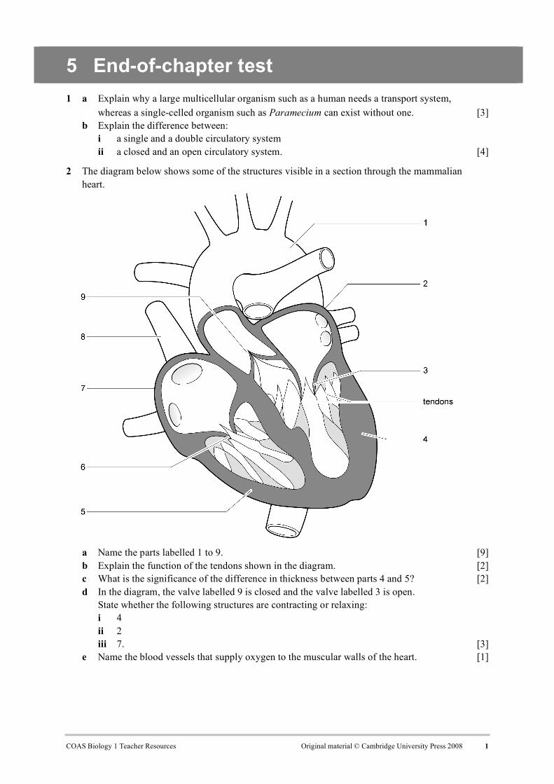
2 The diagram below shows some of the structures visible in a section through the mammalian heart.
a Name the parts labelled 1 to 9. [9] b Explain the function of the tendons shown in the diagram. [2] c What is the significance of the difference in thickness between parts 4 and 5? [2] d In the diagram, the valve labelled 9 is closed and the valve labelled 3 is open.
State whether the following structures are contracting or relaxing: i 4 ii 2 iii 7. [3]
e Name the blood vessels that supply oxygen to the muscular walls of the heart. [1]
3 The diagram below shows a section through a mammalian heart and some of the structures involved in coordination of the heart beat.
Structure X is the origin of waves of electrical excitation that spread through the heart muscle. The time taken for a wave of excitation to spread across the heart from X was measured. Recordings were made at positions A to G. The results are shown in the table below.
Position Time/ms
A 0
B 12
C 32
D 45
E 130
F 150
G 175
a Name the structures labelled X, Y and Z. [3] b Using the information in the diagram and table, explain how the route taken by the wave of
excitation coordinates the contraction of the heart muscle. [6]
4 The diagram below shows the pressure changes in the heart during the cardiac cycle.
a Identify the letters on the graph that correspond to the following events: i the ventricle beginning to contract ii the aortic semilunar valve opening iii the aortic semilunar valve closing iv the bicuspid valve opening. [4]
b From the graph, calculate the heart rate in beats per minute. [2] c Explain what an electrocardiogram (ECG) is. [2]
5 The diagram below shows cross-sections of three different types of mammalian blood vessel, A, B and C.
a Name the three types of blood vessel. [3] b Explain how the middle layer of tissue in blood vessel A is adapted for the function of
the vessel. [2] c State two ways in which blood vessel C is adapted to allow the formation of tissue fluid. [2] d What is the function of tissue fluid? [2] e Approximately 80% of tissue fluid is reabsorbed by the venous end of a capillary bed.
6 Explain how each of the following features of a red blood cell is an adaptation for its function. a its small size (diameter about 7 µm) [2] b its biconcave disc shape [2] c the lack of a nucleus [2] d the presence of the enzyme carbonic anhydrase in the cell [2]
7 The diagram below shows oxygen dissociation curves for human haemoglobin at two partial pressures of carbon dioxide. The effect of a high partial pressure of carbon dioxide on the oxygen dissociation of haemoglobin is called the Bohr effect.
a Explain how the shape of the oxygen dissociation curve at low partial pressure of carbon
dioxide is related to the function of the haemoglobin in carrying oxygen from the lungs to the respiring tissues. [3]
b If the partial pressure of oxygen in the lungs is 11 kPa, use the graph to find the percentage saturation of the haemoglobin with oxygen in the lungs. [1]
c In an actively respiring muscle, the partial pressure of oxygen is 2 kPa. Use the graph to find the percentage saturation of the haemoglobin with oxygen in the muscle tissue. [1]
d Explain why the Bohr effect is of importance to actively respiring tissues. [3] e Copy the axes in the diagram and sketch the curve for haemoglobin at a low partial pressure
of carbon dioxide. On your sketch graph, add another curve that shows the expected dissociation curve for the haemoglobin in the blood of a llama, a mammal species adapted to living at high altitudes. [2]
Total: 70
Score: %
Grade boundaries: 80% A, 70% B, 60% C, 50% D, 40% E