50 ENCORE! Pancreas: Surgical Pathology and Cytopathology of Pancreatic Neoplasms N Adsay MD Michelle Reid MD 2011 Annual Meeting – Las Vegas, NV AMERICAN SOCIETY FOR CLINICAL PATHOLOGY 33 W. Monroe, Ste. 1600 Chicago, IL 60603
Transcript
50 ENCORE! Pancreas: Surgical Pathology and Cytopathology of Pancreatic Neoplasms
N Adsay MD Michelle Reid MD
2011 Annual Meeting – Las Vegas, NV
AMERICAN SOCIETY FOR CLINICAL PATHOLOGY 33 W. Monroe, Ste. 1600
Chicago, IL 60603
50 ENCORE! Pancreas: Surgical Pathology and Cytopathology of Pancreatic Neoplasms With recent advances in imaging and interventional techniques and a dramatic decline in mortality and morbidity of pancreatic operations pancreatic resection specimens are now seen more often by surgical pathologists. Endoscopic ultrasound-guided fine needle aspiration has significantly increased the number of preoperative cytologic specimens reviewed by cytopathologists. Changes in terminology and classifications add to the new information one must now absorb. This session will provide an overview of challenges and practical clues in the diagnosis of pancreatobiliary specimens, with an algorithmic approach to differential diagnosis. Discussions will include: Pancreatic adenocarcinoma and its distinction from its mimics; Differential diagnosis of solid cellular/fleshy tumors of the pancreas; Clinicopathologic characteristics and biologic behavior of cystic tumors of the pancreas; Cytopathologic diagnosis of solid and cystic pancreatic lesions.
• Accurately differentiate problematic cases in surgical pathology of the pancreas, including solid-scirrhous lesions, solid/fleshy circumscribed lesions and cystic and traductal pancreatic tumors.
• Recognize the most common solid and cystic pancreatic lesions/tumors encountered on pancreatic fine needle aspiration; Evaluate the usefulness of ancillary studies in their cytologic diagnosis; Recognize key gastrointestinal contaminants in endoscopic tultrasound-guided pancreatic fine needle aspiration, that may lead to misdiagnosis on cytology.
• Distinguish and diagnose tumors of the ampulla, gallbladder and extrahepatic bile duct; Describe the grossing of pancreatoduodenectomy specimens.
FACULTY: N Adsay MD Michelle Reid MD Practicing Pathologists Surgical Pathology Surgical Pathology (GI, GU, Etc.) 3.0 CME/CMLE Credits Accreditation Statement: The American Society for Clinical Pathology (ASCP) is accredited by the Accreditation Council for Continuing Medical Education to provide continuing medical education (CME) for physicians. This activity has been planned and implemented in accordance with the Essential Areas and Policies of the Accreditation Council for Continuing Medical Education (ACCME). Credit Designation: The ASCP designates this enduring material for a maximum of 3 AMA PRA Category 1 Credits™. Physicians should only claim credit commensurate with the extent of their participation in the activity. ASCP continuing education activities are accepted by California, Florida, and many other states for relicensure of clinical laboratory personnel. ASCP designates these activities for the indicated number of Continuing Medical Laboratory Education (CMLE) credit hours. ASCP CMLE credit hours are acceptable to meet the continuing education requirements for the ASCP Board of Registry Certification Maintenance Program. All ASCP CMLE programs are conducted at intermediate to advanced levels of learning. Continuing medical education (CME) activities offered by ASCP are acceptable for the American Board of Pathology’s Maintenance of Certification Program.
1
EUS‐Guided Fine Needle Aspiration and
Cytopathology of Cystic and Solid Lesions of the Pancreas
Michelle Reid, MDDepartment of PathologyEmory University Hospital
Atlanta, GA
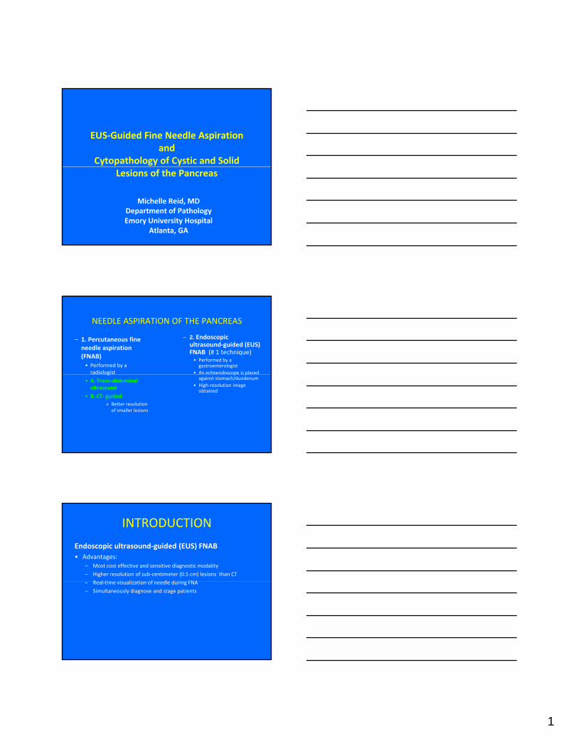
NEEDLE ASPIRATION OF THE PANCREAS
– 1. Percutaneous fine needle aspiration (FNAB) • Performed by a radiologist
– 2. Endoscopic ultrasound‐guided (EUS) FNAB (# 1 technique)• Performed by a gastroenterologist
• An echoendoscope is placed g
• A. Trans‐abdominal ultrasound
• B. CT‐ guided» Better resolution of smaller lesions
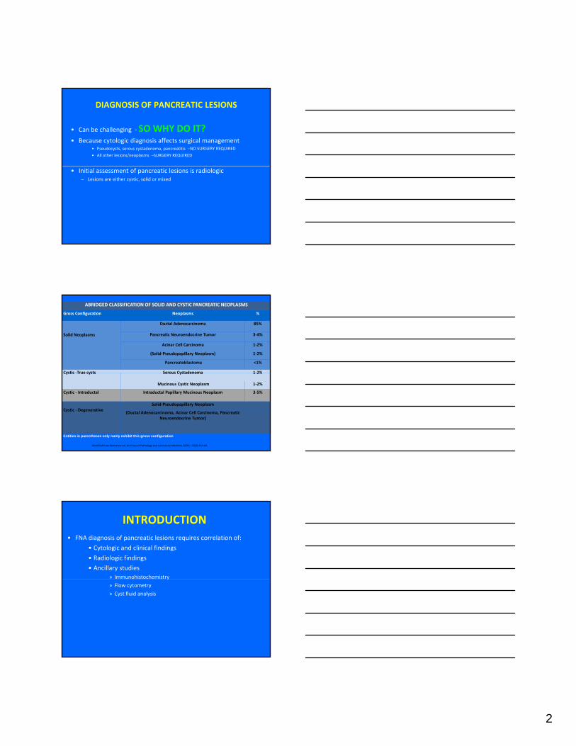
Entities in parentheses only rarely exhibit this gross configuration
Modified from Klimstra et al. Archives of Pathology and Laboratory Medicine 2009; 133(3):454‐64.
INTRODUCTION• FNA diagnosis of pancreatic lesions requires correlation of:
• Cytologic and clinical findings
• Radiologic findings
• Ancillary studies» Immunohistochemistryy
» Flow cytometry
» Cyst fluid analysis
3
INTRODUCTIONAccuracy of FNAB
• Immediate cytologic assessment IS A MUST• Best performed by cytopathologist or cytotechnologist
• Reduces number of passes
• Reduces inadequate samples
• Saves time and money
Accuracy of Pancreatic FNAB• Sensitivity for detecting malignancy:
– 86% ‐ 98% for percutaneous FNAB – 75% ‐ 94% for EUS‐FNAB
• Specificity for both approaches 100% • False‐negative and false‐positive results occur• False negative results more common• False‐negative results more common
SAMPLE PREPARATION AND EVALUATION
• 1. Prepare air‐dried and alcohol‐fixed slidesA. Air‐dried slides
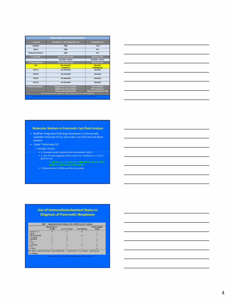
4. Molecular Markers Negative for K‐rasmutationNegative for LOH mutation Low quantity/quality DNA
K‐rasmutation LOH mutation or
High quantity/quality DNACEA, carcinoembryonic antigen; CA, cancer antigen; LOH, loss of heterozygosity.
Modified from Weinstein L, Pancreas. In: Cytology. Diagnostic principles and clinical correlates. 3rd Ed. E. Cibas, B. Ducatman editors
Molecular Markers in Pancreatic Cyst Fluid Analysis
• RedPath Integrated Pathology developed a commercially available molecular kit for pancreatic cyst fluid and cell block analysis
• Called “PathFinderTG”
– Includes 3 tests• 1 k ras gene point mutation (on chromosome 12p12)• 1. k‐ras gene point mutation (on chromosome 12p12)
• 2. Loss of heterozygosity (LOH) analysis for mutations in ≥ 2 of 15 genomic loci
» 1 and 2 are considered high amplitude mutations if they involves > 75% of total DNA content
• 3. Measurement of DNA quantity and quality
Use of Immunohistochemical Stains in Diagnosis of Pancreatic Neoplasms
Table from Klimstra et al. Archives of Pathology and Laboratory Medicine 2009; 133(3):454-64.[14]
5
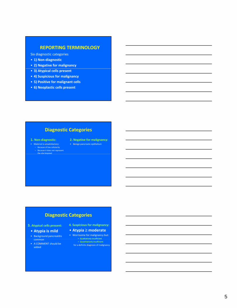
REPORTING TERMINOLOGYSix diagnostic categories
• 1) Non‐diagnostic
• 2) Negative for malignancy
• 3) Atypical cells present
• 4) Suspicious for malignancy
• 5) Positive for malignant cells
• 6) Neoplastic cells present
Diagnostic Categories
1. Non‐diagnostic: • Material is unsatisfactory:
– Because of low cellularity
– Because it does not represent the site biopsied
2. Negative for malignancy: • Benign pancreatic epithelium
p
Diagnostic Categories
3. Atypical cells present:
• Atypia is mild• Background pancreatitis
common
4. Suspicious for malignancy:
• Atypia ≥moderate • Worrisome for malignancy but:
• Qualitatively insufficientcommon
• A COMMENT should be added
Qualitatively insufficient
• Quantitatively insufficient
for a definite diagnosis of malignancy
6
Diagnostic Categories
5. Positive for malignant cells• Cells shows obvious malignant
features
6. Neoplastic cells present: • When the cells are obviously
“neoplastic” but not definitely benign or malignant
• e.g. Mucinous cystse.g. Mucinous cysts
Contaminants in Pancreatic FNAB• EUS‐FNAB introduces gastrointestinal (GI) tract contaminants
• GI tract contaminants include:– 1.Duodenal epithelium
– 2. Gastric epithelium
– 3. GI tract mucin
• Distinguishing GI contaminants from pancreatic ductal adenocarcinoma and neoplastic mucinous cysts can be challenging
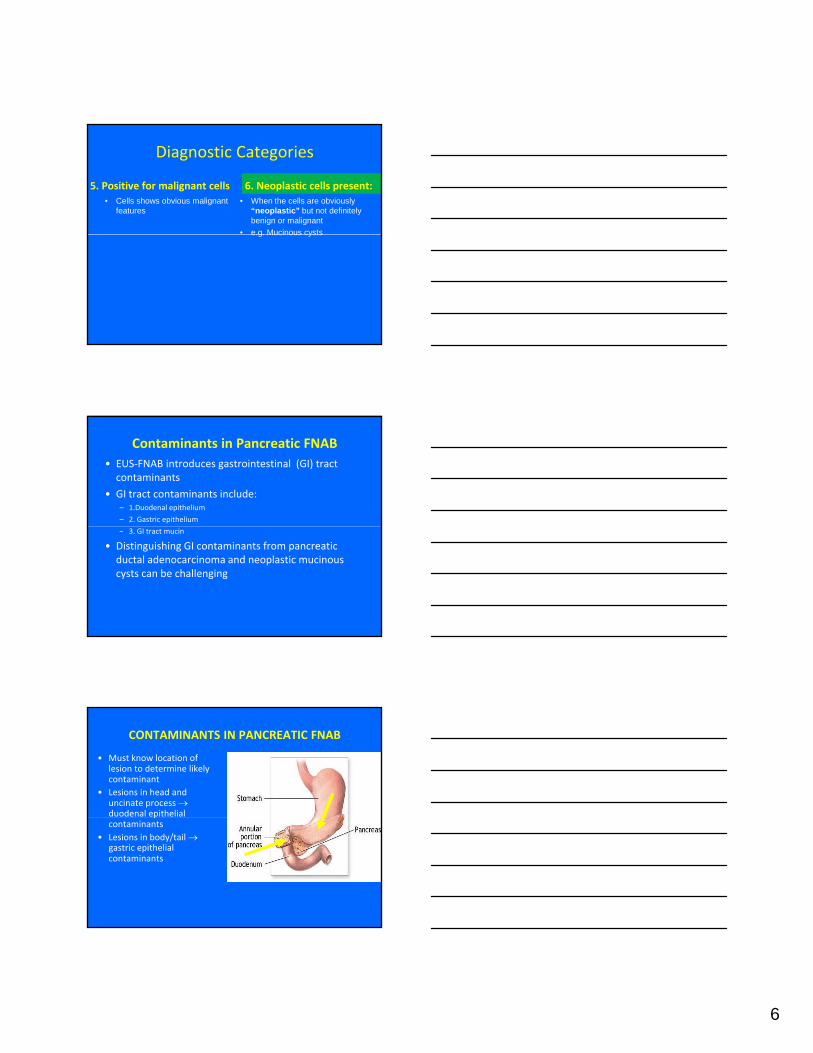
CONTAMINANTS IN PANCREATIC FNAB
• Must know location of lesion to determine likely contaminant
• Lesions in head and uncinate process →duodenal epithelial pcontaminants
• Lesions in body/tail →gastric epithelial contaminants
7
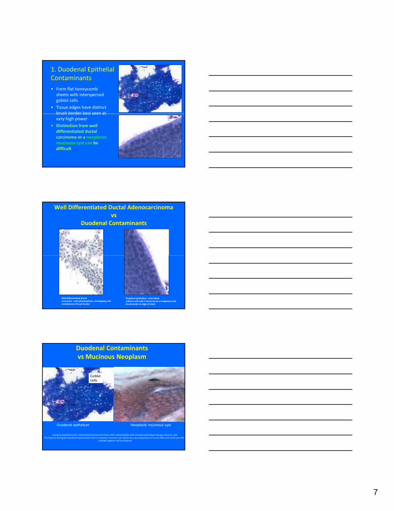
1. Duodenal Epithelial Contaminants
• Form flat honeycomb sheets with interspersed goblet cells
• Tissue edges have distinct brush border best seen atbrush border best seen at very high power
• Distinction from well differentiated ductal carcinoma or a neoplastic mucinous cyst can be difficult
Well Differentiated Ductal Adenocarcinoma vs
Duodenal Contaminants
Well differentiated ductal carcinoma ‐ note pleomorphism, overlapping cells and absence of brush border
Duodenal epithelium –note blanduniform cells with 2‐dimensional arrangement and brush border at edge of sheet
Duodenal Contaminants vs Mucinous Neoplasm
Goblet cells
Duodenal epithelium has 2‐dimensional honeycomb sheets with isolated goblet cells interspersed between benign columnar cellsThis helps to distinguish duodenal contaminants from a neoplastic mucinous cyst which has a pure population of mucin‐filled cells which are often
crowded together and overlapping
Duodenal epithelium Neoplastic mucinous cyst
8
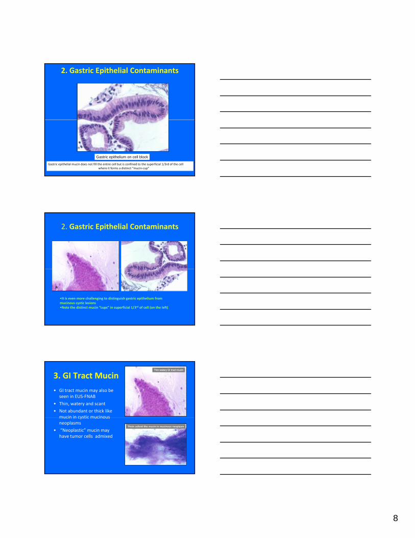
2. Gastric Epithelial Contaminants
Gastric epithelial mucin does not fill the entire cell but is confined to the superficial 1/3rd of the cellwhere it forms a distinct “mucin‐cup”
Gastric epithelium on cell block
2. Gastric Epithelial Contaminants
•It is even more challenging to distinguish gastric epithelium from mucinous cystic lesions•Note the distinct mucin “cups” in superficial 1/3rd of cell (on the left)
3. GI Tract Mucin
• GI tract mucin may also be seen in EUS‐FNAB
• Thin, watery and scant
• Not abundant or thick like mucin in cystic mucinous
Thin watery GI tract mucin
mucin in cystic mucinous neoplasms
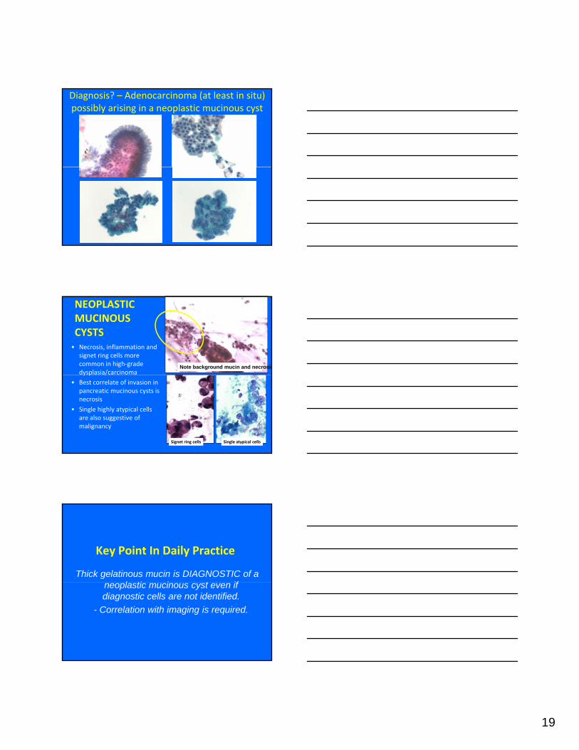
• “Neoplastic” mucin may have tumor cells admixed
Thick colloid-like mucin in mucinous neoplasm
9
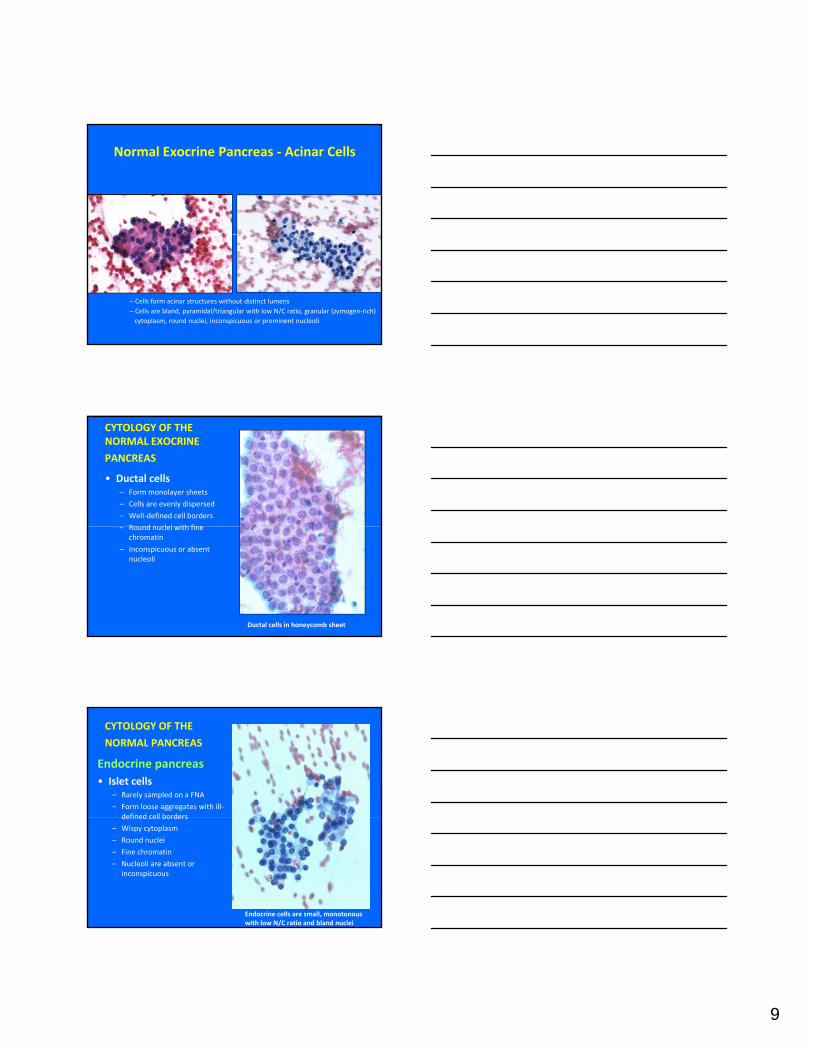
Normal Exocrine Pancreas ‐ Acinar Cells
– Cells form acinar structures without distinct lumens– Cells are bland, pyramidal/triangular with low N/C ratio, granular (zymogen‐rich) cytoplasm, round nuclei, inconspicuous or prominent nucleoli
CYTOLOGY OF THE NORMAL EXOCRINE
PANCREAS
• Ductal cells– Form monolayer sheets
– Cells are evenly dispersed
– Well‐defined cell borders
Round nuclei with fine– Round nuclei with fine chromatin
– Inconspicuous or absent nucleoli
Ductal cells in honeycomb sheet
CYTOLOGY OF THE
NORMAL PANCREAS
Endocrine pancreas• Islet cells
– Rarely sampled on a FNA
– Form loose aggregates with ill‐defined cell bordersdefined cell borders
– Wispy cytoplasm
– Round nuclei
– Fine chromatin
– Nucleoli are absent or inconspicuous
Endocrine cells are small, monotonous with low N/C ratio and bland nuclei
10
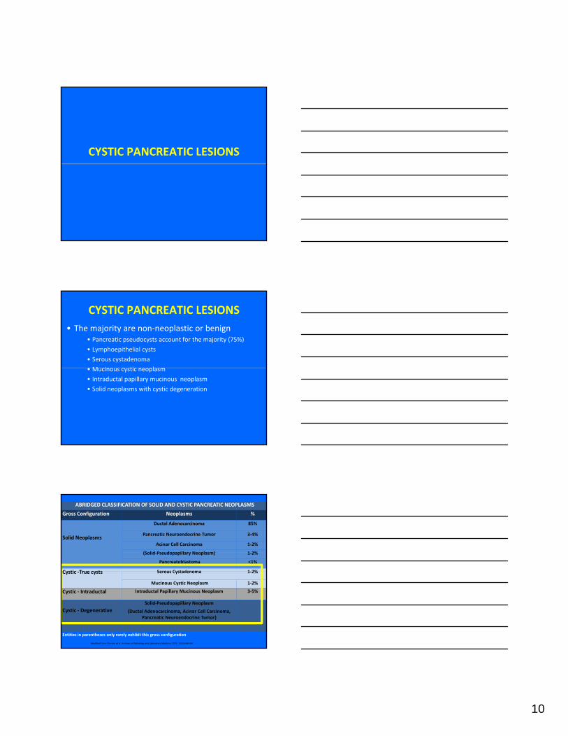
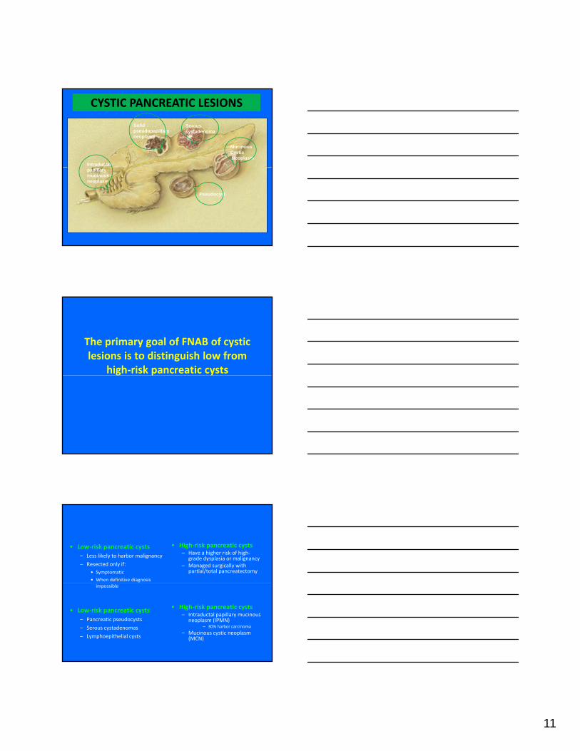
CYSTIC PANCREATIC LESIONS
CYSTIC PANCREATIC LESIONS• The majority are non‐neoplastic or benign
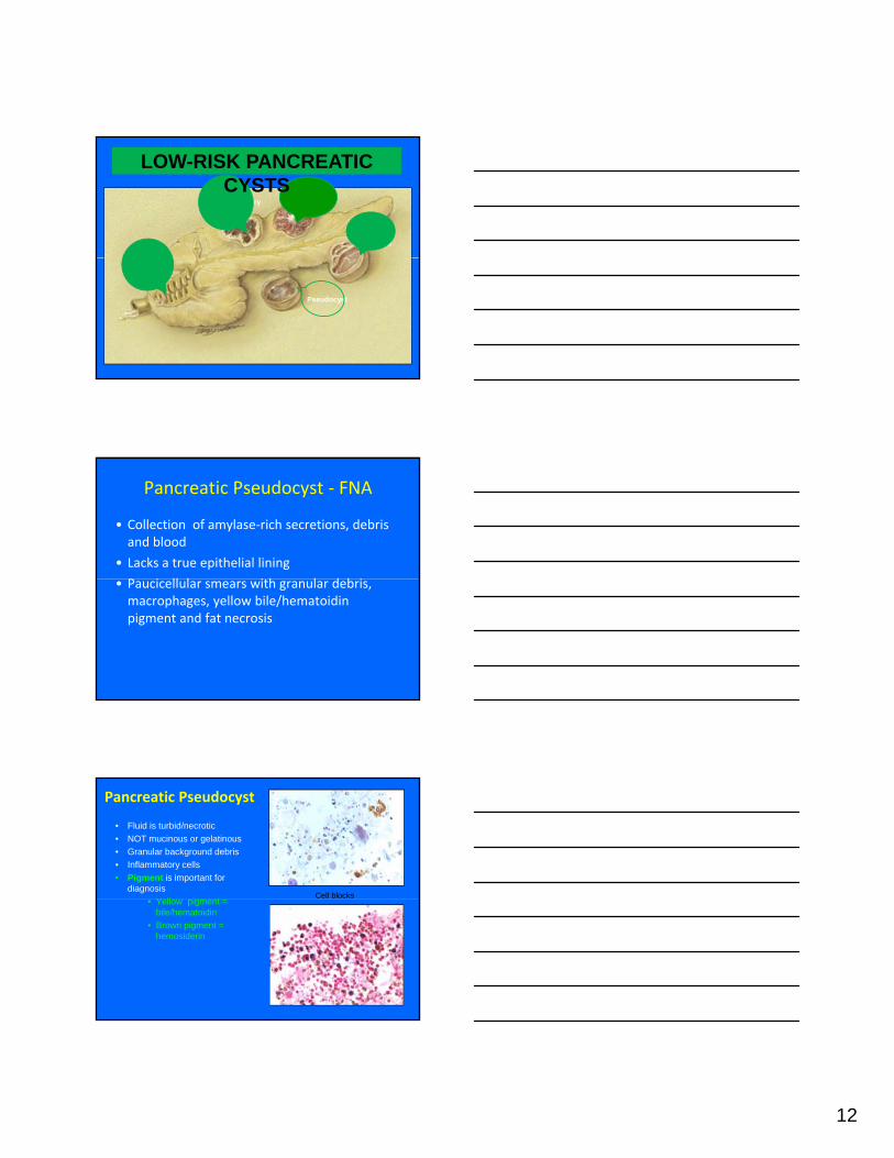
• Pancreatic pseudocysts account for the majority (75%)
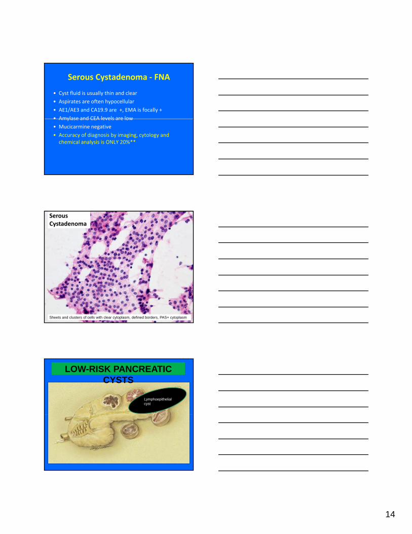
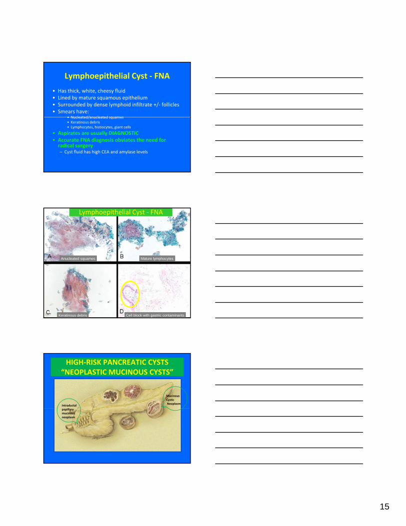
• Lymphoepithelial cysts
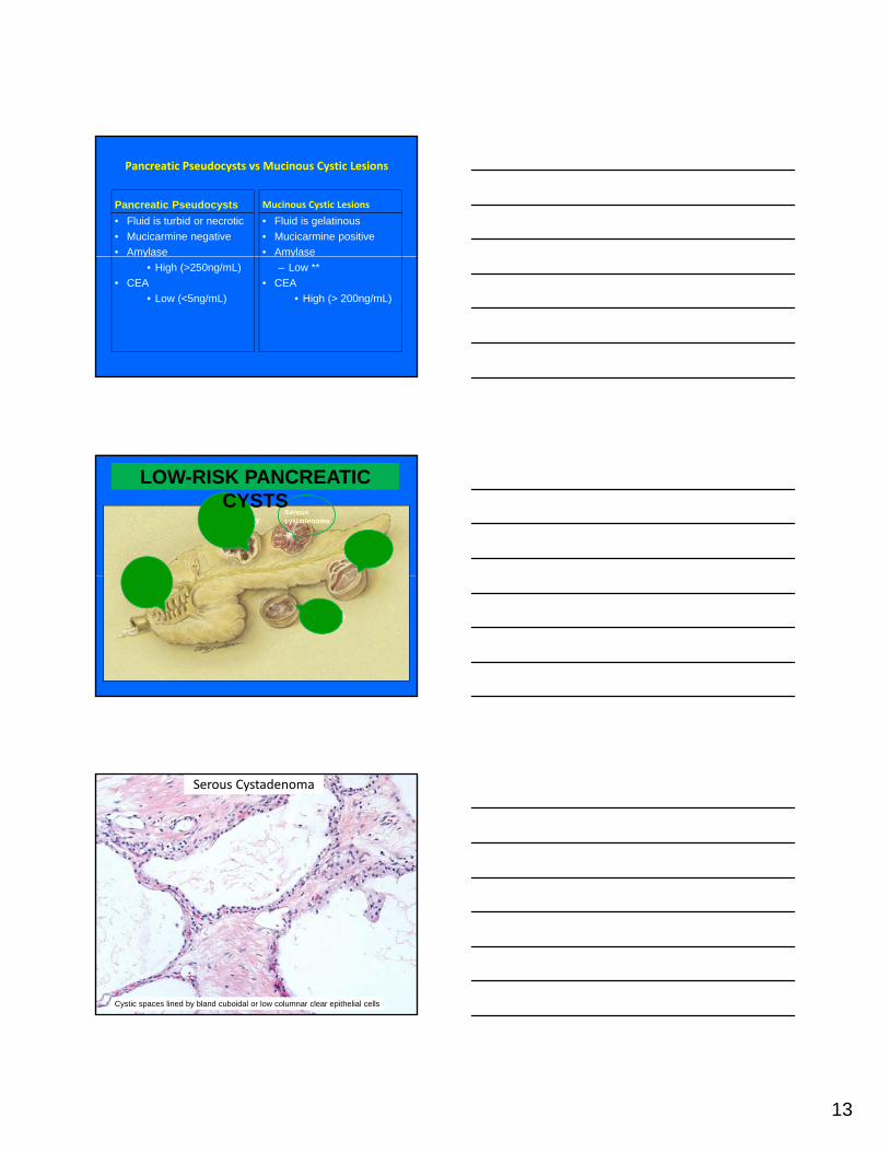
• Serous cystadenoma
M i ti l• Mucinous cystic neoplasm
• Intraductal papillary mucinous neoplasm
• Solid neoplasms with cystic degeneration
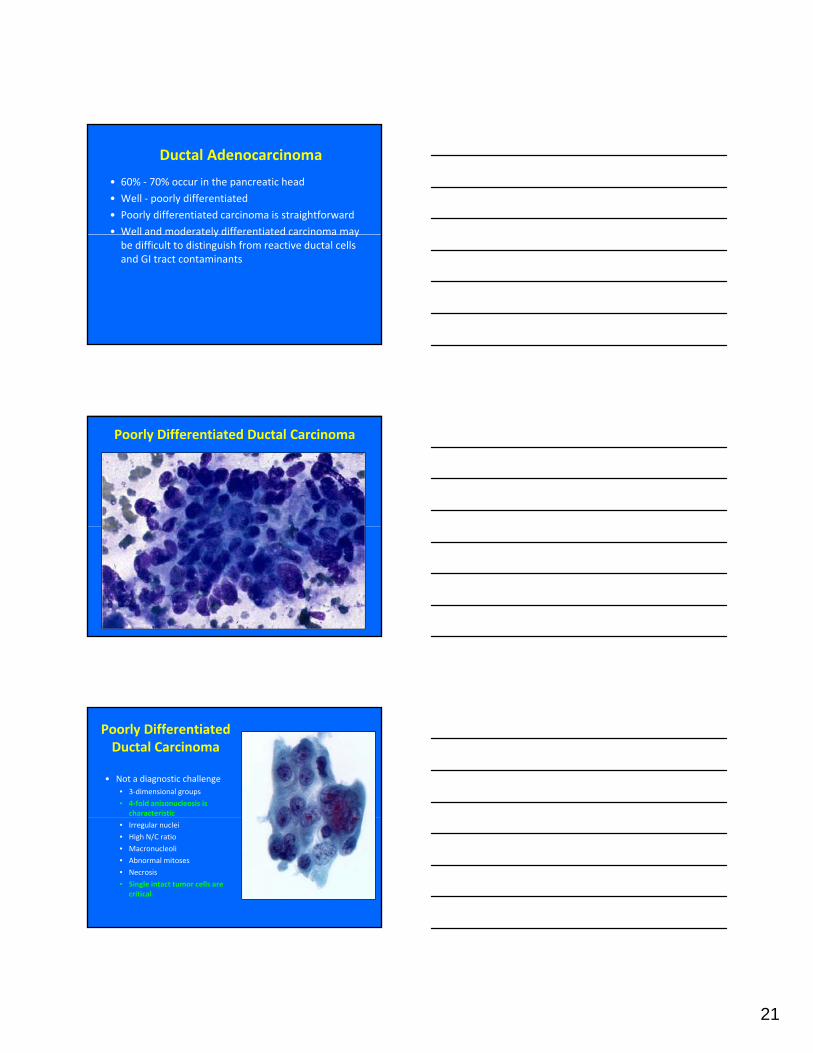
ABRIDGED CLASSIFICATION OF SOLID AND CYSTIC PANCREATIC NEOPLASMS
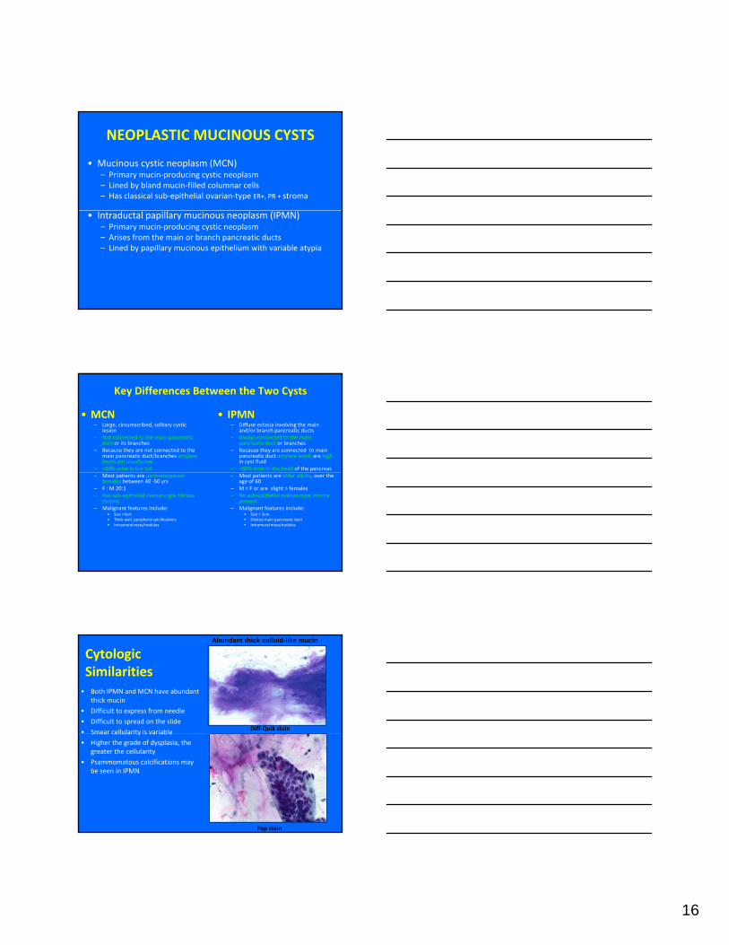
• Mucinous cystic neoplasm (MCN)– Primary mucin‐producing cystic neoplasm – Lined by bland mucin‐filled columnar cells– Has classical sub‐epithelial ovarian‐type ER+, PR + stroma
• Intraductal papillary mucinous neoplasm (IPMN)– Primary mucin‐producing cystic neoplasm – Arises from the main or branch pancreatic ducts– Lined by papillary mucinous epithelium with variable atypia
Key Differences Between the Two Cysts
• MCN– Large, circumscribed, solitary cystic
lesion– Not connected to the main pancreatic
duct or its branches – Because they are not connected to the
main pancreatic duct/branches amylase levels are usually low
– >90% arise in the tail
• IPMN – Diffuse ectasia involving the main
and/or branch pancreatic ducts– Always connected to the main
pancreatic duct or branches – Because they are connected to main
pancreatic duct amylase levels are highin cyst fluid
– >80% arise in the head of the pancreas – Most patients are perimenopausal
females between 40 ‐50 yrs– F : M 20:1 – Has sub‐epithelial ovarian‐type fibrous
Entities in parentheses only rarely exhibit this gross configuration
Modified from Klimstra et al. Archives of Pathology and LaboratoryMedicine 2009; 133(3):454‐64.
21
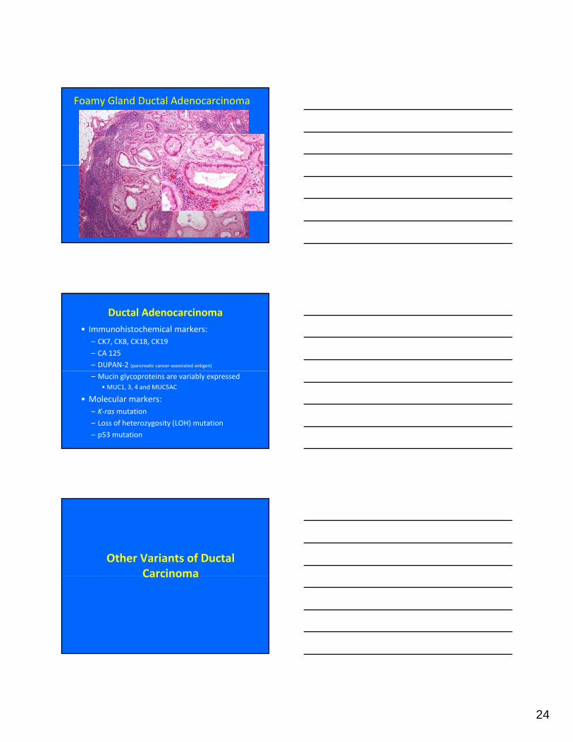
Ductal Adenocarcinoma
• 60% ‐ 70% occur in the pancreatic head
• Well ‐ poorly differentiated
• Poorly differentiated carcinoma is straightforward
• Well and moderately differentiated carcinoma mayWell and moderately differentiated carcinoma may be difficult to distinguish from reactive ductal cells and GI tract contaminants
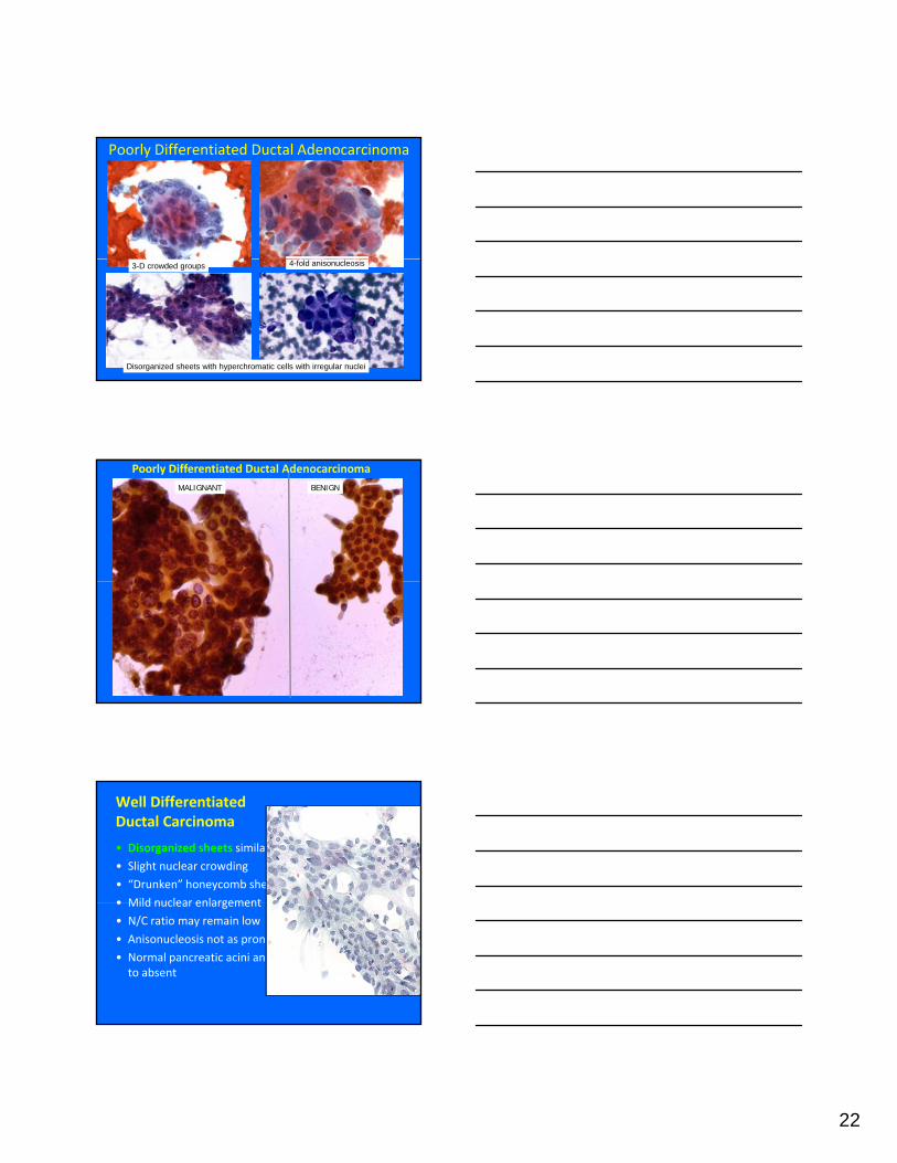
Poorly Differentiated Ductal Carcinoma
Poorly Differentiated Ductal Carcinoma
• Not a diagnostic challenge• 3‐dimensional groups
• 4‐fold anisonucleosis is characteristic
• Irregular nuclei
• High N/C ratio
• Macronucleoli
• Abnormal mitoses
• Necrosis
• Single intact tumor cells are critical
22
Poorly Differentiated Ductal Adenocarcinoma
3-D crowded groups 4-fold anisonucleosis
Disorganized sheets with hyperchromatic cells with irregular nuclei
MALIGNANT BENIGN
Poorly Differentiated Ductal Adenocarcinoma
Well Differentiated Ductal Carcinoma
• Disorganized sheets similar to normal ductal cells
• Associated with ductal adenocarcinoma (40% of cases)• May be focal or predominant
• Prognosis is controversial:• Some say not as dismal as ductal carcinoma
• Others say more aggressive than ductal carcinoma• Others say more aggressive than ductal carcinoma
• Mean survival ≤ 12 months – related to quantity of ductal carcinoma
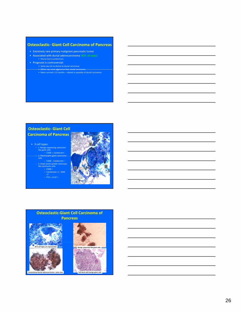
Osteoclastic‐ Giant CellCarcinoma of Pancreas
• 3 cell types:– 1. Benign‐appearing osteoclast‐
like giant cells– CD68 +, Cytokeratin ‐
– 2 Pleomorphic giant carcinoma
3
2. Pleomorphic giant carcinoma cells
– CD68 ‐, Cytokeratin –– 3. Small ovoid‐spindle histiocyte‐
like carcinoma cells – CD68 +– Cytokeratin +/‐, EMA +/‐
– P53 +, ki‐67 +
1 2
Osteoclastic‐Giant Cell Carcinoma of Pancreas
Conventional ductal adenocarcinoma –same case Cell block with benign giant cells
Benign osteoclast‐like giant cellsAll 3 cell types are represented
27
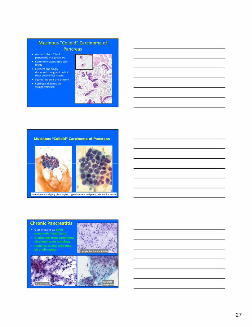
Mucinous “Colloid” Carcinoma of Pancreas
• Accounts for <1% of pancreatic malignancies
• Commonly associated with IPMN
• Clusters and singly dispersed malignant cells indispersed malignant cells in thick colloid‐like mucin
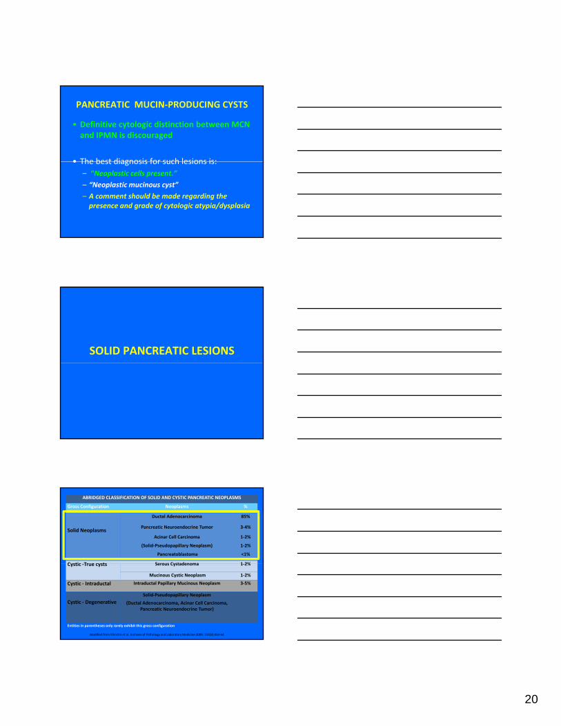
• Signet ring cells are present• Cytologic diagnosis is
straightforward
Mucinous “Colloid” Carcinoma of PancreasMucinous “Colloid” Carcinoma of Pancreas
Note clusters of slightly pleomorphic, hyperchromatic malignant cells in thick mucin
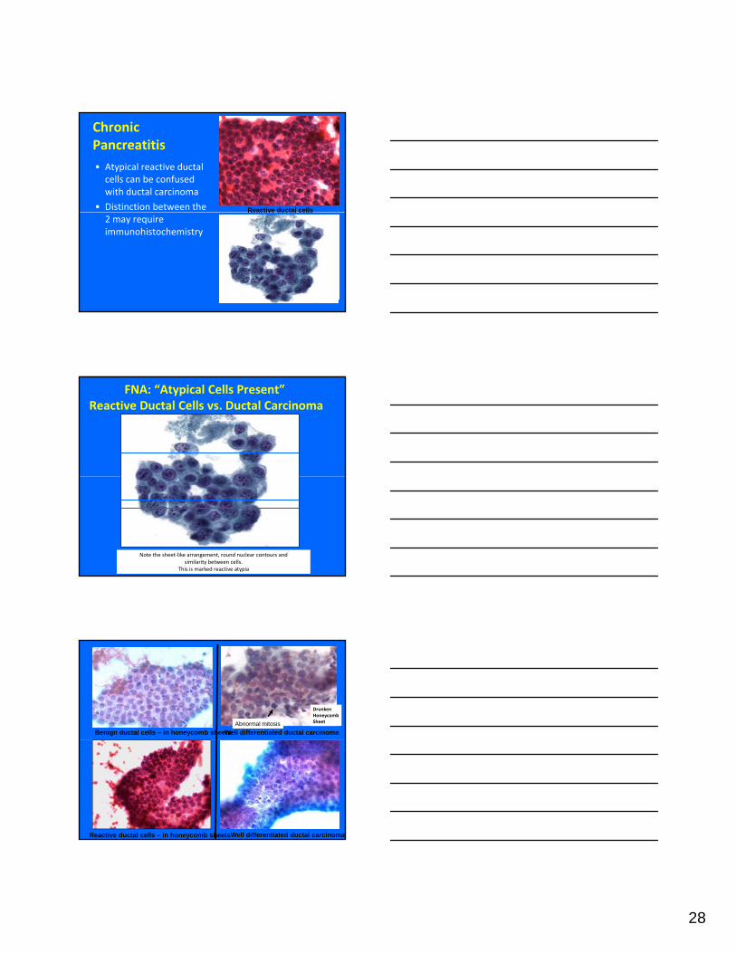
Chronic Pancreatitis• Can present as solid
pancreatic head lesion• Distinction from carcinoma
challenging on radiology• Reactive ductal cells may
be challengingLymphohistiocytic infiltrates
Stromal fragmentsFat necrosis
28
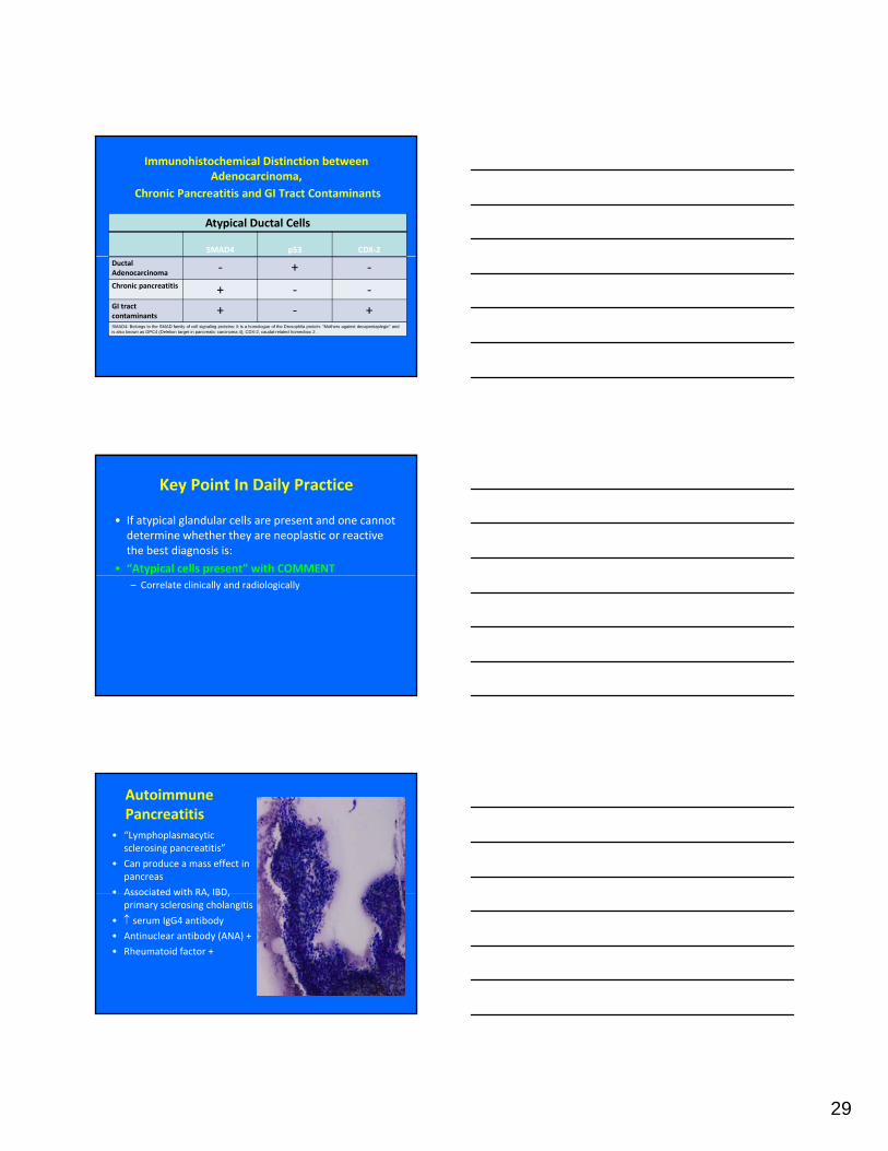
Chronic Pancreatitis• Atypical reactive ductal cells can be confused with ductal carcinoma
• Distinction between the Reactive ductal cells2 may require immunohistochemistry
Reactive ductal cells
FNA: “Atypical Cells Present”Reactive Ductal Cells vs. Ductal Carcinoma
Note the sheet‐like arrangement, round nuclear contours and similarity between cells.
This is marked reactive atypia
Benign ductal cells – in honeycomb sheets
Drunken HoneycombSheet
Well differentiated ductal carcinomaAbnormal mitosis
Reactive ductal cells – in honeycomb sheetsWell differentiated ductal carcinoma
29
Immunohistochemical Distinction between Adenocarcinoma,
Chronic Pancreatitis and GI Tract Contaminants
Atypical Ductal Cells
SMAD4 p53 CDX‐2
Ductal Adenocarcinoma ‐ + ‐Chronic pancreatitis + ‐ ‐GI tract contaminants + ‐ +SMAD4: Belongs to the SMAD family of cell signaling proteins: It is a homologue of the Drosophila protein: “Mothers against decapentaplegic“ and is also known as DPC4 (Deletion target in pancreatic carcinoma 4); CDX-2, caudal-related homeobox 2 .
Key Point In Daily Practice
• If atypical glandular cells are present and one cannot determine whether they are neoplastic or reactive the best diagnosis is:
• “Atypical cells present” with COMMENT– Correlate clinically and radiologically
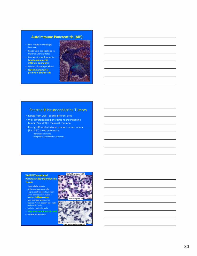
Autoimmune Pancreatitis
• “Lymphoplasmacytic sclerosing pancreatitis”
• Can produce a mass effect in pancreas
• Associated with RA IBDAssociated with RA, IBD, primary sclerosing cholangitis
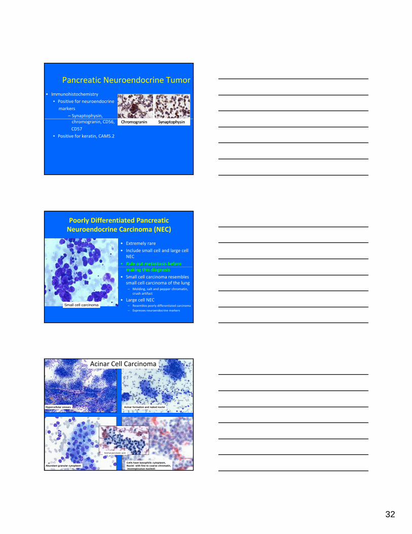
– Pancreatic enzymes:• Lipase, trypsin, chymotrypsin, α 1 anti‐chymotrypsin, elastase and h h li A2phospholipase A2
• Do not confuse trypsin with α‐1‐antitrypsin
• α‐1‐ antitrypsin is not a very useful stain for acinar cells
• Because it also stains solid‐pseudopapillary neoplasm and pancreatic neuroendocrine tumors
Trypsin
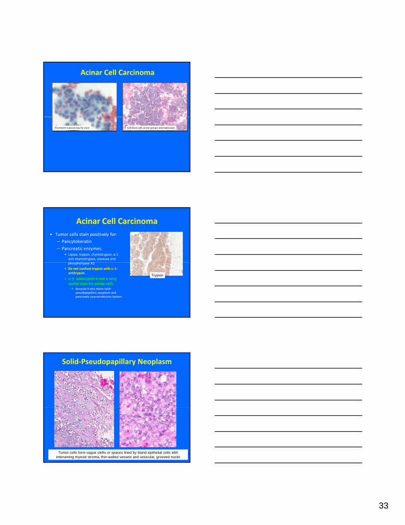
Solid‐Pseudopapillary Neoplasm
Tumor cells form vague clefts or spaces lined by bland epithelial cells with intervening myxoid stroma, thin-walled vessels and vesicular, grooved nuclei
34
Solid‐Pseudopapillary Neoplasm
– Rare low‐ grade solid and cystic pancreatic
– Usually arises in the pancreatic tail
– Almost exclusively in women (F:M 9:1)
– Third decade (mean age 28 years) or adolescence
– Cytologic features are distinctive
– Accurate diagnosis often made before resection
Solid‐Pseudopapillary Neoplasm
Monomorphic small cells with high N/C Complex branching papillae gratio, fine chromatin, nuclear grooves
g
Papillary fronds with central myxoid stroma (on Diff Quik stain) and blood vessel (H&E)
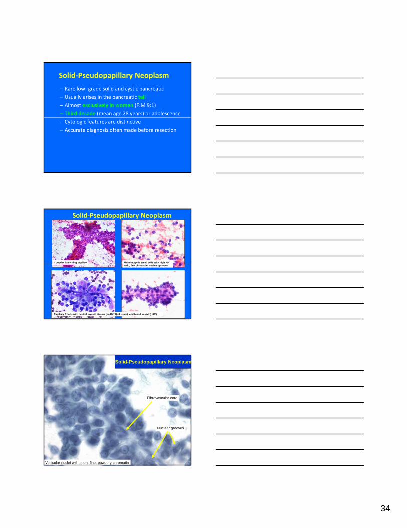
Solid-Pseudopapillary Neoplasm
Fibrovascular core
Vesicular nuclei with open, fine, powdery chromatin
Nuclear grooves
35
Solid‐Pseudopapillary Neoplasm
• Immunohistochemistry is characteristic and diagnostic• Positive for vimentin:
• Frequently negative for cytokeratin
• Positive for:• Neuron specific enolase
• CD56 (variable)
• CD10
• β‐catenin (nuclear)
• Progesterone receptor
• α ‐1‐ antitrypsin is not helpful because it is positive in SPN, acinar cell carcinoma and pancreatic NETs
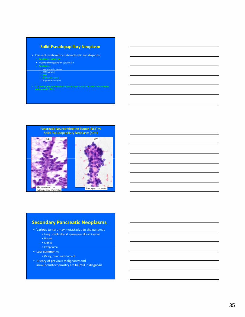
Pancreatic Neuroendocrine Tumor (NET) vs Solid‐Pseudopapillary Neoplasm (SPN)
NET SPN
Fine, open chromatinFibrovascular core Salt-n-pepper chromatin
Secondary Pancreatic Neoplasms• Various tumors may metastasize to the pancreas
• Lung (small cell and squamous cell carcinoma)
• Breast
• Kidney
L h• Lymphoma
• Less commonly:• Ovary, colon and stomach
• History of previous malignancy and immunohistochemistry are helpful in diagnosis
36
SUMMARY ‐ PANCREATIC FNAB
– Cytologic evaluation of pancreatic lesions is complex
– Knowledge of types and location of the most common solid and cystic lesions is helpful in diagnosis
– Correlation with clinical, imaging data is paramount
– Cytopathologist /cytotechnologist’s presence during immediate evaluation improves adequacy and diagnostic yield
– Be mindful that chronic pancreatitis and GI tract contaminants (in EUS‐FNAB) may simulate carcinoma
References• 1. Shen j. Kindelberger dw Pancreas and biliary tree. In: Cibas ES DB, editors. Cytology: Diagnostic Principles and Clinical Correlates. 3rd Ed.
Livingstone Elsevier; 2007 p251‐298.• 3. Centeno BA. Fine needle aspiration biopsy of the pancreas. Clin Lab Med 1998;18(3):401‐27, v‐vi.• 4. Ekberg O, Bergenfeldt M, Aspelin P, Genell S, Lindholm K, Nilsson P, Sigurjonsson S. Reliability of ultrasound‐guided fine‐needle biopsy of
pancreatic masses. Acta Radiol 1988;29(5):535‐9.• 5. Erickson RA, Garza AA. Impact of endoscopic ultrasound on the management and outcome of pancreatic carcinoma. Am J Gastroenterol
2000;95(9):2248‐54.• 6. Pitman MB, Deshpande V. Endoscopic ultrasound‐guided fine needle aspiration cytology of the pancreas: a morphological and multimodal
approach to the diagnosis of solid and cystic mass lesions. Cytopathology 2007;18(6):331‐47.• 7. Mitsuhashi T, Ghafari S, Chang CY, Gu M. Endoscopic ultrasound‐guided fine needle aspiration of the pancreas: cytomorphological
evaluation with emphasis on adequacy assessment, diagnostic criteria and contamination from the gastrointestinal tract. Cytopathology 2006;17(1):34‐41.
• 8. Gonzalez Obeso E, Murphy E, Brugge W, Deshpande V. Pseudocyst of the pancreas: the role of cytology and special stains for mucin. Cancer Cytopathol 2009;117(2):101‐7.
• 9. Brandt KR, Charboneau JW, Stephens DH, Welch TJ, Goellner JR. CT‐ and US‐guided biopsy of the pancreas. Radiology 1993;187(1):99‐104.• 10 David O Green L Reddy V Kluskens L Bitterman P Attal H Prinz R Gattuso P Pancreatic masses: a multi institutional study of 364 fine• 10. David O, Green L, Reddy V, Kluskens L, Bitterman P, Attal H, Prinz R, Gattuso P. Pancreatic masses: a multi‐institutional study of 364 fine‐
needle aspiration biopsies with histopathologic correlation. Diagn Cytopathol 1998;19(6):423‐7.• 11. Di Stasi M, Lencioni R, Solmi L, Magnolfi F, Caturelli E, De Sio I, Salmi A, Buscarini L. Ultrasound‐guided fine needle biopsy of pancreatic
masses: results of a multicenter study. Am J Gastroenterol 1998;93(8):1329‐33.• 12. Robins DB, Katz RL, Evans DB, Atkinson EN, Green L. Fine needle aspiration of the pancreas. In quest of accuracy. Acta Cytol 1995;39(1):1‐
10.• 13. Levin DP, Bret PM. Percutaneous fine‐needle aspiration biopsy of the pancreas resulting in death. Gastrointest Radiol 1991;16(1):67‐9.• 14. Klimstra DS, Pitman MB, Hruban RH. An algorithmic approach to the diagnosis of pancreatic neoplasms. Arch Pathol Lab Med
2009;133(3):454‐64.• 15. Belsley NA, Pitman MB, Lauwers GY, Brugge WR, Deshpande V. Serous cystadenoma of the pancreas: limitations and pitfalls of endoscopic
ultrasound‐guided fine‐needle aspiration biopsy. Cancer 2008;114(2):102‐10.• 16. Jimenez‐Heffernan JA, Vicandi B, Lopez‐Ferrer P, Gonzalez‐Peramato P, Perez‐Campos A, Viguer JM. Fine needle aspiration cytology of
endocrine neoplasms of the pancreas. Morphologic and immunocytochemical findings in 20 cases. Acta Cytol 2004;48(3):295‐301.• 17. Gupta RK, Lallu S, Delahunt B. Fine‐needle aspiration cytology of metastatic clear‐cell renal carcinoma presenting as a solitary mass in the
head of the pancreas. Diagn Cytopathol 1998;19(3):194‐7.• 18. Le Borgne J, de Calan L, Partensky C. Cystadenomas and cystadenocarcinomas of the pancreas: a multiinstitutional retrospective study of
398 cases. French Surgical Association. Ann Surg 1999;230(2):152‐61.• 19. Stelow EB, Bardales RH, Shami VM, Woon C et al. Cytology of pancreatic acinar cell carcinoma. Diagn Cytopathol. 2006 May;34(5):367‐
72. • 20. Cohen MB, Egerter DP, Holly EA et al. Pancreatic adenocarcinoma: regression analysis to identify improved cytologic criteria. Diagn
Cytopathol 1991, 7:341‐345• 21. Mitchell ML, Carney CN. Cytologic criteria for the diagnosis of pancreatic carcinoma. Am J Clin Pathol 1985, 83:171‐176• 22. Robins DB, Katz RL, Evans DB et al. Fine needle aspiration of the pancreas. In quest of accuracy. Acta Cytol 1995. 39:1‐10.


![Pancreatic Cytopathology Cystic Lesions Cytol… · Cystic Lesions Cystic Lesions Of The Pancreas [Practical Issues] ... 1-2% of all pancreatic tumors LMP epithelial tumor of uncertain](https://static.documents.pub/doc/80x56/5f6d9c61a7374f61f46d815c/pancreatic-cytopathology-cystic-lesions-cytol-cystic-lesions-cystic-lesions-of.jpg)

