Page 1
Chapter 5 RESULTS AND DISCUSSION
79
5.1. Extraction
The extraction of Andrographis paniculata and Silybum marianum aerial
part (leaves and stem) was done with 70% ethanol. The percentage yield
obtained was 16.83 and 15.04 for A. paniculata and S. marianum
respectively (Table 5.1; Fig 5.1).
Table 5.1: Percentage yield of hydroalcoholic extract of A. paniculata and S. marianum
Solvent Plant Dry wt.
(g)
Yield
(g)
Time
(h)
Temperature
(˚C) % yield
70%
ethanol
A. paniculata 150 25.25 96 68 16.83
S. marianum 150 22.57 72 68 15.04
Fig. 5.1: Percentage yield of hydroalcoholic extract of A. paniculata and S. marianum
In the present study, hydroalcoholic solvent system was used for
the extraction, which contains 70% ethanol and 30% water. Ethanol is
16.83
15.04
0
5
10
15
20
A. paniculata S. marianum
% yield
Page 2
Chapter 5 RESULTS AND DISCUSSION
80
less polar in nature as compared to water, which is the most polar
solvent on earth. Therefore, both the polar as well as non-polar
compounds present in the plants are extracted out in the same solvent.
The yield of the extract depends, upon the type of solvent (low-polarity
solvents yield more lipophilic compounds, whereas alcoholic extract
yields both polar and non-polar compounds), the time given for
extraction, and the method used for the extraction. Soxhlet extraction
yields better amount of extract as compared to the extraction by flask
method in water bath. The main advantage of the Soxhlet technique is
that, it is an automatic and continuous method that does not require
much manipulation. It has also been shown to be very effective in terms
of extraction yield and therefore, often used as a reference methods.
Page 3
Chapter 5 RESULTS AND DISCUSSION
81
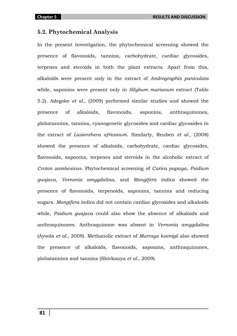
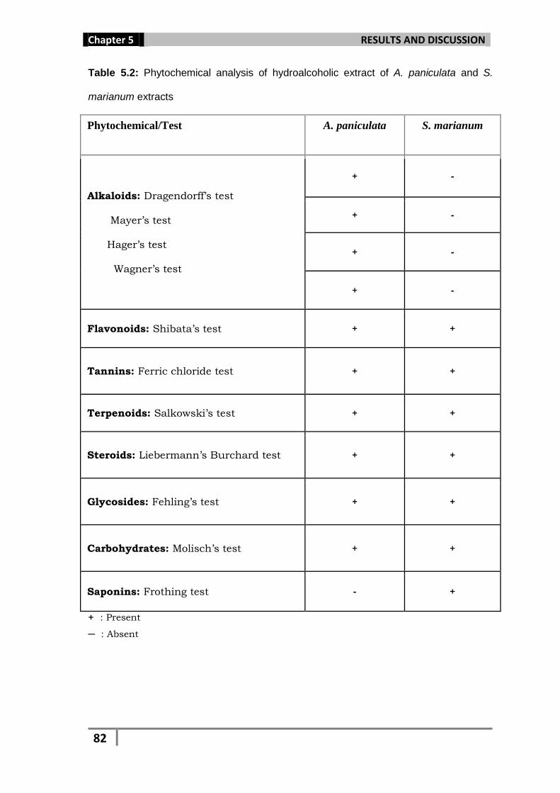
5.2. Phytochemical Analysis
In the present investigation, the phytochemical screening showed the
presence of flavonoids, tannins, carbohydrate, cardiac glycosides,
terpenes and steroids in both the plant extracts. Apart from this,
alkaloids were present only in the extract of Andrographis paniculata
while, saponins were present only in Silybum marianum extract (Table
5.2). Adegoke et al., (2009) performed similar studies and showed the
presence of alkaloids, flavonoids, saponins, anthraquinones,
plobatannins, tannins, cyanogenetic glycosides and cardiac glycosides in
the extract of Lasienthera africanum. Similarly, Reuben et al., (2008)
showed the presence of alkaloids, carbohydrate, cardiac glycosides,
flavonoids, saponins, terpenes and steroids in the alcoholic extract of
Croton zambesicus. Phytochemical screening of Carica papaya, Psidium
guajava, Vernonia amygdalina, and Mangifera indica showed the
presence of flavonoids, terpenoids, saponins, tannins and reducing
sugars. Mangifera indica did not contain cardiac glycosides and alkaloids
while, Psidium guajava could also show the absence of alkaloids and
anthraquinones. Anthraquinone was absent in Vernonia amygdalina
(Ayoola et al., 2008). Methanolic extract of Murraya koenigii also showed
the presence of alkaloids, flavonoids, saponins, anthraquinones,
plobatannins and tannins (Shivkanya et al., 2009).
Page 4
Chapter 5 RESULTS AND DISCUSSION
82
Table 5.2: Phytochemical analysis of hydroalcoholic extract of A. paniculata and S.
marianum extracts
Phytochemical/Test A. paniculata S. marianum
Alkaloids: Dragendorff’s test Mayer’s test Hager’s test Wagner’s test
+ -
+ -
+ -
+ -
Flavonoids: Shibata’s test + +
Tannins: Ferric chloride test + +
Terpenoids: Salkowski’s test + +
Steroids: Liebermann’s Burchard test + +
Glycosides: Fehling’s test + +
Carbohydrates: Molisch’s test + +
Saponins: Frothing test - +
+ : Present
─ : Absent
Page 5
Chapter 5 RESULTS AND DISCUSSION
83
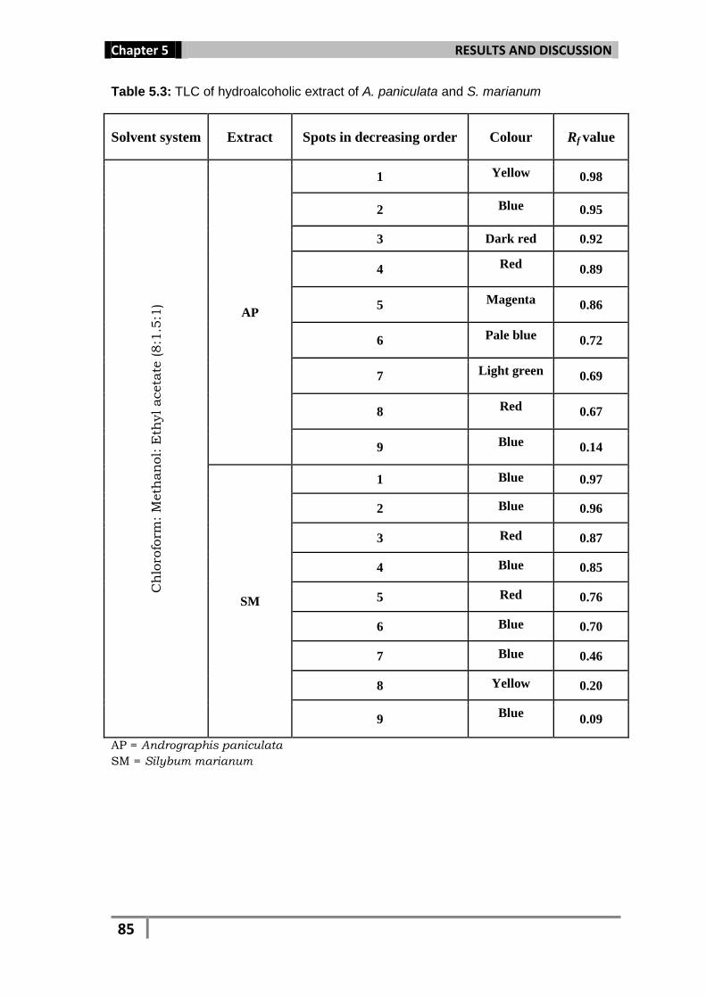
5.3. Thin Layer Chromatography (TLC)
Thin layer chromatography is a separation technique which is used to
separate out the components present in a given mixture. The
phytochemical test for both the extracts reveals the presence of
flavonoid, tannins, glycosides etc. which when subjected to TLC with
different solvent system gave different colored bands on separation. The
Rf value (Relative front) is calculated for each band as this value is
specific for each compound. Just like the Rf value for each amino acid
for a particular solvent is specific and comparable to the unknown
sample run on the same solvent. The result for TLC of both the extracts
showed maximum number of bands i.e., 9 for Andrographis paniculata
with Rf (Relative front) value 0.98, 0.95, 0.92, 0.89, 0.86, 0.72, 0.69,
0.67, and 0.14 respectively, and 8 for Silybum marianum with Rf value
0.97, 0.96, 0.87, 0.85, 0.76, 0.70, 0.46, 0.20, 0.09 respectively, in
Chloroform: Methanol: Ethyl acetate (8:1.5:1) solvent system (Fig. 5.2;
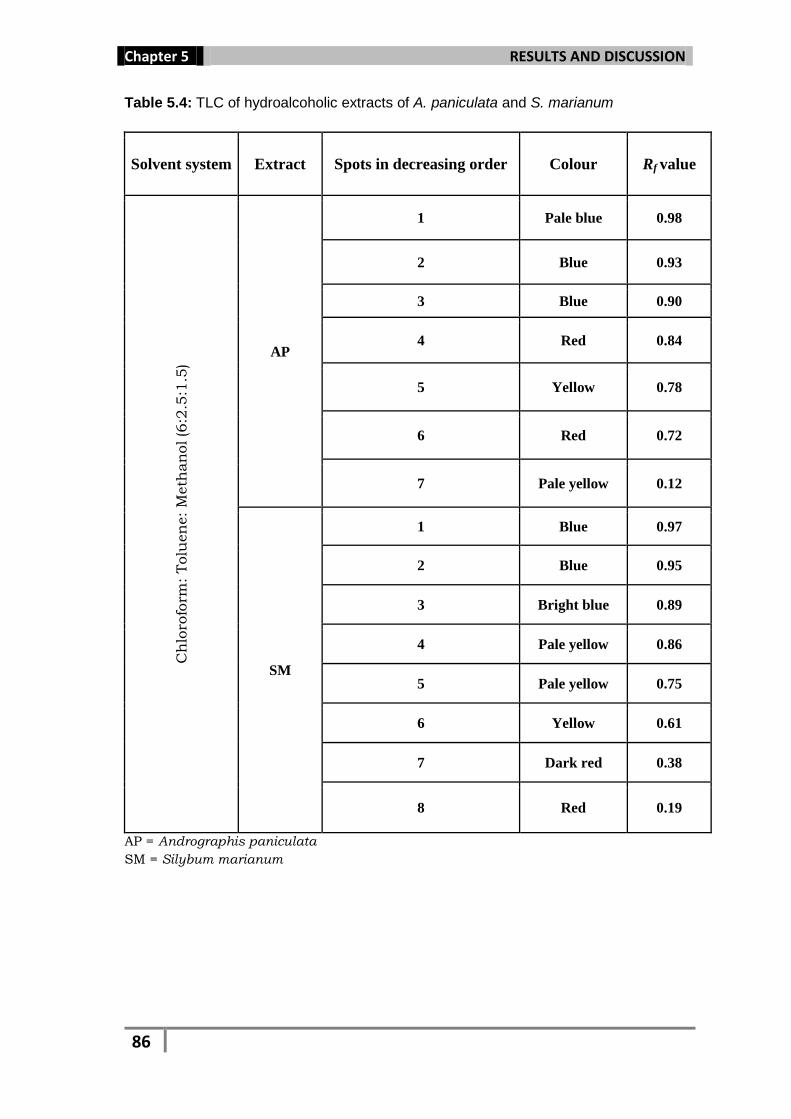
Table 5.3), while Chloroform: Toluene: Methanol (6:2.5:1.5) gave 7 colored
bands for A. paniculata with Rf value 0.98, 0.93, 0.90, 0.84, 0.78, 0.72
and 0.12 respectively and 8 colored bands for S. marianum with Rf value
0.97, 0.95, 0.89, 0.86, 0.75, 0.61, 0.38 and 0.19 respectively (Table 5.4).
Page 6
Chapter 5 RESULTS AND DISCUSSION
84
AP = Andrographis paniculata
SM = Silybum marianum
1 = Chloroform: Toluene: Methanol
2 = Chloroform: Methanol: Ethyl acetate
Fig. 5.2: TLC of hydoalcoholic extracts of A. paniculata and S. marianum in UV light
Page 7
Chapter 5 RESULTS AND DISCUSSION
85
Table 5.3: TLC of hydroalcoholic extract of A. paniculata and S. marianum
Solvent system Extract Spots in decreasing order Colour Rf value
Ch
loro
form
: M
eth
an
ol: E
thyl aceta
te (8:1
.5:1
)
AP
1 Yellow 0.98
2 Blue 0.95
3 Dark red 0.92
4 Red 0.89
5 Magenta 0.86
6 Pale blue 0.72
7 Light green 0.69
8 Red 0.67
9 Blue 0.14
SM
1 Blue 0.97
2 Blue 0.96
3 Red 0.87
4 Blue 0.85
5 Red 0.76
6 Blue 0.70
7 Blue 0.46
8 Yellow 0.20
9 Blue
0.09
AP = Andrographis paniculata
SM = Silybum marianum
Page 8
Chapter 5 RESULTS AND DISCUSSION
86
Table 5.4: TLC of hydroalcoholic extracts of A. paniculata and S. marianum
Solvent system Extract Spots in decreasing order Colour Rf value C
hlo
rofo
rm: Tolu
en
e: M
eth
an
ol (6
:2.5
:1.5
)
AP
1 Pale blue 0.98
2 Blue 0.93
3 Blue 0.90
4 Red 0.84
5 Yellow 0.78
6 Red 0.72
7 Pale yellow 0.12
SM
1 Blue 0.97
2 Blue 0.95
3 Bright blue 0.89
4 Pale yellow 0.86
5 Pale yellow 0.75
6 Yellow 0.61
7 Dark red 0.38
8 Red 0.19
AP = Andrographis paniculata
SM = Silybum marianum
Page 9
Chapter 5 RESULTS AND DISCUSSION
87
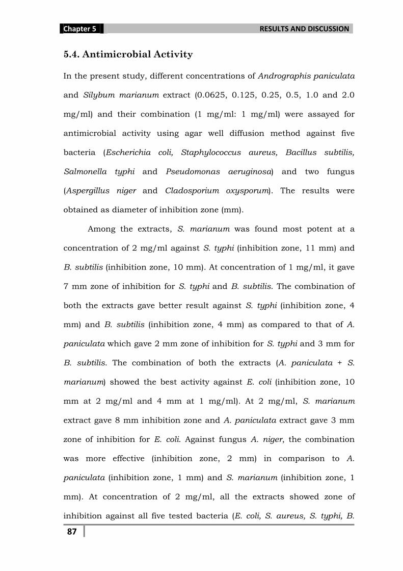
5.4. Antimicrobial Activity
In the present study, different concentrations of Andrographis paniculata
and Silybum marianum extract (0.0625, 0.125, 0.25, 0.5, 1.0 and 2.0
mg/ml) and their combination (1 mg/ml: 1 mg/ml) were assayed for
antimicrobial activity using agar well diffusion method against five
bacteria (Escherichia coli, Staphylococcus aureus, Bacillus subtilis,
Salmonella typhi and Pseudomonas aeruginosa) and two fungus
(Aspergillus niger and Cladosporium oxysporum). The results were
obtained as diameter of inhibition zone (mm).
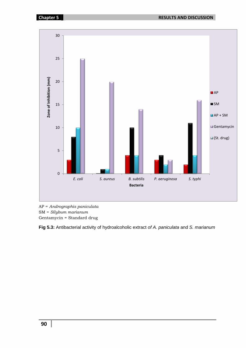
Among the extracts, S. marianum was found most potent at a
concentration of 2 mg/ml against S. typhi (inhibition zone, 11 mm) and
B. subtilis (inhibition zone, 10 mm). At concentration of 1 mg/ml, it gave
7 mm zone of inhibition for S. typhi and B. subtilis. The combination of
both the extracts gave better result against S. typhi (inhibition zone, 4
mm) and B. subtilis (inhibition zone, 4 mm) as compared to that of A.
paniculata which gave 2 mm zone of inhibition for S. typhi and 3 mm for
B. subtilis. The combination of both the extracts (A. paniculata + S.
marianum) showed the best activity against E. coli (inhibition zone, 10
mm at 2 mg/ml and 4 mm at 1 mg/ml). At 2 mg/ml, S. marianum
extract gave 8 mm inhibition zone and A. paniculata extract gave 3 mm
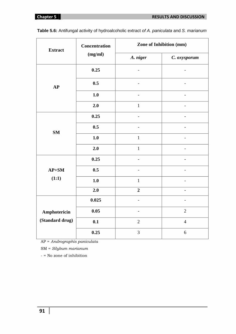
zone of inhibition for E. coli. Against fungus A. niger, the combination
was more effective (inhibition zone, 2 mm) in comparison to A.
paniculata (inhibition zone, 1 mm) and S. marianum (inhibition zone, 1
mm). At concentration of 2 mg/ml, all the extracts showed zone of
inhibition against all five tested bacteria (E. coli, S. aureus, S. typhi, B.
Page 10
Chapter 5 RESULTS AND DISCUSSION
88
subtilis and P. aeruginosa). Similar result was obtained for fungus A.
niger, while none of the extracts were found effective against C.
oxysporum (Fig. 5.3, 5.4; Table 5.5, 5.6). At low concentration (<
1mg/ml) none of the extracts showed significant results against all the
seven tested organisms. From the results obtained, it can be stated that
all the extracts showed a concentration dependent activity. The
antimicrobial effect was in the order of S. marianum > A. paniculata + S.
marianum > A. paniculata. The observations were compared with that of
standard drug Gentamycin (antibacterial) and Amphotericin (antifungal).
Page 11
Chapter 5 RESULTS AND DISCUSSION
89
Table 5.5: Antibacterial activity of hydroalcoholic extracts of A. paniculata and S.
marianum
Extract Concentration
(mg/ml)
Zone of Inhibition (mm)
E. coli S. aureus B. subtilis P. aeruginosa S. typhi
AP
0.25 - - - - -
0.5 - - 1 - -
1.0 2 - 2 - -
2.0 3 1 3 3 2
SM
0.25 3 - 1 - 1
0.5 6 - 4 - 6
1.0 7 - 7 2 7
2.0 8 1 10 4 11
AP+SM
(1:1)
0.25 - - - - -
0.5 - - - - 1
1.0 4 - - - 2
2.0 10 1 4 2 4
Gentamycin
(Standard
drug)
0.025 16 10 6 - 2
0.05 20 15 8 - 3
0.1 22 16 12 1 12
0.25 25 20 14 03 16
AP = Andrographis paniculata
SM = Silybum marianum
- = No zone of inhibition
Page 12
Chapter 5 RESULTS AND DISCUSSION
90
AP = Andrographis paniculata
SM = Silybum marianum
Gentamycin = Standard drug
Fig 5.3: Antibacterial activity of hydroalcoholic extract of A. paniculata and S. marianum
0
5
10
15
20
25
30
E. coli S. aureus B. subtilis P. aeruginosa S. typhi
Zon
e o
f In
hib
itio
n (
mm
)
Bacteria
AP
SM
AP + SM
Gentamycin
(St. drug)
Page 13
Chapter 5 RESULTS AND DISCUSSION
91
Table 5.6: Antifungal activity of hydroalcoholic extract of A. paniculata and S. marianum
Extract Concentration
(mg/ml)
Zone of Inhibition (mm)
A. niger C. oxysporum
AP
0.25 - -
0.5 - -
1.0 - -
2.0 1 -
SM
0.25 - -
0.5 - -
1.0 1 -
2.0 1 -
AP+SM
(1:1)
0.25 - -
0.5 - -
1.0 1 -
2.0 2 -
Amphotericin
(Standard drug)
0.025 - -
0.05 - 2
0.1 2 4
0.25 3 6
AP = Andrographis paniculata
SM = Silybum marianum
- = No zone of inhibition
Page 14
Chapter 5 RESULTS AND DISCUSSION
92
AP = Andrographis paniculata
SM = Silybum marianum
Amphotericin= Standard drug
Fig. 5.4: Antifungal effect of hydroalcoholic extracts of A. paniculata and S. marianum
Antibiotics like Gentamycin, Tetracyclin and Streptomycin inhibit
protein synthesis of the organism, by binding to the 30S ribosome and
freeze the 30S initiation complex (30S-mRNA-tRNA), so that no further
initiation can occur. Antibiotics like Chloramphenicol, Lincomycin and
Clindamycin binds to the 50S ribosome and inhibit peptidyl transferase
activity, while Rifamycin and Rifampicin binds to DNA-dependent RNA
polymerase and inhibit initiation of RNA synthesis. Also, such antibiotic
degrades the peptidoglycan cell wall of bacteria and chitinous cell wall of
the fungus. Similar mode of action may be performed by the plants
0
1
2
3
4
5
6
7
AP SM AP+SM Amphotericin
Zon
e o
f in
hib
itio
n (
mm
)
Extract/drug (mg/ml)
A. niger
C. oxysporum
Page 15
Chapter 5 RESULTS AND DISCUSSION
93
secondary metabolites, against microbial infection, which are present in
both the plant extracts as well as in their combination.
The phenolic compounds (flavonoids and tannins) and terpenoids
present in both the extracts may have inhibited microbial growth by
disrupting the cell wall, binding to the adhesion complex with the cell
wall and also inactivating the enzyme for microbial synthesis (Brownlee
et al., 1990; Rojas et al., 1992; Perrett et al., 1995; Haslam, 1996;
Cichewicz and Thorpe, 1996), while alkaloid present in the extracts may
have intercalated into the cell wall and DNA of the microbes inhibiting
their growth and synthesis (Burdick, 1971; Rahman and Choudhary,
1995). The crude leaf and stem bark extract of Ficus capensis inhibited
the growth of Escherichia coli and Shigella sp. This was mainly due to the
presence of phytochemicals like alkaloids, tannins, carbohydrates,
flavonoids, sterols and terpenes present in the extract. The ethanolic
extract of Olax subscorpioidea could show considerable antibacterial
activity against S. aureus and E. coli due to the presence of alkaloids,
flavonoids and steroids (Ayandele and Adebiyi, 2007). The phytochemical
analysis revealed the presence of alkaloids, saponins, tannins, flavonoids
in the extract of Lasienthera africanum which was tested against
Escherichia coli, Salmonella typhi and Staphylococcus aureus and
inhibited the growth of the tested bacteria (Adegoke et al., 2009).
Combination therapy with two or more plant extracts can be used
in special cases like preventing the emergence of resistant strains, to
treat emergency cases during the period when an etiological diagnosis is
still in progress and also when to take advantage of extract synergism,
Page 16
Chapter 5 RESULTS AND DISCUSSION
94
which occur when the effects of a combination of extract is greater than
the sum of the effects of the individual extract as showed by the
combination of A. paniculata and S. marianum in the case of E.coli and A.
niger. Extract antagonism occurs when one extract, usually the one with
the least effect, interferes with the effect of another extract as seen in the
case of B. subtilis and S. typhi. Further studies can be conducted to
attain a combinational therapy to achieve better synergistic effect.
Page 17
Chapter 5 RESULTS AND DISCUSSION
95
Organism AP Gentamycin
(Standard drug)
E. coli
S. aureus
A. subtilis
P. aeruginosa
S. typhi
AP = Andrographis paniculata
Fig. 5.5: Antibacterial effect of hydroalcoholic extract of A. paniculata and Gentamycin
Page 18
Chapter 5 RESULTS AND DISCUSSION
96
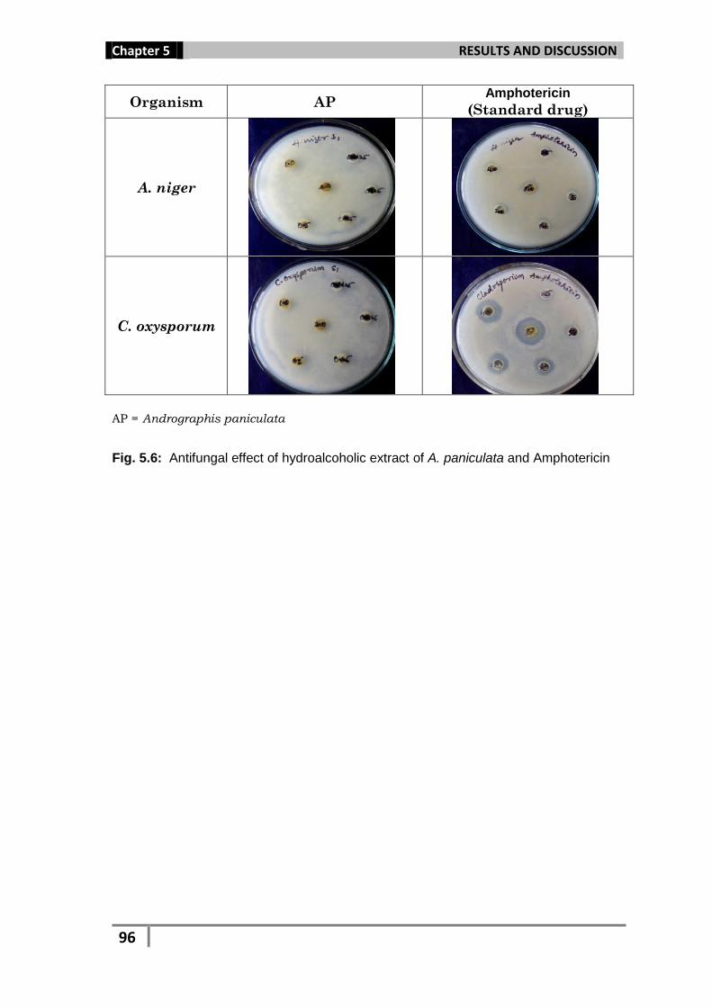
Organism AP Amphotericin
(Standard drug)
A. niger
C. oxysporum
AP = Andrographis paniculata
Fig. 5.6: Antifungal effect of hydroalcoholic extract of A. paniculata and Amphotericin
Page 19
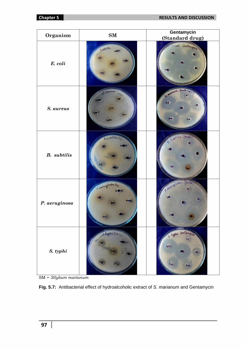
Chapter 5 RESULTS AND DISCUSSION
97
Organism SM Gentamycin
(Standard drug)
E. coli
S. aureus
B. subtilis
P. aeruginosa
S. typhi
SM = Silybum marianum
Fig. 5.7: Antibacterial effect of hydroalcoholic extract of S. marianum and Gentamycin
Page 20
Chapter 5 RESULTS AND DISCUSSION
98
Organism SM Amphotericin
(Standard drug)
A. niger
C. oxysporum
SM = Silybum marianum
Fig. 5.8: Antifungal effect of hydroalcoholic extract of S. marianum and Amphotericin
Page 21
Chapter 5 RESULTS AND DISCUSSION
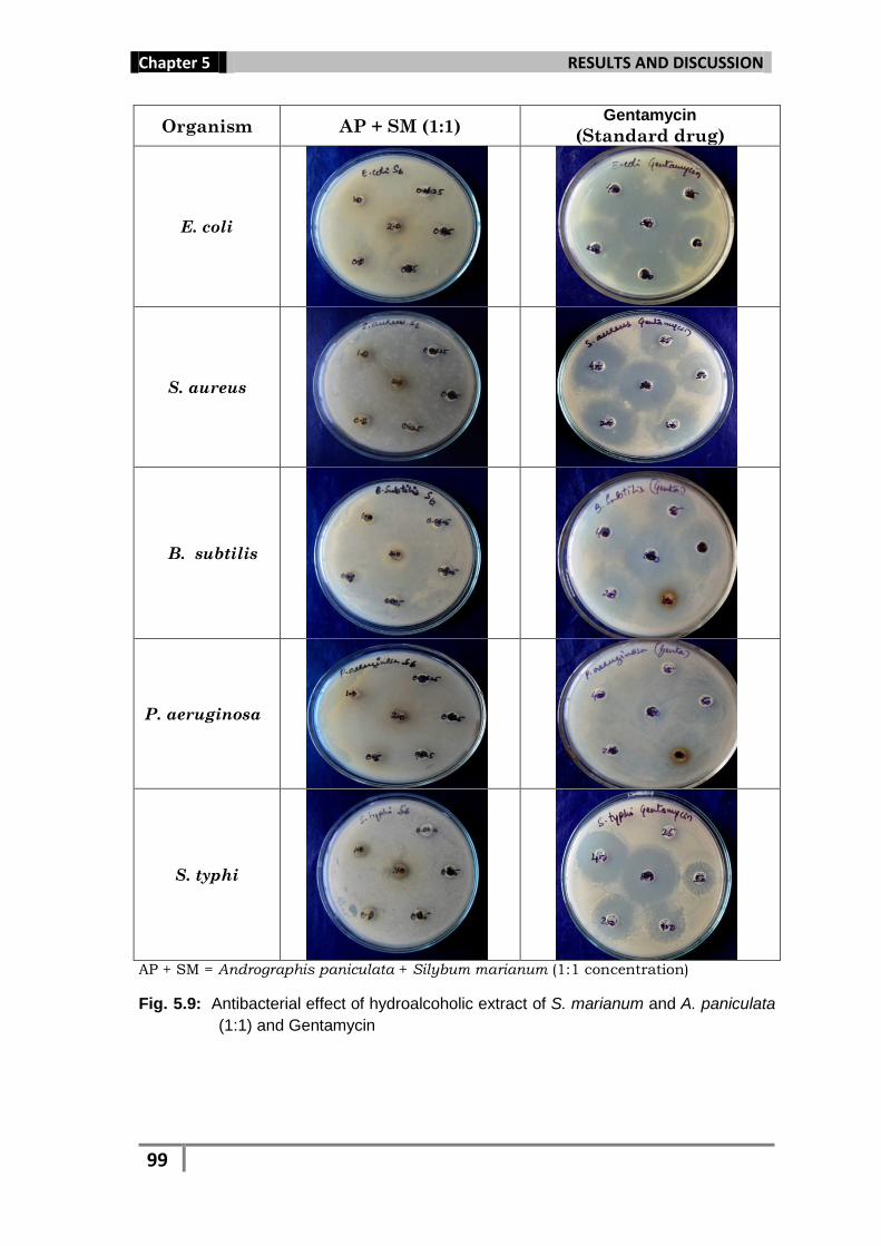
99
Organism AP + SM (1:1) Gentamycin
(Standard drug)
E. coli
S. aureus
B. subtilis
P. aeruginosa
S. typhi
AP + SM = Andrographis paniculata + Silybum marianum (1:1 concentration)
Fig. 5.9: Antibacterial effect of hydroalcoholic extract of S. marianum and A. paniculata
(1:1) and Gentamycin
Page 22
Chapter 5 RESULTS AND DISCUSSION
100
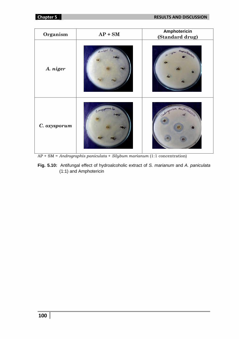
Organism AP + SM Amphotericin
(Standard drug)
A. niger
C. oxysporum
AP + SM = Andrographis paniculata + Silybum marianum (1:1 concentration)
Fig. 5.10: Antifungal effect of hydroalcoholic extract of S. marianum and A. paniculata
(1:1) and Amphotericin
Page 23
Chapter 5 RESULTS AND DISCUSSION
101
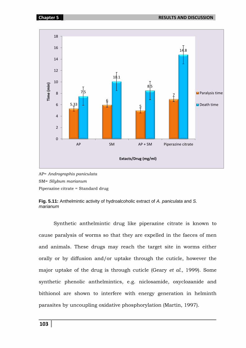
5.5. Anthelmintic Activity
Andrographis paniculata and Silybum marianum and their combination
when analyzed for anthelmintic potential showed a concentration
dependent activity. A. paniculata extract showed better activity by way of
causing the paralysis of the worms at 3.33 min at 40 mg/ml and 5.33
min at 20 mg/ml and death in 5.16 min at 40 mg/ml and 7.50 min at
20 mg/ml. At 40 mg/ml, S. marianum extract caused paralysis at 3.83
min, while death at 7.5 min. Combination with A. paniculata and S.
marianum extract (1:1) was found most potent and caused paralysis of
the worm at 2.83 min and death at 6.33 min. The time taken by the
standard drug (20 mg/ml) for the paralysis and death of the worms was
7.0 and 14.83 min respectively (Table 5.7, Fig 5.11).
Page 24
Chapter 5 RESULTS AND DISCUSSION
102
Table 5.7: Anthelmintic activity of hydroalcoholic extracts of A. paniculata and S. marianum
Extract/Drug Concentration
(mg/ml)
Paralysis time Death time
Min.
AP
20 5.33 ± 0.40 7.50 ± 0.20
40 3.33 ± 0.18 5.16 ± 0.14
SM
20 6 ± 0.23 10.16 ± 0.43
40 3.83 ± 0.14 7.5 ± 0.31
AP + SM
(1:1)
20 5 ± 0.23 8.5 ± 0.20
40 2.83 ± 0.14 6.33 ± 0.18
Piperazine citrate 20 7 ± 0.33 14.83 ± 0.36
Normal Saline (Control) - - -
AP = Andrographis paniculata
SM = Silybum marianum
Piperazine citrate = Standard drug
─ = No activity
Page 25
Chapter 5 RESULTS AND DISCUSSION
103
AP= Andrographis paniculata
SM= Silybum marianum
Piperazine citrate = Standard drug
Fig. 5.11: Anthelmintic activity of hydroalcoholic extract of A. paniculata and S. marianum
Synthetic anthelmintic drug like piperazine citrate is known to
cause paralysis of worms so that they are expelled in the faeces of men
and animals. These drugs may reach the target site in worms either
orally or by diffusion and/or uptake through the cuticle, however the
major uptake of the drug is through cuticle (Geary et al., 1999). Some
synthetic phenolic anthelmintics, e.g. niclosamide, oxyclozanide and
bithionol are shown to interfere with energy generation in helminth
parasites by uncoupling oxidative phosphorylation (Martin, 1997).
5.33 6
5
7 7.5
10.1
8.5
14.8
0
2
4
6
8
10
12
14
16
18
AP SM AP + SM Piperazine citrate
Tim
e (
min
)
Extacts/Drug (mg/ml)
Paralysis time
Death time
Page 26
Chapter 5 RESULTS AND DISCUSSION
104
A. paniculata S. marianum
A. paniculata + S. marianum Piperazine citrate
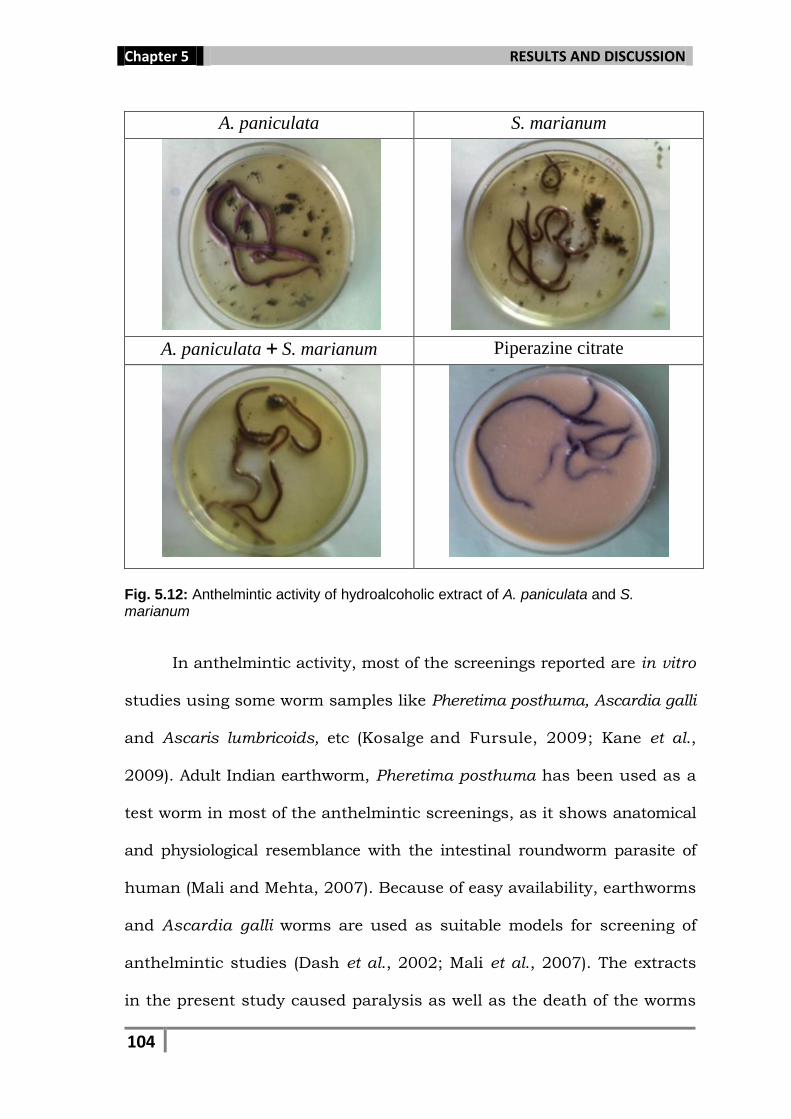
Fig. 5.12: Anthelmintic activity of hydroalcoholic extract of A. paniculata and S. marianum
In anthelmintic activity, most of the screenings reported are in vitro
studies using some worm samples like Pheretima posthuma, Ascardia galli
and Ascaris lumbricoids, etc (Kosalge and Fursule, 2009; Kane et al.,
2009). Adult Indian earthworm, Pheretima posthuma has been used as a
test worm in most of the anthelmintic screenings, as it shows anatomical
and physiological resemblance with the intestinal roundworm parasite of
human (Mali and Mehta, 2007). Because of easy availability, earthworms
and Ascardia galli worms are used as suitable models for screening of
anthelmintic studies (Dash et al., 2002; Mali et al., 2007). The extracts
in the present study caused paralysis as well as the death of the worms
Page 27
Chapter 5 RESULTS AND DISCUSSION
105
may be due to the presence of phenolic compounds. Both the extracts
when taken in combination, showed synergistically better activity as
compared to individual effect. The variation in activity of the plant
extract might be due to the difference in the proportion of the active
compounds responsible for the anthelmintic property (Eguale et al.,
2006). The active constituents may be the phenolics, such as flavonoids
and tannins, present in both the plant extracts may be the reason for
causing paralysis and death of the worms. In the present investigation,
all the extracts were shown to possess tannins which were previously
reported to produce anthelmintic activities (Niezen et al., 1995). The
possible action of tannins is that they can bind to free proteins in the
gastrointestinal tract of the host animal (Athnasiadou et al., 2001) or
glycoprotein on the cuticle of the parasite (Thompson and Geary, 1995)
and may cause death. The results of the combinational study showed
synergistic effect and it may be suggested that both the plants when
consumed together can show better effect against intestinal worms
rather then taken individually.
Page 28
Chapter 5 RESULTS AND DISCUSSION
106
5.6. Hepatoprotective activity
In the present study, the effect of different concentrations of
Andrographis paniculata and Silybum marianum (100, 200 and 400
mg/ml) and their different combinations (1:1, 1:2 and 2:1 to make final
concentration of 400 mg/ml) were assayed on CCl4 induced liver damage
in Wistar rats. After assessment of the biochemical parameters, CCl4
treated animals showed significant increase in the levels of AST (187.22
U/L) and ALT (90.66 U/L), while decrease in the level of total protein
(1.57 mg/dL) as compared to the normal control group (54.91 U/L,
31.65 U/L and 4.89 mg/dL for AST, ALT and total protein, respectively).
Whereas, animals treated with different extracts at a dose of 400 mg/kg
BW, showed significant decrease in the levels of serum marker enzymes
and significant increase in the total protein to the near normal value
which are comparable to that of standard drug Liv 52 (65.31 U/L, 42.31
U/L and 5.51 mg/dL for AST, ALT and total protein, respectively),
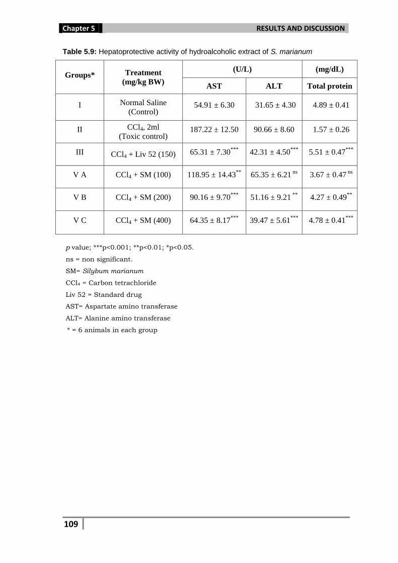
indicating the recovery of hepatic cells against the damage. At 200
mg/ml, S. marianum extract lowered the level of AST to 90.16 U/ml, ALT
to 51.16 U/ml and increased the total protein level to 4.27 U/mg of
protein), while, at a dose of 400 mg/kg it significantly reduced the AST
level to 64.35 U/ml and ALT level to 39.47, while increased the total
protein to 4.78 mg/dL and was found to be more potent among all the
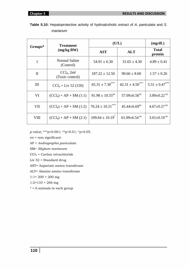
extracts. S. marianum and its combination with A. paniculata in the
concentration ratio of 2:1 (S. marianum 266 mg and A. paniculata 133
mg) was found to be most effective among all the three combinations and
reduced the AST level to 76.24 U/ml, ALT to 45.44 and increased the
Page 29
Chapter 5 RESULTS AND DISCUSSION
107
total protein to 4.67 mg/dL. This combination was also found better
when compared to that of A. paniculata at a dose of 400 mg/ml (AST
level 78.32 U/ml, ALT 46.49 and total protein 4.34 mg/dL). At a
concentration of 100 mg/ml for all the extracts, could not show
significant changes in the level of AST, ALT and total protein. Significant
results for all the three extracts were obtained at higher concentration
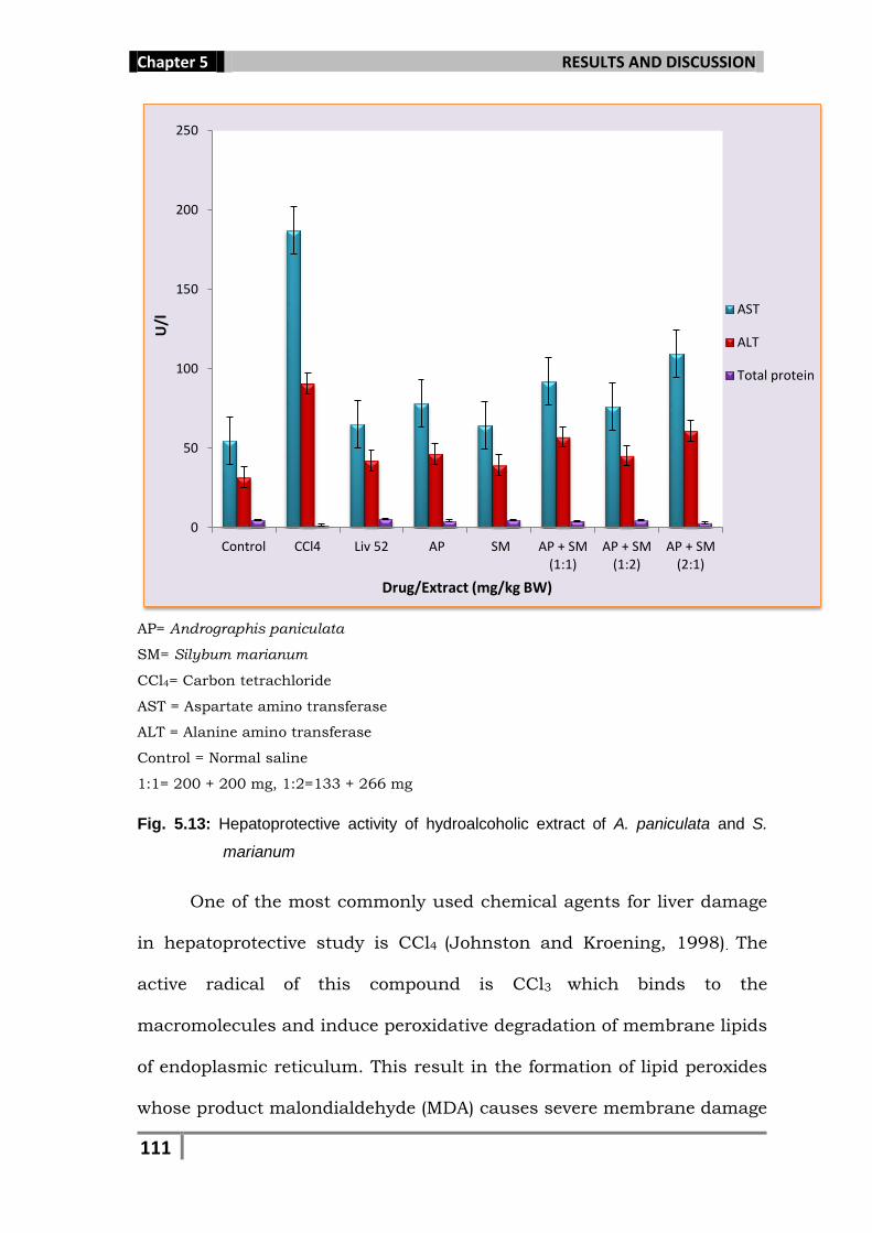
(400 mg/ml). The present finding showed that both the extracts and
their combinations were found effective against the CCl4 induced liver
damage in rats (Table 5.8, 5.9, 5.10; Fig. 5.13).
Page 30
Chapter 5 RESULTS AND DISCUSSION
108
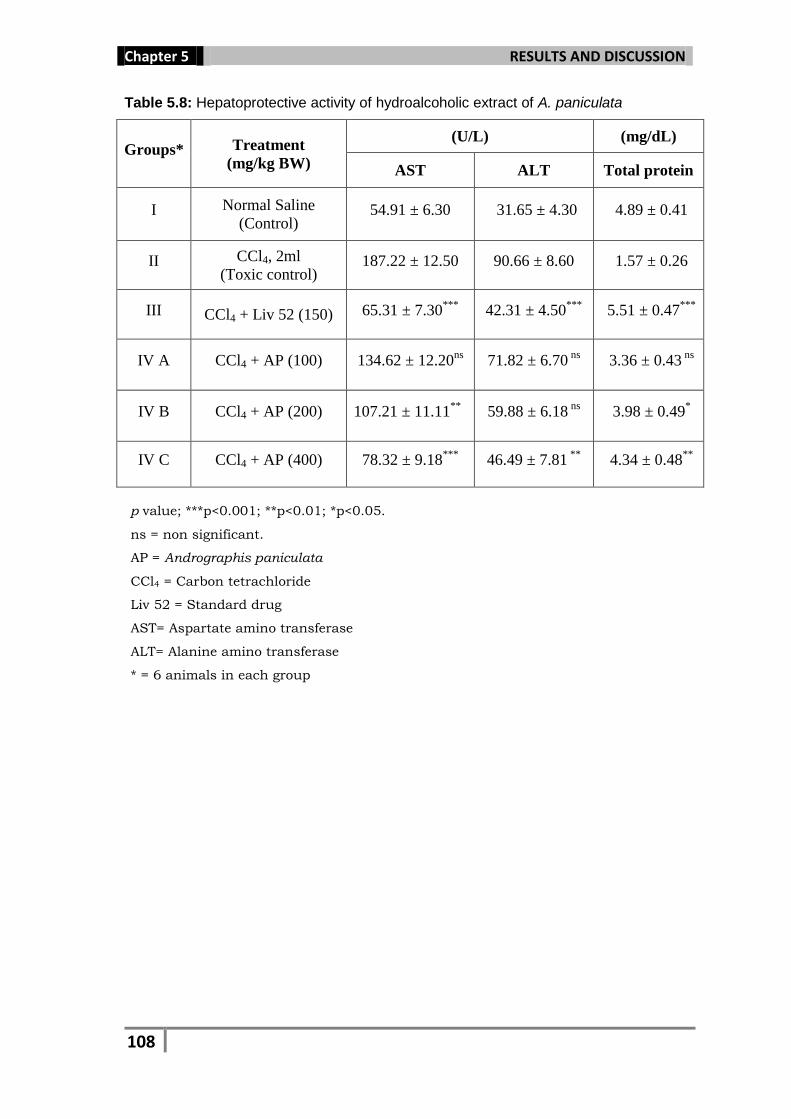
Table 5.8: Hepatoprotective activity of hydroalcoholic extract of A. paniculata
Groups* Treatment
(mg/kg BW)
(U/L) (mg/dL)
AST ALT Total protein
I Normal Saline
(Control) 54.91 ± 6.30 31.65 ± 4.30 4.89 ± 0.41
II CCl4, 2ml
(Toxic control) 187.22 ± 12.50 90.66 ± 8.60 1.57 ± 0.26
III CCl4 + Liv 52 (150) 65.31 ± 7.30***
42.31 ± 4.50***
5.51 ± 0.47***
IV A CCl4 + AP (100) 134.62 ± 12.20ns
71.82 ± 6.70 ns
3.36 ± 0.43 ns
IV B CCl4 + AP (200) 107.21 ± 11.11**
59.88 ± 6.18 ns
3.98 ± 0.49*
IV C CCl4 + AP (400) 78.32 ± 9.18***
46.49 ± 7.81 **
4.34 ± 0.48**
p value; ***p<0.001; **p<0.01; *p<0.05.
ns = non significant.
AP = Andrographis paniculata
CCl4 = Carbon tetrachloride
Liv 52 = Standard drug
AST= Aspartate amino transferase
ALT= Alanine amino transferase
* = 6 animals in each group
Page 31
Chapter 5 RESULTS AND DISCUSSION
109
Table 5.9: Hepatoprotective activity of hydroalcoholic extract of S. marianum
Groups* Treatment
(mg/kg BW)
(U/L) (mg/dL)
AST ALT Total protein
I Normal Saline
(Control) 54.91 ± 6.30 31.65 ± 4.30 4.89 ± 0.41
II CCl4, 2ml
(Toxic control) 187.22 ± 12.50 90.66 ± 8.60 1.57 ± 0.26
III CCl4 + Liv 52 (150) 65.31 ± 7.30***
42.31 ± 4.50***
5.51 ± 0.47***
V A CCl4 + SM (100) 118.95 ± 14.43**
65.35 ± 6.21 ns
3.67 ± 0.47 ns
V B CCl4 + SM (200) 90.16 ± 9.70***
51.16 ± 9.21 **
4.27 ± 0.49**
V C CCl4 + SM (400) 64.35 ± 8.17***
39.47 ± 5.61***
4.78 ± 0.41***
p value; ***p<0.001; **p<0.01; *p<0.05.
ns = non significant.
SM= Silybum marianum
CCl4 = Carbon tetrachloride
Liv 52 = Standard drug
AST= Aspartate amino transferase
ALT= Alanine amino transferase
* = 6 animals in each group
Page 32
Chapter 5 RESULTS AND DISCUSSION
110
Table 5.10: Hepatoprotective activity of hydroalcoholic extract of A. paniculata and S.
marianum
Groups* Treatment
(mg/kg BW)
(U/L) (mg/dL)
AST ALT Total
protein
I Normal Saline
(Control) 54.91 ± 6.30 31.65 ± 4.30 4.89 ± 0.41
II CCl4, 2ml
(Toxic control) 187.22 ± 12.50 90.66 ± 8.60 1.57 ± 0.26
III CCl4 + Liv 52 (150) 65.31 ± 7.30***
42.31 ± 4.50***
5.51 ± 0.47***
VI (CCl4) + AP + SM (1:1) 91.98 ± 10.55ns
57.09±6.56ns
3.89±0.22 ns
VII (CCl4) + AP + SM (1:2) 76.24 ± 10.21***
45.44±6.69ns
4.67±0.27 ns
VIII (CCl4) + AP + SM (2:1) 109.64 ± 10.19* 61.09±6.54
ns 3.01±0.19
ns
p value; ***p<0.001; **p<0.01; *p<0.05.
ns = non significant.
AP = Andrographis paniculata
SM= Silybum marianum
CCl4 = Carbon tetrachloride
Liv 52 = Standard drug
AST= Aspartate amino transferase
ALT= Alanine amino transferase
1:1= 200 + 200 mg
1:2=133 + 266 mg
* = 6 animals in each group
Page 33
Chapter 5 RESULTS AND DISCUSSION
111
AP= Andrographis paniculata
SM= Silybum marianum
CCl4= Carbon tetrachloride
AST = Aspartate amino transferase
ALT = Alanine amino transferase
Control = Normal saline
1:1= 200 + 200 mg, 1:2=133 + 266 mg
Fig. 5.13: Hepatoprotective activity of hydroalcoholic extract of A. paniculata and S.
marianum
One of the most commonly used chemical agents for liver damage
in hepatoprotective study is CCl4 (Johnston and Kroening, 1998). The
active radical of this compound is CCl3 which binds to the
macromolecules and induce peroxidative degradation of membrane lipids
of endoplasmic reticulum. This result in the formation of lipid peroxides
whose product malondialdehyde (MDA) causes severe membrane damage
0
50
100
150
200
250
Control CCl4 Liv 52 AP SM AP + SM(1:1)
AP + SM(1:2)
AP + SM(2:1)
U/l
Drug/Extract (mg/kg BW)
AST
ALT
Total protein
Page 34
Chapter 5 RESULTS AND DISCUSSION
112
(Cotran et al., 1994; Kaplowitz et al., 1986; Deleve and Kaplowitz, 1995;
Farrel 1998). The extent of hepatic damage is assessed by the elevated
levels of serum marker enzyme AST and ALT which is significantly
lowered by the administration of A. paniculata, S. marianum and their
combinational extracts in the tested groups showing their hepato
protective potential. Apart from liver, AST and ALT are also concentrated
in heart muscles, brain, gastric mucosa, adipose tissue, skeletal muscle
and kidneys. When these organs are damaged or destroyed by free
radicals or any other carcinogenic substances, these enzymes are
released from the damaged cells and their concentration is increased in
the blood. Elevated levels of the enzymes can signal myocardial
infarction, hepatic disease, muscular dystrophy, and organ damage. The
total protein estimation is also useful in hepatoprotective study as its
decreased level indicates severe non viral liver cell damage (Shenoy et al.,
2001).
The hepatoprotective potential of a drug depends upon its ability
in reducing the harmful effects caused by a hepatotoxin (Manjunatha et
al., 2008). In the present study, these phytoconstituents play a vital role
in inducing microsomal enzymes thereby accelerating the excretion of
CCl4, or inhibiting the lipid peroxidation induced by CCl4 (Mehta et al.,
1999). Phytoconstituents such as alkaloids (Vijyan et al., 2003) and
flavonoids (Baek et al., 1996) have been found effective in the
hepatoprotection against CCl4 induced liver damage. The hydroalcoholic
extract of both the plants showed the presence of alkaloids and
flavonoids, which may be responsible for their hepatoprotective
Page 35
Chapter 5 RESULTS AND DISCUSSION
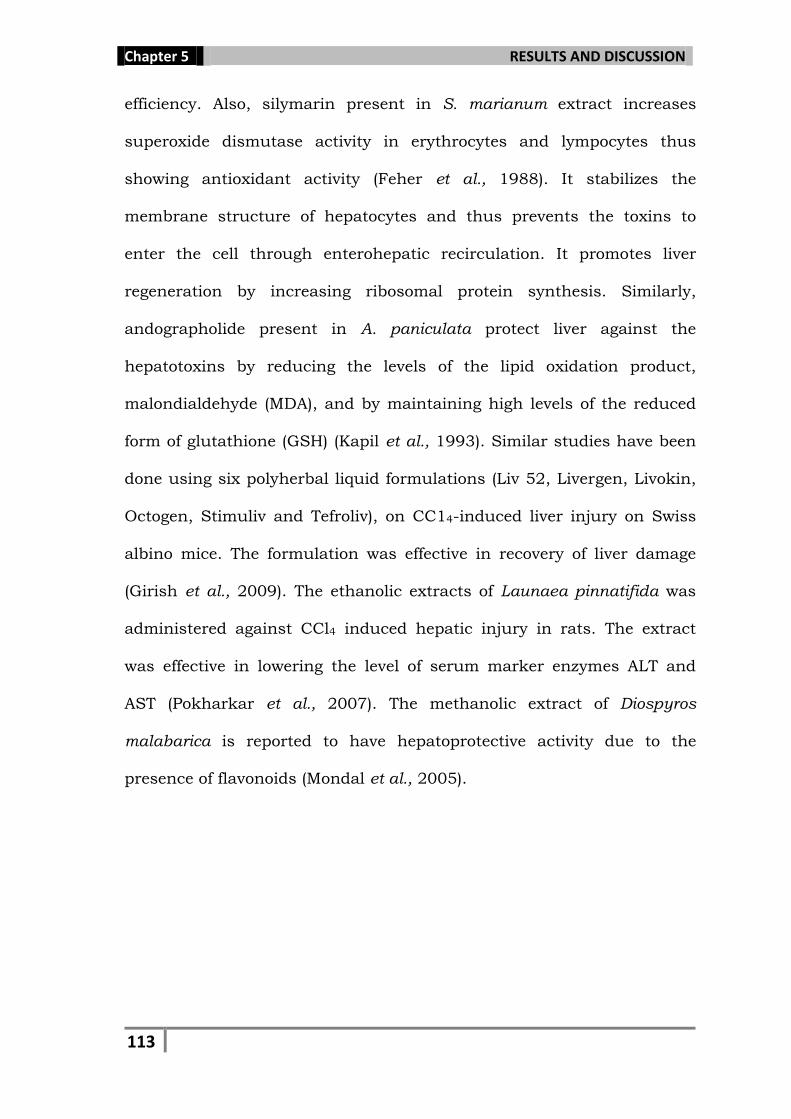
113
efficiency. Also, silymarin present in S. marianum extract increases
superoxide dismutase activity in erythrocytes and lympocytes thus
showing antioxidant activity (Feher et al., 1988). It stabilizes the
membrane structure of hepatocytes and thus prevents the toxins to
enter the cell through enterohepatic recirculation. It promotes liver
regeneration by increasing ribosomal protein synthesis. Similarly,
andographolide present in A. paniculata protect liver against the
hepatotoxins by reducing the levels of the lipid oxidation product,
malondialdehyde (MDA), and by maintaining high levels of the reduced
form of glutathione (GSH) (Kapil et al., 1993). Similar studies have been
done using six polyherbal liquid formulations (Liv 52, Livergen, Livokin,
Octogen, Stimuliv and Tefroliv), on CC14-induced liver injury on Swiss
albino mice. The formulation was effective in recovery of liver damage
(Girish et al., 2009). The ethanolic extracts of Launaea pinnatifida was
administered against CCl4 induced hepatic injury in rats. The extract
was effective in lowering the level of serum marker enzymes ALT and
AST (Pokharkar et al., 2007). The methanolic extract of Diospyros
malabarica is reported to have hepatoprotective activity due to the
presence of flavonoids (Mondal et al., 2005).
Page 36
Chapter 5 RESULTS AND DISCUSSION
114
5.7. Antioxidant activity
In the present study, the hydroalcoholic extract of Andrographis
paniculata and Silybum marianum at different concentrations (100, 200
and 400 mg/ml) and their different combinations (1:1, 1:2 and 2:1 to
make final concentration of 400 mg/ml) were assayed for antioxidant
activity, analyzing SOD (Superoxide dismutase), CAT (Catalase) and GPx
(Glutathione peroxidase). After assessing of the biochemical parameters,
CCl4 treated animals showed significant decrease in the levels of SOD,
CAT and GPx (11.21, 27.57 and 25.34 U/mg of protein, respectively) as
compared to the normal control group (19.30 for SOD, 52.43 for CAT
and 25.34 for GPx). Whereas, animals treated with different extracts at
the dose of 400 mg/kg BW showed significant increase in the levels of
the enzymes to the near normal value which are comparable to the
values observed for standard drug, Liv 52 (18.71, 48.19 and 23.14 U/mg
of protein for SOD, CAT and GPx, respectively). At a dose of 200 mg/ml,
S. marianum increased the activity of SOD to 16.35 U/mg protein, CAT
to 38.58 and GPx to 20.10. However, at a dose of 400 mg/kg BW, it
significantly increased the level of SOD to 17.42 U/mg protein, CAT to
45.24 and GPx to 21.96 U/mg protein, while, the combination of S.
marianum with A. paniculata in the concentration ratio of 2:1 was found
most effective among all the three combinations, which increased the
SOD level to 16.26 U/mg protein, CAT level to 48.88 and GPx 21.41
U/mg protein. This combination was also better when compared to that
of A. paniculata at a dose of 400 mg/ml (16.64, 40.19 and 20.34 for
SOD, CAT and GPx level, respectively). At a concentration of 100 mg/ml
Page 37
Chapter 5 RESULTS AND DISCUSSION
115
for all the extracts, no significant changes were observed in the levels of
SOD, CAT and GPx. Significant results for all the three extracts were
obtained only at higher concentration of 400 mg/ml. (Table 5.11, 5.12,
5.13 and Fig. 5.14).
Table 5.11: Antioxidant activity of hydroalcoholic extract of A. paniculata
Groups* Treatment
(mg/kg BW)
(U/mg protein)
SOD CAT GPx
I Normal Saline
(Control) 19.30 ± 1.30 52.43 ± 4.38 25.34 ± 0.48
II CCl4, 2ml
(Toxic control) 11.21 ± 0.94 27.57 ± 1.43 17.14 ± 0.26
III CCl4 + Liv 52 (150) 18.71 ± 1.12***
48.19 ± 3.87***
23.14 ± 0.46***
IV A CCl4 + AP (100) 11.34 ± 0.90ns
25.12 ± 1.33ns
12.98 ± 0.38ns
IV B CCl4 + AP (200) 15.67 ± 0.78* 35.01 ± 1.25
ns 18.23 ± 0.38
ns
IV C CCl4 + AP (400) 16.64 ± 0.65**
40.19 ± 2.87**
20.34 ± 0.39***
p value; ***p<0.001; **p<0.01; *p<0.05.
ns = non significant.
AP = Andrographis paniculata
CCl4 = Carbon tetrachloride
Liv 52 = Standard drug
SOD = Superoxide dismutase
CAT = Catalase
GPx = Glutathione peroxidase
* = 6 animals in each group
Page 38
Chapter 5 RESULTS AND DISCUSSION
116
Table 5.12: Antioxidant activity of hydroalcoholic extract of S. marianum
Groups* Treatment
(mg/kg BW)
(U/mg protein)
SOD CAT GPx
I Normal Saline
(Control) 19.30 ± 1.30 52.43 ± 4.38 25.34 ± 0.48
II CCl4, 2ml
(Toxic control) 11.21 ± 0.94 27.57 ± 1.43 17.14 ± 0.26
III CCl4 + Liv 52 (150) 18.71 ± 1.12***
48.19 ± 3.87***
23.14 ± 0.46***
V A CCl4 + SM (100) 12.10 ± 0.79ns
28.11 ± 1.59ns
16.32 ± 0.38**
V B CCl4 + SM (200) 16.35 ± 0.71**
38.58 ± 1.64**
20.10 ± 0.41***
V C CCl4 + SM (400) 17.42 ± 0.63***
45.24 ± 1.84***
21.96 ± 0.39***
p value; ***p<0.001; **p<0.01; *p<0.05.
ns = non significant.
SM= Silybum marianum
CCl4 = Carbon tetrachloride
Liv 52 = Standard drug
SOD = Superoxide dismutase
CAT = Catalase
GPx = Glutathione peroxidase
* = 6 animals in each group
Page 39
Chapter 5 RESULTS AND DISCUSSION
117
Table 5.13: Antioxidant activity of hydroalcoholic extract of A. paniculata and S.
marianum
Groups* Treatment
(mg/kg BW)
(U/mg protein)
SOD CAT GPx
I Normal Saline
(Control) 19.30 ± 1.30 52.43 ± 4.38 25.34 ± 0.48
II CCl4, 2ml
(Toxic control) 11.21 ± 0.94 27.57 ± 1.43 17.14 ± 0.26
III CCl4 + Liv 52 (150) 18.71 ± 1.12***
48.19 ± 3.87***
23.14 ± 0.46***
VI (CCl4) + AP + SM (1:1) 14.52±0.91ns
37.91±1.67ns
19.21±0.28ns
VII (CCl4) + AP + SM (1:2) 16.26±0.77 ns
48.88± 2.09***
21.41±0.31ns
VIII (CCl4) + AP + SM (2:1) 13.33±0.67 ns
32.17±1.28ns
17.21±0.33 ns
p value; ***p<0.001; **p<0.01; *p<0.05.
ns = non significant.
AP = Andrographis paniculata
SM= Silybum marianum
CCl4 = Carbon tetrachloride
Liv 52 = Standard drug
SOD = Superoxide dismutase
CAT = Catalase
GPx = Glutathione peroxidase
1:1= 200 + 200 mg
1:2=133 + 266 mg
* = 6 animals in each group
Page 40
Chapter 5 RESULTS AND DISCUSSION
118
AP= Andrographis paniculata
SM= Silybum marianum
Normal control= Normal saline
Liv. 52= Standard drug
CCl4= Carbon tetrachloride
CAT = Catalase
SOD= Superoxide dismutase
GPx= Glutathione peroxidase
1:1= 200 + 200 mg
1:2=133 + 266 mg
Fig. 5.14: Antioxidant activity of hydroalcoholic extract of A. paniculata and S. marianum
Antioxidants are intimately involved in the prevention of cellular
damage which is the common pathway for cancer, aging, and a variety of
diseases. Free radicals are atoms or groups of atoms with an odd
(unpaired) number of electrons which can be formed when oxygen
interacts with certain molecules. Once formed these highly reactive
0
10
20
30
40
50
60
NormalControl
CCl4 Liv 52 AP SM AP + SM(1:1)
AP + SM(1:2)
AP + SM(2:1)
U/m
g p
rote
in
Drug/Extract (mg/kg BW)
SOD
CAT
GPx
Page 41
Chapter 5 RESULTS AND DISCUSSION
119
radicals can start a chain reaction. Their chief danger comes from the
damage they can do when they react with important cellular components
such as DNA, or the cell membrane. The body has a defense system of
antioxidants to prevent free radical damage. They are the molecules
which can safely interact with free radicals and terminate the chain
reaction before vital molecules are damaged. Catalase (CAT) an
antioxidant enzyme, like Superoxide dismutase (SOD) and Glutathione
peroxidase (GPx) is produced naturally within the body. It helps the body
to convert hydrogen peroxide into water and oxygen. It also uses
hydrogen peroxide to break down potentially harmful toxins in the body,
including alcohol, phenol, and formaldehyde. When our body uses
oxygen it produces free radicals that damage cell membranes, proteins
and DNA. Catalase works closely with superoxide dismutase to prevent
free radical damage to the body. SOD converts the dangerous superoxide
radical to hydrogen peroxide which is converted to harmless water and
oxygen by CAT and GPx. When the level of these enzymes decreases in
the body, the antioxidant system cannot function properly.
The results obtained in the present study showed that both the
extracts and their combinations were found effective in increasing SOD,
CAT and GPx activity. It also indicates that these extracts may be
associated with decreased oxidative stress, free radical-mediated tissue
damage and prevent the accumulation of excessive free radicals and
protects the liver from CCl4 induced liver damage in rats. Flavonoids and
other phenolic compounds of plant origin have been reported as
scavengers of free radicals (Formica and Regelson, 1995; Rice et al.,
Page 42
Chapter 5 RESULTS AND DISCUSSION
120
1997). The results obtained in the present investigation of phytochemical
studies show that both A. paniculata and S. marianum are rich in
flavonoids and phenolic compounds. Varga et al., (2001) reported that
silymarin increases superoxide dismutase activity in erythrocytes. It’s
mechanism of action for hepatoprotection appears from its antioxidant
effect to scavenge free radicals and inhibit lipid peroxidation, which is
similar with the case of andrographolide present in A. paniculata (Flora
et al., 1998). Similar study was performed for evaluating antioxidant
effect of the methanolic extract of Eupatorium ayapana leaves in CCl4
induced liver damage in Wistar albino rats. SOD, CAT, GSH and protein
analysis revealed that the extract contains antioxidant activity (Bose et
al., 2007). Carica papaya, Psidium guajava, Vernonia amygdalina, and
Mangifera indica also possess potent antioxidant activity due to the
presence of various phytochemicals, like flavonoids, terpenoids and
tannins (Ayoola et al., 2008). The methanolic extract of Bauhinia
racemosa stem bark was investigated for the antioxidant effect in
paracetamol induced liver damage in Wistar albino rats. The extract
showed antioxidant effects on FeCl2-ascorbate-induced lipid peroxidation
in rat liver homogenate and on superoxide scavenging activity (Gupta et
al., 2004). The methanolic extract of Momordica cymbalaria was used
against CCl4 treated rats for antioxidant activity. The extract restored the
altered catalase level in CCl4 intoxicated rats (Rajshekhar et al., 2009).
Page 43
Chapter 5 RESULTS AND DISCUSSION
121
4.8. Anticancer study
In the present study, an attempt was made to investigate the anticancer
potential of the extracts of A. paniculata and S. marianum and their
combination (1:1) at a concentration of 100 µg/ml against five human
cancer cell lines the human breast adenocarcinoma (MCF-7), the human
cervix (SiHa), colon (HT-29), liver (HepG2) and ovary cancer cell line
(ovcar-5). The result of in vitro, anticancer study suggest that S.
marianum extract showed the best cytotoxic activity against all given cell
lines (percentage inhibition was 21.34, 32.30, 46.56, 59.58, 36.20 for
MCF 7, SiHa, HT, Ovcar and HepG2, respectively), while significant one
was against HT and Ovcar. Andrographis paniculata extract was found
most effective against Ovcar cell line (51.12%). Again, the combination
(1:1) of both the plants showed an intermediate result for most of the cell
line but, the combination was most effective against the liver cell line
(HepG2) which showed better activity (42.76%), as compared with the
activity of both the plant extracts which gave individually (28.22 and
36.20% for A. paniculata and S. marianum respectively) (Table 5.14, Fig.
5.15).
Page 44
Chapter 5 RESULTS AND DISCUSSION
122
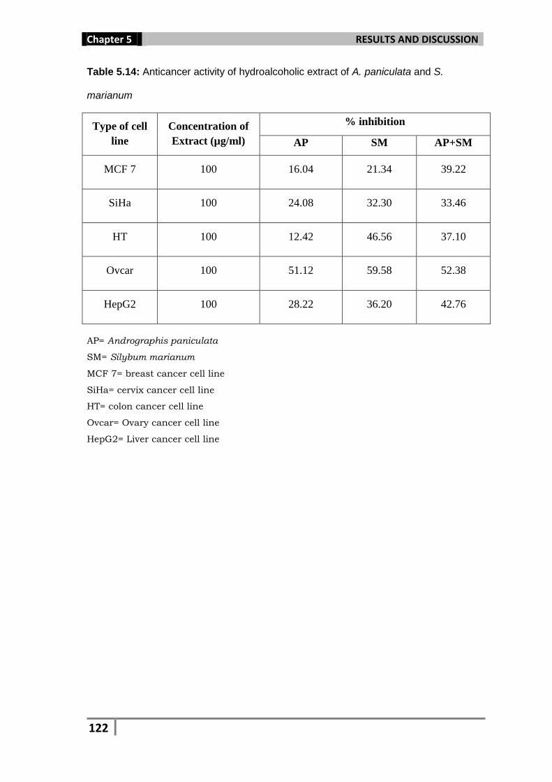
Table 5.14: Anticancer activity of hydroalcoholic extract of A. paniculata and S.
marianum
Type of cell
line
Concentration of
Extract (µg/ml)
% inhibition
AP SM AP+SM
MCF 7 100 16.04 21.34 39.22
SiHa 100 24.08 32.30 33.46
HT 100 12.42 46.56 37.10
Ovcar 100 51.12 59.58 52.38
HepG2 100 28.22 36.20 42.76
AP= Andrographis paniculata
SM= Silybum marianum
MCF 7= breast cancer cell line
SiHa= cervix cancer cell line
HT= colon cancer cell line
Ovcar= Ovary cancer cell line
HepG2= Liver cancer cell line
Page 45
Chapter 5 RESULTS AND DISCUSSION
123
AP= Andrographis paniculata
SM= Silybum marianum
MCF 7= breast cancer cell line
SiHa= cervix cancer cell line
HT= colon cancer cell line
Ovcar= Ovary cancer cell line
HepG2= Liver cancer cell line
Fig. 5.15: Anticancer activity of hydroalcoholic extract of A. paniculata and S. marianum
0
10
20
30
40
50
60
MCF 7 SiHa HT Ovcar HepG2
% in
hib
itio
n
Cell line
% inhibition AP
% inhibition SM
% inhibition AP+SM
Page 46
Chapter 5 RESULTS AND DISCUSSION
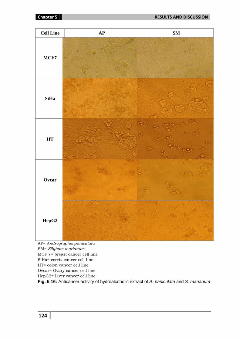
124
Cell Line AP SM
MCF7
SiHa
HT
Ovcar
HepG2
AP= Andrographis paniculata
SM= Silybum marianum
MCF 7= breast cancer cell line
SiHa= cervix cancer cell line
HT= colon cancer cell line
Ovcar= Ovary cancer cell line
HepG2= Liver cancer cell line
Fig. 5.16: Anticancer activity of hydroalcoholic extract of A. paniculata and S. marianum
Page 47
Chapter 5 RESULTS AND DISCUSSION
125
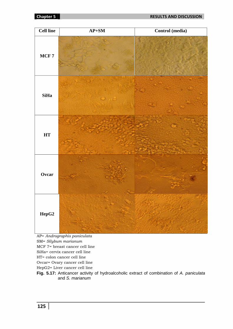
Cell line AP+SM Control (media)
MCF 7
SiHa
HT
Ovcar
HepG2
AP= Andrographis paniculata
SM= Silybum marianum
MCF 7= breast cancer cell line
SiHa= cervix cancer cell line
HT= colon cancer cell line
Ovcar= Ovary cancer cell line
HepG2= Liver cancer cell line
Fig. 5.17: Anticancer activity of hydroalcoholic extract of combination of A. paniculata and S. marianum
Page 48
Chapter 5 RESULTS AND DISCUSSION
126
Medicinal plants maintain the health and vitality of individuals, and also
cure various diseases, including cancer without causing toxicity. These
medicinal plants possess good immunomodulatory and antioxidant
properties, leading to anticancer activities. The antioxidant
phytochemicals protect the cells from oxidative damage. Thus,
consuming a diet rich in antioxidant plant material can provide health-
protective effects. These natural products are supposed to minimize DNA
damage by reacting with free radicals and in this way they can prevent
cancer.
Phytochemicals, such as flavonols and flavonoids were
investigated to determine chemoprevention activity against cancer
(Conese and Blasi, 1995). Phenols, polyphenols, flavonoids and their
derivatives, are ubiquitous in plants and have been found associated
with the inhibition of atherosclerosis and cancer (Cirla and Mann, 2003).
Recent studies have reported antitumor effects of the flavonoids,
quercetin, genistein, and baicalein obtained from plant extracts
(Trangnos et al., 1992; Descher et al., 1991; Yoshida et al., 1990).
Similarly, alkaloids like schischkinnin and montamine have been
isolated from the seeds of Centaurea schischkinii and Centaurea montana
which showed anticancer property (Shoeb et al., 2006). Yang and Wang
(1993) reported the anticancer effect of polyphenols against tumor
formation and growth. Flavonoids isolated from Plantago species were
able to strongly inhibit the proliferation of human cancer cell lines
(Galvez et al., 2003). Aqueous extracts of Larrea tridentata and Juniperus
communis significantly decreased the growth of MCF-7 breast cancer
Page 49
Chapter 5 RESULTS AND DISCUSSION
127
cells (Slambrouck et al., 2007). Flavopiridol is a synthetic flavone,
derived from the plant alkaloid rohitukine, which was isolated from
Dysoxylum binectariferum (Kellard et al., 2000). It is currently in phase I
and phase II clinical trials against a broad range of tumors, including
leukemia, lymphomas and solid tumors (Christian et al., 1997).
Synthetic agent roscovitine derived from natural product olomucine,
originally isolated from Raphanus sativus, is in Phase II clinical trials in
Europe (Cragg and Newman, 2005; Meijer et al., 2003).