High-frequency Duplex Ultrasound Imaging System for Biomedical Applications Using 30 MHz Linear
Arrays
Xiaochen Xu1,2, Lequan Zhang1, Lei Sun1, Jesse T. Yen1, Jonathan M. Cannata1, K. Kirk Shung1 1NIH Resource on Medical Ultrasonic Transducer Technology, University of Southern California, Los Angeles, U.S.A.
2Medical Business Unit, Texas Instruments, Dallas, Texas 75243, U.S.A. Email: [email protected]
Abstract— High-frequency (HF) ultrasound imaging has been shown to be useful for imaging anatomical structures of the eye and small animals in biological and pharmaceutical research, achieving good spatial resolution at an affordable price. Cardiovascular research utilizing mice requires not only real-time B-scan imaging, but also ultrasound Doppler to evaluate both movements and blood flow of the mouse heart. In this paper, we report the development of the first real-time duplex HF ultrasound system with both B-scan imaging and Doppler imaging, using a 30 MHz 64-element linear array. The system included a HF pulsed-wave Doppler module, a 16-channel HF analog beamformer module, a PC with a 200MS/s 14-bit PCI A/D card, and real-time Labview software. Both a wire phantom and a micro flow phantom were used to evaluate system performance. The system has a lateral resolution better than 160 um and is capable of measuring motion velocity as low as 0.1 mm/s and as high as 1 m/s. B-scan images of excised rabbit eyes have been achieved, as well as clear blood flow velocity profiles in mouse superficial vessels with diameters of 200 µm and major aortas. The system is able to acquire real-time B-mode and Doppler images. The system will also have the capability of acquiring >400 B-mode images per second. In vivo zebrafish and mouse experiment results show a promising future of this system in small animal research.
I. INTRODUCTION High-frequency (HF) ultrasound imaging, capable of
achieving good spatial resolution at a low cost, has been shown to be useful for imaging small animals in biological and pharmaceutical research [1, 2]. State-of-the-art HF ultrasound systems can obtain a spatial resolution better than 15 µm [3], and detect blood velocities less than 0.5 mm/s in capillaries as small as 20 µm in diameter [4]. Commercial UBMs have been utilized in preclinical cancer and cardiovascular research [2]. The mechanical nature and fixed focal point of UBM, however, limit the further improvement of image quality and frame rate (>200 frame/s) in a wider field of view in small animal cardiac studies [5, 6]. As with clinical ultrasound systems, a better solution would be a system that utilizes HF ultrasonic linear arrays (>30 MHz) which have been successfully developed [7, 8]. Preliminary studies of B-scan imaging with HF linear arrays have been reported in [5], [9]. Cardiovascular and tumor research utilizing mice requires not only B-scan imaging, but also ultrasound Doppler to evaluate blood flows [10]. In this
paper, we report the development of a duplex HF ultrasound system with both real-time B-mode and Doppler imaging features, using a 30 MHz linear array.
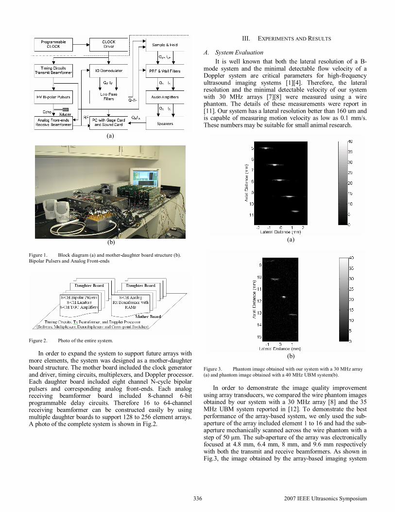
II. SYSTEM DESCRIPTION A block diagram of the duplex system with HF arrays is
illustrated in Fig.1.
As shown in Fig. 1, the system included two separated blocks: a 16-channel analog beamformer block in the dash-dot line box, and a HF directional pulsed-wave Doppler block in the dash line box. The main clock of the system was generated by a programmable clock generator ECS-P83/P85 (ECS, Inc. International, Kansas City, KS). The two blocks were synchronized by clocks which were distributed by a clock driver CD328 (Texas Instruments, Dallas, TX). The 16-channel analog beamformer block consisted of: a 30MHz array [8], timing circuits, 16-channel transmit beamformer, N-cycle bipolar pulsers, analog front-ends, and a 16-channel programmable analog receive beamformer. The beamformed echoes were first filtered by a band-pass filter BBP-30 (Mini-Circuits, Brooklyn, NY), and then fed to the HF directional pulsed-wave Doppler block. The Doppler block included an in-phase and quadrature (IQ) demodulator MIQC-60WD (Mini-Circuits, Brooklyn, NY), low-pass filters BLP-10.7 (Mini-Circuits, Brooklyn, NY), sample-and-hold circuits AD783 (Analog Devices, Norwood, MA), PRF filters MAX291 (Maxim Integrated Products, Sunnyvale, CA), wall filters MAX7490 (Maxim Integrated Products, Sunnyvale, CA), and audio amplifiers LM386 (National Semiconductor Corp., Santa Clara, CA). Doppler audio signals were played by two stereo speakers.
The RF data from the receive beamformer was digitized by a Gage A/D card CS14200 (Gage Applied Technologies Inc., Montreal, Quebec, Canada). The Doppler audio signals were digitized by an integrated sound card in PC. Then the digitized signals were processed and converted into either B-mode images or directional spectrogram by Labview software (National Instruments Corp., Austin, TX) in real time. B-mode and Doppler mode images can be easily switched from one to the other in experiments. Further off-line analysis was conducted using Matlab (The MathWorks, Inc., Natick, MA) based software.
Figure 1. Block diagram (a) and mother-daughter board structure (b). Bipolar Pulsers and Analog Front-ends
Figure 2. Photo of the entire system.
In order to expand the system to support future arrays with more elements, the system was designed as a mother-daughter board structure. The mother board included the clock generator and driver, timing circuits, multiplexers, and Doppler processor. Each daughter board included eight channel N-cycle bipolar pulsers and corresponding analog front-ends. Each analog receiving beamformer board included 8-channel 6-bit programmable delay circuits. Therefore 16 to 64-channel receiving beamformer can be constructed easily by using multiple daughter boards to support 128 to 256 element arrays. A photo of the complete system is shown in Fig.2.
III. EXPERIMENTS AND RESULTS
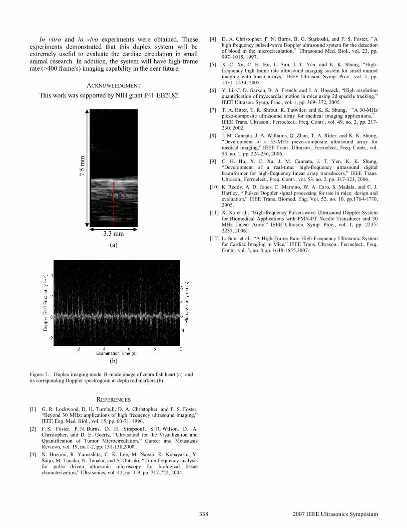
A. System Evaluation It is well known that both the lateral resolution of a B-
mode system and the minimal detectable flow velocity of a Doppler system are critical parameters for high-frequency ultrasound imaging systems [1][4]. Therefore, the lateral resolution and the minimal detectable velocity of our system with 30 MHz arrays [7][8] were measured using a wire phantom. The details of these measurements were report in [11]. Our system has a lateral resolution better than 160 um and is capable of measuring motion velocity as low as 0.1 mm/s. These numbers may be suitable for small animal research.
(a)
(b)
Figure 3. Phantom image obtained with our system with a 30 MHz array (a) and phantom image obtained with a 40 MHz UBM system(b).
In order to demonstrate the image quality improvement using array transducers, we compared the wire phantom images obtained by our system with a 30 MHz array [8] and the 35 MHz UBM system reported in [12]. To demonstrate the best performance of the array-based system, we only used the sub-aperture of the array included element 1 to 16 and had the sub-aperture mechanically scanned across the wire phantom with a step of 50 µm. The sub-aperture of the array was electronically focused at 4.8 mm, 6.4 mm, 8 mm, and 9.6 mm respectively with both the transmit and receive beamformers. As shown in Fig.3, the image obtained by the array-based imaging system
2007 IEEE Ultrasonics Symposium336
can achieve more constant lateral resolutions, while the image obtained by the UBM can only obtain a good lateral resolution at its focal point. This comparison demonstrated the advantages of the HF ultrasound imaging system with arrays. In addition, the lateral resolution of this array-based system can be further improved by expanding the sub-aperture from 16 elements to 64 elements.
B. In Vitro Experiments In vitro B-mode imaging studies were conducted as the
method described above. The pig eye and rabbit eye were imaged. The sub-aperture of the array was scanned across the eyes with a step of 100 µm.
Fig. 4 shows the B-mode images of the pig eye and rabbit eye. Satisfactory image quality was achieved.
C. In vivo Experiments At the Health Science Campus at USC, in vivo were
carried out on zebra fish and mice under approved protocols. After anesthetizing, either zebrafish or mice were fixed on a custom-designed stage. Water was used as a coupling medium.
We measured the Doppler signals from several major mouse arteries which were closed to its mouse heart. The obtained spectrograms illustrated in Fig. 5 shows different hemodynamics in different arteries and are valuable for cardiovascular studies. Compared these results to those reported in [10], the measuring points may include the carotid, mitral and aorta. Fig.6 also shows blood flow velocity profiles in mouse superficial vessels with diameters of about 200 µm.
Figure 5. Doppler spectrograms from major mouse heart arteries.
(a) (b)
Figure 6. (a) B-mode image obtained by the UBM in [12]. (b) Measured Doppler signals from these vessels by our system.
Duplex mode was used to image zebra fish heart. First real-time B-mode images were obtained to identify either heart or vessels, and then the system was switched to pulsed-wave Doppler mode to measure blood flow velocities. Due the current system setup, only Doppler signals at a particular location (i.e. at the central B-mode image line with a depth of 8 mm) can be detected and measured. The experiments results are shown in Fig. 7. Flexible and programmable Doppler measuring features are being added as well as high-frame imaging capability.
IV. CONCLUSION AND FUTURE WORK As an initial attempt, the first real-time HF duplex system
with 30 MHz linear arrays was implemented. Lateral resolutions of the array were measured from 160 µm to 310 µm at different depths. The system is capable of detecting motion velocity of the wire phantom as low as 0.1 mm/s.
Vessels
2007 IEEE Ultrasonics Symposium337
In vitro and in vivo experiments were obtained. These experiments demonstrated that this duplex system will be extremely useful to evaluate the cardiac circulation in small animal research. In addition, the system will have high-frame rate (>400 frame/s) imaging capability in the near future.
ACKNOWLEDGMENT This work was supported by NIH grant P41-EB2182.
(a)
(b)
Figure 7. Duplex imaging mode. B-mode image of zebra fish heart (a) and its corrsponding Doppler spectrogram at depth red markers (b).
REFERENCES [1] G. R. Lockwood, D. H. Turnbull, D. A. Christopher, and F. S. Foster,
“Beyond 30 MHz: applications of high frequency ultrasound imaging,” IEEE Eng. Med. Biol., vol. 15, pp. 60-71, 1996.
[2] F. S. Foster, P. N. Burns, D. H. Simpson1, S. R. Wilson, D. A. Christopher, and D. E. Goertz, “Ultrasound for the Visualization and Quantification of Tumor Microcirculation,” Cancer and Metastasis Reviews, vol. 19, no.1-2, pp. 131-138,2000.
[3] N. Hozumi, R. Yamashita, C. K. Lee, M. Nagao, K. Kobayashi, Y. Saijo, M. Tanaka, N. Tanaka, and S. Ohtsuki, “Time-frequency analysis for pulse driven ultrasonic microscopy for biological tissue characterization,” Ultrasonics, vol. 42, no. 1-9, pp. 717-722, 2004.
[4] D. A. Christopher, P. N. Burns, B. G. Starkoski, and F. S. Foster,“A high frequency pulsed-wave Doppler ultrasound system for the detection of blood in the microcirculation,”Ultrasound Med. Biol., vol. 23, pp. 997–1015, 1997.
[5] X. C. Xu, C. H. Hu, L. Sun, J. T. Yen, and K. K. Shung, “High-frequency high frame rate ultrasound imaging system for small animal imaging with linear arrays,” IEEE Ultrason. Symp. Proc., vol. 1, pp. 1431- 1434, 2005.
[6] Y. Li, C. D. Garson, B. A. French, and J. A. Hossack, “High resolution quantification of myocardial motion in mice using 2d speckle tracking,” IEEE Ultrason. Symp, Proc., vol. 1, pp. 369- 372, 2005.
[7] T. A. Ritter, T. R. Shrout, R. Tutwiler, and K. K. Shung, “A 30-MHz piezo-composite ultrasound array for medical imaging applications,” IEEE Trans. Ultrason., Ferroelect., Freq. Contr., vol. 49, no. 2, pp. 217-230, 2002.
[8] J. M. Cannata, J. A. Williams, Q. Zhou, T. A. Ritter, and K. K. Shung, “Development of a 35-MHz piezo-composite ultrasound array for medical imaging,” IEEE Trans. Ultrason., Ferroelect., Freq. Contr., vol. 53, no. 1, pp. 224-236, 2006.
[9] C. H. Hu, X. C. Xu, J. M. Cannata, J. T. Yen, K. K. Shung, “Development of a real-time, high-frequency ultrasound digital beamformer for high-frequency linear array transducers,” IEEE Trans. Ultrason., Ferroelect., Freq. Contr., vol. 53, no. 2, pp. 317-323, 2006.
[10] K. Reddy, A. D. Jones, C. Martono, W. A. Caro, S. Madala, and C. J. Hartley, “ Pulsed Doppler signal processing for use in mice: design and evaluation,” IEEE Trans. Biomed. Eng. Vol. 52, no. 10, pp.1764-1770, 2005.
[11] X. Xu et al., “High-frequency Pulsed-wave Ultrasound Doppler System for Biomedical Applications with PMN-PT Needle Transducer and 30 MHz Linear Array,” IEEE Ultrason. Symp. Proc., vol. 1, pp. 2235- 2237, 2006.
[12] L. Sun, et al., “A High-Frame Rate High-Frequency Ultrasonic System for Cardiac Imaging in Mice,” IEEE Trans. Ultrason., Ferroelect., Freq. Contr., vol. 5, no. 8,pp. 1648-1655,2007.