242
CA01.1- CA01.1- Introduction Cardiovascular System Lecture I Stephen Bagley, M.D. Resident Physician University of Pennsylvania 1
CA01.1- CA01.1-
Introduction Cardiovascular System Lecture I
Stephen Bagley, M.D. Resident Physician University of Pennsylvania
1

CA01.1- CA01.1-
To understand the following topics and how they may be
tested on USMLE Step 1 – Cardiac physiology
• Fluid dynamics • The cardiac cycle • Electrophysiology
– Heart sounds – Biochemistry of lipids – Ischemic heart disease and cardiomyopathies – Congenital heart disease – Vasculitis
Cardiovascular System Course Objectives:
2

CA01.1- CA01.1-
– Beta and alpha adrenergic receptors and
their role in the cardiovascular system – Cardiac output – Resistance, pressure, and flow – Cardiac function curve
Learning Objectives
3

CA01.2-
Kaplan Pharmacology 2011 : Table II-3-2
FA 2012: 263.1 • FA 2011: 236 • FA 2010: 232 • ME 3e: 169 1

CA01.3-
KNOW THE FICK PRINCIPLE
CO = O2 Consumption / (Arterial O2 – Venous O2)
• O2 Consumpsion Æ often given • Arterial O2 = [Hgb] x [1.34] x [Arterial O2 Saturation] • Venous O2 = [Hgb] x [1.34] x [Venous O2 Saturation]
FA 2012: 280.2 • FA 2011: 254.2 • FA 2010: 250 • ME 3e: 249
1

CA01.3-
Mean Arterial Pressure (MAP)… MAP = (CO) X (TPR) MAP = (2/3 Diastolic Pressure) + (1/3 Systolic Pressure) When heart rate increases, so does the cardiac output and the mean arterial pressure, all things being equal.
FA 2012: 280.2 • FA 2011: 254.2 • FA 2010: 250 • ME 3e: 248
2

CA01.3-
What changes cardiac output... By changing:
Contractility
Preload
Afterload
FA 2012: 281.1 • FA 2011: 255.1 • FA 2010: 251 • ME 3e: 243
3

CA01.3-
Kaplan Pharmacology 2011 : Figure III-4-2
FA 2012: 281.1 • FA 2011: 255.1 • FA 2010: 251 • ME 3e: 271
Ca2+ +
(SERCA)
4

CA01.4-
Kaplan Physiology 2011 : Figure IV-1-9
FA 2012: 281.1 • FA 2011: 255.1 • FA 2010: 251 • ME 3e: 251
1

CA01.4-
PRELOAD = Left Ventricular EDV
FA 2012: 281.2 • FA 2011: 255.2 • FA 2010: 251 • ME 3e: 242
2

CA01.4-
AFTERLOAD = Mean Arterial Pressure (MAP)
FA 2012: 281.2 • FA 2011: 255.2 • FA 2010: 251 • ME 3e: 242
3

CA01.4-
Kaplan Physiology 2011 : Figure VI-1-1
FA 2012: 281.2 • FA 2011: 255.2 • FA 2010: 251 • ME 3e: 242
4

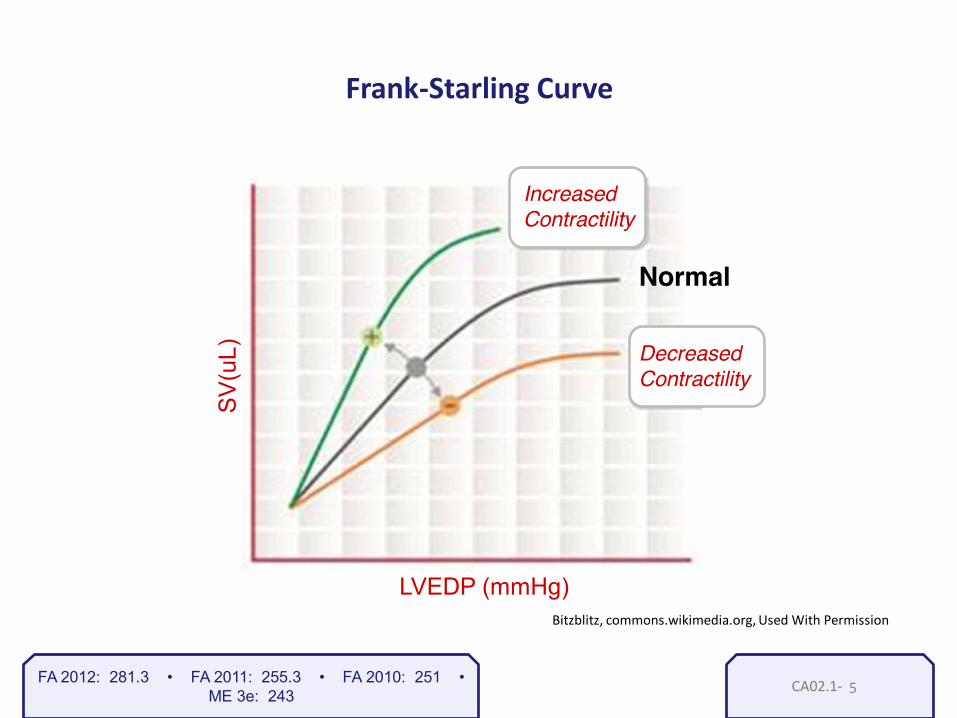
CA01.4-
Bitzblitz, commons.wikimedia.org, Used With Permission
Frank-Starling Curve
Increased Contractility
Decreased Contractility
SV
(uL)
LVEDP (mmHg)
Normal
FA 2012: 281.3 • FA 2011: 255.3 • FA 2010: 251 • ME 3e: 242
5

CA01.4-
NEED TO KNOW !
EF tells us about the heart’s contractility EF = SV / EDV = (EDV-ESV) / EDV A decrease in EF is seen in heart failure Echocardiograms are often used to diagnose heart failure by measuring ejection fraction!
FA 2012: 282.1 • FA 2011: 255.4 • FA 2010: 251 • ME 3e: 242
6

CA01.5-
Resistance (R) = 8(viscosity) x Length π r 4
As viscosity increases, resistance increases Examples: Polycythemia, multiple myeloma
As radius (r) increases, resistance decreases The aorta has less resistance to flow than a capillary
FA 2012: 282.2 • FA 2011: 256.1 • FA 2010: 252 • ME 3e: 248
1

CA01.5-
FA 2012: 282.2 • FA 2011: 256.1 • FA 2010: 252 • ME 3e: 248
2

CA01.5-
Kaplan Physiology 2011 : Figure V-1-5
FA 2012: 282.2 • FA 2011: 256.1 • FA 2010: 252 • ME 3e: 249
Velocity = Q/CSA Q = flow
CSA = cross sectional area
3

CA01.5-
Kaplan Physiology 2011 : Figure V-1-4
FA 2012: 282.2 • FA 2011: 256.1 • FA 2010: 252 • ME 3e: 249
4

CA01.5-
Cardiac and Vascular Function Curves
commons.wikimedia.org Used with permission
FA 2012: 282.3 • FA 2011: 256.2 • FA 2010: 252 • ME 3e: 243
filling pressure
5

CA02.1- CA02.1-
Lecture II • Cardiac cycle • Maintenance of MAP
Stephen Bagley, M.D. Resident Physician University of Pennsylvania
The Cardiac Cycle
1

CA02.1-
Kaplan Physiology 2011 : Figure VI -1-6
FA 2012: 283.1 • FA 2011: 257.1 • FA 2010: 253 • ME 3e: 245 2
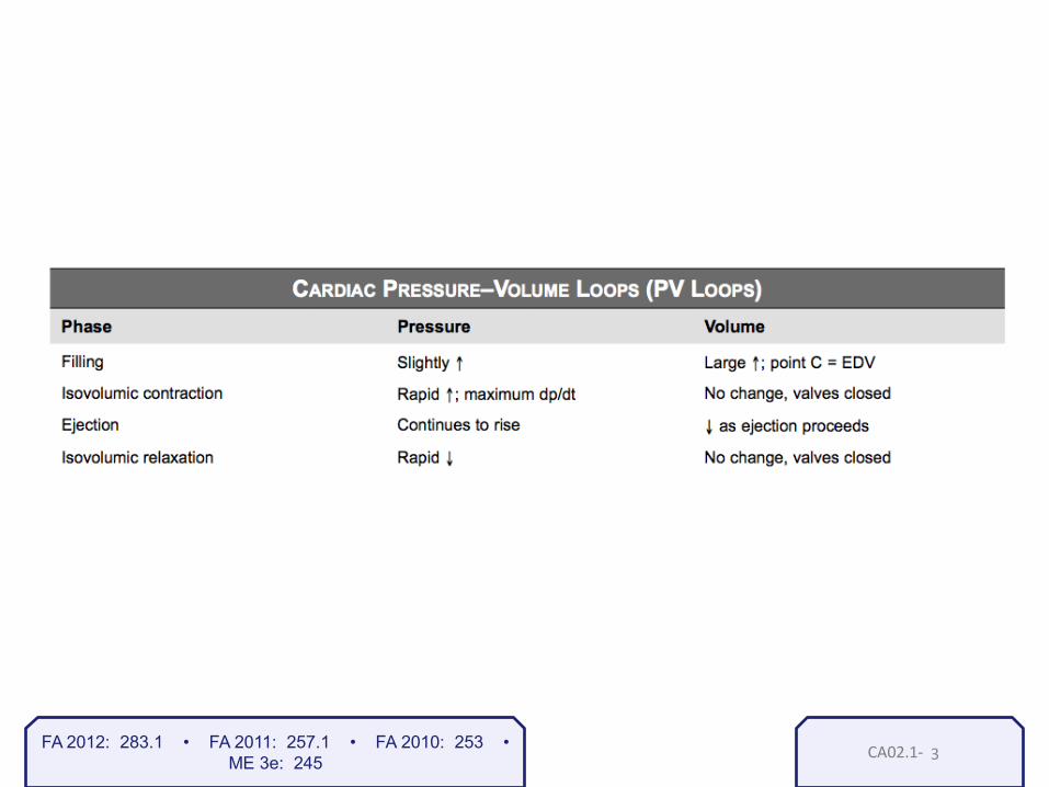
CA02.1- FA 2012: 283.1 • FA 2011: 257.1 • FA 2010: 253 • ME 3e: 245 3

CA02.1-
Kaplan Physiology 2011 : Figure VI -1-6
FA 2012: 283.1 • FA 2011: 257.1 • FA 2010: 253 • ME 3e: 245 4
CA02.1-
Bitzblitz, commons.wikimedia.org, Used With Permission
Increased Contractility
Decreased Contractility
SV
(uL)
LVEDP (mmHg)
Normal
Frank-Starling Curve
FA 2012: 281.3 • FA 2011: 255.3 • FA 2010: 251 • ME 3e: 243 5

CA02.1-
Points: – Greater pressure needed to
open aortic valve – Smaller stroke volume – An example of this would be Æ increased in systemic blood pressure
FA 2012: 283.1 • FA 2011: 257.1 • FA 2010: 253 • ME 3e: 246 6

CA02.1-
Points: – Phase 4 longer – Larger EDV – Wider curve – Increased PVL area – Increased SV and CO
FA 2012: 283.1 • FA 2011: 257.1 • FA 2010: 253 • ME 3e: 245 7

CA02.1-
Increasing Afterload Increasing Preload
FA 2012: 283.1 • FA 2011: 257.1 • FA 2010: 253 • ME 3e: 246 8

CA02.1-
Kaplan Physiology 2011 : Figure VI -1-6
FA 2012: 283.1 • FA 2011: 257.1 • FA 2010: 253 • ME 3e: 245 9

CA02.2-
Kaplan Physiology 2011 : Figure VI -1-1 & Figure VI -1-3
FA 2012: 283.1 • FA 2011: 257.1 • FA 2010: 253 • ME 3e: 244 1

CA02.3-
Lecture II continued Cardiac cycle Maintenance of MAP
Cardiology
1

CA02.3-
Lecture II continued
Maintenance of MAP
Cardiology
2

CA02.3-
Kaplan Physiology 2011 : Fig V-1-18
FA 2012: n/a • FA 2011: 265.1 • FA 2010: 261 • ME 3e: 253 3

CA02.3-
Kaplan Physiology 2011 : Fig X-4-9
FA 2012: n/a • FA 2011: 265.1 • FA 2010: 261 • ME 3e: 253 4

CA02.3-
Atrial Natriuretic Peptide
FA 2012: n/a • FA 2011: 265.1 • FA 2010: 261 • ME 3e: 316 5

CA02.3-
Return to this chart later for review…
FA 2012: 290.3 • FA 2011: 265.2 • FA 2010: 261 • ME 3e: 253 6

CA02.3-
Kaplan Pathology 2011 : Fig 5-5
FA 2012: 290.3 • FA 2011: 265.2 • FA 2010: 261 • ME 3e: 253 7

CA02.3-
Return to this chart later for review…
FA 2012: 290.3 • FA 2011: 265.2 • FA 2010: 261 • ME 3e: 253 8

CA02.3-
Kaplan Physiology 2011 : Fig VII-3-8
FA 2012: 290.3 • FA 2011: 265.2 • FA 2010: 261 • ME 3e: 288 9

CA02.3-
Don’t get tricked with Cushing’s Triad…
ICP Via sympathetic NS Hypertension Via sympathetic NS
Inappropriate response
Appropriate response
Bradycardia
TRIAD = Hypertension + Bradycardia + Respiratory Depression
FA 2012: 290.3 • FA 2011: 265.2 • FA 2010: 261 • ME 3e: 288 10

CARDIO3.1- CARDIO3.1-
Lecture III • Autoregulation of blood flow • Capillary fluid exchange • Sympathomimetic drugs
Regulation of blood flow and fluid exchange
Stephen Bagley, M.D. Resident Physician University of Pennsylvania
1

CARDIO3.1-
Nevit Dilmen, commons.wikimedia.org, Used With Permission
FA 2012: 291.1 • FA 2011: 265.3 • FA 2010: 261 • ME 3e: 252
2

CARDIO3.1-
FA 2012: 291.3 • FA 2011: 266.2 • FA 2010: 262 • ME 3e: 252
Autoregulation
Kaplan Physiology 2011 : Figure V-2-2
What factors control local blood flow?
• Heart: O2, adenosine, NO
• Brain: arterial pCO2/pH
• Kidney: tubuloglomerular feedback
• Lungs: hypoxic vasoconstriction (next slide)
• Skeletal muscle: lactate, adenosine, potassium
3

CARDIO3.1-
Pneumonia = No Oxygen
Nevit Dilmen, commons.wikimedia.org, Used With Permission
FA 2012: 291.3 • FA 2011: 266.2 • FA 2010: 262 • ME 3e: 252
4

CARDIO3.1-
Skin • Temperature influences local
blood flow
Kaplan Pharmacology 2011 : Figure V-2-4
FA 2012: 291.3 • FA 2011: 266.2 • FA 2010: 262 • ME 3e: 252
5

CARDIO3.1-
FA 2012: 291.4 • FA 2011: 266.3 • FA 2010: 262 • ME 3e: 250
6

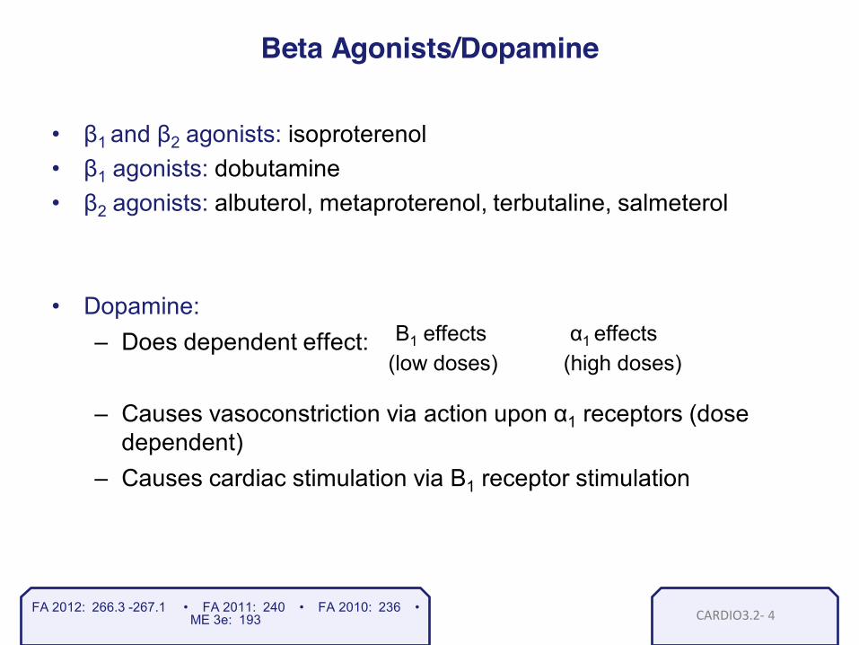
CARDIO3.2-
DIRECT SYMPATHOMIMETICS
Norepinephrine
Dobutamine
Dopamine
Epinephrine
Isoproterenol Phenylephrine
FA 2012: n/a • FA 2011: 240 • FA 2010: 236 • ME 3e: 193
1

CARDIO3.2-
Kaplan Pharmacology 2011 : Figure II-3-5c
FA 2012: n/a • FA 2011: 240 • FA 2010: 236 • ME 3e: 193
2

CARDIO3.2-
Kaplan Pharmacology 2011 : Figure II-3-4
FA 2012: n/a • FA 2011: 240 • FA 2010: 236 • ME 3e: 193
3
CARDIO3.2-
Beta Agonists/Dopamine
• β1 and β2 agonists: isoproterenol • β1 agonists: dobutamine • β2 agonists: albuterol, metaproterenol, terbutaline, salmeterol
• Dopamine: – Does dependent effect:
– Causes vasoconstriction via action upon α1 receptors (dose
dependent) – Causes cardiac stimulation via B1 receptor stimulation
FA 2012: 266.3 -267.1 • FA 2011: 240 • FA 2010: 236 • ME 3e: 193
Β1 effects α1 effects (low doses) (high doses)
4

CARDIO3.2-
Kaplan Pharmacology 2011 : Figure II-3-2
FA 2012: 266.3 • FA 2011: 240 • FA 2010: 236 • ME 3e: 193 5

CARDIO3.2-
FA 2012: 267.1 • FA 2011: 240 • FA 2010: 236 • ME 3e: 194
• Both amphetamines and cocaine are indirect sympathomimetics.
• Both increase endogenous norepinephrine in the synapse.
6

CARDIO3.3-
Clinical Pearls… Clonidine often used to treat opiate withdrawal
– Dampens the sympathetic response that occurs during opiate withdrawal
α-methyldopa indicated in hypertension in pregnancy
FA 2012: 267.2 • FA 2011: 241.1 • FA 2010: 237 • ME 3e: 193
1

CARDIO3.4-
Chikumaya, commons.wikimedia.org, Used With permission
FA 2012: 291.2 • FA 2011: 266.1 • FA 2010: 262 • ME 3e: 249
1

CA04.1 - CA04.1 -
Lecture IV • Normal electrophysiology
Cardiac Electrophysiology
Stephen Bagley, M.D. Resident Physician University of Pennsylvania
1

CA04.1 -
Kaplan Physiology 2011 : Figure VI -1-6
FA 2012: n/a • FA 2011: 260.1 • FA 2010: 256 • ME 3e: 232
2
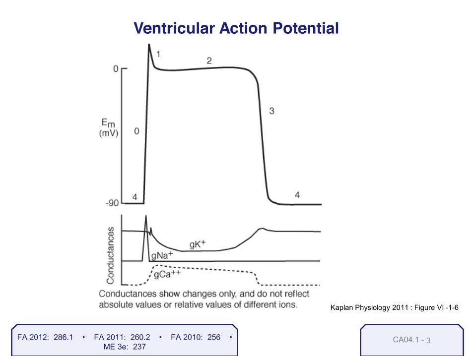
CA04.1 -
Ventricular Action Potential
Kaplan Physiology 2011 : Figure VI -1-6
FA 2012: 286.1 • FA 2011: 260.2 • FA 2010: 256 • ME 3e: 237
3
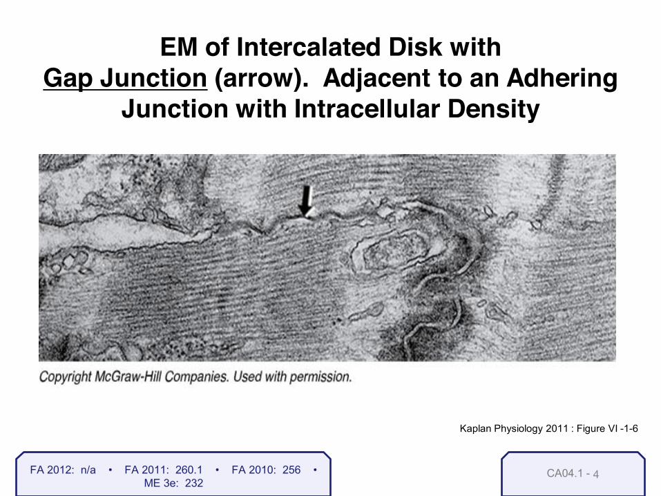
CA04.1 -
EM of Intercalated Disk with
Gap Junction (arrow). Adjacent to an Adhering Junction with Intracellular Density
Kaplan Physiology 2011 : Figure VI -1-6
FA 2012: n/a • FA 2011: 260.1 • FA 2010: 256 • ME 3e: 232
4

CA04.1 -
Cardiac Action Potentials in Fast-Response Fibers
Kaplan Physiology 2011 : Figure VI -1-6
FA 2012: 286.1 • FA 2011: 260.2 • FA 2010: 256 • ME 3e: 237
5

CA04.1 -
Mechanism of Action of Voltage-Gated Na+ Channels
FA 2012: 286.1 • FA 2011: 260.2 • FA 2010: 256 • ME 3e: 237
6

CA04.1 -
Cardiac Action Potentials in Fast-Response Fibers
Kaplan Physiology 2011 : Figure VI -1-6
FA 2012: 286.1 • FA 2011: 260.2 • FA 2010: 256 • ME 3e: 237
7

CA04.1 -
Cardiac Action Potentials in Slow-Response Fibers – SA and AV Nodes
FA 2012: 287.1 • FA 2011: 261.1 • FA 2010: 257 • ME 3e: 237
8

CA04.1 -
Sympathetic Effects
Parasympathetic Effects
Kaplan Physiology 2011 : Figure VI -1-6
FA 2012: 287.1 • FA 2011: 261.1 • FA 2010: 257 • ME 3e: 237
9

CA04.1 -
Cardiac Action Potentials in Slow-Response Fibers – SA and AV Nodes
FA 2012: 287.1 • FA 2011: 261.1 • FA 2010: 257 • ME 3e: 237
10

CA04.2 -
Glenlarson, commons.wikimedia.org, Used With permission
Normal ECG Demonstrating Sinus Rhythm
FA 2012: 288.1 • FA 2011: 262.1 • FA 2010: 258 • ME 3e: 239
1

CA04.2 -
EKG Leads
FA 2012: 288.1 • FA 2011: 262.1 • FA 2010: 258 • ME 3e: 241
2

CA04.2 -
commons.wikimedia.org Used with permission
FA 2012: 288.1 • FA 2011: 262.1 • FA 2010: 258 • ME 3e: 241
3

CA04.2 -
Normal Pattern of an ECG
Kaplan Physiology 2011 : Figure VI -1-6
FA 2012: 288.1 • FA 2011: 262.1 • FA 2010: 258 • ME 3e: 238
4

CA05.1- CA05.1-
Arrhythmias
Lecture V – Arrhythmias – Antiarrhythmic drugs
Stephen Bagley, M.D. Resident Physician University of Pennsylvania
1

CA05.1-
Lecture V Arrhythmias
Cardiovascular System
2

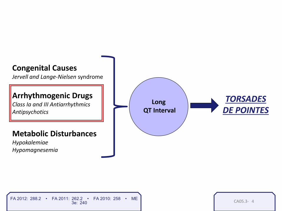
CA05.1-
TORSADES DE POINTES
Long QT Interval
Congenital Causes Jervell and Lange-Nielsen syndrome
Arrhythmogenic Drugs Class Ia and III Antiarrhythmics Antipsychotics
Metabolic Disturbances Hypokalemia, hypomagnesia,
FA 2012: 288.2 • FA 2011: 262.2 • FA 2010: 258 • ME 3e: 240 3

CA05.1- FA 2012: n/a • FA 2011: 263.1 • FA 2010: 259 • ME 3e: 236
DeltaWave09.jpg, commons.wikimedia.org, Used With Permission
Delta Waves
WPW.jpeg, commons.wikimedia.org, Used With Permission
Two-way
Shortened PR interval
Two-way accessory pathway
4

CA05.1- FA 2012: 288.1 • FA 2011: 262.1 • FA 2010: 259 • ME 3e: 236
Arrhythmias Atrial fibrillation:
Atrial flutter:
5

CA05.1-
Arrhythmias: AV Block 1st Degree
2nd Degree
3rd Degree
Progressive prolongation of the PR interval
A blocked beat is not preceded by a change in PR interval
FA 2012: 288.1 • FA 2011: 264.1 • FA 2010: 260 • ME 3e: 236
•Slowed conduction through the AV node
•Some impulses are not transmitted through the AV node
•No impulses are conducted from the atria to ventricles
6

CA05.1-
Arrhythmias: Ventricular Ventricular fibrillation :
FA 2012: 288.1 • FA 2011: 262.1 • FA 2010: 259 • ME 3e: 236 7

CA05.2-
Lecture V
Antiarrhythmic drugs
Cardiovascular System
1

CA05.3- 1
Cardiac Action Potentials in Fast-Response Fibers
Kaplan Physiology 2011 : Figure VI -1-6
FA 2012: 310.1 • FA 2011: 284.1 • FA 2010: 280 • ME 3e: 237

CA05.3-
Mechanism of Action of Voltage-Gated Na+ Channels
Kaplan Physiology 2011 : Figure VI -1-6
FA 2012: 310.1 • FA 2011: 284.1 • FA 2010: 280 • ME 3e: 267 2

CA05.3-
Cardiac Action Potentials in Fast-Response Fibers
Kaplan Physiology 2011 : Figure VI -1-6
• Class IA drugs block Na+ channels and increase AP duration
• Examples: quinidine, procainamide, disopyramide
• Should not be used in patients with a long QT interval
FA 2012: 310.1 • FA 2011: 284.1 • FA 2010: 280 • ME 3e: 237 3
CA05.3-
TORSADES DE POINTES
Long QT Interval
Congenital Causes Jervell and Lange-Nielsen syndrome
Arrhythmogenic Drugs Class Ia and III Antiarrhythmics Antipsychotics
Metabolic Disturbances Hypokalemiae Hypomagnesemia
FA 2012: 288.2 • FA 2011: 262.2 • FA 2010: 258 • ME 3e: 240 4

CA05.3-
Antiarrhythmic toxicities are heavily tested on
USMLE Step 1…
Class Ia Class Ib Class Ic • Thrombocytopenia • Torsades de pointes • Reversible lupus-like syndrome (procainamide) • Cinchonism (quinidine)
• CNS stimulation • CV depression
• Proarrhythmic • Contraindicated after myocardial infarction
The toxicity is increased in all class I antiarrhythmics with HYPERkalemia.
FA 2012: 310.1 • FA 2011: 284.1 • FA 2010: 280 • ME 3e: 267 5

CA05.3-
Cardiac Action Potentials in Fast-Response Fibers
Kaplan Physiology 2011 : Figure VI -1-6
• Class IB drugs decrease AP duration; lidocaine, mexiletine, phenytoin
• Class IC drugs have no effect on AP duration; flecanide, encainide, propafenone; pro-arrhythmic
FA 2012: 310.1 • FA 2011: 284.1 • FA 2010: 280 • ME 3e: 237 6

CA05.3-
Antiarrhythmic toxicities are heavily tested on
USMLE Step 1…
Class Ia Class Ib Class Ic • Thrombocytopenia • Torsades de pointes • Reversible lupus-like syndrome (procainamide) • Cinchonism (quinidine)
• CNS Stimulation • CV depression
• Proarrhythmic • Contraindicated after myocardial infarction
The toxicity is increased in all class I antiarrhythmics with HYPERkalemia.
FA 2012: 310.1 • FA 2011: 284.1 • FA 2010: 280 • ME 3e: 267 7

CA05.4- FA 2012: 311.1 • FA 2011: 285.1 • FA 2010: 281 • ME 3e: 269
Hypertension Angina Chronic heart failure (carvedilol, labetalol, metoprolol) Arrhythmia (propranolol, acebutolol, esmolol) Glaucoma (timolol) Migraine, tremor, thyrotoxicosis (propranolol)
Decreased libido
1

CA05.4-
Cardiac Action Potentials in Slow-Response Fibers
FA 2012: 311.1 • FA 2011: 285.1 • FA 2010: 281 • ME 3e: 237 2

CA05.4-
• Beta Blockers – Clinical Uses:
• Supraventricular tachycardia • Atrial fibrillation • Atrial flutter
– Toxicities:
• Impotence in men • Exacerbation of asthma with use of non-selective -blockers • Can mask signs of hypoglycemia in diabetics
FA 2012: 311.1 • FA 2011: 285.1 • FA 2010: 281 • ME 3e: 269 3

CARDIO5_5 - 1
Cardiac Action Potentials in Fast-Response Fibers
Kaplan Physiology 2011 : Figure VI -1-6
• Class III drugs block K+ channels; sotalol, amiodarone
FA 2012: 310.1 • FA 2011: 284.1 • FA 2010: 280 • ME 3e: 237

CARDIO5_5 - 2
Amiodarone toxicity is commonly tested
Remember to check…
LFTs = Liver Function Tests
PFTs = Pulmonary Function Tests
TFTs = Thyroid Function Tests
commons.wikimedia.org, Used With permission
FA 2012: 311.2 • FA 2011: 285.2 • FA 2010: 281 • ME 3e: 268

CA05.6
Cardiac or Vascular Selectivity of Major Ca2+-Channel Blockers
FA 2012: 312.1 • FA 2011: 286.1 • FA 2010: 282 • ME 3e: 268
(Ca channel blockers which are not used as anti-arrhythmics)
1

CA05.7- FA 2012: 312.2 • FA 2011: 286.2 • FA 2010: 282 • ME 3e: 268
• Clinical Pearls… • Adenosine increases K+ efflux from cells Æ Causes hyperpolarization
• K+ is provided to patients suffering from diabetic ketoacidosis because they are grossly hypokalemic and at risk for arrhythmias.
• Mg+ is used to treat and prevent cardiac arrhythmias and seizures in pre-eclampsia!
1
CARDIO06_1a- CARDIO06_1-
Heart Sounds
Cardiovascular System
1
Stephen Bagley, M.D. Resident Physician University of Pennsylvania

CARDIO6_2c- 1
Abnormal Splitting of the Second Heart Sound (S2)
FA 2012: 284.1 • FA 2011: 258.1 • FA 2010: 254 • ME 3e: 254

CARDIO6_2c- 2
COMMON FORMS OF ACYANOTIC CONGENITAL HEART DISEASE
FA 2012: 284.1 • FA 2011: 258.1 • FA 2010: 254 • ME 3e: 254

CARDIO6_2c- 3
Abnormal Splitting of the Second Heart Sound (S2)
FA 2012: 284.1 • FA 2011: 258.1 • FA 2010: 254 • ME 3e: 255

CARDIO6_3- 1
Projection of Heart Valve Sounds on Anterior Chest Wall
FA 2012: 284.2 • FA 2011: 258.2 • FA 2010: 254 • ME 3e: 234

CARDIO6_4- 1
Auscultation of Heart Murmurs
FA 2012: 285.1 • FA 2011: 259.1 • FA 2010: 255 • ME 3e: 234
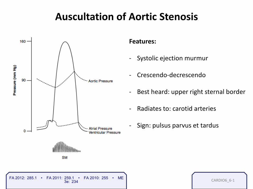
CARDIO6_6- 1 FA 2012: 285.1 • FA 2011: 259.1 • FA 2010: 255 • ME 3e: 234
Auscultation of Aortic Stenosis Features: - Systolic ejection murmur - Crescendo-decrescendo
- Best heard: upper right sternal border
- Radiates to: carotid arteries
- Sign: pulsus parvus et tardus

CARDIO6_7a- 1
Forms of Acyanotic Congenital Heart Disease
FA 2012: 285.1 • FA 2011: 259.1 • FA 2010: 255 • ME 3e: 231

CARDIO6_8a- 1
National Institute of Health, commons.wikimedia.org, Used With permission
FA 2012: 285.1 • FA 2011: 259.1 • FA 2010: 255 • ME 3e: 231

CARDIO6_9a- 1
Aortic Insufficiency (Regurgitation)
Features: - Diastolic flow murmur
- Best heard: lower left sternal border
- Wide pulse pressure
- Bounding pulses
- Causes: HTN (most common), RF, IE
FA 2012: 285.1 • FA 2011: 259.1 • FA 2010: 255 • ME 3e: 247

CARDIO6_10a- 1
Mitral Stenosis
Features: - Diastolic murmur
- Best heard: at the cardiac apex
- Opening snap
- Cause: Rheumatic heart disease
(most common)
FA 2012: 285.1 • FA 2011: 259.1 • FA 2010: 255 • ME 3e: 247

CARDIO6_10a- 2
Features: - Diastolic murmur
- Best heard: at the cardiac apex
- Opening snap
- Cause: Rheumatic heart disease
(most common)
Mitral Stenosis
FA 2012: 285.1 • FA 2011: 259.1 • FA 2010: 255 • ME 3e: 247

CARDIO6_11a- 1
Ductus Arteriosus
FA 2012: 285.1 • FA 2011: 259.1 • FA 2010: 255 • ME 3e: 254

CARDIO6_12- 1
S3
FA 2012: 285.1 • FA 2011: 257.1 • FA 2010: 253 • ME 3e: 244

CARDIO6_12c- 1
S4
FA 2012: 285.1 • FA 2011: 257.1 • FA 2010: 253 • ME 3e: 244

CARDIO6_12e- 1
Auscultation of Heart Murmurs
FA 2012: 285.1 • FA 2011: 259.1 • FA 2010: 255 • ME 3e: 247

CARDIO7.1- CARDIO7.1-
Lecture VII – Hypertension and antihypertensive
medications
Hypertension
Stephen Bagley, M.D. Resident Physician University of Pennsylvania
1

CARDIO7.1-
Hypertension Basics
1. Majority of HTN is “essential” = idiopathic
2. Risk factors affect not only the risk of HTN, but affect the first-
line treatment
3. HTN is a problem because of its long-term consequences Æ stroke, LVH, retinopathy, etc.
FA 2012: 294.2 • FA 2011: 269.2 • FA 2010: 265 • ME 3e: 265 2

CARDIO7.2-
Angiogram of Renal Artery Stenosis
FA 2012: 294.2 • FA 2011: 269.2 • FA 2010: 265 • ME 3e: 265 1

CARDIO7.2- FA 2012: 294.2 • FA 2011: 269.2 • FA 2010: 265 • ME 3e: 265 2

CARDIO7.3-
What’s Happening in Hypertension
1. Decreased number of arterioles
2. Increased arteriolar wall thickness
3. Increased total peripheral resistance
FA 2012: 294.2 • FA 2011: 269.2 • FA 2010: 265 • ME 3e: 265 1

CARDIO7.4-
Patrick J. Lynch, commons.wikimedia.org, Used With Permission
Left Ventricular Hypertrophy Secondary To Longstanding HTN
FA 2012: 294.2 • FA 2011: 269.2 • FA 2010: 265 • ME 3e: 265 1

CARDIO7_5- 1 FA 2012: 294.2 • FA 2011: 269.2 • FA 2010: 265 • ME 3e: 264
Arteriolosclerosis and Onion-Skinning
Note the grossly thickened wall of this arteriole
Tdvorak; commons.wikimedia.org, Used With Permission

CARDIO7_5-
FA 2012: 294.4 • FA 2011: 269.4 • FA 2010: 265 • ME 3e: 264 2

CARDIO7_6-
Location of Aneurysms
FA 2012: 295.3 • FA 2011: 270.2 • FA 2010: 265 • ME 3e: 265 1

CARDIO7_6-
KGH, commons.wikimedia.org, Used With Permission
This is a section of aorta that contains myxomatous
degeneration that led to an aortic dissection
FA 2012: 295.3 • FA 2011: 270.2 • FA 2010: 265 • ME 3e: 265 2

CARDIO7_6-
J Heuser, commons.wikimedia.org, Used With Permission
Widened mediastinum seen on Chest x-ray in aortic dissection
FA 2012: 295.3 • FA 2011: 270.2 • FA 2010: 265 • ME 3e: 265 3

CARDIO7_7-
When treating Hypertension, the USMLE often asks, “What’s the drug of choice…”
• Essential hypertension? •Diuretics (1st ine) •ACE Inhibitors or ARBs •Calcium channel blockers
•The Hypertensive CHF patient? •: Diuretics (e.g. furosemide, hydrochlorothiazide)
• Potassium-sparring diuretics (e.g. spironolactone) • Nesiritide: an analogue of brain natriuretic peptide (BNP) • ACE inhibitors or ARBs: decrease BP and cardiac remodeling • -blockers: used in compensated (not acute) CHF
• The Hypertensive Diabetic? •ACE inhibitors or ARBs (1st line): are reno-protective, slowing progression to nephropathy •2nd line agents:
• Diuretics • Calcium channel blockers • Beta-Blockers
•Notable side effect: •Dyslipidemia (metoprolol, HCTZ,)
FA 2012: 306.1 • FA 2011: 280.1 • FA 2010: 276 • ME
3e: 269 1

CARDIO7_7- FA 2012: 306.2 • FA 2011: 280.2 • FA 2010: 276 • ME 3e: 269 2

CARDIO7_7-
Selectivity of Major Ca2+-Channel Blockers
FA 2012: 306.3 • FA 2011: 280.3 • FA 2010: 277 • ME 3e: 269 3

CARDIO7_7- FA 2012: 306.2 • FA 2011: 280.2 • FA 2010: 277 • ME 3e: 269 4

CARDIO7_7- FA 2012: 268.1 • FA 2011: 241.2 • FA 2010: 237 • ME 3e: 269
α1-antagonist: Nonselective Selective
Phentolamine (reversible) Phenoxybenzamine (irreversible) α1 - prazosin,terazosin,doxazosin α2 - mirtazapine
Blocks α1 receptors on arterioles and venules and/or α2 receptors in the CNS
α2 antagonists: used to treat depression
Non-selective antagonists: used to treat pheochromocytoma
α1 antagonists: used to treat benign prostatic hyperplasia
Side effects: α1 blockade: reflex tachycardia, orthostatic hypotension and syncope α2 blockade:
α2-agonists:
Clonidine
Methyldopa
Decrease sympathetic outflow by stimulating α2 receptors in the CNS
Rebound hypertension, dry mouth, sedation, bradyarrhythmias
Sedation, positive Coombs test: it is a prodrug that is converted to α-methyl norepinephrine
5

CARDIO7_7- FA 2012: 269.1 • FA 2011: 242.1 • FA 2010: 238 • ME 3e: 269
Characteristics of Some Beta Blockers
6

CARDIO8_1- CARDIO8_1-
Lecture VIII – Biochemistry of lipids – Primary dyslipidemias – Lipid-lowering agents
Lipid metabolism
1
Stephen Bagley, M.D. Resident Physician University of Pennsylvania

CARDIO8_2- 1
Overview of Lipoprotein Metabolism
FA 2012: 120.1 • FA 2011: 114.1 • FA 2010: 114 • ME 3e: 53

CARDIO8_2- 2 FA 2012: 350.1 • FA 2011: 320.7 • FA 2010: 314 • ME 3e: 351
Composition water, bilirubin (bile pigments), bile salts, phospholipids, cholesterol
Function Form micelles around dietary lipids to facilitate absorption of fats, cholesterol, and fat-soluble vitamins
Synthesis
primary bile salts
primary bile salts
glycine / taurine conjugation liver
hepatocytes
Brief Review of Bile:
cholesterol primary bile acids
secondary bile salts
gut bacteria

CARDIO8_2- 3
Dietary Lipids
Lipid breakdown by enterocytes at the brush border:
Triglycerides
Cholesterol esters
Phospholipids
pancreatic lipase 2-monoglyceride + fatty acids
cholesterol esterase cholesterol + fatty acid
Absorbable Lipids
lysolecithin + fatty acid phospholipase A2
FA 2012: 120.1 • FA 2011: 114.1 • FA 2010: 114 • ME 3e: 51

CARDIO8_2- 4
Overview of Lipoprotein Metabolism
FA 2012: 120.1 • FA 2011: 114.1 • FA 2010: 114 • ME 3e: 53

CARDIO8_2- 5
Enzyme Triglycerides (TGs) degraded Pancreatic lipase Dietary TGs in small intestine Lipoprotein lipase TGs circulating in chylomicrons and VLDL Hepatic triglyceride lipase TGs that remain in IDL Hormone-sensitive lipase TGs stored in adipocytes (most active
during fasting)
Lipase enzymes catalyze breakdown of triglycerides into free fatty acids
FA 2012: 120.1 • FA 2011: 114.1 • FA 2010: 114 • ME 3e: 51
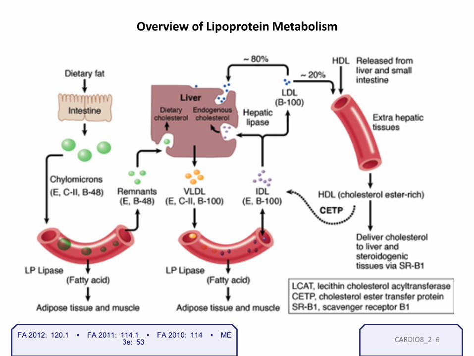
CARDIO8_2- 6
Overview of Lipoprotein Metabolism
FA 2012: 120.1 • FA 2011: 114.1 • FA 2010: 114 • ME 3e: 53

CARDIO8_3- 1
Apoproteins Function
apoA-1 Activates lecithin cholesterol acyltransferase (LCAT) to produce cholesterol esters
apoB-100 Binds LDL receptor to facilitate uptake by the liver Mediates VLDL secretion
apoC-II Cofactor for lipoprotein lipase
apoB-48
Mediates chylomicron secretion from intestinal enterocytes into lymphatic vessels
apoE Mediates remnant uptake
FA 2012: 120.2 • FA 2011: 114.2 • FA 2010: 114 • ME 3e: 53

CARDIO8_4- 1
Classes of Lipoproteins and Important Apoproteins
FA 2012: 121.1 • FA 2011: 115.1 • FA 2010: 115 • ME 3e: 53

CARDIO8_5-
Type
Deficiency
Lipid Elevated in Blood
Lipoprotein Elevated in Blood
Comments
I Familial lipoprotein lipase (rare) apoC-II (rare) Autosomal recessive
Triglyceride Chylomicrons Red-orange eruptive xanthomas (eyelids) Fatty liver Acute pancreatitis Abdominal pain after fatty meals
IIa Familial hypercholesterolemia Autosomal dominant (Aa 1/500, AA 1/106) LDL-receptor deficiency
Cholesterol LDL High risk of atherosclerosis and coronary artery disease Homozygous condition, usually death <20 years Xanthomas of Achilles tendon Tuberous xanthomas on elbows Xanthelasmas Corneal arcus
IV Hepatic overproduction of VLDL
VLDL TG Pancreatitis
1
Primary Hyperlipidemias
FA 2012: 121.2 • FA 2011: 115.2 • FA 2010: 115 • ME 3e: 53
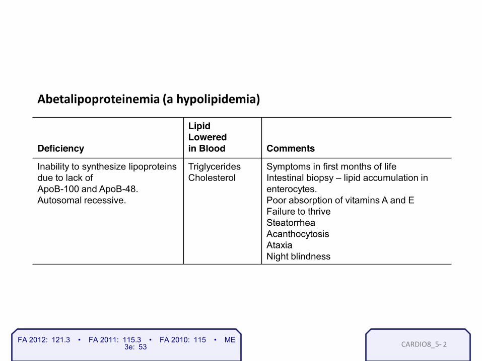
CARDIO8_5- 2
Deficiency
Lipid Lowered in Blood
Comments
Inability to synthesize lipoproteins due to lack of ApoB-100 and ApoB-48. Autosomal recessive.
Triglycerides Cholesterol
Symptoms in first months of life Intestinal biopsy – lipid accumulation in enterocytes. Poor absorption of vitamins A and E Failure to thrive Steatorrhea Acanthocytosis Ataxia Night blindness
Abetalipoproteinemia (a hypolipidemia)
FA 2012: 121.3 • FA 2011: 115.3 • FA 2010: 115 • ME 3e: 53

CARDIO8_6- 1
Statins
FA 2012: 308.1 • FA 2011: 282.1 • FA 2010: 278 • ME 3e: 52

CARDIO8_6- 2
Drug Class/Agent Mechanisms Side Effects/Comments
HMG-CoA Reductase Inhibitors (“-statins”: lovastatin, atorvastatin, fl uvastatin pravastatin, simvastatin, rosuvastatin,)
Inhibit rate-limiting step in cholesterol synthesis
-receptor
expression
• Myalgia, myopathy (check creatine kinase) • Rhabdomyolysis • aminotransferases • Teratogenic • P450 inhibitors can risk of hepatotoxicity, myopathy
Lipid-Lowering Drugs
FA 2012: 308.1 • FA 2011: 282.1 • FA 2010: 278 • ME 3e: 52

CARDIO8_6- 3
Drug Class/Agent Mechanisms Side Effects/Comments
Niacin Liver: Adipose tissue:
Flushing (and over time), pruritus, hepatotoxicity
Lipid-Lowering Drugs
FA 2012: 308.1 • FA 2011: 282.1 • FA 2010: 278 • ME 3e: 53

CARDIO8_6- 4
Drug Class/Agent Mechanisms Side Effects/Comments
Bile Acid Sequestrants (cholestyramine, colestipol, colesevelam)
Enterohepatic recirculation of bile salts, leading to:
salts by liver
-receptor expression
• GI disturbances • Malabsorption of lipid- soluble vitamins • (e.g., warfarin, thiazides, digoxin, pravastatin, fluvastatin)
Lipid-Lowering Drugs
FA 2012: 308.1 • FA 2011: 282.1 • FA 2010: 278 • ME 3e: 53

CARDIO8_6- 5
Drug Class/Agent Mechanisms Side Effects/Comments
Ezetimibe Blocks intestinal absorption of cholesterol
Possible hepatotoxicity with reductase inhibitors
Lipid-Lowering Drugs
FA 2012: 308.1 • FA 2011: 282.1 • FA 2010: 278 • ME 3e: 53

CARDIO8_6- 6
Drug Class/Agent Mechanisms Side Effects/Comments
Fibrates (gemfibrozil, clofibrate, bezafibrate, fenofibrate)
Ligands for PPAR-αactivation of lipoprotein lipases
Modest
• Gallstones • Myopathy (especially when combined with reductase inhibitors) • Can patients, so often combined with other cholesterol-lowering agents • Hepatotoxicity (rare)
Lipid-Lowering Drugs
FA 2012: 308.1 • FA 2011: 282.1 • FA 2010: 278 • ME 3e: 53

CARDIO9_1- CARDIO9_1-
Lecture IX Atherosclerosis Ischemic heart disease
Atherosclerosis
Stephen Bagley, M.D. Resident Physician University of Pennsylvania
1

CARDIO9_1-
Arterial Supply to the Heart
FA 2012: 280.1 • FA 2011: 254.1 • FA 2010: 250 • ME 3e: 233 2

CARDIO9_1-
Structures of the Mediastinum
FA 2012: 280.1 • FA 2011: 254.1 • FA 2010: 250 • ME 3e: 232 3

CARDIO9_2-
Atheromatous Plaque
FA 2012: 295.1 • FA 2011: 270.1 • FA 2010: 266 • ME 3e: 265 1

CARDIO9_3-
Location of Aneurysms
FA 2012: 295.1 • FA 2011: 270.1 • FA 2010: 266 • ME 3e: 265 1
CARDIO9_3-
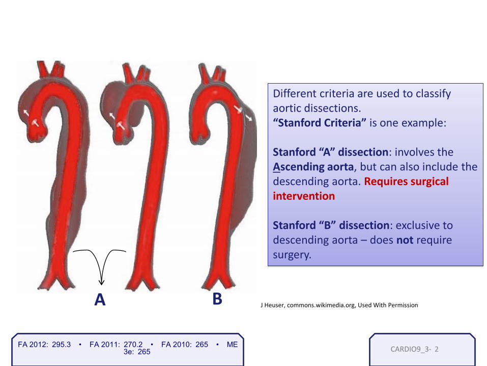
FA 2012: 295.3 • FA 2011: 270.2 • FA 2010: 265 • ME 3e: 265
J Heuser, commons.wikimedia.org, Used With Permission A B
Different criteria are used to classify aortic dissections. “Stanford Criteria” is one example: Stanford “A” dissection: involves the Ascending aorta, but can also include the descending aorta. Requires surgical intervention Stanford “B” dissection: exclusive to descending aorta – does not require surgery.
2

CARDIO9_3-
KGH, commons.wikimedia.org, Used With Permission
Notice the tear between the two layers that has resulted in the formation of a "false lumen".
True lumen
False lumen
FA 2012: 295.3 • FA 2011: 270.2 • FA 2010: 265 • ME 3e: 265 3

CARDIO9_3-
J Heuser, commons.wikimedia.org, Used With Permission A B
Different criteria are used to classify aortic dissections. “Stanford Criteria” is one example: Stanford “A” dissection: involves the Ascending aorta, but can also include the descending aorta. Requires surgical intervention Stanford “B” dissection: exclusive to descending aorta – does not require surgery.
FA 2012: 295.3 • FA 2011: 270.2 • FA 2010: 265 • ME 3e: 265 4

CARDIO9_3-
J Heuser, commons.wikimedia.org, Used With Permission
Widened mediastinum seen on Chest x-ray in aortic dissection
FA 2012: 295.3 • FA 2011: 270.2 • FA 2010: 265 • ME 3e: 265 5

CARDIO9_4- FA 2012: 296.1 • FA 2011: 270.3 • FA 2010: 266 • ME 3e: 256
Ischemic Heart Disease
– Angina • Stable • Unstable • Prinzmetal’s
– Myocardial infarction – Sudden cardiac death – Chronic ischemic heart disease
1

CARDIO10_1- CARDIO10_1-
Lecture X Myocardial infarction Antianginal therapy
Ischemic Heart Disease
1
Stephen Bagley, M.D. Resident Physician University of Pennsylvania

CARDIO10_1- 2
FA 2012: 297.1 • FA 2011: 271.1 • FA 2010: 267 • ME 3e: 257
Time Gross Histologic Complications
1 day Dark mottling. Pale after tetrazolium stain
First 2-4 hours: No visible changes 4-24 hours: Early coagulative necrosis Contraction bands (>4 hrs)
Arrhythmia
2 - 4 days Hyperemia Acute inflammation: Dilated vessels Neutrophils Loss of myocyte nuclei
Arrhythmia
5 - 10 days Yellow-brown softening with hyperemic border
Macrophages Granulation tissue
Free wall rupture Tamponade Papillary muscle rupture Septal rupture
Weeks to months
Gray-white scar Contracted scar Ventricular aneurysm
Tissue Changes in Myocardial Infarction

CARDIO10_1- 3 FA 2012: 298.1 • FA 2011: 272.1 • FA 2010: 268 • ME 3e: 257
Diagnosis of MI
Troponin I first 4 hours 7-10 days
CK-MB 24 hours
cardiac specific
non-specific; released by all muscle injury
Peak Duration
2-3 days
LDH 48 hours
AST non-specific; released by all muscle injury and liver injury
7-10 days
24 hours 2-3 days

CARDIO10_2- 1 FA 2012: 298.2 • FA 2011: 272.2 • FA 2010: 268 • ME 3e: 256
MI
Transmural ST Elevation MI (STEMI)
Subendocardial Non-ST Elevation MI (NSTEMI)
Involves entire thickness of cardiac wall Usually complete occlusion of blood supply (i.e. atherosclerosis of coronary artery) Q Waves at the affected ECG leads
Smaller regions of subendocardial tissue (only innermost layer of wall affected) Affects areas that are highly susceptible to ischemia (high output, low perfusion) Left ventricle, ventricular septum, papillary muscle
CARDIO10_2- 2
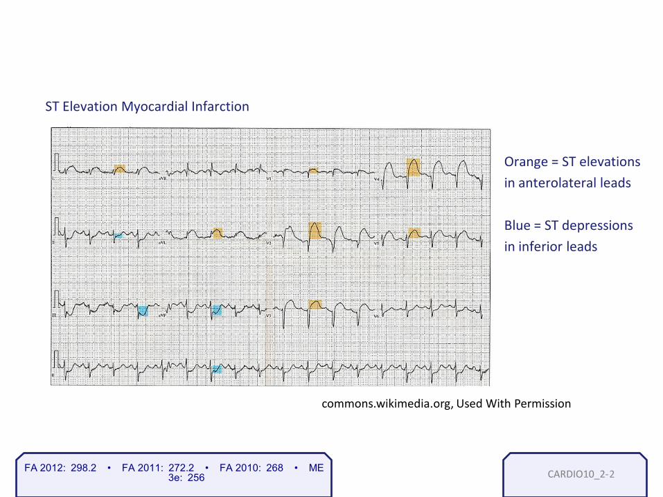
Orange = ST elevations in anterolateral leads Blue = ST depressions in inferior leads
ST Elevation Myocardial Infarction
commons.wikimedia.org, Used With Permission
FA 2012: 298.2 • FA 2011: 272.2 • FA 2010: 268 • ME 3e: 256

CARDIO10_2- 3
FA 2012: 298.3 • FA 2011: 272.3 • FA 2010: n/a • ME 3e: 233
Artery Affected
LAD
Left Main/LCX
RCA
Infarct Location
Relevant Leads (look for ST segment changes and Q waves)
V1-V4
V1-V2
Anterior Wall
Anteroseptal
Anterior Leads
Septal Leads
Lateral Leads Anterolateral
Lateral Wall
Inferior Wall Inferior Leads
V4-V6
I, aVL
II, III, aVF
LCX

CARDIO10_3- 1 FA 2012: 298.4 • FA 2011: 272.4 • FA 2010: 268 • ME 3e: 256
Post-MI Complications – Immediate Risk
Cardiac arrhythmia Ventricular tachycardia/fibrillation Causes sudden cardiac death in 25% of patients with MI Congestive heart failure LV failure with pulmonary edema most common Cardiogenic shock Complete pump failure and hypoperfusion of all organs
Note: These risks are immediate but can persist for months or years.

CARDIO10_3- 2
Post-MI Complications – Delayed Risk
Ventricular rupture 5-10 days post-MI Free wall rupture → tamponade Septum rupture → severe left-to-right shunt via VSD Papillary muscle rupture → severe mitral insufficiency Ventricular aneurysm several weeks to months later Increased risk of mural thrombus (embolism and stroke) Increased risk of arrhythmia
FA 2012: 298.4 • FA 2011: 272.4 • FA 2010: 268 • ME 3e: 256

CARDIO10_3- 3
Post-MI Complications – Pericarditis
Friction rub may be heard in both types.
Post-infarction fibrinous pericarditis Due to inflammation secondary to ischemia Can occur 3-5 days post-MI Dressler’s syndrome (autoimmune pericarditis)
Also fibrinous pericarditis, but due to autoimmune process Can occur 2-10 weeks post-MI
FA 2012: 298.4 • FA 2011: 272.4 • FA 2010: 268 • ME 3e: 256

CARDIO10_3b- 1
Loading…
FA 2012: 298.4 • FA 2011: 272.4 • FA 2010: 268 • ME 3e: 256
This interaction is unavailable on the iPad. To engage with this part of the lesson, please
access this presentation on a Mac or PC that has Adobe Flash installed.

CARDIO10_4- 1 FA 2012: 307.1 • FA 2011: 281.1 • FA 2010: 277 • ME 3e: 271
Antianginal Therapy - Goals
Decrease myocardial oxygen consumption
Primary Goal
Secondary Goal Increase oxygen delivery to myocardium by increasing perfusion
By decreasing: End diastolic volume (preload) Blood pressure (afterload) Heart rate Contractility

CARDIO10_4- 2
Antianginal Therapy – Nitrates
Nitrates
End diastolic volume (preload)
Blood pressure Heart Rate
Contractility
reflex reflex
FA 2012: 307.1 • FA 2011: 281.1 • FA 2010: 277 • ME 3e: 271

CARDIO10_4- 3
Beta Blockers
Antianginal Therapy – Beta Blockers
Nitrates
End diastolic volume (preload)
Blood pressure Heart Rate
Contractility
reflex reflex
FA 2012: 307.1 • FA 2011: 281.1 • FA 2010: 277 • ME 3e: 271
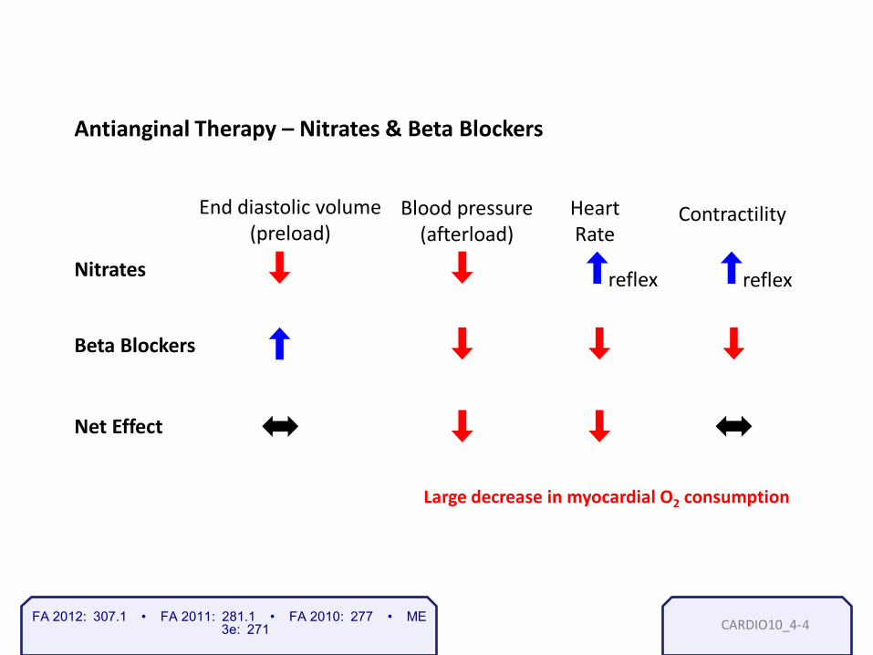
CARDIO10_4- 4
Net Effect
Large decrease in myocardial O2 consumption
Beta Blockers
Antianginal Therapy – Nitrates & Beta Blockers
Nitrates
End diastolic volume (preload)
Blood pressure (afterload)
Heart Rate
Contractility
reflex reflex
FA 2012: 307.1 • FA 2011: 281.1 • FA 2010: 277 • ME 3e: 271

CARDIO10_4- 5
Antianginal Therapy – Calcium Channel Blockers
Do not use beta blockers in patients with reactive airway disease (asthma, COPD)
Instead use:
Nifedipine
Diltiazem Verapamil
(similar to nitrates)
(similar to beta blockers)
End diastolic volume (preload) and Blood pressure (afterload)
Heart rate and Contractility
FA 2012: 307.1 • FA 2011: 281.1 • FA 2010: 277 • ME 3e: 271

CARDIO10_4- 6
FA 2012: 296.1 • FA 2011: 270.3 • FA 2010: 266 • ME 3e: 256
Prinzmetal’s Angina
• Vasospasm causes decreased blood flow through coronary arteries
• Occurs at rest or even wakes patient at night
• Diagnosed with ergonovine test – induces vasospasm
• Transient ST elevations on ECG
• Treated with calcium channel blockers

CARDIO11_1- CARDIO11_1-
Congestive heart failure and cardiomyopathies
1
Stephen Bagley, M.D. Resident Physician University of Pennsylvania
Lecture XI Cardiomyopathies Congestive heart failure

CARDIO11_1- 2 FA 2012: 299.1 • FA 2011: 273.1 • FA 2010: 269 • ME 3e: 260
Dilated Cardiomyopathy
Insult or Injury to
Myocardium
Contractility
Cardiac output
Compensatory neuro-hormonal
signaling
Cardiac remodeling
e.g. Renin- Angiotensin- Aldosterone System
sarcomeres added in
series
Dilated Cardiomyopathy

CARDIO11_1- 3
Dilated Cardiomyopathy
Chronic alcohol abuse
Wet Beriberi (thiamine/B1 deficiency)
Viral myocarditis (Cocksackie B)
Chronic cocaine use
Chagas’ disease (trypanosomes)
Doxorubicin toxicity
Hemochromatosis
Peripartum cardiomyopathy
Important Causes
FA 2012: 299.1 • FA 2011: 273.1 • FA 2010: 269 • ME 3e: 260
CARDIO11_1- 4
Dilated Cardiomyopathy - Signs & Symptoms
• Point of maximal impulse laterally displaced (at axilla)
• S3 heart sound: rapid filling and expansion of ventricle
• Enlarged heart on chest x-ray: cardiac silhouette > ½ chest cavity
• Cavitary dilation of heart on echocardiogram
FA 2012: 299.1 • FA 2011: 273.1 • FA 2010: 269 • ME 3e: 260

CARDIO11_1- 5
Copyright James Van Rhee. Used with permission.
Chest X-Ray
FA 2012: 299.1 • FA 2011: 273.1 • FA 2010: 269 • ME 3e: 260

CARDIO11_2- 1
Cardiac output
Asymmetric septal
thickening
actin/myosin synthesis
concentric hypertrophy:
Sarcomeres added in parallel
Genetic (50% cases) Autosomal dominant
Idiopathic etiology
or Decreased compliance
Poor diastolic
filling
Hypertrophic Cardiomyopathy
FA 2012: 299.1 • FA 2011: 273.1 • FA 2010: 269 • ME 3e: 260

CARDIO11_2- 2
Hypertrophic Cardiomyopathy
Complications
• Decreased compliance diastolic dysfunction (S4 heart sound)
left ventricle outflow obstruction (systolic murmur, exertional syncope)
• Asymmetric septal hypertrophy + Mitral valve anterior leaflet motion
• Arrhythmia sudden cardiac death
Treatments: myomectomy, beta-blockers
FA 2012: 299.1 • FA 2011: 273.1 • FA 2010: 269 • ME 3e: 260

CARDIO11_3- 1
Restrictive Cardiomyopathy
Infiltrative processes can cause stiffening of ventricles, diastolic dysfunction
Causes Sarcoidosis
Amyloidosis
Post-radiation fibrosis
Löffler’s syndrome (endomyocardial fibrosis eosinophilic infiltrate)
Hemochromatosis
FA 2012: 299.1 • FA 2011: 273.1 • FA 2010: 269 • ME 3e: 260

CARDIO11_4- 1
The Failing Heart
FA 2012: 300.1 • FA 2011: 274.1 • FA 2010: 270 • ME 3e: 258
CARDIO11_4- 2
Heart Failure - Signs & Symptoms
• Dyspnea on exertion
• Pulmonary edema → orthopnea, paroxysmal nocturnal dyspnea → hemosiderin-laden macrophages in lungs
• Hepatomegaly, central venous congestion (“nutmeg liver”)
• Peripheral edema (ankles, sacrum) • Jugular venous distention (after right heart failure)
FA 2012: 300.1 • FA 2011: 274.1 • FA 2010: 270 • ME 3e: 258

CARDIO11_5- 1 FA 2012: 302.3 • FA 2011: 276.2 • FA 2010: 272 • ME 3e: 261
Cardiac Tamponade
Compression of heart by fluid in pericardium (blood, effusion)
Signs & Symptoms
• Diastolic pressure equal in all four heart chambers
• Hypotension, jugular venous distention, increased heart rate
• Distant heart sounds
• Pulsus paradoxus (Kussmaul’s pulse): pulse strength during inspiration

CARDIO11_6- 1
Mechanism of Action of Inotropes
FA 2012: n/a • FA 2011: 283.1 • FA 2010: 279 • ME 3e: 271

CARDIO11_6- 2 FA 2012: 309.1 • FA 2011: 283.2 • FA 2010: 279 • ME 3e: 271
Mechanism Effects Uses Toxicities
Inhibits Na+/K+ ATPase
+ + gradient +/Ca2+ exchange
2+
• Increased myocardial Ca2+ increased contractility • Delayed conduction at AV node.
• CHF (contractility) • Atrial fibrillation (to conduction)
Yellow vision, nausea, vomiting, diarrhea, anorexia, hallucination, life-threatening arrhythmias
Notes: • Hypokalemia enhances toxicity • Quinidine • Digoxin antibodies (Fab fragments) used in overdose • Digitalis does not improve survival following MI
Cardiac Glycosides - Digoxin & Digitalis
CARDIO12_1- CARDIO12_1-
Lecture XII Embryology of the heart Congenital Heart Disease
Congenital heart disease
1
Stephen Bagley, M.D. Resident Physician University of Pennsylvania

CARDIO12_1- 2
Fate: (A) Ascending aorta / pulmonary
trunk
(B) Smooth part of L and R ventricles (outflow tracts)
(C) Trabeculated L and R ventricle
(D) Trabeculated L and R atrium
(E) Left horn: coronary sinus Right horn: smooth right atrium
A
B
C
D
E
Heart tube from lateral plate mesoderm
FA 2012: 130.1 • FA 2011: 123.1 • FA 2010: 123 • ME 3e: 230
Inflow
Outflow
Gray476.png, commons.wikimedia.org. Used with permission.
Venous system during cardiac development

CARDIO12_1- 3
Truncus Arteriosus
Migrate into truncal/bulbar
region
Neural crest cells
Spiral and fuse: form
aorticopulmonary septum
(days 27-33)
FA 2012: 130.2 • FA 2011: 123.2 • FA 2010: 123 • ME 3e: 231
Arterial outflow

CARDIO12_1- 4
Interventricular Septum
FA 2012: 130.3 • FA 2011: 123.3 • FA 2010: 123 • ME 3e: 230
Kaplan Anatomy: Figure III-2-16.

CARDIO12_2- 1
Endocardial Cushion Defects
Failure of dorsal and ventral endocardial cushions to fuse → persistent common AV canal
Ebstein’s anomaly • Weak, under-developed right ventricle (“atrialized” right ventricle) • Associated with lithium use by the mother Tricuspid atresia • Tricuspid valve does not form completely • Under-developed right side of heart
FA 2012: 130.3 • FA 2011: 123.3 • FA 2010: 123 • ME 3e: 230

CARDIO12_3- 1 FA 2012: 292.2 • FA 2011: 267.2 • FA 2010: 263 • ME 3e: 231
Eisenmenger’s Syndrome
Uncorrected Left-to-Right
Shunt
(e.g. ASD,VSD,PDA)
Pressure in pulmonary vasculature
Hypertrophy of pulmonary vasculature
and Resistance
Pressure in R side of
heart
L-to-R shunt reverses, becomes
R-to-L shunt
Late Cyanosis

CARDIO12_4- 1
Formation of Atrial Septum
FA 2012: 131.1 • FA 2011: 124.1 • FA 2010: 124 • ME 3e: 230
Kaplan Anatomy: Figure III-2-13.

CARDIO12_4- 2
Secundum and Primum Atrial Septal Defect
Kaplan Anatomy: Figure III-2-15.
FA 2012: 131.1 • FA 2011: 124.1 • FA 2010: 124 • ME 3e: 230

CARDIO12_5- 1 FA 2012: 131.2 • FA 2011: 124.2 • FA 2010: 124 • ME 3e: 232
Fetal Erythropoiesis
Fetal hemoglobin: α2γ2 Adult hemoglobin α2β2
3-8 weeks Yolk sac
6-30 weeks Liver and Spleen
> 28 weeks Bone marrow

CARDIO12_5- 2
Fetal Circulation and Shunts
FA 2012: 132.1 • FA 2011: 125.1 • FA 2010: 125 • ME 3e: 232
Kaplan Anatomy: Figure III-2-12

CARDIO12_6a- 1 FA 2012: 292.1 • FA 2011: 267.1 • FA 2010: 263 • ME 3e: 231
Congenital Heart Disease
Cyanotic (Right-to-Left Shunts) Non-cyanotic (Left-to-Right Shunts)
Tetralogy of Fallot
Transposition of great vessels
Truncus arteriosus
Tricuspid atresia
Total anomalous pulmonary venous return (TAPVR)
Ventricular septal defect (VSD)
Atrial septal defect (ASD)
Patent ductus arteriosus (PDA)
(late cyanosis)

CARDIO12_6a- 2
Persistent Truncus Arteriosus
Kaplan Anatomy: Figure III-2-21.
FA 2012: 292.1 • FA 2011: 267.1 • FA 2010: 263 • ME 3e: 231

CARDIO12_6a- 3
Tricuspid Atresia
Kaplan Pathology: Figure 14-3
Aorta
Atrial Septal defect
Closed tricuspid valve
Under- developed right ventricle
C. Tricuspid Atresia
FA 2012: 292.1 • FA 2011: 267.1 • FA 2010: 263 • ME 3e: 231

CARDIO12_6a- 4
1. Ventricular septal defects 2. Atrial septal defect 3. Patent ductus arteriosus
FA 2012: 292.1 • FA 2011: 267.1 • FA 2010: 263 • ME 3e: 230
CARDIO12_6a- 5
Tetralogy of Fallot
FA 2012: 292.3 • FA 2011: 267.3 • FA 2010: 263 • ME 3e: 231
Kaplan Anatomy: Figure III-2-19.
CARDIO12_6a- 6
Transposition of the Great Vessels
FA 2012: 293.1 • FA 2011: 268.1 • FA 2010: 264 • ME 3e: 231
Kaplan Anatomy: Figure III-2-20.
CARDIO12_6a- 7
Coarctation of the Aorta
FA 2012: 293.2 • FA 2011: 268.2 • FA 2010: 264 • ME 3e: 231
Kaplan Pathology: Figure 14-2.

CARDIO12_6a- 8
Ductus Arteriosus
FA 2012: 293.3 • FA 2011: 268.3 • FA 2010: 264 • ME 3e: 232
Kaplan Anatomy: Figure III-2-17.

CARDIO12_6b- 1
Loading…
FA 2012: 293.3 • FA 2011: 268.3 • FA 2010: 264 • ME 3e: 232

CARDIO12_7- 1 FA 2012: 294.1 • FA 2011: 269.1 • FA 2010: 264 • ME 3e: 230
Congenital Heart Disease - Associations
22q11 syndromes
Down syndrome
Congenital rubella
Turner syndrome
Marfan’s syndrome
Infant of diabetic mother
Truncus arteriosus, Tetralogy of Fallot
ASD, VSD, endocardial cushion defect
PDA, septal, pulmonary artery stenosis
Coarctation of aorta (preductal)
Aortic insufficiency
Transposition of great vessels
CARDIO13_1- CARDIO13_1-
Lecture XIII – Endocarditis and associated bacteria – Rheumatic heart disease – Syphilitic heart disease
Infection-related heart disease
1
Stephen Bagley, M.D. Resident Physician University of Pennsylvania

CARDIO13_1- FA 2012: 155.1 • FA 2011: 144.1 • FA 2010: 146 • ME 3e: 113
Manske, commons.wikimedia.org, Used With Permission
Gram positive bacteria have this thick outer peptidoglycan layer which retains a blue crystal violet stain Also note the absence of the outer cell membrane seen in gram negative bacteria.
Inne
r cel
l m
embr
ane
Pept
idog
lyca
n la
yer
2

CARDIO13_1-
Huckfinne, commons.wikimedia.org, Used With Permission
3
Streptococcus Staphylococcus
S.epidermis
Novobiocin sensitive
S. saprophyticus
Novobiocin resistant
S. pyogenes Group A, bacitracin sensitive
S. agalactiae Group B, bacitracin resistant
Enterococcus
E. faecalis, E. faecium
S. pneumoniae optochin sensitive, bile soluble, capsule (quellung +)
S. viridans, S.mutans, S.sanguis optochin resistant, not bile soluble no capsule
FA 2012: 155.1 • FA 2011: 144.1 • FA 2010: 146 • ME 3e: 533
Corynebacterium

CARDIO13_1-
Bill Branson, commons.wikimedia.org, Used With Permissio
Plain Agar Plate Beta - Hemolysis
4 FA 2012: 155.1 • FA 2011: 144.1 • FA 2010: 146 • ME 3e: 116

CARDIO13_1- FA 2012: n/a • FA 2011: 145.1 • FA 2010: 147 • ME 3e: 533
Huckfinne, commons.wikimedia.org, Used With Permission
5
Streptococcus Staphylococcus
S. epidermis
Novobiocin sensitive
S. saprophyticus
Novobiocin resistant
pyogenesGroup A, bacitracin sensitive
agalactiaeGroup B, bacitracin resistant
Enterococcus
E. faecalis, E. faecium
Pneumoniae optochin sensitive, bile soluble, capsule (quellung +)
Viridans mutans, sanguis optochin resistant, not bile soluble no capsule

CARDIO13_1- 6 FA 2012: n/a • FA 2011: 145.1 • FA 2010: 147 • ME 3e: 533
CARDIO13_2-
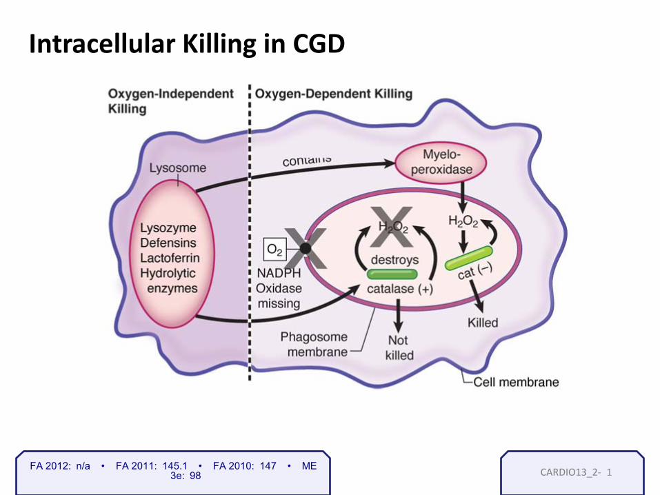
Intracellular Killing in CGD
1 FA 2012: n/a • FA 2011: 145.1 • FA 2010: 147 • ME 3e: 98

CARDIO13_3- 1 FA 2012: n/a • FA 2011: 145.1 • FA 2010: 147 • ME 3e: 533

CARDIO13_4- FA 2012: 156.2 • FA 2011: 145.2 • FA 2010: 147 • ME 3e: 116
Gram positive cocci in clusters
Y tambe, commons.wikimedia.org, Used With Permission
1
S. aureus

CARDIO13_4-
Superantigen Activation Superantigen activation: Notice that
there is no complementarity between the TCR and the MHC/peptide complex.
2 FA 2012: 156.2 • FA 2011: 145.2 • FA 2010: 147 • ME 3e: 92
CARDIO13_5-
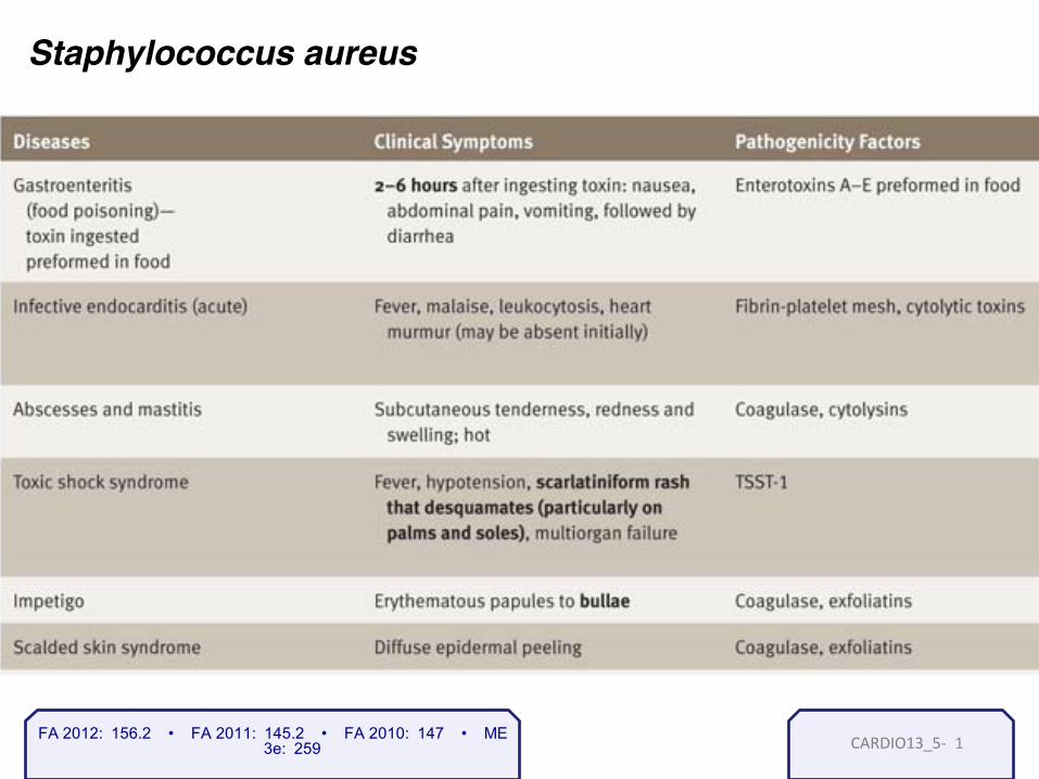
Staphylococcus aureus
1 FA 2012: 156.2 • FA 2011: 145.2 • FA 2010: 147 • ME 3e: 259

CARDIO13_5-
FA 2012: 156.3 • FA 2011: 145.3 • FA 2010: n/a • ME 3e: 259
Mirko Junge M.D., commons.wikimedia.org, Used With Permission
Prosthetic Valves Catheters
Staphylococcus epidermidis seeds prosthetic devices …
2

CARDIO13_6- FA 2012: 156.5 • FA 2011: 145.5 • FA 2010: 147 • ME 3e: 259
Mike Miller M.D., commons.wikimedia.org, Used With Permission
Streptococcus viridans
Endocarditis!
Cavities!
1

CARDIO13_6-
• Enterococci are inherently resistant to penicillins
– Initial treatment is vancomycin – This has led to Vancomycin-Resistant-Enterococci (VRE) – Treat VRE with linezolid
• Streptococcus bovis – Endocarditis in colon cancer patients !!! (USMLE pearl)
FA 2012: 157.3 • FA 2011: 146.3 • FA 2010: 148 • ME 3e: 534
BOTH are group D streptococci Differentiating test Æ Plate with 6.5% NaCl Enterococci will grow; Bovis will not grow
2

CARDIO13_7-
FA 2012: 301.1 • FA 2011: 275.1 • FA 2010: 271 • ME 3e: 259
© Richard Usatine, M.D. Used with permission.
Roth Spots Janeway lesions
1

CARDIO13_7- 2
users
FA 2012: 301.1 • FA 2011: 275.1 • FA 2010: 271 • ME 3e: 259

CARDIO13_8- 1 FA 2012: 301.1 • FA 2011: 275.1 • FA 2010: 271 • ME 3e: 114

CARDIO13_9-
Mitral
Tricuspid
More frequently affected
Commonly affected in IV drug abuse
1 FA 2012: 301.1 • FA 2011: 275.1 • FA 2010: 271 • ME 3e: 259

CARDIO13_10-
Libman-Sacks Endocarditis
FA 2012: n/a • FA 2011: 275.2 • FA 2010: 271 • ME 3e: 259
Sterile vegetations
Usually benign
Vegetations on both sides of valve
Can attack both tricuspid and mitral valves.
1

CARDIO13_11- FA 2012: 302.4 • FA 2011: 276.3 • FA 2010: 272 • ME 3e: 421
Stages of Syphilis
1
CARDIO13_12- FA 2012: 302.1 • FA 2011: 276.1 • FA 2010: 272 • ME 3e: 257
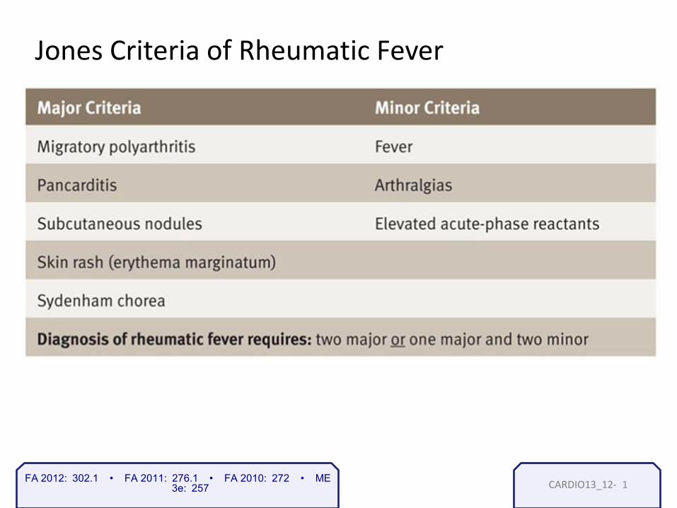
Jones Criteria of Rheumatic Fever
1

CARDIO14_1- CARDIO14_1-
Lecture XIV Vasculitis Cardiac and vascular tumors
Vasculitis and cardiovascular tumors
1
Stephen Bagley, M.D. Resident Physician University of Pennsylvania

CARDIO14_1-
Varicose Veins
• Dilated torturous superficial veins
• Associated with prolonged standing or sitting, and pregnancy
• Occur in areas of high venous pressure (lower extremities)
• Rarely a source of thromboembolism
2 FA 2012: 303.2 • FA 2011: 277.2 • FA 2010: 273 • ME 3e: 265
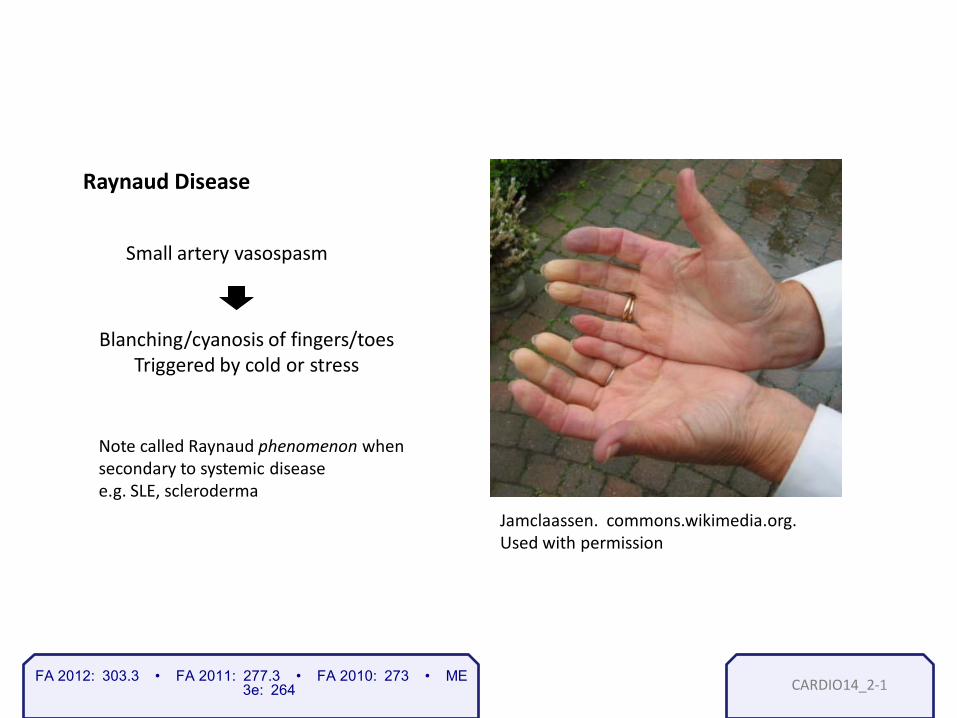
CARDIO14_2- FA 2012: 303.3 • FA 2011: 277.3 • FA 2010: 273 • ME 3e: 264
Jamclaassen. commons.wikimedia.org. Used with permission
Raynaud Disease
Small artery vasospasm
Blanching/cyanosis of fingers/toes Triggered by cold or stress
Note called Raynaud phenomenon when secondary to systemic disease e.g. SLE, scleroderma
1

CARDIO14_3- FA 2012: 304.1 • FA 2011: 277.4 • FA 2010: 274 • ME 3e: 263
Wegener’s granulomatosis
Necrotizing granulomatous inflammation
Vasculitis (small vessels)
Glomerulonephritis lung upper airway
Signs and symptoms:
• Hemoptysis
• Cavitary lung lesions
• Hematuria
• Chronic sinusitis
• Nasal septum perforation
• Otitis media
• Mastoiditis
• Cough, dyspnea
1

CARDIO14_3-
Malittle. commons.wikimedia.org. Used With Permission
2 FA 2012: 304.1 • FA 2011: 277.4 • FA 2010: 274 • ME 3e: 263

CARDIO14_3-
FA 2012: n/a • FA 2011: 277.5 • FA 2010: 274 • ME 3e: 262
Malittle. commons.wikimedia.org. Used With Permission
3

CARDIO14_3-
Wegener’s granulomatosis
• Associated with c-ANCA
• Treated with cyclophosphamide (for renal disease) & corticosteroids
4 FA 2012: 304.1 • FA 2011: 277.4 • FA 2010: 274 • ME 3e: 263

CARDIO14_4-
Other ANCA-associated vasculitides
Microscopic Polyangiitis
Primary pauci-immune crescentic glomerulonephritis
Churg-Strauss syndrome
(all small vessel diseases)
(p-ANCA)
(p-ANCA)
• Affects small vessels in multiple organs but without granulomas
• Fever, malaise, myalgia, weight loss (skin rash in 50%)
• Vasculitis limited to kidney
• Granulomatous vasculitis with eosinopilia - can also involve kidneys, heart, lung
• Asthma, sinusitis, skin lesions, peripheral neuropathy (wrist drop/foot drop)
1 FA 2012: n/a • FA 2011: 277.5 • FA 2010: 274 • ME 3e: 262

CARDIO14_5- FA 2012: 305.1 • FA 2011: 277.6 • FA 2010: 274 • ME 3e: 263 1
Sturge-Weber disease (capillary-sized vessels)
Congenital capillary malformations
Signs and Symptoms • Port-wine stain (nevus flammeus) on face
• Leptomeningeal angiomatosis (intracerebral AVM)
• Seizures
• Early-onset glaucoma
Gaillard. commons.wikimedia.org. Used With Permission

CARDIO14_6- FA 2012: 305.1 • FA 2011: 278.1 • FA 2010: 274 • ME 3e: 262
Henoch-Schönlein Purpura (small vessels)
Signs and Symptoms
• Most common childhood systemic vasculitis
• IgA-mediated vasculitis, circulating IgA complexes
• Linked to parvovirus
• Typically develops after URI
• “Palpable purpura” skin rash (buttocks and legs)
• Arthralgia (wrists, ankles)
• Intestinal involvement: hemorrhage, melena
• Nephritis – IgA nephropathy
• Normal complement levels
1

CARDIO14_6-
Crescent formation in rapidly progressive glomerulonephritis, as seen with trichrome stain
Kaplan Pathology: Figure 16-3.
2 FA 2012: 305.1 • FA 2011: 278.1 • FA 2010: 274 • ME 3e: 262

CARDIO14_7- FA 2012: 304.1 • FA 2011: 278.2 • FA 2010: 275 • ME 3e: 262
Buerger’s disease - thromboangiitis obliterans (small and medium vessels in the extremities)
Signs and Symptoms • Heavy smokers
• Rapidly progressive thrombosing vasculitis
• Claudication (vascular insufficiency)
• Cold sensitivity (Raynaud’s phenomenon)
• Ulceration, gangrene, autoamputation
Treatment: improves rapidly with smoking cessation
Geirunited. commons.wikimedia.org. Used With Permission
1

CARDIO14_8- FA 2012: 304.1 • FA 2011: 278.3 • FA 2010: 275 • ME 3e: 262
Kawasaki disease (small and medium vessels)
Signs and Symptoms
• Acute, segmental necrotizing vasculitis
• Commonly affects infants/children (<4 yo)
• Associated with Japanese heritage
• Fever
• Conjunctivitis
• Erythema of oral mucosa (“strawberry tongue”)
• Lymphadenopathy
• Desquamative skin rash
• Risk of coronary aneurysms
Treatment: IV immunoglobulin and aspirin Soo Kim. commons.wikimedia.org. Used With Permission
1

CARDIO14_9-
FA 2012: 304.1 • FA 2011: 278.4 • FA 2010: 275 • ME 3e: 263
Polyarteritis Nodosa (small and medium arteries)
Signs and Symptoms
• Immune complex-mediated transmural vasculitis
• Lesions at different stages (acute, healing, healed) - ”beads on a string”
• Abdominal pain, diarrhea, GI bleeding
• Fever, weight loss, malaise
• Hematuria, renal failure, hypertension
• Important association with Hepatitis B antigen
• Sometimes associated w/ p-ANCA
1

CARDIO14_10- FA 2012: 304.1 • FA 2011: 278.5 • FA 2010: 275 • ME 3e: 263
Ly. commons.wikimedia.org. Used With Permission
1
Takayasu’s arteritis (medium and large arteries, aortic arch and major branches)
Signs and Symptoms
• Granulomatous vasculitis with heavy intimal thickening
• Often affects young to middle-aged Asian women
• Fever, night sweats, myalgias/arthralgias, weight loss
• Weak pulse in upper extremities (pulseless disease)
• Visual loss or other neurologic abnormalities
• Increased ESR
Ly. commons.wikimedia.org. Used With Permission

CARDIO14_11-
FA 2012: 305.1 • FA 2011: 278.1 • FA 2010: 275 • ME 3e: 263
Temporal arteritis (giant cell arteritis)
Multinucleated giant cell Destroyed artery from giant cell arteritis
Marvin and Nguyen. commons.wikimedia.org. Used With Permission
1

CARDIO14_11-
Temporal arteritis (giant cell arteritis) (medium and large arteries)
Signs and Symptoms
• Most common large-vessel vasculitis (granulomatous)
• Commonly affects branches of carotid artery
• Patient > 50 yo and ESR > 50
• Unilateral headache
• Unilateral jaw claudication (pain on chewing)
• Unilateral visual disturbance
• Associated with polymyalgia rheumatica (proximal muscles)
Treatment: corticosteroids
2 FA 2012: 305.1 • FA 2011: 278.1 • FA 2010: 275 • ME 3e: 263

CARDIO14_12- FA 2012: 305.2 • FA 2011: 279.2 • FA 2010: 276 • ME 3e: 266
Hemangioma
Kaplan Pathology: Figure 13-3.
1

CARDIO14_13-
Cystic Hygromas
• Most commonly associated with Turner Syndrome
• Usually found in the posterior aspect of the neck
Kaplan Pathology: Figure 6-4.
1 FA 2012: 305.2 • FA 2011: 279.2 • FA 2010: 276 • ME 3e: 266

CARDIO14_14-
KGH. commons.wikimedia.org. Used With Permission
Angiosarcoma Kaposi’s Sarcoma
1 FA 2012: 305.2 • FA 2011: 279.2 • FA 2010: 276 • ME 3e: 266

CARDIO14_15-
FA 2012: 303.1 • FA 2011: 277.1 • FA 2010: 273 • ME 3e: 261
James Moon. commons.wikimedia.org. Used With Permission
Atrial Myxoma
Myxoma
Mitral valve
ventricle
(Ball valve obstruction)
1

CARDIO14_15-
Nephron. commons.wikimedia.org. Used With Permission
Atrial Myxoma
• Stellate cells
• Myxoid background
2 FA 2012: 303.1 • FA 2011: 277.1 • FA 2010: 273 • ME 3e: 261
CARDIO14_16-
Rhabdomyoma
• Most common primary cardiac tumor in children
• Patients with tuberous sclerosis often develop these tumors
1 FA 2012: 303.1 • FA 2011: 277.1 • FA 2010: 273 • ME 3e: 261


