January 2018 6-1 Device-associated Module PNEU Pneumonia (Ventilator-associated [VAP] and non-ventilator-associated Pneumonia [PNEU]) Event Introduction: In 2011, an estimated 157,000 healthcare-associated pneumonias occurred in acute care hospitals in U.S.; 39% of these pneumonias were ventilator- associated (VAP). 1 Patients receiving invasive mechanical ventilation are at risk for numerous complications, including pneumonia. Ventilator-associated pneumonia (VAP) and other healthcare-associated pneumonias are important, common healthcare- associated infections, but national surveillance for VAP has long been a challenge because of the lack of objective, reliable definitions. Due to these challenges, in January 2013 the National Healthcare Safety Network (NHSN) replaced surveillance for ventilator-associated pneumonia (VAP) in adult inpatient locations with surveillance for ventilator-associated events (VAE). 2 Based on discussions with an expert working group in 2012-2013, NHSN also discontinued in-plan VAP surveillance in neonatal locations. As of January 2014, in-plan VAP surveillance is only available in pediatric inpatient locations. Settings: Surveillance may occur in any inpatient pediatric location where denominator data can be collected, such as critical/intensive care units (pedICUs), specialty care areas (SCA), step-down units, wards, and long term care units. In-plan surveillance for ventilator-associated pneumonia (pedVAP) using the criteria found in this chapter is restricted to patients of any age in pediatric locations (excludes neonatal locations). In- plan surveillance conducted for mechanically-ventilated patients in adult locations (regardless of age) will use the Ventilator-Associated Event (VAE) protocol (see VAE chapter). The PNEU definitions are still available for those units seeking to conduct off- plan PNEU surveillance for mechanically-ventilated adult, pediatric and neonatal patients and non-ventilated adults, pediatric or neonatal patients. A complete listing of inpatient locations and instructions for mapping can be found in the CDC Locations and Descriptions chapter. Note: If you are following pedVAP in your monthly reporting plan it is not required to monitor for VAPs after the patient is discharged from the facility. However, if discovered, any VAPs with event date on the day of discharge or day after discharge should be reported to NHSN (see Transfer Rule below). No additional ventilator days are reported. Definitions: Present on Admission (POA): Infections that are POA, as defined in Chapter 2, are not considered HAIs and therefore are never reported to NHSN. Note: POA reporting exception for PNEU/VAP: One eligible chest imaging test is acceptable to satisfy the imaging parameter for PNEU/VAP-POA determinations regardless of whether the patient has underlying pulmonary or cardiac disease.
Transcript
January 2018 6-1
Device-associated Module
PNEU
Pneumonia (Ventilator-associated [VAP] and non-ventilator-associated
Pneumonia [PNEU]) Event
Introduction: In 2011, an estimated 157,000 healthcare-associated pneumonias
occurred in acute care hospitals in U.S.; 39% of these pneumonias were ventilator-
associated (VAP).1 Patients receiving invasive mechanical ventilation are at risk for
numerous complications, including pneumonia. Ventilator-associated pneumonia (VAP)
and other healthcare-associated pneumonias are important, common healthcare-
associated infections, but national surveillance for VAP has long been a challenge
because of the lack of objective, reliable definitions. Due to these challenges, in January
2013 the National Healthcare Safety Network (NHSN) replaced surveillance for
ventilator-associated pneumonia (VAP) in adult inpatient locations with surveillance for
ventilator-associated events (VAE).2 Based on discussions with an expert working group
in 2012-2013, NHSN also discontinued in-plan VAP surveillance in neonatal locations.
As of January 2014, in-plan VAP surveillance is only available in pediatric inpatient
locations.
Settings: Surveillance may occur in any inpatient pediatric location where denominator
data can be collected, such as critical/intensive care units (pedICUs), specialty care areas
(SCA), step-down units, wards, and long term care units. In-plan surveillance for
ventilator-associated pneumonia (pedVAP) using the criteria found in this chapter is
restricted to patients of any age in pediatric locations (excludes neonatal locations). In-
plan surveillance conducted for mechanically-ventilated patients in adult locations
(regardless of age) will use the Ventilator-Associated Event (VAE) protocol (see VAE
chapter). The PNEU definitions are still available for those units seeking to conduct off-
plan PNEU surveillance for mechanically-ventilated adult, pediatric and neonatal patients
and non-ventilated adults, pediatric or neonatal patients. A complete listing of inpatient
locations and instructions for mapping can be found in the CDC Locations and
Descriptions chapter.
Note: If you are following pedVAP in your monthly reporting plan it is not required to
monitor for VAPs after the patient is discharged from the facility. However, if
discovered, any VAPs with event date on the day of discharge or day after discharge
should be reported to NHSN (see Transfer Rule below). No additional ventilator days are
reported.
Definitions:
Present on Admission (POA): Infections that are POA, as defined in Chapter 2, are not
considered HAIs and therefore are never reported to NHSN.
Note: POA reporting exception for PNEU/VAP: One eligible chest imaging test is
acceptable to satisfy the imaging parameter for PNEU/VAP-POA determinations
regardless of whether the patient has underlying pulmonary or cardiac disease.
Abbreviations used in the PNEU laboratory criteria:
BAL–bronchoalveolar lavage
EIA–enzyme immunoassay
IFA–immunofluorescent antibody
LRT–lower respiratory tract
PMN–polymorphonuclear leukocyte
RIA–radioimmunoassay
Reporting Instructions:
There is a hierarchy of specific categories within the major site pneumonia. If the
patient meets criteria for more than one specific site during the infection window
period or the RIT, report only one:
o If a patient meets criteria for both PNU1 and PNU2, report PNU2.
o If a patient meets criteria for both PNU2 and PNU3, report PNU3.
o If a patient meets criteria for both PNU1 and PNU3, report PNU3.
Pathogens and secondary bloodstream infections can only be reported for PNU2 and
PNU3 specific events.
Report concurrent LUNG and PNEU with at least one matching organism(s) as
PNEU.
January 2018 6-5
Device-associated Module
PNEU
Table 1: Specific Site Algorithms for Clinically Defined Pneumonia (PNU1)
Imaging Test
Evidence
Signs/Symptoms/Laboratory
Two or more serial chest
imaging test results with
at least one of the
following1,2,14:
New and persistent
or
Progressive and
persistent
Infiltrate
Consolidation
Cavitation
Pneumatoceles, in
infants ≤1 year old
Note: In patients
without underlying
pulmonary or cardiac
disease (for example: respiratory distress
syndrome,
bronchopulmonary
dysplasia, pulmonary
edema, or chronic
obstructive pulmonary
disease), one definitive
imaging test result is
acceptable.1
For ANY PATIENT, at least one of the following:
Fever (>38.0°C or >100.4°F)
Leukopenia (≤4000 WBC/mm3) or leukocytosis (>12,000 WBC/mm3)
For adults >70 years old, altered mental status with no other recognized cause
And at least two of the following:
New onset of purulent sputum3 or change in character of sputum4, or increased
respiratory secretions, or increased suctioning requirements
New onset or worsening cough, or dyspnea, or tachypnea5
Rales6 or bronchial breath sounds
Worsening gas exchange (for example: O2 desaturations (for example: PaO2/FiO2
<240)7, increased oxygen requirements, or increased ventilator demand)
ALTERNATE CRITERIA, for infants <1 year old:
Worsening gas exchange (for example:2 desaturations [for example pulse oximetry
<94%], increased oxygen requirements, or increased ventilator demand)
And at least three of the following:
Temperature instability
Leukopenia (≤4000 WBC/mm3) or leukocytosis (>15,000 WBC/mm3) and left shift
(>10% band forms)
New onset of purulent sputum3 or change in character of sputum4, or increased
respiratory secretions or increased suctioning requirements
Apnea, tachypnea5 , nasal flaring with retraction of chest wall or nasal flaring with
grunting
Wheezing, rales6, or rhonchi
Cough
Bradycardia (<100 beats/min) or tachycardia (>170 beats/min)
ALTERNATE CRITERIA, for child >1 year old or ≤12 years old, at least three of the
following:
Fever (>38. 0°C or >100. 4°F) or hypothermia (<36. 0°C or <96. 8°F)
Leukopenia (≤4000 WBC/mm3) or leukocytosis (≥15,000 WBC/mm3)
New onset of purulent sputum3 or change in character of sputum4, or increased
respiratory secretions, or increased suctioning requirements
New onset or worsening cough, or dyspnea, apnea, or tachypnea5.
Rales6 or bronchial breath sounds
Worsening gas exchange (for example: O2 desaturations [for example pulse
oximetry <94%], increased oxygen requirements, or increased ventilator demand)
January 2018 6-6
Device-associated Module
PNEU
Table 2: Specific Site Algorithms for Pneumonia with Common Bacterial or Filamentous
Fungal Pathogens and Specific Laboratory Findings (PNU2)
Imaging Test
Evidence
Signs/Symptoms Laboratory
Two or more serial chest
imaging test results with at
least one of the
following1,2,14:
New and persistent
or
Progressive and persistent
Infiltrate
Consolidation
Cavitation
Pneumatoceles, in
infants ≤1 year old
Note: In patients without
underlying pulmonary or
cardiac disease (for example: respiratory
distress syndrome,
bronchopulmonary
dysplasia, pulmonary
edema, or chronic
obstructive pulmonary
disease), one definitive
chest imaging test result is
acceptable.1
At least one of the following:
Fever (>38.0°C or >100.4°F)
Leukopenia (≤4000 WBC/mm3)
or leukocytosis (>12,000
WBC/mm3)
For adults >70 years old, altered
mental status with no other
recognized cause
And at least one of the following:
New onset of purulent sputum3 or
change in character of sputum4, or
increased respiratory secretions,
or increased suctioning
requirements
New onset or worsening cough, or
dyspnea or tachypnea5
Rales6 or bronchial breath sounds
Worsening gas exchange (for
example: O2 desaturations [for
example: PaO2/FiO2 <240]7,
increased oxygen requirements, or
increased ventilator demand)
At least one of the following:
• Organism identified from blood8,13
• Organism identified from pleural fluid9,13
• Positive quantitative culture or corresponding semi-quantitative culture result9 from minimally-contaminated LRT
specimen (specifically, BAL, protected
specimen brushing or endotracheal
aspirate)
• ≥5% BAL-obtained cells contain intracellular bacteria on direct microscopic exam (for example: Gram’s
stain)
• Positive quantitative culture or corresponding semi-quantitative culture result9 of lung tissue
• Histopathologic exam shows at least one
of the following evidences of pneumonia:
o Abscess formation or foci of consolidation with intense PMN accumulation in bronchioles and alveoli
o Evidence of lung parenchyma invasion by fungal hyphae or pseudohyphae
January 2018 6-7
Device-associated Module
PNEU
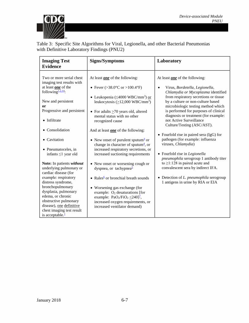
Table 3: Specific Site Algorithms for Viral, Legionella, and other Bacterial Pneumonias
with Definitive Laboratory Findings (PNU2)
Imaging Test
Evidence
Signs/Symptoms Laboratory
Two or more serial chest
imaging test results with
at least one of the
following1,2,14:
New and persistent
or
Progressive and persistent
Infiltrate
Consolidation
Cavitation
Pneumatoceles, in
infants ≤1 year old
Note: In patients without
underlying pulmonary or
cardiac disease (for example: respiratory
distress syndrome,
bronchopulmonary
dysplasia, pulmonary
edema, or chronic
obstructive pulmonary
disease), one definitive
chest imaging test result
is acceptable.1
At least one of the following:
Fever (>38.0°C or >100.4°F)
Leukopenia (≤4000 WBC/mm3) or
leukocytosis (>12,000 WBC/mm3)
For adults >70 years old, altered
mental status with no other
recognized cause
And at least one of the following:
New onset of purulent sputum3 or
change in character of sputum4, or
increased respiratory secretions, or
increased suctioning requirements
New onset or worsening cough or
dyspnea, or tachypnea5
Rales6 or bronchial breath sounds
Worsening gas exchange (for
example: O2 desaturations [for
example: PaO2/FiO2 <240]7,
increased oxygen requirements, or
increased ventilator demand)
At least one of the following:
Virus, Bordetella, Legionella,
Chlamydia or Mycoplasma identified
from respiratory secretions or tissue
by a culture or non-culture based
microbiologic testing method which
is performed for purposes of clinical
diagnosis or treatment (for example:
not Active Surveillance
Culture/Testing (ASC/AST).
Fourfold rise in paired sera (IgG) for
pathogen (for example: influenza
viruses, Chlamydia)
Fourfold rise in Legionella
pneumophila serogroup 1 antibody titer
to ≥1:128 in paired acute and
convalescent sera by indirect IFA.
Detection of L. pneumophila serogroup
1 antigens in urine by RIA or EIA
January 2018 6-8
Device-associated Module
PNEU
Table 4: Specific Site Algorithm for Pneumonia in Immunocompromised Patients
(PNU3)
Imaging Test
Evidence
Signs/Symptoms Laboratory
Two or more serial chest
imaging test results with
at least one of the
following1,2,14:
New and persistent
or
Progressive and
persistent
Infiltrate
Consolidation
Cavitation
Pneumatoceles, in
infants ≤1 year old
Note: In patients
without underlying
pulmonary or cardiac
disease (for example:
respiratory distress
syndrome,
bronchopulmonary
dysplasia, pulmonary
edema, or chronic
obstructive pulmonary
disease), one definitive
chest imaging test result
is acceptable.1
Patient who is
immunocompromised (see
definition in footnote 10 ) has at
least one of the following:
Fever (>38.0°C or >100.4°F)
For adults >70 years old, altered
mental status with no other
recognized cause
New onset of purulent sputum3,
or change in character of
sputum4, or increased respiratory
secretions, or increased
suctioning requirements
New onset or worsening cough,
or dyspnea, or tachypnea5
Rales6 or bronchial breath sounds
Worsening gas exchange (for
example: O2 desaturations [for
example: PaO2/FiO2 <240]7,
increased oxygen requirements,
or increased ventilator demand)
Hemoptysis
Pleuritic chest pain
At least one of the following:
Identification of matching Candida spp.
from blood and one of the following:
sputum, endotracheal aspirate, BAL or
protected specimen brushing.11,12,13
Evidence of fungi from minimally-
contaminated LRT specimen (specifically
BAL, protected specimen brushing or
endotracheal aspirate) from one of the
following:
Direct microscopic exam
Positive culture of fungi
Non-culture diagnostic laboratory test
OR
Any of the following from:
LABORATORY CRITERIA DEFINED
UNDER PNU2
January 2018 6-9
Device-associated Module
PNEU
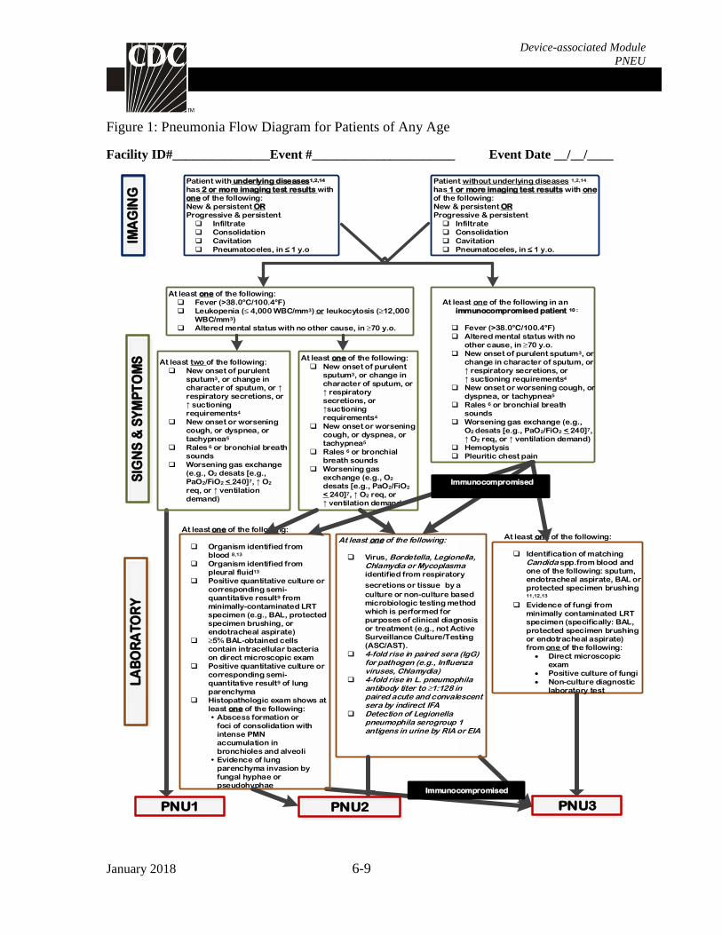
Figure 1: Pneumonia Flow Diagram for Patients of Any Age
Facility ID#_______________Event #______________________ Event Date __/__/____
Patient with underlying diseases1,2,14 has 2 or more imaging test results with one of the following:New & persistent ORProgressive & persistent
q Infiltrate
q Consolidation
q Cavitation
q Pneumatoceles, in ≤ 1 y.o
At least one of the following:
q Fever (>38.0°C/100.4°F)
q Leukopenia (≤ 4,000 WBC/mm3) or leukocytosis (≥12,000 WBC/mm3)
q Altered mental status with no other cause, in ≥70 y.o.
At least two of the following:
q New onset of purulent sputum3, or change in character of sputum, or ↑ respiratory secretions, or ↑ suctioning requirements4
q New onset or worsening cough, or dyspnea, or tachypnea5
q Rales 6 or bronchial breath sounds
q Worsening gas exchange (e.g., O2 desats [e.g.,
PaO2/FiO2 < 240]7, ↑ O2
req, or ↑ ventilation demand)
At least one of the following:
q New onset of purulent sputum3, or change in character of sputum, or ↑ respiratory secretions, or ↑suctioning requirements4
q New onset or worsening cough, or dyspnea, or tachypnea5
q Rales 6 or bronchial breath sounds
q Worsening gas exchange (e.g., O2 desats [e.g., PaO2/FiO2 < 240]7, ↑ O2 req, or ↑ ventilation demand
Patient without underlying diseases 1,2,14
has 1 or more imaging test results with one of the following:New & persistent OR Progressive & persistent
q Infiltrate
q Consolidation
q Cavitation
q Pneumatoceles, in ≤ 1 y.o.
At least one of the following in an immunocompromised patient 10 :
q Fever (>38.0°C/100.4°F)
q Altered mental status with no other cause, in ≥70 y.o.
q New onset of purulent sputum3, or change in character of sputum, or ↑ respiratory secretions, or ↑ suctioning requirements4
q New onset or worsening cough, or dyspnea, or tachypnea5
q Rales 6 or bronchial breath sounds
q Worsening gas exchange (e.g., O2 desats [e.g., PaO2/FiO2 < 240]7, ↑ O2 req, or ↑ ventilation demand)
q Hemoptysis
q Pleuritic chest pain
At least one of the following:
q Identification of matching Candida spp.from blood and one of the following: sputum, endotracheal aspirate, BAL or protected specimen brushing 11,12,13
q Evidence of fungi from minimally contaminated LRT specimen (specifically: BAL, protected specimen brushing or endotracheal aspirate) from one of the following:
Direct microscopic exam
Positive culture of fungi
Non-culture diagnostic laboratory test
At least one of the following:
q Virus, Bordetella, Legionella, Chlamydia or Mycoplasma identified from respiratory
secretions or tissue by a
culture or non-culture based microbiologic testing method which is performed for purposes of clinical diagnosis or treatment (e.g., not Active Surveillance Culture/Testing (ASC/AST).
q 4-fold rise in paired sera (IgG) for pathogen (e.g., Influenza viruses, Chlamydia)
q 4-fold rise in L. pneumophila antibody titer to ≥1:128 in paired acute and convalescent sera by indirect IFA
q Detection of Legionella pneumophila serogroup 1 antigens in urine by RIA or EIA
PNU3PNU2PNU1
At least one of the following:
q Organism identified from blood 8,13
q Organism identified from pleural fluid13
q Positive quantitative culture or corresponding semi-quantitative result9 from minimally-contaminated LRT specimen (e.g., BAL, protected specimen brushing, or endotracheal aspirate)
q ≥5% BAL-obtained cells contain intracellular bacteria on direct microscopic exam
q Positive quantitative culture or corresponding semi-quantitative result9 of lung parenchyma
q Histopathologic exam shows at least one of the following:
• Abscess formation or foci of consolidation with intense PMN accumulation in bronchioles and alveoli
• Evidence of lung parenchyma invasion by fungal hyphae or pseudohyphae
LA
BO
RA
TO
RY
S
IGN
S &
SY
MP
TO
MS
IMA
GIN
G
Immunocompromised
Immunocompromised
January 2018 6-10
Device-associated Module
PNEU
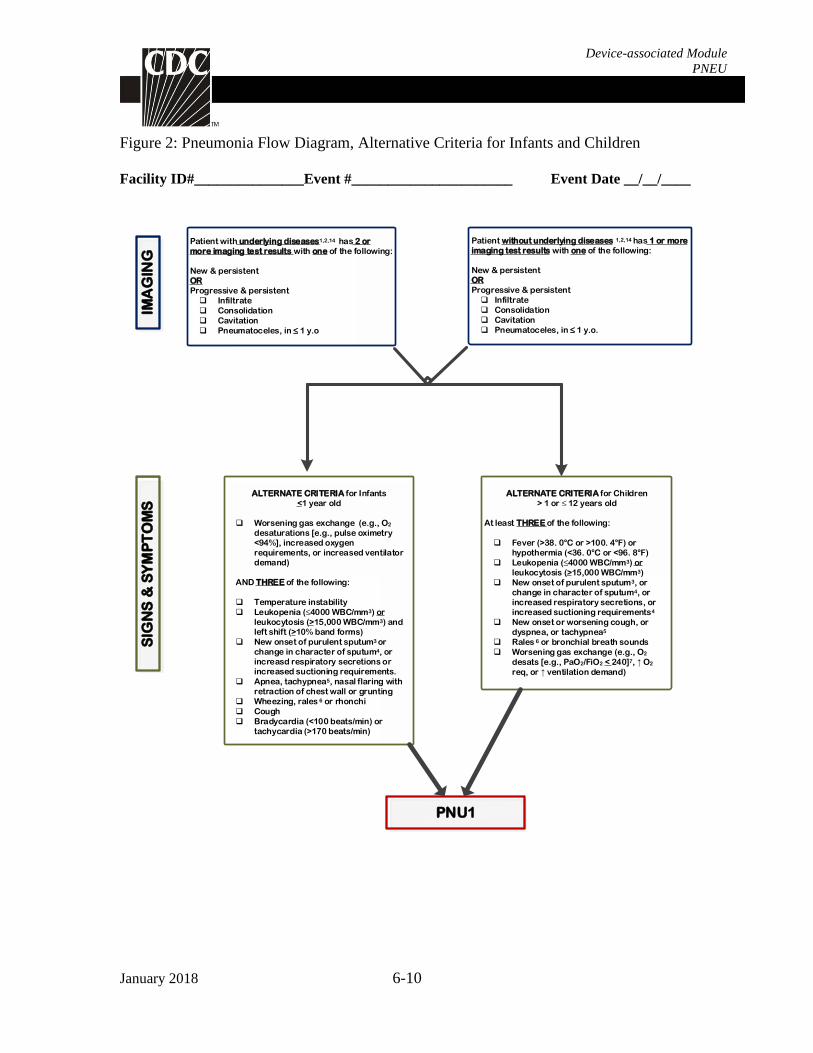
Figure 2: Pneumonia Flow Diagram, Alternative Criteria for Infants and Children
Facility ID#_______________Event #______________________ Event Date __/__/____
SIG
NS
& S
YM
PT
OM
SIM
AG
ING
ALTERNATE CRITERIA for Infants <1 year old
q Worsening gas exchange (e.g., O2 desaturations [e.g., pulse oximetry <94%], increased oxygen requirements, or increased ventilator demand)
AND THREE of the following:
q Temperature instability
q Leukopenia (≤4000 WBC/mm3) or leukocytosis (>15,000 WBC/mm3) and left shift (>10% band forms)
q New onset of purulent sputum3 or change in character of sputum4, or increasd respiratory secretions or increased suctioning requirements.
q Apnea, tachypnea5, nasal flaring with retraction of chest wall or grunting
q Wheezing, rales 6 or rhonchi
q Cough
q Bradycardia (<100 beats/min) or tachycardia (>170 beats/min)
ALTERNATE CRITERIA for Children
> 1 or ≤ 12 years old
At least THREE of the following:
q Fever (>38. 0°C or >100. 4°F) or hypothermia (<36. 0°C or <96. 8°F)
q Leukopenia (≤4000 WBC/mm3) or leukocytosis (>15,000 WBC/mm3)
q New onset of purulent sputum3, or change in character of sputum4, or increased respiratory secretions, or increased suctioning requirements4
q New onset or worsening cough, or dyspnea, or tachypnea5
q Rales 6 or bronchial breath sounds
q Worsening gas exchange (e.g., O2
desats [e.g., PaO2/FiO2 < 240]7, ↑ O2
req, or ↑ ventilation demand)
PNU1
Patient with underlying diseases1,2,14 has 2 or more imaging test results with one of the following:
New & persistent ORProgressive & persistent
q Infiltrate
q Consolidation
q Cavitation
q Pneumatoceles, in ≤ 1 y.o
Patient without underlying diseases 1,2,14 has 1 or more imaging test results with one of the following:
New & persistent OR Progressive & persistent
q Infiltrate
q Consolidation
q Cavitation
q Pneumatoceles, in ≤ 1 y.o.
January 2018 6-11
Device-associated Module
PNEU
Footnotes to Algorithms and Flow Diagrams:
1. Occasionally, in non-ventilated patients, the diagnosis of healthcare-associated pneumonia may be quite
clear on the basis of symptoms, signs, and a single definitive chest imaging test result. However, in patients
with pulmonary or cardiac disease (for example: interstitial lung disease or congestive heart failure), the
diagnosis of pneumonia may be particularly difficult. Other non-infectious conditions (pulmonary edema
from decompensated congestive heart failure) may simulate the presentation of pneumonia. In these more
difficult cases, serial chest imaging test results must be examined to help separate infectious from non-
infectious pulmonary processes. To help confirm difficult cases, it may be useful to review multiple imaging
test results spanning over several calendar days. Pneumonia may have rapid onset and progression, but does
not resolve quickly. Imaging test evidence of pneumonia will persist. Rapid imaging resolution suggests that
the patient does not have pneumonia, but rather a non-infectious process such as atelectasis or congestive
heart failure.
2. Note that there are many ways of describing the imaging appearance of pneumonia. Examples include, but
are not limited to, “air-space disease”, “focal opacification”, “patchy areas of increased density”. Although
perhaps not specifically delineated as pneumonia by the radiologist, in the appropriate clinical setting these
alternative descriptive wordings should be seriously considered as potentially positive findings.
3. Purulent sputum is defined as secretions from the lungs, bronchi, or trachea that contain >25 neutrophils
and <10 squamous epithelial cells per low power field (x100). Refer to the table below if your laboratory
reports these data semi-quantitatively or uses a different format for reporting Gram stain or direct examination
results (for example: “many WBCs” or “few squamous epithelial cells”). This laboratory confirmation is
required since written clinical descriptions of purulence are highly variable.
How do I use the purulent respiratory
secretions criterion if …
Instruction
My laboratory reports counts of “white blood
cells” or “polymorphonuclear leukocytes” or
“leukocytes” rather than counts of
“neutrophils”?
Assume that counts of cells identified by these other
descriptors (for example “white blood cells”) are
equivalent to counts of neutrophils, unless the
laboratory tells you this is not the case.
My laboratory reports semi-quantitative
results (not quantitative results) for numbers
of neutrophils and squamous epithelial cells?
Check with the laboratory to get information about
what quantitative ranges the semi-quantitative reports
correspond to.
My laboratory cannot provide additional
information on how its semi-quantitative
reporting corresponds to quantitative
reporting ranges for neutrophils and
squamous epithelial cells?
Use the following direct examination results to meet
the purulent respiratory secretions criterion: heavy, 4+,
or ≥25 neutrophils per low power field (lpf) [x100],
AND rare, occasional, few, 1+ or 2+, or ≤10 squamous
epithelial cells per lpf [x100] [19].
My laboratory reports only the numbers of
neutrophils present, without reporting the
number of squamous epithelial cells?
In this situation, the purulent secretions criterion may
be met using the specified quantitative and semi-
quantitative thresholds for neutrophils alone
(specifically heavy, 4+, or ≥25 neutrophils per lpf
[x100]).
My laboratory uses different reporting
thresholds for neutrophils and squamous
epithelial cells (for example: maximum
report of ≥ 20 neutrophils per low power field
[x100], or minimum report of ≤ 15 squamous
epithelial cells per low power field [x100])?
In this situation, the purulent secretions criterion may
be met using the laboratory’s specified maximum
quantitative threshold for neutrophils, and/or minimum
quantitative threshold for squamous epithelial cells.
January 2018 6-12
Device-associated Module
PNEU
4. Change in character of sputum refers to the color, consistency, odor and quantity.
5. In adults, tachypnea is defined as respiration rate >25 breaths per minute. Tachypnea is defined as >75
breaths per minute in premature infants born at <37 weeks gestation and until the 40th week; >60 breaths per
minute in patients <2 months old; >50 breaths per minute in patients 2-12 months old; and >30 breaths per
minute in children >1 year old.
6. Rales may be described as “crackles”.
7. This measure of arterial oxygenation is defined as the ratio of the arterial tension (PaO2) to the inspiratory
fraction of oxygen (FiO2).
8. Coagulase-negative Staphylococcus species, Enterococcus species and Candida species or yeast not
otherwise specified that are identified from blood cannot be deemed secondary to a PNEU, unless the
organism was also identified from pleural fluid (where specimen was obtained during thoracentesis or initial
placement of chest tube and NOT from an indwelling chest tube) or lung tissue. Identification of matching
Candida spp. from blood and sputum, endotracheal aspirate, BAL or protected specimen brushing can be used
to satisfy PNU3 definition for immunocompromised patients.
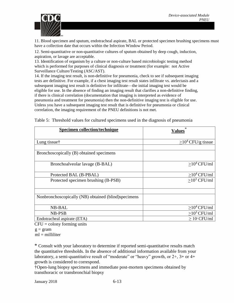
9. Refer to threshold values for cultured specimens with growth of eligible pathogens. (Table 5).
Notes:
A specimen that is not obtained through an artificial airway (specifically endotracheal tube or
tracheostomy) is not considered minimally contaminated and is not eligible for use in meeting the
laboratory criteria for PNU2. Sputum is not a minimally-contaminated specimen.
Because they are an indication of commensal flora of the oral cavity or upper respiratory tract, the
following organisms can only be used to meet PNEU definitions when identified from pleural fluid
obtained during thoracentesis or initial placement of chest tube (not from an indwelling chest tube) or
lung tissue:
o Coagulase-negative Staphylococcus species
o Enterococcus species
o Candida species or yeast not otherwise specified. Exception: identification of matching
Candida spp. from blood and sputum, endotracheal aspirate, BAL or protected specimen
brushing can be used to satisfy PNU3 definition for immunocompromised patients.
10. Immunocompromised patients include only:
those with neutropenia defined as absolute neutrophil count or total white blood cell count (WBC)
<500/mm3 those with leukemia, lymphoma or who are HIV positive with CD4 count <200 those who have undergone splenectomy those who have a history of solid organ or hematopoietic stem cell transplant those on cytotoxic chemotherapy those on steroids (excluding inhaled steroids) daily for >2 weeks.
My laboratory processes respiratory
specimens such as bronchoalveolar lavage
fluid using a centrifugation procedure (for
example, “cytospin”), and there is no
quantitation or semi-quantitation of
neutrophils or white blood cells in the direct
examination report?
In this situation, a report indicating the presence of
white blood cells, without quantitation, is sufficient to
meet the purulent secretions criterion.
January 2018 6-13
Device-associated Module
PNEU
11. Blood specimen and sputum, endotracheal aspirate, BAL or protected specimen brushing specimens must
have a collection date that occurs within the Infection Window Period.
12. Semi-quantitative or non-quantitative cultures of sputum obtained by deep cough, induction,
aspiration, or lavage are acceptable.
13. Identification of organism by a culture or non-culture based microbiologic testing method
which is performed for purposes of clinical diagnosis or treatment (for example: not Active
Surveillance Culture/Testing (ASC/AST).
14. If the imaging test result, is non-definitive for pneumonia, check to see if subsequent imaging
tests are definitive. For example, if a chest imaging test result states infiltrate vs. atelectasis and a
subsequent imaging test result is definitive for infiltrate—the initial imaging test would be
eligible for use. In the absence of finding an imaging result that clarifies a non-definitive finding,
if there is clinical correlation (documentation that imaging is interpreted as evidence of
pneumonia and treatment for pneumonia) then the non-definitive imaging test is eligible for use.
Unless you have a subsequent imaging test result that is definitive for pneumonia or clinical
correlation, the imaging requirement of the PNEU definitions is not met.
Table 5: Threshold values for cultured specimens used in the diagnosis of pneumonia