Chapter 6 Viruses. 6.1 General Properties of Viruses 6.2 Structure of viruses 6.3 The cultivation of viruses 6.4 General Features of Virus Reproduction 6.5 one-step growth curve 6.6 Temperate Bacteriophages: Lysogeny and Lambda 6.7 viral classification 6.8 Overview of Animal Viruses - PowerPoint PPT Presentation
67
6.1 General Properties of Viruses 6.2 Structure of viruses 6.3 The cultivation of viruses 6.4 General Features of Virus Reprod uction 6.5 one-step growth curve 6.6 Temperate Bacteriophages: Lysoge ny and Lambda 6.7 viral classification 6.8 Overview of Animal Viruses 6.9 plant viruses 6.10 Viroids and Prions Chapter 6 Viruses
Transcript
6.1 General Properties of Viruses 6.2 Structure of viruses6.3 The cultivation of viruses6.4 General Features of Virus Reproduction6.5 one-step growth curve6.6 Temperate Bacteriophages: Lysogeny and Lambda6.7 viral classification6.8 Overview of Animal Viruses6.9 plant viruses6.10 Viroids and Prions
Chapter 6 Viruses
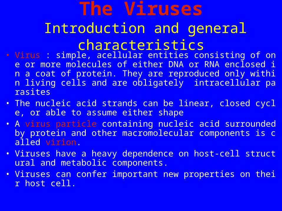
The Viruses Introduction and general characteristics
• Virus : simple, acellular entities consisting of one or more molecules of either DNA or RNA enclosed in a coat of protein. They are reproduced only within living cells and are obligately intracellular parasites
• The nucleic acid strands can be linear, closed cycle, or able to assume either shape
• A virus particle containing nucleic acid surrounded by protein and other macromolecular components is called virion.
• Viruses have a heavy dependence on host-cell structural and metabolic components.
• Viruses can confer important new properties on their host cell.
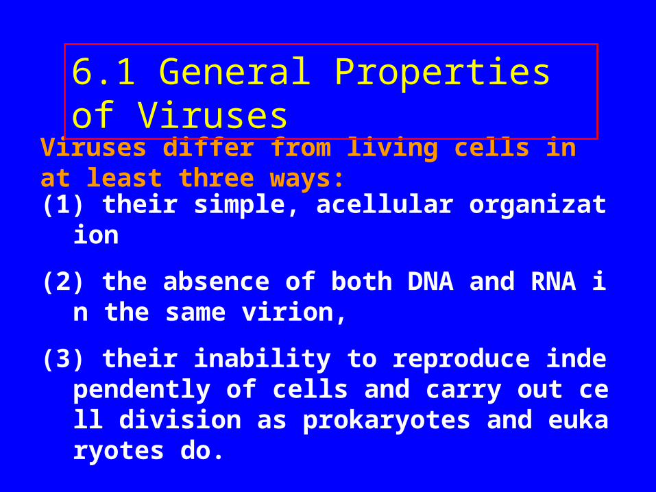
(1) their simple, acellular organization
(2) the absence of both DNA and RNA in the same virion,
(3) their inability to reproduce independently of cells and carry out cell division as prokaryotes and eukaryotes do.
Viruses differ from living cells in at least three ways:
6.1 General Properties of Viruses
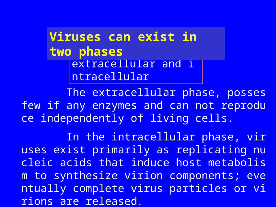
The extracellular phase, posses few if any enzymes and can not reproduce independently of living cells.
In the intracellular phase, viruses exist primarily as replicating nucleic acids that induce host metabolism to synthesize virion components; eventually complete virus particles or virions are released.
extracellular and intracellular
Viruses can exist in two phases
Hosts and size
Three main classes - animal viruses, bacterial viruses (bacteriophages), and plant viruses.
The particular host range of a virus is determined by the virus's requirements for its specific attachment to the host cell and the availability within the potential host of cellular factors required for viral multiplication.
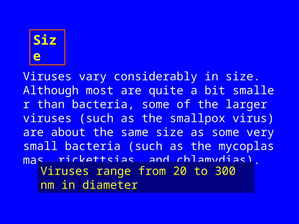
Viruses vary considerably in size. Although most are quite a bit smaller than bacteria, some of the larger viruses (such as the smallpox virus) are about the same size as some very small bacteria (such as the mycoplasmas, rickettsias, and chlamydias).
Size
Viruses range from 20 to 300 nm in diameter
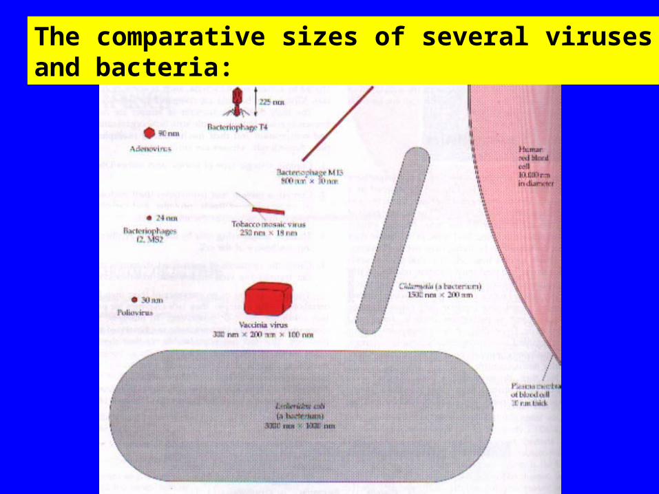
The comparative sizes of several viruses and bacteria:
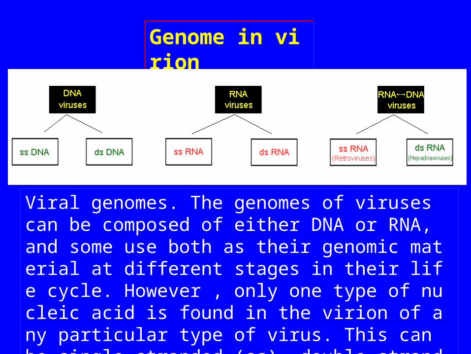
Genome in virion
Viral genomes. The genomes of viruses can be composed of either DNA or RNA, and some use both as their genomic material at different stages in their life cycle. However , only one type of nucleic acid is found in the virion of any particular type of virus. This can be single-stranded (ss), double-stranded (ds), or in the case of the hepadnaviruses, partially double-stranded.
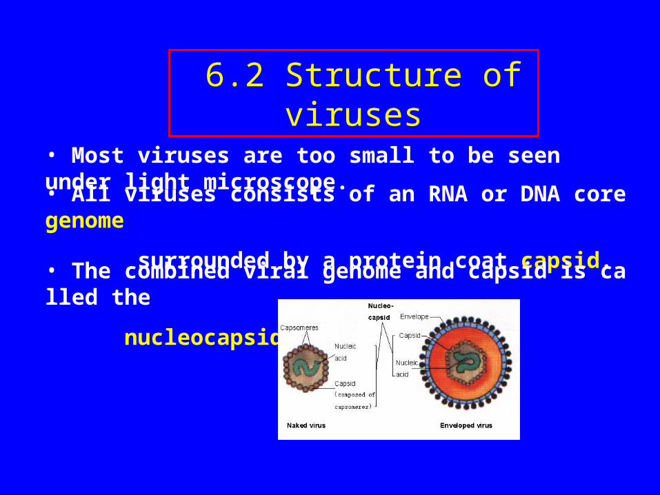
• Most viruses are too small to be seen under light microscope.
• All viruses consists of an RNA or DNA core genome
surrounded by a protein coat capsid.• The combined viral genome and capsid is called the
nucleocapsid.
6.2 Structure of viruses
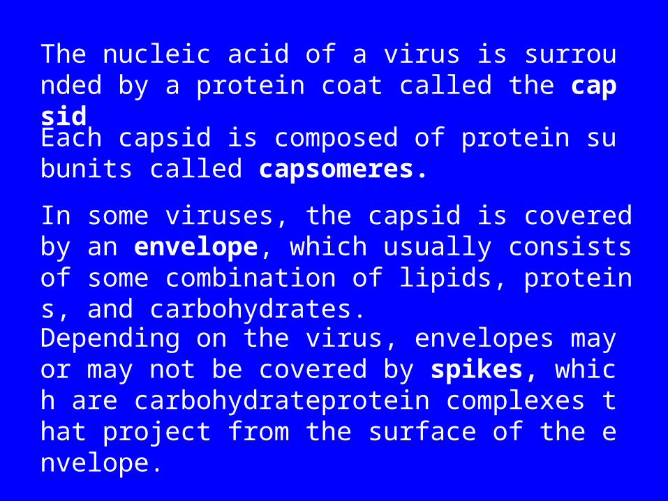
The nucleic acid of a virus is surrounded by a protein coat called the capsid
Each capsid is composed of protein subunits called capsomeres.
In some viruses, the capsid is covered by an envelope, which usually consists of some combination of lipids, proteins, and carbohydrates.
Depending on the virus, envelopes may or may not be covered by spikes, which are carbohydrateprotein complexes that project from the surface of the envelope.
Viruses may be classified into several morphological types on the basis of their capsid architecture as revealed by electron microscopy and a technique called x-ray crystallography.
GENERAL MORPHOLOGY
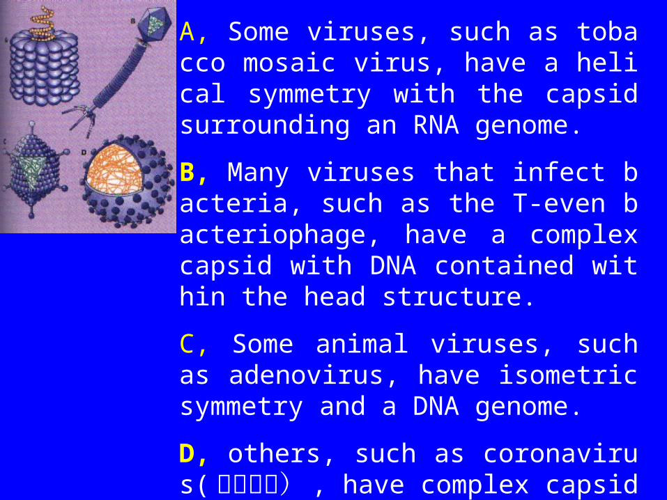
A, Some viruses, such as tobacco mosaic virus, have a helical symmetry with the capsid surrounding an RNA genome.
B, Many viruses that infect bacteria, such as the T-even bacteriophage, have a complex capsid with DNA contained within the head structure.
C, Some animal viruses, such as adenovirus, have isometric symmetry and a DNA genome.
D, others, such as coronavirus(冠状病毒) , have complex capsids and an envelope with protruding proteins surrounding an RNA genome.
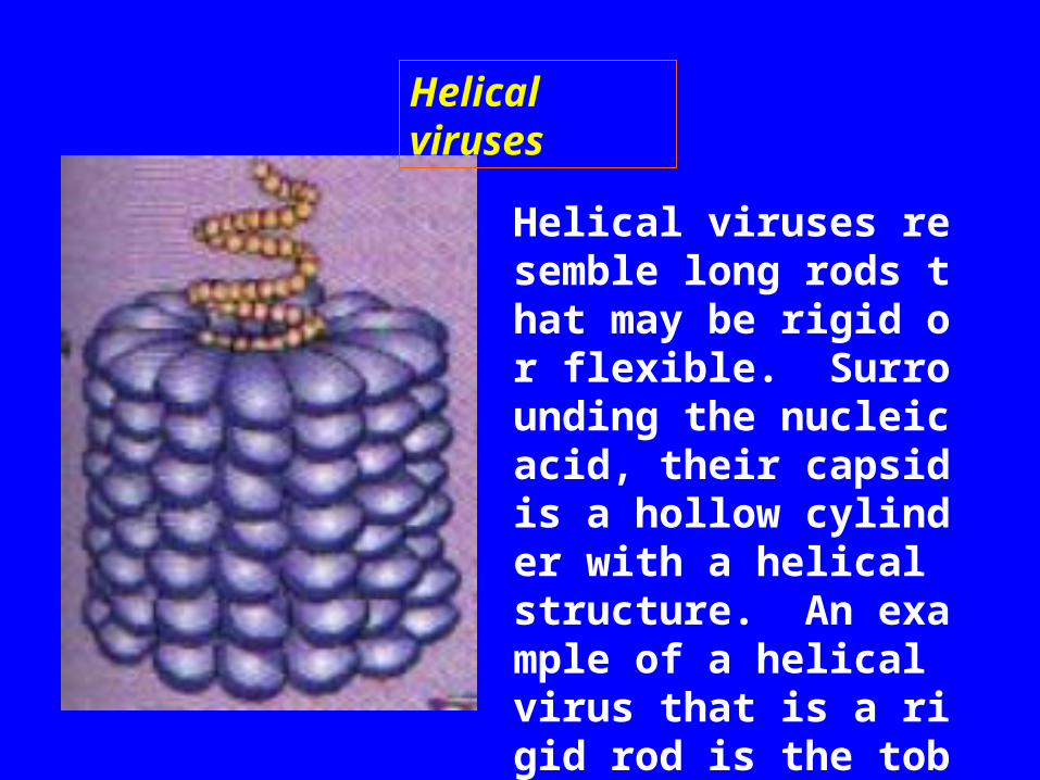
Helical viruses resemble long rods that may be rigid or flexible. Surrounding the nucleic acid, their capsid is a hollow cylinder with a helical structure. An example of a helical virus that is a rigid rod is the tobacco mosaic virus
Helical viruses
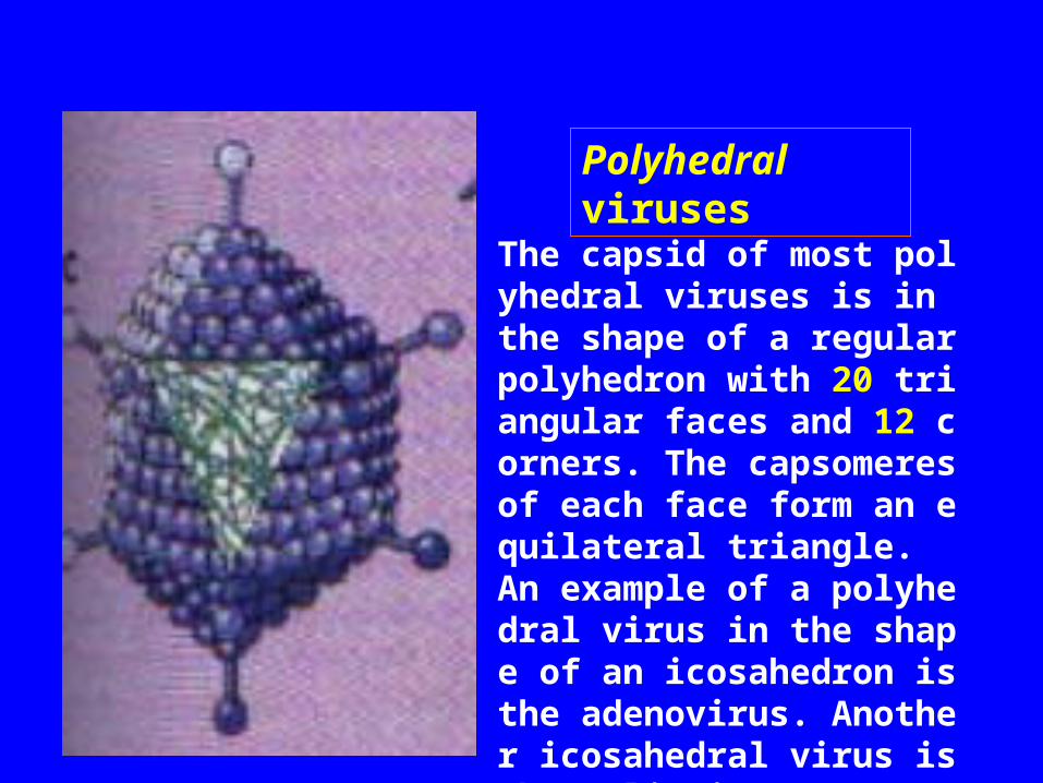
The capsid of most polyhedral viruses is in the shape of a regular polyhedron with 20 triangular faces and 12 corners. The capsomeres of each face form an equilateral triangle. An example of a polyhedral virus in the shape of an icosahedron is the adenovirus. Another icosahedral virus is the poliovirus.
Polyhedral viruses
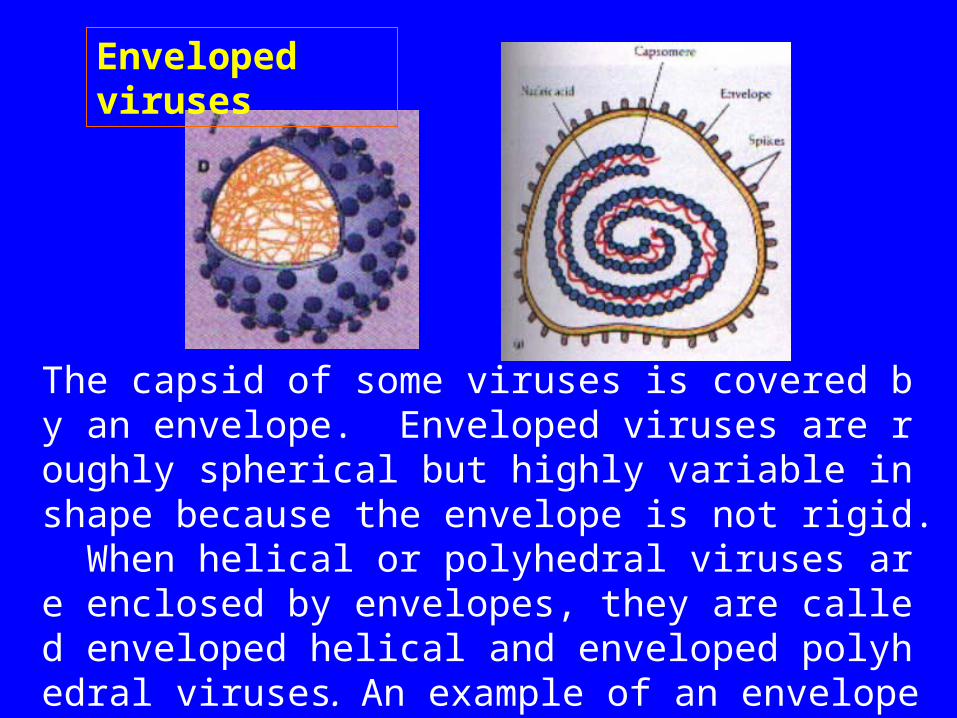
The capsid of some viruses is covered by an envelope. Enveloped viruses are roughly spherical but highly variable in shape because the envelope is not rigid. When helical or polyhedral viruses are enclosed by envelopes, they are called enveloped helical and enveloped polyhedral viruses. An example of an enveloped helical virus is the influenza virus.
Enveloped viruses
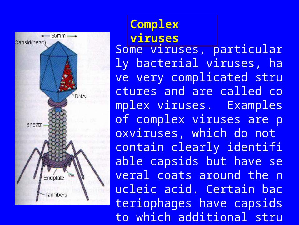
Some viruses, particularly bacterial viruses, have very complicated structures and are called complex viruses. Examples of complex viruses are poxviruses, which do not contain clearly identifiable capsids but have several coats around the nucleic acid. Certain bacteriophages have capsids to which additional structures are attached.
Complex viruses
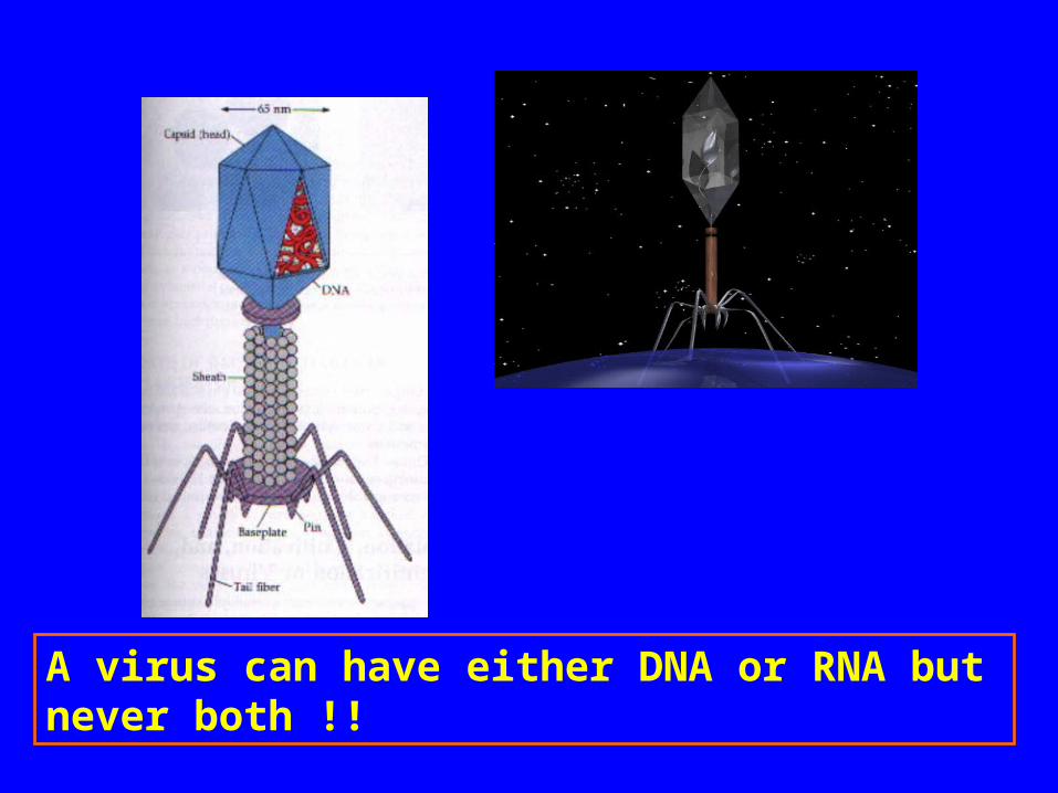
A virus can have either DNA or RNA but never both !!
6.3 Cultivating Viruses
• Bacteriophage: bacterial Cultures• Animal viruses: Tissue or cell cultures• Plant viruses: tissue or separated cell cult
ures, or cultures of protoplasts
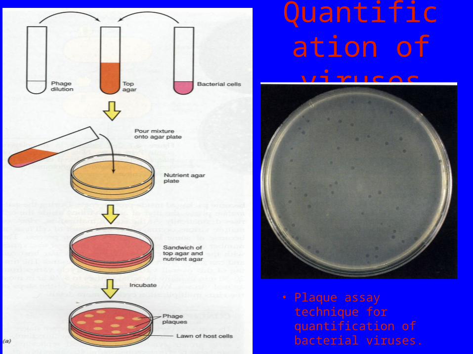
Quantification of viruses
• Plaque assay technique for quantification of bacterial viruses.
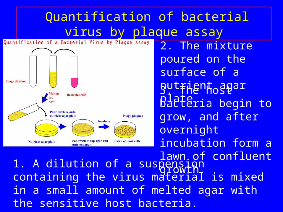
1. A dilution of a suspension containing the virus material is mixed in a small amount of melted agar with the sensitive host bacteria.
3. The host bacteria begin to grow, and after overnight incubation form a lawn of confluent growth.
Quantification of bacterial virus by plaque assay
2. The mixture poured on the surface of a nutrient agar plate.
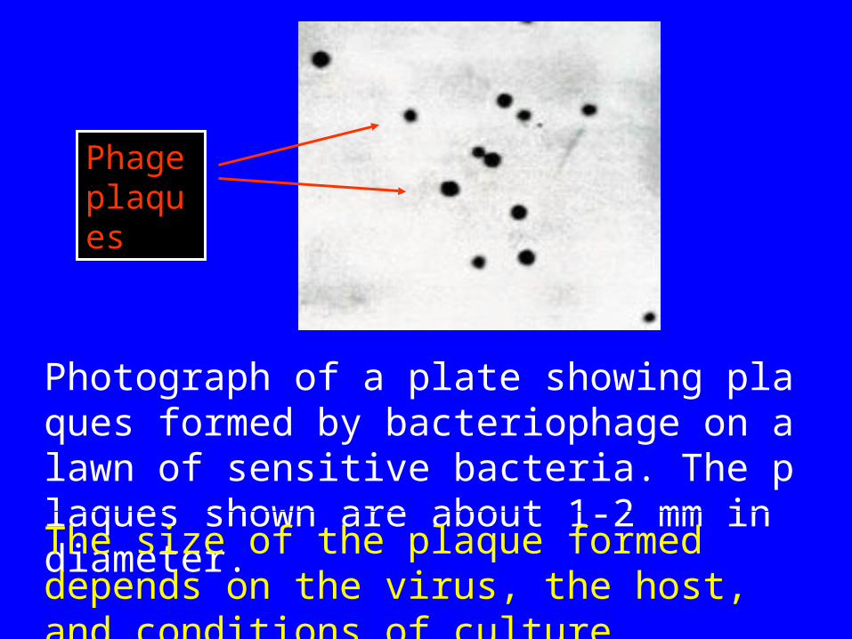
Photograph of a plate showing plaques formed by bacteriophage on a lawn of sensitive bacteria. The plaques shown are about 1-2 mm in diameter.
The size of the plaque formed depends on the virus, the host, and conditions of culture.
Phage plaques
Attentions
• Young, actively growing bacterial cells• Plaque appearance often is characteristic of
the phage being cultivated.• Plaque-forming units(PFU): each plaque
in a layer of bacterial or animal cells is assumed to have arisen from the reproduction of a single virus particle.
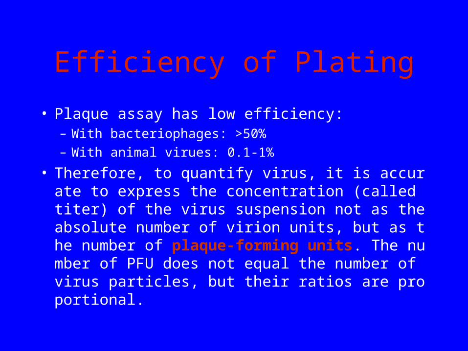
Efficiency of Plating
• Plaque assay has low efficiency:– With bacteriophages: >50%– With animal virues: 0.1-1%
• Therefore, to quantify virus, it is accurate to express the concentration (called titer) of the virus suspension not as the absolute number of virion units, but as the number of plaque-forming units. The number of PFU does not equal the number of virus particles, but their ratios are proportional.
The cultivation of animal viruses
• For many years researchers have cultivated animal viruses by inoculating suitable host animals or embryonated eggs-fertilized chicken eggs incubated about 6 to 8 days after laying.
• More recently animal viruses have been grown in tissue(cell) culture on monolayers of animal cells. This technique is made possible by the development of growth media for animal cells and by the advent of antibiotics that can prevent bacterial and fungal contamination.
• Animal viruses can cause microscopic or macroscopic degenerative changes or abnormalities in host cells and in tissues called cytopathic effects.
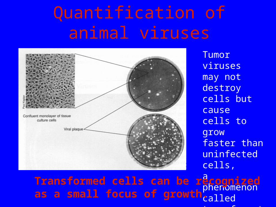
Quantification of animal viruses
Tumor viruses may not destroy cells but cause cells to grow faster than uninfected cells, a phenomenon called transformation.
Transformed cells can be recognized as a small focus of growth.
6.4 Viral Multiplication
For a virus to multiply, it must invade a host cell and take over the host's metabolic machinery. A single virus can give rise to several or even thousands of similar viruses in a single host cell. This process drastically changes the host cell and often causes its death.
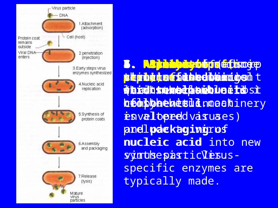
1. Attachment (adsorption) of the virion to a susceptible host cell.
2. Penetration (injection) of the virion or its nucleic acid into the cell.
3. Early steps in replication during which the host cell biosynthetic machinery is altered as a prelude to virus nucleic acid synthesis. Virus-specific enzymes are typically made.
Multiplication of bacteriophages
4. Replication of the virus nucleic acid.
5. Synthesis of proteins used as structural subunits of the virus coat.
6. Assembly of structural subunits (and membrane components in enveloped viruses) and packaging of nucleic acid into new virus particles.
7. Release of mature virions from the cell.
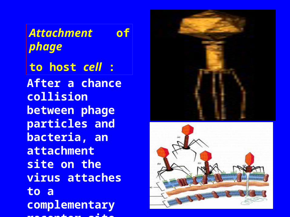
Attachment of phage
to host cell :
After a chance collision between phage particles and bacteria, an attachment site on the virus attaches to a complementary receptor site on the bacterial cell.
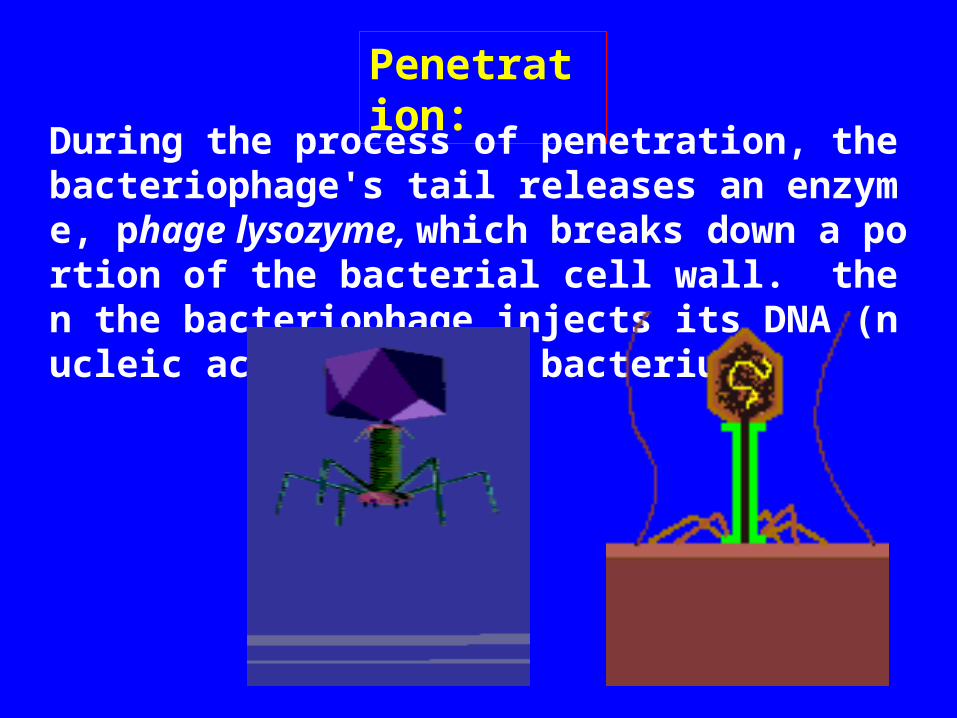
Penetration:
During the process of penetration, the bacteriophage's tail releases an enzyme, phage lysozyme, which breaks down a portion of the bacterial cell wall. then the bacteriophage injects its DNA (nucleic acid) into the bacterium.
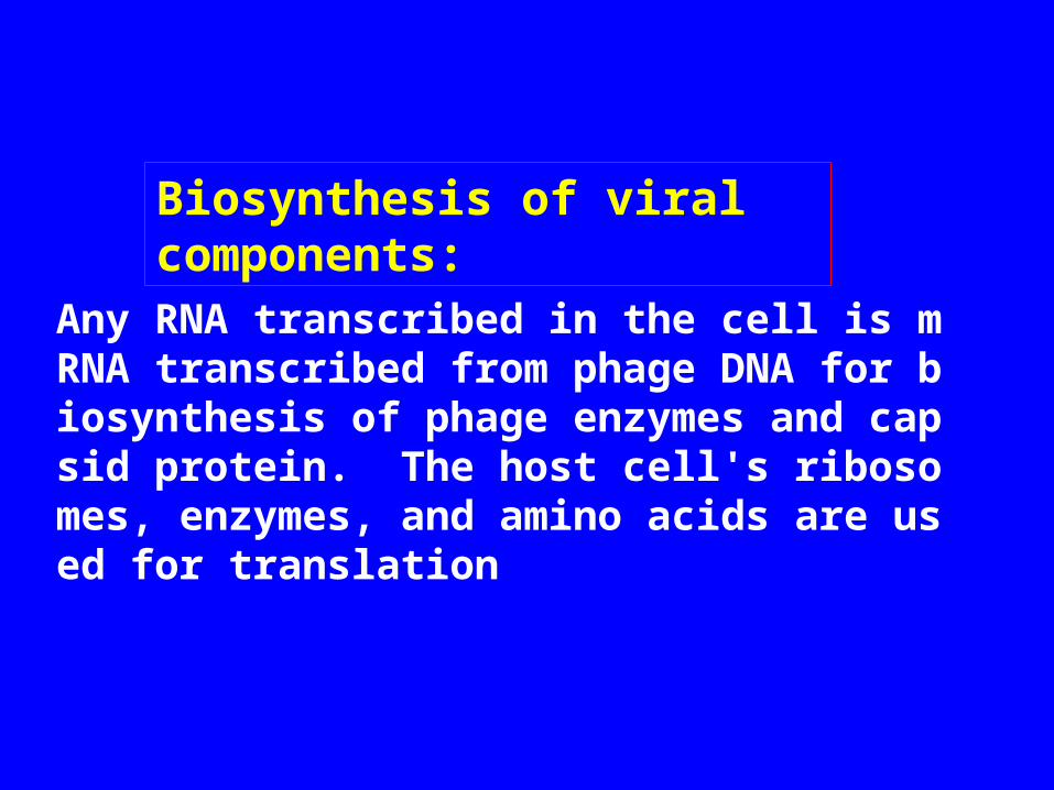
Biosynthesis of viral components:
Any RNA transcribed in the cell is mRNA transcribed from phage DNA for biosynthesis of phage enzymes and capsid protein. The host cell's ribosomes, enzymes, and amino acids are used for translation
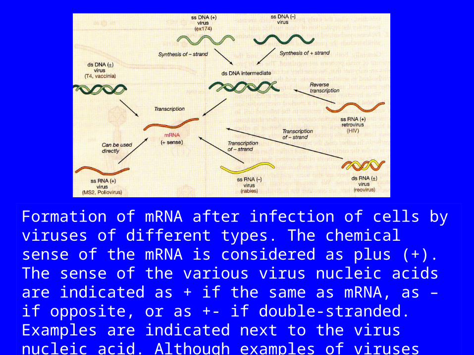
Formation of mRNA after infection of cells by viruses of different types. The chemical sense of the mRNA is considered as plus (+). The sense of the various virus nucleic acids are indicated as + if the same as mRNA, as – if opposite, or as +- if double-stranded. Examples are indicated next to the virus nucleic acid. Although examples of viruses containing SS DNA of the - sense are known, none are discussed in the text.
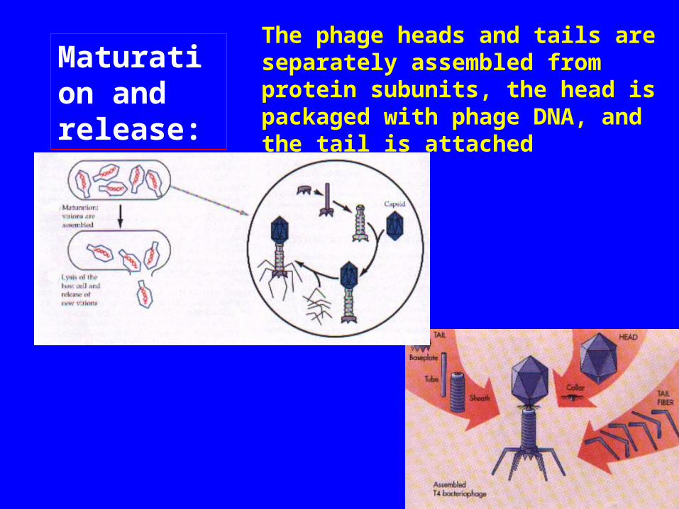
Maturation and release:
The phage heads and tails are separately assembled from protein subunits, the head is packaged with phage DNA, and the tail is attached
Release:
Lysozyme, whose code is provided by a phage gene, is synthesized within the cell. This enzyme causes a breakdown of the bacterial cell wall, and the newly produced bactedophages are released from the host cell.
The number of newly synthesized phage particles released from a single cell usually ranges from about 50 to 200.
1. Attachment (adsorption) of the virion to a susceptible host cell.
2. Penetration (injection) of the virion or its nucleic acid into the cell.
3. Early steps in replication during which the host cell biosynthetic machinery is altered as a prelude to virus nucleic acid synthesis. Virus-specific enzymes are typically made.
4. Replication of the virus nucleic acid.5. Synthesis of proteins used as structural subunits of the virus coat.
6. Assembly of structural subunits (and membrane components in enveloped viruses) and packaging of nucleic acid into new virus particles.
7. Release of mature virions from the cell
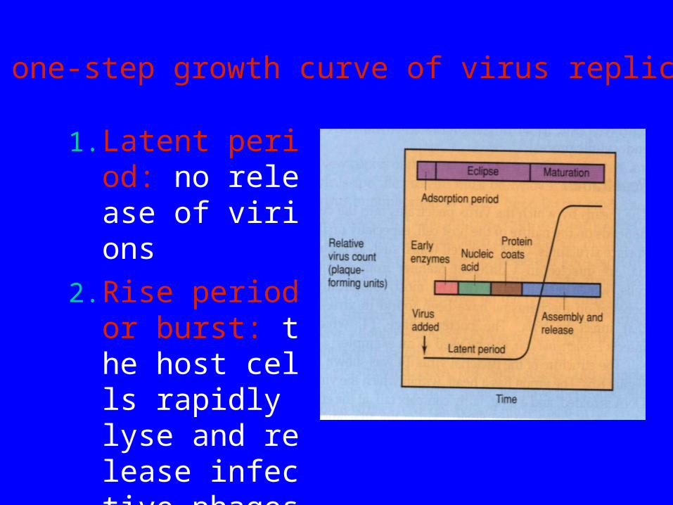
6.5 one-step growth curve
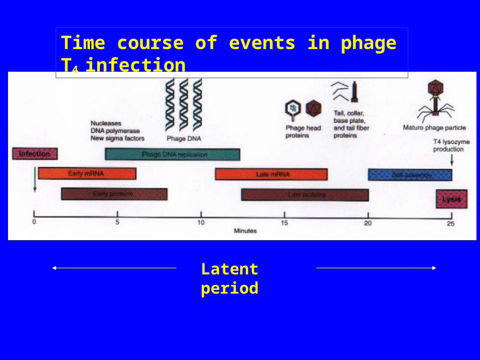
1. Latent period: no release of virions
2. Rise period or burst: the host cells rapidly lyse and release infective phages
The one-step growth curve of virus replication
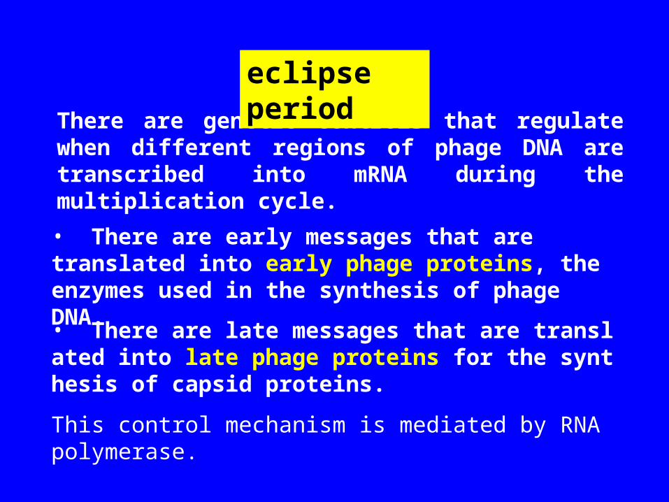
There are genetic controls that regulate when different regions of phage DNA are transcribed into mRNA during the multiplication cycle.
• There are early messages that are translated into early phage proteins, the enzymes used in the synthesis of phage DNA.
• There are late messages that are translated into late phage proteins for the synthesis of capsid proteins.
This control mechanism is mediated by RNA polymerase.
eclipse period
Time course of events in phage T4 infection
Latent period
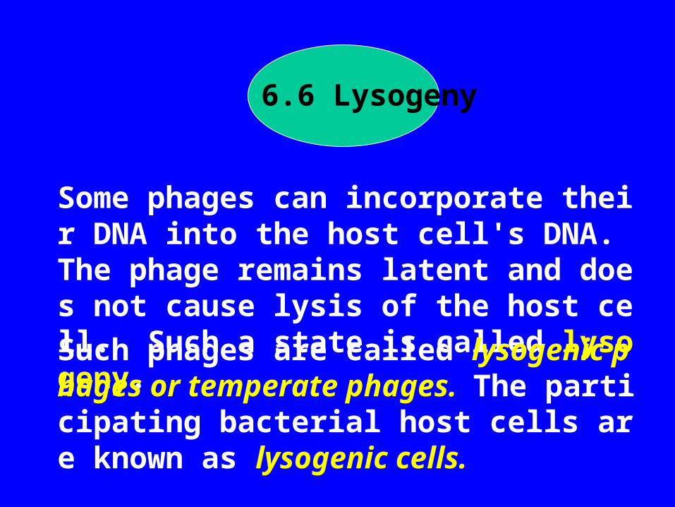
6.6 Lysogeny
Some phages can incorporate their DNA into the host cell's DNA. The phage remains latent and does not cause lysis of the host cell. Such a state is called lysogeny.Such phages are called lysogenic phages or temperate phages. The participating bacterial host cells are known as lysogenic cells.
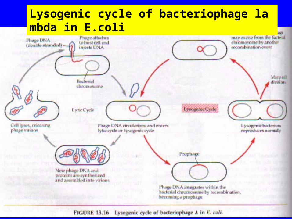
Lysogenic cycle of bacteriophage lambda in E.coli
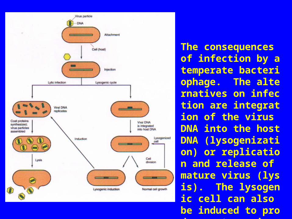
The consequences of infection by a temperate bacteriophage. The alternatives on infection are integration of the virus DNA into the host DNA (lysogenization) or replication and release of mature virus (lysis). The lysogenic cell can also be induced to produce mature virus and lyse.
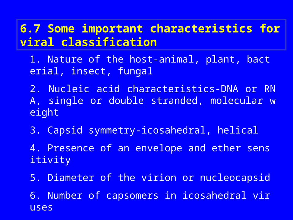
1. Nature of the host-animal, plant, bacterial, insect, fungal
2. Nucleic acid characteristics-DNA or RNA, single or double stranded, molecular weight
3. Capsid symmetry-icosahedral, helical
4. Presence of an envelope and ether sensitivity
5. Diameter of the virion or nucleocapsid
6. Number of capsomers in icosahedral viruses
7. Immunologic properties
8. Intracellular location of viral replication
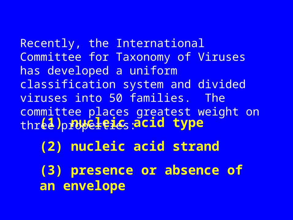
6.7 Some important characteristics for viral classification
(1) nucleic acid type
(2) nucleic acid strand
(3) presence or absence of an envelope
Recently, the International Committee for Taxonomy of Viruses has developed a uniform classification system and divided viruses into 50 families. The committee places greatest weight on three properties:
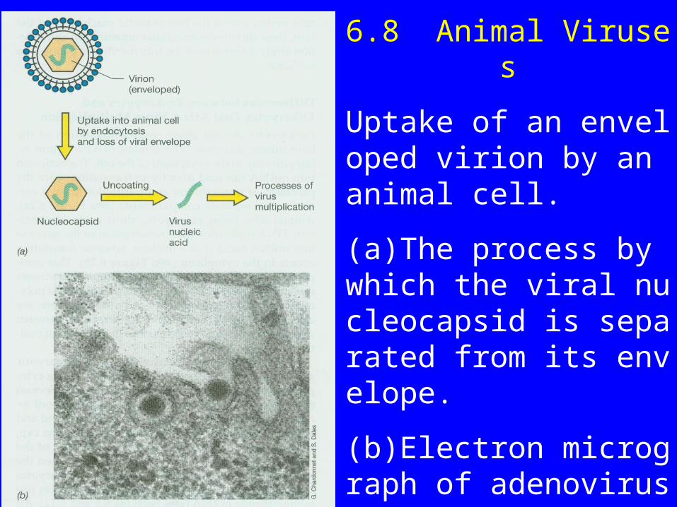
(a)The process by which the viral nucleocapsid is separated from its envelope.
(b)Electron micrograph of adenovirus virions entering a cell. Each particle is about 70 nm in diameter.
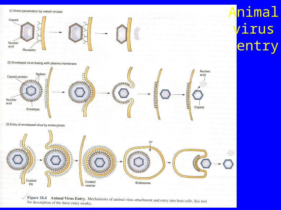
Animal virus entry
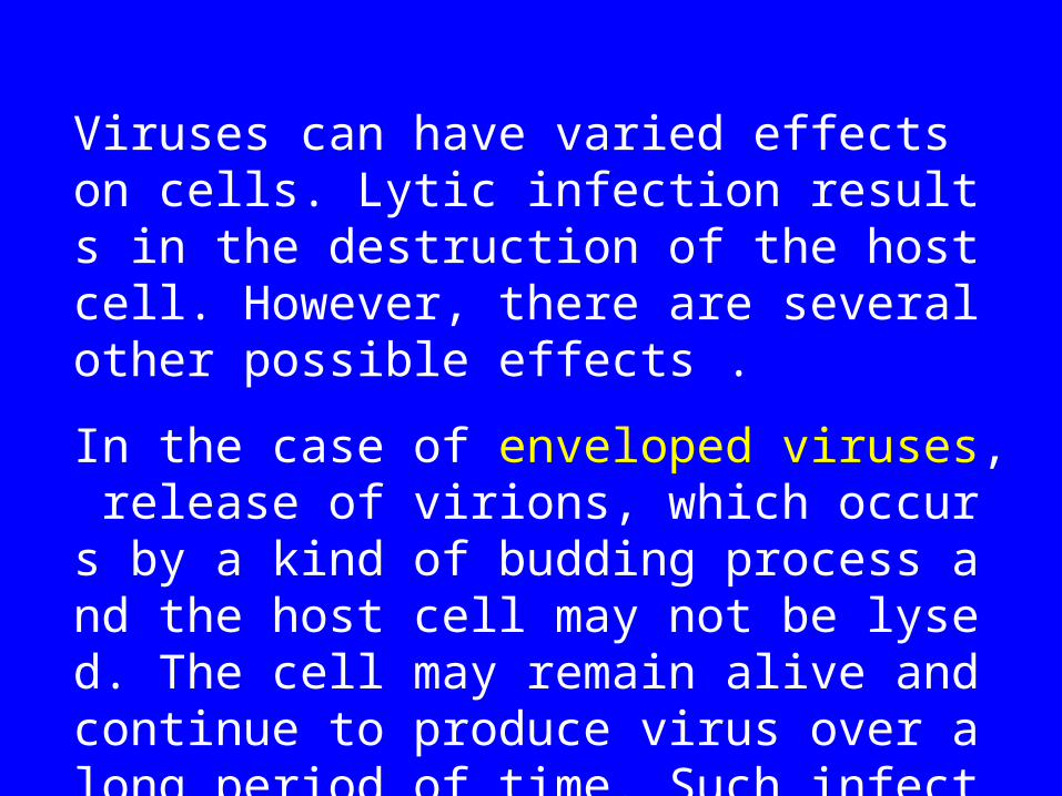
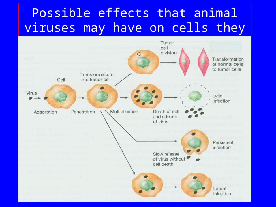
Viruses can have varied effects on cells. Lytic infection results in the destruction of the host cell. However, there are several other possible effects .
In the case of enveloped viruses, release of virions, which occurs by a kind of budding process and the host cell may not be lysed. The cell may remain alive and continue to produce virus over a long period of time. Such infections are referred to as persistent infections
Possible effects that animal viruses may have on cells they effect
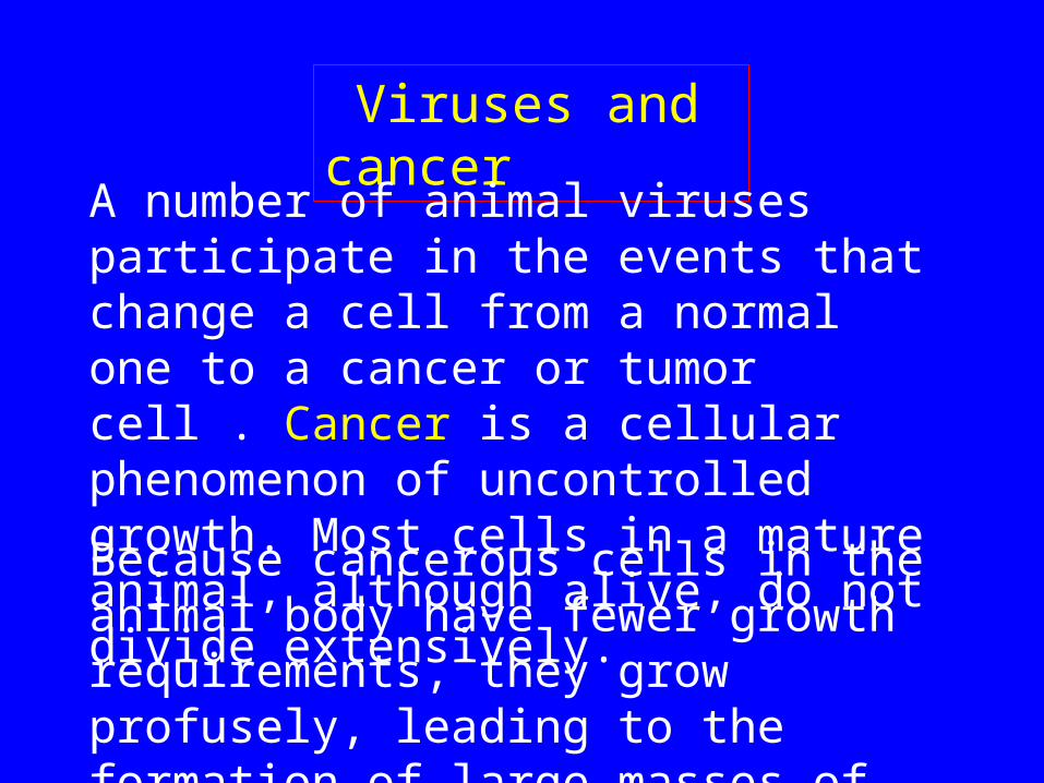
Viruses and cancer
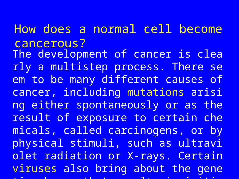
A number of animal viruses participate in the events that change a cell from a normal one to a cancer or tumor cell . Cancer is a cellular phenomenon of uncontrolled growth. Most cells in a mature animal, although alive, do not divide extensively.
Because cancerous cells in the animal body have fewer growth requirements, they grow profusely, leading to the formation of large masses of cells, called tumors.
The development of cancer is clearly a multistep process. There seem to be many different causes of cancer, including mutations arising either spontaneously or as the result of exposure to certain chemicals, called carcinogens, or by physical stimuli, such as ultraviolet radiation or X-rays. Certain viruses also bring about the genetic change that results in initiation of tumor formation.
How does a normal cell become cancerous?
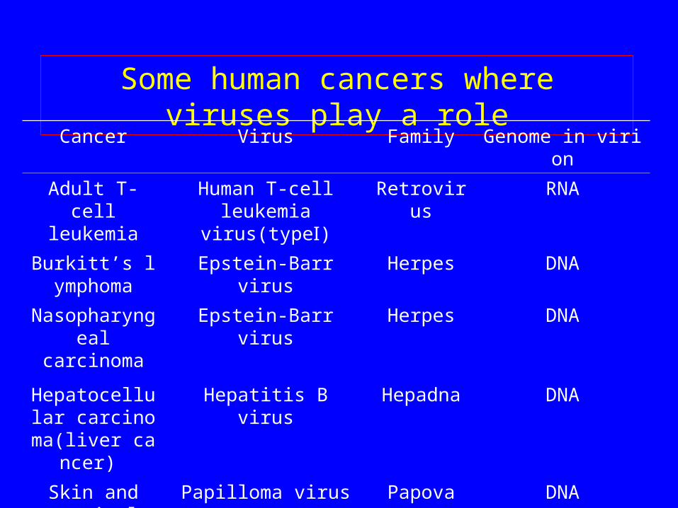
Some human cancers where viruses play a role
Cancer Virus Family Genome in virionAdult T-cell
leukemiaHuman T-cell leukemia
virus(type )ⅠRetrovirus RNA
Burkitt’s lymphoma
Epstein-Barr virus Herpes DNA
Nasopharyngeal carcinoma
Epstein-Barr virus Herpes DNA
Hepatocellular carcinoma(liver c
ancer)
Hepatitis B virus Hepadna DNA
Skin and cervical cancers
Papilloma virus Papova DNA
6.8.1 Pox Viruses
The most complex and largest animal viruses known and have some characteristics that approach those of primitive cells.
The pox viruses are not able to metabolize and thus depend on the host for the complete machinery of protein synthesis.
These viruses are also unique in that they are DNA viruses that replicate in the cytoplasm.
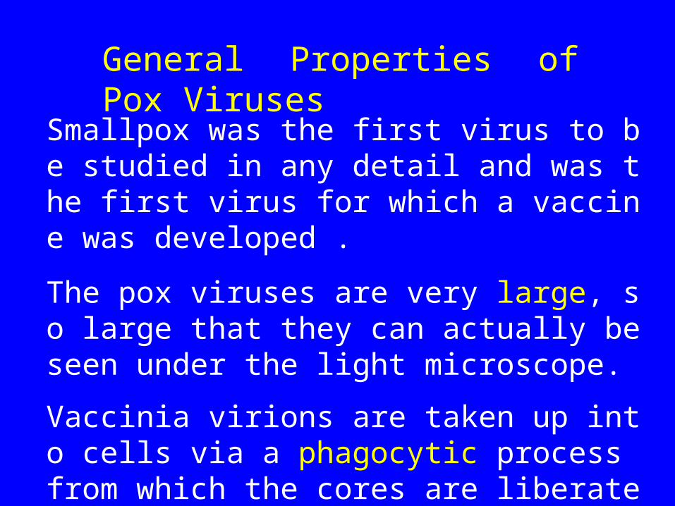
Smallpox was the first virus to be studied in any detail and was the first virus for which a vaccine was developed .
The pox viruses are very large, so large that they can actually be seen under the light microscope.
Vaccinia virions are taken up into cells via a phagocytic process from which the cores are liberated into the cytoplasm.
General Properties of Pox Viruses
6.8.2 Adenoviruses
The genomes of the adenoviruses consist of linear double-stranded DNA of about 36 kilobase pairs.
Attached in covalent linkage to the 5'-terminus of the DNA is a protein component essential for infectivity of the DNA.
The DNA has inverted terminal repeats of 100-1800 base pairs (this varies with the virus strain).
The DNA of the adenoviruses is six to seven times the size of the DNA of the papovavirus SV40.
Replication of the viral DNA occurs in the nucleus.
After the virus particle has been transported to the nucleus, the core is released and converted to a viral DNA-histone complex.
Early transcription is carried out by an RNA polymerase of the host, and a number of primary transcripts are made.
The transcripts are spliced, capped, and polyadenylated, giving sever al different mRNAs.
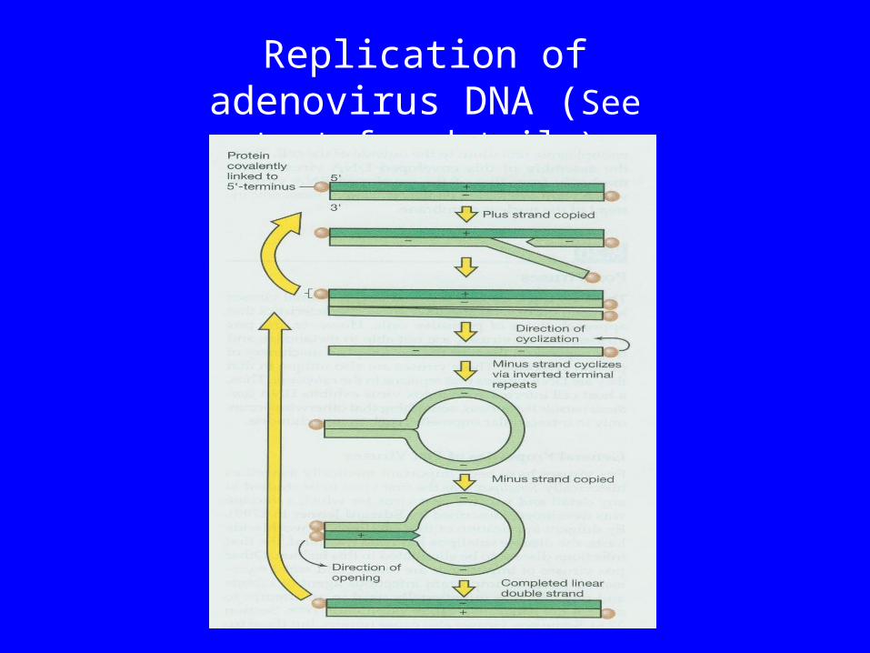
Replication of adenovirus DNA (See text for details)
6.8.3 Retroviruses
One important group of animal viruses, those called the retroviruses, have both an RNA and a DNA phase of replication.
The retroviruses are RNA viruses, but they replicate by means of a DNA intermediate using the enzyme reverse transcriptase.
3. The enzyme reverse transcriptase has become a major tool in genetic engineering.
2. One retrovirus, the one causing acquired immunodeficiency syndrome (AIDS) .
1. They were the first viruses shown to cause cancer and have been studied most extensively for their carcinogenic characteristics.
Importance
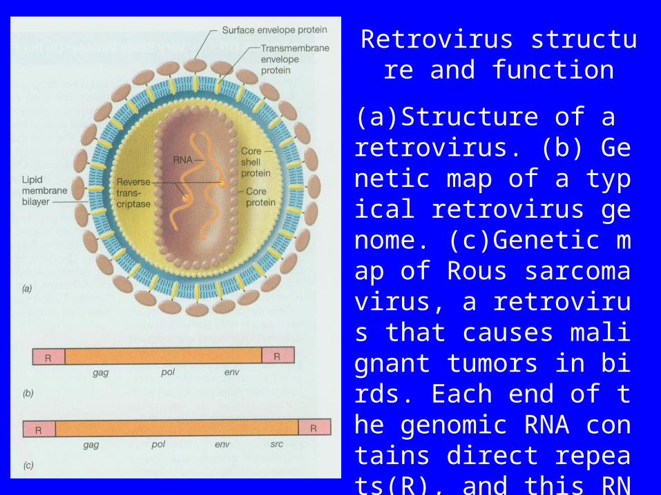
Retrovirus structure and function
(a)Structure of a retrovirus. (b) Genetic map of a typical retrovirus genome. (c)Genetic map of Rous sarcoma virus, a retrovirus that causes malignant tumors in birds. Each end of the genomic RNA contains direct repeats(R), and this RNA also has a 5’-cap and a 3’-poly-A tail. See text for more details.
6.8.4 Insect viruses• Many insect virus infections are accomanied by the format
ion of inclusion bodies within the infected cells. 1. cytoplasmic polyhedrosis 2. nuclear polyhedrosis 3. cytoplasmic and nuclear polyhedrosis• The inclusion bodies are protein in nature and enclose one
or more virions. Polyhedral bodies protect the virions against heat, low pH, and many chemicals.
• Insect larvae are infected when they feed on leaves contaminated with inclusion bodies. When in insect gut contents, inclusion bodies dissolve to liberate the virions.
Insect viruses as biological control agents
• Baculoviruses have received the most attention for at least three reasons:
1. They attack only invertebrates and have considerable host specificity.
2. They are encased in protective inclusion bodies. 3. They are well suited for commercial production since t
hey often reach extremely hegh concentrations in larval tissue.
• Cotton bollworm. Douglas fir tussock moth, gypsy moth, alfalfa looper
• Inclusion bodies are sprayed on foliage consumed by the target insects
6.9 Plant Viruses :
Virion morphology: 1. Helical capsids 2. Icosahedral•Almost all plant viruses are RNA viruses, either single stranded or double stranded.•Taxomomy standard: nucleic acid type, strandedness, capsid symmetry and size, envelope presence•Transmission of plant viruses: leaves are mechanically damaged.
6.10 Viroids and PrionsViroids are small, circular, single-stranded RNA molecules that are the smallest known pathogens.
• about 250 to 370 nucleotides long. • Viroids are found principally in the nucleolus o
f infected cells; 200-2000 copies• They do not act as mRNAs to direct protein syn
thesis. How to cause disease symptoms• Could be replicated by an RNA-dependent RN
A polymerase, a rolling-circle type mechanism
Prions represent the other extreme from viroids. They have a distinct extracellular form, but the extracellular form seems to be entirely protein. It apparently does not contain any nucleic acid, or if it does, the molecule is not long enough to encode the single kind of protein of which the prion is composed.
Prions • Proteinaceous infectious particle• Cause a degenerative disorder of the central nervo
us system.• No nucleic acid has yet been detected in the agent,
it seems to be a 33 to 35 kDa hydrophobic membrane protein, often called PrP.
• The PrP is bound to the surface of neurons, presumably an altered PrP is at least partly responsible for the disease.