DMD # 69823 1 Title Page Biotransformation and Rearrangement of Laromustine Alaa-Eldin F. Nassar, Adam V. Wisnewski, Ivan King School of Medicine, Department of Internal Medicine, Yale University, New Haven, CT (AFN and AVW) , Department of Chemistry, 55 N. Eagleville Rd., University of Connecticut, Storrs, CT (AFN), Metastagen, Inc., Wilmington, DE, United States (IK). This article has not been copyedited and formatted. The final version may differ from this version. DMD Fast Forward. Published on June 8, 2016 as DOI: 10.1124/dmd.116.069823 at ASPET Journals on May 17, 2020 dmd.aspetjournals.org Downloaded from
Transcript
DMD # 69823
1
Title Page
Biotransformation and Rearrangement of Laromustine
Alaa-Eldin F. Nassar, Adam V. Wisnewski, Ivan King
School of Medicine, Department of Internal Medicine, Yale University, New Haven, CT
(AFN and AVW) , Department of Chemistry, 55 N. Eagleville Rd., University of
Connecticut, Storrs, CT (AFN), Metastagen, Inc., Wilmington, DE, United States (IK).
This article has not been copyedited and formatted. The final version may differ from this version.DMD Fast Forward. Published on June 8, 2016 as DOI: 10.1124/dmd.116.069823
This article has not been copyedited and formatted. The final version may differ from this version.DMD Fast Forward. Published on June 8, 2016 as DOI: 10.1124/dmd.116.069823
Drug Administration; FMO, flavin-containing monooxygenase; LC-MS/MS, liquid
chromatography-tandem mass spectrometry; NADPH, nicotinamide adenine
dinucleotide phosphate, LC-MSn; Liquid chromatography-multi-stage mass
spectrometry, idiosyncratic drug reactions, IDRs; glutathione; GSH, N-acetylecysteine;
NAC, cysteine; CYS, and hydrogen-deuterium exchange; H-D.
This article has not been copyedited and formatted. The final version may differ from this version.DMD Fast Forward. Published on June 8, 2016 as DOI: 10.1124/dmd.116.069823
This review highlights the recent research into the biotransformations and
rearrangement of the sulfonylhydrazine alkylating agent laromustine. Incubation of
[14C]laromustine with rat, dog, monkey and human liver microsomes produced eight
radioactive components (C-1 to C-8). There was little difference in the metabolite profile
among the species examined, partly because nicotinamide adenine dinucleotide
phosphate (NADPH) was not required for the formation of most components, but rather
involved decomposition and/or hydrolysis. The exception was C-7, a hydroxylated
metabolite, largely formed by CYP2B6 and CYP3A4/5. Liquid chromatography-multi-
stage mass spectrometry (LC-MSn) studies determined that collision-induced
dissociation, and not biotransformation or enzyme catalysis, produced the unique mass
spectral rearrangement. Accurate mass measurements performed with a Fourier-
transform ion cyclotron resonance mass spectrometer (FTICR-MS) significantly aided
determination of the elemental compositions of the fragments, and in the case of
laromustine, revealed the possibility of rearrangement. Further, collision-induced
dissociation produced the loss of nitrogen (N2), methylsulfonyl and methyl isocyanate
moieties. The rearrangement, metabolite/decomposition products and conjugation
reactions were analyzed utilizing hydrogen-deuterium exchange (H-D), exact mass, 13C-
labeled laromustine, nuclear magnetic resonance spectroscopy (NMR) and LC-MSn
experiments to assist with the assignments of these fragments and possible
mechanistic rearrangement. Such techniques produced valuable insights into these
This article has not been copyedited and formatted. The final version may differ from this version.DMD Fast Forward. Published on June 8, 2016 as DOI: 10.1124/dmd.116.069823
functions: (1) P450 is involved in C-7 formation but plays little or no role in the
conversion of [14C]laromustine to C-1 through C-6 and C-8; (2) the relative abundance
of individual degradation/metabolite products was not species dependent; and (3)
laromustine produces several reactive intermediates that may produce the toxicities
seen in the clinical trials.
This article has not been copyedited and formatted. The final version may differ from this version.DMD Fast Forward. Published on June 8, 2016 as DOI: 10.1124/dmd.116.069823
and alkyl sulfonates (e.g., busulfan). Originally, alkylating agents were best known for
their use as mustard gas and related chemical weapons in World War I, due to their
toxicity. These agents are thought to react with the N7 position of guanine or any
nitrogen base in each of the double strands of DNA, resulting in damage to the DNA
(Alkylating Agents, 2014). They are harmful to normal cells, especially cells that divide
frequently, such as those in the gastrointestinal tract, bone marrow, testicles and
ovaries, which can cause loss of fertility. As early as the 1940’s, it was discovered that
they could be used as part of chemotherapy in different types of cancers (Scott 1970,
Wiedemann et.al., 1996, Goodman et al., 1946). One early example is busulfan, a
cancer drug in use since 1959; in 1999 it was approved by the US Food and Drug
Administration (FDA) for treatment of chronic myeloid leukemia (CML). Busulfan was
the main chemotherapeutic for treatment of chronic myeloid leukemia (CML) until it was
displaced by imatinib, but it is still in use because of its low cost. Alkylating agents are
commonly used to treat many different cancers, including leukemia, lymphoma,
Hodgkin’s disease, multiple myeloma, and sarcoma, as well as cancers of the lung,
This article has not been copyedited and formatted. The final version may differ from this version.DMD Fast Forward. Published on June 8, 2016 as DOI: 10.1124/dmd.116.069823
breast, and ovary. The risk with these drugs is that, at higher doses, they can cause
long-term damage to the bone marrow.
One key to successful drug design and development lies in finding the right
combination of diverse properties such as activity, toxicity, exposure, etc. Continued
development of any drug candidate hinges on the research team being able to
determine, and then optimize, these exposure-activity-toxicity relationships Therefore,
the goal of drug metabolism research is to optimize plasma half-life, drug/metabolic
clearance, metabolic stability, and the ratio of metabolic to renal clearance.
Concurrently, the researcher must be aware of the need to minimize or eliminate issues
such as gut/hepatic-first-pass metabolism, inhibition/induction of drug metabolizing
enzymes by metabolites, biologically active metabolites, metabolism by polymorphically
expressed drug metabolizing enzymes, and formation of reactive metabolites. A
successful outcome is a safer drug that undergoes predictable metabolic inactivation or
even undergoes no metabolism. Approaches available to the drug design team, as they
seek to meet the above goals, include active metabolites, prodrugs, hard and soft
drugs; these will be discussed below, with some examples from recent case studies.
The prodrug approach is commonly used in drug design. Prodrugs undergo an
enzymatic and/or chemical transformation in vivo to release the active drug. They help
researchers to improve the physicochemical, biopharmaceutical or pharmacokinetic
properties of pharmacologically potent drugs. About 10% of drugs approved worldwide
can be classified as prodrugs (Rautio et al., 2008). Prodrugs were discovered when it
was demonstrated that the antibacterial agent protosil was active in vivo only when it
This article has not been copyedited and formatted. The final version may differ from this version.DMD Fast Forward. Published on June 8, 2016 as DOI: 10.1124/dmd.116.069823
was metabolized to the actual drug sulfanilamide. Prodrugs most commonly use either
oxidative or reductive activation; for example, protosil is activated by reduction of its azo
linkage to the amine sulfa drug. Another example of a prodrug is the antipyretic agent
phenacetin, which becomes active upon conversion to acetaminophen. Carbamazepine
is an anticonvulsant drug that is the metabolic precursor of the active agent
carbamazepine 10,11-oxide. Minoxidil, originally developed as a potent vasodilator, also
induces hypertrichosis of facial and body hair (Buhl et al., 1990). Research data shows
that sulfation is a critical step for the hair-growth effects of minoxidil and that it is the
sulfated metabolite that directly stimulates hair follicles.
The various structural modifications of ampicillin demonstrate how the prodrug
strategy can improve pharmacokinetics (PK) properties. Pivampicillin, talampicillin, and
bacampicillin are prodrugs of ampicillin, each of which is produced from the
esterification of the polar carboxylate group to form lipophilic, enzymatically labile esters
(Sjövall et al., 1978, Ensink et al., 1996, Ehrnebo et al., 1979). These prodrugs result in
nearly total absorption, whereas that of ampicillin is <50%. A randomized cross-over
study on 11 healthy volunteers to determine the pharamcokinetics of bacampicillin,
ampicillin and pivampicillin showed that the relative bioavailability of bacampicillin (%F =
87-95) and pivampicillin (%F = 82-89) was comparable, whereas ampicillin was less
than 2/3 that of the others (%F = 47-49). Additionally, the mean of the individual peak
concentrations in serum was 8.3 μg/ml for bacampicillin, 7.1 μg/ml for pivampicillin and
3.7 μg/ml for ampicillin.
This article has not been copyedited and formatted. The final version may differ from this version.DMD Fast Forward. Published on June 8, 2016 as DOI: 10.1124/dmd.116.069823
(methylamino)carbonylhydrazine] is an active member of a relatively new class of
sulfonylhydrazine prodrugs under development as antineoplastic alkylating agents
(Penketh et al., 2004; Baumann et al., 2005). Figure 1a shows the chemical structure of
laromustine and its active degradation product VNP4090CE (90CE). Two distinct types
of reactive intermediates, 90CE and methylisocyanate, are generated as shown in Fig.
1b. Hard chloroethylating (DNA-reactive) species such as 90CE alkylate DNA at the O6-
position of guanine residues that progress to G-C interstrand cross-links as shown in
Fig. 1b (Penketh et al., 2000, 2004). Methylisocyanate, a soft electrophilic
carbamoylating agent, binds preferentially and stoichiometrically to sulfhydryl groups
and inhibits a number of enzymes, including O6-alkylguanine-DNA-alkyltransferase
This article has not been copyedited and formatted. The final version may differ from this version.DMD Fast Forward. Published on June 8, 2016 as DOI: 10.1124/dmd.116.069823
(AGT), a DNA repair enzyme. Laromustine surpasses other similar agents in that it
generates both 90CE and methylisocyanate (Baumann et al., 2005). It is thought that
the antineoplastic effect of 90CE and related chloroethylating species augments the
release of methylisocyanate and its inhibition of DNA repair by AGT, likely producing the
significant improvement of antineoplastic activity of laromustine relative to that of other
sulfonylhydrazine alkylating agents. It showed anticancer activity against a broad
spectrum of transplanted tumors (Finch et al., 2001) and exhibited antileukemic activity
in initial clinical trials (Giles et al., 2004). The average human plasma Cmax for
laromustine is approximately 25 μM (Nassar et al., 2009).
Elderly patients with de novo poor-risk acute myelogenous leukemia (AML)
achieved durable complete remissions with Laromustine injection; thus it offers an
important therapeutic option for such patients. As an example, eighty-five patients age
60 years or older (median age, 72 years; range, 60 to 87 years) with previously
untreated poor-risk AML were treated with laromustine. Nearly all (96%) had at least
two risk factors, with 39% having four or more risk factors. Median overall survival was
3.2 months, with a 1-year survival of 21%. Among those who achieved complete
response (CR)/CR with incomplete platelet recovery the median duration of survival for
(CRp) was 12.4 months, and 1-year survival 52% (Schiller et.al., 2010).
The use of mass spectrometry has become vital to assessment of structure
information in complex biological matrices (Baillie 1992; Watt et al., 2003; Mutlib et al.,
1995; Pochapsky and Pochapsky 2001; Nassar et al., 2003a and 2003b). Time-of-flight
MS, FTICR-MS, and Oribtrap mass spectrometers are among the most effective for this
This article has not been copyedited and formatted. The final version may differ from this version.DMD Fast Forward. Published on June 8, 2016 as DOI: 10.1124/dmd.116.069823
work. With recent advances in FTICR-MS, it has become routine to generate mass
spectra with ppm mass accuracy and exceptional resolving power, two metrics which
are critical to determining fragmentation ions for metabolite identification for drug
metabolism studies. The online H-D method was developed for use with small
molecules where online exchange on column without any further sample preparation is
employed for metabolite identification and characterization. H-D has become common
practice for online analysis of metabolite identification of complex matrices; when
combined with modern mass spectrometry, it has proven ideal for the task of structure
confirmation (Nassar et al., 2003a, 2003b and 2004).
Herein we summarize and review several in vitro studies that were designed to
examine the biotransformation and rearrangement of laromustine. High mass accuracy
and ultrahigh resolution measurements (Nassar et al., 2010a and 2010b), H-D, stable-
isotope labeled analogue (13C-labeled laromustine), NMR, and detailed analyses of the
LC-MSn experiments were used to assist with the assignments of these fragments and
possible mechanistic rearrangement. This work was valuable in this research because it
provided significantly enhanced guidance into the mechanistic rearrangement of
laromustine. These studies revealed that laromustine undergoes rearrangement,
dehalogenation, and hydrolysis at physiological pH to form active moieties. Laromustine
(VNP40101M) produces several reactive metabolites which were trapped by glutathione
(GSH), N-acetylecysteine (NAC), and cysteine (CYS) in the in vitro systems (Nassar et
al., 2011).
This article has not been copyedited and formatted. The final version may differ from this version.DMD Fast Forward. Published on June 8, 2016 as DOI: 10.1124/dmd.116.069823
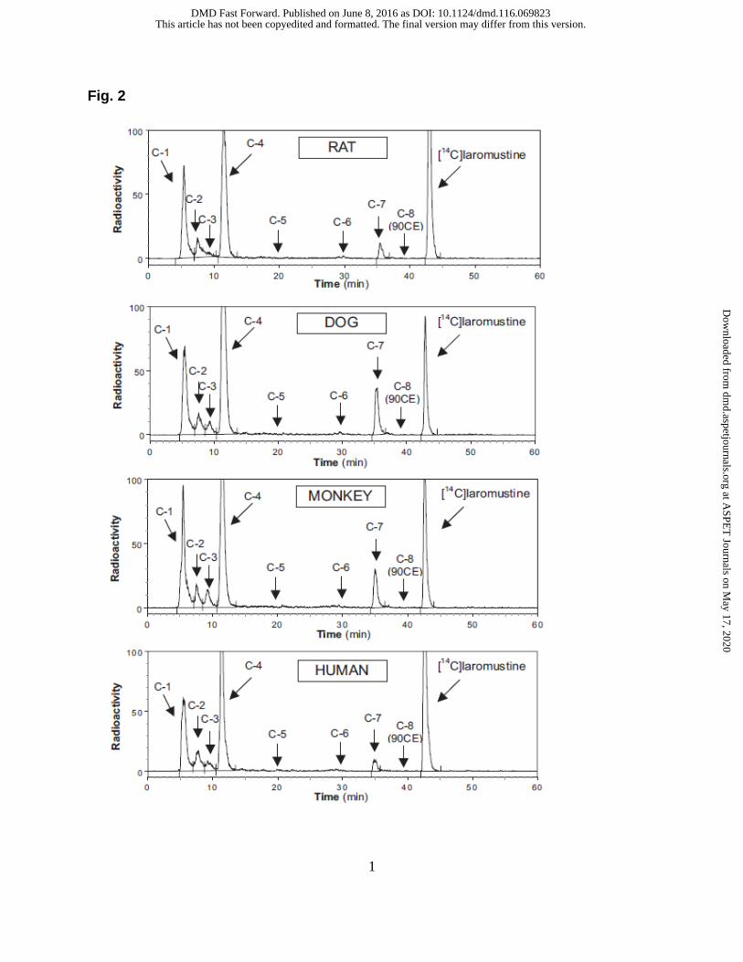
1.1 In vitro metabolism of [14C]laromustine. The in vitro profile of
[14C]laromustine metabolites/degradation products formed by liver microsomes from
rats, dogs, monkeys and humans is shown in Fig. 2. When [14C]laromustine (100 µM)
was incubated with these four liver microsomes in the presence of NADPH, eight
radioactive components (C-1 through C-8) were detected after 60 minutes of incubation
(Fig. 2). The metabolite profile was similar for all species with two notable exceptions:
First, rat liver microsomes formed C-3 at low levels or not at all, and second, dog and
monkey liver microsomes produced considerably higher levels of C-7 compared with rat
and human liver microsomes. Component C-8 cochromatographed (RT ~ 39 min) with
an authentic standard of 90CE; it was a major component at early time points and at low
substrate concentrations regardless of the source of liver microsomes. Formation of
90CE (C-8) is associated with formation of methylisocyanate, but formation of
methylisocyanate from [14C]laromustine was not detected by radiometric HPLC because
it lacked the 14C-label, which is part of the chloroethyl moiety, as shown in Fig. 1a.
The differences between the presence and absence of NADPH are summarized
in Supplemental Tables 1 through 3. In the presence of NADPH, after 60 min of
incubation, the loss of substrate for rat, dog, monkey and human liver microsomes was
63, 82, 76 and 64% respectively, and mass balance ranged from 91.0 – 99.3%. In the
absence of NADPH, after 60 min of incubation with [14C]laromustine (100 µM), the loss
of substrate for rat, dog, monkey and human liver microsomes was 59, 53, 61 and 59%
respectively, and mass balance ranged from 100.6 – 116.4%. For rat, dog, monkey and
This article has not been copyedited and formatted. The final version may differ from this version.DMD Fast Forward. Published on June 8, 2016 as DOI: 10.1124/dmd.116.069823
human liver microsomes, the total formation (sum of C-1, C-2, C-3, C-4 and C-7) was
54, 81, 70 and 55% respectively in the presence of NADPH and 59, 69, 66 and 64%,
respectively, in the absence of NADPH.
Of the radioactive components detected, only the formation of C-7 was
dependent on NADPH. Formation of C-7 involved methyl-hydroxylation of laromustine
(+16 amu) on the methylisocyanate moiety. The time course of C-7 formation by liver
microsomes from each species indicates that once formed, C-7 is further metabolized or
degraded. The rate of formation of C-7 followed the rank order (from fastest to slowest):
Dog ≈ monkey > rat ≈ human, which corresponds to the same rank order for the overall
rate of laromustine consumption. Formation of C-7 did not account for the loss of parent
compound. The loss of substrate was largely caused by non-enzymatic chemical
degradation. In contrast, formation of C-7 was not observed in zero-cofactor (no
NADPH) and zero-protein samples, suggesting its formation was enzymatic.
In vitro studies were designed to determine the role of P450 and other HLM
enzymes on laromustine metabolism. Incubations of [14C] laromustine were performed
with and without human liver microsomes and with boiled (denatured) microsomes, with
and without NADPH. The data suggested that [14C] laromustine undergoes extensive
chemical degradation rather than metabolism by cytochrome P450s. It was also
determined that reactions stopped with methanol, acetonitrile and perchloric acid
generated similar degradation profiles. This suggests that the sample preparation
methods did not contribute to the degradation products.
This article has not been copyedited and formatted. The final version may differ from this version.DMD Fast Forward. Published on June 8, 2016 as DOI: 10.1124/dmd.116.069823
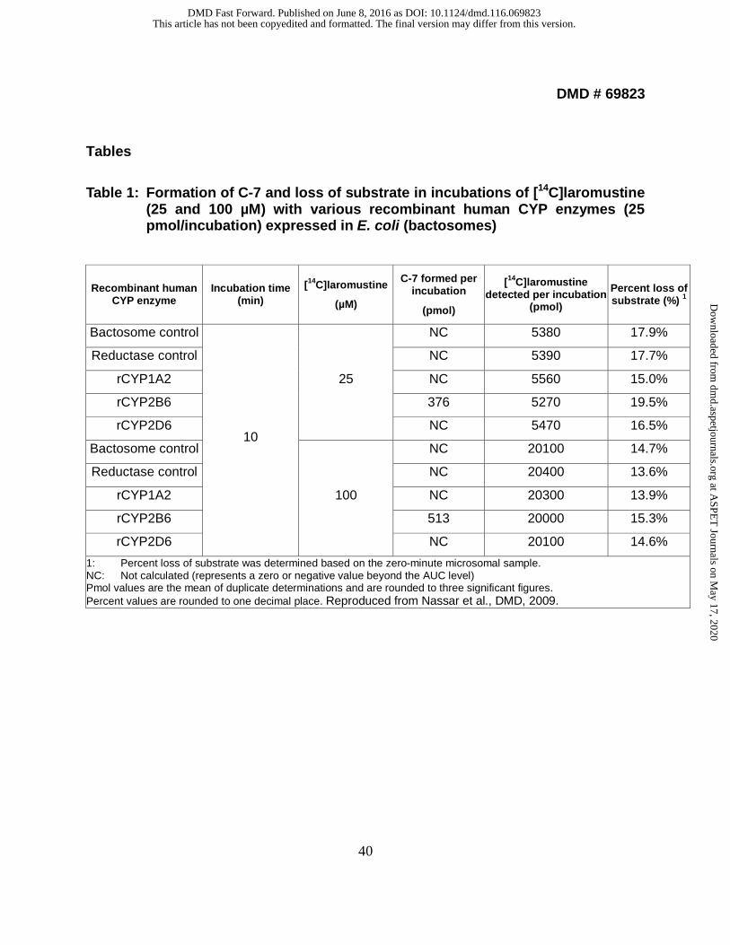
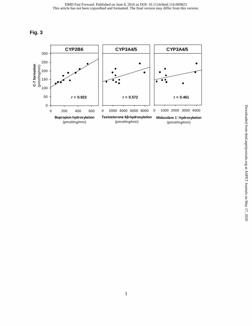
1.2 Identification of human CYP enzymes involved in the metabolism of
[14C]laromustine. To identify the human CYP enzyme or enzymes responsible for C-7
formation, laromustine (25 and 100 µM) was incubated with a bank (n = 10) of individual
samples of human liver microsomes (correlation analysis) and a panel of recombinant
human enzymes. Of the recombinant human enzymes evaluated, only CYP2B6 and
CYP3A4/5 converted laromustine to C-7, and did so at comparable rates. With the
recombinant CYP enzymes, C-7 formation was evaluated at a single time point (10 min)
based on time-course experiments with human liver microsomes. We cannot exclude
the possibility that one or more recombinant CYP enzymes formed C-7 at a later or
earlier time point. As shown in Fig. 3, the sample-to-sample variation in the conversion
of [14C]laromustine (25 µM) to C-7 correlated strongly (r = 0.923) with CYP2B6
(bupropion hydroxylase) activity. It also correlated weakly with CYP3A4/5 activity based
on testosterone 6β-hydroxylation (r = 0.572) and midazolam 1′-hydroxylation (r = 0.461).
The linear regression lines for CYP2B6 and CYP3A4/5 did not pass through or near the
origin, suggesting that both enzymes are involved in the formation of C-7. The
correlation between C-7 formation and CYP2B6 activity (r = 0.923) improved when the
variation in CYP3A4/5 activity was also taken into consideration (r = 0.945).
[14C]laromustine (25 and 100 µM) was incubated with a panel of recombinant
human CYP enzymes (rCYP1A2, 2A6, 2B6, 2C8, 2C9, 2C19, 2D6 and 3A4) and
recombinant human FMO3 for zero and 10 min, as summarized in Table 1, and
Supplemental Tables 4 and 5. Among the panel of recombinant human CYP and FMO
enzymes evaluated, formation of C-7 was only observed in incubations with
This article has not been copyedited and formatted. The final version may differ from this version.DMD Fast Forward. Published on June 8, 2016 as DOI: 10.1124/dmd.116.069823
recombinant CYP2B6 and CYP3A4. Loss of parent compound was observed in all
incubations including the control samples (i.e., samples containing membranes with no
human CYP enzyme). Formation of C-7 was observed in incubations containing
recombinant human CYP2B6 and CYP3A4 at both substrate concentrations. After 10-
min incubations of [14C]laromustine at concentrations of 25 and 100 µM with
recombinant human CYP2B6, the [14C] quantities of C-7 were 376 and 513 pmol,
respectively. After 10-min incubations of [14C]laromustine at concentrations of 25 and
100 µM with recombinant human CYP3A4, the [14C] levels of C-7 were 362 and
549 pmol, respectively. The formation of the radioactive components increased with
respect to incubation time with the exception of C-7. The formation of C-7 tended to
increase with increasing incubation time and protein concentration; however, formation
of C-7 was not linear with respect to either of these parameters. In addition, the
formation of C-7 was not proportional to substrate concentration. The non-linearity of C-
7 formation suggests that, once formed, this component was further metabolized or
degraded.
2. Novel Rearrangement
2.1 Unanticipated loss of N2 from laromustine by collision-induced
dissociation. During metabolism studies of laromustine utilizing liquid chromatography-
mass spectrometry, a novel mass spectral rearrangement by collision-induced
dissociation in the positive ion mode was observed. Accurate mass measurements were
performed with a Fourier transform ion cyclotron resonance-mass spectrometer (FTICR-
This article has not been copyedited and formatted. The final version may differ from this version.DMD Fast Forward. Published on June 8, 2016 as DOI: 10.1124/dmd.116.069823
MS) to determine the elemental compositions of the fragments. During the
fragmentation of m/z 171 to m/z 143 direct cleavage fragment ions were not observed,
leading to the speculation that laromustine undergoes rearrangement. This suggested
the loss of nitrogen (N2), methylsulfonyl and methyl isocyanate moieties from
laromustine by collision-induced dissociation. Additionally, the rearrangement was
analyzed utilizing hydrogen-deuterium exchange (H-D), a stable-isotope labeled
analogue (13C-labeled laromustine), and LC-MSn experiments to assist with the
assignments of these fragments and possible mechanistic rearrangements. A
mechanism for this rearrangement was proposed on the basis of fragmentation ions. H-
D exchange methods are useful for determination of the presence, number, and position
of H-D exchangeable functional groups on metabolite structures and thus serve as an
aid for structural elucidation of metabolites, as well as aiding in differentiation of
compounds (Nassar 2003a). It has been reported that H-D exchange can be used to
discriminate between N- or S-oxide formation and monohydroxylation; also, conjugation
such as glucuronide can be easily identified with this technique. However, in this study
the samples were dried under a nitrogen stream and then reconstituted in D2O mobile
phase for the H-D exchange. Stable isotopes have the same number of protons as
common elements, and consequently share the same physicochemical properties, but
they differ in mass due to a difference in the number of neutrons. Stable isotope labeling
involves the use of non-radioactive isotopes that can be used to confirm metabolites.
Stable isotope labeling provides great improvements and enhanced confidence in
metabolite identification and characterization (Kassahun et al., 2001). The combination
This article has not been copyedited and formatted. The final version may differ from this version.DMD Fast Forward. Published on June 8, 2016 as DOI: 10.1124/dmd.116.069823
of these two approaches (H-D and stable isotopes) provides a comprehensive
understanding of structure elucidation.
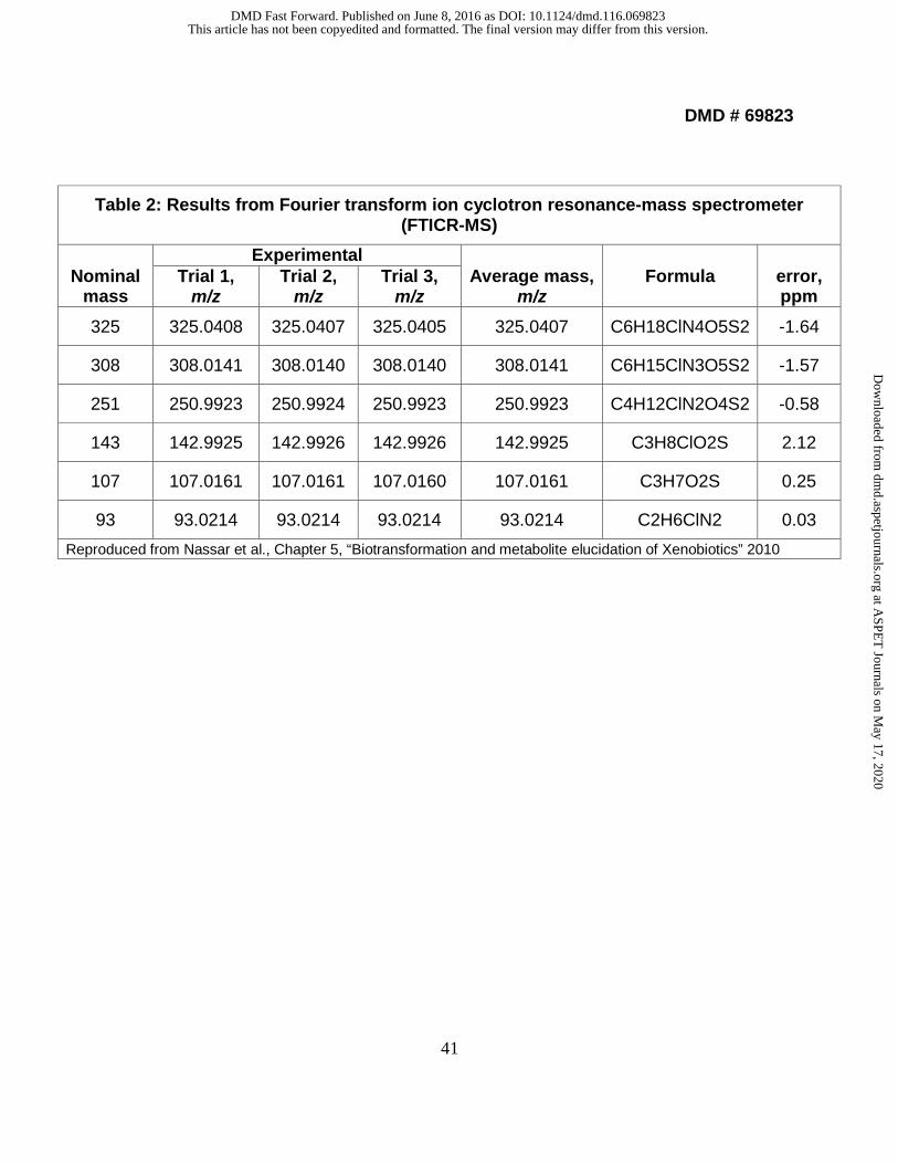
2.2 Analysis with Fourier transform ion cyclotron resonance-mass
spectrometry (FTICR-MS). Each sample was analyzed in triplicate. Exact mass
measurement of the peak of interest (observed at m/z 325) was initially collected.
Subsequently, the peak of interest was selected and fragmented by collision induced
dissociation (CID) to produce product ions. Bruker Daltonics DataAnalysis software (v.
3.4) was used to analyze the data and assignments were made based on exact mass
measurements and fit of isotopic peaks to that of theoretical isotopic patterns. After
generation of the m/z mass spectrum for this peak, data was deconvoluted to determine
monoisotopic masses. The data are shown in Table 2 and Fig. 4. The mass errors were
between -1.64 to 2.12 ppm as shown in Table 2. These data allowed us to determine
the elemental compositions of the fragmentation ions of laromustine and provided
unambiguous fragmentation ion pathways.
2.3 Confirmation of the fragmentation ions of laromustine. Sequential LC-
MSn experiments were carried out on a LTQ ion trap mass spectrometer to gain
extensive structural information for laromustine in full scan MS mode. The fragmentation
ions from the initial MSn experiments on laromustine using LTQ suggested that
rearrangement of laromustine took place. Supplemental Figure 1 shows the sequential
MSn experiments up to MS5. Under the experimental conditions presented (Nassar et
This article has not been copyedited and formatted. The final version may differ from this version.DMD Fast Forward. Published on June 8, 2016 as DOI: 10.1124/dmd.116.069823
al., 2010b), and on the basis of the MS5 results, direct cleavage fragment ions were not
observed; then it was speculated that laromustine undergoes rearrangement during the
fragmentation of ion m/z 171 to m/z 143. The MS signature of chlorine isotope (35Cl and
37Cl) in laromustine was observed and verified in the fragmentation ions; this unique
signature confirmed the formation of m/z 143. Also the fragmentation ions were further
confirmed using H-D exchange and stable-isotope experiments.
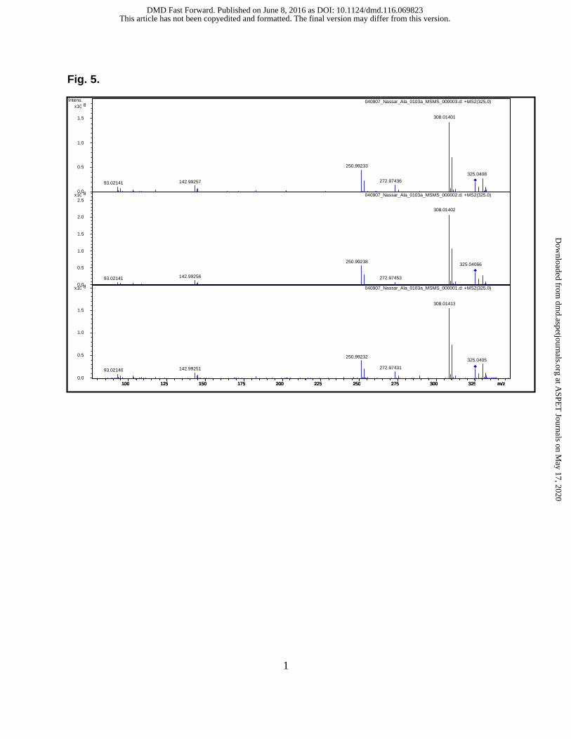
The proposed mechanism of formation for the fragmentation ions m/z 251, m/z
171, m/z 143, m/z 107, m/z 81, and m/z 63 is shown in Fig. 5. The product-ion spectra
of m/z 325 showed the fragment ions of m/z 308, 273, 251, 143, 107, 93, 81, and 63.
The fragment ion at m/z 308 resulted from the loss of ammonia from m/z 325. The
fragment ion at m/z 273 resulted from the losses of HCl from m/z 308. The fragment ion
at m/z 251 resulted from cleavage of the C-N bond. The fragment ion at m/z 143
resulted from the loss of CH3SO2H and N2 from m/z 251. The fragment ion at m/z 107
was due to losses of HCl from m/z 143. The fragment ion at m/z 81 was due to losses of
C2H3Cl from m/z 143. The product-ion spectra of m/z 327 (laromustine, 13C2) showed
the fragment ions of m/z 310, 253, 145, 109, 95 and 65. The product-ion spectra of m/z
330 using H-D exchange experiments showed the fragment ions of m/z 310, 253, 144,
and 95. Detailed analyses of the LC-MSn mass spectra suggest that the fragmentation
process involves a rearrangement. This rearrangement fragment provided important
clues about the location and identity of functional groups of laromustine. This was very
useful for elucidating ionic structures of laromustine. Proposed fragmentation
This article has not been copyedited and formatted. The final version may differ from this version.DMD Fast Forward. Published on June 8, 2016 as DOI: 10.1124/dmd.116.069823
mechanisms for m/z 325 (laromustine), m/z 327 (laromustine, 13C2), and m/z 330
(laromustine, H-D) are shown in Fig. 6.
3. Identification and characterization of in vitro metabolite/decomposition
products of laromustine in HLM. Eight metabolite/decomposition products along with
the parent drug were detected following incubation of laromustine with human liver
microsomes for 60 min in the presence of NADPH. The major decomposition products
were designated C-1 and C-4. Table 3 shows the molecular formula, molecular weights,
retention times, results from H-D exchange experiments, N-rule data and RDBE data of
the metabolite/decomposition products of laromustine formed in HLM incubations. Peak
C-2 was not identified because the MS spectrum of C-2 was not well ionized under the
conditions utilized in this study. C-6 product was not reported because it is one of the
potential impurities in laromustine (Nassar et al., 2010a). The total conversion (sum of
C-1, C-2, C-3, C-4 and C-7) of laromustine in HLM was 55%, in the presence of NADPH
after 60 min; data are shown graphically in Fig. 2. In vitro studies using HLM have
shown that C-7 formation is mediated primarily by CYP3A4/5 and CYP2B6 (Nassar et
al., 2009).
3.1. Identification and characterization of C-1: The MS spectrum of C-1 was
acquired, which revealed an ammonium adducted, protonated molecular ion [M+NH4]+
at m/z 142. Comparison of the full-scan mass spectrum of undeuterated/deuterated C-1
enabled the determination of the number of labile protons. The full-scan mass spectrum
This article has not been copyedited and formatted. The final version may differ from this version.DMD Fast Forward. Published on June 8, 2016 as DOI: 10.1124/dmd.116.069823
of undeuterated/deuterated C-1 gave ions at m/z 142/147 respectively, which suggested
C-1 has only one labile proton. The stable-isotopic experiments gave a full-scan ion at
m/z 144, which suggested that the structure of C-1 contains 13C2. The product-ion
spectra of m/z 142 from sequential MSn experiments showed the fragment ions of m/z
125, 107, 81, and 79. The fragment ion at m/z 125 resulted from the loss of ammonia
from m/z 142. The fragment ion at m/z 107 was due to the loss of water from m/z 125.
The fragment ion at m/z 79 resulted from cleavage of the C-S bond. The data in Table 3
show that C-1 contains no nitrogen (N) atoms, has only one labile proton and RDBE is
equal to 0 which agrees with the proposed structure (2-(methylsulfonyl)ethanol).
3.2 Identification and characterization of C-3 and C-4: The MS spectra of C-3
and C-4 were acquired, which revealed an ammonium adducted, protonated molecular
ion [M+NH4]+ at m/z 232 (Supplemental Figures 2 and 3 for components C-3 and C-4
respectively). The data in Table 3 show that C-3 and C-4 contain an even number of
nitrogen (N) atoms and RDBE is equal to 1. Comparison of the full-scan mass spectrum
of undeuterated/deuterated C-3 and C-4 enabled the determination of the number of
labile protons. The full-scan mass spectrum of undeuterated/deuterated C-3 and C-4
gave ions at m/z 232/237 respectively, which suggested C-3 and C-4 have one labile
proton. The stable-isotopic experiments gave a full-scan ion at m/z 234 for C-3 and C-4,
which suggested that the structure of C-3 and C-4 contains 13C2. Stable-isotopic
experiments, in addition to supporting data including N-rule, RDBE, and MSn, provided
evidence to support the structural characterization of C-3 and C-4. The product-ion
This article has not been copyedited and formatted. The final version may differ from this version.DMD Fast Forward. Published on June 8, 2016 as DOI: 10.1124/dmd.116.069823
This article has not been copyedited and formatted. The final version may differ from this version.DMD Fast Forward. Published on June 8, 2016 as DOI: 10.1124/dmd.116.069823
3.4 Identification and characterization of C-7: This component was detected
when laromustine was incubated with HLM in the presence of NADPH, suggesting that
this is a metabolite rather than a decomposition product (Fig. 2). The fact that C-7
decreased over the incubation time suggests that C-7 continued to metabolize or
decompose. The MS spectrum of C-7 was acquired, which revealed an ammonium
adducted, protonated molecular ion [M+NH4]+ at m/z 341, 16 Da (dalton) more than the
parent drug, suggesting the addition of oxygen on laromustine. The presence of
pseudomolecular ions [M+NH4]+ at m/z 341/343 in a ratio of 3:1 suggested the presence
of chlorine on the molecule. The MS signature of chlorine in C-7 was verified by multi-
stage MS. The product-ion spectra of m/z 341 from sequential MSn experiments
showed the fragment ions of m/z 306, 294, 251, 227, 148, 143, and 93. The fragment
ion at m/z 306 resulted from the loss of ammonia and water from m/z 341. The fragment
ion at m/z 227 was due to the loss of CH3SO2 moiety from m/z 306. The fragment ion at
m/z 148 was due to the loss of CH3SO2 moiety from m/z 227. The fragment ion at m/z
251 resulted from cleavage of the C-N bond. The fragment ion at m/z 143 resulted from
the loss of CH3SO2H and N2 moieties from m/z 251. The stable-isotopic experiments
gave a full-scan ion at m/z 343. These observations support the proposed structure of
C-7. The unique MS signature of chlorine isotope ratios that differs in mass by 2 Da
greatly facilitated the structure characterization. The proposed structure and
fragmentation is shown in Fig. 7.
This article has not been copyedited and formatted. The final version may differ from this version.DMD Fast Forward. Published on June 8, 2016 as DOI: 10.1124/dmd.116.069823
3.5 Buffer incubations. Following incubation of laromustine with phosphate
buffer (50 mM) at pH 7.4 for 60 min, decomposition products along with the parent drug
were detected, and were similar to those detected in HLM in the absence of NADPH.
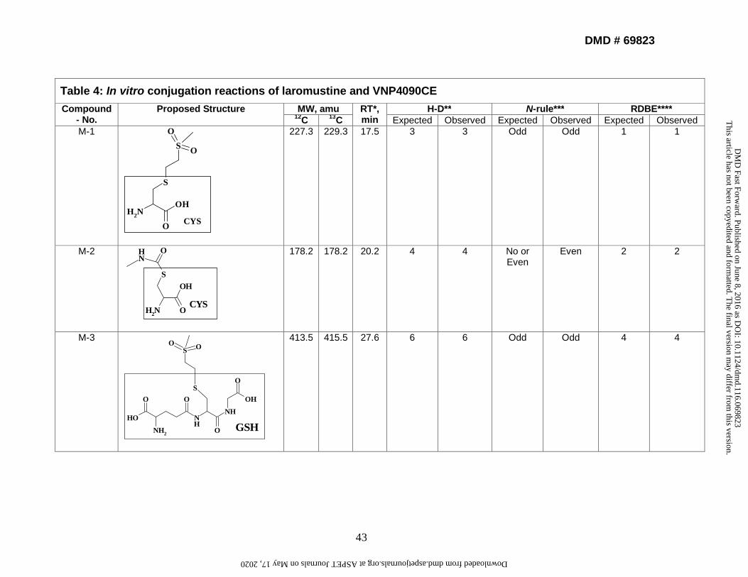
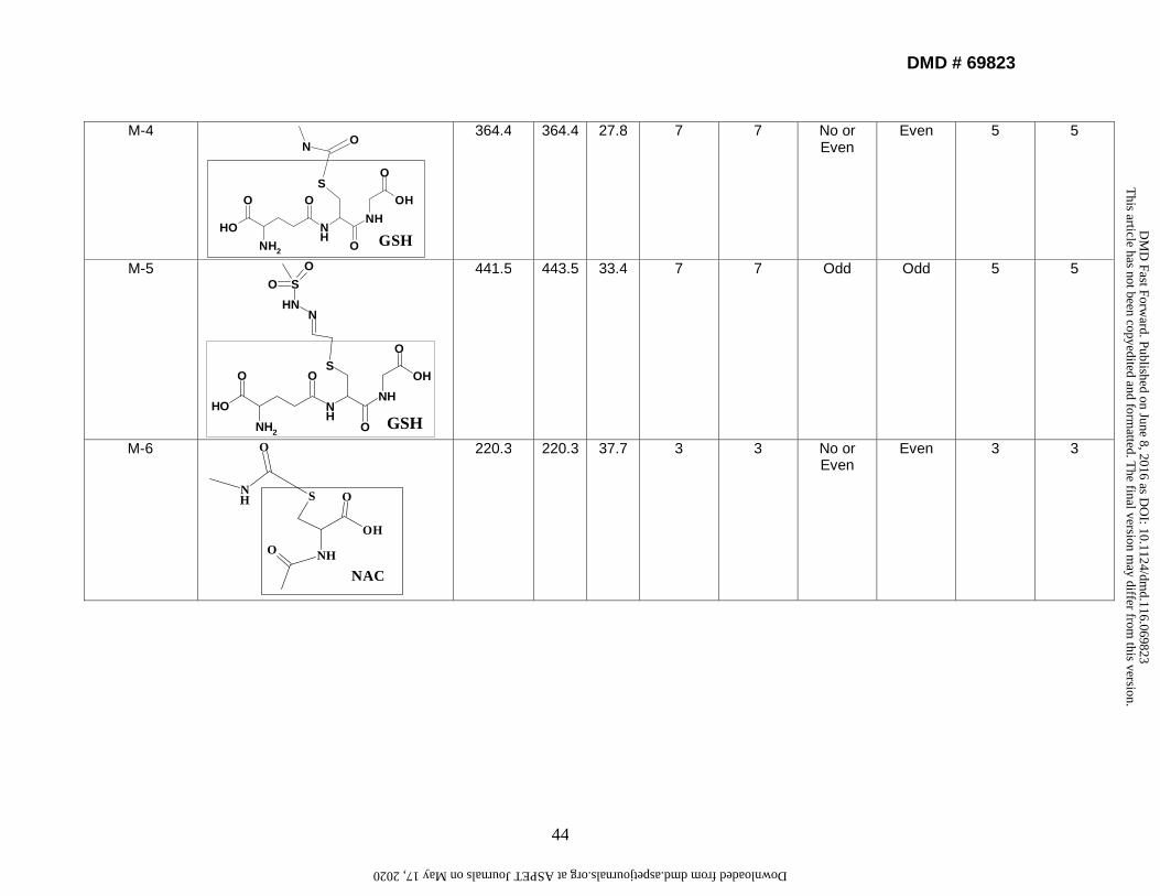
4. Identification and characterization of in vitro conjugation reactions of
laromustine. The retention times of conjugation reactions were between 16 and 48
min with adequate separation efficiency. Each structure was assigned a numerical
notation (M-1, M-2, etc.) based on the order of HPLC elution. MSn data were acquired
for each structure. Following incubation of laromustine with human liver microsomes for
60 min in the presence of NADPH, GSH, CYS, and NAC, eight conjugation reactions
were detected (M-1 to M-8). Following incubation of VNP4090CE with human liver
microsomes for 60 min in the presence of NADPH, GSH, CYS, and NAC, five distinct
conjugation reactions were detected (M-1, M-3, M-5, M-7 and M-8). M-2, M-4 and M-6
were not detected because VNP4090CE does not contain a methylformamide group
(Fig. 1a). Reactive moieties (groups) formed from laromustine and VNP4090CE in HLM
or buffer incubations are shown in Supplemental Table 7. The CH3SO2CH2CH2- and
CH3NHCO- groups formed conjugates with GSH, CYS, and NAC. The
CH3SO2NHN=CHCH2- group formed conjugates with GSH and NAC but it did not form a
conjugate with CYS under the experimental conditions evaluated, suggesting that CYS
has low trapping efficiency with the CH3SO2NHN=CHCH2- group and/or this conjugate
did not ionize well under the MS conditions. Table 4 shows the chemical structure,
molecular weight, retention time, and results from H-D experiments on the conjugation
This article has not been copyedited and formatted. The final version may differ from this version.DMD Fast Forward. Published on June 8, 2016 as DOI: 10.1124/dmd.116.069823
reactions. These conjugates were formed in buffer solution with or without HLM,
indicating that the formation of active moieties from laromustine and VNP4090CE was
enzyme independent. The MS data from buffer incubations gave a stronger MSn signal
because the buffer has less isobaric interferences than HLM.
4.1. Identification and characterization of M-3 as an example of how we
identified M1-M8. The LC-MS retention time of M-3 was approximately 27.6 min, and
the mass spectrum revealed a protonated molecular ion [M+H]+ at m/z 414, suggesting
that the CH3SO2CH2CH2- group was covalently linked to the sulfur atom of the GSH
moiety (Supplemental Figure 6). The absence of pseudomolecular ions [M+H]+ at m/z
414/416 in a ratio of 3:1 suggested that M-3 does not contain chlorine. The stable-
isotopic experiments (13C-labeled) gave a full-scan ion at m/z 416, suggesting that M-3
contains [13C]. The H-D experiments gave a full-scan ion at m/z 421, suggesting there
are 6 labile protons on M-3. The product-ion spectra of m/z 414 showed the fragment
ions of m/z 339, 285, 268, 250, 240, 193, 113, and 107 (Supplemental Figure 6). The
fragment ion at m/z 339 resulted from the loss of C2H5NO2 from m/z 414. The fragment
ion at m/z 285 was due to the loss of the glutamate moiety from m/z 414. The fragment
ion at m/z 268 resulted from the loss of NH3 from m/z 285. The fragment ion at m/z 250
resulted from the loss of H2O from m/z 268. The fragment ion at m/z 240 resulted from
the loss of CO from m/z 268. The fragment ion at m/z 193 resulted from the loss of
C2H5NO2 from m/z 268. The fragment ion at m/z 113 resulted from the loss of CH3SO2H
from m/z 193. The fragment ion at m/z 107 resulted from cleavage of the C-S bond.
This article has not been copyedited and formatted. The final version may differ from this version.DMD Fast Forward. Published on June 8, 2016 as DOI: 10.1124/dmd.116.069823
These observations support the proposed structure of M-3 and suggest that an unusual
rearrangement may occur after dehalogenation of VNP4090CE, resulting in the
formation of a novel reactive intermediate. The CH3SO2CH2CH2- group was trapped by
GSH to form M-3. The proposed structure and fragmentation ions of M-3 based on
supported data from LC-MSn, N-rule, RDBE, stable isotope experiments, and H-D
experiments are shown in Supplemental Figure 7.
5. Discussion
These studies indicate that laromustine readily undergoes base-catalyzed (non-
enzymatic) conversion to methylisocyanate and 90CE, which further degrade to
additionnal chloroethylating derivatives. Fig. 1b shows the proposed formation of
decomposition/metabolite product pathways of laromustine formed in in vitro systems.
The rate at which [14C]laromustine (100 µM) produces its metabolites/degradation
products in the presence of NADPH-fortified human liver microsomes was essentially
the same as that in the absence of NADPH or the presence of boiled (heat-denatured)
microsomes. Additionally, it remained consistent with the non-enzymatic rate of
laromustine degradation in potassium phosphate buffer (Nassar et al., 2009). This
suggests that laromustine metabolism/degradation is not significantly affected by human
liver microsomal CYP enzymes and carboxylesterases.
Only the formation of C-7 required the presence of NADPH. C-7 appears to be
decomposed or metabolized further, which suggests that cytochrome P450 is a factor in
C-7 formation but plays little or no role in the conversion of [14C]laromustine to other
This article has not been copyedited and formatted. The final version may differ from this version.DMD Fast Forward. Published on June 8, 2016 as DOI: 10.1124/dmd.116.069823
radioactive products. The combination of the correlation analysis and recombinant
human CYP enzyme experiments points to a role for both CYP2B6 and CYP3A4 in the
formation of metabolite C-7. The formation of C-7 correlated most strongly with CYP2B6
activity, with moderate correlation to CYP2A6, CYP2D6 and CYP3A4 activity in 10
individual samples of NADPH-fortified human liver microsomes. As shown by the
recombinant human CYP data, rCYP3A4 and rCYP2B6 were capable of forming C-7,
whereas formation of C-7 was not observed with rCYP2A6 and rCYP2D6. Laromustine
was shown to be a competitive inhibitor for both CYP2B6 and CYP3A4, which indicates
that it is a substrate for these enzymes (Nassar et. al, 2009).
It was undertaken to determine whether one or more CYP enzymes in human
liver microsomes can potentially contribute 25% or more to the clearance of
laromustine. We examined the role of CYP2B6 and CYP3A4/5 in the hydroxylation of
laromustine to C-7, which is one of eight radioactive components detected when
[14C]laromustine is incubated with human liver microsomes. Studies with a panel of
recombinant human CYP enzymes and correlation analysis with a bank of human liver
microsomes implicated CYP2B6, and showed that C-7 is the only component whose
formation is dependent on NADPH. However, inhibition of CYP2B6 and CYP3A4/5 will
not impact the pharmacokinetics of laromustine. This is because the rate of loss of
[14C]laromustine (100 µM) in the presence of NADPH-fortified human liver microsomes
was essentially the same as that in the absence of NADPH or the presence of boiled
(heat-denatured) microsomes. This rate was also consistent with the non-enzymatic
rate of laromustine degradation in potassium phosphate buffer. Therefore, although
This article has not been copyedited and formatted. The final version may differ from this version.DMD Fast Forward. Published on June 8, 2016 as DOI: 10.1124/dmd.116.069823
CYP2B6 and CYP3A4/5 convert laromustine to C-7, metabolism by CYP (or any other
microsomal enzyme) does not contribute substantially to the overall rate of in vitro
clearance, which is largely determined by non-enzymatic degradation (Nassar et al.,
2009 and 2010c).
Laromustine undergoes a rearrangement which was determined by a
combination of different methods such as exact mass measurements, stable isotope, H-
D exchange and MSn experiments. H-D exchange experiments were useful to
determine the number of exchangeable protons in the fragmentation ions. The data
from FTICR-MS facilitated the determination of the elemental compositions of the
fragmentation ions of laromustine and provided unambiguous fragmentation ion
pathways. The fragmentation processes are typically categorized as direct cleavage or
rearrangement. Cleavage reactions are simply the breaking of a bond to produce two
fragments. These reactions usually produce an even electron ion. Rearrangements are
more complex reactions that involve both making and breaking bonds. These reactions
are thermodynamically favorable because they require less energy. However they also
require a mechanism that is not as kinetically favorable when compared to a simple
cleavage reaction (McLafferty and Turecek, 1993). The distinct rearrangement fragment
of laromustine was observed and provided important clues about the location and
identity of its functional groups. This was very useful for elucidating the unique
structures of laromustine reactions.
Laromustine undergoes extensive decomposition/metabolism in in vitro systems.
Following incubation of [14C]laromustine with HLM in the presence of NADPH, eight
This article has not been copyedited and formatted. The final version may differ from this version.DMD Fast Forward. Published on June 8, 2016 as DOI: 10.1124/dmd.116.069823
metabolite/decomposition products along with the parent drug were detected. Most of
the identified structures resulted from the chemical decomposition of laromustine and
were not P450-mediated. The major decomposition products (C-3, C-4 and C-5) from
the incubation of laromustine in HLM were found to be the products of dehalogenation,
rearrangement, and hydrolysis. Their formations do not require the involvement of any
enzymes. H-D exchange, 13C-labeled laromustine, NMR, and MSn techniques were
applied to identify and characterize the metabolite/decomposition components of
laromustine. H-D exchange combined with MS is an efficient tool for studying metabolite
identification. A combination of these techniques appears promising for maximizing
structural information for laromustine.
The mechanisms of formation for the decomposition components were proposed.
The major decomposition components were not P450-mediated. C-7 was only detected
when laromustine was incubated with HLM in the presence of an NADPH-generating
system, suggesting that this is a metabolite rather than a decomposition product.
Dechlorination and hydrolysis of the methyl isocyanite moiety from laromustine does not
require P450 enzymes and occurs at biological pH. A mechanism for the formation of
these metabolite/decomposition products from laromustine was proposed. The
structural characterizations of the MW 214 components (C-3 and C-4) were verified
using NMR. NMR data suggested that peak (C-3) is composed of the free base form
with a carbon-nitrogen double bond. The free base (C-3) was found to exist as a mixture
of cis and trans isomers with the trans isomer typically more abundant. The unusual
observation that the methylene protons adjacent to the double bond are exchangeable
This article has not been copyedited and formatted. The final version may differ from this version.DMD Fast Forward. Published on June 8, 2016 as DOI: 10.1124/dmd.116.069823
in CD3OD suggests that the double bond can shift in the free base to give a carbon-
carbon double bond. In the presence of even weak acids, the free base form is
converted to the salt which is present almost entirely in the trans form and does not
have exchangeable methylene protons, indicating that the double bond does not shift
between the carbons in the salt. The salt (C-4) elutes slightly later than the free base as
shown in Fig. 2. The third MW 214 peak (C-5) could not be isolated (for NMR analysis)
due to its apparent instability. This component has no exchangeable protons,
suggesting that the double bond may shift to reside between the nitrogen atoms. Based
on the NMR data, C-3 was determined to be the free base and C-4 was determined to
be the salt form of m/z 232. The proposed structures and fragmentations ions for C-4
and C-5 are shown in Supplemental Figures 4 and 5. A proposed mechanism of
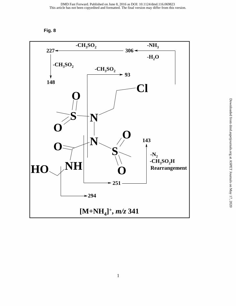
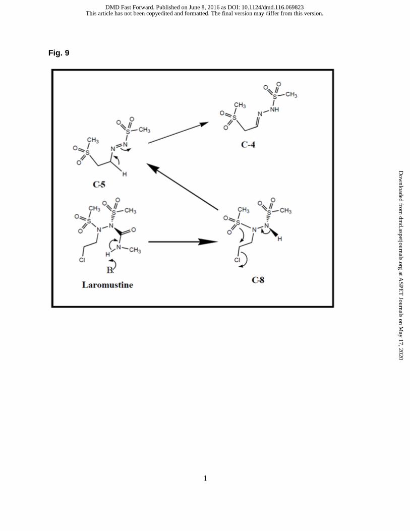
formation of C-4 and C-5 is shown in Figure 8. It has been proposed that laromustine
undergoes hydrolysis to form C-8 which then produces C-5 after dehalogenation and
rearrangement. C-5 undergoes further rearrangement to form C-3 (free base) or C-4
(salt form).
Conjugation reactions of laromustine and VNP4090CE occurred after incubation
of laromustine or VNP4090CE with pooled HLM and co-factors NADPH, GSH, NAC,
and CYS. Eight distinct conjugation reactions (M-1 to M-8) were identified and
characterized by H-D, 13C-labeled laromustine, and MSn experiments. M-4 and M-5
were further confirmed by NMR. The short-lived CH3SO2CH2CH2-, methylformamide
and CH3SO2NHN=CHCH2- moieties were generated from laromustine. The reactive
intermediates CH3SO2CH2CH2- and methylformamide formed conjugates with GSH,
This article has not been copyedited and formatted. The final version may differ from this version.DMD Fast Forward. Published on June 8, 2016 as DOI: 10.1124/dmd.116.069823
CYS, and NAC. The CH3SO2NHN=CHCH2- moiety formed conjugates with GSH and
NAC. M-2, M-4 and M-6 were only detected from the incubation of laromustine because
VNP4090CE does not contain a methylformamide group. All other conjugates were
formed by both laromustine and VNP4090CE. Laromustine and VNP4090CE produced
the same decomposition product-conjugates in buffer solution with or without HLM,
which suggests that the decomposition of laromustine and VNP4090CE to active
moieties does not involve P450 metabolism. These in vitro studies found that
laromustine and VNP4090CE undergo activation via chemical decomposition in buffer
solution in the presence or absence of HLM and with or without NADPH.
It is possible that these active moieties may alter cellular functions when they
form conjugates with biomolecules. The degradation of laromustine produces methyl
isocyanate (MIC), which is extremely toxic. MIC can induce damage by inhalation,
ingestion, and contact in quantities as low as 0.4 ppm. Symptoms may include
coughing, chest pain, dyspnea, asthma, irritation of the eyes, nose, and throat as well
as skin damage. Higher levels of exposure, over 21 ppm, can result in pulmonary or
lung edema, emphysema, and hemorrhaging, bronchial pneumonia, and death. Studies
were conducted to evaluate both dose- and time-dependent (N = 3) response using N –
succinimidyl N -methylcarbamate, a chemical entity that mimics the effects of methyl
isocyanate in vitro. These studies demonstrated that isocyanates induce neutrophil
apoptosis via activation of mitochondrial-mediated pathways along with reactive oxygen
species production; depletion in antioxidant defense states; and elevated prion
flammatory cytokine response (Mishra et al., 2008 and 2010; Ennever and Rosenkranz
This article has not been copyedited and formatted. The final version may differ from this version.DMD Fast Forward. Published on June 8, 2016 as DOI: 10.1124/dmd.116.069823
1987; Bucher 1987). This study showed that laromustine produces several reactive
intermediates that may play a role in toxicities seen in the clinical trials. Combined,
these findings provide an understanding of both the beneficial and potentially harmful
aspects of the metabolism of laromustine.
Acknowledgements
These studies were done at XenoTech, LLC (now Sekisui XenoTech, LLC), for
Vion Pharmaceuticals Inc. We would like to thank Drs Paris B L, Haupt L, Ndikum-
Moffor F, Campbell R, Usuki E, Skibbe J, Brobst D, Ogilvie B, and Parkinson A, for
performing and supervising these studies.
Authorship Contributions
Participated in research design: AFN, IK
Wrote or contributed to writing of the manuscript: IV, AVW, AFN
This article has not been copyedited and formatted. The final version may differ from this version.DMD Fast Forward. Published on June 8, 2016 as DOI: 10.1124/dmd.116.069823
"Alkylating Agents". (2014) US National Library of Medicine.
http://livertox.nih.gov/AlkylatingAgents.htm
Baillie TA (1992) Advances in the application of mass spectrometry to studies of drug
metabolism, pharmacokinetics and toxicology. Int. J. Mass Spectrom. Ion Proc.
118/119, 289-314.
Baumann RP, Seow HA, Shyam K, Penketh PG, and Sartorelli AC (2005) The
antineoplastic efficacy of the prodrug Cloretazine is produced by the synergistic
interaction of carbamoylating and alkylating products of its activation. Oncol Res
15(6):313-25.
Brundrett RB, Cowens JW, Colvin M (1976), Chemistry of Nitrosoureas. Decomposition
of Deuterated 1,3-Bis(2-chloroethyl)-1-nitrosourea. Journal of Medicinal
Chemistry, Vol. 19, 7, 958-961
Bucher JR (1987) The toxicity of methyl isocyanate: Where do we stand? Environ
Health Perspect. 72, 197–198
Buhl AE, Waldon DJ, Baker CA, and Johnson GA (1990) Minoxidil sulfate is the active
metabolite that stimulates hair follicles. J. Invest. Dermatol. 95, 553-557
Colvid M, Cowens W, Brundrett RB, Kramer BS, Ludlum DB (1974).
DECOMPOSITIONO F BCNU (1,3-BIS(2-CBLORORTRYL)-L-NITROSOURRA)
IN AQUEOU SOLUTION. BIOCHEMICAL AND BIOPHYSICAL RESEARCH
COMMUNICATIONS. 60, 2, 515-520
This article has not been copyedited and formatted. The final version may differ from this version.DMD Fast Forward. Published on June 8, 2016 as DOI: 10.1124/dmd.116.069823
Kassahun K, Pearson PG, Tang W, McIntosh I, Leung K, Elmore C, Dean D, Wang R,
Doss G, Baillie TA. (2001) Studies on the metabolism of troglitazone to reactive
intermediates in vitro and in vivo; Evidence for novel biotransformation pathways
This article has not been copyedited and formatted. The final version may differ from this version.DMD Fast Forward. Published on June 8, 2016 as DOI: 10.1124/dmd.116.069823
involving quinone methide formation and thiazolidinedione ring scission. Chem.
Res. Toxicol. 14(1):62-70.
McLafferty FW and Turecek F (1993) "Interpretation of Mass Spectra" Chapter 4, Fourth
Edition, University Science Books, 1993, page 51-83.
Mishra PK, Panwar H, Bhargava A, Gorantla VR, Jain SK, Banerjee S, and Maudar KK
(2008) Isocyanates induces DNA damage, apoptosis, oxidative stress, and in
flammation in cultured human lymphocytes. J. Biochem. Mol. Toxicol. 22 (6), 429
Mishra PK, Khan S, Bhargava A, Panwar H, Banerjee S, Jain SK, and Maudar KK
(2010) Regulation of isocyanate-induced apoptosis, oxidative stress, and in
flammation in cultured human neutrophils: isocyanate-induced neutrophils
apoptosis. Cell Biol. Toxicol.26 (3), 279
Mutlib AE, Strupczewski JT, and Chesson SM (1995) Application of hyphenated
LC/NMR and LC/MS techniques in rapid identification of in vitro and in vivo
metabolites of iloperidone. Drug Metab Dispos 23: 951–964.
Nassar AEF (2003a) Online hydrogen-deuterium exchange and a tandem quadrupole
time-of-flight mass spectrometer coupled with liquid chromatography for
metabolite identification in drug metabolism. J. Chromatogr. Sci., 41, 398–404.
Nassar AEE and Adams PE (2003b) Metabolite characterization in drug discovery
utilizing robotic liquid-handling, quadrupole time-of-flight mass spectrometry and
in silico prediction. Curr. Drug Metab., 4, 259–271.
Nassar AEF and Talaat R (2004) Strategies for dealing with metabolite elucidation in
drug discovery and development. Drug Discov. Today, 9, 317–327.
This article has not been copyedited and formatted. The final version may differ from this version.DMD Fast Forward. Published on June 8, 2016 as DOI: 10.1124/dmd.116.069823
John Wiley & Sons, Chapter 5, “Biotransformation and metabolite elucidation of
xenobiotics”
Nassar AE, King I and Du J (2010c). “In vitro profiling and mass balance of the anti-
cancer agent laromustine [14C]-VNP40101M by rat, dog, monkey and human liver
microsomes” The Open Drug Metabolism Journal, 4, 1-9
Nassar AE, King I and Du J (2011) Characterization of Short-Lived Electrophilic
Metabolites of the Anticancer Agent Laromustine (VNP40101M). Chem. Res.
Toxicol., 24 (4), pp 568–578
Ortiz de Montellano PA (2013) Cytochrome P450-activated prodrugs. Future Med
Chem.; 5(2): 213–228.
This article has not been copyedited and formatted. The final version may differ from this version.DMD Fast Forward. Published on June 8, 2016 as DOI: 10.1124/dmd.116.069823
Scott RB (1970). "Cancer chemotherapy--the first twenty-five years". Br Med J. 4
(5730): 259–265
Sjövall J, Magni L, and Bergan T (1978) Pharmacokinetics of Bacampicillin Compared
with Those of Ampicillin, Pivampicillin, and Amoxycillin. Antimicrob Agents
Chemother., 13, 90–96
This article has not been copyedited and formatted. The final version may differ from this version.DMD Fast Forward. Published on June 8, 2016 as DOI: 10.1124/dmd.116.069823
Watt AP, Mortishire-Smith RJ, Gerhard U, and Thomas SR, (2003) Metabolite
identification in drug discovery. Curr Opin Drug Discov Devel. 1, 57-65.
Wiedemann GJ, Robins HI, Gutsche S, Mentzel M, Deeken M, Katschinski DM,
Eleftheriadis S, Crahé R, Weiss C, and Storer B, Wagner T. (1996). "Ifosfamide,
carboplatin and etoposide (ICE) combined with 41.8 degrees C whole body
hyperthermia in patients with refractory sarcoma" European Journal of Cancer
32A (5): 888–92.
This article has not been copyedited and formatted. The final version may differ from this version.DMD Fast Forward. Published on June 8, 2016 as DOI: 10.1124/dmd.116.069823
Materials and Methods. For detailed information on these studies, please see
the References (Nassar et al., 2009; Nassar et al., 2010a; Nassar et al., 2010b; Nassar
et al., 2010c; and Nassar et al., 2011) and Supplementary Data.
In Memoriam to Dr. Alan C. Sartorelli. Alan C. Sartorelli, Ph.D was a professor
of pharmacology for over 50 years at Yale School of Medicine, director of the Yale
Comprehensive Cancer Center and chair of the Department of Pharmacology. His
career highlights include receiving the Yale Comprehensive Cancer Center Lifetime
Achievement Award in 2011, the Otto Krayer Award in Pharmacology in 2002, the
Experimental Therapeutics Award from the American Society of Pharmacology and
Experimental Therapeutics, also in 2002, the Bruce F. Cain Memorial Award in 2001,
and the Mike Hogg Award from the UTMD Anderson Cancer Center in 1989. Dr.
Sartorelli authored and co-authored over 700 papers published in various scientific
journals, and was the founding editor of Pharmacology and Therapeutics, and co-
inventor on 16 U.S. patents for anti-cancer therapy. Laromustine is one of the
compounds which was invented in his laboratory and has entered into advanced clinical
trials.
This article has not been copyedited and formatted. The final version may differ from this version.DMD Fast Forward. Published on June 8, 2016 as DOI: 10.1124/dmd.116.069823
Scheme 1. The pathway of activation of cyclophosphamide and ifosfamide Scheme 2. Chemical structures of carmustine and lomustine and their degradation products Figure 1. (a) Chemical structures of laromustine and VNP4090CE and (b) Chemical structure of laromustine and its metabolite/degradation products (Reproduced from Nassar et al., DMD, 2009) Figure 2. Chromatograms from incubation of [14C]laromustine (100 uM) with rat, dog, monkey, and human liver microsomes in the presence of NADPH (Reproduced from Nassar et al., DMD, 2009) Figure 3. Correlation between the rate of formation of C-7 from [14C]laromustine (25 μM) and CYP2B6 and CYP3A4/5 activity in a bank of human liver microsomes (n = 10) (Reproduced from Nassar et al., DMD, 2009) Figure 4. FTMS results for MS/MS of m/z 325 peak using 9.4T Bruker Qe FTICR-MS, the same sample were injected three times to confirm the fragmentation ions. (Reproduced from Nassar et al., Chapter 5, “Biotransformation and metabolite elucidation of xenobiotics” 2010) Figure 5. Proposed mechanism of formation fragmentation ions of m/z 251, 171, 143, 107, 81 and 63 (Reproduced from Nassar et al., Chapter 5, “Biotransformation and metabolite elucidation of xenobiotics” 2010) Figure 6. Proposed fragmentation ions of laromustine of m/z 325, m/z 327 and m/z 330 (Reproduced from Nassar et al., Chapter 6, “Biotransformation and metabolite Elucidation of xenobiotics” 2010) Figure 7. Proposed structure and fragmentation ions for C-7(Reproduced from Nassar
et al., Chapter 6, “Biotransformation and metabolite elucidation of xenobiotics” 2010) Figure 8. Proposed formation mechanism of MW 214 (C-4 and C-5) (Reproduced from Nassar et al., Chapter 6, “Biotransformation and metabolite elucidation of xenobiotics” 2010)
This article has not been copyedited and formatted. The final version may differ from this version.DMD Fast Forward. Published on June 8, 2016 as DOI: 10.1124/dmd.116.069823
Table 1: Formation of C-7 and loss of substrate in incubations of [14C]laromustine
(25 and 100 µM) with various recombinant human CYP enzymes (25 pmol/incubation) expressed in E. coli (bactosomes)
Recombinant human CYP enzyme
Incubation time (min)
[14C]laromustine
(µM)
C-7 formed per incubation
(pmol)
[14C]laromustine detected per incubation
(pmol)
Percent loss of substrate (%) 1
Bactosome control
10
25
NC 5380 17.9%
Reductase control NC 5390 17.7%
rCYP1A2 NC 5560 15.0%
rCYP2B6 376 5270 19.5%
rCYP2D6 NC 5470 16.5%
Bactosome control
100
NC 20100 14.7%
Reductase control NC 20400 13.6%
rCYP1A2 NC 20300 13.9%
rCYP2B6 513 20000 15.3%
rCYP2D6 NC 20100 14.6%
1: Percent loss of substrate was determined based on the zero-minute microsomal sample. NC: Not calculated (represents a zero or negative value beyond the AUC level) Pmol values are the mean of duplicate determinations and are rounded to three significant figures. Percent values are rounded to one decimal place. Reproduced from Nassar et al., DMD, 2009.
This article has not been copyedited and formatted. The final version may differ from this version.DMD Fast Forward. Published on June 8, 2016 as DOI: 10.1124/dmd.116.069823
Reproduced from Nassar et al., Chapter 5, “Biotransformation and metabolite elucidation of Xenobiotics” 2010
This article has not been copyedited and formatted. The final version may differ from this version.DMD Fast Forward. Published on June 8, 2016 as DOI: 10.1124/dmd.116.069823
MW= Molecular-Weight 13C2= stable isotope (13C-labeled laromustine) *=If a compound has an odd mass it definitely has a N atom in its structure, and contains an odd number of N atoms (1, 3, 5 etc.). If a compound has an even mass, it has either no N or an even number of N atoms in its structure (0, 2, 4 etc.). **=Ring Double Bond Equivalents (RDBE) ***= the number of labile protons on the molecule
****=[14C]2-chloroethanol peak (C-4a) overlaps with the component C-4; 2-chloroethanol peak was detected by GC-MS ND=No Data available from LC-MS Reproduced from Nassar et al., Chapter 6, “Biotransformation and metabolite elucidation of xenobiotics” 2010
This article has not been copyedited and form
atted. The final version m
ay differ from this version.
DM
D Fast Forw
ard. Published on June 8, 2016 as DO
I: 10.1124/dmd.116.069823 at ASPET Journals on May 17, 2020 dmd.aspetjournals.org Downloaded from
MW= Molecular-Weight, *=approximate retention time on LC-MS, **= the number of labile protons on the molecule; the protonating species in the H-D experiments is D and not H, ***=If a compound has an odd mass it has an N atom in its structure, and contains an odd number of N atoms (1, 3, 5 etc.). If a compound has an even mass, it has either no N or an even number of N atoms in its structure (0, 2, 4 etc.); the nitrogen rule only applies to unprotonated, intact molecules, ****=Ring Double Bond Equivalents (RDBE), sulfone oxygen atoms (i.e., those double-bonded to sulfur) do not count toward rings and double bonds, 13C=[chloroethyl-1,2], Reproduced from Nassar et al., Chem. Res. Toxicol, 2011
This article has not been copyedited and form
atted. The final version m
ay differ from this version.
DM
D Fast Forw
ard. Published on June 8, 2016 as DO
I: 10.1124/dmd.116.069823 at ASPET Journals on May 17, 2020 dmd.aspetjournals.org Downloaded from
This article has not been copyedited and formatted. The final version may differ from this version.DMD Fast Forward. Published on June 8, 2016 as DOI: 10.1124/dmd.116.069823
This article has not been copyedited and formatted. The final version may differ from this version.DMD Fast Forward. Published on June 8, 2016 as DOI: 10.1124/dmd.116.069823
This article has not been copyedited and formatted. The final version may differ from this version.DMD Fast Forward. Published on June 8, 2016 as DOI: 10.1124/dmd.116.069823
This article has not been copyedited and formatted. The final version may differ from this version.DMD Fast Forward. Published on June 8, 2016 as DOI: 10.1124/dmd.116.069823
This article has not been copyedited and formatted. The final version may differ from this version.DMD Fast Forward. Published on June 8, 2016 as DOI: 10.1124/dmd.116.069823
This article has not been copyedited and formatted. The final version may differ from this version.DMD Fast Forward. Published on June 8, 2016 as DOI: 10.1124/dmd.116.069823
This article has not been copyedited and formatted. The final version may differ from this version.DMD Fast Forward. Published on June 8, 2016 as DOI: 10.1124/dmd.116.069823
This article has not been copyedited and formatted. The final version may differ from this version.DMD Fast Forward. Published on June 8, 2016 as DOI: 10.1124/dmd.116.069823
This article has not been copyedited and formatted. The final version may differ from this version.DMD Fast Forward. Published on June 8, 2016 as DOI: 10.1124/dmd.116.069823
This article has not been copyedited and formatted. The final version may differ from this version.DMD Fast Forward. Published on June 8, 2016 as DOI: 10.1124/dmd.116.069823