68
October 11-14, 2007 Dublin, Ireland 6th International Symposium on Translational Research in Oncology This program is supported by educational grants from

October 11-14, 2007Dublin, Ireland
6th International Symposium on Translational Research in Oncology
This program is supported by educational grants from
Dennis J. Slamon, MD, PhDChief, Division of Hematology/OncologyDavid Geffen School of Medicine at UCLALos Angeles, California
John Crown, MD, MPHHead, Medical Oncology ResearchSt Vincent’s HospitalElm ParkDublin, Ireland
6th International Symposium on Translational Research in Oncology
Image crop is 3.5 x 5
clinicaloptions.com/oncology
6th International Symposium on Translational Research in Oncology
Program Overview
Now in its sixth year, this annual symposium has a firmly established reputation as a premier meeting at which the world’s leading researchers gather to present and discuss new directions in oncology research with a focus on translating the most recent laboratory developments into improved clinical outcomes for cancer patients. Under the direction of John Crown, MD, MPH, and Dennis J. Slamon, MD, PhD, the program includes didactic presentations and interactive discussions. Faculty are carefully selected from among the researchers at the forefront of the translational work in the topic, whether from academia, government, or industry. The program encourages networking and interaction between the attendees and the renowned faculty members.
clinicaloptions.com/oncology
6th International Symposium on Translational Research in Oncology
DisclaimerThe materials published on the Clinical Care Options Web site reflect the views of the authors, not those of Clinical Care Options, LLC, the CME providers, or the companies providing educational grants. The materials may discuss uses and dosages for therapeutic products that have not been approved by the United States Food and Drug Administration. A qualified healthcare professional should be consulted before using any therapeutic product discussed. Readers should verify all information and data before treating patients or using any therapies described in these materials.
Users are encouraged to include these slides in their own presentations, but we ask that content and attribution not be changed. Users are asked to honor this intent.
These slides may not be published or posted online or used for any other commercial purpose without written permission from Clinical Care Options.
We are grateful to Gerry Melino, MD, PhD, the Chair of the Session, who aided in the preparation of this slideset.
We are also grateful to Donald W. Nicholson, PhD; Richard A. Knight, MD, PhD; Seamus J. Martin, PhD; Henning Walczak, PhD; and Gerry Melino, MD, PhD, who gave us permission to use a select group of their slides from the meeting to make this presentation possible.
About These Slides
Session III: Apoptosis and Programmed Cell Death
Gerry Melino, MD, PhDProfessorMedical Research CouncilLeicester, United Kingdom
Apoptosis Inhibitors in Cancer Therapy
clinicaloptions.com/oncology
6th International Symposium on Translational Research in Oncology
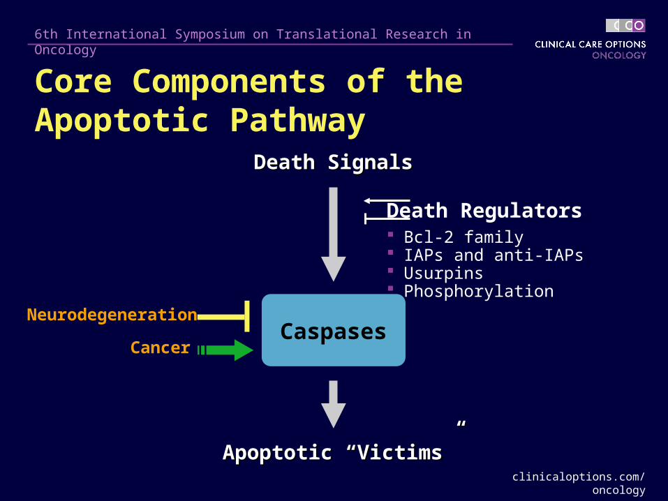
Caspases
Death SignalsDeath Signals
Death Regulators Bcl-2 family IAPs and anti-IAPs Usurpins Phosphorylation
Apoptotic “Victims”Apoptotic “Victims”
Neurodegeneration
Cancer
Core Components of the Apoptotic Pathway
clinicaloptions.com/oncology
6th International Symposium on Translational Research in Oncology
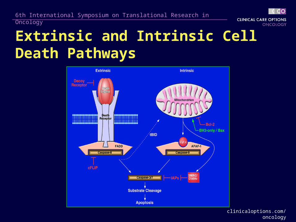
Extrinsic and Intrinsic Cell Death Pathways
clinicaloptions.com/oncology
6th International Symposium on Translational Research in Oncology
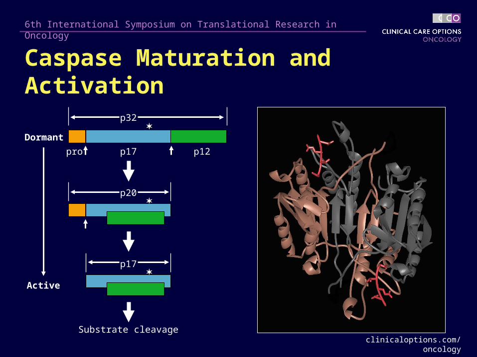
p17 p12pro
p32
Substrate cleavage
Dormant
Active
p20
p17
Caspase Maturation and Activation

clinicaloptions.com/oncology
6th International Symposium on Translational Research in Oncology
proCaspase-3 Levels in Human Colon Cancer
Colonic Adenocarcinoma vs Adjacent Normal Mucosa
N TN T N TN T N TN T
11 22 33 (Patient)(Patient)
p32p32
0
1
2
3
4
Normal Tumor
(Arb
itra
ry U
nit
s/m
g P
rote
in)
pro
Cas
pas
e-3
Co
nte
nt
(averaged data, n = 20)
6x
(Normal, Tumor)(Normal, Tumor)
clinicaloptions.com/oncology
6th International Symposium on Translational Research in Oncology
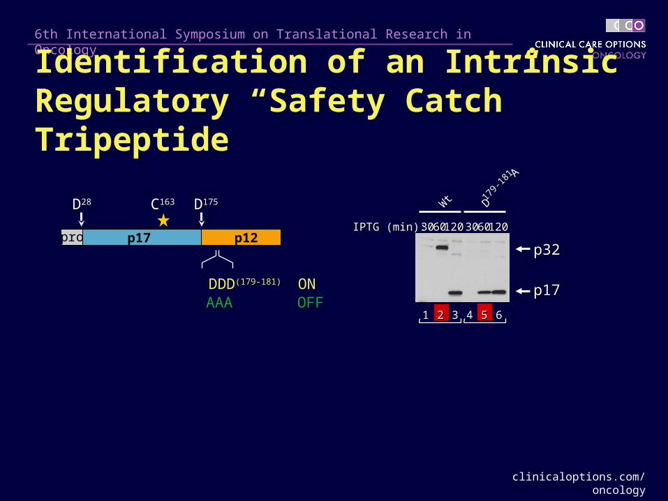
prop32p32
p17p17
p17 p12
DD2828
IPTG (min):IPTG (min):
Wt
Wt
DD17
9-18
1
179-
181 AA
11 33 44 66
30306060120120 30306060120120
22 55
DDD(179-181) ONAAA OFF
Identification of an Intrinsic Regulatory “Safety Catch” Tripeptide
CC163163 DD175175
clinicaloptions.com/oncology
6th International Symposium on Translational Research in Oncology
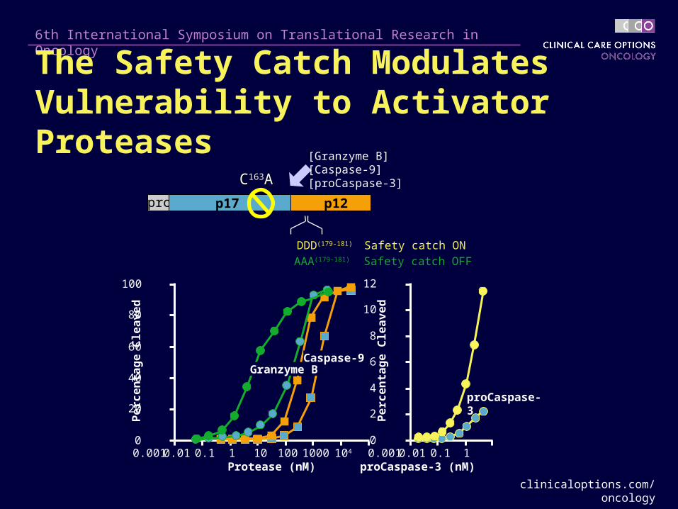
[Granzyme B][Caspase-9][proCaspase-3]
proCaspase-3
0
20
40
60
80
100
0.001 0.01 0.1 1 10 100 1000 104
Pe
rce
nta
ge
Cle
av
ed
Protease (nM)
0
2
4
6
8
10
12
0.001 0.01 0.1 1
Pe
rce
nta
ge
Cle
av
ed
proCaspase-3 (nM)
pro p17 p12
DDD(179-181) Safety catch ON
AAA(179-181) Safety catch OFF
CC163163AA
The Safety Catch Modulates Vulnerability to Activator Proteases
Caspase-9Granzyme B
clinicaloptions.com/oncology
6th International Symposium on Translational Research in Oncology
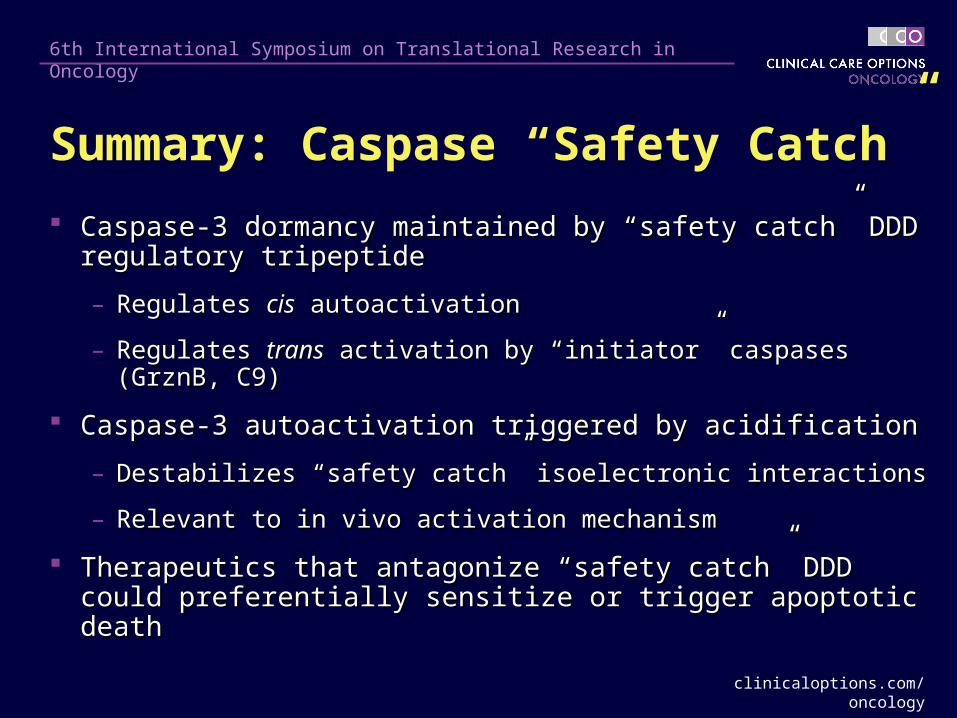
Summary: Caspase “Safety Catch”
Caspase-3 dormancy maintained by “safety catch” DDD Caspase-3 dormancy maintained by “safety catch” DDD regulatory tripeptideregulatory tripeptide
– Regulates Regulates ciscis autoactivation autoactivation
– Regulates Regulates transtrans activation by “initiator” caspases (GrznB, C9) activation by “initiator” caspases (GrznB, C9)
Caspase-3 autoactivation triggered by acidificationCaspase-3 autoactivation triggered by acidification
– Destabilizes “safety catch” isoelectronic interactionsDestabilizes “safety catch” isoelectronic interactions
– Relevant to in vivo activation mechanismRelevant to in vivo activation mechanism
Therapeutics that antagonize “safety catch” DDD could Therapeutics that antagonize “safety catch” DDD could preferentially sensitize or trigger apoptotic deathpreferentially sensitize or trigger apoptotic death
clinicaloptions.com/oncology
6th International Symposium on Translational Research in Oncology
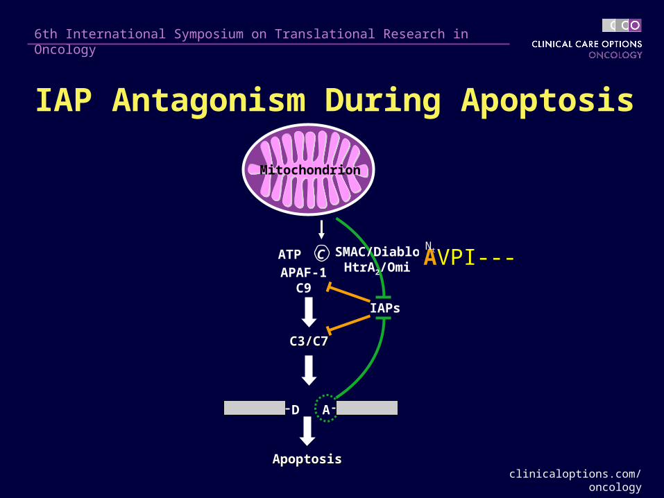
A
Mitochondrion
ATP C
APAF-1C9
C3/C7C3/C7
D
ApoptosisApoptosis
IAPs
SMAC/DiabloHtrA2/Omi AVPI---
Nt
IAP Antagonism During Apoptosis
clinicaloptions.com/oncology
6th International Symposium on Translational Research in Oncology
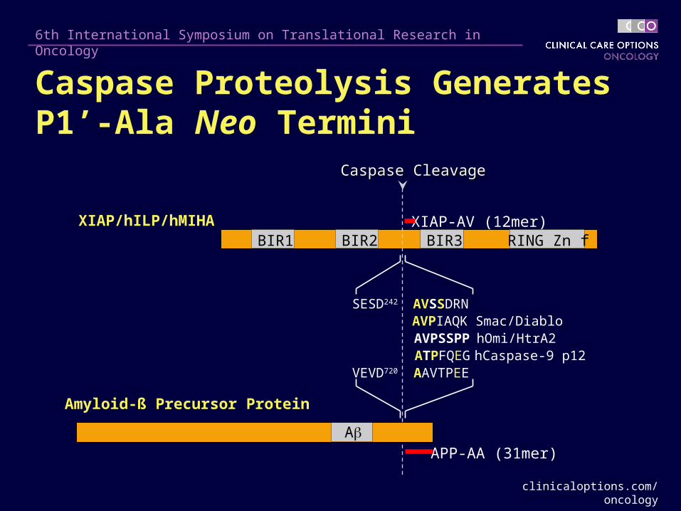
BIR3 RING Zn fBIR2BIR1XIAP/hILP/hMIHA
SESD242 AVSSDRN AVPIAQK
AVPSSPPATPFQEG
VEVD720 AAVTPEE
Smac/Diablo hOmi/HtrA2 hCaspase-9 p12
Amyloid-ß Precursor Protein
APP-AA (31mer)
XIAP-AV (12mer)
A
Caspase CleavageCaspase Cleavage
Caspase Proteolysis Generates P1’-Ala Neo Termini

clinicaloptions.com/oncology
6th International Symposium on Translational Research in Oncology
M MM
SMAC/Diablo APP XIAP
Functional Antagonism of IAPs in Cell-Free Extracts
0
20
40
60
80
100
120
140
160
180
DE
VD
ase
Ac
tiv
ity
(% o
f N
on
inh
ibit
ed A
ctiv
ity)
0 12.5 50 200
Smac-20
0
20
40
60
80
100
120
0 50 200
APP-AA
APP-MGD
EV
Das
e A
cti
vit
y (
% o
f N
on
inh
ibit
ed A
ctiv
ity)
0
10
20
30
40
50
60
70
80
0 50 200 800
XIAP-AV
XIAP-MV
DE
VD
ase
Ac
tiv
ity
(%
of
No
nin
hib
ited
Act
ivit
y)
clinicaloptions.com/oncology
6th International Symposium on Translational Research in Oncology
Summary: IAP AntagonismSummary: IAP Antagonism
IAP proteins are direct inhibitors of caspasesIAP proteins are direct inhibitors of caspases
– Ensure dormancy in healthy cellsEnsure dormancy in healthy cells
– Naturally antagonized by Smac and some caspase Naturally antagonized by Smac and some caspase substrates after caspase proteolysissubstrates after caspase proteolysis
IAP antagonism can be mediated by peptides and small IAP antagonism can be mediated by peptides and small moleculesmolecules
Therapeutics that antagonize IAPs could preferentially Therapeutics that antagonize IAPs could preferentially sensitize or trigger apoptotic deathsensitize or trigger apoptotic death
ITCH Inhibitors
clinicaloptions.com/oncology
6th International Symposium on Translational Research in Oncology
The p53 Family
The p53 family includes 3 genes, encoding for p53, p63, p73 proteins
The oldest protein is p63, whereas p53 is the most recent
– P53 is involved in DNA damage repair
Each protein exists as different isoforms generated by distinct promoters (TA isoforms and N isoforms) or by alternative splicing (, , , etc, isoforms)
– TA isoforms induce cell death (anticarcinogenic, acting as tumor suppressor)
– N isoforms inhibit cell death (procarcinogenic, acting as oncogenes)
p73 and p63, like p53, are involved in DNA damage repair, inhibiting cell cycle progression, and inducing apoptosis
– p73 is rarely mutated in cancer (0.5% mutation rate of p73 versus > 50% mutation rate of p53)
clinicaloptions.com/oncology
6th International Symposium on Translational Research in Oncology
The p53 Family (cont’d)
p73 kills cells by regulating the transcription of genes crucial to the cell cycle (eg, p21) and to apoptosis (eg, bax, PUMA, CD95), thus affecting the sensitivity of cancer cells to chemotherapy
After 30 years of research on p53, clinically exploitable targets are mostly limited to its protein degradation regulation (inhibitors of the ubiquitin E3 ligase MDM2), and ARF (DNA methyltransferase inhibitors) pathways,
clinicaloptions.com/oncology
6th International Symposium on Translational Research in Oncology
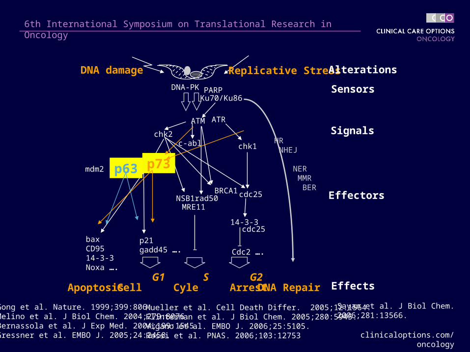
DNA damage Replicative Stress
Cell Cyle ArrestApoptosis
Sensors
Effectors
Signals
Effects
Alterations
ATM ATR
p53
chk1
chk2
p21gadd45 ….
mdm2
c-abl
bax CD9514-3-3Noxa ….
MRE11rad50
BRCA1NSB1
DNA-PK PARP
cdc25
G1 S G2
Ku70/Ku86
14-3-3cdc25
Cdc2 ….
DNA Repair
HR NHEJ
NER MMR BER
p73p63
Mueller et al. Cell Death Differ. 2005;12:1564.Flinterman et al. J Biol Chem. 2005;280:5945.Vigano et al. EMBO J. 2006;25:5105.Rossi et al. PNAS. 2006;103:12753
Gong et al. Nature. 1999;399:806.Melino et al. J Biol Chem. 2004;279:8076.Bernassola et al. J Exp Med. 2004;199:1545.Gressner et al. EMBO J. 2005;24:2458.
Sayan et al. J Biol Chem. 2006;281:13566.
clinicaloptions.com/oncology
6th International Symposium on Translational Research in Oncology
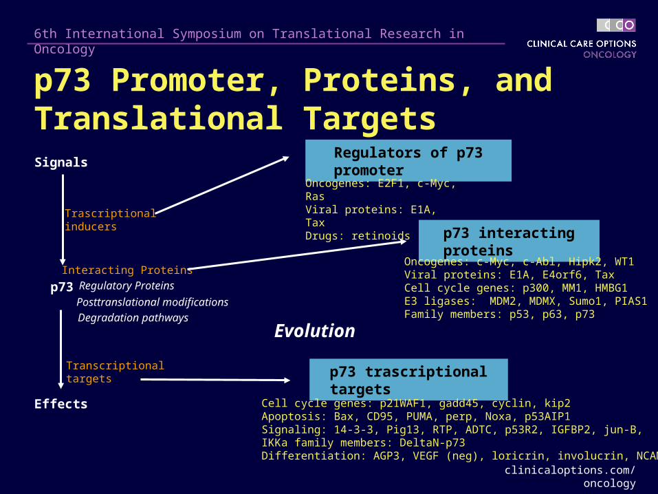
Regulators of p73 promoter
p73 interacting proteins
p73 trascriptional targets
Signals
p73
Effects
Trascriptionalinducers
Transcriptionaltargets
Interacting Proteins
Degradation pathways
Regulatory Proteins
Posttranslational modifications
Oncogenes: E2F1, c-Myc, RasViral proteins: E1A, TaxDrugs: retinoids
Oncogenes: c-Myc, c-Abl, Hipk2, WT1 Viral proteins: E1A, E4orf6, Tax Cell cycle genes: p300, MM1, HMBG1E3 ligases: MDM2, MDMX, Sumo1, PIAS1 Family members: p53, p63, p73
Cell cycle genes: p21WAF1, gadd45, cyclin, kip2 Apoptosis: Bax, CD95, PUMA, perp, Noxa, p53AIP1 Signaling: 14-3-3, Pig13, RTP, ADTC, p53R2, IGFBP2, jun-B, IKKa family members: DeltaN-p73Differentiation: AGP3, VEGF (neg), loricrin, involucrin, NCAM
Evolution
p73 Promoter, Proteins, and Translational Targets
clinicaloptions.com/oncology
6th International Symposium on Translational Research in Oncology
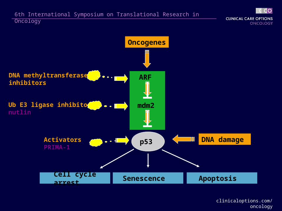
p53
Senescence Apoptosis Cell cycle arrest
Oncogenes
DNA damage
Ub E3 ligase inhibitorsnutlin
ActivatorsPRIMA-1
DNA methyltransferase inhibitors
ARF
mdm2
clinicaloptions.com/oncology
6th International Symposium on Translational Research in Oncology
Protein degradation of both p73 and p63 is regulated by the ubiquitin E3 ligase ITCH, a member of the HECT-containing E3s
In addition to p73 and p63, ITCH controls the degradation of c-jun, Notch, jun-B, and Flip, all involved in oncogenesis and apoptosis
– The prediction is therefore that ITCH regulation affects carcinogenesis and/or chemosensitivity
Modulation of ITCH protein levels regulates chemosensitivity in vitro, suggesting that an inhibitor of ITCH could increase chemosensitivity
How Are p63/p73 Protein Levels Controlled?
clinicaloptions.com/oncology
6th International Symposium on Translational Research in Oncology
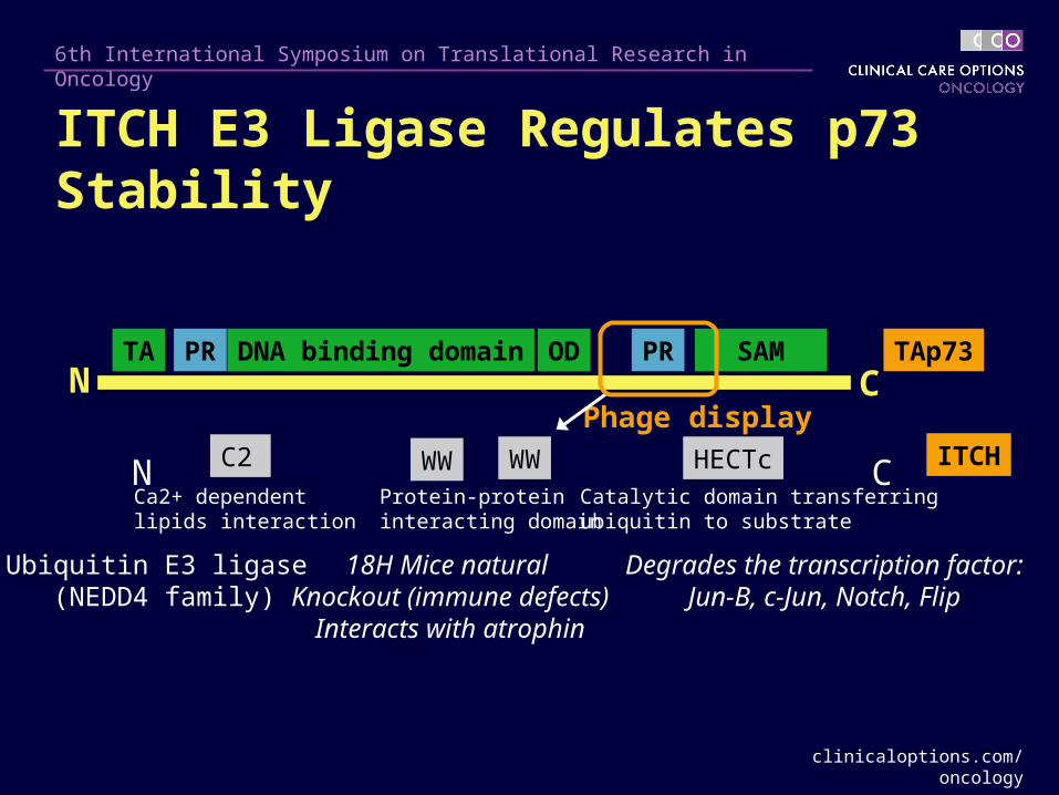
ITCH
Ubiquitin E3 ligase (NEDD4 family)
18H Mice natural Knockout (immune defects)
Interacts with atrophin
Degrades the transcription factor:Jun-B, c-Jun, Notch, Flip
HECTcWW WWC2
Ca2+ dependent lipids interaction
Protein-protein interacting domain
Catalytic domain transferring ubiquitin to substrate
CN
Phage display
TA DNA binding domain OD SAM PR PRCN
TAp73
ITCH E3 Ligase Regulates p73 Stability
clinicaloptions.com/oncology
6th International Symposium on Translational Research in Oncology
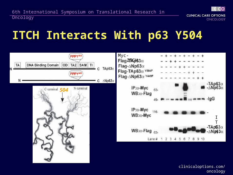
504
ITCH Interacts With p63 Y504
Myc-ITCH
-ITCH
clinicaloptions.com/oncology
6th International Symposium on Translational Research in Oncology
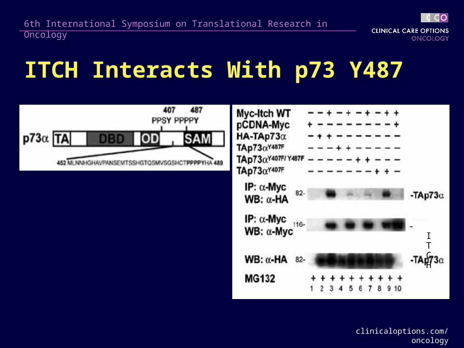
-ITCH
ITCH Interacts With p73 Y487
clinicaloptions.com/oncology
6th International Symposium on Translational Research in Oncology
Is ITCH E3 Activity Regulated?
Summary
The function of ubiquitin E3 ligase ITCH is regulated by a physical interaction with a novel protein called N4BP1
N4BP1 competes with ITCH substrates (p63, p73, c-Jun) by binding on the same region of ITCH, called WW2
N4BP1 physiologically regulates ITCH and its substrates (p73, p63, c-Jun), thereby affecting cell death
clinicaloptions.com/oncology
6th International Symposium on Translational Research in Oncology
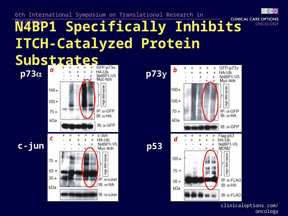
N4BP1 Specifically Inhibits ITCH-Catalyzed Protein Substrates
b
d
p73p73
c-jun p53c
a
clinicaloptions.com/oncology
6th International Symposium on Translational Research in Oncology
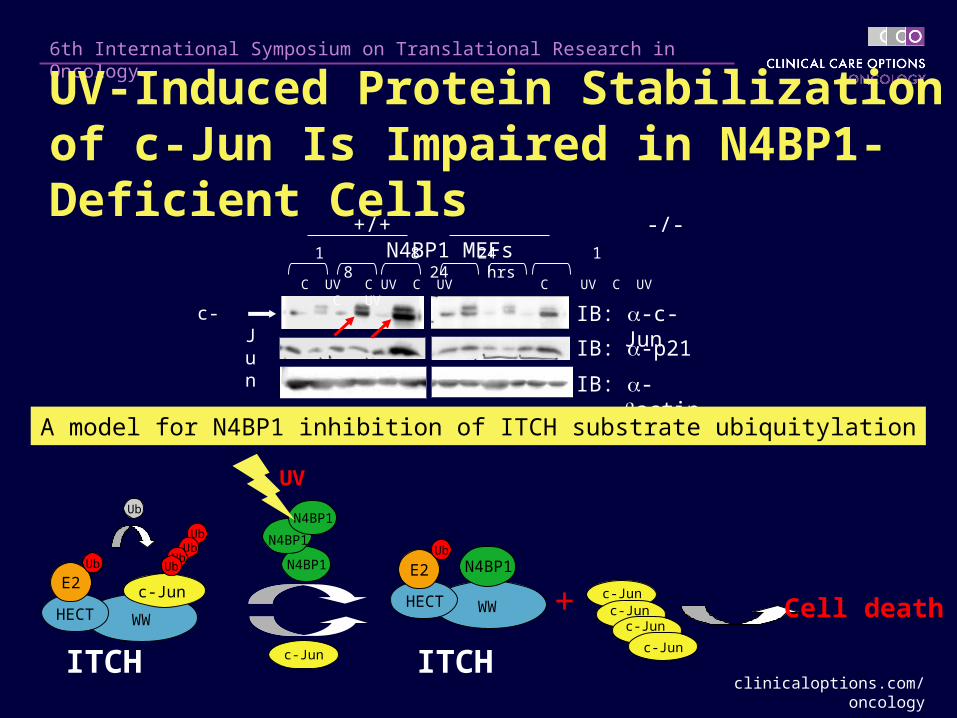
1 8 24 1 8 24 hrs
+/+ -/- N4BP1 MEFs
IB: -c-Junc-Jun
C UV C UV C UV C UV C UV C UV
IB: -p21
IB: -actin
A model for N4BP1 inhibition of ITCH substrate ubiquitylation
WWHECT
E2Ub
UbUb
UbUb
c-Jun
Ub
N4BP1
N4BP1
N4BP1
WWHECT
E2Ub
N4BP1
c-Jun
+
UV
c-Junc-Jun
c-Jun
c-Jun
Cell death
ITCH ITCH
UV-Induced Protein Stabilization of c-Jun Is Impaired in N4BP1-Deficient Cells
clinicaloptions.com/oncology
6th International Symposium on Translational Research in Oncology
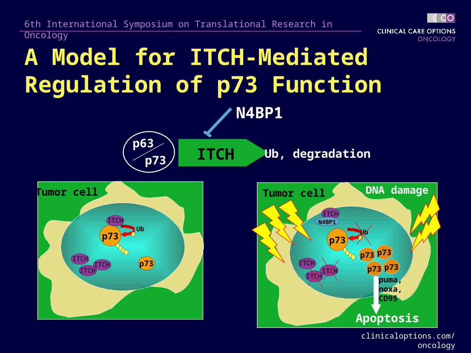
Tumor cell
p73Ub
p73ITCHITCH
ITCH
ITCH
Tumor cell
p73Ub
p73ITCHITCH
ITCH
DNA damage
Apoptosis
ITCH
N4BP1
p73 p73
p73
puma, noxa,CD95
p63ITCH Ub, degradation
N4BP1
p73
A Model for ITCH-Mediated Regulation of p73 Function
clinicaloptions.com/oncology
6th International Symposium on Translational Research in Oncology
Small molecular inhibitors of ubiquitin E3 ligase ITCH have been identified
Potential ITCH inhibitors are currently under evaluation for their anticancer activity
Can We Inhibit ITCH E3 Activity?
clinicaloptions.com/oncology
6th International Symposium on Translational Research in Oncology
p63/p73 are involved in DNA repair/cancer
p63/p73 regulate chemosensitivity in cancer
p63/p73 are ubiquitinated and degraded by ITCH
ITCH is regulated by N4BP1
Low MW ITICH inhibitors are under development
Perspective
ITCH is a candidate therapeutic target (to regulate p63/p73)
Summary
Caspase Substrates and p53
clinicaloptions.com/oncology
6th International Symposium on Translational Research in Oncology
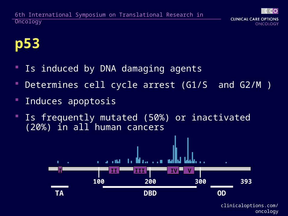
p53
Is induced by DNA damaging agents
Determines cell cycle arrest (G1/S and G2/M )
Induces apoptosis
Is frequently mutated (50%) or inactivated (20%) in all human cancers
I II III IV V
100 200 300 393
TA DBD OD
clinicaloptions.com/oncology
6th International Symposium on Translational Research in Oncology
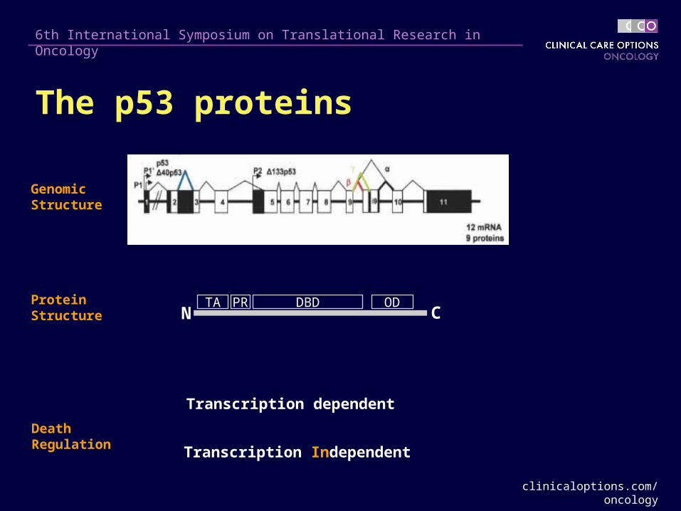
NTA DBD ODPR
C
Transcription dependent
GenomicStructure
ProteinStructure
Transcription Independent
DeathRegulation
The p53 proteins
clinicaloptions.com/oncology
6th International Symposium on Translational Research in Oncology
A - 3 6 7 8
50
37
25
Caspase C - 20 50 100 200 400 nMCaspase 3
1
234
BZ-VAD-fmk +- -Etoposide +- +
50
37
25
D Caspase 3 - + - + - + - +
50
37
25
DO1 1801 FL393 C19
E - + + + +Caspase 3p53 W
T
WT
D21
AD
186A
D21
A,
D18
6A
at D21 and D186
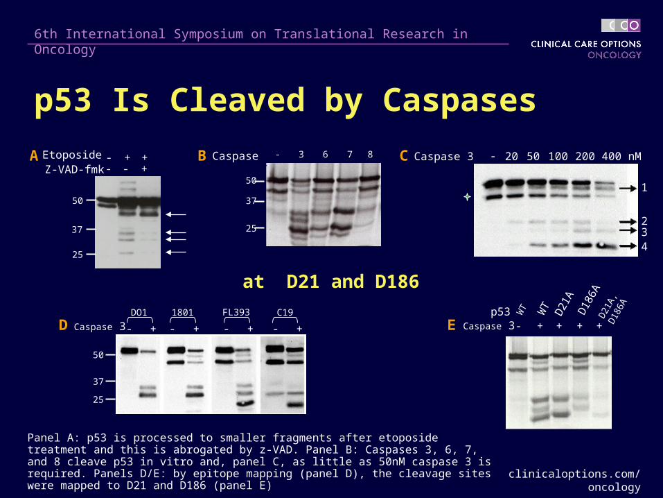
Panel A: p53 is processed to smaller fragments after etoposide treatment and this is abrogated by z-VAD. Panel B: Caspases 3, 6, 7, and 8 cleave p53 in vitro and, panel C, as little as 50nM caspase 3 is required. Panels D/E: by epitope mapping (panel D), the cleavage sites were mapped to D21 and D186 (panel E)
p53 Is Cleaved by Caspases
clinicaloptions.com/oncology
6th International Symposium on Translational Research in Oncology
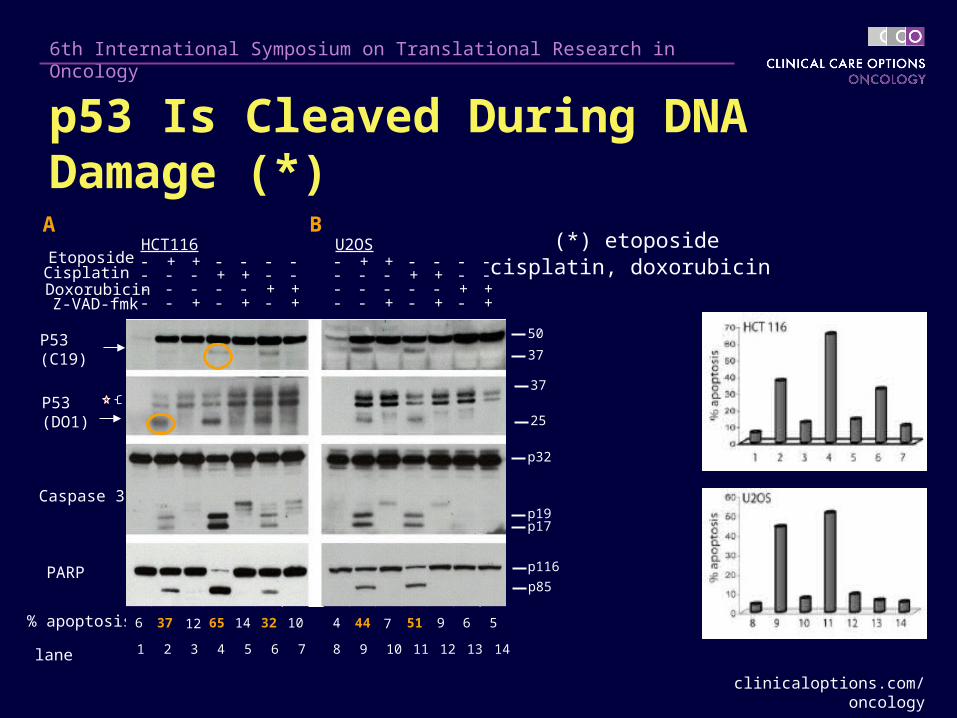
p53 Is Cleaved During DNA Damage (*)
Z-VAD-fmk ++ - + - - - + - +- - - +Doxorubicin -- + + - - - - + +- - - -Cisplatin +- - - - - + + - -- - + -Etoposide -+ - - - + - - - -- + - +
P53 (C19)
HCT116 U2OS
Caspase 3
PARP
53 6 71 2 4 8 9 11 12 13 1410
p32
p19p17
p116
p85
P53 (DO1)
37
25
50
37
% apoptosis 6 37 65 14 32 1012 4 44 51 9 6 57
lane
(*) etoposidecisplatin, doxorubicin
A B
clinicaloptions.com/oncology
6th International Symposium on Translational Research in Oncology
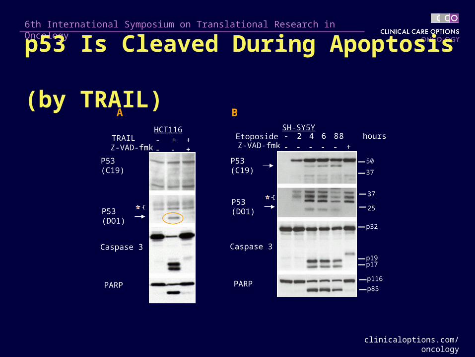
A B
Z-VAD-fmk -- +- - -Etoposide 84 8 hours- 2 6
SH-SY5Y
Caspase 3
PARPp116
p85
p32
p19p17
P53 (C19)
P53 (DO1)
37
25
50
37
TRAIL +- +HCT116
Z-VAD-fmk +- -
Caspase 3
PARP
P53 (C19)
P53 (DO1)
p53 Is Cleaved During Apoptosis (by TRAIL)
clinicaloptions.com/oncology
6th International Symposium on Translational Research in Oncology
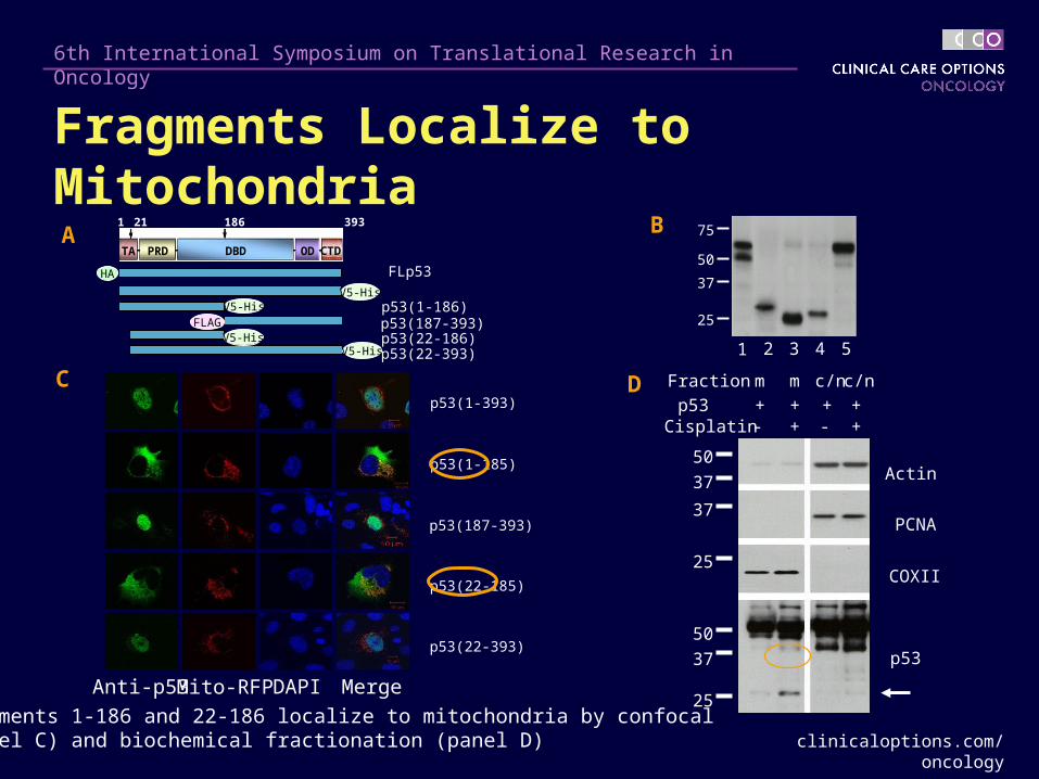
Fragments Localize to MitochondriaA B
FLp53
p53(1-186)p53(187-393)p53(22-186)p53(22-393)
21 186 393
TA PRD DBD OD CTD
1
HA
FLAGV5-His
V5-HisV5-His
V5-His
50
37
25
75
1 2 3 4 5
C
Actin
PCNA
COXII
p53
50
37
37
25
50
37
25
Fraction m m c/nc/n
p53 + + + +Cisplatin - + - +
D
Anti-p53 DAPI MergeMito-RFP
p53(1-393)
p53(1-185)
p53(187-393)
p53(22-185)
p53(22-393)
Fragments 1-186 and 22-186 localize to mitochondria by confocal (panel C) and biochemical fractionation (panel D)
clinicaloptions.com/oncology
6th International Symposium on Translational Research in Oncology
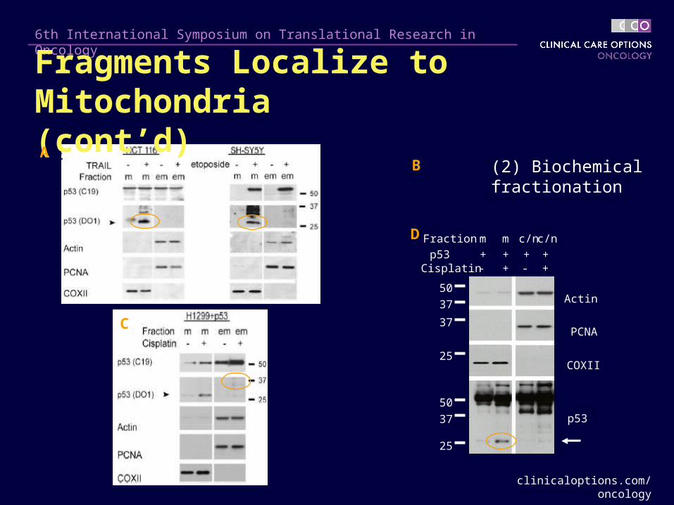
B
Actin
PCNA
COXII
p53
50
37
37
25
50
37
25
Fraction m m c/nc/n
p53 + + + +Cisplatin - + - +
D
C
(2) Biochemicalfractionation
Fragments Localize to Mitochondria (cont’d)A
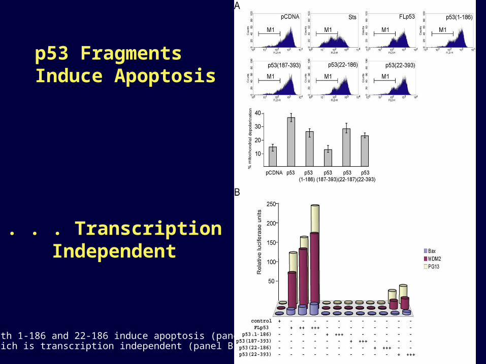
p53 Fragments Induce Apoptosis
. . . TranscriptionIndependent
Both 1-186 and 22-186 induce apoptosis (panel A)which is transcription independent (panel B)
clinicaloptions.com/oncology
6th International Symposium on Translational Research in Oncology
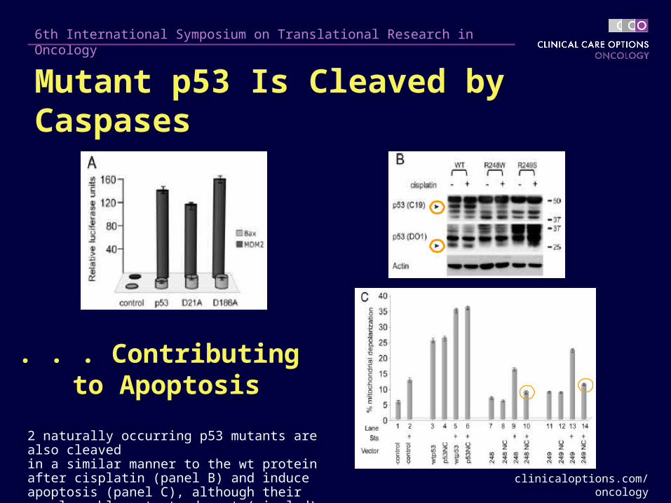
. . . Contributing to Apoptosis
2 naturally occurring p53 mutants are also cleaved in a similar manner to the wt protein after cisplatin (panel B) and induce apoptosis (panel C), although their noncleavable mutants do not (circled)
Mutant p53 Is Cleaved by Caspases
clinicaloptions.com/oncology
6th International Symposium on Translational Research in Oncology
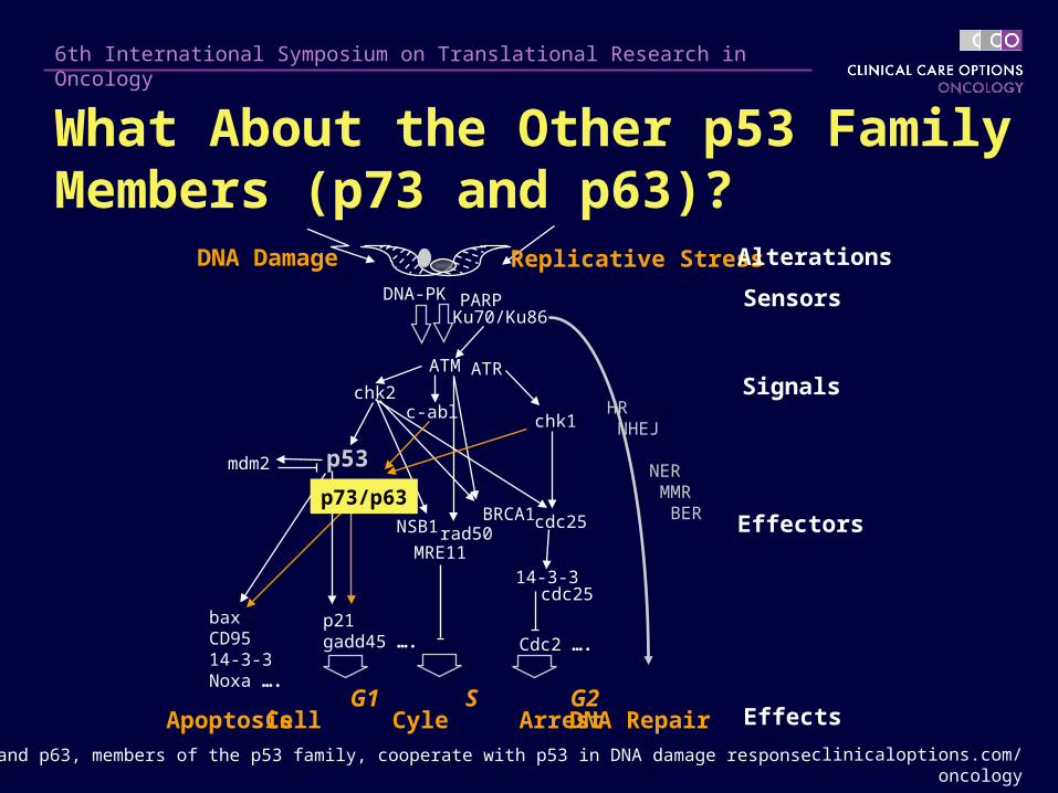
DNA Damage Replicative Stress
Cell Cyle ArrestApoptosis
Sensors
Effectors
Signals
Effects
Alterations
ATM ATR
p53
chk1
chk2
p21gadd45 ….
mdm2
c-abl
bax CD9514-3-3Noxa ….
MRE11rad50
BRCA1NSB1
DNA-PK PARP
cdc25
G1 S G2
Ku70/Ku86
14-3-3cdc25
Cdc2 ….
DNA Repair
HR NHEJ
NER MMR BER
p73 and p63, members of the p53 family, cooperate with p53 in DNA damage response
What About the Other p53 Family Members (p73 and p63)?
p73/p63
clinicaloptions.com/oncology
6th International Symposium on Translational Research in Oncology
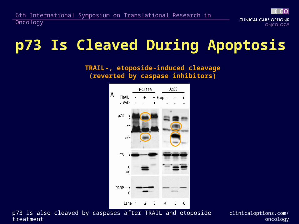
TRAIL-, etoposide-induced cleavage(reverted by caspase inhibitors)
p73 is also cleaved by caspases after TRAIL and etoposide treatment
p73 Is Cleaved During Apoptosis
clinicaloptions.com/oncology
6th International Symposium on Translational Research in Oncology
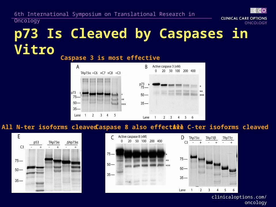
All N-ter isoforms cleaved All C-ter isoforms cleaved
Caspase 3 is most effective
Caspase 8 also effective
p73 Is Cleaved by Caspases in Vitro
clinicaloptions.com/oncology
6th International Symposium on Translational Research in Oncology
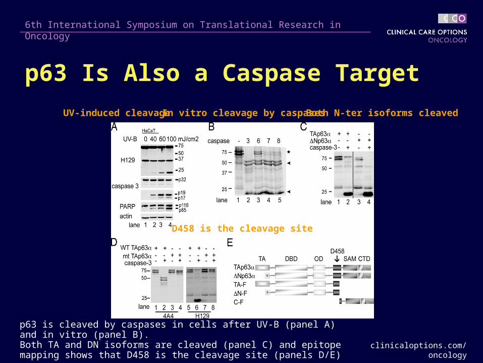
p63 is cleaved by caspases in cells after UV-B (panel A) and in vitro (panel B). Both TA and DN isoforms are cleaved (panel C) and epitope mapping shows that D458 is the cleavage site (panels D/E)
D458 is the cleavage site
UV-induced cleavage In vitro cleavage by caspases Both N-ter isoforms cleaved
p63 Is Also a Caspase Target
clinicaloptions.com/oncology
6th International Symposium on Translational Research in Oncology
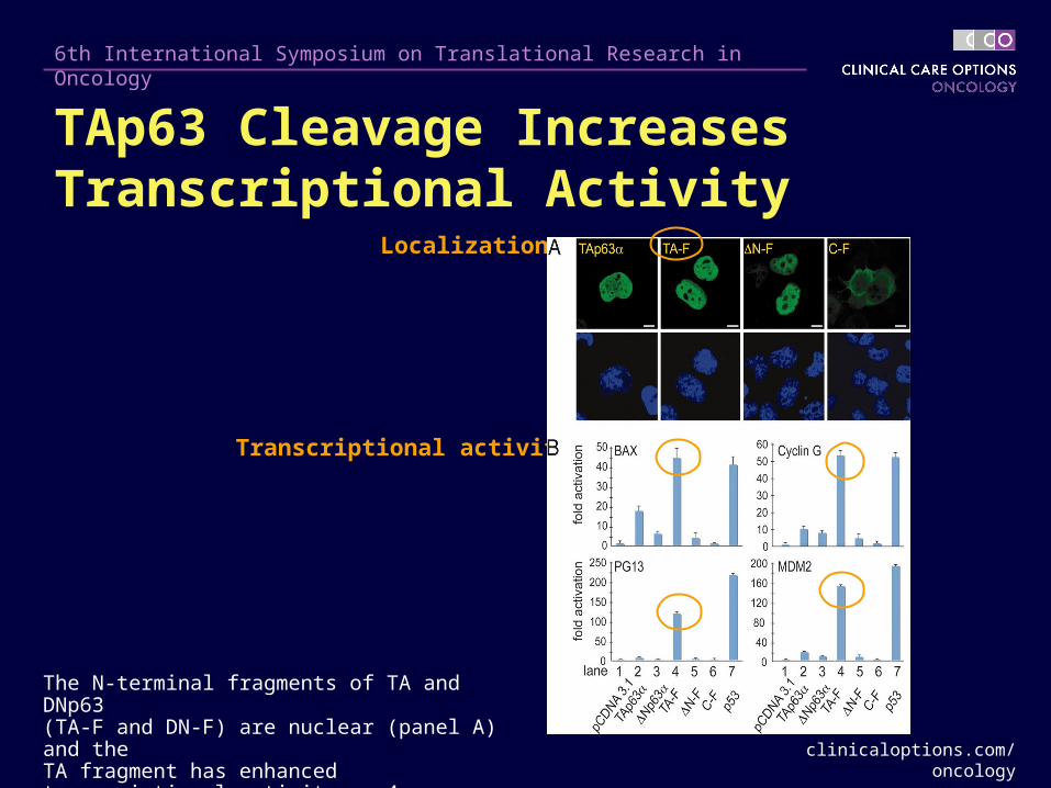
Localization
Transcriptional activity
The N-terminal fragments of TA and DNp63 (TA-F and DN-F) are nuclear (panel A) and the TA fragment has enhanced transcriptional activity on 4 promoters compared to intact TAp63a
TAp63 Cleavage Increases Transcriptional Activity
clinicaloptions.com/oncology
6th International Symposium on Translational Research in Oncology
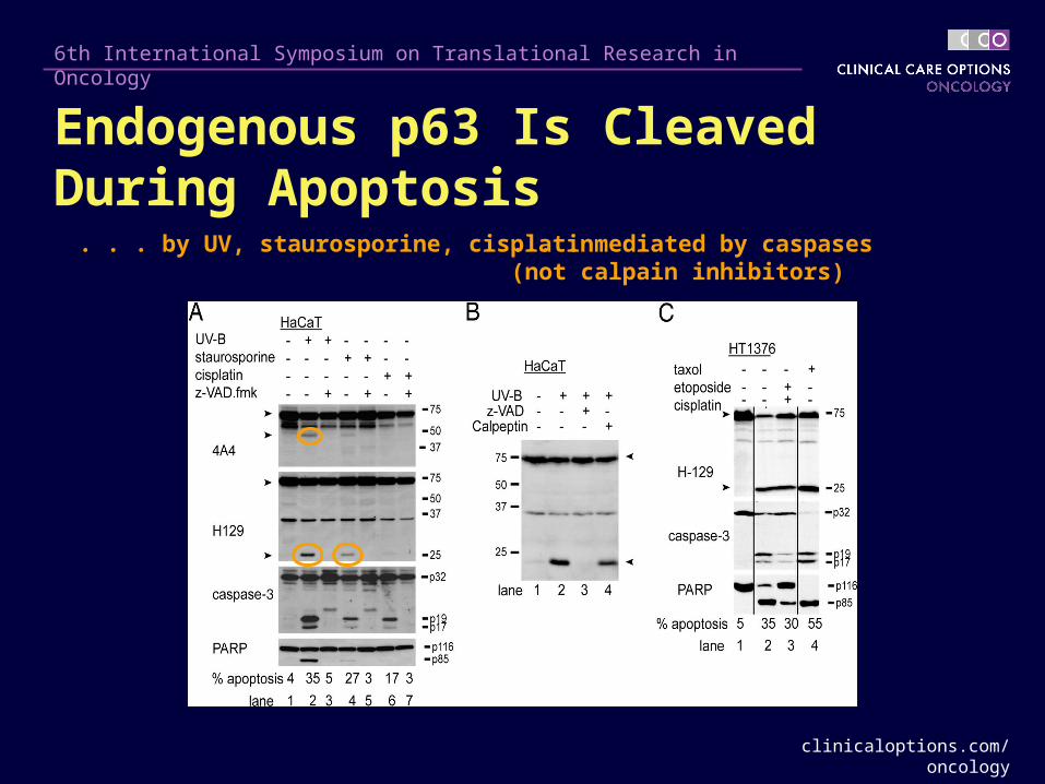
. . . by UV, staurosporine, cisplatin . . . mediated by caspases(not calpain inhibitors)
Endogenous p63 Is Cleaved During Apoptosis
clinicaloptions.com/oncology
6th International Symposium on Translational Research in Oncology
Summary
p53 is cleaved by caspases during apoptosis, contributing to cell death
Transcriptionally inactive natural mutants of p53 can be cleaved by caspases to produce transcriptionally inactive fragments
– Can still induce apoptosis by depolarization of mitochondria
Other members of the p53 family, p63 and p73, are also susceptible to caspase cleavage
Caspase cleavage of p63a isoforms relieves the inhibitory effects of the C-terminal transactivation inhibitory domain, resulting in
– Enhanced transcriptional activity by the TA isoform
– Abrogating the transactivational inhibitory effects of the DNp63 isoform
Granzyme B in Cancer Therapeutics
clinicaloptions.com/oncology
6th International Symposium on Translational Research in Oncology
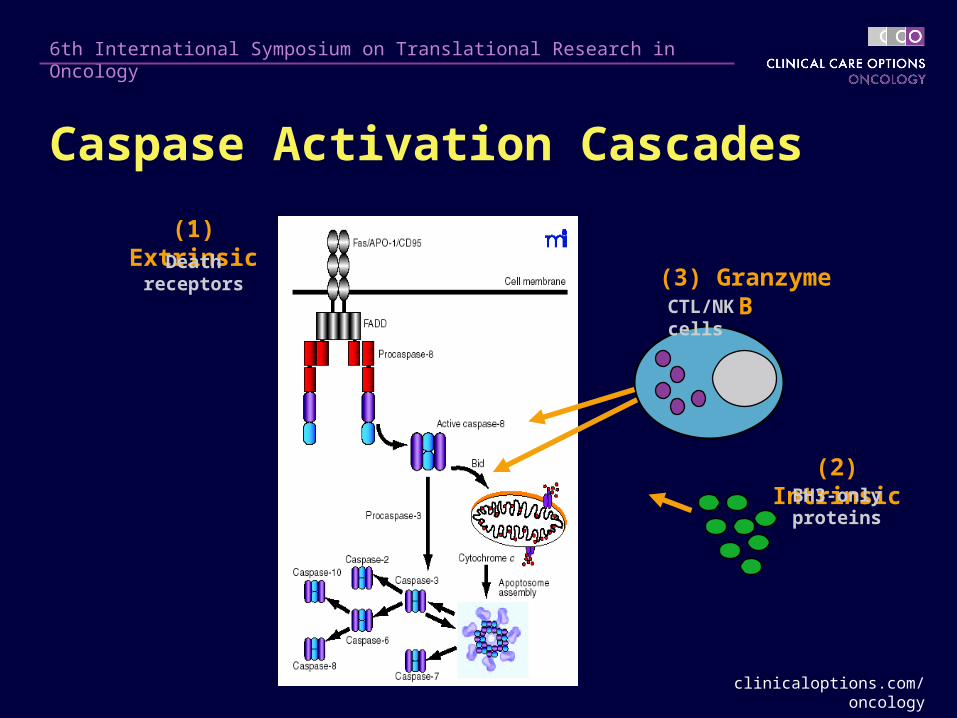
(1) Extrinsic
(2) Intrinsic
(3) Granzyme B
BH3-only proteins
CTL/NK cells
Caspase Activation Cascades
Death receptors

clinicaloptions.com/oncology
6th International Symposium on Translational Research in Oncology
Bad
BimBid
PumaNoxaBmf
Hrk
Bik
p53Caspase-8
Death receptors
Granzyme B
Growth factordeprivation
Ag receptorProteasome inhibition
BH3-Only Proteins: Pathway-Specific Sensors of Stress and Damage
clinicaloptions.com/oncology
6th International Symposium on Translational Research in Oncology
Malignant melanoma notoriously refractory to Malignant melanoma notoriously refractory to chemotherapy chemotherapy
~ 60% to 80% of melanomas display mutations in B-Raf~ 60% to 80% of melanomas display mutations in B-Raf
B-Raf mutations also found in colorectal, thyroid, and B-Raf mutations also found in colorectal, thyroid, and ovarian cancer ovarian cancer
Vast majority of mutations are single point mutations at Vast majority of mutations are single point mutations at V600>EV600>E
B-Raf and Melanoma
clinicaloptions.com/oncology
6th International Symposium on Translational Research in Oncology
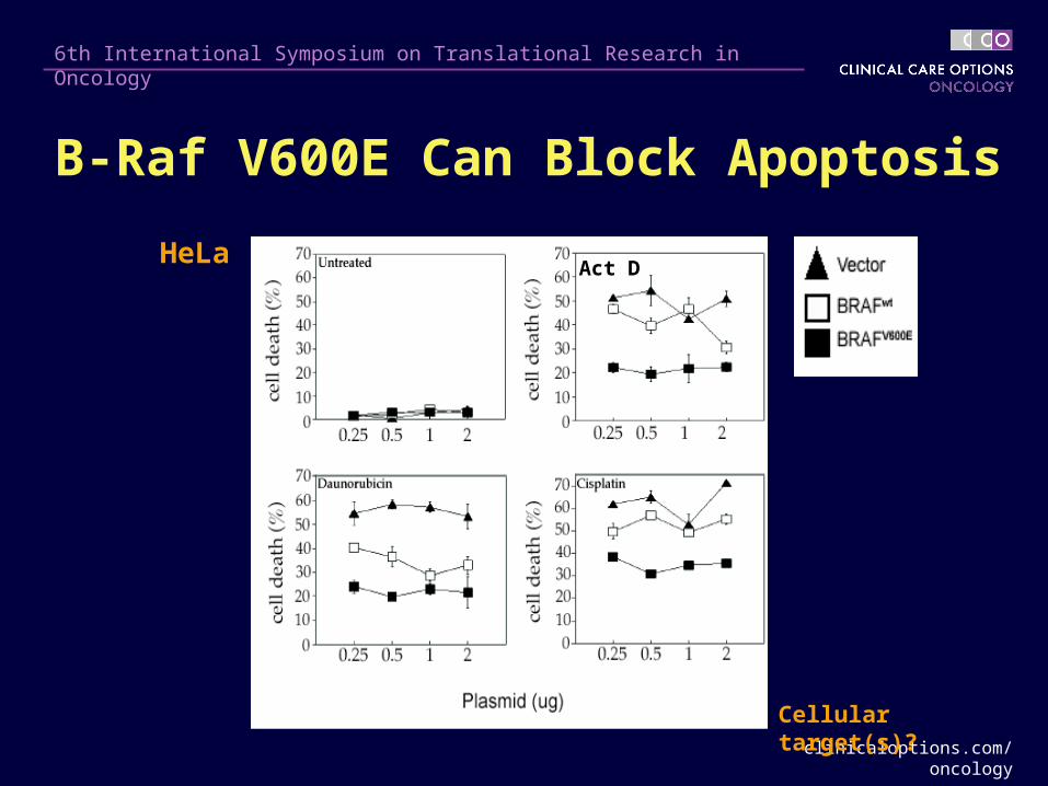
B-Raf V600E Can Block Apoptosis
Act DHeLa
Cellular target(s)?
clinicaloptions.com/oncology
6th International Symposium on Translational Research in Oncology
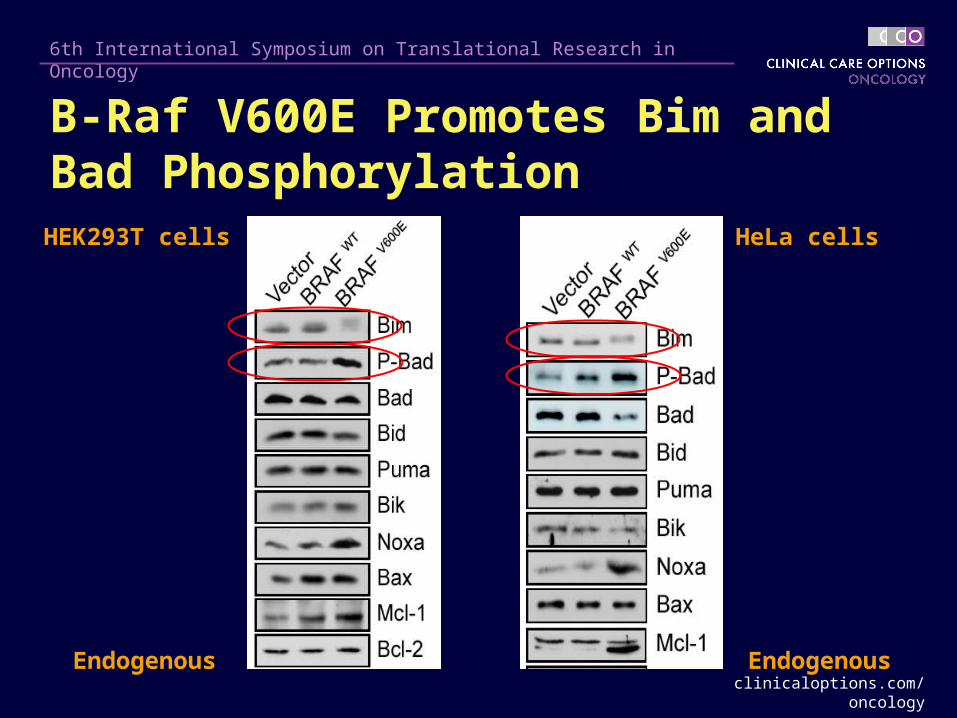
HeLa cells
Endogenous
HEK293T cells
Endogenous
B-Raf V600E Promotes Bim and Bad Phosphorylation
clinicaloptions.com/oncology
6th International Symposium on Translational Research in Oncology
Summary
Oncogenic B-Raf suppresses apoptosis
B-Raf inactivates the BH3-only proteins Bim and Bid via ERK kinase phosphorylation
B-Raf blocks Bim- and Bid-induced cell death and is required for survival
clinicaloptions.com/oncology
6th International Symposium on Translational Research in Oncology
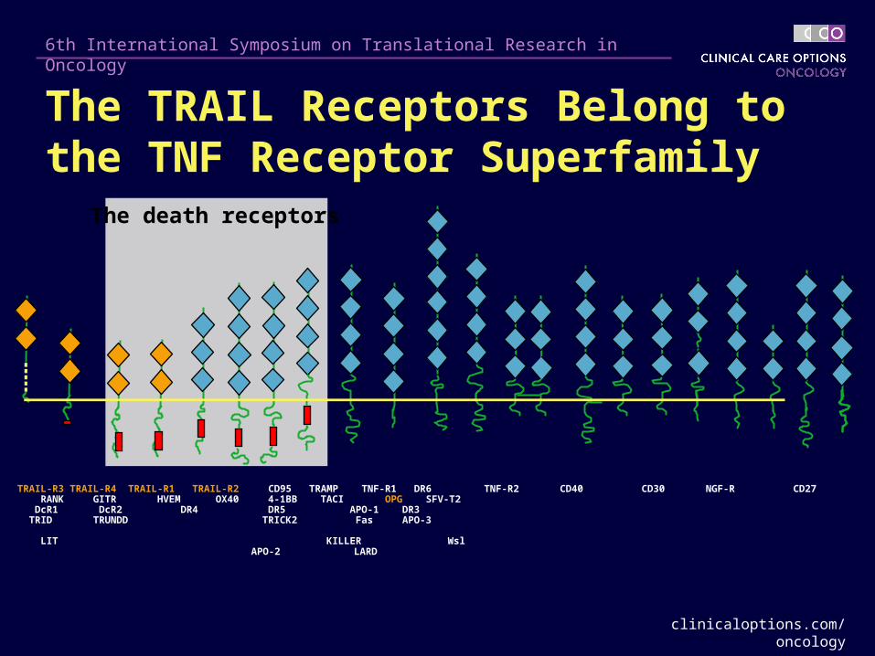
The death receptors
TRAIL-R3 TRAIL-R4 TRAIL-R1 TRAIL-R2 CD95 TRAMP TNF-R1 DR6 TNF-R2 CD40 CD30 NGF-R CD27 RANK GITR HVEM OX40 4-1BB TACI OPG SFV-T2 DcR1 DcR2 DR4 DR5 APO-1 DR3 TRID TRUNDD TRICK2 Fas APO-3 LIT KILLER Wsl
APO-2 LARD
The TRAIL Receptors Belong to the TNF Receptor Superfamily
clinicaloptions.com/oncology
6th International Symposium on Translational Research in Oncology
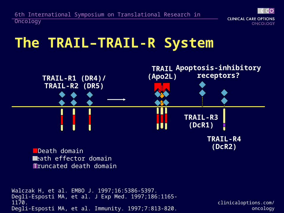
TRAIL-R1 (DR4)/TRAIL-R2 (DR5)
TRAIL-R3(DcR1)
TRAIL-R4(DcR2)
Apoptosis-inhibitoryreceptors?
TRAIL(Apo2L)
Death domainDeath effector domainTruncated death domain
Walczak H, et al. EMBO J. 1997;16:5386-5397.Degli-Esposti MA, et al. J Exp Med. 1997;186:1165-1170.Degli-Esposti MA, et al. Immunity. 1997;7:813-820.
The TRAIL–TRAIL-R System
clinicaloptions.com/oncology
6th International Symposium on Translational Research in Oncology
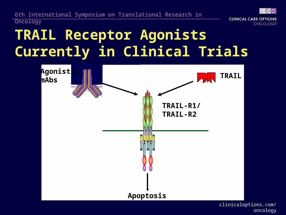
Apoptosis
Agonistic mAbs
TRAIL
TRAIL-R1/TRAIL-R2
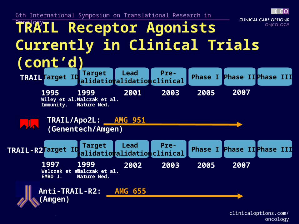
TRAIL Receptor Agonists Currently in Clinical Trials
clinicaloptions.com/oncology
6th International Symposium on Translational Research in Oncology
TRAIL/Apo2L: AMG 951(Genentech/Amgen)
Target validation
Target ID
1995Wiley et al. Immunity.
1999Walczak et al. Nature Med.
Pre-clinical
Phase II Phase IIIPhase ILead
validation
2001 2003 2005
TRAIL
TRAIL-R2
1997Walczak et al. EMBO J.
Target validation
Target IDPre-
clinicalPhase II Phase IIIPhase I
Lead validation
1999Walczak et al. Nature Med.
2002 2003 2005
2007
2007
TRAIL Receptor Agonists Currently in Clinical Trials (cont’d)
Anti-TRAIL-R2: AMG 655(Amgen)
clinicaloptions.com/oncology
6th International Symposium on Translational Research in Oncology
TRAIL/Apo2L – Genentech/Amgen
1 agonistic antibody to TRAIL-R1 (DR4)– Human Genome Sciences
5 agonistic antibodies to TRAIL-R2 (DR5)– Human Genome Sciences
– Genentech
– Amgen
– Sankyo
– Novartis
TRAIL Receptor Agonists Currently in Clinical Trials (cont’d)
clinicaloptions.com/oncology
6th International Symposium on Translational Research in Oncology
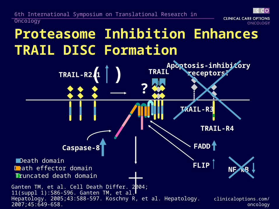
( )
Death domainDeath effector domainTruncated death domain
Ganten TM, et al. Cell Death Differ. 2004;11(suppl 1):S86-S96. Ganten TM, et al. Hepatology. 2005;43:588-597. Koschny R, et al. Hepatology. 2007;45:649-658.
TRAIL-R3
TRAIL-R4
Apoptosis-inhibitoryreceptors?TRAILTRAIL-R2/1
FLIP
Caspase-8
?
FADD
NF-B
Proteasome Inhibition Enhances TRAIL DISC Formation
clinicaloptions.com/oncology
6th International Symposium on Translational Research in Oncology
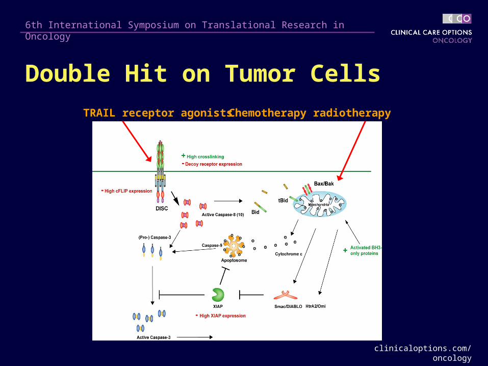
Chemotherapy radiotherapyTRAIL receptor agonists
Double Hit on Tumor Cells
clinicaloptions.com/oncology
6th International Symposium on Translational Research in Oncology
Only limited toxicity, even in combination with chemotherapy
The different TRAIL receptor agonists are unique
– Pharmacokinetics
– Target TRAIL-R1 and/or TRAIL-R2
Produce significant numbers of stable disease; many patients show stable disease
Response to monotherapy is rare
Long-lasting treatment possible
Conclusions From Clinical Studies With TRAIL Receptor Agonists
clinicaloptions.com/oncology
6th International Symposium on Translational Research in Oncology
TRAIL/Apo2L + rituximab in CD20-positive NHL
TRAIL/Apo2L + chemotherapy with and without bevacizumab in NSCLC
Randomized phase 2 in multiple myeloma mapatumumab (anti–TRAIL-R1) with or without bortezomib
Ongoing Interesting Combination Studies (No Data Available Yet)

clinicaloptions.com/oncology
6th International Symposium on Translational Research in Oncology
5 Agonistic Antibodies to TRAIL-R2 (DR5)1. ETR2 Human Genome Sciences
2 phase I studies Phase I study + chemo
2. Apomab GenentechPhase I study
3. AMG 655 AmgenPhase I study
4. LBY135 NovartisPhase I/II trialalone and with capecitabine
5. CS-1008 SankyoPhase I study
clinicaloptions.com/oncology
6th International Symposium on Translational Research in Oncology
More Hematology/Oncology Available Online! Medical Meeting Coverage: key data plus Expert
Analysis panel discussions exploring clinical implications
Treatment Updates: comprehensive programs covering the most important new concepts
Interactive Cases: test your ability to manage patients
clinicaloptions.com/oncology


