Maxillary All-On-Four Therapy Using Angled Implants: A 16-Month Clinical Study of 1110 Implants in 276 Jaws Stuart Graves, DDS, MS a, *, Brian A. Mahler, DDS b , Ben Javid, DDS c , Debora Armell ini, DDS, MD d , Ole T. Jensen, DDS, MS e,f The max illa is a cha llen gin g area for dental impl ant restor ati on. Encroa chment ofanatomic structures such as the sinus and nasal floor make vertical placement diffi- cult. Implants placed at an angle may be used to avoid these anatomic structures or eliminate the need for a bone grafting procedure. The question occasionally arises about the possible detrimental effects of placing implants at an angle. It should be noted that because of bone resorptio n numer ous implants, especial ly in the maxillary anterior, have been placed at significant angles for many years. Anecdotally these ti lt ed impl an ts seem to work, but what ev idence is av ai labl e in the li terature wi th re gard to the efficacy of implants placed at an angle? This article was previously published in the May 2011 issue of Oral and Maxillofacial SurgeryClinics of North America. a Northern Vir ginia Oral Max illofacial & Implant Surger y , Burke Profess ional Center, 5206 Lyngate Court, Burke, VA 22015, USA b Private Practice, 10550 Warwick Avenue, Fairfax, VA 22030, USA c ClearChoice Dental Implant Cen ter– Washington Metro, 11200 Rockville Pike, Suite 115, Bethesda, MD 20852, USA d ClearChoi ce Dental Imp lant Center –Washi ngt on Metr o, 8219 Leesburg Pike, Suite 100, Vienna, VA 22182, USA e Implant Dentis try Ass oci ates of Colorado, 8200 East Bel lev iew Avenue, Sui te 520E, Greenwood Village, CO 80111, USA f Department of Oral and Maxillofacial Surgery, Hebrew University School of Dental Medicine, POB 12272, Jerusalem, 91120, Israel * Corresponding author. E-mail address: [email protected]KEYWORDS Maxillary implants All-on-Four implant protocol Tilted implants Edentulous maxilla Pterygomaxillary implants Zygomatic implants Dent Clin N Am 55 (2011) 779–794 doi:10.1016/j.cden.2011.07.007 dental.theclinics.com 0011-8532/11 /$ – see front matter 2011 Elsevier Inc. All right s reserved.
Transcript
5/16/2018 88729490 Maxillary All on Four Therapy Using Angled Implants - slidepdf.com
M a x i l l a r y A l l - O n - F o u rT h e r a p y U s i n g A n g l e d
I m p l a n t s : A 1 6 - M o n t hC l i n i c a l S t u d y o f 1 1 1 0I m p la n t s i n 2 7 6 J a w s
Stuart Graves, DDS, MSa,*, Brian A. Mahler, DDS
b, Ben Javid, DDSc,
Debora Armellini, DDS, MDd, Ole T. Jensen, DDS, MS
e,f
The maxilla is a challenging area for dental implant restoration. Encroachment of
anatomic structures such as the sinus and nasal floor make vertical placement diffi-
cult. Implants placed at an angle may be used to avoid these anatomic structures
or eliminate the need for a bone grafting procedure. The question occasionally arises
about the possible detrimental effects of placing implants at an angle. It should be
noted that because of bone resorption numerous implants, especially in the maxillary
anterior, have been placed at significant angles for many years. Anecdotally these
tilted implants seem to work, but what evidence is available in the literature with regard
to the efficacy of implants placed at an angle?
This article was previously published in the May 2011 issue of Oral and Maxillofacial Surgery
Clinics of North America.a Northern Virginia Oral Maxillofacial & Implant Surgery, Burke Professional Center, 5206Lyngate Court, Burke, VA 22015, USAb Private Practice, 10550 Warwick Avenue, Fairfax, VA 22030, USAc ClearChoice Dental Implant Center–Washington Metro, 11200 Rockville Pike, Suite 115,Bethesda, MD 20852, USAd ClearChoice Dental Implant Center–Washington Metro, 8219 Leesburg Pike, Suite 100,Vienna, VA 22182, USAe Implant Dentistry Associates of Colorado, 8200 East Belleview Avenue, Suite 520E, Greenwood
Village, CO 80111, USAf Department of Oral and Maxillofacial Surgery, Hebrew University School of Dental Medicine,POB 12272, Jerusalem, 91120, Israel* Corresponding author.E-mail address: [email protected]
A literature search was conducted regarding the placement of off axis implants. It
has been concluded by some using Finite Element Analysis, mathematical models,1–11
and mechanical testing12 that off-axis loading will produce more stress on the implant
and implant/bone interface, although the 2 articles that speculated on the possible
results of these forces believed the forces would be within the physiologic range for
the most part. In other studies13–17 Finite Element Analysis concluded that, under
many common clinical situations, no stress diff erences were apparent between tilted
and nontilted implants. Two animal studies18,19 showed no apparent long-term differ-
ences in hard or soft tissue results around nonaxial implants, although one19 showed
short-term differences in the healing mechanisms.
Although mathematical models, mechanical testing, and animal studies can provide
useful information, long-term human clinical results are required to ensure a procedure
is effective. There have been numerous studies and articles published regarding tilted
implants in humans.
Implants placed into the pterygomaxillary regions were some of the first implantsintentionally tilted. Such implants have been used for more than 20 years. Pterygo-
maxillary implants often allow for the placement of implants in the posterior maxilla
without the use of sinus augmentation procedures or other types of bone grafts.
This method decreases the cost of implant treatment and saves time, eliminating
the need for cantilevers in many cases. Balshi and colleagues20 found the survival
rate of these implants to be comparable to previous studies for implants placed in
the maxillary arch. A subsequently published study21 by the same investigators using
surface-roughened implants in the pterygomaxillary region showed excellent clinical
results. Valero n and Valero n22 followed pterygomaxillary implants for a minimum of
5 years and up to 10 years. These investigators lost only 2 of 152 implants after func-tional loading, and concluded that despite the necessity for inclination, these implants
easily supported functional load. It should be noted that these implants are often
placed into the worst quality bone and under the highest forces possible. The majority
of the implants in most studies were 4.0 mm or less in diameter. All articles on these
off-axis implants in the pterygomaxillary region appear to endorse their use.
Another implant that is intentionally placed at an angle is the zygomatic implant.
These implants have also been used for more than 15 years. Three studies concluded
that these implants are a predictable alternative to extensive bone grafting.23–25 Two
other articles found acceptable results but advocated further studies.26,27 None of
these articles referenced concern regarding adverse outcomes due to the angulationsof these implants.
Implants placed off-axis usually require angle-corrected abutments. Eger and
colleagues28 concluded that implants placed at unfavorable angles may be restored
with angled abutments without compromise of function or esthetics. Sethi and
colleagues29,30 published 2 articles following 3100 angle-corrected restorations over
10 years, concluding that good esthetic and functional results can be achieved.
Koutouzis and Wennstro ¨ m31 compared bone levels of fixed partial dentures restored
on implants at 5 years that used both axial and nonaxial placed implants, and
concluded that implant inclination had no effect on peri-implant bone loss.
Articles have been published using intentionally tilted implants in other locations.Krekmanov and colleagues32 followed cases for up to 5 years that involved the tilting
of implants distally anterior to both the sinus and the mental foramen, and concluded
that this method of treatment for edentulous arches represents an alternative or com-
plementary technique to others mentioned in the literature. The investigators stated
that this technique leads to an improved position of support, and allows for place-
ment of longer implants and/or improved anchorage in dense bone. Biomechanical
Graves et al780
5/16/2018 88729490 Maxillary All on Four Therapy Using Angled Implants - slidepdf.com
measurements showed that the tilting does not have a negative effect on the load
distribution when it is a part of prosthesis support. The advantages are further exten-
sion of the prosthesis in a posterior direction, possible use of longer posterior
implants, and improved bone anchorage. Krekmanov and colleagues concluded
that the technique is relatively easy to perform in any outpatient setting by a surgeon
who is not familiar with bone grafting of the maxillary sinus. Furthermore, it eliminates
the need for such advanced techniques for some patients.
Malo and colleagues33 used implants in the maxilla and mandible in a similar manner
to Krekmanov except that most implants were immediately restored. At 1 year Malo
and colleagues concluded that this treatment modality was highly successful. Four
additional studies34–38 used a similar technique, immediately restoring the maxilla
and/or mandible with full-arch fixed prostheses. All 3 studies found similar bone levels,
and all 3 concluded that tilted implants may be a viable treatment modality.
Rose n and Gynther39 followed implants in the maxilla for 8 to 12 years that were
tilted to avoid grafting procedures, concluding that this was a successful alternativeprocedure to the more resource-demanding techniques involving bone grafting. Cal-
andriello and Tomatis40 showed similar finding in a 1-year follow-up study. Krennmair
and colleagues41 studied 62 patients with overdentures and analyzed the various
angles of the implants placed for optimal restoration. It was concluded that sagittal
inclination should be attributed more importance than axial loading of implants. Apar-
icio and colleagues42 followed fixed implant bridges supported by both axial and tilted
implants for 21 to 87 months after insertion. Fortin and colleagues43,44 followed inten-
tionally placed tilted implants using an image-guided system in the atrophic maxilla
over 4 years. Both of these groups concluded that the use of tilted implants is an effec-
tive and safe alternative to maxillary sinus floor augmentation procedures.
METHODS
The All-on-Four protocol as set forth by Malo and colleagues33 for immediately reha-
bilitating the edentulous maxilla was used for fully edentulous patients as well as being
applied to partially dentate patients who preferred a fixed alternative to an interim
removable denture during implant healing. This series spans a homologous group
treated by the same surgical-prosthetic team over the course of 16 months using
extractions when indicated, simultaneous implant placement, and immediate loading
(within 3–6 hours post surgery) with a fixed acrylic hybrid prosthesis. A total of 1110
implants were placed in 276 maxillas. Nine maxillas were not loaded on the day of
surgery, due to insufficient torque values for immediate loading. Forty-five definitive
prostheses have been delivered to date. All surgeries were completed under intrave-
nous anesthesia.
PATIENT SELECTION AND PREOPERATIVE PROCEDURE
All patients underwent a comprehensive prosthetic examination, presurgical con-
sultation with necessary medical consultations, and an anesthesia evaluation. Only
American Society of Anesthesiologists grades I and II were treated. Patients were
excluded if they demonstrated poorly controlled diabetes mellitus, active neoplastic
disease, and history of bisphosphonate use with a fasting C-terminal telopeptide level
below 150 pg/mL. Presurgical planning included cone-beam computed tomography,
periapical radiographs where indicated, impressions, and records necessary for fabri-
cation of the interim prosthesis before surgery.
All-On-Four Maxilla 781
5/16/2018 88729490 Maxillary All on Four Therapy Using Angled Implants - slidepdf.com
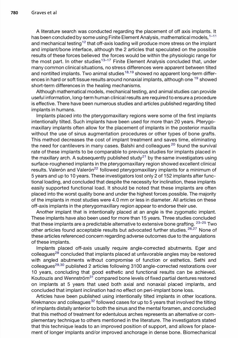
A 59-year old man presented with severe maxillary atrophy ( Fig. 1 ). He first wore
a denture 25 years prior but reported he had been unable to wear a denture for the
last 10 years ( Fig. 2 ). The patient had been to 7 other surgeons, all of whom statedhe needed major grafting. Evaluation of scan revealed less than 5 mm vertical height
of alveolar bone anterior to the tooth to the sinus ( Fig. 3 ). The patient demonstrated
a markedly pneumatized maxillary sinus bilaterally ( Fig. 4 ). He had a small well-
formed tuberosity approximately 8 mm in vertical height bilaterally. A stereolithic
model was obtained to further evaluate the case ( Fig. 5 ). The rest of the medical
history was within normal limits. The patient stated in no uncertain terms that “he
did not want bone graft.”
In preparation for the procedure, model surgery was performed on the stereolithic
model ( Fig. 6 ). From this surgery it was determined that there was adequate volume
and density of bone in the following areas: anteronasal region that includes the vomerbone, the zygomatic buttress arch, and the posterior tuberosity and pterygoid region.
The sizes of the implants from this model surgery were noted, and angled with specific
abutments were planned to correct the severe angles and specific angled abutments
( Fig. 7 ).
One half hour before surgery the patient was given cephalexin orally with a small sip
of water. The patient was kept on this antibiotic for one week after surgery. The
surgery was done under intravenous sedation. Two parallel incisions approximately
3 mm apart were made from tuberosity to tuberosity on the alveolar crest. With sharp
dissection, taking care not to tear the periosteum, the following structures were
exposed: the anteronares, the zygomatic buttress and arch, the tuberosity, retrotu-berosity, and pterygomaxillary, using protocol for active implants. The anteronasal
region was degloved with sharp dissection. The vomer bone identified and 2 parallel
osteotomies preformed for Nobel Active Implants. Both of these implants measured
3.5 Â 10 mm and were torqued to 25 Ncm. Posterior where bilateral pterygoid
implants were placed, care was taken to make sure the 20 Â 2-mm long drill pierced
the pterygoid buttress and easily went into the fossa. Two 3.5 Â 15-mm NobelActive
implants (Nobel Biocare, Zurich, Switzerland) were placed, both of which achieved
a torque of 20 Ncm. Attention was then turned to the zygomatic area. Using the
Fig. 1. A 59-year-old man had worn dentures for 25 years, leading to advanced maxillarybone resorption.
Graves et al782
5/16/2018 88729490 Maxillary All on Four Therapy Using Angled Implants - slidepdf.com
a lateral slot technique was used in both right and leftmaxillary sinuses and two 42.5-mm TiUnite implants (Nobel Biocare) placed. Cover
screws were placed on all implants, and the area was sutured with 3-0 chromic in
a running suture. Hemostasis was good and the patient’s existing upper denture
was relined.
The patient’s mandible was treated in a conventional fashion for an All-on-Four
prosthesis. Parallel crestal incisions approximately 3 mm apart were used to expose
the superior half of the mandible with sharp bisection. The mental foramina were
exposed, the bone tabled to a flat surface, and 4 active implants placed. The bicuspid
area was 15 mm and the lateral incisor area was 13 mm. All lower implants achieved
torque values of approximately 50 Ncm. The incisions were closed with interrupted 3-0chromic. Angled abutments were placed, 30 posteriorly and 17 anteriorly.
Prosthetic Procedure
Following surgery, an interim maxillary denture was soft-lined and delivered. Next,
abutment level impression copings were placed on the mandibular implants and linked
using pattern resin and wire. An impression was made using an open-tray technique.
While the impression was poured, temporary cylinders were attached to the implants
and the previously fabricated lower interim prosthesis was related to the cylinders
using acrylic resin. Care was taken to maintain the horizontal plane orientation and
maintain the preestablished vertical dimension of occlusion by using a closed-
mouth technique. A laboratory reline procedure was completed for the lower pros-
thesis prior to same-day delivery. At interim prosthesis delivery prosthetic screws
Fig. 5. ( A, B) Stereolithic models demonstrate severe atrophy.
Fig. 6. ( A, B) Implants were placed in the models in a “mock surgery.”
Graves et al784
5/16/2018 88729490 Maxillary All on Four Therapy Using Angled Implants - slidepdf.com
were torqued to 15 Ncm and access holes were sealed with gingival retraction cordand “Fermit.” Group function was verified and the patient was advised to maintain
a soft diet during the integration period.
Six months later, second-stage uncovering of the maxillary implants was performed
in the usual fashion (see Fig. 7 ). The appropriate angled abutments were placed so
that the screw access holes did not reach the facies of any teeth. Approximately 1
month later, impressions were made for both upper and lower definitive prostheses.
The completed full-arch prostheses were supported by a milled titanium framework
with cantilevers to allow for maximum posterior occlusion. After appointments veri-
fying the accuracy of the bar and the occlusion, final restorations were delivered 3
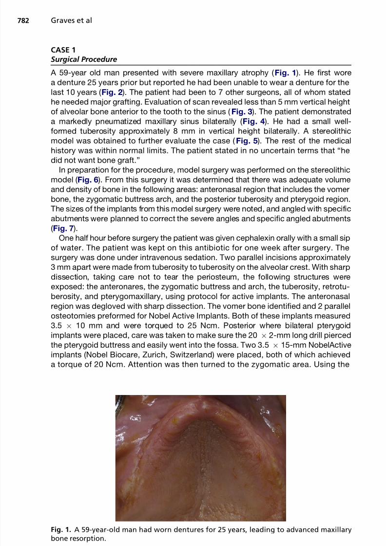
months later ( Figs. 8 and 9 ).
CASE 2
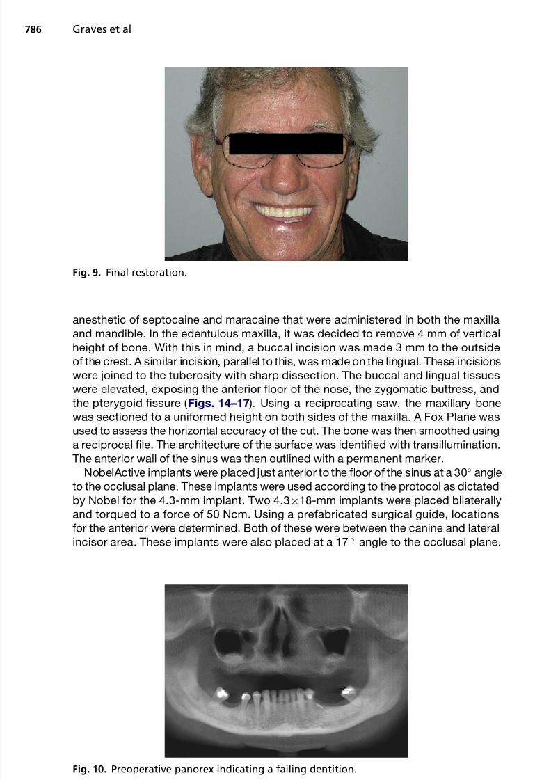
A 42-year old woman had worn dentures for less than 2 years following what she
described as 7 years of aggressive periodontal treatment and subsequent extractions
( Figs. 10 and 11 ). She reported wearing a removable denture that had detrimental
effects on both her social as well as psychological well-being. Upper and lower
implants were planned along with extraction of remaining failing lower teeth ( Figs. 12
and 13 ).
Surgical Procedure
The patient was premedicated with clindamycin, 600 mg, 1 hour before surgery. She
was also given 0.1% chlorhexidine rinse. She was sedated intravenously using a local
Fig. 7. ( A) Zygomatic, vomer, and pterygoid implants were placed as shown on radiograph.(B) The implants were uncovered after 6 months.
Fig. 8. Final restoration.
All-On-Four Maxilla 785
5/16/2018 88729490 Maxillary All on Four Therapy Using Angled Implants - slidepdf.com
anesthetic of septocaine and maracaine that were administered in both the maxilla
and mandible. In the edentulous maxilla, it was decided to remove 4 mm of vertical
height of bone. With this in mind, a buccal incision was made 3 mm to the outside
of the crest. A similar incision, parallel to this, was made on the lingual. These incisions
were joined to the tuberosity with sharp dissection. The buccal and lingual tissues
were elevated, exposing the anterior floor of the nose, the zygomatic buttress, and
the pterygoid fissure ( Figs. 14–17 ). Using a reciprocating saw, the maxillary bonewas sectioned to a uniformed height on both sides of the maxilla. A Fox Plane was
used to assess the horizontal accuracy of the cut. The bone was then smoothed using
a reciprocal file. The architecture of the surface was identified with transillumination.
The anterior wall of the sinus was then outlined with a permanent marker.
NobelActive implants were placed just anterior to the floor of the sinus at a 30 angle
to the occlusal plane. These implants were used according to the protocol as dictated
by Nobel for the 4.3-mm implant. Two 4.3Â18-mm implants were placed bilaterally
and torqued to a force of 50 Ncm. Using a prefabricated surgical guide, locations
for the anterior were determined. Both of these were between the canine and lateral
incisor area. These implants were also placed at a 17
angle to the occlusal plane.
Fig. 9. Final restoration.
Fig. 10. Preoperative panorex indicating a failing dentition.
Graves et al786
5/16/2018 88729490 Maxillary All on Four Therapy Using Angled Implants - slidepdf.com
more bone (6 mm) was tabled on the lower jaw. All of the implants were torqued to 50
Ncm. Abutments were then placed as described for the maxillary arch.
Prosthetic Procedure
Following surgery, abutment level impression copings were placed on the maxillary
implants. Seating of the impression copings was verified by periapical radiographs
( Fig. 18 ). Copings were then linked using pattern resin and wire. An impression was
made using an open-tray technique. While the impression was poured, temporary
cylinders were attached to the implants and the previously fabricated upper and lower
interim prostheses were related to the cylinders using acrylic resin. Care was taken to
maintain the horizontal plane orientation and maintain the preestablished vertical
dimension of occlusion by using a closed-mouth technique. A laboratory reline proce-
dure was completed for both prostheses prior to same-day delivery. At delivery pros-
thetic screws were torqued to 15 Ncm, and access holes were sealed with gingival
retraction cord and Fermit. Anterior occlusion and posterior disclusion were verified,
and the patient was advised to maintain a soft diet during the integration period.
Final impressions for the definitive maxillary and mandibular fixed-hybrid pros-
theses were performed 6 months after surgery ( Fig. 19 ). The completed full-arch pros-
theses were supported by milled titanium frameworks with cantilevers to allow formaximum posterior occlusion.
Fig. 13. Final restoration.
Fig. 14. When there is minimal paranasal bone available for implant placement and sinusanatomy is prominent, sometimes even deflecting anterior of the canine eminence, transsi-nus implants can be considered.
Graves et al788
5/16/2018 88729490 Maxillary All on Four Therapy Using Angled Implants - slidepdf.com
At delivery, occlusion was verified and prosthetic screws were torqued to 15 Ncm.
Access holes were sealed with gingival retraction cord and Fermit. At 3 months after
delivery, after verifying screws were torqued to 15 Ncm, access holes were sealed
with retraction cord and composite.
RESULTS
From July 2009 to November 2010, 276 patients received implant treatment involving
angled implants in the maxilla. Two hundred and sixty-seven patients received fixed
interim prostheses on the day of surgery. Forty-five patients received final prostheses.
The patient population included dentate as well as edentulous individuals. Age,
smoking history, or systemic disease controlled by medication was not a criterion
for discussion. Failure is defined as inability to withstand 35 Ncm of torque 6 months
postoperatively. In all, 1110 implants were placed, with 28 failures and a success rate
of 97.48%.
Anteroposterior spread was measured in all cases. Distance was measured
between a line through the center of the frontmost implants and a line through theposterior implants. The two sides were averaged. The average distance was 15.9
mm. Of note, on mandibular arches in the same patient population this measurement
was less, at 15.25 mm.
Fig. 15. Following sinus membrane elevation, the distal implant is placed transsinus toengage “M” point.
Fig. 16. The exposed implant, including the sinus floor, is then bone grafted using BMP-2.
All-On-Four Maxilla 789
5/16/2018 88729490 Maxillary All on Four Therapy Using Angled Implants - slidepdf.com
3. Elimination or shortening of cantilevers4. Avoidance of various anatomic structures
5. Fewer implants to support the prosthesis.
The placement of angled implants has numerous benefits to patients. The place-
ment of these implants into the patient’s available bone is usually easier for the
surgical dentist than additional grafting procedures.
One possible disadvantage of the tilted placement of conventional dental implants is
that they usually become more difficult to restore, which requires angle-correcting
abutments. These abutments are available in different angles from most implant
manufacturers. Care must be taken to create enough vertical space for the interme-
diate abutment.
REFERENCES
1. Sutpideler M, Eckert SE, Zobitz M, et al. Finite element analysis of effect of pros-
thesis height, angle of force application, and implant offset on supporting bone.
Int J Oral Maxillofac Implants 2004;19(6):819–25.
2. Saab XE, Griggs JA, Powers JM, et al. Effect of abutment angulation on the strain
on the bone around an implant in the anterior maxilla: a finite element study.
J Prosthet Dent 2007;97(2):85–92.
3. Cehreli MC, Iplikcioglu H, Bilir OG. The influence of the location of load transferon strains around implants supporting four unit cement-retained fixed pros-
theses: in vitro evaluation of axial versus non-axial loading. J Oral Rehabil
2002;29(4):394–400.
4. Brosh T, Pilo R, Sudai D. The influence of abutment angulation on strains and
stresses along the implant/bone interface: comparison between 2 experimental
techniques. J Prosthet Dent 1998;79:328–34.
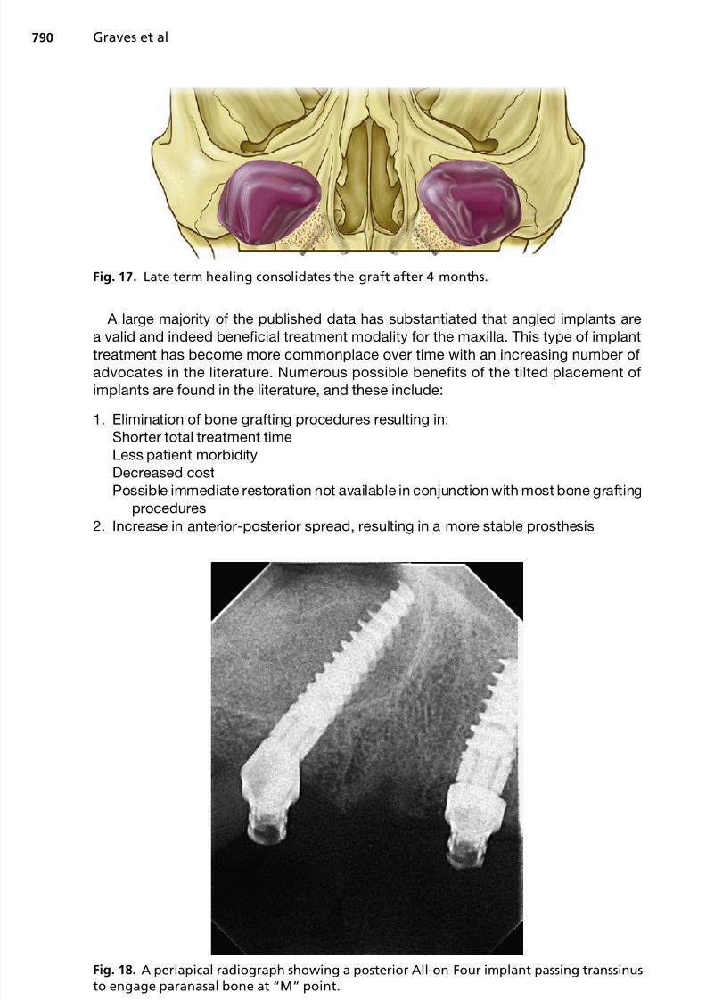
Fig. 19. After 6 months of healing, the implant appears well integrated into consolidatedsinus graft, the tip of the implant engaging into the lateral nasal wall.
All-On-Four Maxilla 791
5/16/2018 88729490 Maxillary All on Four Therapy Using Angled Implants - slidepdf.com
7. O’Mahony A, Bowles Q, Woolsey G, et al. Stress distribution in the single-unit os-
seointegrated dental implant: finite element analyses of axial and off-axial
loading. Implant Dent 2000;9(3):207–18.
8. Hsu ML, Chen FC, Kao HC, et al. Influence of off-axis loading of an anterior maxil-
lary implant: a 3-dimensional finite element analysis. Int J Oral Maxillofac Implants
2007;22(2):301–9.
9. Zampelis A, Rangert B, Heijl L. Tilting of splinted implants for improved prostho-
dontic support: a two-dimensional finite element analysis. J Prosthet Dent 2007;
97(Suppl 6):S35–43.
10. Lin CL, Wang JC, Ramp LC, et al. Biomechanical response of implant systemsplaced in the maxillary posterior region under various conditions of angulation,
bone density, and loading. Int J Oral Maxillofac Implants 2008;23(1):57–64.
11. Kao HC, Gung YW, Chung TF, et al. The influence of abutment angulation on mi-
cromotion level for immediately loaded dental implants: a 3-D finite element anal-
ysis. Int J Oral Maxillofac Implants 2008;23(4):623–30.
12. Clelland NL, Gilat A, McGlumphy EA, et al. A photoelastic and strain gauge anal-
ysis of angled abutments for an implant system. Int J Oral Maxillofac Implants
1993;8(5):541–8.
13. Cruz M, Wassall T, Toledo EM, et al. Finite element stress analysis of dental pros-
theses supported by straight and angled implants. Int J Oral Maxillofac Implants2009;24(3):391–403.
14. Bellini CM, Romeo D, Galbusera F, et al. A finite element analysis of tilted versus
non-tilted implant configurations in the edentulous maxilla. Int J Prosthodont
2009;22(2):155–7.
15. Las Casas EB, Ferreira PC, Cimini CA Jr, et al. Comparative 3D finite element
stress analysis of straight and angled wedge-shaped implant designs. Int J
Oral Maxillofac Implants 2008;23(2):215–25.
16. Markarian RA, Ueda C, Sendyk CL, et al. Stress distribution after installation of
fixed frameworks with marginal gaps over angled and parallel implants: a photoe-
lastic analysis. J Prosthodont 2007;16(2):117–22.17. Bellini CM, Romeo D, Galbusera F, et al. Comparison of tilted versus nontilted
implant-supported prosthetic designs for the restoration of the edentulous mandi-
ble: a biomechanical study. Int J Oral Maxillofac Implants 2009;24(3):511–7.
18. Celletti R, Pameijer CH, Bracchetti G, et al. Histologic evaluation of osseointe-
grated implants restored in nonaxial functional occlusion with preangled abut-
ments. Int J Periodontics Restorative Dent 1995;15(6):562–73.
19. Barbier L, Schepers E. Adaptive bone remodeling around oral implants under
axial and nonaxial loading conditions in the dog mandible. Int J Oral Maxillofac
Implants 1997;12(2):215–23.
20. Balshi SF, Wolfinger GJ, Balshi TJ. Analysis of 356 pterygomaxillary implants inedentulous arches for fixed prosthesis anchorage. Int J Oral Maxillofac Implants
1999;14(3):398–406.
21. Balshi SF, Wolfinger GJ, Balshi TJ. Analysis of 164 titanium oxide-surface
implants in completely edentulous arches for fixed prosthesis anchorage using
the pterygomaxillary region. Int J Oral Maxillofac Implants 2005;20(6):946–52.
Graves et al792
5/16/2018 88729490 Maxillary All on Four Therapy Using Angled Implants - slidepdf.com
22. Valeron JF, Valeron PF. Long-term results in placement of screw-type implants in
the pterygomaxillary-pyramidal region. Int J Oral Maxillofac Implants 2007;22(2):
195–200.
23. Ahlgren F, Størksen K, Tornes K. A study of 25 zygomatic dental implants with 11
to 49 months’ follow-up after loading. Int J Oral Maxillofac Implants 2006;21(3):
421–5.
24. Aparicio C, Ouazzani W, Garcia R, et al. A prospective clinical study on titanium
implants in the zygomatic arch for prosthetic rehabilitation of the atrophic eden-
tulous maxilla with a follow-up of 6 months to 5 years. Clin Implant Dent Relat Res
2006;8(3):114–22.
25. Bedrossian E, Rangert B, Stumpel L, et al. Immediate function with the zygomatic
implant: a graftless solution for the patient with mild to advanced atrophy of the
maxilla. Int J Oral Maxillofac Implants 2006;21(6):937–42.
26. Becktor JP, Isaksson S, Abrahamsson P, et al. Evaluation of 31 zygomatic
implants and 74 regular dental implants used in 16 patients for prosthetic recon-struction of the atrophic maxilla with cross-arch fixed bridges. Clin Implant Dent
Relat Res 2005;7(3):159–65.
27. Farzad P, Andersson L, Gunnarsson S, et al. Rehabilitation of severely resorbed
maxillae with zygomatic implants: an evaluation of implant stability, tissue condi-
tions, and patients’ opinion before and after treatment. Int J Oral Maxillofac
Implants 2006;21(3):399–404.
28. Eger DE, Gunsolley JC, Feldman S. Comparison of angled and standard abut-
ments and their effect on clinical outcomes: a preliminary report. Int J Oral Max-
illofac Implants 2000;15(6):819–23.
29. Sethi A, Kaus T, Sochor P. The use of angulated abutments in implant dentistry:five-year clinical results of an ongoing prospective study. Int J Oral Maxillofac
Implants 2000;15(6):801–10.
30. Sethi A, Kaus T, Sochor P, et al. Evolution of the concept of angulated abutments
in implant dentistry: 14-year clinical data. Implant Dent 2002;11(1):41–51.
31. Koutouzis T, Wennstrom JL. Bone level changes at axial- and non-axial-
positioned implants supporting fixed partial dentures. A 5-year retrospective
longitudinal study. Clin Oral Implants Res 2007;18(5):585–90.
32. Krekmanov L, Kahn M, Rangert B, et al. Tilting of posterior mandibular and maxil-
lary implants for improved prosthesis support. Int J Oral Maxillofac Implants
2000;15(3):405–14.33. Malo P, Nobre Mde A, Petersson U, et al. A pilot study of complete edentulous
rehabilitation with immediate function using a new implant design: case series.
Clin Implant Dent Relat Res 2006;8(4):223–32.
34. Francetti L, Agliardi E, Testori T, et al. Immediate rehabilitation of the mandible
with fixed full prosthesis supported by axial and tilted implants: interim results
of a single cohort prospective study. Clin Implant Dent Relat Res 2008;10(4):
255–63.
35. Testori T, Del Fabbro M, Capelli M, et al. Immediate occlusal loading and tilted
implants for the rehabilitation of the atrophic edentulous maxilla: 1-year interim
results of a multicenter prospective study. Clin Oral Implants Res 2008;19(3):227–32.
36. Capelli M, Zuffettii F, Del Fabbro M, et al. Immediate rehabilitation of the
completely edentulous jaw with fixed prostheses supported by either upright or
tilted implants: a multicenter clinical study. Int J Oral Maxillofac Implants 2007;
22(4):639–44.
All-On-Four Maxilla 793
5/16/2018 88729490 Maxillary All on Four Therapy Using Angled Implants - slidepdf.com