NOVEL ADENOVIRUS DETECTED IN KOWARI(DASYUROIDES BYRNEI) WITH PNEUMONIA
JÁNOS GÁL1, MÍRA MÁNDOKI
2, ENDRE SÓS3, PÉTER KERTÉSZ3, VIKTÓRIA
KOROKNAI3, KRISZTIÁN BÁNYAI
4 and SZILVIA L. FARKAS4*1Department of Exotic Animal and Wildlife Medicine, University of Veterinary Science,
Budapest, Hungary2Department of Pathology, University of Veterinary Science, Budapest, Hungary
3Budapest Zoo and Botanical Garden, Budapest, Hungary4Lendület ‘Pathogen Discovery’ Research Group, Institute for Veterinary MedicalResearch, Centre for Agricultural Research, Hungarian Academy of Sciences,
Budapest, Hungary
(Received: 8 October 2015; accepted: 16 July 2016)
A male kowari (Dasyuroides byrnei) originating from a zoo facility wasdelivered for post mortem evaluation in Hungary. Acute lobar pneumonia withhistopathologic changes resembling an adenovirus (AdV) infection was detected bylight microscopic examination. The presence of an AdV was confirmed by obtainingpartial sequence data from the adenoviral DNA-dependent DNA-polymerase.Although the exact taxonomic position of this novel marsupial origin virus couldnot be determined, pairwise identity analyses and phylogenetic calculations revealedthat it is distantly related to other members in the family Adenoviridae.
Keywords: Kowari adenovirus, brush-tailed marsupial rat, Dasyuroidesbyrnei, pneumonia
Introduction
Adenoviruses (AdVs) are frequently detected double-stranded DNA virusesof vertebrates (mammals, birds, squamata, chelonians, amphibians, and fish)usually exhibiting a narrow host-range and co-evolution with their host [1, 2].AdV infections can be asymptomatic or associated with different clinical man-ifestations including conjunctivitis, respiratory illness, gastroenteritis, cystitis,meningoencephalitis, hepatitis, or even mortality. In most cases, the outcome ofthe infection is substantially influenced by the actual immune status of the host.
Acta Microbiologica et Immunologica Hungarica 64 (1), pp. 81–90 (2017)DOI: 10.1556/030.63.2016.024
Basophilic or eosinophilic nuclear inclusions in the affected susceptible cells arecharacteristic to AdV infections.
The family Adenoviridae is currently divided into five genera: Mastadeno-,Aviadeno-, Atadeno-, Siadeno-, and Ichtadenovirus (International Committee onTaxonomy of Viruses; http://www.ictvonline.org/). A sixth AdV lineage wasdescribed recently in testudinoid turtles in Hungary and USA [3, 4]. Althoughseveral AdVs have been identified in different mammalian species belonging tothe infraclass Placentalia, there is only a single known marsupial origin AdV, thePossum AdV 1, which was described in brushtail possums (Trichosurus vulpe-cula, order Diprotodontia) from New Zealand [5]. Partial sequence data, high A+Tcontent of the determined sequences and phylogenetic calculations clustered thePossum AdV 1 into the genus Atadenovirus.
The kowari or brush-tailed marsupial rat (Dasyuroides byrnei) is a small,14–18 cm long nocturnal carnivorous marsupial native to the region of Lake Eyrein Australia. The kowari belongs to a different order in the infraclass Marsupialia,the Dasyuromorphia. Its diet consists of insects, lizards, rodents, birds, and eggs.There is limited information found in the literature about viral infections diagnosedin this species. Cytomegalic disease was described in several marsupial speciesincluding the kowari; historically, the disease was thought to be caused by acytomegalovirus, but in a recent study gammaherpesviruses were detected inconnection with the disorder [6]. The present work describes the detection, andpreliminary genetic characterization of a distinct, novel AdV in a specimen ofkowari as possible cause of pneumonia.
Pathological and Histological Investigations
On January 10, 2014, a succumbed male kowari (D. byrnei) was delivered toUniversity of Veterinary Science, Department of Exotic Animal and WildlifeMedicine for post mortem evaluation. The kowari originated from a zoo facility inBudapest (Hungary). Routine pathological dissection was carried out with mac-roscopic examination. From organs showing visible pathological lesions (lung,liver, and kidneys), samples were taken for further histopathological, virological,and routine bacteriological investigations. Specimens intended for histopatholog-ical examination were fixed in 8% buffered formaldehyde solution and embeddedin paraffin. Sections (4–5 μm thick) were stained with hematoxylin and eosinaccording to the standard histopathological procedure. Routine bacteriologicalexamination from the organ samples was carried out on blood and Drigalski agarplates; cultures were incubated at 37 °C.
82 GÁL ET AL.
Acta Microbiologica et Immunologica Hungarica 64, 2017
The necropsy performed on the carcass revealed normal fur and skin,without any changes in the natural orifices. The subcutis was intact with mediumamount of subcutaneous fat tissue, indicating normal nutritional status of thekowari. Upon opening the abdominal cavity, the organs were located according toanatomical expectations. The spleen had normal shape and size, and was lightbrownish-red in color. The slightly enlarged liver was normally shaped, butyellowish and moderately easy to tear. The cut surface of the liver had a fattyshine. Neither the gastrointestinal tract nor the kidneys showed any pathologicalalterations. The position of the thoracic organs was normal, but the thoracic cavitycontained 3 ml transparent thin fluid, the pleura visceralis was edematous(Figure 1). The lungs, in general, were normally shaped and sized, but the leftcaudal and the right middle lobes were enlarged. Most of the lung tissue appearedin the normal pale brick-red color, except for the enlarged lobes, which weremottled with darker red areas. The altered lobes had higher liquid content andcontained less air bubbles. Although the heart was rounded, the myocardium andthe chambers did not show any pathological lesions.
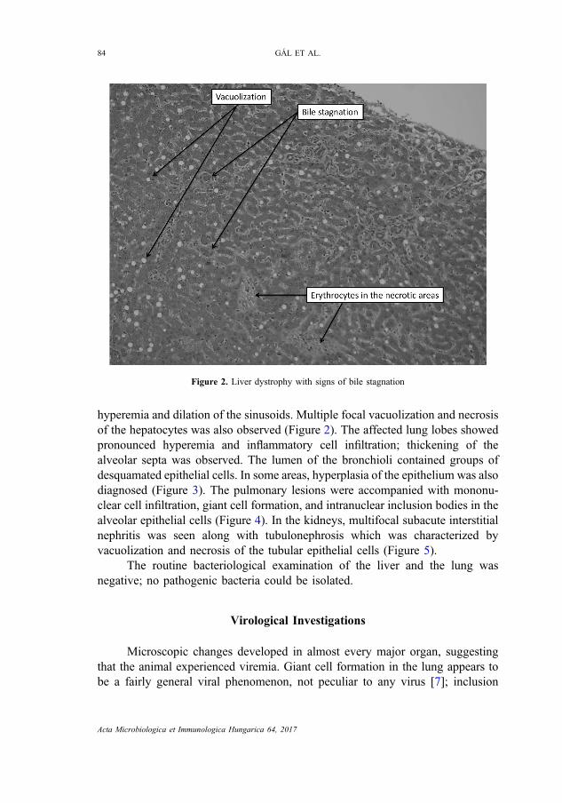
Histological examination of the liver revealed bile stagnation indicated bydeposition of a homogenous brownish-red material in the capillaries, and
Figure 1. Fluid accumulation (hydrothorax) in the thoracic cavity of the kowari(Dasyuroides byrnei)
ADENOVIRUS INFECTION AND PNEUMONIA IN KOWARI 83
Acta Microbiologica et Immunologica Hungarica 64, 2017
hyperemia and dilation of the sinusoids. Multiple focal vacuolization and necrosisof the hepatocytes was also observed (Figure 2). The affected lung lobes showedpronounced hyperemia and inflammatory cell infiltration; thickening of thealveolar septa was observed. The lumen of the bronchioli contained groups ofdesquamated epithelial cells. In some areas, hyperplasia of the epithelium was alsodiagnosed (Figure 3). The pulmonary lesions were accompanied with mononu-clear cell infiltration, giant cell formation, and intranuclear inclusion bodies in thealveolar epithelial cells (Figure 4). In the kidneys, multifocal subacute interstitialnephritis was seen along with tubulonephrosis which was characterized byvacuolization and necrosis of the tubular epithelial cells (Figure 5).
The routine bacteriological examination of the liver and the lung wasnegative; no pathogenic bacteria could be isolated.
Virological Investigations
Microscopic changes developed in almost every major organ, suggestingthat the animal experienced viremia. Giant cell formation in the lung appears tobe a fairly general viral phenomenon, not peculiar to any virus [7]; inclusion
Figure 2. Liver dystrophy with signs of bile stagnation
84 GÁL ET AL.
Acta Microbiologica et Immunologica Hungarica 64, 2017
Figure 3. Hyperemia in the lung and desquamation of the epithelial cells in the airways
Figure 4.Giant cell formation, mononuclear inflammatory cell infiltration, and anisokaryosis in the lung
ADENOVIRUS INFECTION AND PNEUMONIA IN KOWARI 85
Acta Microbiologica et Immunologica Hungarica 64, 2017
bodies are characteristic to AdV infections, therefore AdV-specific nested PCRtargeting the DNA-dependent DNA-polymerase gene was performed as de-scribed previously [8]. To exclude herpes-, circo-, and reovirus infections,consensus PCR assays, routinely used in our laboratory, were performed[9–11]. PCR products of the expected size were excised and then extractedfrom gel for direct sequencing. BLASTn and BLASTx algorithms were used toidentify homologous genes among sequences deposited in GenBank [12]. AdVsequences used in our study were retrieved from GenBank. Multiple sequencealignments were generated using the Multalin online platform (http://multalin.toulouse.inra.fr/multalin/) and sequences were edited in GeneDoc [13]. Phylo-genetic analysis was performed using the MEGA version 6.0. program package[14]. Gene-specific substitution models were evaluated, and the best-fit modelwas selected based on the Bayesian information criterion. A maximum likeli-hood tree was generated, and tree topology was validated by bootstrap analysisas implemented in MEGA6. Pairwise identity comparison based on alignedamino acid (aa) sequences was also calculated in MEGA6 (p-distance, uniformrates, and pairwise deletion).
Figure 5. Tubulonephrosis and multifocal interstitial nephritis
86 GÁL ET AL.
Acta Microbiologica et Immunologica Hungarica 64, 2017
Acta Microbiologica et Immunologica Hungarica 64, 2017
Nucleic acid samples isolated from pooled organ samples (lung, liver, andkidney) were used for the PCR-based detection of numerous pathogens, but only theAdV-specific PCR gave positive result. The sequence data was submitted toGenBank (accession no.: KT696557). The G+C content of the Kowari AdVsequence was relatively high, 65%, but fell in the range observed in other adenoviral
Figure 6.Unrooted phylogenetic tree of partial adenoviral DNA-dependent DNA-polymerase aminoacid sequences showing the clustering of adenoviruses. The length of the branches indicates thephylogenetic distance between the different viruses and the scale bar represents 50 mutations per 100
sequence positions. Bootstrap values are given for 100 data sets
88 GÁL ET AL.
Acta Microbiologica et Immunologica Hungarica 64, 2017
sequences (33.6%–66.9%). BLASTx analysis of the partial, 90 aa long sequence ofthe Kowari AdV revealed that the highest scores were obtained when comparedwith the Bearded dragon AdV 1 (GenBank accession no.: AAS89694) and Centralnetted dragon AdV 1 (GenBank accession no.: AEG65825). Amino acid sequenceidentity values between the Kowari AdV and the studied sequences ranged between41.1% and 61% (Yellow-bellied slider AdV and Porcine AdV 3, GenBankaccession no.: AGB07604 and AC_000189, respectively). Of interest, the ob-served aa sequence identity values along this short genomic region fell, virtually,in the range distinguishing established AdV genera (Table I). Although in thephylogenetic calculations, the Kowari AdV did not cluster with viruses belongingto established AdV genera and appeared on a separate branch, conclusions aboutthe taxonomic position of this novel AdV cannot be drawn based on the shortDNA-polymerase gene sequence (Figure 6). Unfortunately no overlappingsequences with Possum AdV 1 were available in GenBank for comparison, butour data suggests that there is no close genetic relationship between the twomarsupial origin AdVs. Additional sequencing of genes reflecting more preciselythe phylogenetic relationships of AdVs is needed in order to classify this novelvirus.
Conclusions
The pathological and the virological examinations of the kowari’s carcass(D. byrnei) revealed acute lobar pneumonia associated with an AdV infection,pronounced hydrothorax, liver dystrophy with bile stagnation, and subacutemultifocal nephritis and tubulonephrosis. We assume that the direct cause of deathof the animal might have been the respiratory failure and consequential hypoxemiaof the parenchymal organs. The kowari was kept in a cage with a companionanimal, which was supposedly infected by the same virus, but remained healthy.Therefore, it can be presumed that a possible immunocompromised state orunknown stress factors might have exacerbated the AdV infection in the animal,which could lead to viremia and the consequential death of the animal. Althoughthe exact pathogenic role of the novel Kowari AdV strain has not been examined,the pronounced genetic distance of the detected virus from other known AdVsjustifies the further investigations of AdV diversity and pathogenicity in marsupials.
Acknowledgement
This work was supported by the Momentum Program of the HungarianAcademy of Sciences and KUK-2014.
ADENOVIRUS INFECTION AND PNEUMONIA IN KOWARI 89
Acta Microbiologica et Immunologica Hungarica 64, 2017
Conflict of Interest
The authors declare that they have no conflict of interest.
References
1. Benkő, M., Doszpoly, A.: Ichtadenovirus. Adenoviridae. In Tidona, C. A., Darai, G. (eds):The Springer Index of Viruses. Springer, New York, NY, USA, 2011, pp. 29–32.
2. Harrach, B., Benkő, M., Both, G. W., Brown, M., Davison, A. J., Echavarria, M., Hess, M.,Jones, M. S., Kajon, A., Lehmkuhl, H. D., Mautner, V., Mittal, S. K., Wadell, G.: FamilyAdenoviridae. In King, A. M. Q., Adams, M. J., Carstens, E. B., Lefkowitz, E. J. (eds): VirusTaxonomy: Classification and Nomenclature of Viruses: Ninth Report of the InternationalCommittee on Taxonomy of Viruses. Elsevier, San Diego, CA, 2012, pp. 125–141.
3. Doszpoly, A., Wellehan, J. F., Jr., Childress, A. L., Tarján, Z. L., Kovács, E. R., Harrach,B., Benkő, M.: Partial characterization of a new adenovirus lineage discovered intestudinoid turtles. Infect Genet Evol 17, 106–112 (2013).
4. Farkas, S. L., Gál, J.: Adenovirus and mycoplasma infection in an ornate box turtle(Terrapene ornata ornata) in Hungary. Vet Microbiol 138, 169–173 (2009).
5. Thomson, D., Meers, J., Harrach, B.: Molecular confirmation of an adenovirus in brushtailpossums (Trichosurus vulpecula). Virus Res 83, 189–195 (2002).
6. Amery-Gale, J., Vaz, P. K., Whiteley, P., Tatarczuch, L., Taggart, D. A., Charles, J. A.,Schultz, D., Ficorilli, N. P., Devlin, J. M., Wilks, C. R.: Detection and identification of agammaherpesvirus in Antechinus spp. in Australia. J Wildl Dis 50, 334–339 (2014).
7. Adams, J. M., Imagawa, D. T., Yoshimori, M., Huntington, R. W.: Huntington giant cellpneumonia. Pediatrics 18, 888–898 (1956).
8. Wellehan, J. F., Johnson, A. J., Harrach, B., Benkő, M., Pessier, A. P., Johnson, C. M.,Garner, M. M., Childress, A., Jacobson, E. R.: Detection and analysis of six lizardadenoviruses by consensus primer PCR provides further evidence of a reptilian originfor the atadenoviruses. J Virol 78, 13366–13369 (2004).
9. VanDevanter, D. R., Warrener, P., Bennett, L., Schultz, E. R., Coulter, S., Garber, R. L.,Rose, T. M.: Detection and analysis of diverse herpesviral species by consensus primerPCR. J Clin Microbiol 34, 1666–1671 (1996).
10. Wellehan, J. F., Jr., Childress, A. L., Marschang, R. E., Johnson, A. J., Lamirande, E. W.,Roberts, J. F., Vickers, M. L., Gaskin, J. M., Jacobson, E. R.: Consensus nested PCRamplification and sequencing of diverse reptilian, avian, and mammalian orthoreoviruses.Vet Microbiol 133, 34–42 (2009).
11. Halami, M. Y., Nieper, H., Müller, H., Johne, R.: Detection of a novel circovirus in muteswans (Cygnus olor) by using nested broad-spectrum PCR. Virus Res 132, 208–212 (2008).
12. Altschul, S. F., Gish, W., Miller, W., Myers, E. W., Lipman, D. J.: Basic local alignmentsearch tool. J Mol Biol 215, 403–410 (1990).
13. Nicholas, K. B., Nicholas, H. B., Jr., Deerfield, D. W., II: GeneDoc: Analysis andvisualization of genetic variation. EMBNEW News 4, 14 (1997).
14. Tamura, K., Stecher, G., Peterson, D., Filipski, A., Kumar, S.: MEGA6: Molecularevolutionary genetics analysis version 6.0. Mol Biol Evol 30, 2725–2729 (2013).
90 GÁL ET AL.
Acta Microbiologica et Immunologica Hungarica 64, 2017


![2323434 Ethics Volume 90 Issue 4 1980 [Doi 10.2307%2F2380448] Richard J. Arneson -- Mill Versus Paternalism](https://static.documents.pub/doc/80x56/55cf8f88550346703b9d3305/2323434-ethics-volume-90-issue-4-1980-doi-1023072f2380448-richard-j-arneson.jpg)


