Page 1
9/30/2014
1
Copyright © The McGraw-Hill Companies, Inc. Permission required for reproduction or display.
Chapter 10Functional
Organization of
Nervous Tissue
Neuron Network
Functions of the Nervous System
• master controlling & communicating system of body
• Functions
1. Sensory input: detects external & internal stimuli
2. Integration: processes & responds to sensory input
3. Control of Muscles and Glands
4. Homeostasis maintained by regulating other systems
5. Center for Mental Activities
Fig. 10.1
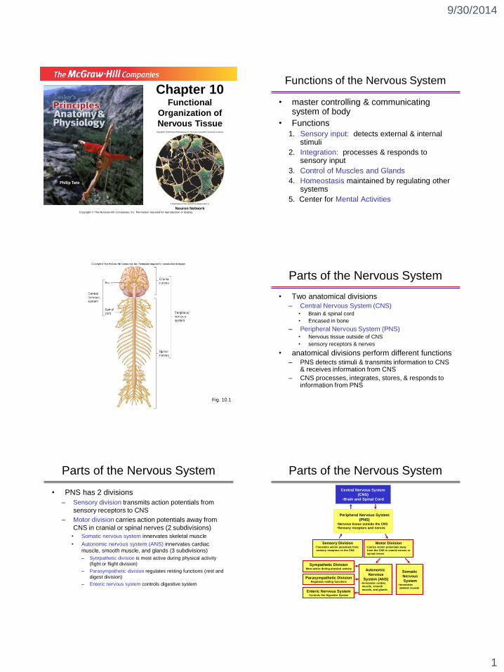
Parts of the Nervous System
• Two anatomical divisions
– Central Nervous System (CNS)• Brain & spinal cord
• Encased in bone
– Peripheral Nervous System (PNS)• Nervous tissue outside of CNS
• sensory receptors & nerves
• anatomical divisions perform different functions– PNS detects stimuli & transmits information to CNS
& receives information from CNS
– CNS processes, integrates, stores, & responds to information from PNS
Parts of the Nervous System
• PNS has 2 divisions
– Sensory division transmits action potentials from
sensory receptors to CNS
– Motor division carries action potentials away from
CNS in cranial or spinal nerves (2 subdivisions)
• Somatic nervous system innervates skeletal muscle
• Autonomic nervous system (ANS) innervates cardiac
muscle, smooth muscle, and glands (3 subdivisions)
– Sympathetic division is most active during physical activity
(fight or flight division)
– Parasympathetic division regulates resting functions (rest and
digest division)
– Enteric nervous system controls digestive system
Central Nervous System
(CNS)
•Brain and Spinal Cord
Peripheral Nervous System
(PNS)•Nervous tissue outside the CNS
•Sensory receptors and nerves
Sensory Division•Transmits action potentials from
sensory receptors to the CNS
Motor Division•Carries action potentials away
from the CNS in cranial nerves or
spinal nerves
Sympathetic DivisionMost active during physical activity
Somatic
Nervous
System• Innervates
skeletal muscle
Autonomic
Nervous
System (ANS)• Innervates cardiac
muscle, smooth
muscle, and glands
Parasympathetic DivisionRegulates resting functions
Enteric Nervous SystemControls the Digestive System
Parts of the Nervous System
Page 2
9/30/2014
2
Fig. 10.2
Cells of the Nervous System
• 2 principal cell types of nervous system
– Neurons: excitable cells that transmit
electrical signals
– Non-neural cells (Glial cells): cells that
surround neurons. >1/2 of brain’s weight
• < 20% is extracellular space
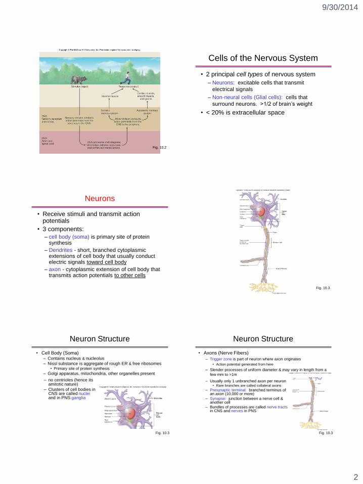
Neurons
• Receive stimuli and transmit action potentials
• 3 components:
– cell body (soma) is primary site of protein synthesis
– Dendrites - short, branched cytoplasmic extensions of cell body that usually conduct electric signals toward cell body
– axon - cytoplasmic extension of cell body that transmits action potentials to other cells
Fig. 10.3
Neuron Structure
• Cell Body (Soma)– Contains nucleus & nucleolus
– Nissl substance is aggregate of rough ER & free ribosomes• Primary site of protein synthesis
– Golgi apparatus, mitochondria, other organelles present
– no centrioles (hence its amitotic nature)
– Clusters of cell bodies in CNS are called nucleiand in PNS ganglia
Fig. 10.3
Neuron Structure
– Usually only 1 unbranched axon per neuron• Rare branches are called collateral axons
– Presynaptic terminal: branched terminus of an axon (10,000 or more)
– Synapse: junction between a nerve cell & another cell
– Bundles of processes are called nerve tractsin CNS and nerves in PNS
Fig. 10.3
• Axons (Nerve Fibers)
– Trigger zone is part of neuron where axon originates
• Action potential generated from here
– Slender processes of uniform diameter & may vary in length from a
few mm to >1m
Page 3
9/30/2014
3
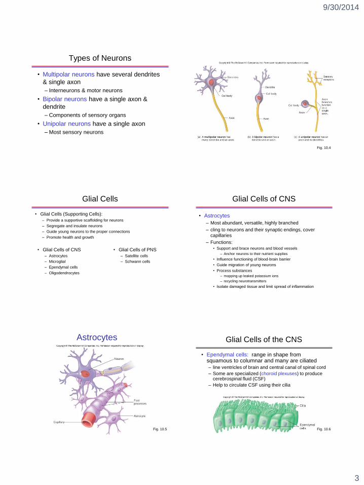
Types of Neurons
• Multipolar neurons have several dendrites
& single axon
– Interneurons & motor neurons
• Bipolar neurons have a single axon &
dendrite
– Components of sensory organs
• Unipolar neurons have a single axon
– Most sensory neurons
Fig. 10.4
Glial Cells
• Glial Cells of CNS
– Astrocytes
– Microglial
– Ependymal cells
– Oligodendrocytes
• Glial Cells of PNS
– Satellite cells
– Schwann cells
• Glial Cells (Supporting Cells):
– Provide a supportive scaffolding for neurons
– Segregate and insulate neurons
– Guide young neurons to the proper connections
– Promote health and growth
Glial Cells of CNS
• Astrocytes
– Most abundant, versatile, highly branched
– cling to neurons and their synaptic endings, cover
capillaries
– Functions:
• Support and brace neurons and blood vessels
– Anchor neurons to their nutrient supplies
• Influence functioning of blood-brain barrier
• Guide migration of young neurons
• Process substances
– mopping up leaked potassium ions
– recycling neurotransmitters
• Isolate damaged tissue and limit spread of inflammation
Fig. 10.5
Astrocytes Glial Cells of the CNS
• Ependymal cells: range in shape from squamous to columnar and many are ciliated
– line ventricles of brain and central canal of spinal cord
– Some are specialized (choroid plexuses) to produce cerebrospinal fluid (CSF)
– Help to circulate CSF using their cilia
Fig. 10.6
Page 4
9/30/2014
4
Glial Cells of the CNS
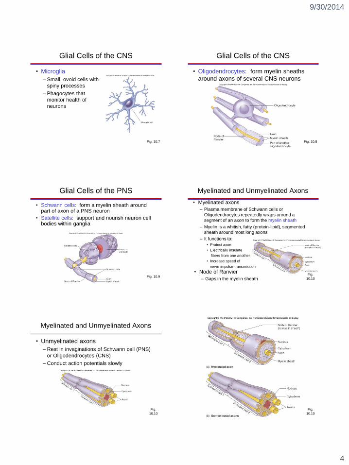
• Microglia
– Small, ovoid cells with
spiny processes
– Phagocytes that
monitor health of
neurons
Fig. 10.7
Glial Cells of the CNS
• Oligodendrocytes: form myelin sheaths
around axons of several CNS neurons
Fig. 10.8
Glial Cells of the PNS
• Schwann cells: form a myelin sheath around part of axon of a PNS neuron
• Satellite cells: support and nourish neuron cell bodies within ganglia
Fig. 10.9
Myelinated and Unmyelinated Axons
• Myelinated axons
– Plasma membrane of Schwann cells or
Oligodendrocytes repeatedly wraps around a
segment of an axon to form the myelin sheath
– Myelin is a whitish, fatty (protein-lipid), segmented
sheath around most long axons
– It functions to:
• Protect axon
• Electrically insulate
fibers from one another
• Increase speed of
nerve impulse transmission
• Node of Ranvier
– Gaps in the myelin sheathFig.
10.10
Myelinated and Unmyelinated Axons
• Unmyelinated axons
– Rest in invaginations of Schwann cell (PNS)
or Oligodendrocytes (CNS)
– Conduct action potentials slowly
Fig.
10.10
Fig.
10.10
Page 5
9/30/2014
5
Organization of Nervous Tissue
• Nervous tissue can be grouped into white matterand gray matter
– White matter • Consists of myelinated axons
• Propagates action potentials
• Forms nerve tracts in CNS and nerves in PNS
– Gray Matter• Collections of neuron cell bodies or unmyelinated axons
• Forms cortex and nuclei in CNS and ganglia in PNS
• Axons synapse with neuron cell bodies, which are functionally site of integration in nervous system
Electric Signals
• Electric signals produced by cells are called action potentials– When action potentials are received from sensory cells it can
result in sensations of sight, hearing, and touch
– Complex mental activities, such as conscious thought, memory, and emotions, result from action potentials
– Contraction of muscles and secretion of certain glands occur in response to action potentials
• Electrical properties of cells result from– Ionic concentration differences across the plasma
membrane
– Permeability characteristics of the plasma membrane
Electric Signals
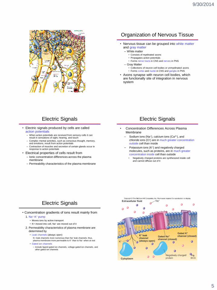
• Concentration Differences Across Plasma
Membrane
– Sodium ions (Na+), calcium ions (Ca2+), and
chloride ions (Cl-) are in much greater concentration
outside cell than inside
– Potassium ions (K+) and negatively charged
molecules, such as proteins, are in much greater
concentration inside cell than outside
• Negatively charged proteins are synthesized inside cell
and cannot diffuse out of it
Electric Signals
• Concentration gradients of ions result mainly from
1. Na+-K+ pump
• Moves ions by active transport
• K+ moved into cell, Na+ are moved out of it
2. Permeability characteristics of plasma membrane are
determined by
• Leak channels (always open)
K+ leak channels more numerous than Na+ leak channels; thus,
plasma membrane more permeable to K+ than to Na+ when at rest
• Gated ion channels
– Include ligand-gated ion channels, voltage-gated ion channels, and
other gated ion channels
Fig.
10.11
Page 6
9/30/2014
6
Electric Signals
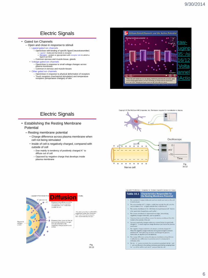
• Gated Ion Channels– Open and close in response to stimuli
• Ligand-gated ion channels– Open/close with binding of specific ligand (neurotransmitter)
» Ligand - molecule that binds to receptor
» Receptor - protein or glycoprotein with receptor site to which a ligand can bind
– Common nervous and muscle tissue, glands
• Voltage-gated ion channels– Open/close in response to small voltage changes across
plasma membrane
– Common in nervous and muscle tissues
• Other gated ion channels– Open/close in response to physical deformation of receptors
– Touch receptors (mechanical stimulation) and temperature receptors (temperature changes) of skin
http://highered.mcgraw-
hill.com/olcweb/cgi/pluginp
op.cgi?it=swf::535::535::/si
tes/dl/free/0072437316/12
0107/anim0013.swf::Volta
ge%20Gated%20Channel
s%20and%20the%20Actio
n%20Potential
Electric Signals
• Establishing the Resting Membrane
Potential
– Resting membrane potential
• Charge difference across plasma membrane when
cell not being stimulated
• Inside of cell is negatively charged, compared with
outside of cell
– Due mainly to tendency of positively charged K+ to
diffuse out of cell
– Opposed by negative charge that develops inside
plasma membrane
Fig.
10.12
Fig.
10.13
Page 7
9/30/2014
7
Electric Signals
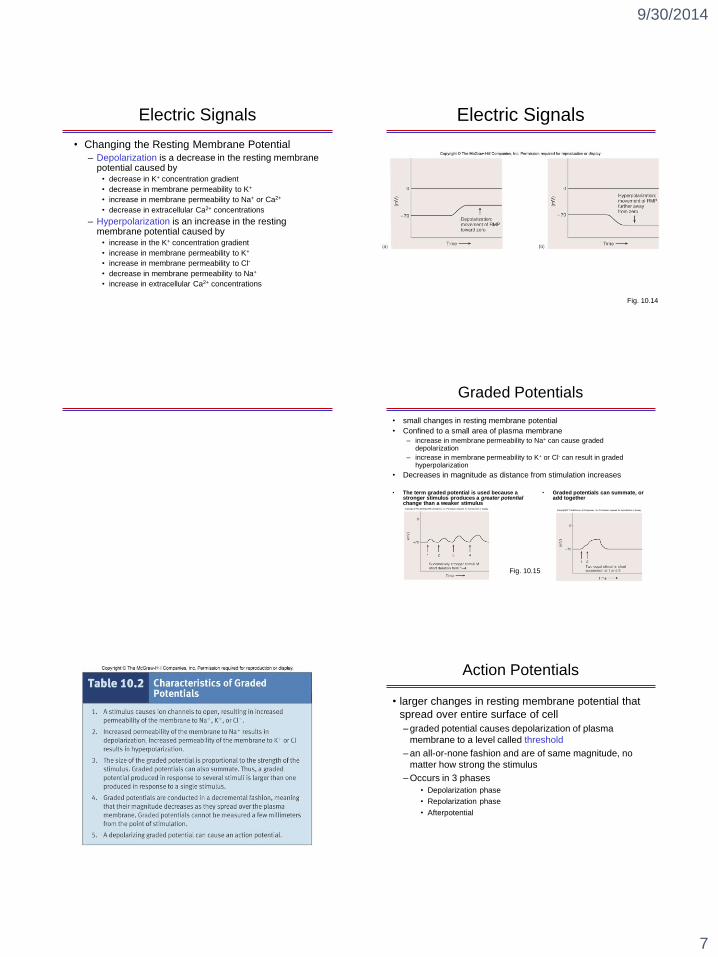
• Changing the Resting Membrane Potential
– Depolarization is a decrease in the resting membrane potential caused by
• decrease in K+ concentration gradient
• decrease in membrane permeability to K+
• increase in membrane permeability to Na+ or Ca2+
• decrease in extracellular Ca2+ concentrations
– Hyperpolarization is an increase in the resting membrane potential caused by
• increase in the K+ concentration gradient
• increase in membrane permeability to K+
• increase in membrane permeability to Cl-
• decrease in membrane permeability to Na+
• increase in extracellular Ca2+ concentrations
Electric Signals
Fig. 10.14
Graded Potentials
• small changes in resting membrane potential
• Confined to a small area of plasma membrane
– increase in membrane permeability to Na+ can cause graded depolarization
– increase in membrane permeability to K+ or Cl- can result in graded hyperpolarization
• Decreases in magnitude as distance from stimulation increases
Fig. 10.15
• The term graded potential is used because a stronger stimulus produces a greater potential change than a weaker stimulus
• Graded potentials can summate, or add together
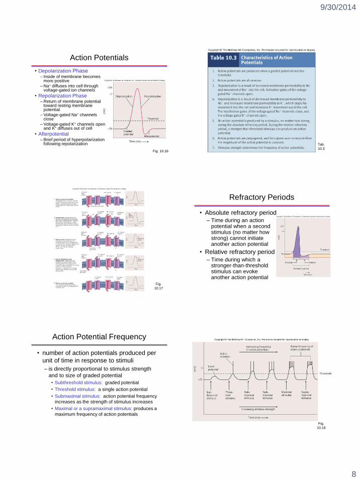
Action Potentials
• larger changes in resting membrane potential that
spread over entire surface of cell
– graded potential causes depolarization of plasma
membrane to a level called threshold
– an all-or-none fashion and are of same magnitude, no
matter how strong the stimulus
– Occurs in 3 phases
• Depolarization phase
• Repolarization phase
• Afterpotential
Page 8
9/30/2014
8
Action Potentials
Fig. 10.16
• Depolarization Phase– Inside of membrane becomes
more positive
– Na+ diffuses into cell through voltage-gated ion channels
• Repolarization Phase– Return of membrane potential
toward resting membrane potential
– Voltage-gated Na+ channels close
– Voltage-gated K+ channels open and K+ diffuses out of cell
• Afterpotential– Brief period of hyperpolarization
following repolarization Tab.
10.3
Fig.
10.17
Refractory Periods
• Absolute refractory period
– Time during an action potential when a second stimulus (no matter how strong) cannot initiate another action potential
• Relative refractory period
– Time during which a stronger-than-threshold stimulus can evoke another action potential
Action Potential Frequency
• number of action potentials produced per
unit of time in response to stimuli
– is directly proportional to stimulus strength
and to size of graded potential
• Subthreshold stimulus: graded potential
• Threshold stimulus: a single action potential
• Submaximal stimulus: action potential frequency
increases as the strength of stimulus increases
• Maximal or a supramaximal stimulus: produces a
maximum frequency of action potentials
Fig.
10.19
Page 9
9/30/2014
9
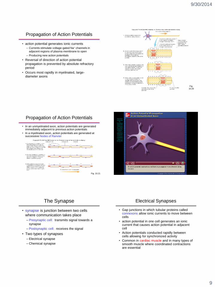
Propagation of Action Potentials
• action potential generates ionic currents
– Currents stimulate voltage-gated Na+ channels in
adjacent regions of plasma membrane to open
– Producing new action potentials
• Reversal of direction of action potential
propagation is prevented by absolute refractory
period
• Occurs most rapidly in myelinated, large-
diameter axons
Fig.
10.20
Propagation of Action Potentials
• In an unmyelinated axon, action potentials are generated immediately adjacent to previous action potentials
• In a myelinated axon, action potentials are generated at successive Nodes of Ranvier
Fig. 10.21
http://highered.m
cgraw-
hill.com/sites/007
2943696/student
_view0/chapter8/
animation__actio
n_potential_prop
agation_in_an_u
nmyelinated_axo
n__quiz_2_.html
http://high
ered.mcgr
aw-hill.com/sit
es/007294
3696/stud
ent_view0/
chapter8/animation_
_action_p
otential_pr
opagation
_in_an_unmyelinated
_axon__q
uiz_2_.htm
l
The Synapse
• synapse is junction between two cells
where communication takes place
– Presynaptic cell: transmits signal towards a
synapse
– Postsynaptic cell: receives the signal
• Two types of synapses
– Electrical synapse
– Chemical synapse
Electrical Synapses
• Gap junctions in which tubular proteins called connexons allow ionic currents to move between cells
• action potential in one cell generates an ionic current that causes action potential in adjacent cell
• Action potentials conducted rapidly between cells allowing for synchronized activity
• Common in cardiac muscle and in many types of smooth muscle where coordinated contractions are essential
Page 10
9/30/2014
10
Chemical Synapses
• Have three anatomical
components
– enlarged ends of axon are
presynaptic terminals
containing synaptic
vesicles
– postsynaptic membranes
contain receptors for
neurotransmitter
– synaptic cleft, space,
separates presynaptic and
postsynaptic membrane
Fig. 10.22
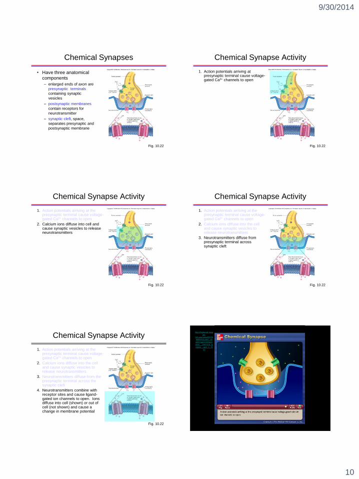
Chemical Synapse Activity
1. Action potentials arriving at presynaptic terminal cause voltage-gated Ca2+ channels to open
Fig. 10.22
Chemical Synapse Activity
1. Action potentials arriving at the presynaptic terminal cause voltage-gated Ca2+ channels to open
2. Calcium ions diffuse into cell and cause synaptic vesicles to release neurotransmitters
Fig. 10.22
Chemical Synapse Activity
1. Action potentials arriving at the presynaptic terminal cause voltage-gated Ca2+ channels to open
2. Calcium ions diffuse into the cell and cause synaptic vesicles to release neurotransmitters
3. Neurotransmitters diffuse from presynaptic terminal across synaptic cleft
Fig. 10.22
Chemical Synapse Activity
1. Action potentials arriving at the presynaptic terminal cause voltage-gated Ca2+ channels to open
2. Calcium ions diffuse into the cell and cause synaptic vesicles to release neurotransmitters
3. Neurotransmitters diffuse from the presynaptic terminal across the synaptic cleft
4. Neurotransmitters combine with receptor sites and cause ligand-gated ion channels to open. Ions diffuse into cell (shown) or out of cell (not shown) and cause a change in membrane potential
Fig. 10.22
http://highered.mcgr
aw-
hill.com/sites/0072495855/student_vie
w0/chapter14/anim
ation__chemical_sy
napse__quiz_1_.ht
ml
Page 11
9/30/2014
11
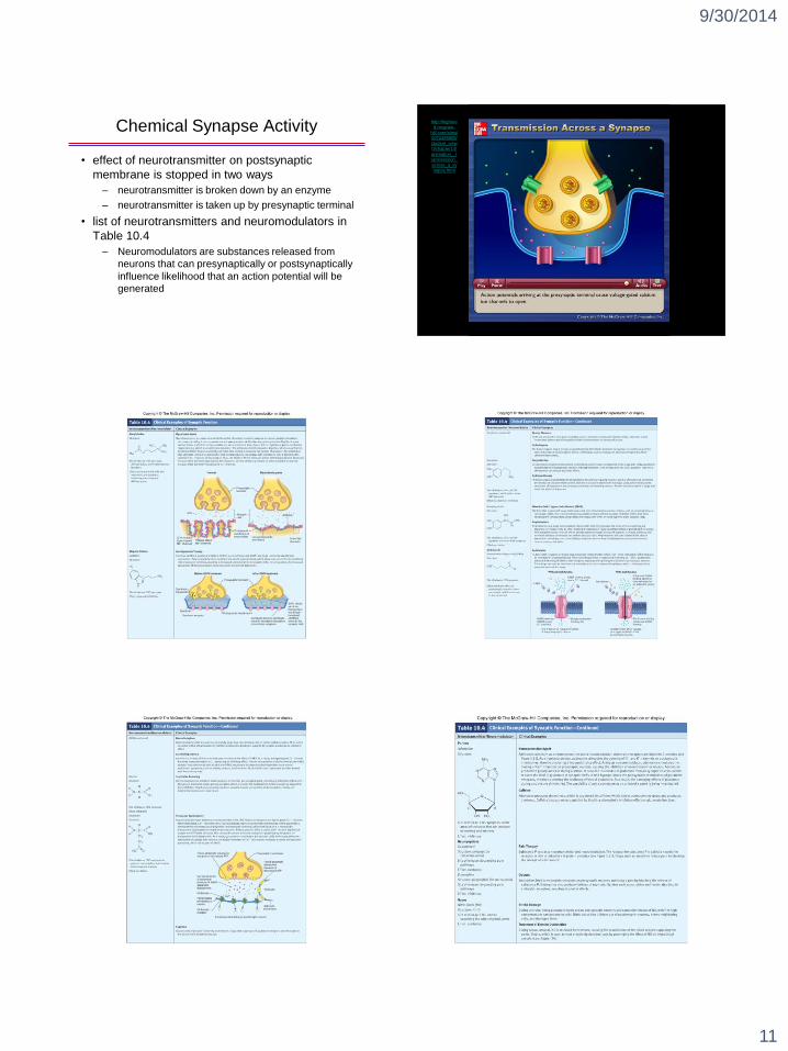
Chemical Synapse Activity
• effect of neurotransmitter on postsynaptic
membrane is stopped in two ways
– neurotransmitter is broken down by an enzyme
– neurotransmitter is taken up by presynaptic terminal
• list of neurotransmitters and neuromodulators in
Table 10.4
– Neuromodulators are substances released from
neurons that can presynaptically or postsynaptically
influence likelihood that an action potential will be
generated
http://highere
d.mcgraw-
hill.com/sites/0072495855/
student_view
0/chapter14/
animation__t
ransmission_across_a_sy
napse.html
Page 12
9/30/2014
12
Chemical Synapse Activity
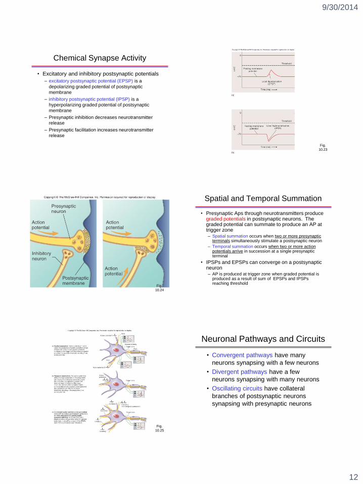
• Excitatory and inhibitory postsynaptic potentials
– excitatory postsynaptic potential (EPSP) is a
depolarizing graded potential of postsynaptic
membrane
– inhibitory postsynaptic potential (IPSP) is a
hyperpolarizing graded potential of postsynaptic
membrane
– Presynaptic inhibition decreases neurotransmitter
release
– Presynaptic facilitation increases neurotransmitter
release
Fig.
10.23
Fig.
10.24
Spatial and Temporal Summation
• Presynaptic Aps through neurotransmitters produce graded potentials in postsynaptic neurons. The graded potential can summate to produce an AP at trigger zone– Spatial summation occurs when two or more presynaptic
terminals simultaneously stimulate a postsynaptic neuron
– Temporal summation occurs when two or more action potentials arrive in succession at a single presynaptic terminal
• IPSPs and EPSPs can converge on a postsynaptic neuron– AP is produced at trigger zone when graded potential is
produced as a result of sum of EPSPs and IPSPs reaching threshold
Fig.
10.25
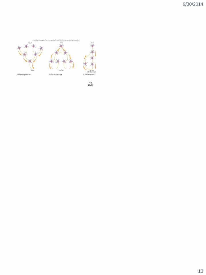
Neuronal Pathways and Circuits
• Convergent pathways have many
neurons synapsing with a few neurons
• Divergent pathways have a few
neurons synapsing with many neurons
• Oscillating circuits have collateral
branches of postsynaptic neurons
synapsing with presynaptic neurons
Page 13
9/30/2014
13
Fig.
10.26