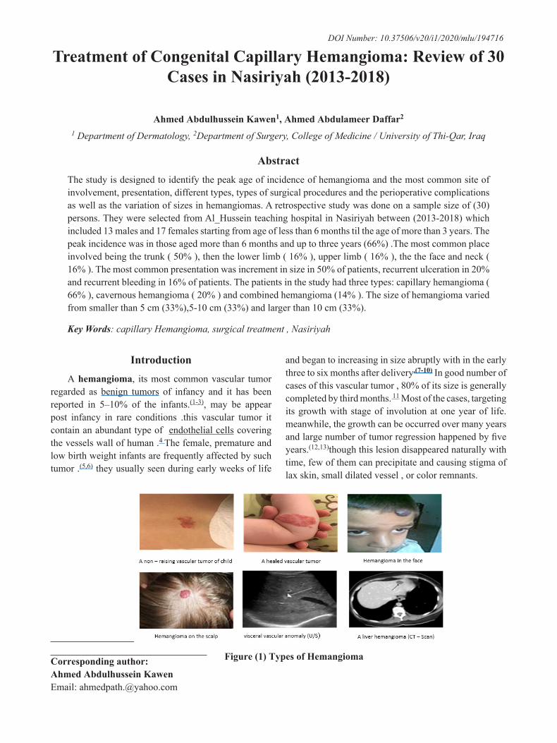
934 Medico-legal Update, January-March 2020, Vol.20, No. 1 Treatment of Congenital Capillary Hemangioma: Review of 30 Cases in Nasiriyah (2013-2018) Ahmed Abdulhussein Kawen 1 , Ahmed Abdulameer Daffar 2 1 Department of Dermatology, 2 Department of Surgery, College of Medicine / University of Thi-Qar, Iraq Abstract The study is designed to identify the peak age of incidence of hemangioma and the most common site of involvement, presentation, different types, types of surgical procedures and the perioperative complications as well as the variation of sizes in hemangiomas. A retrospective study was done on a sample size of (30) persons. They were selected from Al_Hussein teaching hospital in Nasiriyah between (2013-2018) which included 13 males and 17 females starting from age of less than 6 months til the age of more than 3 years. The peak incidence was in those aged more than 6 months and up to three years (66%) .The most common place involved being the trunk ( 50% ), then the lower limb ( 16% ), upper limb ( 16% ), the the face and neck ( 16% ). The most common presentation was increment in size in 50% of patients, recurrent ulceration in 20% and recurrent bleeding in 16% of patients. The patients in the study had three types: capillary hemangioma ( 66% ), cavernous hemangioma ( 20% ) and combined hemangioma (14% ). The size of hemangioma varied from smaller than 5 cm (33%),5-10 cm (33%) and larger than 10 cm (33%). Key Words: capillary Hemangioma, surgical treatment , Nasiriyah Corresponding author: Ahmed Abdulhussein Kawen Email: [email protected]Introduction A hemangioma, its most common vascular tumor regarded as benign tumors of infancy and it has been reported in 5–10% of the infants. (1-3) , may be appear post infancy in rare conditions .this vascular tumor it contain an abundant type of endothelial cells covering the vessels wall of human . 4 The female, premature and low birth weight infants are frequently affected by such tumor . (5,6) they usually seen during early weeks of life and began to increasing in size abruptly with in the early three to six months after delivery .(7-10) In good number of cases of this vascular tumor , 80% of its size is generally completed by third months. 11 Most of the cases, targeting its growth with stage of involution at one year of life. meanwhile, the growth can be occurred over many years and large number of tumor regression happened by five years. (12,13) though this lesion disappeared naturally with time, few of them can precipitate and causing stigma of lax skin, small dilated vessel , or color remnants. Figure (1) Types of Hemangioma DOI Number: 10.37506/v20/i1/2020/mlu/194716
Treatment of Congenital Capillary Hemangioma: Review of 30 Cases in Nasiriyah (2013-2018)
Ahmed Abdulhussein Kawen1, Ahmed Abdulameer Daffar2
1 Department of Dermatology, 2Department of Surgery, College of Medicine / University of Thi-Qar, Iraq
AbstractThe study is designed to identify the peak age of incidence of hemangioma and the most common site of involvement, presentation, different types, types of surgical procedures and the perioperative complications as well as the variation of sizes in hemangiomas. A retrospective study was done on a sample size of (30) persons. They were selected from Al_Hussein teaching hospital in Nasiriyah between (2013-2018) which included 13 males and 17 females starting from age of less than 6 months til the age of more than 3 years. The peak incidence was in those aged more than 6 months and up to three years (66%) .The most common place involved being the trunk ( 50% ), then the lower limb ( 16% ), upper limb ( 16% ), the the face and neck ( 16% ). The most common presentation was increment in size in 50% of patients, recurrent ulceration in 20% and recurrent bleeding in 16% of patients. The patients in the study had three types: capillary hemangioma ( 66% ), cavernous hemangioma ( 20% ) and combined hemangioma (14% ). The size of hemangioma varied from smaller than 5 cm (33%),5-10 cm (33%) and larger than 10 cm (33%).
IntroductionA hemangioma, its most common vascular tumor
regarded as benign tumors of infancy and it has been reported in 5–10% of the infants.(1-3), may be appear post infancy in rare conditions .this vascular tumor it contain an abundant type of endothelial cells covering the vessels wall of human .4 The female, premature and low birth weight infants are frequently affected by such tumor .(5,6) they usually seen during early weeks of life
and began to increasing in size abruptly with in the early three to six months after delivery.(7-10) In good number of cases of this vascular tumor , 80% of its size is generally completed by third months. 11 Most of the cases, targeting its growth with stage of involution at one year of life. meanwhile, the growth can be occurred over many years and large number of tumor regression happened by five years.(12,13)though this lesion disappeared naturally with time, few of them can precipitate and causing stigma of lax skin, small dilated vessel , or color remnants.
Most of the cases of vascular tumors of infancy easily discovered from patient history and direct physical examination.6 In certain conditions need assisted measured like imaging (US) with Doppler, (MRI)) and/or other confirming tools are needed like cytology or histopathology. 14 This vascular tumor (infantile hemangioma IH) are not present at birth in most of cases or faint area, small dilated blood vessel , or dark area may be present. A well developed lesion after delivery predict other diagnosis. The type of vascular tumor with faint red strawberry color it’s a Superficial type that occupied the upper dermis, while the blue and firm textured lesion which occupying the deep dermis and subcutis that indicates deep type of this vascular tumor . Mixed type of lesion contain both proliferative IH which do not substantially proliferate 15 and pig endophytic IH in which obvious increment in size began later and continue for extended period .11A good history of the unique growth of a such condition its very important in the detection of the lesion . In the early weeks of after delivery this vascular abnormality proliferate very abruptly with increasing mass more than its size . After that accompanied by a decreasing growth for t up to 6–9 months, while the majority of growth about 80% is achieved by 3 months. Lastly , this lesion regressed over a period of years.11 If the diagnosis is not reached from physical examination, so other added measures like imaging or histopathology is helpful .
(4,14) On Doppler ultrasound the diagnosis is achieved by showing a high flow soft-tissue mass usually without direct arteriovenous shunting for the proliferative phase. On MRI, showing the increased signal intensity and increase up take after gadolinium injections. There are fast flow vessels. (14,15) specimen for diagnosis can be taken from the lesion by different invasive methods for microscopic evaluation. 16 Under the microscope and tissue can be processed for the detection of this tumor cells , abundant well capsulated endothelial cell . 17 The \GLUT-1 histochemical marker also may be help full in differentiation from other vascular abnormalities. 18
ResultsTable (1) Frequency of age groups in patients
and site of hemangioma
Variable Age group Frequency PercentSmaller than 6 Months 5 16.6%6 Months - 12 Months 10 33.3%1 Year - 3 Years 10 33.3%Older than 3 Years 5 16.6%Total 30 100%
Site Frequency PercentFace 2 6.66%Neck 3 10.0%Trunk 15 50.0%Lower Limb 5 16.6%Upper Limb 5 16.6%Total 30 100 %
DisscusionAt present work the female to male ratio was 1.3:1
unlike the study done in Goldenberg DC, et al. Plast Reconstruct Surg. 2016 which showed that the female-to-male ratio was 5.7:1.
In our study the most common site was the trunk then limbs followed by the face and neck unlike the study done in Goldenberg which showed that the face and limbs are the most common site .The presentation differed in our study from recurrent bleeding to increament in size to recurrent ulceration like the study done in Goldenberg DC and in Department of Pediatric Surgery, La Paz Children´s Hospital, Spain .
in our present study, the most common type is capillary hemangioma and this is similar to the study
in Department of Pediatric Surgery, La Paz Children´s Hospital.
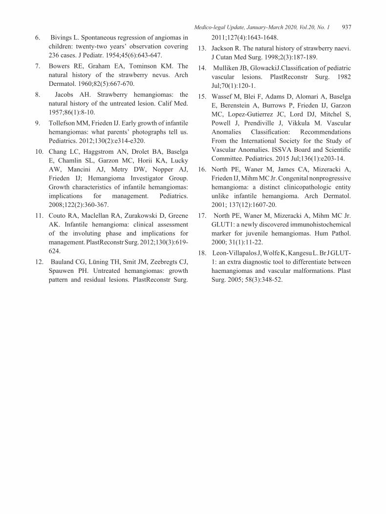
The size of hemangioma ranged from less than 5 cm in one third of patients to more than 10 cm in one third of patients who underwent surgery and this is similar to the studies made by others.
Conclusion Hemangiomas actually resolve spontaneously and
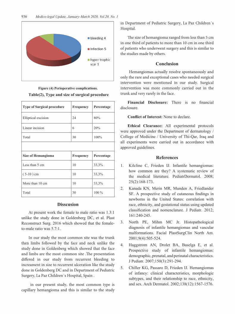
only the rare and exceptional cases who needed surgical intervention were mentioned in our study. Surgical intervention was more commonly carried out in the trunk and very rarely in the face.
Financial Disclosure: There is no financial disclosure.
Conflict of Interest: None to declare.
Ethical Clearance: All experimental protocols were approved under the Department of dermatology / College of Medicine / University of Thi-Qar, Iraq and all experiments were carried out in accordance with approved guidelines.
how common are they? A systematic review of the medical literature. PediatrDermatol. 2008; 25(2):168-173.
2. Kanada KN, Merin MR, Munden A, Friedlander SF. A prospective study of cutaneous findings in newborns in the United States: correlation with race, ethnicity, and gestational status using updated classification and nomenclature. J Pediatr. 2012; 161:240-245.
3. North PE, Mihm MC Jr. Histopathological diagnosis of infantile hemangiomas and vascular malformations. Facial PlastSurgClin North Am. 2001;9(4):505-524.
4. Haggstrom AN, Drolet BA, Baselga E, et al. Prospective study of infantile hemangiomas: demographic, prenatal, and perinatal characteristics. J Pediatr. 2007;150(3):291-294.
5. Chiller KG, Passaro D, Frieden IJ. Hemangiomas of infancy: clinical characteristics, morphologic subtypes, and their relationship to race, ethnicity, and sex. Arch Dermatol. 2002;138(12):1567-1576.
15. Wassef M, Blei F, Adams D, Alomari A, Baselga E, Berenstein A, Burrows P, Frieden IJ, Garzon MC, Lopez-Gutierrez JC, Lord DJ, Mitchel S, Powell J, Prendiville J, Vikkula M. Vascular Anomalies Classification: Recommendations From the International Society for the Study of Vascular Anomalies. ISSVA Board and Scientific Committee. Pediatrics. 2015 Jul;136(1):e203-14.
16. North PE, Waner M, James CA, Mizeracki A, Frieden IJ, Mihm MC Jr. Congenital nonprogressive hemangioma: a distinct clinicopathologic entity unlike infantile hemangioma. Arch Dermatol. 2001; 137(12):1607-20.
17. North PE, Waner M, Mizeracki A, Mihm MC Jr. GLUT1: a newly discovered immunohistochemical marker for juvenile hemangiomas. Hum Pathol. 2000; 31(1):11-22.
18. Leon-Villapalos J, Wolfe K, Kangesu L. Br J GLUT-1: an extra diagnostic tool to differentiate between haemangiomas and vascular malformations. Plast Surg. 2005; 58(3):348-52.