492


Problem BasedNeurosurgery
7830tp.indd 1 12/9/10 4:55 PM

This page intentionally left blankThis page intentionally left blank

N E W J E R S E Y • L O N D O N • S I N G A P O R E • B E I J I N G • S H A N G H A I • H O N G K O N G • TA I P E I • C H E N N A I
World Scientific
Sam EljamelThe University of Dundee, UK
Problem BasedNeurosurgery
7830tp.indd 2 12/9/10 4:55 PM

British Library Cataloguing-in-Publication DataA catalogue record for this book is available from the British Library.
For photocopying of material in this volume, please pay a copying fee through the CopyrightClearance Center, Inc., 222 Rosewood Drive, Danvers, MA 01923, USA. In this case permission tophotocopy is not required from the publisher.
ISBN-13 978-981-4317-07-8ISBN-10 981-4317-07-1
Typeset by Stallion PressEmail: [email protected]
All rights reserved. This book, or parts thereof, may not be reproduced in any form or by any means,electronic or mechanical, including photocopying, recording or any information storage and retrievalsystem now known or to be invented, without written permission from the Publisher.
Copyright © 2011 by World Scientific Publishing Co. Pte. Ltd.
Published by
World Scientific Publishing Co. Pte. Ltd.
5 Toh Tuck Link, Singapore 596224
USA office: 27 Warren Street, Suite 401-402, Hackensack, NJ 07601
UK office: 57 Shelton Street, Covent Garden, London WC2H 9HE
Printed in Singapore.
PROBLEM BASED NEUROSURGERY
JQuek - Problem Based Neurosurgery.pmd 3/2/2011, 4:40 PM1

Disclaimer
The author provided a summary of information he thought rele-vant to students, doctors in training and other health careprofessionals learning about neurosurgical disorders. The authorhad made no attempt to set a standard of care and common senseshould prevail. The author had made no attempt to update theinformation after the date of publication. The author took everyprecaution to accurately present the information, but errors oromissions may have occurred. Any therapeutic drug dosages orrecommendations contained in this book should be verifiedbefore use and local policies, procedures, guidelines and nationalrecommendations should be checked before use.
b1009 Problem Based Neurosurgery
b1009_FM.qxd 11/12/2010 10:48 AM Page v

b1009 Problem Based Neurosurgery
b1009_FM.qxd 11/12/2010 10:48 AM Page vi
This page intentionally left blankThis page intentionally left blank

Dedication
I dedicate this book to my wonderful family: my supportive andloving wife “Adora”, my eldest daughter “Sarah” who inspiredme to do this work as she progressed through the medical course,my youngest daughter “Sana” who proof-read every word of thisbook on top of her busy schedule studying medicine and my son“Sam Jr” who kept me going to finish this project. To my parentswho gave me the opportunity to pursue my career. To my teach-ers, my students, my colleagues and my patients who gave me theexperience and wisdom that culminated in this project.
b1009 Problem Based Neurosurgery
b1009_FM.qxd 11/12/2010 10:48 AM Page vii

b1009 Problem Based Neurosurgery
b1009_FM.qxd 11/12/2010 10:48 AM Page viii
This page intentionally left blankThis page intentionally left blank
ix
b1009 Problem Based Neurosurgery
Contents
Preface xiii
Chapter 1: History and Physical Exam 1
Problem 1-1: How to get the patient to tell you what is wrong. 1(The smart way of taking a succinct completehistory of any illness)
Problem 1-2: How to elicit neurological signs effectively, 12demonstrate them with confidence and makea lasting impression. (The smart way of performingneurological physical examination 1)
Problem 1-3: How to examine the first two cranial nerves 23efficiently, with confidence and make a lastingimpression. (The smart way of performingneurological physical examination 2)
Problem 1-4: How to examine the third, fourth and sixth cranial 32nerves efficiently, with confidence and make alasting impression. (The smart way of performingneurological physical examination 3)
Problem 1-5: How to examine the face (fifth and seventh 45cranial nerves) efficiently, with confidence andmake a lasting impression. (The smart way ofperforming neurological physical examination 4)
Problem 1-6: How to examine the eighth, ninth & tenth cranial 57nerves efficiently, with confidence and make alasting impression. (The smart way of performingneurological physical examination 5)
b1009_FM.qxd 11/12/2010 10:48 AM Page ix

x Contents
b1009 Problem Based Neurosurgery
Problem 1-7: How to examine the 11th and 12th cranial nerves 69efficiently, with confidence and make a lastingimpression. (The smart way of performingneurological physical examination 6)
Problem 1-8: How to examine the motor system efficiently, 73with confidence and make a lasting impression.(The smart way of performing neurologicalphysical examination 7)
Problem 1-9: How to examine the sensory system, coordination 96and gait efficiently and make a lasting impression.(The smart way of performing neurological physicalexamination 8)
Chapter 2: Neurological Investigations 109
Problem 2-1: Computerised tomographic scan (CT): How to 109interpret CT-based images?
Problem 2-2: Magnetic resonance imaging (MRI): How to 120interpret MRI-based images?
Problem 2-3: Non-radiological neuro-investigations. How to 134interpret non-radiological neuro-investigationsin a smart way?
Chapter 3: Trauma (Head and Spinal Injured Patients) 145
Problem 3-1: Head injuries and head trauma. How to manage 145a patient presenting with a head injury?
Problem 3-2: Spinal trauma and traumatic spinal cord syndromes. 163How to manage a patient following spinal trauma?
Chapter 4: Sudden Headache or Collapse (SAH, ICH, 185Seizures)
Problem 4-1: Sudden headache and subarachnoid haemorrhage. 185How to manage a patient presenting with suddenheadache?
Problem 4-2: Collapse and sudden focal neurological deficits. 209How to manage a patient presenting with suddenfocal neurological deficit or collapse?
b1009_FM.qxd 11/12/2010 10:48 AM Page x

Contents xi
b1009 Problem Based Neurosurgery
Problem 4-3: Collapse, seizures, fits and funny turns. How to 220manage a patient presenting with seizure orfunny turn?
Chapter 5: Raised ICP (Tumours, Abscess and 233Hydrocephalus)
Problem 5-1: Raised ICP and primary malignant brain tumours. 233How to manage a patient presenting with raisedICP due to primary malignant brain tumours(PMBT)?
Problem 5-2: Raised ICP and secondary brain tumours. How to 258manage a patient presenting with raised ICP dueto secondary brain tumour (SBT)?
Problem 5-3: Raised ICP, brain abscess and CNS infections. 265How to manage a patient presenting with raisedICP due to CNS infection?
Problem 5-4: Raised ICP and hydrocephalus. How to manage 281a patient presenting with raised ICP due tohydrocephalus?
Chapter 6: Visual Symptoms (Meningiomas, Pituitary 307Adenomas)
Problem 6-1: Visual failure and intracranial meningiomas. 307How to manage a patient presenting withcompressive optic neuropathy?
Problem 6-2: Visual symptoms and pituitary adenomas. 324How to manage a patient presenting with visualfield defect?
Chapter 7: Hearing Loss, Ataxia, Vertigo and Facial Pain 345(CPA Lesions)
Problem 7-1: Hearing loss and cerebellopontine angle (CPA) 345lesions. How to manage a patient presentingwith hearing loss?
Problem 7-2: Facial pain and trigeminal neuralgia. How to 356manage a patient presenting with facial pain?
b1009_FM.qxd 11/12/2010 10:48 AM Page xi

xii Contents
b1009 Problem Based Neurosurgery
Chapter 8: Tremor (Parkinson’s Disease and Dystonia) 369
Problem 8-1: Tremor and Parkinson’s disease. How to manage 369a patient presenting with tremor?
Problem 8-2: Movement disorders and dystonia. How to 376manage a patient with dystonia?
Chapter 9: Para-/Tetraparesis (Spinal Compression) 381
Problem 9-1: Bilateral limb paresis (malignant spinal 381compression). How to manage suspected malignantspinal compression?
Problem 9-2: Bilateral limb paresis (benign spinal compression). 396How to manage suspected benign spinalcompression?
Problem 9-3: Bilateral limb paresis (spinal infections). How to 410manage suspected spinal infection?
Chapter 10: Pain, Weakness or Numbness in a Limb 421(Radiculopathy, Myelopathy and PeripheralNerve Pathologies)
Problem 10-1: Brachalgia, myelopathy and cervical disc 421prolapse. How to manage a patient presentingwith arm pain or myelopathy?
Problem 10-2: Sciatica, cauda equina and lumbar disc prolapse. 429How to manage a patient presenting with leg painor cauda equina?
Problem 10-3: Hands and feet numbness and peripheral nerves. 443How to manage a patient presenting with hand orfoot numbness?
Appendices 451
Index 473
b1009_FM.qxd 11/12/2010 10:48 AM Page xii

Preface
Problem based neurosurgery is a systematic approach to diagnosis, under-standing and management of neurosurgical diseases based on symptomsand signs of disease and using common sense and the art of applyingscientific knowledge to practice.
In producing this book I took the common sense approach, mypatients presented me with a set of symptoms and signs creating a prob-lem that needed diagnosis and management plans. My students andresidents had asked me questions. It is these presentations and questionsthat formed the foundation problems in this book. I concentrated on coreand common neurosurgical problems that constituted the majority of neu-rosurgical practice. When one’s goal is to be concise, it is not possible toinclude every detail in this text. I envisaged that the main users of thisbook will be those studying neurosurgery, and neurology and those train-ing in neurosurgery, emergency medicine, ENT, ophthalmology, generalmedicine, general surgery, orthopaedic surgery, and radiology and doctorsin their foundation years and those practicing in the community.
Thanks for using this text.
Professor Sam Eljamel, MD, FRCS(Ed,Ir,NS)Consultant Neurosurgeon
xiii
b1009 Problem Based Neurosurgery
b1009_FM.qxd 11/12/2010 10:48 AM Page xiii

b1009 Problem Based Neurosurgery
b1009_FM.qxd 11/12/2010 10:48 AM Page xiv
This page intentionally left blankThis page intentionally left blank

Chapter 1: History and Physical Exam
Problem 1-1: How to get the patient to tell you what is wrong.(The smart way of taking a succinct complete history of any illness)
The main questions on every patient’smind when (s)he walks into a doctor’soffice or when (s)he seeks a doctor’sadvice are:
What is wrong with me doctor?What can you do about what is wrong?andCan you cure it?
The key to unlock the mystery to these questions is to take a succinct com-plete history, and analyse the information you gather instantly to guidefurther questions and clinical examination. Throughout this book you willfind examples of real life problems that patients presented with and youwill learn how to take a smart history, elicit clinical signs and performintelligent analysis of the symptoms and signs to reach a diagnosis andanswer patient’s questions in full, request appropriate investigations andmanage the patient effectively.
To find out the cause of each symptom of the patient and manage yourpatient effectively, you need to collect the following essential informationabout each symptom. You need to know:
• The anatomical location of the symptom (where is it coming from?),• Its mode of onset (how did it start?),• Its duration (how long it has been there for?), and• Its course (what happened to it since it began?).
It is important to ask open-ended questions to get as much accurate infor-mation from the patient as possible. Using closed-ended questions such as
1
b1009 Problem Based Neurosurgery
Problem based tool box:How to take smart history?How to examine HMF?How to examine cranial nerves?How to examine motor system?How to examine sensory
system?
b1009_Chapter-01.qxd 11/12/2010 10:48 AM Page 1

those requiring “Yes” or “No” answers, is counterproductive and likely tolead to avoidable errors. This kind of question may become necessary toconfirm the answers to open-ended questions, to narrow down the diag-nosis or for systemic enquiry. Let us study few examples to understand thedifference between open- and closed-ended questions.
Problem case scenario (PCS) 1-1-1:
A 35-year-old woman walks into a doctor’s office and complains that shehad facial pain.As soon as such a patient turns up in my office I will be rounding twomain causative suspects straightway: idiopathic trigeminal neuralgia(ITN) or atypical facial pain syndrome caused by structural lesion insidethe skull, at the skull base or in the face.
I will ask the patient the following four open-ended basic questions:
1. Where exactly was this pain? (Exact location)2. How did the pain start? (Mode of onset)3. How long was the pain there for? (Duration of the pain) and4. What happened to the pain since it started? (Course of the pain)
Note that all my questions were open-ended questions beginning with“Where”, “How”, and “What”. Any question that starts with a verb is aclosed-ended question requiring a “Yes” or “No” answer and may lead toan incomplete history that takes longer to elicit. For example, if I hadasked the patient: “Can you tell me where the pain is?” The patient mayjust answer: “Yes, I can”, and I have to ask the patient another question:“Where is it?” or ask the patient to tell me where the pain was. It wouldhave been much easier and quicker if I went ahead and asked the straight-forward question: Where exactly was the pain?
Another commonly encountered method of taking history is givingthe patient a multiple choice question (MCQ). MCQ invites guessing andguessing leads to avoidable mistakes. This often leads to avoidable errorsbecause some patients may feel that they have no option but to choose oneof the choices the doctor have given. For example, instead of asking thepatient the more reasonable recommended open-ended question “How did
2 Chapter 1
b1009 Problem Based Neurosurgery
b1009_Chapter-01.qxd 11/12/2010 10:48 AM Page 2

the pain start?”, you asked “Did the pain start suddenly or gradually?”. Ifyou ask such closed-ended question you are more likely to get the wronginformation and miss the mode of onset.
Suppose the patient answered the four basic open-ended questions asfollows: “the pain was in the right jaw, started suddenly three weeks agoand it was episodic”. These answers make me think that this patient wasmore likely to be suffering from typical (idiopathic) trigeminal neuralgia(ITN). As I knew what the other features of ITN are, I would supplementmy original four questions by the following three questions:
• What makes this pain worse? (Aggravating factors)• What makes this pain better? (Relieving factors)• What was the character of the pain? (Description of type of pain)
If my provisional diagnosis of ITN was correct, the patient would havesaid “the pain was made worse by laughing, brushing my teeth or expo-sure to cold wind, it was made better by carbamazepine or nothing at alland it was lancinating in nature”.
Once I had finished with the facial pain symptom as above, I wouldgo on and ask the fifth basic question:
5. Apart from this pain, what other symptoms do you have?
Note that I had used in my fifth question some of the information I hadalready gathered (pain). By including the word pain in my question I amtelling the patient two things: I had finished with the pain and I had lis-tened to the patient’s complaint and noted it.
For every other symptom the patient comes up with, I would use thesame four basic questions again and again followed by any supplementaryquestions and finally by the fifth basic question and keep repeating thecycle until the patient says “No doctor, I do not have any more problems”.
PCS1-1-2:
A 35-year-old woman came in with facial pain. The answers to the fourbasic questions were: “the pain was around my left eye, it started suddenly
History and Physical Exam 3
b1009 Problem Based Neurosurgery
b1009_Chapter-01.qxd 11/12/2010 10:48 AM Page 3

yesterday and it had been slowly getting worse”. I had asked the fifthbasic question: “apart from this pain around the left eye what other symp-toms do you have?” The patient replied that her eyelid came down and shecould not open her left eye. I would ask the same four basic questionsabout the eyelid closure: Location: which eyelid? Onset: how did it start?Duration: how long was the eyelid closed for? Course: what happenedto the eyelid since it closed?
If the patient answered: “it was my left eyelid that closed suddenly atthe same time as my pain started and that it remained closed since”. Usingthis additional information I will be thinking that a painful closure of theleft eyelid can be caused by left Oculomotor (III) nerve palsy caused byan intracranial aneurysm at the posterior communicating artery (PComA),left orbital cellulites or left cavernous sinus pathology. I would ask the fol-lowing supplementary questions:
If you opened the left eye what happens? Looking for double vision(diplopia).
What other problems have you noticed with the left eye? Looking forredness, discharge, eye deviation or dilatation of the pupil.
The next question in the last case scenario would be: Apart from thepain and the left eyelid closure, what else?
If this patient comes with another symptom, e.g. double vision, I wouldask the same four basic questions in the same way followed by myfifth question as described: Which direction did you note the doublevision? For how long did you have the double vision? How did it start?What happened to the double vision since it started? If the patientsaid that the double vision started just before the eyelid closed, itstarted suddenly and was maximum looking to the right and it disap-peared when the eyelid closed, this information is confirmatory ofpainful III nerve palsy likely to be caused by intracranial PComAaneurysm and my clinical examination would be designed to confirmthis. Figure 1-1 sums up the concept of the basic questions and supple-mentary ones.
4 Chapter 1
b1009 Problem Based Neurosurgery
b1009_Chapter-01.qxd 11/12/2010 10:48 AM Page 4

If however the patient said: “I do not have any other symptoms”, thenext step would be to continue the history as follows:
Step 1: Ask questions that will narrow down the diagnosis. These arequestions that will confirm or rule out other features of the diagnosisunder consideration. These questions are closed-ended questions and any“Yes” answer requires going through the four basic questions for any pos-itive symptom followed by any other supplementary questions. Forexample, in the last case scenario, if I am considering orbital cellulites asa diagnosis I would ask: “Did you have discharge from the left eye?” Ifthe answer was “Yes” then I would have asked, “What type of discharge?How long has the discharge been there for? How did it start? And whathappened to the discharge since it started?”
Another example: if a patient came in with headache, I would haveasked about nausea and vomiting, and blurring of vision as I would bethinking of raised intracranial pressure (ICP) as the cause of the headache.
Step 2: Ask questions about the organ that is most likely to be involved inthe illness to elicit any other symptoms the patient might have dismissed.For example in the two previous case scenarios I would be asking ques-tions to elicit any symptoms associated with dysfunction of the brain.These symptoms can be summarised as follows:
Symptoms of raised ICP: Any
1. Headache.2. Nausea and vomiting.3. Blurred vision.
History and Physical Exam 5
b1009 Problem Based Neurosurgery
Figure 1-1: Summary of basic and supplementary questions.
b1009_Chapter-01.qxd 11/12/2010 10:48 AM Page 5

Higher mental functions symptoms: Any problems with
1. Alertness.2. Speech.3. Memory.4. Intellect.
Cranial nerves symptoms: Any problems with
1. Senses of smell, vision, or hearing.2. Swallowing or voice change.3. Balance.4. Any vertigo or tinnitus.5. Facial weakness, twitching or altered sensation.
Motor functions symptoms: Any
1. Weakness.2. Rigidity.3. Slowness.4. Stiffness.5. Tremor.6. Abnormal movements.
Sensory function symptoms:
1. Any lack of or abnormal sensation anywhere in the body.
Other symptoms:
1. Neck stiffness.2. Photophobia.3. Sphincter disturbance.
Step 3: Ask questions about the cardinal symptoms of other systems thatare likely to be affected by or implicated in the causation of the diagnosisunder consideration. For example if I am dealing with a patient likely tohave suffered from cerebral infract (stroke) then I would ask questionsabout the cardiovascular system that is likely to be the source of thrombo-embolism: I would ask about palpitations, chest pain, ankle swelling, andshortness of breath. On the other hand if my patient had difficulty swal-lowing because of a stroke or tumor in the brain stem then I would be
6 Chapter 1
b1009 Problem Based Neurosurgery
b1009_Chapter-01.qxd 11/12/2010 10:48 AM Page 6

asking questions about the respiratory system to find out if the patient hadaspirated: I would ask about cough, chest pain, and shortness of breath.A full list of the cardinal symptoms of all the body systems is listed inFigure 1-2. Imagine that the systems of the body were organised in acircle, if you start the circle at one point and follow the circle around in onedirection (clockwise or anticlockwise), you will finish in the same pointyou have started from, without forgetting any system. Remember that theduty of care of a doctor goes beyond providing a diagnosis, treatment andprognosis. Doctors are also responsible for early detection, screening andprevention of disease as well as health promotion. The purpose of sys-tematic systemic review is to not only narrow down the diagnosis, but isalso to fulfil the doctor’s obligations to patients and the community interms of health promotion, disease prevention and early detection.
Step 4: Elicit past history. Past history often clinches the diagnosis. I usethe following simple four opening questions:
1. Have you had any similar symptoms in the past? If the answer was “Yes”then I would proceed by asking: Where? How often? How long ago?
History and Physical Exam 7
b1009 Problem Based Neurosurgery
Figure 1-2: The circle of systemic review.
b1009_Chapter-01.qxd 11/12/2010 10:48 AM Page 7

2. Have you been admitted to hospital before? If the answer was “Yes”then I would proceed by asking: When? Why? For how long?
3. Have you had any operations in the past? If the answer was “Yes”then I would proceed by asking: What? When? How long ago?
4. Do you suffer from any long term condition? If the answer was “Yes”then I would proceed by asking: What? For how long? and Whattreatment?
For example a patient suffering from left-sided weakness involving armand leg of sudden onset with diabetes mellitus, hypercholesterolaemia andchronic hypertension might be suffering from thrombotic stroke; a patientsuffering the same thing but with previous mitral valve replacement oratrial fibrillation might be suffering from embolic stroke.
Step 5: Take drug history: Are you on any medications? Is a very goodquestion to start with, if the answer was “Yes” then I would proceed byasking: What drug? What dose? How often? For how long? Who pre-scribed it? For example, a patient coming in with gradual slowlyprogressive left-sided weakness associated with headaches and was onanticoagulants might be suffering from subdural haematoma.
Step 6: Take family history. Because certain diseases can run in familiesand may have a genetic aetiology, doctors need to explore any family his-tory of diabetes mellitus, hypertension, strokes, heart attacks, angina,asthma or epilepsy. Some patients might go away with the wrong idea thatthese illnesses are transmissible or contagious and they need an explana-tion that they are not. I use the following questions to open this sectionwith patients:
Did you or any of your family members suffer from chronic illness?This question combines past history with family history and saves timeduring history gathering. Similarly the question: Did you or any memberof your family suffer from the following illnesses: diabetes, asthma, etc.?will save time by combining past and family histories together. It isimportant to explain to the patient what you mean by members of family.You are looking for family history in the patient’s parents, children, broth-ers, sisters, grandparents, uncles and aunts, not in partners or distantrelatives (Figure 1-3).
8 Chapter 1
b1009 Problem Based Neurosurgery
b1009_Chapter-01.qxd 11/12/2010 10:48 AM Page 8

Step 7: Elicit social history. The main purpose of this section is healthpromotion and screening. The two main areas you wish to cover underthis section are smoking and alcohol consumption. Exploring patient’slifestyles might be useful in diagnosis and management. For example achronic smoking patient coming with symptoms and signs of raised ICPand cough might be suffering from metastatic lung cancer in the brain. Ioften use the following opening questions in this section:
1. Did you or anybody in your household smoke? If the answer was“No”, or (s)he had stopped smoking. I would use positive rein-forcement and say “Well done! That is really good for you”. If theanswer was “Yes”, then I would ask: How often? For how long?Followed by: Have you considered giving up smoking? and directthe patient to where help for smoking cessation can be found. If thepatient had stopped smoking I explore how long ago did he smoke,as that might be relevant, for example if the patient no longersmokes but had smoked heavily for 20 years, is very relevant todisease causation.
2. Do you drink alcohol? If the answer was “Yes”, I would ask: Howmuch? For how long? If I encounter an excessive drinking patient,
History and Physical Exam 9
b1009 Problem Based Neurosurgery
Figure 1-3: Drawing of relevant family history: illnesses in the shaded family tree ofthe patient are relevant, while in the unshaded is not.
b1009_Chapter-01.qxd 11/12/2010 10:48 AM Page 9

I give advice about the importance to stop drinking and refer thepatient to counselling services.
At this point of history gathering, I would be able to summarise thepatient’s history into three or four sentences: for example in PCS1-1-1 tosum up I would say “A 35-year-old woman presented with suddenepisodic lancinating left mandibular pain with no significant past, socialor drug history”. In PCS1-1-2 summing up would be as follows: “A35-year-old woman presented with sudden left painful III nerve palsy withhistory of hypertension and smoked 30 cigarettes per day”. Summing upfocuses your thoughts about what is the underlying problem was andmakes you think about what to look for during the physical examination.
Review questions:
1- What does a patient want from the healthcare provider?a. Diagnosis.b. Reassurance.c. Treatment.
2- What are the five basic questions in history taking?a. Where exactly the symptom (location)?b. How did it start (mode of onset)?c. How long it has been there for (duration)?d. What happened to it since it started (course)?e. Apart from this symptoms above, anything else?
3- What are the obligations of the healthcare provider?a. Make a diagnosis.b. Discuss all treatment options that apply to them.c. Inform patients if you do not know what is wrong
with them.d. Involve the patient in the decision process.e. Health screening.f. Early diagnosis.g. Health promotion.
10 Chapter 1
b1009 Problem Based Neurosurgery
b1009_Chapter-01.qxd 11/12/2010 10:48 AM Page 10

Your personal notes:
.....................................................................................................................
.....................................................................................................................
.....................................................................................................................
.....................................................................................................................
.....................................................................................................................
.....................................................................................................................
.....................................................................................................................
.....................................................................................................................
.....................................................................................................................
.....................................................................................................................
History and Physical Exam 11
b1009 Problem Based Neurosurgery
b1009_Chapter-01.qxd 11/12/2010 10:48 AM Page 11

Problem 1-2: How to elicit neurological signs effectively,demonstrate them with confidence and make a lasting impression.(The smart way of performing neurological physical examination 1)
The purpose of physical examinationis to elicit clinical signs of the diseaseto reach or narrow down the diagno-sis, order appropriate investigations,reassure the patient, and provideappropriate treatment, advice andfollow-up. Smart physical examinationis based on smart history. Smart physical examination will not replace orsubstitute poor history and vice versa. A confident clinician makes alasting impression when (s)he performs physical examination based onsmart history and performs it with confidence. Confidence can only beachieved by practice and practice (Practice makes perfect), so everymoment and every patient in hospital wards is an educational opportunity,so practice by examining patients over and over again. Patients are yourbest teachers and nothing can replace or substitute this experience.
Common sense dictates that physical examination should proceed inthe following order:
• Examine the organ affected by the disease first. It does not make anysense to the patient if the presenting complaint was left arm weaknessand the doctor starts to examine the cranial nerves. Although it isessential to examine the cranial nerves at some point, it makes moresense to start the examination with the left arm first for two reasons:
� By examining the organ or area complained of first, the doctor isin effect telling the patient that (s)he had been listened to.
� Patients are more likely to be more cooperative and finish theexamination when they know that attention was given to theorgan complained of at the outset of the examination. Patients areat their best at the beginning of the physical examination.
• Examine the system affected or implicated in the disease immediatelyafter examining the organ affected. In the left arm weakness case
12 Chapter 1
b1009 Problem Based Neurosurgery
Problem based tool box:GCS Speech assessmentMMSE Intellect assessmentOrder of physical examination
b1009_Chapter-01.qxd 11/12/2010 10:48 AM Page 12

scenario I would examine the nervous system after I had examined theleft arm.
• Examine the system or systems that are more likely to be affected orimplicated in the disease process next. For example in the left armweakness scenario I would examine the musculo-skeletal system afterI had examined the left arm and the nervous system. In someone whocomplained of swallowing difficulty, I would examine the back of thethroat and neck first, followed by the cranial nerves, the nervous sys-tem, the respiratory system, cardiovascular system and so on.
• Examine the rest of body systems for early detection of disease,screening, or possibly detection of the source of the disease. In thecase of systemic infection to detect the focus of primary infection orin the case of cancer by discovering the site of the primary cancer.
• Examination should begin with observation, followed by palpationand then by special bedside tests. The LFT principle: Look-Feel-Test.Look before you feel, and feel before you perform special tests. Forexample in someone presenting with leg pain, I would observe thepatient’s posture, and legs looking for signs of severe pain, musclespasm, muscle wasting or fasciculation looking for signs of severepain, so I would be very careful not to cause more pain during the restof physical examination.
The nervous system examination can be divided into the following sets:
1- Higher mental functions examination.2- Cranial nerves examination.3- Motor functions examination.4- Sensory functions examination.5- Other neurological signs.
1-2-1 Higher mental functions examination (HMF):
It makes a lot of sense to determine the patient’s higher mental functionsfrom the outset of any consultation, even before taking the history becauseimpairment of consciousness, speech or memory significantly affect theconduct of history and physical examination. For example, confused
History and Physical Exam 13
b1009 Problem Based Neurosurgery
b1009_Chapter-01.qxd 11/12/2010 10:48 AM Page 13

patients, patients with memory problems or dysphasic patients would notbe able to provide a reliable history and their physical examination needsto be modified to take account of these deficits. HMF examinationincludes examination of:
i- Conscious level.ii- Speech and language.
iii- Memory.iv- Intelligence.v- Handedness.
1-2-1i- Assessment of level of consciousness:
Level of consciousness is assessed at the bedside by the Glasgow ComaScale (GCS). The GCS consists of observing the patient’s responses toverbal or painful stimulation. Three responses are observed: Best EyeOpening Response (BEOR), Best Verbal Response (BVR) and Best MotorResponse (BMR).
a- Best Eye Opening Response (BEOR):There are four possible BEOR in any patient: 1 = no eye opening to anystimulus, 2 = eye opening to painful stimuli, 3 = eye opening to verbalstimuli, and 4 = eye opening spontaneously (Table 1-1). One drawback ofthe BEOR assessment is that it cannot be assessed in patients who havebilateral complete III nerve palsies or bilateral orbital haematomas. Inthese patients, the BEOR should be recorded as “C” for closed eyes ratherthan “1” for no BEOR. In patients who have either of these abnormalitiesin one eye only, the response of the better eye should be recorded for thepurpose of level of consciousness assessment.
b- Best Verbal Response (BVR):There are five possible responses under BVR: 1 = no verbal response toany stimulus, 2 = incomprehensible sounds, 3 = uttering words, 4 = con-fused, and 5 = oriented in time, place and person. It would not be possibleto assess BVR in patients who are dysphasic and it should be recorded as“D” for dysphasia rather than “1” for no BVR. Similarly patients who are
14 Chapter 1
b1009 Problem Based Neurosurgery
b1009_Chapter-01.qxd 11/12/2010 10:48 AM Page 14

artificially ventilated via endotracheal tube or tracheotomy cannot beassessed for BVR and it should be recorded as “T” for tube rather than “1”for no BVR (Table 1-1).
c- Best Motor Response (BMR):There are six possible responses within this category: 1 = no motorresponse to any stimulus, 2 = extension to pain, 3 = abnormal flexion topain, 4 = flexion to pain, 5 = localising pain, and 6 = obeying simple com-mands (Table 1-1). It would not be easy to assess BMR in patients withspinal cord injury leading to tetraplegia although if they can obey simplecommands, BMR can be assessed by observing motor responses in theface area, e.g. closing the eyes or showing the tongue. If the BMR couldnot be assessed because of paralysis of the limbs due to injury or becauseof sedative or muscle relaxant drugs, the BMR should be recorded as “P”for paralysis rather than “1” for no BMR.
Coma is defined on the GCS as any patient who fulfils all the follow-ing three criteria: 1- no eye opening response to any stimulus (score of 1only), 2- no comprehensible sound (score of 1 or 2) and 3- not obeyingsimple commands (score of 1 to 5). Therefore coma is GCS of 8 or lessprovided that the patient does not obey commands, does not utter anywords and does not open eyes to any stimulus.
Although each response of the GCS carries a number against it, theaggregated numbers should not be used to describe patients as thatleads to avoidable misunderstandings and misinterpretations of thenumbers. For example a GCS of 8 could mean a patient with BEOR1,
History and Physical Exam 15
b1009 Problem Based Neurosurgery
Table 1-1: The Glasgow Coma Scale (GCS)
Score BEOR BVR BMR Definitions
1 None None None Any patient within
2 To speech Sounds Extension to pain these shaded
3 To pain Words Abnormal flexion to painareas is in
4 Spontaneous Confused Flexion to painCOMA.
5 Orientated Localising pain
6 Obeys simple commands Not in coma
b1009_Chapter-01.qxd 11/12/2010 10:48 AM Page 15

BVR2 and BMR5 (patient is comatose) or it could mean BEO2, BVR3and BMR3 (patient is not comatose). It would be far better to usethe actual description of the patient’s response in each category ratherthan the numbers. Aggregate numbers are useful in determining trendsof progress and to perform statistical analysis in research and auditstudies.
1-2-1ia How to assess the BEOR?Observe the patient, if at least one eye is open without stimulation, thenBEOR is spontaneous (BEOR4), if at least one eye opens only in responseto speech, then the BEOR is to speech or drowsy (BEOR3), if both eyesremain closed despite verbal stimuli but at least one eye opens to painfulstimulus then BEOR is open to pain (BEOR2) and if there is no BEOR topain the BEOR is none (BEOR1). There are two ways to assess responsesto pain:
1. By exerting pressure on the supra-orbital nerve by putting the thumbparallel to the eye brow, feel the supra-orbital notch and exert pressure(Figure 1-4),
16 Chapter 1
b1009 Problem Based Neurosurgery
Figure 1-4: Eyebrow painful stimulus.
b1009_Chapter-01.qxd 11/12/2010 10:48 AM Page 16

2. By exerting pressure on the nail bed of any of the fingers using a pen-cil or a pen (Figure 1-5). The supra-orbital location is the preferredoption as it makes the distinction of the BMR easier.
1-2-1ib How to assess the BVR:To assess the BVR, I start by asking the patient the following simple ques-tions to assess orientation in place and time: Where are you right now?What sort of place are you in? What city are you in? And what country areyou in? If the patient could answer correctly at least two of these ques-tions, i.e. named hospital, and city or city and country or hospital andcountry, then the patient is oriented in place. Then I would ask the patient:What day of the week is today? What month of the year is it? What yearis it? If the patient got at least two of these correct (day and month, monthand year or day and year) then the patient is oriented in time. If the patientwas oriented in time and place their BVR is oriented (BVR5). If thepatient was disoriented in place and time, but was speaking in sentencesthat does not make sense, then the patient is confused (BVR4). However,you have to be careful in situations where the patient has suffered fromspeech difficulty, particularly nominal dysphasia or aphasia. If the patientwas only uttering words, then the patient is uttering words only (BVR3).If the patient was uttering sounds only and was unable to say any words
History and Physical Exam 17
b1009 Problem Based Neurosurgery
Figure 1-5: Nail bed painful stimulus.
b1009_Chapter-01.qxd 11/12/2010 10:48 AM Page 17

then the BVR is uttering incomprehensible sounds (BVR2). If the patientwas not uttering any sounds, the BVR will be no verbal response (BVR1).
1-2-1ic How to assess the BMR:When assessing the BMR, I use simple commands such as: Squeeze myfingers please, show me your tongue please, or close your eyes please.Simple commands do not require sophisticated brain processes to carrythem out and patients can perform these commands even in the presenceof cognitive or higher executive dysfunctions. On the other hand thecommand: Show me your little finger of the right hand please, or put yourright ring finger in your left ear please, are very complex commands thatdemand higher mental functions and likely to fail in patients who werefully conscious but had significant cognitive impairment. If the patientwas unable to obey simple commands, then the next stage is to simulatethe patient with pain as described under BEOR. By stimulating the supra-orbital nerve, the patient should be able to bring his hand up towards thestimulus to remove it. If the hand came to a level above the chin level, theBMR would be localising pain (BMR5), if the hand flexed but did notreach the level of the chin then the BMR would be flexion to pain(BMR4), if the elbow flexed, the shoulder extended and the forearmpronated then the BMR would be abnormal flexion to pain (BMR3), if theupper limb extends at shoulder and elbow and pronated then the BMRwould be extension to pain (BMR2), and if there was no motor responseto pain then the BMR would be none (BMR1). Figure 1-6 demonstratedthe BMR from 1 to 5.
1-2-1ii- How to assess speech and language?
Assessment of speech should include assessments of speech perceptionprimarily controlled in the angular gyrus of the dominant parietal lobe andspeech expression primarily controlled in the frontal opercula of the dom-inant hemisphere (Broca’s area). From the aforementioned historygathering and assessment of conscious level, one would have a good ideaof whether the patient understands and expresses speech, and if the patientwas fully oriented by answering the questions appropriately. If the patientwas unable to respond appropriately to questions during history taking
18 Chapter 1
b1009 Problem Based Neurosurgery
b1009_Chapter-01.qxd 11/12/2010 10:48 AM Page 18

and consciousness level assessment, then the doctor should evaluate thespeech in more detail to make sure that there was no speech impairmentleading to this difficulty. Simple questions such as: What is your name?Where are you? What day of the week is it? How old are you? And howare you?, are often sufficient to determine if the patient understandsspeech. In those patients who are unable to speak, one can assess under-standing of speech by asking the patient to perform simple tasks, e.g.show me your tongue, close your eyes or squeeze my hand. If the patientwas unable to understand speech altogether, the reasons could be that thepatient was unable to understand the language, had receptive dysphasiaor mute. Mutism can occur in bilateral subfrontal pathology, receptivedysphasia can occur due to damage of the angular gyrus on the domi-nant parietal lobe (Wernike’s dysphasia) and patients who merely donot speak the language often respond in their mother tongue. If thepatient can understand speech but seems to be confused or unable torespond appropriately in a verbal manner, the doctor needs to evaluatespeech expression in more detail. This can be evaluated by showing thepatient several objects and asking the patient to name them, e.g. show apen, a tie, a watch, a buckle, a strap, a spoon and a fork. If the patient wasunable to speak at all but seems to understand the language, then the
History and Physical Exam 19
b1009 Problem Based Neurosurgery
Figure 1-6: BMR to painful stimulus at the supra-orbital notch in patients not obeyingsimple commands.
b1009_Chapter-01.qxd 11/12/2010 10:48 AM Page 19

patient has an expressive aphasic. On the other hand if the patient wasable to speak but unable to name objects correctly then the patient hadnominal dysphasia. Junior clinicians often confuse nominal dysphasiawith confusion, the difference is that confused patients are able to nameobjects presented to them, while patients with nominal dysphasia cannot(Figure 1-7).
Assessment of language should include reading and writing. Readingcan be assessed by asking the patient to read simple sentences e.g. carry-ing a written request such as close your eyes or reading a written sentencealoud. Writing can be assessed by asking the patient to write his (her)name and address.
1-2-1iii- How to assess memory?
Memory can be divided into short- and long-term. Short-term memoryincludes remembering recent events and recall, and it is the most vulner-able memory after brain injury or disease. Long-term memory is moreresistant to insults. Memory requires the functions of the limbic systemparticularly the fornix and the medial temporal structures, the hippocampus
20 Chapter 1
b1009 Problem Based Neurosurgery
Figure 1-7: Dominant cerebral hemisphere cortex.
b1009_Chapter-01.qxd 11/12/2010 10:48 AM Page 20

and parahippocampus. Memories can also be divided into verbal andvisual memories. For simple bedside assessment of memory I often usethe following questions for long-term memory: What is your name? Whenwere you born? What is your address or where do you live? Or factualknowledge that the patient is expected to know, e.g. date of wedding ifmarried, names of the children if they had any, etc.
Short-term memory assessment can be performed by giving thepatient the names of three items (spoon, ball and car) and asking thepatient to repeat them (assessment of immediate recall), and askingthe patient to repeat the same words after five minutes or so (recall) andasking patients simple questions about recent events, e.g. When did theycome to hospital? How did they come into hospital? What did they havefor dinner, breakfast, etc? A more thorough evaluation of verbal andvisual memories could be performed by more sophisticated tests. Forexample, patients with subtle memory deficits and those who are workedup for temporal lobe surgery are examined by experts to assess their ver-bal and visual memories in detail.
1-2-1iv How to assess intelligence?
Intellect and IQ can be assessed by very sophisticated tests that aredesigned to assess IQ. A normal IQ should be a score of 70 or above.However, at the bedside a simple screening test such as the subtraction of7s from 100 or spelling the word “world” backwards would suffice. Anaverage patient would be able to subtract 7s from 100 fairly quickly bysaying “100, 93, 86, 79, 72, 65, 58, 51…” and so on or spell the word“world” backwards as “D-L-R-O-W”. Problems with calculation andspelling may indicate problems with memory, recall or pathology in thedominant parietal lobe. When dyscalculia is associated with fingeragnosia and dygraphia it is said that the patient suffers from Gerstmann’ssyndrome and that the lesion is located in the dominant parietal lobe.1 Onthe other hand a lesion in the non-dominant parietal lobe leads to disori-entation in space and loss of body image.2
Orientation, registration, attention, concentration, recall and languagecan be assessed at the bedside using the mini-mental state examination(MMSE). Normal individuals score 30/30 on this scale (Appendix I).
History and Physical Exam 21
b1009 Problem Based Neurosurgery
b1009_Chapter-01.qxd 11/12/2010 10:48 AM Page 21
1-2-1v How to assess handedness?
Assessment of handedness is very important to differentiate dominantfrom non-dominant hemispheric lesions. Handedness is dictated by thedominant hemisphere. In the vast majority of people, the left hemisphereis dominant.3 To determine which hand is dominant simply ask thepatient: Are you right or left handed? Most patients would know. If how-ever the patient does not know, you can ask the following questions:Which hand do you use to write? Which hand do you use to hold a knifeand cut your food? Or which foot do you use to kick a football?
Your personal notes:
.....................................................................................................................
.....................................................................................................................
.....................................................................................................................
.....................................................................................................................
.....................................................................................................................
.....................................................................................................................
.....................................................................................................................
.....................................................................................................................
.....................................................................................................................
.....................................................................................................................
22 Chapter 1
b1009 Problem Based Neurosurgery
b1009_Chapter-01.qxd 11/12/2010 10:48 AM Page 22

Problem 1-3: How to examine the first two cranial nervesefficiently, with confidence and make a lasting impression. (Thesmart way of performing neurological physical examination 2)
There are 12 cranial nerves that needto be examined. To be able to examinecranial nerves effectively the doctorneeds to know what each nerve does,where each cranial nerve originatesfrom and its course from origin todestination. Knowing these anatomicaland physiological facts will help thedoctor localise pathological processesmore precisely.
1-3-1 Where does each cranial nerve originate from?
I use a simple formula to remind myself where each cranial nerve arisesfrom: 2C + 2MB = 4P + 4MO. This means that the first two cranialnerves [Olfactory (I = 1st) and Optic (II = 2nd) nerves] originate in thecerebrum, the next two [Oculomotor (III = 3rd) and Trochlear (IV =4th)] cranial nerves arise from the midbrain, the next four [Trigeminal(V = 5th), Abducens (VI = 6th), Facial (VII = 7th) and Vestibulocochlear(VIII = 8th)] cranial nerves arise from the pons and the last four[Glossopharyngeal (IX = 9th), Vagus (X = 10th), Accessory (XI = 11th)and Hypoglossal (XII = 12th)] cranial nerves arise from the medullaoblongata.
1-3-2 What are the special features and functions of each nerve?
I- The first cranial nerve (Olfactory nerve):The olfactory nerve is responsible for the sense of smell, its tiny nervefibres pass through the cribriform plate to reach the olfactory pulp on eachside. Because of this fact the olfactory nerve is very susceptible at thecribriform plate to injury in anterior skull base fractures.
History and Physical Exam 23
b1009 Problem Based Neurosurgery
Problem based tool box:How to assess VA and CV?How to perform VF?
Fundoscopy?How to examine olfaction?How to localise visual
pathway lesion?
b1009_Chapter-01.qxd 11/12/2010 10:48 AM Page 23

Symptoms:
Olfactory nerve dysfunction manifests as lack of smell (anosmia).Abnormal sensation of smell does not arise from olfactory nerve dys-function; it originates from the temporal lobe in psychogenic seizures.Some patients with olfactory nerve dysfunction may also present withchange in their taste.
1-3-2i How to examine the first cranial nerve?
The sense of smell can be assessed by asking patients directly if therewere any problems with their sense of smell and can be confirmed byexamining each nerve independently by closing one nostril at a time andasking the patient to identify the aroma of coffee, tea, an apple, cloves, orperfume.
Examination of the olfactory nerve is the most commonly omittedpart of cranial nerves’ examination by junior clinicians. It is howeveressential to assess the sense of smell in every patient requiring neurolog-ical examination because it could be the only abnormal physical sign inpatients with olfactory groove meningiomas or after head injuries. Loss ofsense of smell carries significant implications to patients as they wouldnot be able to detect dangerous smells, e.g. gas leak at home and they needto be made aware of their deficit to take extra precautions.
II- The second cranial nerve (Optic nerve):The optic nerve is responsible for vision; the optic nerve enters the orbitthrough the optic canal making it susceptible to compression in thisregion. The medial (nasal) fibres cross over in the optic chiasm. Thereforea lesion in the optic chiasm leads to bitemporal hemianopia. Some ofthese nasal fibres loop forwards within the distal contralateral optic nerve.As a result of this anatomical feature, a pure lesion of one optic nerve maylead to junctional scotoma in the opposite visual field. The temporal fibresof one optic nerve and the nasal fibres of the contralateral optic nerve joinbehind the chiasm to form the optic tract that loops around the ipsilateralcerebral peduncle and just underneath the ipsilateral globus pallidus inter-nal (Gpi), thus over-stimulation of the Gpi may produce flashing lights in
24 Chapter 1
b1009 Problem Based Neurosurgery
b1009_Chapter-01.qxd 11/12/2010 10:48 AM Page 24

both eyes in patients with pallidal deep brain stimulators. The optic tractin turn ends into the ipsilateral lateral geniculate body (LGB) of the thal-amus before forming the optic radiation. The optic radiation is mainlylocated in the ipsilateral parietal lobe, except for Meyer’s loop whichloops anteriorly into the ipsilateral temporal lobe. The optic radiation fin-ishes in the visual cortex in the ipsilateral occipital lobe. The visual cortexserving the central vision (the macula) has a dual blood supply and lesionsin the occipital lobe often spare the macula (central vision).
What are the symptoms of optic nerve dysfunction?The most common symptoms related to optic nerve dysfunction areblurred vision caused by optic nerve swelling, transient obscuration ofvision arising from transient ischaemic neuropathy (Amaurosis fugax),loss of vision due to visual field (VF) deficit, visual hallucinations:colours originate from the occipital lobe while well formed images origi-nate from the temporal lobe, pain on eye movements in retrobulbar opticneuritis, and photopsia, photophobia and loss of colour vision.
1-3-2ii How can I localise a lesion along the visual pathways?
The most useful test to locate the exact location of a lesion along thevisual pathways is accurate and thorough VF examination:
1- VF loss in one eye means the lesion is located in the anterior seg-ment of the optic nerve or the eye itself, e.g. glaucoma in one eye,optic neuritis in one eye, ischaemic optic neuropathy in one eye,central retinal artery occlusion in one eye, central retinal veinocclusion in one eye, retinal detachment in one eye, or compressiveoptic neuropathy due to sphenoid wing meningioma (Figure 1-8,Lesions 1 and 2).
2- VF loss involving the whole VF in one eye and junctional scotoma inthe other eye means the lesion is affecting the posterior segment ofthe optic nerve such as that due to compressive optic neuropathy(Figure 1-8, Lesion 3).
3- Lateral chiasm lesion will cause ipsilateral nasal VF defect (Figure 1-8,Lesion 10).
History and Physical Exam 25
b1009 Problem Based Neurosurgery
b1009_Chapter-01.qxd 11/12/2010 10:48 AM Page 25

4- VF defect in both eyes means the lesion is located in the chiasm orbehind the chiasm as follows:
a. Bitemporal VF defect means an optic chiasm compression frompituitary adenoma, craniopharyngioma, suprasellar meningioma,aneurysm or similar lesions in the same location (Figure 1-8,Lesion 4).
b. Homonymous VF defect means the lesion lies behind the optic chi-asm contralateral to the side of the VF, i.e. left sided homonymous
26 Chapter 1
b1009 Problem Based Neurosurgery
Figure 1-8: The visual pathways and visual field defects.
b1009_Chapter-01.qxd 11/12/2010 10:48 AM Page 26

VF defect means a lesion in the right visual pathways behind theoptic chiasm:
i. Homonymous superior quadrantanopia means a lesion in thecontralateral posterior temporal lobe involving Meyer’s loop,sometimes described as pie in the sky (Figure 1-8, Lesion 6).
ii. Homonymous hemianopia without macular sparing means alesion in the contralateral optic tract or contralateral parietallobe (Figure 1-8, Lesions 5 and 8).
iii. Homonymous hemianopia sparing the central vision means alesion in the contralateral occipital lobe (Figure 1-8, Lesion 9).
iv. Some parietal lobe lesions may cause contralateral homony-mous inferior quadrantanopia (Figure 1-8, Lesion 7).
1-3-2iii How to examine the optic nerve?
Examination of the optic nerve is not complete unless the visual acuity(VA), colour vision (CV), visual field (VF) and fundoscopy are per-formed. VA is assessed using Snellen’s chart for distant vision and fornear vision by using reading charts. CV is assessed by using Ischiharaplates (Figures 1-9a and 1-9b) to detect colour blindness. VFs are
History and Physical Exam 27
b1009 Problem Based Neurosurgery
Figure 1-9a: Ischihara Plate 1 (normal patients should be able to read the number 6).
b1009_Chapter-01.qxd 11/12/2010 10:48 AM Page 27

assessed at the bedside by confrontation method and by perimetery suchas Goldman’s (Figure 1-10). Fundoscopy is performed by direct oph-thalmoscopy at the bedside or by slit lamp examination in the eyedepartment.
28 Chapter 1
b1009 Problem Based Neurosurgery
Figure 1-9b: Ischihara Plate 2 (normal patients should be able to read the number 8,colour-blind patients may read this plate as 3).
Figure 1-10: Perimetery VF examination results.
b1009_Chapter-01.qxd 11/12/2010 10:49 AM Page 28

1-3-2iv How to assess vision?
VA: distant VA should be examined without and with corrective glassesand expressed as 6/6 (normal = the patient could see what a normal indi-vidual could see at 6 feet) or 20/20 (normal = the patient could see whata normal person could see at 20 metres). 6/12 means that a patient couldsee at 6 feet what a normal person could see at 12 feet and 10/20 meansa patient could see at 10 metres what a normal individual could see at20 metres. Near vision is assessed with and without reading glasses and isexpressed as N5, N4 etc. where the N represents the size of font thepatient could read.
CV: assessment of colour vision is important as many conditions affectingthe retina and optic nerves could affect colour vision. Similarly patientswith X-linked colour blindness would not be able to differentiate betweengreen and red.
1-3-2v How to assess VF with confrontation?
To assess the patient’s VF at the bedside, the confrontation method is used.The most common way of testing VF is to seat the patient on a chair andthe doctor sits in front of the patient at an arm’s length (50–60 cm away).I ask the patient to close one eye and look at the tip of my nose, I close myopposite eye, i.e. if the patient was asked to close his (her) left eye, I closemy right eye and vice versa. I also focus on the patient’s tip of the nose. Ioutstretch my left arm half way between the patient and myself and instructthe patient to keep looking at the tip of my nose while I bring my wigglingfingers from laterally to medially asking the patient to indicate when (s)hespots my moving fingers. I examine the four quadrants individually andcompare the patient’s VF with mine using my left hand to examine thetemporal VF of the patient’s right eye and compare it to my left temporalVF and I use my right hand to compare the patient’s left nasal VF with myright nasal VF. I examine the other eye’s VF in a similar fashion then exam-ine both eyes at the same time to detect any visual inattention that can arisein parietal lobe lesions. You may use a pin with white head instead of fin-gers to test VF and if the pin head is small you could detect any significantscotomas and any enlarged blind spot. Using a red head pin helps the VF
History and Physical Exam 29
b1009 Problem Based Neurosurgery
b1009_Chapter-01.qxd 11/12/2010 10:49 AM Page 29

examination of central vision (Macular vision or colour vision). Rememberthat if the VA is poor VF examination would not be reliable.
1-3-2vi How to perform fundoscopy?
Direct fundoscopy requires the use of an ophthalmoscope. To performdirect ophthalmoscopy without dilating the pupils you need to seat thepatient in a chair so that if possible you can access both sides, darken theroom as much as possible, ask the patient to focus on a distant object andavoid looking into the light. These simple measures allow the pupil todilate enough allowing direct ophthalmoscopy to be performed without eyedrops. I carry out ophthalmoscopy in a systematic fashion as follows anduse it to perform both ophthalmoscopy and examine the eyes themselves:
A normal fundus appearance to demonstrated in Figure 1-11.
1- I start with detecting the red reflex. From a 30–40 cm distance thepupil should appear red in an eye with clear media (cornea, lens andvitreous). If the pupil is black and the red reflex is absent then youcannot look at the retina. Absent red reflex could be due to: cornealopacity, exudates or pus in the anterior chamber, cataract, vitreousopacity or haemorrhage.
2- I then examine the anterior media starting with convex lens of 10dand examine the cornea, the anterior chamber, the iris, the lens andthen the vitreous.
3- I then examine the optic disc systematically: disc margin (disc mar-gins will be blurred in papilloedema and secondary optic atrophy),optic cup (this will be lost in papilloedema and enlarged in glau-coma), optic disc colour (this will be pale in optic atrophy), and opticdisc swelling (this will be present in papilloedema and papillitis).
4- I then examine the retinal vessels: venous pulsations will be absentand the veins will be engorged in papilloedema. The arteries mayshow silver lining, nipping or atheroma in patients with chronichypertension. Neovascularisation may be present in retinopathy.
5- I then examine the rest of the retina looking for:
a. Haemorrhages in the subhyaloid layer (in subarachnoid haemor-rhage), and splinter haemorrhages in hypertension.
30 Chapter 1
b1009 Problem Based Neurosurgery
b1009_Chapter-01.qxd 11/12/2010 10:49 AM Page 30

b. Exudates in hypertension and diabetes mellitus.c. Other abnormalities such as nevus, pigmentation, pallor, or
detachment.
Your personal notes:
.....................................................................................................................
.....................................................................................................................
.....................................................................................................................
.....................................................................................................................
.....................................................................................................................
.....................................................................................................................
.....................................................................................................................
.....................................................................................................................
.....................................................................................................................
.....................................................................................................................
History and Physical Exam 31
b1009 Problem Based Neurosurgery
Figure 1-11: Photograph of a fundus demonstrating normal optic disc.
b1009_Chapter-01.qxd 11/12/2010 10:49 AM Page 31

Problem 1-4: How to examine the third, fourth and sixth cranialnerves efficiently, with confidence and make a lasting impression.(The smart way of performing neurological physical examination 3)
The third, fourth and sixth cranialnerves control eye movements.During undergraduate examinations,and postgraduate examinations inneurology, general medicine, ophthal-mology and neurosurgery, candidatesare often asked to demonstrate thephysical examination of these musclesand nerves. Patients with abnormalities of these nerves are loved byexaminers and they often feature as short or long cases during theseassessments. Therefore mastering the physical examination of thesenerves not only helps in the evaluation and diagnosis of patients but it isalso helpful during these examinations.
III- The third cranial nerve (Oculomotor):The third cranial nerve consists of somatic motor fibres and parasympa-thetic fibres. It innervates all the extra-ocular muscles except the lateralrectus and the superior oblique muscles. It supplies the superior, inferiorand medial rectus muscles, and the inferior oblique muscle. It also sup-plies the majority of the elevator muscle of the superior eyelid. The thirdnerve parasympathetic fibres supply the constrictor muscles of the pupiland the ciliary muscle. Therefore complete third nerve palsy manifestswith ipsilateral: ptosis, diplopia and dilated fixed pupil. The double visionis present in all directions of gaze. The two images are separated widelyon looking laterally to the opposite side of third nerve palsy and theimages come nearer to each other in lateral gaze to the same side as thepalsy. The diplopia is not crossed, i.e. the right image belongs to the righteye and the images are side by side with slight tilt (Figure 1-12).
Where does the third nerve originate?The third nerve originates in the midbrain (remember the formula 2C +2MB = 4P + 4MO). It has two nuclei: the motor and the parasympathetic
32 Chapter 1
b1009 Problem Based Neurosurgery
Problem based tool box:How to examine eye
movements?How to evaluate diplopia?How to evaluate the pupils?How to evaluate ptosis?
b1009_Chapter-01.qxd 11/12/2010 10:49 AM Page 32

(Edinger Westphal) nuclei (Figure 1-13). The important anatomical loca-tions where the third nerve is susceptible to compression that help localisethe site of the lesion are summarised in Figure 1-13.
These sites include:
1- The superior orbital fissure: leading to complete ophthalmoplegia andophthalmic trigeminal neuropathy as the third, fourth and sixth andthe ophthalmic division of the trigeminal nerve (V1) enter the orbitvia superior orbital fissure. Tumours either primary or secondary areprimary causes of third nerve palsy at this location. In bigger andextensive tumours this syndrome can be associated with proptosis orcompressive optic neuropathy.
2- The cavernous sinus (CS): the third nerve passes in the lateral wall ofthe CS together with the fourth nerve and ophthalmic (V1) and themaxillary (V2) divisions of the trigeminal nerve. So a CS syndromeleads to complete ophthalmoplegia associated with trigeminal nervedysfunction in V1 and V2, and often proptosis due to obstruction ofvenous drainage of the orbit. The eye may be pulsatile and red incarotid-cavernous fistula (CCF).
History and Physical Exam 33
b1009 Problem Based Neurosurgery
Figure 1-12: Uncrossed diplopia in third nerve palsy.
b1009_Chapter-01.qxd 11/12/2010 10:49 AM Page 33

3- The tentorial edge: the third nerve passes underneath the uncus of thetemporal lobe near the tentorial edge and is often compressed by theherniated uncus during transtentorial herniation. Third nerve palsy isa true localising sign in raised ICP, meaning that any expanding lesioncausing uncal herniation is located on the same side as the third nervepalsy. If the transtentorial herniation was severe it eventually pushesthe contralateral cerebral peduncle against the contralateral tentorial
34 Chapter 1
b1009 Problem Based Neurosurgery
Figure 1-13: The course of the third nerve.
b1009_Chapter-01.qxd 11/12/2010 10:49 AM Page 34

edge leading to hemiparesis on the same side as the third nerve palsy(the hemiparesis is a false localising sign).
4- The posterior communicating artery (PComA) origin: the third nervepasses very close to the PComA origin from the internal carotid artery(ICA). Expanding PComA aneurysms are one of the commonestcauses of painful third nerve palsy.
5- The interpeduncular cistern (IPC): the third nerve passes between theposterior cerebral artery (PCA) superiorly and the superior cerebellarartery (SCA) inferiorly in the IPC. An aneurysm of the SCA is a rarecause of third nerve palsy.
6- The midbrain: a lesion in the midbrain at the level of the thirdnerve nucleus leads to ipsilateral third nerve palsy and contralateralhemiparesis.
1-4-1 How to examine the third nerve?
i- Inspection may reveal ptosis, deviation of the eye laterally and down-wards, and anisocorea (unequal pupils).
ii- Extra-ocular muscle movement examination (EOMME):
EOMME can be performed by manually opening the closed eye, ask-ing the patient to focus on an object held at 30–40 cm in front of thepatient, and asking if one or two images of the object are seen. If thepatient can only see one image, ask the patient to let you know as soonas two images are seen. If the patient can see two images, ask thepatient to describe the images. One image is usually clear and the otherimage less clear, in third nerve palsy the images are side by side. Askthe patient to describe when the images come together or further apartas you move the object in the directions of gaze. In third nerve palsythe images come closer together as you move the object laterally on theside of third nerve palsy and further apart as you move the object lat-erally on the contralateral side. I first move the object laterally to thefar right then straight up and down while the right eye is abducted.Then I move the object horizontally to the far left and move the objectup and down from that position. The value of this technique is theexamination of each extra-ocular muscle individually (Figure 1-14).
History and Physical Exam 35
b1009 Problem Based Neurosurgery
b1009_Chapter-01.qxd 11/12/2010 10:49 AM Page 35

iii- Cover-uncover test:
When I discover that a patient has a squint or a strabismus (eye devi-ation with or without diplopia) I perform the Hirschberg test using apenlight to determine the type and degree of strabismus, the cross-over test to reveal latent as well as manifest strabismus and thecover-uncover test which reveals latent strabismus.
a) Hirschberg test: using a small penlight torch, direct the light to theeyes and ask the patient to look at it and observe the reflection oflight in the eyes. Normal eyes with no strabismus show the reflec-tion of the light in the centre of the pupils. Any deviation of morethan one degree is considered abnormal.
b) Cross-over test: using the same light use an occluder to transfer fromone eye to the other without interval and observe if the exposed eyemoves. This will reveal any obvious or latent strabismus.
c) Cover-uncover test: in this test you cover and uncover the sameeye sequentially and observe the eye as it moves in and out if stra-bismus was present (Figure 1-15).
These three tests are performed in that order.
36 Chapter 1
b1009 Problem Based Neurosurgery
Figure 1-14: Schematic representation of extra-ocular muscle movements.
b1009_Chapter-01.qxd 11/12/2010 10:49 AM Page 36
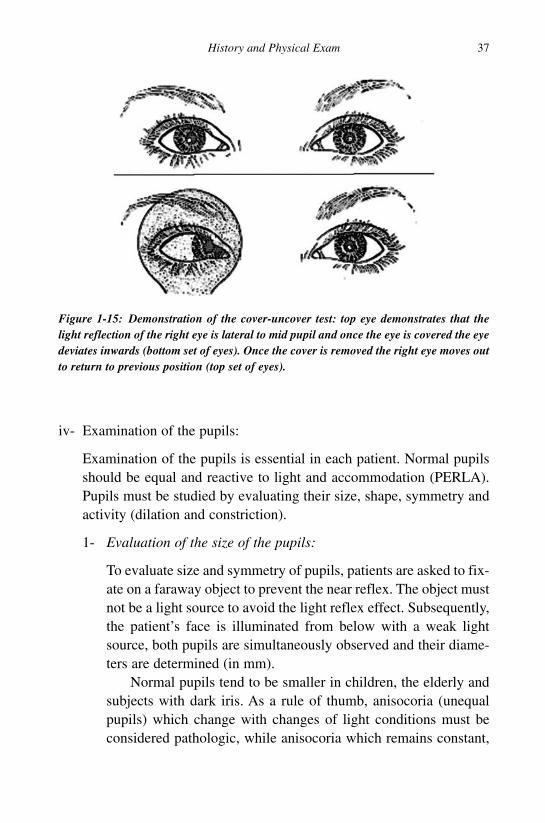
iv- Examination of the pupils:
Examination of the pupils is essential in each patient. Normal pupilsshould be equal and reactive to light and accommodation (PERLA).Pupils must be studied by evaluating their size, shape, symmetry andactivity (dilation and constriction).
1- Evaluation of the size of the pupils:
To evaluate size and symmetry of pupils, patients are asked to fix-ate on a faraway object to prevent the near reflex. The object mustnot be a light source to avoid the light reflex effect. Subsequently,the patient’s face is illuminated from below with a weak lightsource, both pupils are simultaneously observed and their diame-ters are determined (in mm).
Normal pupils tend to be smaller in children, the elderly andsubjects with dark iris. As a rule of thumb, anisocoria (unequalpupils) which change with changes of light conditions must beconsidered pathologic, while anisocoria which remains constant,
History and Physical Exam 37
b1009 Problem Based Neurosurgery
Figure 1-15: Demonstration of the cover-uncover test: top eye demonstrates that thelight reflection of the right eye is lateral to mid pupil and once the eye is covered the eyedeviates inwards (bottom set of eyes). Once the cover is removed the right eye moves outto return to previous position (top set of eyes).
b1009_Chapter-01.qxd 11/12/2010 10:49 AM Page 37

independent from the degree of light and is associated with a sym-metrical direct light reflex, is physiologic anisocoria.
2- Direct pupillary light reflex (DPLR):
The DPLR is tested by asking the patient to fixate on a farawayobject in a darkened room and shining a torch-light directly intothe pupil of each eye in turn. The normal reaction is a pupillaryconstriction (myosis). It is an important test for assessing pres-ence or absence of brainstem response in comatose patients and itis the only test used in patients who are artificially ventilated andsedated during treatment of a number of nervous, cardiovascular,respiratory and systemic diseases. Each reflex consists of fivecomponents: stimulus, afferent, centre, efferent and effecter.The stimulus in the DPLR is the light, the afferent is the ipsilat-eral optic nerve, the centre of the reflex is located in the tectum ofmidbrain at the level of the superior colliculus, the efferent is theipsilateral third nerve and the effecter is the pupillary constrictormuscles (Figure 1-16). Normal pupils should constrict at the samespeed and to the same extent unless there is a relative afferentpupillary defect (RAPD) due to visual impairment on the side ofthe sluggish pupil.
If neither pupil constricts to light the patient might be blind,has bilateral third nerve palsies, has damage of the midbrain orhas paralysis of the constrictor muscles due instillation of mydri-atic eye-drops.
3- Indirect pupillary light reflex or consensual light reflex (CPLR):
To test the CPLR, light is shone in one eye and the pupil reactionof the other eye is observed. Normal pupils constrict when the lightis shone in the other eye. The stimulus of this reflex is the light, theafferent is the ipsilateral optic nerve, the centre is the tectum of themidbrain, the efferent is the contralateral third nerve and effecter isthe pupillary constrictors of the contralateral pupil (Figure 1-16).
If the reflex arc is intact, the DPLR must be equal to theCPLR (due to the double decussating pupillary fibres in the mid-brain as well as the decussation of the nasal visual fibres in the
38 Chapter 1
b1009 Problem Based Neurosurgery
b1009_Chapter-01.qxd 11/12/2010 10:49 AM Page 38

optic chiasm). The amplitude, latency and speed of pupillary con-striction after a light stimulus are generally correlated to thevisual acuity of the patient, except in cases in which the visualdefects are secondary to a circumscribed foveal lesion or a bilat-eral cerebral lesion above the LGB (parietal, or occipital), inwhich pupillary activity is normal.
4- Near pupillary reflex (NPR):
The NPR is analysed by asking the patient to fixate on a farawayobject and then to fixate on a near object positioned in front ofthe nose. Normal pupils constrict symmetrically and both eyesconverge.
5- Evaluation of relative afferent pupillary defect (RAPD):
The presence of a relative afferent pupillary reflex (RAPD) isone of the most important signs in the assessment of visual
History and Physical Exam 39
b1009 Problem Based Neurosurgery
Figure 1-16: Diagram demonstrating the DPLR and CPLR arcs. A == Normal DPLRand CPLR in both eyes; B == normal CPLR in the right eye and normal DPLR in the lefteye; C == normal CPLR in the left eye and absent DPLR in the right eye.
b1009_Chapter-01.qxd 11/12/2010 10:49 AM Page 39

pathways as it provides objective evidence of damage to theanterior visual pathways. When light is positioned in front of ahealthy eye, both pupils constrict and then slowly dilate; whilein an affected eye, the constriction is reduced or absent, but thesubsequent dilation is immediately obvious. To correctly searchfor the presence of a RAPD it is necessary to begin with bothpupils in the dark. Each pupil is then rapidly illuminated in analternating way (for a maximum of three seconds) passingabove the nose. A unilateral lesion of the optic nerve is practi-cally always associated with RAPD, while a bilateral lesion isnot associated with RAPD unless the lesions were stronglyasymmetrical. On the other hand, retinal pathology (e.g. largeretinal detachment) may be associated with RAPD. SlightRAPD may be present in some large macular lesions and incases of amblyopia. It is generally not present in acute papil-loedema, severe refractive defects, cataract, non-organic visualloss, and cerebral lesions.
Examination of the pupils is very helpful in diagnosis andmanagement of patients (Table 1-2). Dissociation of light pupil-lary reflexes and NPR indicates the presence of midbrainpathology (Parinaud’s syndrome, Argyll-Robertson’s pupil) or theinvolvement of postganglionic parasympathetic fibres (Adie’stonic pupil).
40 Chapter 1
b1009 Problem Based Neurosurgery
Table 1-2: Pupillary signs and their interpretation
Pupillary reactions
Pupils’ sizes Right Left Conclusions
Equal Reactive Reactive Intact arc, normalLarge fixed Large fixed Blindness, bilateral third palsiesSmall fixed Small fixed Pontine haemorrhage
Unequal Large fixed Reactive Right third palsyUnequal Absent DPLR and Reactive Right optic neuropathy
present CLR
b1009_Chapter-01.qxd 11/12/2010 10:49 AM Page 40

v- Assessment of ptosis:
Droopy eyelids are common in the old age and may involve one orboth eyelids. The drooping appearance may be due to: true “eyelidptosis”, “excess eyelid skin”, “brow ptosis”, “contralateral eyelidretraction”, “enophthalmos”(sunken eye) or, a combination of theabove. The treatments for these various problems are different and itis essential to distinguish which of the contributing factors are presentin each patient so that an appropriate diagnosis and management plancan be provided.
IV- The fourth nerve (Trochlear):The trochlear nerve arises from the midbrain (2C + 2MB = 4P + 4MO). Itis the only cranial nerve that emerges from the back of the brain stem. Itpasses anterior around the ipsilateral cerebral peduncle into the anterioredge of the tentorium in the lateral wall of the CS before entering the orbitthrough the superior orbital fissure. Its slender shape, long intracranialcourse and posterior exit from the midbrain make it susceptible to injuries.It is also involved in superior orbital fissure and cavernous sinus syn-dromes. The fourth nerve supplies only the superior oblique muscle thatdepresses the eyeball. The causes of fourth nerve palsy can be broadlyclassified as congenital or acquired. Isolated congenital fourth nervepalsies may be heralded by head-tilting to the opposite side of the affectednerve in early childhood. In others, a congenital palsy may go unnoticedbecause of a compensatory mechanism allowing for alignment of the eyeswhen focusing on an object. Isolated acquired trochlear nerve palsies canbe the result of numerous disorders. In most cases an underlying causecannot be found (idiopathic palsy). Due to its long course within the cra-nium, the fourth nerve is susceptible to injury following head trauma.Depending on the site of nerve compression during trauma one or bothnerves may be affected. Aneurysms at the superior cerebellar artery4 orbrain tumours may directly compress or result in an increase of ICP result-ing in fourth nerve palsies. Disorders such as myasthenia gravis, diabetes,meningitis, microvascular disease (atherosclerotic vascular disease) orany cause of raised ICP may result in trochlear nerve palsy. In patientswith compensated fourth nerve palsy, the removal of a cataract may
History and Physical Exam 41
b1009 Problem Based Neurosurgery
b1009_Chapter-01.qxd 11/12/2010 10:49 AM Page 41
restore clear vision to both eyes allowing the patient to become aware oftheir double vision. A child with a congenital palsy may be found doing ahead-tilt by his or her parents or relatives. Children will very rarely com-plain of double vision. Adults with a new onset fourth nerve palsy willnote two images, one on top of the other or angled in position when botheyes are open. Covering one eye, no matter which one is covered, willresolve the diplopia. The double vision will worsen when looking downor away from the affected side. If both nerves are affected (s)he may expe-rience a horizontal diplopia (two images side by side) when lookingdownward. If a decompensated palsy is suspected, one should review oldphotographs to document a pre-existing head-tilt to support the diagnosis.Diagnosing fourth nerve palsy is for the most part a clinical one. Carefulhistory taking and examination is the key to diagnosis. The Bielchowskyhead-tilt test is one commonly used and reliable technique to diagnose iso-lated trochlear nerve palsies. Review of patient’s old photographs canprove indispensable in diagnosing a decompensated palsy, obviating theneed for additional testing.4
VI- Sixth cranial nerve examination (Abducens):The abducens nerve arises from the pons (2C + 2MB = 4P + 4MO). Itpasses anterior into the CS before entering the orbit through the superiororbital fissure. Its long intracranial course makes it susceptible to injuries.It is also involved in superior orbital fissure and cavernous sinus syn-dromes. The sixth nerve supplies only the lateral rectus muscle thatabducts the eyeball. It causes diplopia in the lateral gaze. The diplopia iscrossed where the left image belongs to the right eye and vice versa(Figure 1-17). Sixth nerve palsy is considered a false localising sign inhead injuries and raised ICP, however lesions at the tip of the petrous tem-poral bone may result in sixth nerve palsy (Gradenigo’s syndrome –Appendix I).5
Examination of conjugate eye movements:Conjugate eye movements are a very important part of the extra-ocularmuscle assessment. These include examination of pursuit and saccade eyemovements in addition to the aforementioned examination of third, fourthand sixth cranial nerves.
42 Chapter 1
b1009 Problem Based Neurosurgery
b1009_Chapter-01.qxd 11/12/2010 10:49 AM Page 42

1- Smooth Pursuit eye movements:
The patient is asked to follow a finger as it is slowly moved to theleft and to the right, and up and down. Make sure that the patientcan see the finger clearly and do not exceed 60 degrees in total arc or40 degrees per second. Normal eye tracking of a slowly moving dis-crete object generates a smooth eye movement that the examiner caneasily see. Cerebellar or brain stem disease can cause saccadic eyetracking in which the patient repeatedly loses the target and thencatches up with a small saccade. In most cases, abnormal pursuit isnon-localising within the nervous system, although ipsilateral loss ofpursuit can be ascribed to parietal lobe lesions.
2- Saccades eye movements:
The patient is asked to look back and forth between two outstretchedfingers held about 12 inches apart in the horizontal and vertical plane.The latency of onset, speed, accuracy, and conjugate movement areobserved. Saccadic eye movements are refixation movements thatinvolve the frontal lobes (voluntary saccades), brain stem reticularformation (voluntary and involuntary saccades), and nuclei of third,fourth and sixth cranial nerves. Delayed saccades are seen in corticaland brainstem lesions, and slow saccades accompany brainstem dis-ease. Inaccurate saccades (especially overshoots) are associated with
History and Physical Exam 43
b1009 Problem Based Neurosurgery
Figure 1-17: Sixth nerve palsy with crossed diplopia.
b1009_Chapter-01.qxd 11/12/2010 10:49 AM Page 43

lesions of the cerebellar vermis and fastigial nuclei. Dysconjugate eyemovements with slowing of the adducting eye and overshoots of theabducting eye imply medial longitudinal fasciculus (MLF) pathologyfrequently associated with multiple sclerosis.
Your personal notes:
.....................................................................................................................
.....................................................................................................................
.....................................................................................................................
.....................................................................................................................
.....................................................................................................................
.....................................................................................................................
.....................................................................................................................
.....................................................................................................................
.....................................................................................................................
.....................................................................................................................
44 Chapter 1
b1009 Problem Based Neurosurgery
b1009_Chapter-01.qxd 11/12/2010 10:49 AM Page 44

Problem 1-5: How to examine the face (fifth and seventh cranialnerves) efficiently, with confidence and make a lasting impression.(The smart way of performing neurological physical examination 4)
The fifth cranial nerve (Trigeminal) isresponsible for facial sensation andthe seventh cranial nerve (Facial) isresponsible for facial movements.During undergraduate examinations,and postgraduate examinations inneurology, general medicine, ophthal-mology and neurosurgery candidatesare often asked to demonstrate thephysical examination of the face. Patients with abnormalities of thesenerves are loved by examiners and they often feature as short or longcases during these assessments. Therefore mastering this physical exami-nation of these nerves not only helps in the evaluation and diagnosis ofpatients but is also helpful during these examinations.
V- The fifth cranial nerve (Trigeminal):The fifth cranial nerve consists of somatic sensory and motor fibres. Itinnervates muscles of mastication (masseter, temporalis, medial and lat-eral pterygoids), tensor tympani, tensor veli palatini, mylohyoid andposterior belly of digastric muscle and is responsible for facial sensation.It consists of three divisions: ophthalmic (V1), maxillary (V2) andmandibular (V3).
1-5-1 Where does the fifth nerve originate?
The fifth nerve originates in the pons (remember the formula 2C + 2MB =4P + 4MO). It has motor and sensory nuclei (Figure 1-18). The trigeminalsensory nucleus extends throughout the entire brainstem, from the mid-brain to the medulla, and continues into the cervical cord, where it mergeswith dorsal horn cells of the spinal cord. The nucleus is divided anatomi-cally into three parts. From caudal (Medulla) to rostral (Midbrain) theyare the spinal trigeminal nucleus and tract (Descending Nucleus = DN),
History and Physical Exam 45
b1009 Problem Based Neurosurgery
Problem based tool box:How to examine the face?How to examine the facial
nerve?How to examine the fifth
nerve?
b1009_Chapter-01.qxd 11/12/2010 10:49 AM Page 45

the main trigeminal nucleus,5 and the mesencephalic trigeminal nucleusand tract (Ascending Nucleus = AN) (Figure 1-18).
The three parts of the trigeminal nucleus receive different types ofsensory information. The spinal trigeminal (DN) nucleus receivespain/temperature fibres. The main trigeminal nucleus5 receivestouch/position fibres. The mesencephalic nucleus (AN) receives proprio-ception and mechanoreception fibres from the jaws and teeth. The spinaltrigeminal nucleus contains a pain/temperature sensory map of the faceand mouth. From the spinal trigeminal nucleus, secondary order neuron
46 Chapter 1
b1009 Problem Based Neurosurgery
Figure 1-18: Cranial nerve nuclei in the brain stem: 3 == 3rd nerve nucleus, 4 == 4thnerve nucleus in midbrain, 5 == 5th nerve main nucleus with AN == ascending or mesen-cephalic 5th nerve nucleus and tract and DN == descending or spinal 5th nucleus andtract, 6 == sixth nerve, 8c == cochlear nucleus, 8v == vestibular nucleus, 9 == 9th nervenucleus, 10 == 10th dorsal nucleus, 11 == accessory nucleus, 12 == hypoglossal nucleus, TS ==tractus solitarius, NA == nucleus ambigious, and MLF == medial longitudinal fasciculus.
b1009_Chapter-01.qxd 11/12/2010 10:49 AM Page 46

fibres cross the midline and ascend in the trigeminothalamic tract (TTT)to the contralateral thalamus. The TTT runs parallel to the spinothalamictract (STT), which carries pain/temperature information from the rest ofthe body. Pain/temperature fibres are sent to thalamic nuclei. The centralprocessing of pain/temperature information is markedly different from thecentral processing of touch/position information. Within the spinaltrigeminal nucleus, information is represented in an onion skin fashion.The lowest levels of the nucleus (in the upper cervical cord and lowermedulla) represent peripheral areas of the face (scalp, ears and chin).Higher levels (in the upper medulla) represent more central areas (nose,cheeks, lips). The highest levels (in the pons) represent the mouth, teeth,and pharyngeal cavity. The onion skin distribution is entirely differentfrom the dermatome distribution of the peripheral branches of the fifthnerve. Lesions that destroy lower areas of the spinal trigeminal nucleus(but which spare higher areas) preserve pain/temperature sensation in thenose (V1), upper lip (V2) and mouth (V3) while removing pain/temperaturesensation from the forehead (V1), cheeks (V2) and chin (V3). Analgesia inthis distribution is “nonphysiologic” in the traditional sense, because itcrosses over several dermatomes (Figure 1-19). Nevertheless, analgesia inexactly this distribution is found in humans after surgical sectioning of thespinal tract of the trigeminal nucleus. The spinal trigeminal nucleus sendspain/temperature information to the thalamus. It also sends informationto the mesencephalon and the reticular formation of the brainstem. The
History and Physical Exam 47
b1009 Problem Based Neurosurgery
Figure 1-19: The onion ring map of facial sensation in the trigeminal nucleus in thebrain stem and the V1, V2, V3 innervation of the face.
b1009_Chapter-01.qxd 11/12/2010 10:49 AM Page 47

latter pathways are analogous to the spinomesencephalic and spinoreticu-lar tracts of the spinal cord, which send pain/temperature informationfrom the rest of the body to the same areas. The mesencephalon modulatespainful input before it reaches the level of consciousness. The reticularformation is responsible for the automatic (unconscious) orientation of thebody to painful stimuli. The main trigeminal nucleus representstouch/position sensation from the face. It is located in the pons, close tothe entry site of the fifth nerve. Fibres carrying touch/position informationfrom the face and mouth (via cranial nerves V, VII, IX, and X) are sent tothe main trigeminal nucleus when they enter the brainstem. The maintrigeminal nucleus contains a touch/position sensory map of the face andmouth, just as the spinal trigeminal nucleus contains a complete pain/tem-perature map. The main nucleus is analogous to the dorsal column nuclei(the gracile and cuneate nuclei) of the spinal cord, which contain atouch/position map of the rest of the body. From the main trigeminalnucleus, secondary fibres cross the midline and ascend in the trigeminallemniscus to the contralateral thalamus. The trigeminal lemniscus runsparallel to the medial lemniscus, which carries touch/position informationfrom the rest of the body to the thalamus. Some sensory information fromthe teeth and jaws is sent from the main trigeminal nucleus to the ipsilat-eral thalamus, via the small dorsal trigeminal tract. Thus touch/positioninformation from the teeth and jaws is represented bilaterally in the thal-amus (and hence in the cortex). The mesencephalic trigeminal nucleus(AN) is not really a “nucleus” it is a sensory ganglion (like the trigeminalganglion) that happens to be imbedded in the brainstem. The mesen-cephalic “nucleus” is the sole exception to the general rule that sensoryinformation passes through peripheral sensory ganglia before entering thecentral nervous system. Only certain types of sensory fibres have cell bod-ies in the mesencephalic nucleus: proprioception fibres from the jaw andmechanoreceptor fibres from the teeth. Some of these incoming fibres goto the motor nucleus of fifth nerve, thus entirely bypassing the pathwaysfor conscious perception. The jaw jerk reflex is an example. Tapping thejaw elicits a reflex closure of the jaw, in exactly the same way that tappingthe knee elicits a reflex kick of the leg. Other incoming fibres from theteeth and jaws go to the main nucleus of fifth nerve. This informationis projected bilaterally to the thalamus. It is available for conscious
48 Chapter 1
b1009 Problem Based Neurosurgery
b1009_Chapter-01.qxd 11/12/2010 10:49 AM Page 48

perception. Activities like biting, chewing and swallowing require sym-metrical, simultaneous coordination of both sides of the body. They areessentially automatic activities, to which we pay little conscious attention.They involve a sensory component (feedback about touch/position) that isprocessed at a largely unconscious level. The unusual anatomy of “mes-encephalic fifth” has been found in all vertebrates, with the exception oflampreys and hagfishes. Lampreys and hagfishes are the only vertebrateswithout jaws. It is evident therefore, that information about biting, chew-ing and swallowing is singled out for special processing in the vertebratebrainstem, specifically in the mesencephalic nucleus.
1-5-2 How to localise fifth nerve lesion?
It is important to understand the common anatomical locations where thefifth nerve is susceptible to compression. Knowing these facts will helpyou localise the site of the lesion in clinical patient presentations.
1. The superior orbital fissure: leading to complete ophthalmoplegia andophthalmic trigeminal (V1) neuropathy as the third, fourth, sixth andV1 enter the orbit via the superior orbital fissure. Primary or second-ary tumours are the main causes of fifth nerve palsies at this location.In bigger and extensive tumours, this syndrome can be associatedwith proptosis or compressive optic neuropathy.
2. The cavernous sinus (CS): V1 and V2 pass in the lateral wall of theCS together with the third and fourth cranial nerves. So a CS syn-drome leads to complete ophthalmoplegia associated with trigeminalnerve dysfunction in V1 and V2, and often proptosis due to obstruc-tion of venous drainage of the orbit. The eye may be pulsatile and redin carotid-cavernous fistula (CCF).
3. Foramen ovale: where the sensory V3 enters and the motor V3 exitsthe skull base. Skull base tumours are the most common cause of V3neuropathy at this location.
4. Meckel’s cave: where the sensory trigeminal ganglion is located andthe most common causes of lesions at this location are benigntumours in the form of meningioma or schwannoma leading to sen-sory impairment in all three divisions.
History and Physical Exam 49
b1009 Problem Based Neurosurgery
b1009_Chapter-01.qxd 11/12/2010 10:49 AM Page 49

5. The cerebellopontiane angle (CPA): where the fifth nerve lies superiorand can be affected in acoustic neuroma, CPA meningioma, epider-moid cyst and other tumours.
6. The root entry zone at the fifth nerve junction with the pons: com-pression at this location most often due to vascular compressionleading to trigeminal neuralgia.
7. In the pons: leading to trigeminal neuropathy and contralateral hemi-paresis, usually due to a cavernoma, arteriovenous malformation(AVM) or ischaemia.
8. In the medulla: causes loss of pain and temperature sensation in theface and the contralateral part of the body (Wallenberg syndrome).
1-5-3 How to examine the fifth nerve?
i- Test light touch in the three divisions of the trigeminal nerve usinglight cotton wisp. If the face is hairy avoid using stroke movementsand use light touch only.
ii- Test for pain using sharp pin such as neurotip (Figure 1-20). The neu-rotip had two ends: blunt and sharp ends. Ask the patient to closehis/her eyes and randomly test by the blunt and sharp ends of the neu-rotip. If the patient can identify the sharp end correctly each time thenthe patient had no impairment of pin prick (PP). Test all the divisionsindividually and systematically. If you find an area of impaired sen-sation, chart it by testing from abnormal to normal.
iii- Corneal reflex: the corneal reflex is an important reflex protectsagainst corneal abrasions. The stimulus is light touch, the afferent isV1, the centre is in the pons, the efferent is facial nerve and the
50 Chapter 1
b1009 Problem Based Neurosurgery
Figure 1-20: Neurotip used to test pin prick sensation (left end sharp and the right endis blunt).
b1009_Chapter-01.qxd 11/12/2010 10:49 AM Page 50
effecter is the orbicularis oculi. Ask the patient to stare at a far object,use a wisp of sterile cotton, approach the eye from the lateral side toavoid the menace reflex (the menace reflex uses the optic nerve asafferent and is used to distinguish true blindness from functionalvisual symptoms), touch the cornea lightly, normal response leads tosudden closure of the eye.
iv- Test the strength of muscles of mastication by asking the patient toclose the lower jaw tightly and feel the masseter and temporalis mus-cles, ask the patient to move the jaw from side to side againstresistance to test the power of the pterygoids and to open the jawagainst resistance to test the jaw openers.
VII- The seventh cranial nerve (Facial):The seventh cranial nerve consists of somatic motor and gustatory fibres.It innervates muscles of facial expression, stapedius muscle and taste sen-sation from the anterior two thirds of the tongue. The taste sensation iscarried by the chorda tympani that joins V3, passes in the middle ear, joinsthe facial in the facial canal in the temporal bone and finally as the nervusintermedius.
1-5-4 Where does the seventh nerve originate?
The seventh nerve originates in the pons (remember the formula 2C +2MB = 4P + 4MO). It has motor and gustatory nuclei (Figure 1-21). Thefacial nucleus is located in the anterior part of the pons, its fibres pass pos-terior in the pons and loop around the sixth nerve nucleus before passingforward in the pons and exiting anterior towards the internal auditorymeatus (IAM) through the CPA. In the IAM it joins the cochlear, and thesuperior and inferior vestibular nerves (Figure 1-21).
The relationship of the seventh and eighth nerves at the IAM is veryimportant during surgery at this region. The seventh nerve occupies theanterior superior quadrant, the cochlear occupies the anterior inferiorquadrant, the superior vestibular occupies the posterior superior quadrantand the inferior vestibular nerve occupies the posterior inferior quadrant(Figure 1-22).
History and Physical Exam 51
b1009 Problem Based Neurosurgery
b1009_Chapter-01.qxd 11/12/2010 10:49 AM Page 51

The principal muscles supplied by the facial nerve are the frontalis,orbicularis oculi, buccinator, orbicularis oris, platysma, the posterior bellyof the digastric muscle, and the stapedius muscle. In nuclear or infranu-clear (“peripheral”) lesions (lower motor neuron lesion = LMN), there isa partial to complete facial paralysis with smoothing of the brow, openeye, flat nasolabial fold, and drooping of the mouth ipsilateral to thelesion. Supranuclear (“central”) lesions (upper motor neuron lesion =UMN) spares the eyebrow and eyelid musculature; there is flattening of
52 Chapter 1
b1009 Problem Based Neurosurgery
Figure 1-21: The course of the facial nerve and localisation of lesions (PAN == posteriorauricular nerve, MLF == medial longitudinal fasciculus, ML == medial lemniscus, NI ==nervus intermedius).
Figure 1-22: Schematic representation of the IAM and its contents.
b1009_Chapter-01.qxd 11/12/2010 10:49 AM Page 52

the nasolabial fold and drooping of the mouth contralateral to the lesion.This distinction of LMN from UMN is very important as that will helplocalise the pathology. For example in a patient presenting with facialweakness that spares the upper half of the face (UMN) is more likely tobe due to a lesion above the pons, and a patient presenting with a lesioninvolving all the face is likely to have a lesion in the pons outwardstowards the face. The secretory and vasomotor fibres of the facial nervego to the lachrymal gland, the mucous membranes of the nose and mouth,and the submandibular and sublingual salivary glands, and cutaneoussensory to the external auditory meatus and the region at the back of theear. Abnormalities of taste include ageusia (lack of taste); hypogeusia(diminished taste acuity); and dysgeusia (unpleasant, obnoxious, or per-verted taste).
1-5-5 How to examine the seventh nerve?
Careful and thoughtful observation is the key to discerning subtle signs offacial weakness. Note the blink, the nasolabial folds, and the corners ofthe mouth. Any asymmetry is the clue to unilateral facial weakness and isbest perceived during conversation when the patient is unaware of beingobserved.
a. The blink reflex: The eyelid on the affected side closes just a tracelater than the opposite eyelid.
b. The nasolabial folds: The weak one is flatter.c. The mouth: The affected side droops and participates manifestly less
in speaking.d. Ask the patient to look up or wrinkle the forehead; inspect for
asymmetry.e. Ask the patient to close the eyes tightly. Look for incomplete closure
or incomplete “burying” of the eyelashes on the affected side.Observe the nasolabial folds and mouth while the patient is concen-trating on the eyes. As the orbicularis oculi contract tightly, there aremilder associated contractions of muscles about the mouth and nose;these milder contractions are better suited to displaying slight weak-ness than when these muscles are tested directly.
History and Physical Exam 53
b1009 Problem Based Neurosurgery
b1009_Chapter-01.qxd 11/12/2010 10:49 AM Page 53

f. Ask the patient to smile or to show his/her teeth. Look for asymmetryabout the mouth (Figure 1-23).
g. The most subtle signs of mild facial weakness are the blink reflex andincomplete lid closure. Observe the blink reflex during conversation,or tap gently on the glabella with your index finger or reflex hammerin an attempt to bring out a mild asymmetry of blink.
h. If you strongly suspect but are having difficulty confirming a mildfacial weakness, ask the patient to lie flat on the examination couchwith face up. Slide the patient’s head off the examination couch sothe head is below the body. This forces the eyelids to work againstgravity. Now ask the patient to close both eyes and inspect for incom-plete closure. Tap on the glabella and note asymmetry of the blinkreflex.
i. Test for taste: The four primary tastes are bitter, sweet, sour, and salty.Screen for disorders of sweet or salty taste with salt and sugar. Withthe patient’s eyes closed and tongue protruded, take a tongue spatulaand smear a small amount of salt or sugar on the lateral surface andside of the tongue. Instruct the patient to tell you the identity of thesubstance. Rinse the mouth thoroughly and repeat the test on the otherside, using a different substance.
j. Facial nerve reflexes:
i. Corneal reflex: Stimulation of the cornea with a wisp of cotton pro-duces reflex closure of both ipsilateral (strongest) and contralateral
54 Chapter 1
b1009 Problem Based Neurosurgery
Figure 1-23: Photograph of right facial nerve palsy, demonstrating drooping of the sideof the mouth.
b1009_Chapter-01.qxd 11/12/2010 10:49 AM Page 54

eyelids. The fifth nerve carries the afferent impulses and the facialnerve the efferent impulses.
ii. Glabellar reflex: Tapping the glabella leads to both eyes to close,with the contralateral response being weaker. The trigeminalnerve is the afferent side and the facial nerve the efferent side ofthe reflex. Light and sound can also produce the reflex, with theoptic and acoustic nerves providing the afferent side. Theresponse is weak or abolished in LMN facial weakness, present orexaggerated in UMN lesions, and exaggerated in Parkinsonismand cannot be voluntarily inhibited.
iii. Palpebral-oculogyric reflex and Bell’s phenomenon: The eyeballsdeviate upward when the eyes are closed, when awake and asleep.The afferent arc is proprioception carried through the seventhnerve to the medial longitudinal fasciculus (MLF). The thirdnerve to the superior rectus muscles forms the efferent side. Inperipheral and nuclear lesions an exaggeration of this reflex isknown as Bell’s phenomenon.
iv. Orbicularis oris reflex: Percussion on the side of the nose or theupper lip causes ipsilateral elevation of the angle of the mouth andupper lip. The reflex arc is composed of the fifth and seventhnerves. Synonyms: nasomental, buccal, oral, or perioral reflex.This reflex disappears after about the first year of life, recurringwith supranuclear facial nerve lesions and with extra-pyramidaldiseases, such as Parkinsonism.
v. Snout reflex: Tapping the upper lip lightly causes bilateral contrac-tion of the muscles around the mouth and base of the nose. Themouth resembles a snout. This is an exaggeration of the orbicularisoris reflex. It is present with bilateral supranuclear lesions and indiffuse cerebral diseases, such as various causes of dementia.
vi. Suck reflex: Sucking movements of lips, tongue, and mouth arebrought about by lightly touching or tapping on the lips. At timesmerely bringing an object near the lips produces the reflex.Occurs in patients with diffuse cerebral lesions. The snout reflexoccurs in similar circumstances.
vii. Palmomental reflex: A stimulus of the thenar area of the handcauses a reflex contraction ipsilaterally of the orbicularis oris and
History and Physical Exam 55
b1009 Problem Based Neurosurgery
b1009_Chapter-01.qxd 11/12/2010 10:49 AM Page 55

mentalis muscles. A number of normal individuals have thisreflex, and also patients with diffuse cerebral disease. It is signif-icant when other similar reflexes are also present.
1-5-6 How to localise facial nerve lesions?
1- UMN facial weakness alone or with upper limb weakness occurs incontralateral lesions of the motor cortex.
2- UMN facial weakness with hemiparesis occurs in contralateral lesionsof internal capsule, or in midbrain.
3- LMN facial weakness with contralateral hemiparesis occurs in pon-tine lesions.
4- LMN facial weakness with sensori-neural deafness and vertigo occurin lesions of the CPA, or the IAM.
5- LMN facial weakness with conductive deafness and vertigo occur inlesions of the middle ear.
6- LMN facial weakness alone occurs in contralateral lesions in thefacial canal, stylomastoid foramen and peripherally.
Your personal notes:
.....................................................................................................................
.....................................................................................................................
.....................................................................................................................
.....................................................................................................................
.....................................................................................................................
.....................................................................................................................
.....................................................................................................................
.....................................................................................................................
.....................................................................................................................
.....................................................................................................................
56 Chapter 1
b1009 Problem Based Neurosurgery
b1009_Chapter-01.qxd 11/12/2010 10:49 AM Page 56

Problem 1-6: How to examine the eighth, ninth & tenth cranialnerves efficiently, with confidence and make a lasting impression.(The smart way of performing neurological physical examination 5)
The eighth cranial nerve (Vestibulo-Cochlear) is responsible for hearingand balance, the ninth cranial nerve(Glossopharyngeal) is responsible forswallowing and taste from posteriorone third of the tongue, and the tenthcranial nerve (Vagus) is responsible forvocal cord movements and parasym-pathetic supply to the heart and gut.During undergraduate examinations,and postgraduate examinations inneurology, general medicine, ophthal-mology and neurosurgery candidatesare often asked to demonstrate the physical examination of the eighth cra-nial nerve. Patients with deafness are loved by examiners and they oftenfeature as short or long cases during these assessments. Therefore master-ing this physical examination of these nerves not only helps in theevaluation and diagnosis of patients but it is also helpful during theseexaminations.
VIII- The eighth cranial nerve (Vestibulo-Cochlear):The eighth cranial nerve consists of three nerves: the cochlear and thesuperior and inferior vestibular nerves. The cochlear nerve is responsiblefor hearing and the vestibular nerves for balance.
1-6-1 Where does the eighth nerve originate?
The eighth nerve originates in the pons (remember the formula 2C + 2MB =4P + 4MO). It has cochlear and vestibular nuclei (Figure 1-18). It emergesfrom the anterior lateral aspect of the pons and heads towards the IAMwhere it joins the facial nerve (Figure 1-22).
History and Physical Exam 57
b1009 Problem Based Neurosurgery
Problem based tool box:How to examine 8, 9 & 10
nerves?How to perform Weber’s
test?How to perform Rinné’s test?How to perform the gag
reflex?How to assess vestibular
functions?
b1009_Chapter-01.qxd 11/12/2010 10:49 AM Page 57

1-6-2 How to examine the eighth nerve?
Patients with eighth nerve problems complain of deafness, tinnitus or ver-tigo. The hearing level (HL) is quantified relative to “normal” hearing indecibels (dB), with higher dB indicating worse hearing. Hearing loss isoften described as:
• Normal hearing: less than 25 dB in adults and 15 dB in children.• Mild hearing loss: 25–40 dB.• Moderate hearing loss: 41–65 dB.• Severe hearing loss: 66–90 dB.• Profound hearing loss: 90 dB or above.• 100 dB hearing loss is nearly equivalent to complete deafness for that
particular frequency. A score of 0 is normal. It is possible to havescores less than 0, which indicates better than average hearing.
Testing of hearing at the bedside:These tests may be used for initial assessment, but formal audiometry ispreferable. It is essential to mask the other ear by putting a finger in theother ear and rubbing the tragus at the same time. Alternatively, continu-ous rubbing of a piece of paper between thumb and index finger near themasked ear to produce a consistent broadband sound would be sufficient.However, to test for loud noises, you need to use a Barany noise box.Tuning forks are often used to test hearing at chosen frequencies, butwhisper, rubbed fingers and a ticking watch can be used.
1- Whispered voice test:
This is a useful screening test but may not be sensitive in children.The examiner stands at an arm’s length (0.6 metres) behind the seatedpatient (to prevent lip reading) and whispers a combination of num-bers and letters (for example, “6-E-4”), and then asks the patient torepeat the sequence. The examiner should quietly exhale before whis-pering to ensure as quiet a voice as possible. If the patient respondsincorrectly, the test is repeated using a different number/letter combi-nation. The patient is considered to have passed the screening testif they repeat at least three out of a possible six numbers or letters
58 Chapter 1
b1009 Problem Based Neurosurgery
b1009_Chapter-01.qxd 11/12/2010 10:49 AM Page 58
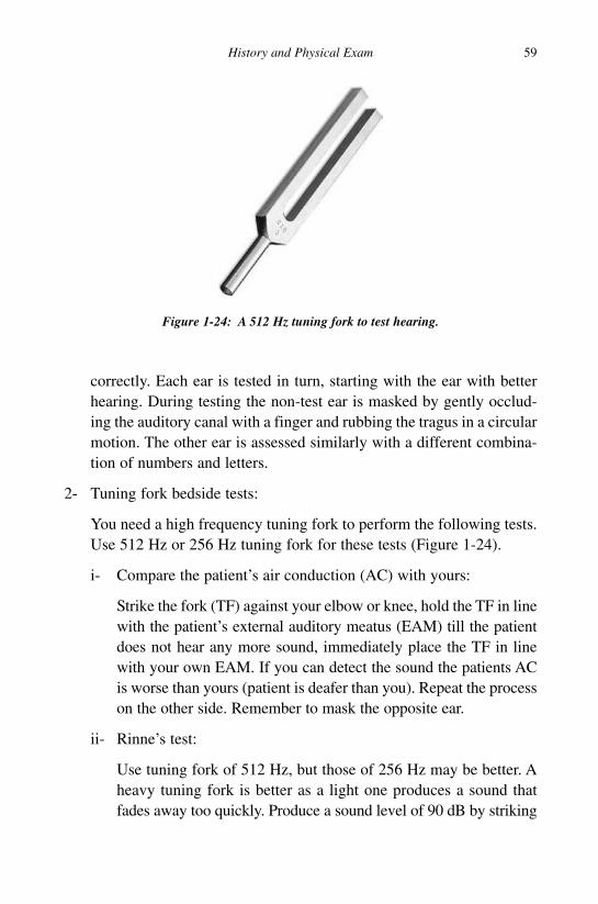
correctly. Each ear is tested in turn, starting with the ear with betterhearing. During testing the non-test ear is masked by gently occlud-ing the auditory canal with a finger and rubbing the tragus in a circularmotion. The other ear is assessed similarly with a different combina-tion of numbers and letters.
2- Tuning fork bedside tests:
You need a high frequency tuning fork to perform the following tests.Use 512 Hz or 256 Hz tuning fork for these tests (Figure 1-24).
i- Compare the patient’s air conduction (AC) with yours:
Strike the fork (TF) against your elbow or knee, hold the TF in linewith the patient’s external auditory meatus (EAM) till the patientdoes not hear any more sound, immediately place the TF in linewith your own EAM. If you can detect the sound the patients ACis worse than yours (patient is deafer than you). Repeat the processon the other side. Remember to mask the opposite ear.
ii- Rinne’s test:
Use tuning fork of 512 Hz, but those of 256 Hz may be better. Aheavy tuning fork is better as a light one produces a sound thatfades away too quickly. Produce a sound level of 90 dB by striking
History and Physical Exam 59
b1009 Problem Based Neurosurgery
Figure 1-24: A 512 Hz tuning fork to test hearing.
b1009_Chapter-01.qxd 11/12/2010 10:49 AM Page 59

the TF against your knee or elbow. To test AC, hold the TFdirectly in line with the external auditory canal. When testingbone conduction, place the flat end of the stem of the TF againstthe mastoid bone using firm pressure (loudness varies by up to15 dB with different pressures). Hold the patient’s head steadywith your other free hand. When AC is louder than BC, this isRinne’s positive and it means either normal or sensorineural deaf-ness. If bone conduction (BC) is louder than AC, it indicatesconductive deafness. The Rinne test will reliably detect a con-duction defect with an AC-BC gap of at least 30–40 dB. However,it is not a substitute for pure tone audiometry.
iii- Weber’s test:
A 512 Hz TF is placed in the midline of the patient’s forehead. Ifthe sound is louder on one side than the other, the patient mayhave either an ipsilateral (the ear hears the sound loudest) con-ductive hearing loss or a contralateral sensorineural hearing loss.
iv- Speech discrimination test (SDT):
SDT helps determine how well a person hears and understandsspeech. Spondee or spondaic words are the speech stimuli used toobtain the speech reception threshold (SRT). A spondee is definedas a two-syllable word spoken with equal stress. The SRT is thesoftest intensity level at which a patient can correctly repeat 50%of the words. Word recognition scoring is a common clinicalapproach to evaluate a person’s ability to hear and understandspeech. Lists of 20 to 50 words are presented to the patient atsupra-threshold levels, usually 30 dB above threshold. The list isphonetically balanced, which means it has speech sounds thatoccur as often as they would in everyday conversation. Out of thislist, a percentage is calculated based on correctly repeated words.Word recognition scores of 90% or higher are considered normalwhile scores below this level indicate a problem with word recog-nition. Patients with conductive hearing loss usually showexcellent word recognition. Patients with cochlear lesions havepoorer discrimination. Patients with retrocochlear lesions usually
60 Chapter 1
b1009 Problem Based Neurosurgery
b1009_Chapter-01.qxd 11/12/2010 10:49 AM Page 60
have even poorer discrimination scores which sometimes may beexacerbated by the phenomenon of “rollover”. Rollover isthought to occur as a result of changes in code intensity due toloss of monotonic stimulation and may be indicative of retro-cochlear pathology.
1-6-3 How to test vestibular functions (balance)?
Problems with the vestibular system cause central vertigo. This sensationmay be described by patients in a variety of ways: spinning, tilting,pushed to one side, light headedness, clumsiness, or even blacking out. Ifblackout was documented, a peripheral cause of the dizziness is rarely ifever at fault. Vertigo of vestibular origin is characterised by worsening onsudden movements of the head and change in posture.
1- Observe for spontaneous nystagmus:
Ask the patient to fixate on a stationary target in the neutral gaze positionwith best corrected vision (glasses or contact lenses in place). Observe fornystagmus. If nystagmus is observed, note its amplitude, direction, andeffect of target fixation. Lesions of the labyrinth and eighth vestibular (8v)produce intense, direction-fixed, horizontal-rotary nystagmus that isenhanced under Fresnel lenses. (These lenses were invented by Frenchphysicist Augustin-Jean Fresnel. Originally developed for lighthouses, thedesign enables the construction of lenses of large aperture and short focallength without the weight and volume of material required in conventionallens design. Fresnel lens therefore, is thinner, passing more light andallowing lighthouses to be visible over much longer distances. In nystag-mus, these lenses are used to make the image less clear and enhancenystagmus.) The nystagmus also intensifies when gazing in the directionof the fast phase (Alexander’s law). This pattern can be seen in lesionscausing irritation (beating toward the affected ear) and destruction (beat-ing toward the unaffected ear) of the labyrinth, 8v, or (rarely) the8v-nuclei. In contrast, lesions of the brain stem, cerebellum, and cerebrumcause less intense, direction-changing horizontal, vertical, torsional, orpendular nystagmus that is diminished under Fresnel lenses. Examples
History and Physical Exam 61
b1009 Problem Based Neurosurgery
b1009_Chapter-01.qxd 11/12/2010 10:49 AM Page 61
include periodic alternating nystagmus (PAN), congenital nystagmus, andlesions of the vermis.
2- Look for gaze nystagmus:
The patient is asked to look at a target placed 20 to 30 degrees to the leftor right off centre for 20 seconds. Gaze-evoked nystagmus is observedand change in nystagmus direction, type, or intensity of spontaneous nys-tagmus. The ability to maintain eccentric gaze is under control of thebrainstem and the vermis (flocculonodular lobes). When these mecha-nisms fail to hold the eyes in the eccentric position, the eyes drift towardthe midline (exponentially decreasing velocity), followed by refixationsaccades toward the target. Such gaze-evoked nystagmus is central in ori-gin and always beats in the direction of intended gaze. In contrast,enhancement of peripheral spontaneous nystagmus (linear slow compo-nent velocity) occurs without direction change when gazing in thedirection of the fast phase. Causes of gaze-evoked nystagmus include adrug effect (sedatives, anti-epileptics), alcohol, brain tumours, and cere-bellar degenerative syndromes.
3- Fixation Suppression Test:
The patient is asked to fixate on his/her own index finger held out in frontat an arm’s length. The examination chair is rotated up to 2 Hz while thepatient stares at his/her finger. The examiner observes for a decrease in thevisual-vestibular nystagmus that was evoked during rotation without ocu-lar fixation. The modulation of nystagmus invoked by rotation is a centralnervous system phenomenon heavily dependent on the cerebellar floccu-lus. Failure of fixation suppression in the presence of adequate visualacuity implies floccular dysfunction. This test is similar in nature to thefixation suppression performed after caloric stimulation during electro-oculography.
4- Head Thrust Test (Head Impulse Test):
The patient is asked to fixate on a target on the wall in front of them whilethe examiner moves the patient’s head rapidly to each side. The examinerlooks for any movement of the pupil during the head thrust and a refixa-tion saccade back to the target. Either direct observation of pupillary
62 Chapter 1
b1009 Problem Based Neurosurgery
b1009_Chapter-01.qxd 11/12/2010 10:49 AM Page 62

movement or the use of an ophthalmoscope is employed to document eyemovement. This test was introduced by Halmagyi and Curthoys (1988).The head impulse test was described as a reliable sign of reduced vestibu-lar function in the plane of rotation for the ear ipsilateral to the head thrust.The observation of eye movement during the manoeuvre is a sign ofdecreased neural input from the ipsilateral ear to the vestibulo-ocularreflex (VOR) because the contralateral ear is in inhibitory “saturation”and cannot supply enough neural activity to stabilise gaze. In suchinstances, the eye travels with the head during the high-velocity move-ment, and a refixation saccade is necessary to fixate the target. Bilateralrefixation movements are seen frequently in cases of ototoxicity.
5- Postheadshake Nystagmus:
The patient’s head is tilted forward 30 degrees and the examiner shakesthe patient’s head in the horizontal plane at 2 Hz for 20 seconds andobserves for postheadshake nystagmus and notes the direction and anyreversal of nystagmus. Fresnel lenses are used to avoid fixation and makethe nystagmus more obvious. The manoeuvre may be repeated in the ver-tical direction. Postheadshake nystagmus is considered a pathologic signof imbalance in the vestibular inputs in the plane of rotation. In nystagmuscaused by peripheral causes the nystagmus is directed toward the strongerear with small reversal phase sometimes observed. Signs of central aeti-ology include prolonged nystagmus, vertical nystagmus followinghorizontal headshake (“cross coupling”), and dysconjugate nystagmus.
6- Vestibular Caloric Test (VCT):
The VCT is a test of the vestibulo-ocular reflex (VOR) that involves irri-gating cold or warm water or air into the external auditory canal. It iscommonly used by audiologists and other trained professionals to validatea diagnosis of asymmetric function in the peripheral vestibular system.Caloric test is usually a subtest of the electronystagmography (ENG) bat-tery of tests. It is one of several tests which can be used to test forbrainstem death (BSD). Cold (not more than seven degrees less than nor-mal, 30°C) or warm (not more than seven degrees above normal, 44°C)water or air is irrigated into the external auditory canal, usually using asyringe. In patients with an intact cerebrum: if the water is cold relative to
History and Physical Exam 63
b1009 Problem Based Neurosurgery
b1009_Chapter-01.qxd 11/12/2010 10:49 AM Page 63

body temperature (30°C), the eyes turn toward the ipsilateral ear, withhorizontal nystagmus (quick horizontal eye movements) to the contralat-eral ear. If the water is warm (44°C) the eyes turn toward the contralateralear, with horizontal nystagmus to the ipsilateral ear. Absent reactive eyemovement suggests vestibular weakness of the horizontal semicircularcanal of the side being stimulated. In comatose patients with cerebraldamage, the fast phase of nystagmus will be absent as this is controlled bythe cerebrum. As a result, using cold water irrigation will result in devia-tion of the eyes toward the ear being irrigated. If both phases are absent,this suggests the patient’s brainstem reflexes are also damaged and carriesa very poor prognosis. The acronym COWS is used to remember: “Coldwater = fast phase of nystagmus to the side Opposite from the cold waterfilled ear, Warm water = fast phase of nystagmus to the Same side as thewarm water filled the ear”.
IX- The ninth cranial nerve (Glossopharyngeal):The ninth cranial nerve supplies the stylopharyngeal muscle and carriessensation from the back of the tongue and the larynx, is responsible fortaste sensation from the posterior third of the tongue, and supplies theparotid gland.
1-6-4 Where does the ninth nerve originate?
The ninth nerve originates in the medulla oblongata (remember the formula2C + 2MB = 4P + 4MO). Its sensory fibres end up in the spinal tract of thetrigeminal nerve. The ninth nerve can be regarded as a purely sensory nerve.
1-6-5 How to examine the ninth cranial nerve?
The ninth nerve can only be evaluated as such. The main tests used are:
1- Pharyngeal sensation:
Touch the back of the throat gently with an orange stick on either side ofthe midline and ask the patient to compare both sides. If in doubt you canuse a long pin to test for pain.
64 Chapter 1
b1009 Problem Based Neurosurgery
b1009_Chapter-01.qxd 11/12/2010 10:49 AM Page 64

2- The gag reflex (GR):
The GR is a reflex contraction of the back of the throat, evoked by touch-ing the back of throat. The GR helps prevent choking. The afferent ofthe reflex is the ninth nerve, which inputs to the nucleus solitarius(Figure 1-18), and the efferent is the vagus nerve from the nucleusambiguus. Absence of the gag reflex can be a symptom of a number ofdamages to the ninth nerve, the tenth nerve, or brainstem. However, stud-ies indicated that up to one-third of healthy people do not have a GR. TheGR sometimes is triggered intentionally to induce vomiting, by those whosuffer from bulimia nervosa.
X- The tenth cranial nerve (Vagus):The vagus nerve is the only nerve that starts in the brainstem and extends,through the jugular foramen, down below the head, to the neck, chest andabdomen, where it contributes to the innervations of the viscera. Besidesoutput to the various organs in the body, the tenth nerve conveys sensoryinformation about the state of the body’s organs to the central nervous sys-tem. The left tenth nerve is mainly sensory and the right is mainly motor,however 80–90% of the fibres are sensory.
1-6-6 Where does the tenth nerve originate?
The tenth nerve originates in the medulla oblongata (remember the for-mula 2C + 2MB = 4P + 4MO). It passes through the jugular foramentogether with the internal jugular-sigmoid sinus junction, the ninth and11th cranial nerves, and then passes in the carotid sheath where they arejoined by the internal carotid artery (Figure 1-25).
In the superior mediastinum, the tenth nerve gives out its recurrentlaryngeal nerve (RLN). The RLN supplies the laryngeal muscles that movethe vocal cords. The left RLN loops around the arch of the aorta while theright RLN loops around the subclavian artery. Therefore the left RLN is lesslikely to be stretched during anterior cervical surgery (longer and mediallylocated), e.g. during approach to cervical discs where the larynx andpharynx are retracted to the right during left-sided approach and to theleft during right-sided approach. Hence hoarseness is more common after
History and Physical Exam 65
b1009 Problem Based Neurosurgery
b1009_Chapter-01.qxd 11/12/2010 10:49 AM Page 65

right-sided approaches. Damage to the vagus causes dysphonia or hoarse-ness, while stimulation of the vagus nerve produces bradycardia. Becausethe right vagus is mainly motor, stimulation of the right vagus producesbradycardia and diarrhoea, while stimulation of the left which is purely sen-sory does not have these side effects and is used to treat epilepsy anddepression. The vagus nerve supplies motor parasympathetic fibres to allthe organs from the neck down to the second segment of the transversecolon, except the suprarenal (adrenal) glands. The vagus also controls a fewskeletal muscles, namely: the cricothyroid, the levator veli palatini, salpin-gopharyngeus, palatoglossus, palatopharyngeus, and the superior, middleand inferior pharyngeal constrictor muscles of the larynx (for speech). Thismeans that the vagus nerve is responsible for such varied tasks as heart rate,gastrointestinal peristalsis, sweating, and quite a few muscle movements inthe mouth, including speech (via the RLN) and keeping the larynx open forbreathing. It also receives some sensation from the outer ear, via the auric-ular branch (Alderman’s nerve) and part of the meninges. The right tenthnerve innervates the sinoatrial node. The vagus nerve has three nuclei asso-ciated with cardiovascular control: the dorsal motor nucleus, the nucleusambiguus and the solitary nucleus. The parasympathetic output to the heart
66 Chapter 1
b1009 Problem Based Neurosurgery
Figure 1-25: Diagram of the ninth and tenth cranial nerves (G == ganglion).
b1009_Chapter-01.qxd 11/12/2010 10:49 AM Page 66

comes mainly from neurons in the nucleus ambiguus and to a lesser extentfrom the dorsal motor nucleus. The solitary nucleus receives sensory inputabout the state of the cardiovascular system, being an integration hub for thebaroreflex. Anticholinergics such as atropine and scopolamine are calledvagolytic because they inhibit the action of the vagus on the heart, gas-trointestinal tract and other organs. Anticholinergics increase heart rate andare used to treat bradycardia and asystole. Vagus nerve stimulation (VNS)therapy using a pacemaker-like device implanted in the chest is a treatmentused since 1997 to control seizures in epilepsy patients and has recentlybeen approved for treating drug-resistant cases of clinical depression. VNSmay also be achieved by one of the vagus manoeuvres: holding the breathfor a few seconds, dipping the face in cold water, coughing, or tensing thestomach muscles as if to bear down to have a bowel movement (valsalvamanoeuvre). Patients with supraventricular tachycardia (STV), atrial fibril-lation (AF), and other illnesses may be trained to perform vagusmanoeuvres (or find one or more on their own). Vagus nerve blocking vago-tomy (cutting of the vagus nerve) is a now-obsolete therapy that wasperformed for peptic ulcer. Vagotomy is currently being researched as a lessinvasive alternative weight loss procedure to gastric bypass surgery.Excessive activation of the vagus nerve during emotional stress can causevasovagal syncope because of a sudden drop in blood pressure and heartrate. Vasovagal syncope affects young children and women more often. Itcan also lead to temporary loss of bladder control under moments ofextreme fear. Vagus damage leads to loss of the GR and deviation of theuvula away from the side of lesion, and there is failure of palate elevation.
1-6-7 How to examine the tenth cranial nerve?
1- Inspection of the uvula:
The uvula should be midline in normal patients and deviated to the otherside of vagus lesion. Ask the patient to say “Aah” and watch the uvulamovements.
2- Gag reflex (GR):
The gag reflex was discussed under the ninth cranial nerve.
History and Physical Exam 67
b1009 Problem Based Neurosurgery
b1009_Chapter-01.qxd 11/12/2010 10:49 AM Page 67

Your personal notes:
.....................................................................................................................
.....................................................................................................................
.....................................................................................................................
.....................................................................................................................
.....................................................................................................................
.....................................................................................................................
.....................................................................................................................
.....................................................................................................................
.....................................................................................................................
.....................................................................................................................
68 Chapter 1
b1009 Problem Based Neurosurgery
b1009_Chapter-01.qxd 11/12/2010 10:49 AM Page 68

Problem 1-7: How to examine the 11th and 12th cranial nervesefficiently, with confidence and make a lasting impression. (Thesmart way of performing neurological physical examination 6)
The 11th cranial nerve (accessory)is responsible for innervating thesternocleidomastoid (SCM) and thetrapezium muscles and the 12th cra-nial nerve (hypoglossal) is responsiblefor tongue.
During undergraduate examina-tions, and postgraduate examinationsin neurology, general medicine, ophthalmology and neurosurgery candi-dates are often asked to demonstrate the physical examination of thesetwo nerves. Patients with tongue paralysis or torticollis are loved byexaminers and they often feature as short or long cases during theseassessments. Therefore mastering this physical examination of thesenerves not only helps in the evaluation and diagnosis of patients but it isalso helpful during these examinations.
XI- The eleventh cranial nerve (Accessory):Anatomically, the 11th nerve consists of two components: cranial part thateventually joins the tenth nerve and spinal part that supplies the SCM andtrapezium muscles. The 11th nerve supplies also the palatoglossus muscleof the tongue, the only muscle of the tongue not supplied by the 12th nerve.The cranial component however is now considered part of the vagus and notpart of the accessory nerve proper. Therefore in practice when you are askedto examine the 11th nerve, concentrate on examining the spinal component.
1-7-1 Where does the 11th nerve originate?
The 11th nerve originates in the medulla (remember the formula 2C +2MB = 4P + 4MO). However, what we consider in practice as the 11thnerve arises from the cervical spinal cord, it ascends up through the fora-men magnum and out through the jugular foramen joining the vagus andglossopharyngeal nerves (Figure 1-26).
History and Physical Exam 69
b1009 Problem Based Neurosurgery
Problem based tool box:How to examine the tongue?How to examine the 11th nerve?How to examine the 12th nerve?How to localise lesions of 11th
and 12th cranial nerves?
b1009_Chapter-01.qxd 11/12/2010 10:49 AM Page 69

1-7-2 How to examine the 11th nerve?
Patients with 11th nerve problems complain of torticollis or drooping ofthe shoulder. The sternocleidomastoid (SCM) muscle tilts and rotates thehead, so torticollis is either caused by spasm ipsilateral to the torticollis orweakness of the contralateral SCM. The trapezium muscle has severalactions on the scapula, including shoulder elevation and adduction. Rangeof motion and strength testing of the neck and shoulders can be measuredduring a neurological examination to assess function of the spinal acces-sory nerve. Limited range of motion or poor muscle strength is suggestiveof damage to the 11th nerve. Damage of the 11th nerve can be caused byinjury to the spinal accessory nerve most commonly iatrogenic duringhead and neck surgery.
• Inspect the SCM and trapezium muscles looking for wasting, fascic-ulation or deformity of the neck.
• To assess the SCM power, ask the patient to turn the head to one sideagainst resistance.
• To assess the strength of trapezium muscle, ask the patient to shrugthe shoulders against resistance.
70 Chapter 1
b1009 Problem Based Neurosurgery
Figure 1-26: The 11th cranial nerve (NA == nucleus ambiguus).
b1009_Chapter-01.qxd 11/12/2010 10:49 AM Page 70

XII- The twelfth nerve (Hypoglossal):The 12th cranial nerve supplies all the tongue muscles apart from the palato-glossus which is supplied by the 11th cranial nerve as mentioned before.
1-7-3 Where does the 12th nerve originate?
The 12th nerve arises from the hypoglossal nucleus in the medulla oblon-gata (remember the formula 2C + 2MB = 4P + 4MO) and emerges in thepre-olivary sulcus separating the olive and the pyramid. It then passesthrough the hypoglossal canal. On emerging from the hypoglossal canal,it gives off a small meningeal branch and picks up a branch from the ante-rior ramus of C1. It spirals behind the vagus nerve and passes between theinternal carotid artery and internal jugular vein lying on the carotid sheath.After passing deep to the posterior belly of the digastric muscle, it passesto the submandibular region to enter the tongue. It supplies motor fibresto all of the muscles of the tongue, except the palatoglossus muscle whichis innervated by the 11th nerve, which runs in part with the vagus nerve.
1-7-4 How to examine the 12th cranial nerve?
To test the function of the 12th nerve, the patient is asked to stick out theirtongue. If there was loss of function on one side (unilateral paralysis) the
History and Physical Exam 71
b1009 Problem Based Neurosurgery
Figure 1-27: Photograph of a right paralysed hypoglossal.
b1009_Chapter-01.qxd 11/12/2010 10:49 AM Page 71

tongue will appear atrophic on the paralysed side and may have some fas-ciculations. The tongue points towards the affected side (Figure 1-27).
The strength of the tongue can be tested by getting the patient to pokethe inside of his/her cheek, and feeling how strongly the patient can pusha hand pushed against the patient’s cheek.
72 Chapter 1
b1009 Problem Based Neurosurgery
b1009_Chapter-01.qxd 11/12/2010 10:49 AM Page 72

Problem 1-8: How to examine the motor system efficiently, withconfidence and make a lasting impression. (The smart way ofperforming neurological physical examination 7)
Examination of the motor system isvery important in all patients, particu-larly in the upper and lower limbs.Symptoms related to dysfunction of themotor system include the following:
• Weakness.• Stiffness of the limbs.• Slowness.• Tremor or shaking.• Abnormal movements.• Poor hand grip.• Leg gives way.• Falls.
To examine the motor system you need to assess the following components:
1- Muscle shape and mass.2- Abnormal movements.3- Muscle tone.4- Muscle power.5- Reflexes.
Always observe first (muscle mass and shape), inspect second (individualmuscles for abnormal movements), palpate third (muscle tenderness, toneand power), and use specials bedside tests last (reflexes).
• Observe: Carefully observe the patient to detect any twitches,tremors, dyskinesia, other involuntary movements, or any unusualpaucity of movements. Note the patient’s posture and gait.
• Inspect: Next closely inspect several individual muscles for musclewasting, hypertrophy or fasciculation. Fasciculations are best
History and Physical Exam 73
b1009 Problem Based Neurosurgery
Problem based tool box:How to examine the motor
system?How to assess muscle tone and
power?What is the MRC grades?How to demonstrate deep
reflexes?How to localise a lesion along
the motor system pathways?
b1009_Chapter-01.qxd 11/12/2010 10:49 AM Page 73

observed in the small muscles of the hand, shoulder girdle muscles,and the thigh muscles.
• Palpate: Palpate for muscle tenderness if you suspect myositis.
1-8-1 How to assess muscle tone?
• Muscle tone: To assess the muscle tone, ask the patient to relax, flex andextend the patient’s fingers, wrist, and elbow, flex and extend patient’sankle and knee. Normally there is a small, continuous resistance to pas-sive movement. Observe for decreased (flaccid) or increased (rigid/spastic) muscle tone. Abnormal muscle tone can be described as:
1- Hypotonia:
Hypotonia means decreased muscle tone. It can either be congenitalor acquired. Congenital hypotonia is called benign congenital hypo-tonia and is usually detected during infancy. An infant with hypotoniaexhibits a floppy quality (rag doll) feeling when (s)he is held. Infantswith this problem lag behind in acquiring certain fine and gross motordevelopmental milestones that enable a baby to hold his/her head upwhen placed prone, balance themselves or sit upright and remainseated without falling over. Since the muscles that support the bonejoints are so soft, there is a tendency for hip, jaw and neck dislocationsin these infants and their joints can be hyperextended. Some childrenwith hypotonia may have trouble feeding if they are unable to suck orchew for long periods. A child with hypotonia may also have prob-lems with speech or exhibit shallow breathing. Acquired hypotoniamay occur in muscular dystrophy.
2- Flaccidity (flaccid tone):
Flaccidity also means reduced or absent muscle tone. This is oftenindicative of LMN lesions in the anterior horn cells, e.g. in motor neu-ron disease (MND) or poliomyelitis, peripheral nerve such as inperipheral nerve injuries, radiculopathy or peripheral nerve compres-sion. In acute spinal cord injury (spinal shock) the muscle tone can beflaccid despite the lesion being UMN in nature.
74 Chapter 1
b1009 Problem Based Neurosurgery
b1009_Chapter-01.qxd 11/12/2010 10:49 AM Page 74
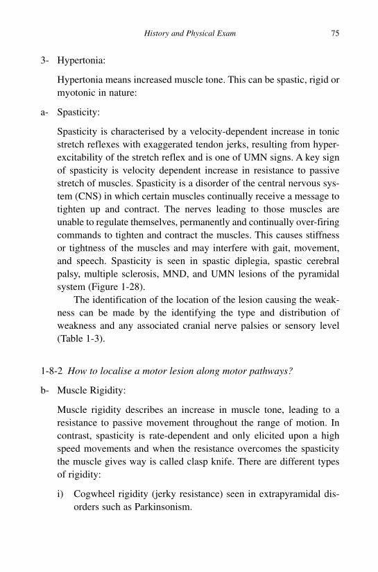
3- Hypertonia:
Hypertonia means increased muscle tone. This can be spastic, rigid ormyotonic in nature:
a- Spasticity:
Spasticity is characterised by a velocity-dependent increase in tonicstretch reflexes with exaggerated tendon jerks, resulting from hyper-excitability of the stretch reflex and is one of UMN signs. A key signof spasticity is velocity dependent increase in resistance to passivestretch of muscles. Spasticity is a disorder of the central nervous sys-tem (CNS) in which certain muscles continually receive a message totighten up and contract. The nerves leading to those muscles areunable to regulate themselves, permanently and continually over-firingcommands to tighten and contract the muscles. This causes stiffnessor tightness of the muscles and may interfere with gait, movement,and speech. Spasticity is seen in spastic diplegia, spastic cerebralpalsy, multiple sclerosis, MND, and UMN lesions of the pyramidalsystem (Figure 1-28).
The identification of the location of the lesion causing the weak-ness can be made by the identifying the type and distribution ofweakness and any associated cranial nerve palsies or sensory level(Table 1-3).
1-8-2 How to localise a motor lesion along motor pathways?
b- Muscle Rigidity:
Muscle rigidity describes an increase in muscle tone, leading to aresistance to passive movement throughout the range of motion. Incontrast, spasticity is rate-dependent and only elicited upon a highspeed movements and when the resistance overcomes the spasticitythe muscle gives way is called clasp knife. There are different typesof rigidity:
i) Cogwheel rigidity (jerky resistance) seen in extrapyramidal dis-orders such as Parkinsonism.
History and Physical Exam 75
b1009 Problem Based Neurosurgery
b1009_Chapter-01.qxd 11/12/2010 10:49 AM Page 75

76 Chapter 1
b1009 Problem Based Neurosurgery
Figure 1-28: Schematic representation of the motor pathways (pyramidal system).
b1009_Chapter-01.qxd 11/12/2010 10:49 AM Page 76

ii) Lead-pipe rigidity (continuous rigidity). These various forms ofrigidity can be seen in different forms of movement disorderssuch as Parkinson’s.
iii) Decerebrate rigidity: this is spasticity in extensor musclesdescribed in animals with brain stem transaction (Figure 1-29).
iv) Decorticate rigidity: this spasticity in the flexor muscles describedin patients with diffuse axonal injury (DAI) (Figure 1-29).
1-8-3 How to assess muscle power?
• Muscle power:
Test muscle strength by having the patient move against your resistance.Make sure that you test across one joint at a time by holding the limb by
History and Physical Exam 77
b1009 Problem Based Neurosurgery
Table 1-3: Localisation of lesions causing motor weakness: correlate these lesions withdiagram in Figure 1-28 above
Associated AssociatedLesion location Type Distribution nerve palsy sensory level
1- Motor cortex UMN One limb None None2- Subcortical UMN Hemiparesis None None3- Int. capsule UMN Hemiparesis UMN- Facial None4- Midbrain UMN Hemiparesis LMN- Third None5- Pons UMN Hemiparesis LMN- Facial None6- Medulla UMN Hemiparesis LMN- 12th None7- Cervical cord UMN Tetraparesis None At C3 to C88- Thoracic cord UMN Paraparesis None At T1 to T129- Conus Mixed Paraparesis None At L1 to L5
10- Cauda equina LMN Paraparesis None At L1 to S1
Weakness
Figure 1-29: Decerebrate and decorticate rigidities.
b1009_Chapter-01.qxd 11/12/2010 10:49 AM Page 77

the other hand, e.g. testing the power across the elbow joint ask the patientto flex the elbow, hold the upper arm by one hand and the forearm by theother hand and ask patient to flex against your resistance then extendagainst resistance (Figure 1-30). Always compare one side to the otherand grade muscle strength on MRC (Medical Research Council) scalefrom 0 to 5 (Table 1-4).
Test the following muscle groups:
1- Finger abduction (C8, T1, ulnar nerve): ask the patient to spread theirfingers apart and keep them apart against your resistance. Use yourtwo index fingers to exert the resistance (Figure 1-31).
78 Chapter 1
b1009 Problem Based Neurosurgery
Figure 1-30: Diagram demonstrating how to examine muscle strength in the upper andlower limbs.
Table 1-4: MRC scale for grading muscle power
Grade Description
0 No muscle movement1 Visible muscle movement, flicker, but no movement at the joint2 Movement at the joint, but not against gravity3 Movement against gravity, but not against added resistance4 Movement against resistance, but less than normal5 Normal strength
b1009_Chapter-01.qxd 11/12/2010 10:49 AM Page 78

2- Opposition of the thumb (C8, T1, median nerve): ask the patient tobring his/her thumb towards the little finger against resistance or askthe patient to oppose the thumb and index together and prevent youopening the formed ring (Figure 1-32).
3- Hand grip: ask the patient to squeeze two of your fingers as hard aspossible (C7, C8, T1) (Figure 1-33).
4- Extension at the wrist (C6, C7, C8, radial nerve): ask the patient tomake a fist and exert pressure to overcome it (Figure 1-34).
History and Physical Exam 79
b1009 Problem Based Neurosurgery
Figure 1-31: Finger abduction.
Figure 1-32: Thumb opposition.
b1009_Chapter-01.qxd 11/12/2010 10:49 AM Page 79

5- Flexion at the elbow (C5, C6, biceps): ask patient to flex the elbowagainst resistance (Figure 1-30).
6- Extension at the elbow (C6, C7, C8, triceps): ask the patient to extendthe elbow against your resistance (Figure 1-35).
7- Shoulder abduction (mainly C5, deltoid): ask patient to abduct theshoulders to 90 degrees with the elbow flexed but hands apart, exertpressure down to abduct the shoulders asking patient to resist yourpressure (Figure 1-36).
80 Chapter 1
b1009 Problem Based Neurosurgery
Figure 1-33: Assessment of hand grip.
Figure 1-34: Wrist extension.
b1009_Chapter-01.qxd 11/12/2010 10:49 AM Page 80

8- Flexion at the hip (L2, L3, L4, iliopsoas): ask the patient to elevate theknee against resistance (Figure 1-37).
9- Adduction at the hips (L2, L3, L4, adductors): ask the patient to flexthe hip and knee by 90 degrees and bring the knee to the opposite sideagainst resistance.
10- Abduction at the hips (L4, L5, S1, gluteus medius and minimus): fromthe same position above ask patient to move the knee outwardsagainst resistance.
11- Extension at the hips (S1, gluteus maximus): ask patient to resist yourattempt to elevate the thigh.
History and Physical Exam 81
b1009 Problem Based Neurosurgery
Figure 1-35: Elbow extension.
Figure 1-36: Shoulder abduction.
b1009_Chapter-01.qxd 11/12/2010 10:49 AM Page 81

12- Extension at the knee (L2, L3, L4, quadriceps): with the knee and hipflexed 90 degrees ask patient to extend the knee against resistance(Figure 1-38).
13- Flexion at the knee (L4, L5, S1, S2, hamstrings): from the sameprevious position ask patient to flex the knee against resistance(Figure 1-39).
14- Dorsiflexion at the ankle (L4, L5): ask patient to dorsiflex the footagainst resistance (Figure 1-40).
15- Plantar flexion (S1): ask patient to push against your hand as if push-ing an accelerator of a vehicle (Figure 1-41).
16- Extensor hallucis longus (L5): ask patient to dorsiflex the big toeagainst resistance (Figure 1-42).
17- Pronator drift: Ask the patient to out-stretch both upper limbs straightforward for 20–30 seconds with palms up, and eyes closed. Instructthe patient to keep the arms still while you tap them briskly down-ward. The patient will not be able to maintain extension andsupination and drifts into pronation with UMN disease.
82 Chapter 1
b1009 Problem Based Neurosurgery
Figure 1-37: Illustration of hip flexion.
b1009_Chapter-01.qxd 11/12/2010 10:49 AM Page 82

History and Physical Exam 83
b1009 Problem Based Neurosurgery
Figure 1-38: Knee extension.
Figure 1-39: Knee flexion.
b1009_Chapter-01.qxd 11/12/2010 10:49 AM Page 83

1-8-4 How to elicit the reflexes?
• Reflexes:
Normally when a muscle tendon is tapped briskly, the muscle immedi-ately contracts. This is an involuntary two-neuron reflex arc involvingthe spinal or brainstem segment that innervates the muscle. The afferentneuron whose cell body lies in a dorsal root ganglion or sensory ganglion
84 Chapter 1
b1009 Problem Based Neurosurgery
Figure 1-40: Dorsiflexion of the foot.
Figure 1-41: Plantar flexion of the foot.
b1009_Chapter-01.qxd 11/12/2010 10:49 AM Page 84

of the cranial nerves, innervates the muscle or Golgi tendon organ asso-ciated with the muscle;6 the efferent neuron is an alpha motor-neuron inthe anterior horn of the spinal cord or the motor nucleus of a cranialnerve. The cerebral cortex and a number of brainstem nuclei exert influ-ence over the sensory input of the muscle spindles by means of thegamma motor-neurons that are located in the anterior horn of the spinalcord; these neurons supply a set of muscle fibres that control the lengthof the muscle spindle itself. The reflexes could be either normal, hypo-or hyperreflexia:
Hyporeflexia: is an absent or diminished response to tapping. It usuallyindicates a disease that involves one or more of the components of thetwo-neuron reflex arc itself.
Hyperreflexia: refers to hyperactive or repeating (clonic) reflexes. Theseusually indicate an interruption of corticospinal and other descendingpathways that influence the reflex arc due to a UMN lesion, that is, alesion above the level of the reflex pathways.
By convention the deep tendon reflexes are graded as in Table 1-5.
History and Physical Exam 85
b1009 Problem Based Neurosurgery
Figure 1-42: Extensor hallucis longus examination.
b1009_Chapter-01.qxd 11/12/2010 10:49 AM Page 85

Whether the 1+ and 3+ responses are normal depends on what theywere previously, what the other reflexes are, and analysis of associatedfindings such as muscle tone, muscle strength, or other evidence of dis-ease. Asymmetry of reflexes suggests an abnormality.
Methods: In a screening examination you will usually find it more con-venient to integrate the reflex examination into the rest of the examinationof that part of the body, e.g. do the upper extremity reflexes when exam-ining the rest of the upper extremity. When an abnormality of the reflexesis suspected or discovered, the reflexes should be examined as a groupwith careful attention paid to the method of the examination. Valid testresults are best obtained in a fully relaxed patient and not thinking aboutwhat you are doing. After a general explanation, mingle the specificinstructions with questions or comments designed to distract the patient totalk at some length about some unrelated topic. If you cannot get anyresponse with a specific reflex, have the patient strongly contract a mus-cle not being tested, e.g.
• In the upper extremity ask the patient to clench their teeth tightly orhave the patient make a fist with one hand while the opposite extrem-ity is being tested.
• If the reflex being tested is in the lower limbs, have the patient per-form the Jendrassik manoeuvre, to reinforce the reflex. The patient’sfingers of each hand are hooked together so each arm can forcefullypull against the other. The split second before you are ready to tap thetendon, say pull.7,8
86 Chapter 1
b1009 Problem Based Neurosurgery
Table 1-5: Grading of the deep tendon reflexes
Grade Description Interpretation
0 No response Always abnormal1+ Present with reinforcement May or may not be abnormal2+ Brisk Normal3+ Very brisk May or may not be abnormal4+ Clonic (repeating) response Always abnormal5+ Sustained clonus Always abnormal
b1009_Chapter-01.qxd 11/12/2010 10:49 AM Page 86

The best position to perform these reflexes is for the patient to be sitting onthe side of the bed or examination couch. Use a reflex hammer (Figure 1-43).Hold the hammer so that movement occurs at the wrist joint of your hand notat your elbow. Use a brisk but not painful tap. Use your wrist, not your arm,for the action. In an extremity a useful manoeuvre is to elicit the reflex fromseveral different positions, rapidly shifting the limb and performing the test.
Use varying force and note any variance in the response. Note the fol-lowing features of the reflex response: the amount of hammer forcenecessary to obtain contraction, the velocity of contraction, the strengthand duration of the muscle contraction, the duration of relaxation phase,and the response of other muscles nearby. When a reflex is hyperactive,that muscle often will respond to the testing of a nearby muscle. A goodexample is reflex activity of a hyperactive biceps or finger reflex when thebrachioradialis tendon is tapped. This is termed overflowing of a reflex.After obtaining the reflex on one side, always go immediately to theopposite side for the same reflex so that you can compare them. These arethe reflexes tested normally:
Jaw Jerk (JJ): Place the tip of your index finger on a relaxed jaw, onethat is about one-third open. Tap briskly on your index finger and note thespeed as the mandible is flexed.
Pectoralis Jerk (PJ): The chest is exposed, feel the pectoris major mus-cle tendon, ask patient to relax, place index and middle fingers over thetendon and tap it with the reflex hammer. Observe the muscle contraction.
Deltoid Jerk (DJ): Feel the tendon of the deltoid about 1/3 the lateralaspect of the upper arm, put two fingers over the tendon, tap gently withreflex hammer, and watch the deltoid muscle for any contraction.
History and Physical Exam 87
b1009 Problem Based Neurosurgery
Figure 1-43: The reflex hammer.
b1009_Chapter-01.qxd 11/12/2010 10:49 AM Page 87

Biceps Jerk (BJ): The forearm should be supported, either resting on thepatient’s thighs or resting on the forearm of the examiner. The arm is mid-way between flexion and extension. Place your thumb or index firmlyover the biceps tendon, with your fingers curling around the elbow, andtap briskly (Figure 1-44). The forearm will flex at the elbow.
Triceps Jerk (TJ): Support the patient’s forearm by cradling it with yoursor by placing it on the thigh, with the arm midway between flexion andextension. Identify the triceps tendon at its insertion on the olecranonprocess, and tap just above the insertion (Figure 1-45). There is extensionof the forearm.
Brachioradialis (Supinator) Jerk (SJ): The patient’s arm should be sup-ported. Identify the brachioradialis tendon at the wrist. It inserts at the baseof the styloid process of the radius, usually about 1 cm lateral to the radialartery. If in doubt, ask the patient to hold the arm as if in a sling (flexed at
88 Chapter 1
b1009 Problem Based Neurosurgery
Figure 1-44: Demonstration of the biceps jerk.
b1009_Chapter-01.qxd 11/12/2010 10:49 AM Page 88

the elbow and halfway between pronation and supination) and then flex theforearm at the elbow against resistance from you. The brachioradialis andits tendon will then stand out. Place the thumb of the hand supporting thepatient’s elbow on the biceps tendon while tapping the brachioradialis ten-don with the other hand (Figure 1-46). Observe three potential reflexes asyou tap. Brachioradialis reflex: flexion and supination of the forearm.
History and Physical Exam 89
b1009 Problem Based Neurosurgery
Figure 1-45: Triceps jerk.
Figure 1-46: Demonstration of the brachioradialis jerk.
b1009_Chapter-01.qxd 11/12/2010 10:49 AM Page 89

Biceps reflex: flexion of the forearm. You will feel the biceps tendon con-tract if the biceps reflex is stimulated by the tap on the brachioradialistendon. Finger jerk: flexion of the fingers. The usual pattern is for only thebrachioradialis reflex to be stimulated. But in the presence of a hyperactivebiceps or finger jerk reflex, these reflexes may be stimulated also.
Finger Jerk (FJ): Have the patient gently curl their fingers over your indexfinger, much as a bird curls its claws around the branch of a tree. Then raiseyour hand, with the patient’s hand now being supported by the curled fin-gers. Tap briskly on your fingers so that the force will transmit to thepatient’s curled fingers. The response is a flexion of the patient’s fingers.
Patellar (Knee) Jerk (KJ): Let the knees swing free by the side of thebed or couch, and place one hand on the quadriceps so you can feel itscontraction. If the patient is in bed, slightly flex the knee by placing yourforearm under both knees by contraction of the quadriceps with extensionof the leg. If the reflex is hyperactive there is sometimes concomitantadduction of the ipsilateral thigh. Adduction of the opposite thigh andextension of the opposite leg also can occur simultaneously if thosereflexes are hyperactive. Note that this so-called crossed thigh adductionor leg extension tells you that the reflexes in the opposite leg are hyper-active. They tell you nothing about the state of the reflex in the leg beingtested. Use the Jendrassik manoeuvre if there is no response (Figure 1-47).The KJ is mediated by the L3 and L4 nerve roots, mainly L4.
Ankle Jerk (AJ): With the patient sitting, place one hand underneath thesole and dorsiflex the foot slightly. Then tap on the Achilles tendon justabove its insertion on the calcaneus. If the patient is in bed, flex the kneeand invert or evert the foot somewhat, cradling the foot and lower leg inyour arm. Then tap on the tendon. If no response is obtained, have thepatient face a chair and kneel on it with the knees resting against the backof the chair, the elbows on the top of the back, and the feet projecting overthe seat. First dorsiflex the foot slightly and tap on the tendon.9 Use theJendrassik manoeuvre if this does not work. This position is well suited toobserving the relaxation phase of the reflex in patients with suspected thy-roid disease (Figure 1-48).
90 Chapter 1
b1009 Problem Based Neurosurgery
b1009_Chapter-01.qxd 11/12/2010 10:49 AM Page 90

1-8-5 How to interpret reflex findings?
Clinical significance: Absent stretch reflexes indicate a lesion in the reflexarc itself. Associated symptoms and signs make localisation possible inmost cases:
• Absent reflexes and sensory loss in the distribution of the nerve sup-plying the reflex means that the lesion involves the afferent arc of thereflex; either the nerve or the dorsal horn.
History and Physical Exam 91
b1009 Problem Based Neurosurgery
Figure 1-47: The knee jerk.
Figure 1-48: Ankle jerk.
b1009_Chapter-01.qxd 11/12/2010 10:49 AM Page 91

• Absent reflex with paralysis, muscle atrophy, and fasciculation meansthe lesion involves the efferent arc; the anterior horn cells (AHC) orefferent nerve, or both. Peripheral neuropathy is the most commoncause of absent reflexes.
The causes include diabetes mellitus, alcoholism, amyloidosis, uraemia;vitamin deficiencies, remote cancer; and toxins including lead, arsenic,isoniazid, vincristine, and diphenylhydantoin. Neuropathies can be pre-dominantly sensory, motor, or mixed and therefore can affect any or allcomponents of the reflex arc. Muscle diseases do not produce a disturbanceof the stretch reflex unless the muscle is rendered too weak to contract.This occasionally occurs in polymyositis and muscular dystrophy.
• Hyperactive stretch reflexes are seen when there is interruption of thecortical supply to the LMN, an UMN lesion. The interruption can beanywhere above the segment of the reflex arc. Analysis of associatedfindings enables localisation of the lesion.
• Pendular stretch reflexes: insult to the cerebellum may lead to pendu-lar reflexes. A pendular response is not brisk but involves lessdamping of the limb movement than is usually observed when a deeptendon reflex is elicited. Patients with cerebellar lesions may have aKJ that swings forwards and backwards several times. A normal orbrisk KJ would have little more than one swing forward and one back.Pendular reflexes are best observed when the patient’s legs areallowed to hang and swing freely off the end of an examination couch.
The deep tendon reflexes are useful localising signs to determine the siteof the lesion (Table 1-6).
Other reflexes:A number of other reflexes are examined at the same time as examiningthe tendon jerks. These include:
Cremasteric reflex (CR): Elicited in males by stroking the medial side ofthe thigh and observing elevation of the ipsilateral testicle. CR is absentin L1 and L2 lesions.
92 Chapter 1
b1009 Problem Based Neurosurgery
b1009_Chapter-01.qxd 11/12/2010 10:49 AM Page 92

Posterior tibial reflex (PTR): Elicited by taping the tibialis posterior ten-don, just behind the lateral malleolus. The reflex is absent in L5 nervelesion.
Plantar reflex (PR): The plantar response is an important reflex to masterand understand its significance. Stroking the lateral aspect of the sole ofeach foot with the end of a reflex hammer or a key will produce flexionof the toes. Extension of the big toe with fanning of the other toes isabnormal (a positive Babinski). The afferent reflex arc is S1 nerve root(Figure 1-49). An absent PR means LMN of the S1 nerve root or anteriorhorn cell of S1. Babinski positive means there was an UMN lesion.However Babinski positive will also be observed in drowsy individuals,comatose patients and in infants.
The abdominal reflex: Use a blunt object such as a key or tongue depres-sor spatula. Stroke the abdomen lightly on each side in an inward anddownward direction above (T8, T9, T10) and below the umbilicus (T10,T11, T12). Note the contraction of the abdominal muscles and deviationof the umbilicus towards the stimulus (Figure 1-50).
Anal wink (AW): Elicited by striking the skin around the anus and observ-ing the anus reaction. The afferent arc is innervated by S3-5.
History and Physical Exam 93
b1009 Problem Based Neurosurgery
Table 1-6: Deep tendon reflexes and localisation
Abbreviation Name of reflex Level/site
JJ Jaw jerk Trigeminal nervePJ Pectoralis jerk C4DJ Deltoid jerk C5BJ Biceps jerk Mainly C6SJ Supinator jerk Mainly C6TJ Triceps jerk Mainly C7FJ Finger jerk C8 and T1KJ Knee jerk L3 and L4AJ Ankle jerk S1
b1009_Chapter-01.qxd 11/12/2010 10:49 AM Page 93

94 Chapter 1
b1009 Problem Based Neurosurgery
Figure 1-49: Eliciting the plantar response.
Figure 1-50: Diagram of the abdominal reflex. Number 1 denotes the direction of theabdominal stroke. Number 2 denotes the normal direction of movements of the umbili-cus in a normal reflex arc.
b1009_Chapter-01.qxd 11/12/2010 10:49 AM Page 94

Your personal notes:
.....................................................................................................................
.....................................................................................................................
.....................................................................................................................
.....................................................................................................................
.....................................................................................................................
.....................................................................................................................
.....................................................................................................................
.....................................................................................................................
.....................................................................................................................
.....................................................................................................................
History and Physical Exam 95
b1009 Problem Based Neurosurgery
b1009_Chapter-01.qxd 11/12/2010 10:49 AM Page 95
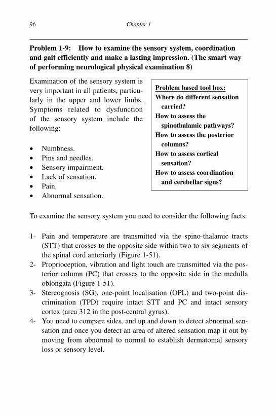
Problem 1-9: How to examine the sensory system, coordinationand gait efficiently and make a lasting impression. (The smart wayof performing neurological physical examination 8)
Examination of the sensory system isvery important in all patients, particu-larly in the upper and lower limbs.Symptoms related to dysfunctionof the sensory system include thefollowing:
• Numbness.• Pins and needles.• Sensory impairment.• Lack of sensation.• Pain.• Abnormal sensation.
To examine the sensory system you need to consider the following facts:
1- Pain and temperature are transmitted via the spino-thalamic tracts(STT) that crosses to the opposite side within two to six segments ofthe spinal cord anteriorly (Figure 1-51).
2- Proprioception, vibration and light touch are transmitted via the pos-terior column (PC) that crosses to the opposite side in the medullaoblongata (Figure 1-51).
3- Stereognosis (SG), one-point localisation (OPL) and two-point dis-crimination (TPD) require intact STT and PC and intact sensorycortex (area 312 in the post-central gyrus).
4- You need to compare sides, and up and down to detect abnormal sen-sation and once you detect an area of altered sensation map it out bymoving from abnormal to normal to establish dermatomal sensoryloss or sensory level.
96 Chapter 1
b1009 Problem Based Neurosurgery
Problem based tool box:Where do different sensation
carried?How to assess the
spinothalamic pathways?How to assess the posterior
columns?How to assess cortical
sensation?How to assess coordination
and cerebellar signs?
b1009_Chapter-01.qxd 11/12/2010 10:49 AM Page 96
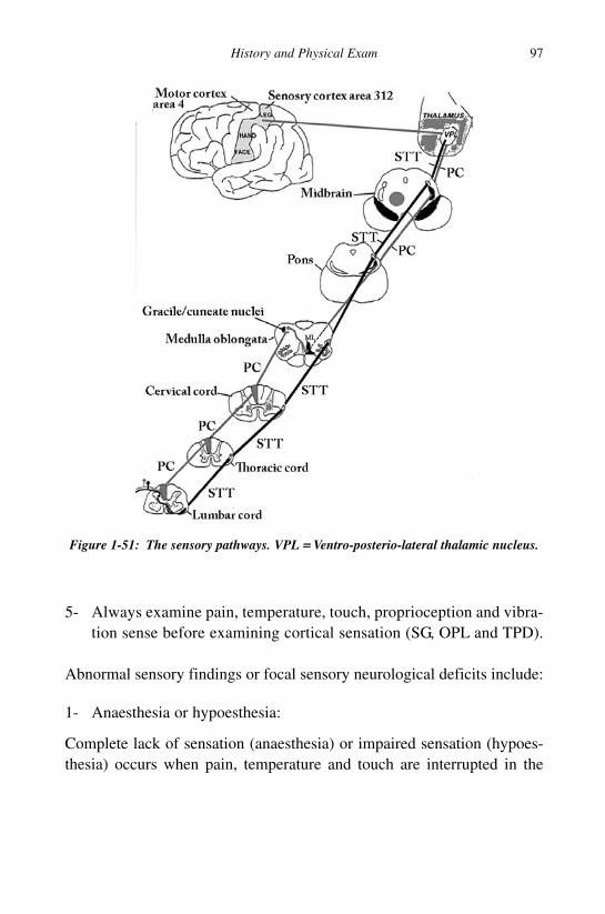
5- Always examine pain, temperature, touch, proprioception and vibra-tion sense before examining cortical sensation (SG, OPL and TPD).
Abnormal sensory findings or focal sensory neurological deficits include:
1- Anaesthesia or hypoesthesia:
Complete lack of sensation (anaesthesia) or impaired sensation (hypoes-thesia) occurs when pain, temperature and touch are interrupted in the
History and Physical Exam 97
b1009 Problem Based Neurosurgery
Figure 1-51: The sensory pathways. VPL == Ventro-posterio-lateral thalamic nucleus.
b1009_Chapter-01.qxd 11/12/2010 10:49 AM Page 97

area of skin or mucous membrane in a peripheral nerve, nerve trunk, nerveroot, the STT or VPL nucleus of the thalamus.
2- Paraesthesia:
Paraesthesia means abnormal sensation such as hyperesthesia, burningsensation, or strong pins and needles.
3- Dissociate anaesthesia:
This means that pain and temperature are intact on one side and proprio-ception is absent in the same area or vice versa.
4- Sensory level:
This denotes the level of lack or abnormal sensation. For example if wesay patient X had a sensory level at T10, we mean that patient X is unableto feel the sensation below the level of T10 (the umbilicus).
1-9-1 How to localise a lesion along the sensory pathway?
To test and identify the correct sensory level you need to know the sen-sory landmarks on the body (Table 1-7 and Figure 1-52).
1-9-2 How to examine the spinothalamic sensory pathways?
1-9-2i Examination of light touch:
Use a wisp of cotton to test for light touch; do not stroke the skin if theskin is hairy as that would not be testing light touch. Gently touch the skinwith the patient’s eyes open in normal area first so that the patient under-stands what is being tested. Then examine touch in systematic fashionwith the patient’s eyes closed. Map any abnormal area by moving fromabnormal to normal.
1-9-2ii Examination for pain and temperature:
Because pain and temperature are transmitted in the STT, testing painoften is sufficient in most clinical scenarios. To test pain sensation, use a
98 Chapter 1
b1009 Problem Based Neurosurgery
b1009_Chapter-01.qxd 11/12/2010 10:49 AM Page 98

neurotip (Figure 1-20). Use similar technique to that used to test trigem-inal sensation. Use the sharp end and the blunt end in normal area first sothe patient understands what you are testing and ask the patient to distin-guish the two sensations. With the patient’s eyes closed test the skin in asystematic fashion using random sharp and blunt pricks and ask thepatient to identify the sensation by uttering the word “sharp” if (s)hethinks that the sharp end was used and the word “blunt” if (s)he thinksthat the blunt end was used. Map from abnormal to normal. Testing tem-perature is often omitted if pain sensation was normal. However, you canuse a tuning fork (TF) heated or cooled by water and ask the patient toidentify “hot” or “cold”. Test the following areas: shoulders (C4), (T1)and outer (C5) aspects of the arms, thumbs (C6), middle fingers (C7) andlittle fingers (C8), front of both thighs (L2), front of the knees (L3),medial (L4) and lateral (L5) aspect of both calves and lateral border ofthe feet (S1).
History and Physical Exam 99
b1009 Problem Based Neurosurgery
Table 1-7: Localisation of sensory dermatomes and sensory levels
Dermatome/Level Description of the area
C2 The occipital region of the scalp.C3 The lateral aspect of the neck.C4 The shoulder area.C5 The lateral area of the upper arm around the deltoid.C6 The thumb and extends above the wrist in the forearm.C7 The middle finger and extends above the wrist.C8 The little finger and extends above the wrist in the forearm.T1 The arm pit.T4 The nipple area.T6 The xiphoid level.T10 The umbilicus.T12 The symphysis pubis.L2 Lateral thigh.L3 Around the front of the knee.L4 Medial calf/ medial malleolus.L5 The big toe.S1 Lateral border of the foot.S3–5 Saddle area.
b1009_Chapter-01.qxd 11/12/2010 10:49 AM Page 99

1-9-3 How to assess integrity of posterior columns?
1-9-3i Examination of joint position and joint movements:
To examine for joint position and joint movement (JPM) hold the distalphalanx of the big toe between your thumb and index, holding the sidesof the toe rather than superior-inferior surface to avoid the use of othersensation such as pressure. Move the phalanx up or down, show the
100 Chapter 1
b1009 Problem Based Neurosurgery
Figure 1-52: Sensory dermatomes and sensory levels.
b1009_Chapter-01.qxd 11/12/2010 10:49 AM Page 100
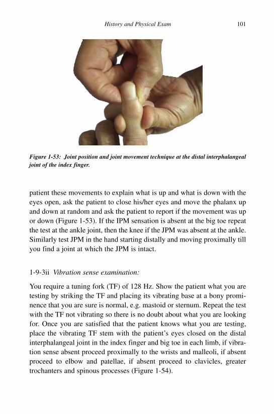
patient these movements to explain what is up and what is down with theeyes open, ask the patient to close his/her eyes and move the phalanx upand down at random and ask the patient to report if the movement was upor down (Figure 1-53). If the IPM sensation is absent at the big toe repeatthe test at the ankle joint, then the knee if the JPM was absent at the ankle.Similarly test JPM in the hand starting distally and moving proximally tillyou find a joint at which the JPM is intact.
1-9-3ii Vibration sense examination:
You require a tuning fork (TF) of 128 Hz. Show the patient what you aretesting by striking the TF and placing its vibrating base at a bony promi-nence that you are sure is normal, e.g. mastoid or sternum. Repeat the testwith the TF not vibrating so there is no doubt about what you are lookingfor. Once you are satisfied that the patient knows what you are testing,place the vibrating TF stem with the patient’s eyes closed on the distalinterphalangeal joint in the index finger and big toe in each limb, if vibra-tion sense absent proceed proximally to the wrists and malleoli, if absentproceed to elbow and patellae, if absent proceed to clavicles, greatertrochanters and spinous processes (Figure 1-54).
History and Physical Exam 101
b1009 Problem Based Neurosurgery
Figure 1-53: Joint position and joint movement technique at the distal interphalangealjoint of the index finger.
b1009_Chapter-01.qxd 11/12/2010 10:49 AM Page 101

1-9-4 How to examine cortical sensation?
Proceed to examine these if the aforementioned sensations were normaland you suspect a parietal lobe problem.
1-9-4i Stereognosis:
To check stereognosis place a familiar object in the patient’s hand (coin,paper clip, pencil, etc.). Ask the patient to identify the object.
1-9-4ii One-point localisation:
With the patient’s eyes closed touch the skin with a pen to make a mark,give the pen to the patient in the untested hand and ask him (her) to makea similar mark where (s)he thought (s)he felt you had made your mark.Measure the distance between the two marks. On the distal phalanx shouldbe few millimetres; on the trunk should be about 10 mm (Figure 1-55).
1-9-4iii Two-point discrimination:
Use an opened paper clip or a specific calliper that has two points to touchthe patient’s finger pads in two places simultaneously. Randomly alternatethe test by touch with one or two points, ask the patient to identify “one”
102 Chapter 1
b1009 Problem Based Neurosurgery
Figure 1-54: Testing vibration in the interphalangeal joint.
b1009_Chapter-01.qxd 11/12/2010 10:49 AM Page 102

or “two”. Find the minimal distance at which the patient can discriminate.On the tip of the fingers it should be very few millimetres, while on thepalm could be 5–6 mm.
1-9-4iv Graphesthesia:
With the blunt end of a pen or pencil, draw a large number (2, 5, 6, 9 etc.)in the patient’s palm. Ask the patient to identify the number.
1-9-5 How to assess coordination?
Coordination is dependent on the integrity of proprioception, visual cues,vestibular system and the cerebellum. The following tests are often usedto assess coordination.
1-9-5i Finger nose test:
Ask the patient to touch your finger then the tip of his/her nose repeti-tively and observe over shooting (Figure 1-56).
1-9-5ii Heel-shin test:
Ask the patient to slide the heel of one leg over the front of the shin of theother (Figure 1-57).
History and Physical Exam 103
b1009 Problem Based Neurosurgery
Figure 1-55: One point localisation on the palm compared to the tip index.
b1009_Chapter-01.qxd 11/12/2010 10:49 AM Page 103

1-9-5iii Disdiadokinesis:
Ask the patient to stretch their hands supine in front and then to rapidlymove in pronation/supination movements. Alternatively ask the patient tomimic playing the piano with the fingers, or ask the patient to tap the dis-tal thumb with the tip of the index finger as fast as possible, or ask thepatient to strike one hand on the thigh, raise the hand, turn it over, and then
104 Chapter 1
b1009 Problem Based Neurosurgery
Figure 1.56: Finger nose test, patient tries to touch examiner’s finger than his/her noserepetitively while the examiner changes the position of his finger.
Figure 1-57: The heel-shin test.
b1009_Chapter-01.qxd 11/12/2010 10:49 AM Page 104

strike it back down as fast as possible, or ask the patient to tap your handwith the ball of each foot as fast as possible. Observe the speed and sym-metry of movements (Figure 1-58).
1-9-5iv Romberg test:
Please be aware that if this test is positive, i.e. patient is dependent almostentirely on their vision to steady themselves in space and have no propri-oception the patient may fall to the ground, so be prepared to catch thepatient if they are unstable. To perform the test, ask the patient to standwith the feet together (Figure 1-59). Ask the patient to close his/her eyesfor five to ten seconds without support, while you are ready to support thepatient. The test is said to be positive if the patient becomes unstable(indicating a vestibular or proprioception problem).
1-9-5v How to assess gait?
Ask the patient to walk across the room, turn and come back, observe pos-ture, hand swing and steadiness. Then ask the patient to walk heel-to-toe(Figure 1-60) in a straight line (tandem gait) and observe any unsteadi-ness. Ask the patient to walk on their toes in a straight line and then ontheir heels in a straight line. Finally ask the patient to hop in place on eachfoot and do a shallow knee bend rise from a sitting position.
1-9-5vi How to assess inattention?
Some patients may have inattention for sensory information from one sideof the body often seen in parietal lobe lesions. To look for inattention you
History and Physical Exam 105
b1009 Problem Based Neurosurgery
Figure 1-58: Alternative tapping of the palm and dorsum of the right hand.
b1009_Chapter-01.qxd 11/12/2010 10:49 AM Page 105
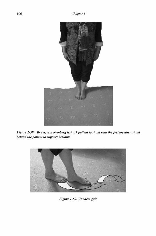
106 Chapter 1
b1009 Problem Based Neurosurgery
Figure 1-59: To perform Romberg test ask patient to stand with the feet together, standbehind the patient to support her/him.
Figure 1-60: Tandem gait.
b1009_Chapter-01.qxd 11/12/2010 10:49 AM Page 106

have to present the patient with simultaneous stimulation of the left andright sides of the body as follows:
• Visual inattention: If the visual fields were tested individually andboth were normal, look for inattention by presenting visual cuessimultaneously in the two visual fields. Ask the patient to focus on adistant object, use both hands and present moving index fingers in thetemporal field of each eye simultaneously and ask the patient to pointto the moving finger. Inattention is present when the patient alwayspoints to one side only when both fingers were moving.
• Sensory inattention: With the patient’s eyes closed, ask the patientto say “right” when the right palm is touched and “left” when theleft palm is touched. Sensory inattention is present when the patientidentifies only one side when both palms were touched at the sametime.
• Line bisection test: Draw a line on a paper and ask the patient to drawa line across the middle of the line. Patient with inattention will bisectthe line to one side (Figure 1-61).
References
1. Mayer E, Martory MD, Pegna AJ et al. A pure case of Gerstmann syndrome
with a subangular lesion. Brain 1999; 122: 1107–1120.
History and Physical Exam 107
b1009 Problem Based Neurosurgery
Figure 1-61: Line bisection and inattention: 1 normal midline bisection, 2 in right hemineglect, and 3 in left hemi neglect.
b1009_Chapter-01.qxd 11/12/2010 10:49 AM Page 107

2. Bornstein B, Kidron DP, Maschiach I. Right parietal syndrome spatial inat-
tention, dressing apraxia and topographical disorientation, following right
carotid artery thrombosis. Psychiatr Neurol 1959; 137: 310–324.
3. Knecht S, Drager B, Deppe M et al. Handedness and hemispheric language
dominance in healthy humans. Brain 2000; 123: 2512–2518.
4. Collins T, Mehalic E, Thomas F et al. Trochlear nerve palsy as the sole ini-
tial sign of an aneurysm of the superior cerebellar artery. Neurosurgery 1992;
30: 258–261.
5. Bléry M, Chagnon S, Picard A et al. A report on four cases, including a
Gradenigo-Lannois syndrome. J Radiol 1980; 61: 677–681.
6. Garnit R. The functional role of the muscle spindles: facts and hypotheses.
Brain 1975; 98: 531–556.
7. Delwaide PJ, Toulouse P. The Jendrassik manoeuvre: quantitative analysis of
reflex reinforcement by remote voluntary muscle contraction. Adv Neurol
1983; 39: 661–669.
8. Gassel MM, Diamantopoulos E. The Jendrassik manoeuvre. Neurology
1964; 14: 555–560, 640–642.
9. Impallomeni M, Kenny RA, Flynn MD et al. The elderly and their ankle
jerks. Lancet 1984; 1: 670–672.
Your personal notes:
.....................................................................................................................
.....................................................................................................................
.....................................................................................................................
.....................................................................................................................
.....................................................................................................................
.....................................................................................................................
.....................................................................................................................
.....................................................................................................................
.....................................................................................................................
.....................................................................................................................
108 Chapter 1
b1009 Problem Based Neurosurgery
b1009_Chapter-01.qxd 11/12/2010 10:49 AM Page 108

Chapter 2: Neurological Investigations
Problem 2-1: Computerised tomographic scan (CT): How tointerpret CT-based images?
Computerised tomographic scan(CT) is a method used to generatethree-dimensional image of anobject, e.g. the brain from a series oftwo-dimensional X-ray images takenaround a single rotational axis(Figure 2-1).
Each image is a computer-generated image of a volume of an object ororgan based on X-ray absorption (electron density). By convention theright side of the body is projected on the left side of the image as if youare looking into a mirror. Each CT image (slice) is 1.25 to 10 mm thickand related to an axis on the scout view (Figure 2-2).
2-1-1: How can I read a plain CT image of the brain?
The easy way to read an image is to view the image systematicallystarting from one location, e.g. one corner and scan the whole imagesquare by square and note any abnormalities. To spot abnormalities youneed to be familiar with the normal anatomy of the organ under inves-tigation. Normal CT brain image demonstrates the following anatomy(Figure 2-3):
1- Brain tissue as a shade of grey (1).2- CSF in the ventricles as shade of dark (2) hypodense compared to
brain which means it is darker than brain.3- Bone (skull) as a shade of white (3) hyperdense = whiter than
brain.
109
b1009 Problem Based Neurosurgery
Problem based tool box:CT pros and consHyperdense lesionsIsodense lesionsHypodense lesionsRing lesions
b1009_Chapter-02.qxd 11/12/2010 10:49 AM Page 109

110 Chapter 2
b1009 Problem Based Neurosurgery
Figure 2-1: Diagram of CT scanner. A == The axis of rotation of X-ray source and detec-tors, B == schematic representation of X-ray tube and detectors rotation, C == image ofthe CT scanner gantry housing the X-ray tubes and detectors, and D == motorised tableto feed the patient into the scanner gantry.
Figure 2-2: CT scan slice of the head 1.5 mm thick, the exact location of the image isshown on the scout view (A).
b1009_Chapter-02.qxd 11/12/2010 10:49 AM Page 110
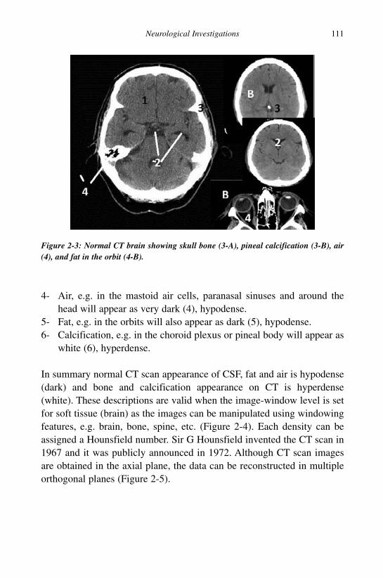
4- Air, e.g. in the mastoid air cells, paranasal sinuses and around thehead will appear as very dark (4), hypodense.
5- Fat, e.g. in the orbits will also appear as dark (5), hypodense.6- Calcification, e.g. in the choroid plexus or pineal body will appear as
white (6), hyperdense.
In summary normal CT scan appearance of CSF, fat and air is hypodense(dark) and bone and calcification appearance on CT is hyperdense(white). These descriptions are valid when the image-window level is setfor soft tissue (brain) as the images can be manipulated using windowingfeatures, e.g. brain, bone, spine, etc. (Figure 2-4). Each density can beassigned a Hounsfield number. Sir G Hounsfield invented the CT scan in1967 and it was publicly announced in 1972. Although CT scan imagesare obtained in the axial plane, the data can be reconstructed in multipleorthogonal planes (Figure 2-5).
Neurological Investigations 111
b1009 Problem Based Neurosurgery
Figure 2-3: Normal CT brain showing skull bone (3-A), pineal calcification (3-B), air(4), and fat in the orbit (4-B).
b1009_Chapter-02.qxd 11/12/2010 10:49 AM Page 111

2-1-2 What are the uses of CT scan?
CT scan of the head is used to evaluate the following conditions:
• Head trauma:
In head trauma CT is proved to be a very valuable tool. It can detect skullfractures, fluid levels in paranasal sinuses, diffuse axonal injury, brain
112 Chapter 2
b1009 Problem Based Neurosurgery
Figure 2-4: Axial CT head using bone window demonstrating the skull inner and outertables (hyperdense) and the scalp slightly hyperdense.
Figure 2-5: CT scan reconstructions in sagittal plane.
b1009_Chapter-02.qxd 11/12/2010 10:49 AM Page 112
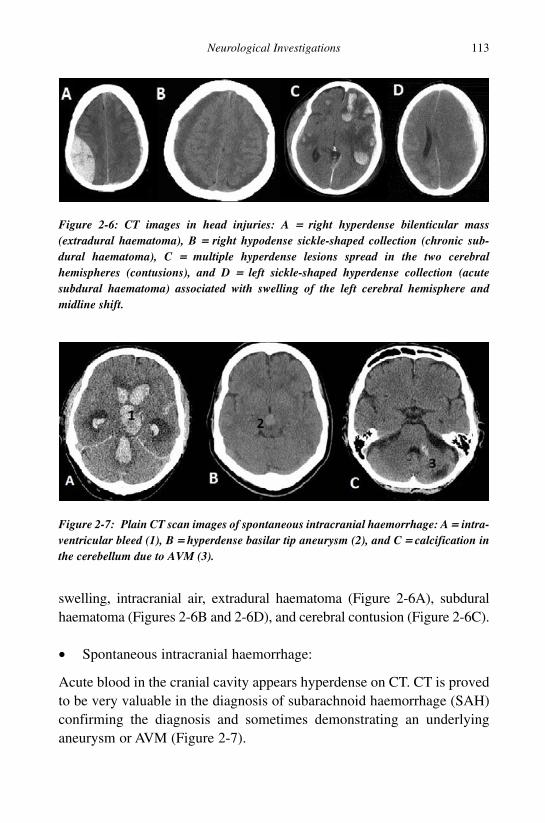
swelling, intracranial air, extradural haematoma (Figure 2-6A), subduralhaematoma (Figures 2-6B and 2-6D), and cerebral contusion (Figure 2-6C).
• Spontaneous intracranial haemorrhage:
Acute blood in the cranial cavity appears hyperdense on CT. CT is provedto be very valuable in the diagnosis of subarachnoid haemorrhage (SAH)confirming the diagnosis and sometimes demonstrating an underlyinganeurysm or AVM (Figure 2-7).
Neurological Investigations 113
b1009 Problem Based Neurosurgery
Figure 2-6: CT images in head injuries: A == right hyperdense bilenticular mass(extradural haematoma), B == right hypodense sickle-shaped collection (chronic sub-dural haematoma), C == multiple hyperdense lesions spread in the two cerebralhemispheres (contusions), and D == left sickle-shaped hyperdense collection (acutesubdural haematoma) associated with swelling of the left cerebral hemisphere andmidline shift.
Figure 2-7: Plain CT scan images of spontaneous intracranial haemorrhage: A == intra-ventricular bleed (1), B == hyperdense basilar tip aneurysm (2), and C == calcification inthe cerebellum due to AVM (3).
b1009_Chapter-02.qxd 11/12/2010 10:49 AM Page 113

• Strokes and transient ischaemic attacks:
CT scan is used in patients suspected to have stroke or cerebral ischaemia.An established stroke appears as hypodense (Figure 2-8). If there was ahaemorrhage in the infarct, the haemorrhage would appear as hyperdense.
• Spinal trauma:
CT is proved to be very valuable in assessing patients with spinal traumaparticularly the cervical spine when an area is obscured by the shoulders,e.g. the cervico-thoracic junction. It is also very valuable in reconstruct-ing the spine to evaluate its stability (Figure 2-9).
114 Chapter 2
b1009 Problem Based Neurosurgery
Figure 2-8: Plain CT scan images of cerebellar stroke (A), B == haemorrhage in theright frontal lobe with some blood layering in the left occipital horn.
Figure 2-9: CT scan images of the spine: A == 3D reconstruction of cervical spine, B ==axial image of C7 with fracture in the left lamina, and C == burst fracture of thoracicvertebra with bilateral haemothorax.
b1009_Chapter-02.qxd 11/12/2010 10:49 AM Page 114

Figure 2-10: CT scan images of patients with raised ICP: A == posterior cranial fossatumour (1) with hydrocephalus, B == butterfly glioma (2), and C == multiple abscesses,one in the left thalamus (3).
• Suspected raised intracranial pressure (ICP):
Patients suspected to have raised ICP such as those presenting withheadache, nausea and vomiting, patients with papilloedema, patientswith focal neurological deficits, patients with seizures and patients withreduced consciousness or comatose (Figure 2-10).
• Stereotactic targeting:
CT can be used in conjunction with stereotactic frames or frameless imageguided technology to biopsy lesions in the brain, insert electric leads inthe brain, insert catheters, chemicals or cells, and localise lesions duringsurgery (Figure 2-11).
• Surgical planning:
Neurosurgery is image dependent and almost no neurosurgical procedureis carried out without neuro-image guidance. From 1990s most neurosur-gical procedures are carried out using image guided systems that are ableto track the surgical field and display the corresponding location on animage space created on the computer workstation.
• Assessment of paranasal sinuses, temporal bone, skull bone and facialbones and their lesions.
Neurological Investigations 115
b1009 Problem Based Neurosurgery
b1009_Chapter-02.qxd 11/12/2010 10:49 AM Page 115

• Radiotherapy planning.• Staging of malignancies by performing CT scan of chest, abdomen
and pelvis.
IV contrast is used after plain CT to highlight breakdown in the bloodbrain barrier (BBB). Some lesions enhance well (homogenous enhance-ment), others have mixed enhancement and others do not enhance at all.
2-1-3 What enhancement patterns are seen on CT?
1- Homogenous enhancement is seen in Figure 2-12:
a. Meningiomas.b. Vestibular schwannomas.
116 Chapter 2
b1009 Problem Based Neurosurgery
Figure 2-11: Plain CT scan images planning for stereotactic biopsy: A == axial slice withnine fiducials and B == scout view showing the stereotactic frame ring.
Figure 2-12: Examples of homogenous enhancing lesion: A, B, C, and D menin-giomas; C before contrast and D after contrast injection.
b1009_Chapter-02.qxd 11/12/2010 10:49 AM Page 116

c. Haemangioblastoma nodules.d. Pilocytic astrocytoma.e. Haemangiopericytoma.f. Ependymoma.
2- Mixed enhancement is seen in Figure 2-13:
a. High grade gliomas.b. Metastases.c. Medulloblastoma.d. Pituitary adenomas.e. Craniopharyngiomas.f. Inflammatory lesions.
3- Peripheral enhancement is seen in Figure 2-14:
a. Abscess (ring enhancing lesion).b. Cystic metastasis.c. Cystic high grade glioma.d. Luxury perfusion in acute infarct (2% after two days).e. Cystic astrocytoma.f. Inflammatory cyst.
Neurological Investigations 117
b1009 Problem Based Neurosurgery
Figure 2-13: Examples of non-uniform mixed enhancing lesions: A metastasis andB glioblastoma multiforme (GBM).
b1009_Chapter-02.qxd 11/12/2010 10:49 AM Page 117

4- No enhancement is seen:
a. Infarction.b. Arachnoid cyst.c. Cholesterol cyst.d. Lipoma.e. Grade I astrocytoma.f. Epidermoid cyst.
2-1-4 What are the advantages of CT?
CT has higher resolution and ability to image inside the skull. It candemonstrate bone very well, e.g. skull lesions, bony involvement andbone density. It can also demonstrate haemorrhages. Modern CT scannershave the added advantage of 3D reconstruction.
2-1-5 What are the disadvantages of CT?
One of the disadvantages of CT is the fact that it utilises ionising radiationand is considerate moderate to high risk investigation. Although CT
118 Chapter 2
b1009 Problem Based Neurosurgery
Figure 2-14: Examples of ring enhancing lesion (metastasis).
b1009_Chapter-02.qxd 11/12/2010 10:49 AM Page 118

represents about 7% of all radiological investigations using X-rays it isresponsible for 47% of total collective radiation dose.1 Due to the rise inusing CT, the total medical radiation had increased despite that it hadreduced in other areas (Table 2-1).
Another disadvantage of CT is reaction to contrast materials. Allergicreaction can occur and contrast material can lead to kidney failure, partic-ularly in those who had pre-existing renal insufficiency or diabetesmellitus.
Neurological Investigations 119
b1009 Problem Based Neurosurgery
Table 2-1: Comparison of radiation dose of CT and other X-rays
Investigation mSv (Effective dose) Millirem
Chest X-ray 0.1 10Head CT 1.5 150Chest CT 5.8 580Abdominal CT 5.3 530Chest, abdomen and pelvis CT 9.9 990
b1009_Chapter-02.qxd 11/12/2010 10:49 AM Page 119
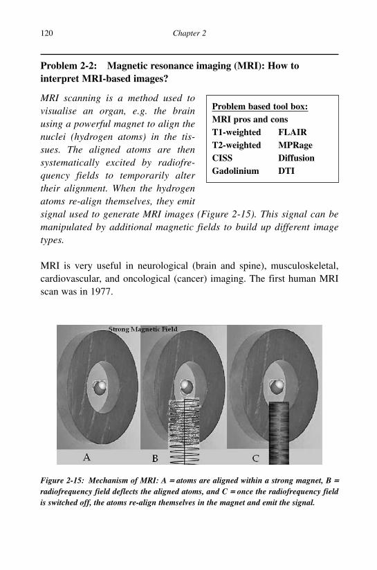
Problem 2-2: Magnetic resonance imaging (MRI): How tointerpret MRI-based images?
MRI scanning is a method used tovisualise an organ, e.g. the brainusing a powerful magnet to align thenuclei (hydrogen atoms) in the tis-sues. The aligned atoms are thensystematically excited by radiofre-quency fields to temporarily altertheir alignment. When the hydrogenatoms re-align themselves, they emitsignal used to generate MRI images (Figure 2-15). This signal can bemanipulated by additional magnetic fields to build up different imagetypes.
MRI is very useful in neurological (brain and spine), musculoskeletal,cardiovascular, and oncological (cancer) imaging. The first human MRIscan was in 1977.
120 Chapter 2
b1009 Problem Based Neurosurgery
Problem based tool box:MRI pros and consT1-weighted FLAIRT2-weighted MPRageCISS DiffusionGadolinium DTI
Figure 2-15: Mechanism of MRI: A == atoms are aligned within a strong magnet, B ==radiofrequency field deflects the aligned atoms, and C == once the radiofrequency fieldis switched off, the atoms re-align themselves in the magnet and emit the signal.
b1009_Chapter-02.qxd 11/12/2010 10:49 AM Page 120
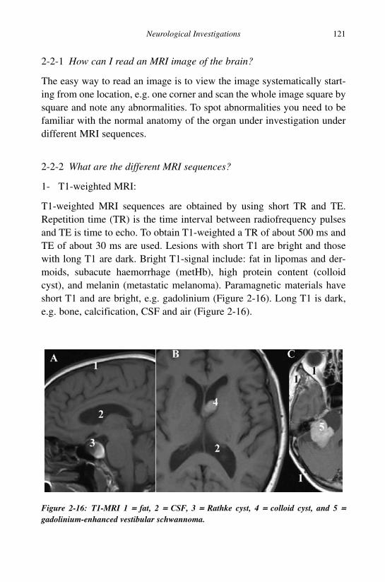
2-2-1 How can I read an MRI image of the brain?
The easy way to read an image is to view the image systematically start-ing from one location, e.g. one corner and scan the whole image square bysquare and note any abnormalities. To spot abnormalities you need to befamiliar with the normal anatomy of the organ under investigation underdifferent MRI sequences.
2-2-2 What are the different MRI sequences?
1- T1-weighted MRI:
T1-weighted MRI sequences are obtained by using short TR and TE.Repetition time (TR) is the time interval between radiofrequency pulsesand TE is time to echo. To obtain T1-weighted a TR of about 500 ms andTE of about 30 ms are used. Lesions with short T1 are bright and thosewith long T1 are dark. Bright T1-signal include: fat in lipomas and der-moids, subacute haemorrhage (metHb), high protein content (colloidcyst), and melanin (metastatic melanoma). Paramagnetic materials haveshort T1 and are bright, e.g. gadolinium (Figure 2-16). Long T1 is dark,e.g. bone, calcification, CSF and air (Figure 2-16).
Neurological Investigations 121
b1009 Problem Based Neurosurgery
Figure 2-16: T1-MRI 1 == fat, 2 == CSF, 3 == Rathke cyst, 4 == colloid cyst, and 5 ==gadolinium-enhanced vestibular schwannoma.
b1009_Chapter-02.qxd 11/12/2010 10:49 AM Page 121

2- T2-weighted MRI:
T2-weighted MRI sequences are obtained by using long TR and TE. Toobtain T2-weighted a TR of about 2000 ms and TE of about 100 ms areused. Lesions with short T2 are bright and those with long T2 are dark.Bright T2-signal include: fat in lipomas and dermoids, acute haemorrhage(deoxyHb), haemosidrin, physiological iron, and mucinous material. CSFis also bright on T2 (Figure 2-17). Long T2 is dark, e.g. bone, calcifica-tion, flowing blood (flow void in arteries and AVMs) and air (Figure 2-17).
The easy way to recognise T1 and T2 is to look at the ventricles, if theventricles are bright then it is almost certainly T2-weighted and if they aredark it is most likely T1-weighted.
3- MPRage sequence:
Magnetisation Prepared Rapid Gradient Echo is a fast 3D gradient echopulse sequence using a magnetisation preparation pulse likeTurboFLASH. Only one segment or partition of a 3D data record isobtained per inversion preparation pulse. After the acquisition, for allrows a delay time (TD) is used to prevent saturation effects.MPRage is designed for rapid acquisition with T1-weighted domi-nance. Fast gradient echoes are characterised by their rapid sampling
122 Chapter 2
b1009 Problem Based Neurosurgery
Figure 2-17: T2-weighted images 1 and 3 == flow void, 2 == haemosidrin, and 4 == CSF.
b1009_Chapter-02.qxd 11/12/2010 10:49 AM Page 122

time, high signal intensity and image contrast while approaching steadystate (the echo is collected during the time when tissues are experienc-ing T1 relaxation). The rapid speed of the acquisition makes it anexcellent alternative to breath-hold abdominal imaging, neuro,dynamic bolus, MR angiography and cardiac imaging. MPRage is veryuseful in delineating the basal ganglia and other internal brain struc-tures because of better contrast between the grey and white matters(Figure 2-18).
4- FLAIR sequence:
Fluid attenuated inversion recovery (FLAIR) produces stronglyT2-weighted image and suppressed CSF signal. This is accomplished by180° inversion pulse. A relatively long TI is used to allow the longitudinalmagnetisation of CSF to return to the null point before SE imaging. Thus,the CSF signal is completely suppressed for cortical or periventricularareas, and lesions with typical T2 prolongation in the brain that are adja-cent to spinal fluid become much more conspicuous compared withconventional T2 imaging (Figure 2-19). FLAIR sequence is very useful inacute multiple sclerosis and acute infarction. FLAIR removes CSF partialvolume artifact that obscures these lesions on T2-weighted images.2
Neurological Investigations 123
b1009 Problem Based Neurosurgery
Figure 2-18: MPRage, 1 == caudate nucleus, 2 == putamen, 3 == globus pallidum, 4 ==anterior limb of internal capsule (IC), 5 == posterior limb of IC, 6 == thalamus, 8 == exter-nal capsule, 9 == insular cortex, 10 == lesion in the pallidum, and 11 == third ventricle.
b1009_Chapter-02.qxd 11/12/2010 10:49 AM Page 123

5- CISS sequence:
Constructive Interference Steady State (CISS) is a stimulated T2 echo, wheretwo true fast spin echo sequences are acquired with differing RF pulses andthen combined for strong T2-weighted high resolution 3D images. Thesespinecho sequences are normally affected by dark phase dispersion bands,which are caused by patient induced local field inhomogeneities and madeprominent by the relatively long TR used. The different excitation pulseregimes offset these bands in the two sequences. Combining these imagesresult in a picture free of banding. The image combination is performed auto-matically after data collection, adding some more time to the reconstructionprocess. The advantage of the 3D CISS sequence is its combination of highsignal levels and extremely high spatial resolution. CISS is used to image theinner ear, cranial nerves and cerebellum, e.g. in small acoustic or vascularcompression in trigeminal neuralgia (Figures 2-20 and 2-21).
6- Diffusion sequences:
Various pulse sequences are modified to enhance signal loss resulting fromwater molecules that show significant diffusion versus those with more
124 Chapter 2
b1009 Problem Based Neurosurgery
Figure 2-19: FLAIR images demonstrating periventricular high signal lesions in theright hemisphere (2) and very dark signal of suppressed CSF (1). It also demonstrateshigh signal in clotted aneurysm (3).
b1009_Chapter-02.qxd 11/12/2010 10:49 AM Page 124

restricted diffusion. Certain pathologies show restriction of diffusion, suchas cytotoxic oedema and demyelination. With a diffusion-weighted pulsesequence, these abnormalities can be made more obvious (Figure 2-22).
Depending on the particular gradient/gradients used, anisotropic dif-fusion can be detected, as occurs normally in white matter tracts. This canbe used in fibre tracking: diffusion tensor imaging (DTI) (Figure 2-23).
7- STIR sequences:
STIR (short TI inversion recovery) sequence can be used for fat suppres-sion, where a relatively short inversion time is used to null the fat signalwhile maintaining water and soft tissue signal. This sequence is useful to
Neurological Investigations 125
b1009 Problem Based Neurosurgery
Figure 2-20: CISS image demonstrating small right vestibular schwannoma (1) andclear VII/VIII nerves and cochlea (2) on the left.
Figure 2-21: CISS image demonstrating trigeminal nerves (1). Note the vascular com-pression on the right and clear nerve on the left (1).
b1009_Chapter-02.qxd 11/12/2010 10:49 AM Page 125

distinguish lipomas from other types of tumours or to suppress fat sur-rounding a lesion making it more obvious. One drawback of this sequenceis the partial loss of proton signal during the TI time. Also the TR timemust be longer than that of a spin echo sequence for recovery of longitu-dinal magnetisation.
126 Chapter 2
b1009 Problem Based Neurosurgery
Figure 2-22: Diffusion MRI image demonstrating restricted diffusion of water mole-cules in an acute infarction (1).
Figure 2-23: DTI image of fibre tracking around a brain tumour (1) identifying a sur-gical corridor through which the tumour was removed without affecting the whitematter tracts.
b1009_Chapter-02.qxd 11/12/2010 10:49 AM Page 126

2-2-3 What are the uses of MRI?
MRI scan of the head is used to evaluate the following conditions:
• Head trauma:
CT scan is the main neuroimaging modality in head injuries. However,MRI can provide the same or even better information in head injuries, butsignificant numbers of patients with head injuries requiring brain imagingare ventilated at the time of the scan making MRI less desirable. When CTis not available MRI is often used (Figure 2-24).
• Spontaneous intracranial haemorrhage:
Acute blood in the cranial cavity appears high signal on MRI. AlthoughCT is the main brain imaging used in spontaneous intracerebralhaematoma and SAH, MRI proved very valuable in the diagnosis ofdelayed subarachnoid haemorrhage, aneurysms and AVMs (Figure 2-25).
Neurological Investigations 127
b1009 Problem Based Neurosurgery
Figure 2-24: MRT T2-weighted image in a patient with bilateral chronic subduralhaematomas (1 and 2) who had MRI because the CT was out of commission for serv-ice. Note the layering of the blood on the right side (1) indicating that there was fresherbleed on that side.
b1009_Chapter-02.qxd 11/12/2010 10:49 AM Page 127

• Strokes and transient ischaemic attacks:
MRI is a better imaging modality and more sensitive in acute ischaemiaparticularly using diffusion images (Figure 2-22).
• Spinal conditions:
MRI scan is the investigation of choice in the spine and virtuallyreplaced CT and myelography. MRI is excellent in demonstrating bony,
128 Chapter 2
b1009 Problem Based Neurosurgery
Figure 2-25: Different MRI sequences in vascular causes of spontaneous intracranialhaemorrhage: A == multiple aneurysms one in right middle cerebral artery and one inanterior com artery, B == cavernoma in the left frontal region, C == cavernoma in the leftinsula, D == AVM in left cerebellum, and E == giant aneurysm in tip of basilar artery.Dark signals denote flow void (A and D) or haemosiderin (B, C, and E) due to previoushaemorrhage (B and C).
b1009_Chapter-02.qxd 11/12/2010 10:49 AM Page 128

disc, dural, extradural, intradural or intramedullary lesions in the spine(Figure 2-26).
• Suspected raised intracranial pressure (ICP):
Patients suspected to have raised ICP such as those presenting withheadache, nausea and vomiting, patients with papilloedema, patients withfocal neurological deficits, and patients with seizures are best investigatedby MRI (Figure 2-27).
• Stereotactic targeting:
MRI is used in conjunction with stereotactic frames or frameless imageguided technology to biopsy lesions in the brain, insert electric leads inthe brain, insert catheters, chemicals or cells, and localise lesions duringsurgery (Figure 2-28).
Neurological Investigations 129
b1009 Problem Based Neurosurgery
Figure 2-26: MRI images of the spine: A == T2-weighted demonstrating L4/5 disc pro-lapse, B == T2 demonstrating L4 hemivertebra and epidermoid, C == STIR image ofT11/12 discitis, and D == T2 demonstrating screw plate fixation of C3 to C7 andintradural gliosis.
b1009_Chapter-02.qxd 11/12/2010 10:49 AM Page 129
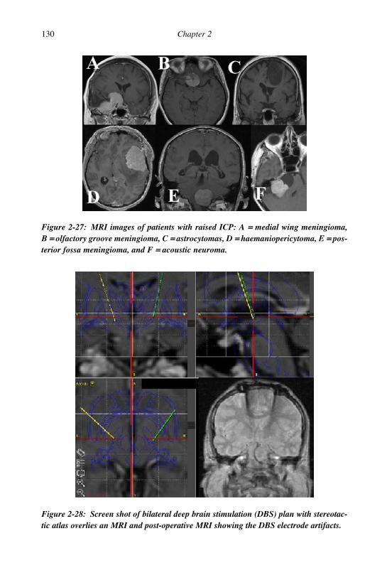
130 Chapter 2
b1009 Problem Based Neurosurgery
Figure 2-27: MRI images of patients with raised ICP: A == medial wing meningioma,B == olfactory groove meningioma, C == astrocytomas, D == haemaniopericytoma, E == pos-terior fossa meningioma, and F == acoustic neuroma.
Figure 2-28: Screen shot of bilateral deep brain stimulation (DBS) plan with stereotac-tic atlas overlies an MRI and post-operative MRI showing the DBS electrode artifacts.
b1009_Chapter-02.qxd 11/12/2010 10:49 AM Page 130

• Surgical planning:
MRI scan is the main planning modality of brain operations using imageguided systems, e.g. tumour surgery (Figure 2-29), gamma knife or cyber-knife stereotactic radiosurgery.
2-2-4 What lesions enhance on MRI scan?
IV contrast is used after plain T1-weighted imaging using gadolinium tohighlight breakdown in the BBB. Some lesions enhance well (homoge-nous enhancement), others have mixed enhancement and others do notenhance at all.
Neurological Investigations 131
b1009 Problem Based Neurosurgery
Figure 2-29: Screen shot of pre-operative surgical plan for transsphenoidal pituitarysurgery.
b1009_Chapter-02.qxd 11/12/2010 10:49 AM Page 131

1- Homogenous enhancement is seen in:
a. Meningiomas.b. Vestibular schwannomas.c. Haemangioblastoma nodules.d. Pilocytic astrocytoma.e. Haemangiopericytoma.f. Ependymoma.
2- Mixed enhancement is seen in:
a. High grade gliomas.b. Metastases.c. Medulloblastoma.d. Pituitary adenomas.e. Craniopharyngiomas.f. Inflammatory lesions.
3- Peripheral enhancement is seen in:
a. Abscess (ring enhancing lesion).b. Cystic metastases.c. Cystic high grade gliomas.d. Luxury perfusion in acute infarcts (2% in two days).e. Cystic astrocytoma.f. Inflammatory cysts.
4- No enhancement is seen in:
a. Infarction.b. Arachnoid cyst.c. Cholesterol cyst.d. Lipoma.e. Grade I astrocytoma.f. Epidermoid cyst.
2-2-5 What are the advantages of MRI?
In summary MRI has several advantages: excellent tissue contrast and res-olution, multiplaner imaging capability, no-ionising radiation and abilityto detect motion of water molecules.
132 Chapter 2
b1009 Problem Based Neurosurgery
b1009_Chapter-02.qxd 11/12/2010 10:49 AM Page 132

2-2-6 What are the disadvantages of MRI?
The main disadvantage of MRI imaging is its inability to overcome metaland motion artifacts that can limit its use. It cannot be used in patients whohave pacemaker, neurostimulator or a pump, metal clips on intracranialvessels or metal fragments in the eyes. Some patients also find the MRIset-up too claustrophobic and are unable to have it. Some of the patientswho suffer from claustrophobia can have MRI scan in upright or openMRI machines.
Neurological Investigations 133
b1009 Problem Based Neurosurgery
b1009_Chapter-02.qxd 11/12/2010 10:49 AM Page 133

Problem 2-3: Non-radiological neuro-investigations. How tointerpret non-radiological neuro-investigations in a smart way?
Most diagnostic investigations ofneuro-surgical conditions involveneuro-radiological imaging. Howevera number of other investigations areused in neuro-surgery for diagnosisand monitoring. Therefore some know-ledge of the underlying principles ofthese investigations will help maximise their utility, diagnostic value andinterpretation.
2-3-1 How can I perform and interpret CSF examination?
CSF analysis is usually required for diagnostic purposes to rule out poten-tial life-threatening conditions such as bacterial meningitis orsubarachnoid haemorrhage. CSF fluid analysis can also aid in the diagno-sis of various other conditions, such as demyelinating diseases andcarcinomatous meningitis. To obtain CSF for analysis a lumbar puncture(L/P), cisternal puncture or ventricular puncture is required. L/P is themost commonly used method to obtain CSF and it should never be per-formed before thorough neurological examination and it should neverdelay life-saving treatments. L/P should only be used after ruling out masslesions on CT scan. L/P is also sometimes performed for therapeutic rea-sons, such as the treatment of pseudotumour cerebri (Benign IntracranialHypertension — BIH), communicating hydrocephalus after SAH, normalpressure hydrocephalus (NPH), to relax the brain during surgery, e.g. dur-ing clipping of intracranial aneurysm, for infusion test in NPH, forintrathecal injection of antibiotics, baclofen, ziconamide, morphine, andchemotherapeutic agents and to control CSF leakage from post-operativewounds or assist neurosurgical repair of CSF fistulae, e.g. after transsphe-noidal, translabrynthine and transnasal surgery. L/P is absolutelycontraindicated in:
1- Unequal pressures between supra- and infra-tentorial compartments,usually inferred by characteristic CT findings: midline shift, loss of
134 Chapter 2
b1009 Problem Based Neurosurgery
Problem based tool box:Lumbar puncture and CSFEEG NCSEMG AEPSSP VEP
b1009_Chapter-02.qxd 11/12/2010 10:49 AM Page 134

suprachiasmatic and basilar cisterns, posterior fossa mass, loss ofsuperior cerebellar cistern, and loss of quadrigeminal cistern.
2- Infected skin over the needle entry site.
L/P should also be avoided in the presence of: increased intracranialpressure (ICP) and coagulopathy. CT is always recommended beforeL/P and L/P should only be performed if the perceived benefits out-weighed its risks in patients who are fully consciousness, had no focalneurology and had no papilloedema. CT should always be performedbefore L/P in all patients over 60 years, immunocompromised, CNSlesions, a seizure within one week of presentation, reduced level of con-sciousness, and focal neurological findings or papilloedema seen onphysical examination.
2-3-2 How L/P is performed?
Explain the procedure of L/P to the patient carefully, including its risksand benefits. In experienced hands, L/P is a relatively safe procedure;post-L/P headaches, infection, intrathecal bleed, radicular pain, nausea,photophobia and intraspinal haematomas can occur.
Patient position is extremely important for successful L/P. The patientis asked to lie on his/her side or sitting up, with the back arched towardthe examiner and as close to the edge of the coach as possible and thencurl into a ball. This involves the patient flexing his/her neck and lowerspine, whilst drawing up his/her thighs toward the chest. The shouldersand pelvis should be vertically aligned without any tilt. In 94% of adultsthe spinal cord terminates at L1. In 6% of adults the spinal cord extendsto L2/L3. Therefore L/P is generally performed at or below L3/L4 inter-space. As a general anatomical rule, a line drawn between the posterioriliac crests often corresponds closely to L3-L4 (Figure 2-30). The inter-space is identified after palpation of the spinous processes at each lumbarlevel.
To avoid infection, the examiner puts on a mask and sterile gloves andthe skin is cleansed with alcohol or iodine-based disinfectant and the areais draped with sterile drapes. A local anaesthetic, commonly 1% ligno-caine, is injected into the subcutaneous area — this should be ideally done
Neurological Investigations 135
b1009 Problem Based Neurosurgery
b1009_Chapter-02.qxd 11/12/2010 10:49 AM Page 135

at least five minutes prior to insertion of the lumbar puncture needle. Thelumbar puncture needle is typically a 20–22 gauge needle (use 18-16gauge for therapeutic L/P) and it is inserted into the target area and slowlyadvanced. The bevel of the needle is maintained in a horizontal position(with the flat portion of the bevel pointing up) and it should be parallel tothe direction of the dural fibres. In most cases the needle is advanced4–5 cm before the subarachnoid space is reached — this is characteristicallyrecognised by a sudden decrease in resistance (give-way) and sometimesa “popping” sound may be heard. Once the subarachnoid space has beenreached, a manometer can be attached to the needle to record the openingpressure. Fluid is then usually obtained for collection in three subsequentuniversal containers. Five millilitres of CSF in each container is usuallysufficient. In most cases CSF will be colourless, but it can often be bloodstained. To differentiate between true blood-stained CSF, e.g. secondary toSAH and traumatic tap, observe the intensity of colour in the three sam-ples; if the CSF becomes clear with each subsequent sample, it is mostlikely to be traumatic and traumatic tap does not have xanthochromia.
2-3-3 What should CSF analysis include?
In all CSF samples sent to the laboratory the following tests are per-formed: cell count, CSF glucose, CSF protein, smear Gram stainexamination and culture. In special circumstances on the request of thetreating physician the following tests are also performed: acid-fast bacilli
136 Chapter 2
b1009 Problem Based Neurosurgery
Figure 2-30: Site of L/P needle at L3/4 level.
b1009_Chapter-02.qxd 11/12/2010 10:49 AM Page 136

smear and culture, antigen tests and serology (e.g. cryptococcal antigen,latex agglutination, limulus lusate tests), PCR (polymerase chain reaction)tests to amplify DNA or RNA of micro-organisms, antibody tests, immu-noelectrophoresis, and cytology.
2-3-4 What is the differential of abnormal CSF analysis?
Table 2-2 summarises the different CSF findings in the most commonconditions compared to normal CSF analysis. When interpreting the glu-cose level in CSF it is important to compare it to blood glucose level(BGL). Therefore it is important to send blood for BGL at the same timeas the L/P.
2-4-1 How can I interpret an EEG?
EEG (electroencephalography) is recording of electrical brain activityproduced by the firing of neurons. Clinically EEG refers to recording ofbrain’s spontaneous electrical activity over a short period of time (20–40minutes) as recorded from multiple electrodes placed on the scalp. Themain diagnostic application of EEG is in the case of epilepsy, as epileptic
Neurological Investigations 137
b1009 Problem Based Neurosurgery
Table 2-2: CSF analysis results
Condition Cells Glucose Protein (g/L) Others
Normal 0–5 2/3 BGL 0.15–0.45 Normal pressure,*clear
Bacterial Polymorph- Very low 1–10 High pressure, turbid,meningitis leukocytosis bacteria
Viral CNS Monocytosis Normal 0.4–1.0 Normal, clearinfection colourless
Subarachnoid RBCs Normal Raised High ICP, red,haemorrhage xanthochromia
Tuberculosis Lymphocytosis Low 0.6–7 High ICP, yellow,infect acid-fast bacteria
Guillain-Barre 0–5 Normal 0.5–10 Normal ICP, clearsyndrome
* Normal pressure = 70–180 mmH2O.
b1009_Chapter-02.qxd 11/12/2010 10:49 AM Page 137

activity can create clear abnormalities on a standard EEG study.3 A sec-ondary clinical use of EEG is in diagnosis of coma, encephalopathies, andbrain death. EEG was used in the past to diagnose tumours, stroke andother focal brain disorders, but it is no longer used for this purpose withthe advent of CT and MRI scanners. EEG is still used intra-operatively tolocate an epileptic focus during resective epilepsy surgery.
Evoked potentials (EP), e.g. somatosensory potentials (SSP),visual evoked responses (VER) and auditory evoked potentials (AEP)(Figure 2-31) are derivatives of the EEG technique, which involves aver-aging the EEG activity time-locked to the presentation of a stimulus.Event-related potentials (ERP) refer to averaged EEG responses that aretime-locked to more complex processing of stimuli; this technique is usedin cognitive science, cognitive psychology, and psychophysiologicalresearch.
2-4-2 How is EEG generated?
EEG reflects correlated synaptic activity caused by post-synaptic poten-tials of cortical neurons. The ionic currents involved in the generation offast action potentials may not contribute much to the averaged field poten-tials representing the EEG.3 Scalp electrical potentials that produce EEG
138 Chapter 2
b1009 Problem Based Neurosurgery
Figure 2-31: AEP during removal of an acoustic neuroma, this generated by summa-tion of EEG activity in response to auditory clicking stimulus.
b1009_Chapter-02.qxd 11/12/2010 10:49 AM Page 138

are generally thought to be caused by the extracellular ionic currentscaused by dendritic electrical activity, whereas the fields producing mag-neto encephalographic signals (MEG)4 are associated with intracellularionic currents. The electric potentials generated by single neurons are fartoo small to be picked up by EEG or MEG. EEG activity therefore alwaysreflects the summation of the synchronous activity of thousands or mil-lions of neurons that have similar spatial orientation, radial to the scalp.Currents that are tangential to the scalp are not picked up by the EEG. TheEEG therefore benefits from the parallel, radial arrangement of apicaldendrites in the cortex. Because voltage fields fall off with the fourthpower of the radius, activity from deep sources is more difficult to detectthan currents near the skull.
2-4-3 What are the different types of EEG waves?
Scalp EEG activity shows oscillations at a variety of frequencies. Severalof these oscillations have characteristic frequency ranges, spatial distribu-tions and are associated with different states of brain functioning (awakeand various sleep stages). These oscillations represent synchronised activ-ity over a network of neurons. The neuronal networks underlying some ofthese oscillations are understood (e.g. thalamocortical resonance underly-ing sleep spindles), while many others are not (e.g. the system thatgenerates the posterior basic rhythm). Figure 2-32 depicts the differenttypes of EEG waves and Table 2-3 summarises their significance.
2-5-1 How can I interpret results of nerve conduction studies (NCS)?
NCS are measuring the conduction velocity of electricity along a nerve todiagnose peripheral neuropathy and peripheral nerve entrapment syn-dromes. NCS are used mainly for evaluation of paraesthesia (numbness,tingling, burning) or weakness of the arms and legs. The type of studyrequired is dependent in part on the symptoms presented. A physicalexamination and thorough history also help to direct NCS, e.g. if the sen-sory deficit extends beyond the wrist there is no point looking for carpaltunnel (CTS) or cubital tunnel (UNC = ulnar nerve compression) syn-dromes. Some of the common disorders which can be diagnosed by nerve
Neurological Investigations 139
b1009 Problem Based Neurosurgery
b1009_Chapter-02.qxd 11/12/2010 10:49 AM Page 139

conduction studies include CTS, UNC, peripheral neuropathy, Guillain-Barré syndrome, Facioscapulohumeral muscular dystrophy, andradiculopathy. NCS include sensory NCS, motor NCS, F-wave study andH-reflex study.
140 Chapter 2
b1009 Problem Based Neurosurgery
Figure 2-32: Different EEG waves and rhythms.
Table 2-3: Types of EEG waves and rhythms and their significance
EEG wave/rhythm Location Significance
Delta Frontal in adults during slow Can be seen in subcortical lesions,up 4 Hz wave sleep and posterior in diffuse lesions, deep midline
children and babies. lesions and metabolicencephalopathies.
Theta Seen in young children, Seen in focal subcortical lesions,4–7 Hz drowsiness or arousal in adults. deep midline lesions, metabolic
encephalopathies,hydrocephalus.
Alpha Seen posteriorly during relaxation Can be seen in coma.8–12 Hz eyes closed.
Beta Seen bilaterally more frontally in Can be seen with benzodiazepines.12–30 Hz active busy states.
Gamma Seen in certain cognitive and30–>100 Hz motor functions.
b1009_Chapter-02.qxd 11/12/2010 10:49 AM Page 140

2-5-2 Motor NCS
Motor NCS are performed by electrical stimulation of a peripheral nerveand recording from a muscle supplied by the nerve. The time it takes forthe electrical impulse to travel from the stimulation to the recording siteis measured. This value is called the latency and is measured in millisec-onds (msec). The size of the response (amplitude) is also measured. Motoramplitudes are measured in millivolts (mV). By stimulating in two ormore different locations along the same nerve, the nerve conductionvelocity (NCV) across different segments can be determined. Calculationsare performed using the distance between the different stimulating elec-trodes and the difference in latencies.
2-5-3 Sensory NCS
Sensory NCS are performed in a similar fashion to motor NCS by electri-cal stimulation of a peripheral nerve and recording from a purely-sensoryportion of the nerve, such as on a finger. Like the motor NCS, sensorylatencies are on the scale of msec. Sensory amplitudes are much smallerthan the motor amplitudes, usually in the microvolt (µV) range. The sen-sory NCV is calculated based upon the latency and the distance betweenthe stimulating and recording electrode.
2-5-4 What is F-wave study?
F-wave is the second of two voltage changes observed after electricalstimulation is applied to the skin surface above the distal region of anerve. F-waves are particularly useful for evaluating conduction prob-lems in the proximal region of nerves. It is called F wave because it wasinitially recorded in the foot muscles. In a typical F-wave study, a strongelectrical stimulus is applied to the skin surface above the distal portion ofa nerve so that the impulse travels both distally (towards muscle fibres)and proximally (back to motor neurons of spinal cord), are also known asorthodromic and antidromic respectively. When the orthodromic stimulusreaches the muscle fibre, it elicits a strong M-wave indicative of musclecontraction. When the antidromic stimulus reaches the motor neurons, a
Neurological Investigations 141
b1009 Problem Based Neurosurgery
b1009_Chapter-02.qxd 11/12/2010 10:49 AM Page 141

small portion of the motor neurons backfires and orthodromic wave trav-els back down the nerve towards the muscle. This reflected stimulusevokes small proportion of the muscle fibres causing a small, secondwave called the F-wave. Because a different population of anterior horncells is stimulated by each stimulus, each F-wave has a slightly differentshape, amplitude and latency. NCV is derived by measuring the limblength in millimetres from the stimulation site to the corresponding spinalsegment (C7 spinous process to wrist crease for median nerve). This ismultiplied by two as it goes to the cord and returns to the muscle (2D); 2Dis divided by the latency difference between mean F and M waves and onemillisecond subtracted (F − M − 1). The formula for NCV during F-wavestudy is 2D/(F − M − 1) mm/msec.
2-5-5 H-reflex study
H-reflex study uses stimulation of a nerve and recording the reflex elec-trical discharge from a muscle in the limb. This test evaluates conductionbetween a limb and spinal cord, but afferent impulses are in sensorynerves while efferent impulses are in motor nerves.
2-5-6 What is the normal F-wave response?
A normal F-wave response has a latency of 25–32 msec in the upperlimbs and 45–56 msec in the lower extremities and with repeated stim-ulation normal F-wave should persist more than 50% of the time(80–100%).
While interpretation of NCS is complex, slowing of NCV usuallyindicates damage to the myelin sheath, slowing across the wrist formotor and sensory latencies of the median nerve indicates CTS, andslowing of all nerve conductions in more than one limb indicates periph-eral polyneuropathy.
References
1. Hart D, Wall BF. UK population dose from medical X-ray examinations. Eur
J Radiol 2004; 50: 285–291.
142 Chapter 2
b1009 Problem Based Neurosurgery
b1009_Chapter-02.qxd 11/12/2010 10:49 AM Page 142

2. De Coene B, Hajnal JV, Gatehouse P et al. MR of the brain using fluid-
attenuated inversion recovery (FLAIR) pulse sequences. Am J Neuroradiol
1992; 13: 1555–1564.
3. Abou-Khalil B, Musilus, KE. Atlas of EEG and Seizure Semiology. Elsevier,
2006.
4. Creutzfeldt OD, Watanabe S, Lux HD. Relations between EEG phenomena
and potentials of single cortical cells. I. Evoked responses after thalamic and
epicortical stimulation. Electroencephalogr Clin Neurophysiol 1966; 20: 1–18.
Neurological Investigations 143
b1009 Problem Based Neurosurgery
b1009_Chapter-02.qxd 11/12/2010 10:49 AM Page 143

b1009 Problem Based Neurosurgery
b1009_Chapter-02.qxd 11/12/2010 10:49 AM Page 144
This page intentionally left blankThis page intentionally left blank

Chapter 3: Trauma (Head and SpinalInjured Patients)
Problem 3-1: Head injuries and head trauma. How to manage apatient presenting with a head injury?
Any patient presenting with headinjury should be assessed carefully todetect any intracranial pathology andprovide appropriate therapy to pre-vent secondary brain damage. It isalso important to keep a high index ofsuspicion to avoid missing associatedspinal injuries.
PCS3-1-1:
A 20-year-old female driver of a car that was involved in a collision withanother vehicle. The impact was sustained at the driver’s side of her vehi-cle. An ambulance arrived and she was found with no eye opening, noverbal response and no motor response (GCS = 3) at the scene of the acci-dent. She had an airway obstruction and was intubated at the scene. Therewas no spontaneous respiration. Distal pulses were felt and blood pressurewas 160/70. Pulse was 95/min. Her cervical spine was then cleared byX-ray with no fractures. aPO2 on 8L oxygen/minute = 5.5 kPa, aPCO2 =4.8 kPa and chest was clear. CT of head demonstrated traumatic sub-arachnoid haemorrhage with blood in lateral ventricles. Extensive skullfractures of vertex and base of skull which crossed midline to left carotidcanal. After resuscitation her pupils were reactive to light but sluggish, herneurological status improved to eye opening spontaneously, extending topain and no verbal response (GCS = 7). Na 142, K 4.3, Ur 4.6, Creat 69.Patient was managed on the neuro-intensive care unit with ventilation andICP monitoring.
145
b1009 Problem Based Neurosurgery
Problem based toolkit:Acute subdural haematomaCerebral contusionChronic subdural haematomaDiffuse axonal injuryEpidural haematoma
b1009_Chapter-03.qxd 11/12/2010 10:49 AM Page 145

3-1-2 What is the incidence of head injuries?
The incidence of head injuries vary from country to country depending onroad safety and health safety at work, e.g. head injuries in the UK affect6–10 per 100,000 of the population and 20% of head injuries attendingemergency departments in the UK end up admitted to hospital with 0.2%mortality. To reduce morbidity, secondary brain injury and mortality fromhead injuries several guidelines and protocols were developed and imple-mented. The National Institute of Clinical Excellence (NICE)1 and theScottish Intercollegiate Guidelines Network (SIGN)2 are just a few to listin this short synopsis.
3-1-3 What is the rationale of head injury management?
The rationale of head injury management is to prevent secondary braindamage and maximise the best chances of recovery from primary braindamage that occurred at the time of the head injury. The main causes ofsecondary brain damage are: hypoxia, hypotension and raised intracranialpressure. The main causes of raised intracranial pressure after primaryhead injury are: extradural haematoma (EDH) (Figure 3-1), acute subdural
146 Chapter 3
b1009 Problem Based Neurosurgery
Figure 3-1: Extradural haematoma (EDH) (a) after a head injury. Axial CT-imagedemonstrating right parietal high density bilenticular clot (a). There is also a very thinsmall acute subdural on the left side (arrow).
b1009_Chapter-03.qxd 11/12/2010 10:49 AM Page 146

Trauma (Head and Spinal Injured Patients) 147
b1009 Problem Based Neurosurgery
Figure 3-2: Acute subdural haematoma (ASDH) (b) after a head injury. Axial CT-image showing high density blood clot concave towards the brain and convex towardsthe inner table of the skull (b).
haematoma (ASDH) (Figure 3-2), burst lobe and contusions (Figure 3-3)or brain oedema (Figure 3-4).
3-1-4 When would you refer a patient to hospital after a head injury?
When referring a patient to hospital accident and emergency room youneed to take full history and perform full neurological examination. In thehistory you need to know the circumstances and timing of the injury,whether the patient lost consciousness or not, what was the last thing thepatient had remembered before the injury, and what was the first thinghe/she remembered after the injury. Amnesia prior to the injury is calledpre-traumatic or retrograde amnesia. Amnesia after the injury is calledpost-traumatic or antegrade amnesia. History of persistent headaches,nausea and vomiting, weakness, sensory disturbance, loss of vision, hear-ing, sense of smell or balance, diplopia, seizures or speech impairment areimportant to note. Clinical examination should include assessment of
b1009_Chapter-03.qxd 11/12/2010 10:49 AM Page 147

148 Chapter 3
b1009 Problem Based Neurosurgery
Figure 3-3: Burst temporal lobe after head injury (brain contusion) (C). Axial CT-image demonstrating mixed density area in the brain (C). This was associated with acutesubdural clot (b) on the left and over the tentorium with shift of the ventricles ( f ++ g)from the midline (e).
Figure 3-4: Diffuse axonal injury (DAI) of the left hemisphere (D) with small acutesubdural haematoma after head injury causing brain swelling as shown on axialCT-image and complete effacement of the left lateral ventricle and dilatation of the con-tralateral ventricle (g) due to brain shift and obstruction of the III ventricle.
b1009_Chapter-03.qxd 11/12/2010 10:49 AM Page 148

level of consciousness, short-term memory, speech and language, vision,hearing, motor power, sensory function and co-ordination. It is alsoimportant to note any external bruises on the face and the scalp. Bruisingaround the mastoid is called battle sign and indicates a temporal skull basefracture and may be associated with blood in the middle ear (haemotym-panum), conductive hearing loss, facial palsy or otorrhoea (CSF leakthrough the ear) or even paradoxical rhinorrhoea (CSF leak through thenose if the tympanic membrane was intact and CSF flows through thefracture into the middle ear, via the Eustachian tube into the nostril).Bilateral black eyes or panda eyes indicate anterior cranial fossa basalskull fracture and may be associated with CSF rhinorrhoea. Scalp lacera-tions may overly depress a skull fracture that needs wound cleaning andfracture elevation. The level of consciousness is assessed by the GlasgowComa Scale (GCS).
Refer a patient who had a head injury to the emergency room using anemergency ambulance service if:
1. The person presents with an altered level of consciousness. Fully con-scious individuals should be able to open their eyes spontaneously,should be orientated in time place and person and should be able toobey simple commands. Any person who is not fully conscious at thetime of examination after head injury must be referred to an emer-gency room.
2. There has been a seizure since the head injury.3. There was evidence of a basal skull fracture, such as CSF leak from the
nose or ear, black eyes with no damage around the eyes (panda eyes);bleeding from one or both ears; haemotympanum (blood behind theear drum); or bruising around both or one mastoid (battle sign).
4. There was evidence of focal neurological deficit, such as doublevision, speech impairment, problems with balance, loss of musclepower; or sensory disturbance of the extremities.
5. Any person with deteriorating level of consciousness.6. Presence of associated other injuries such as chest or abdominal
trauma; limb or pelvic trauma; or there was significant vascularinjury.
7. If the injured did not have access to adequate transportation to hospital.
Trauma (Head and Spinal Injured Patients) 149
b1009 Problem Based Neurosurgery
b1009_Chapter-03.qxd 11/12/2010 10:49 AM Page 149

A head injured patient should be referred to hospital if any of the follow-ing is present:
a- Impaired consciousness (not fully conscious) at any time since injury. b- Amnesia for the incident or subsequent events.c- Presence of any neurological symptoms, e.g. severe and persistent
headache, nausea and vomiting, irritability or altered behaviour,seizure, speech impairment, limb weakness or double vision.
d- Clinical evidence of a skull fracture, e.g. CSF leak from nose or ear,periorbital haematoma (panda eyes), battle sign, or haemotympanum.
e- Significant extracranial injuries.f- A mechanism of injury suggesting: high energy injury (e.g. road traf-
fic accident, fall from height), possible penetrating brain injury,possible non-accidental injury (in a child) or continuing uncertaintyabout the diagnosis after first assessment.
g- Presence of medical co-morbidity, e.g. anticoagulant treatment (war-farin), antiplatelet therapy (aspirin, clopedigrol, persantin), drugabuse or alcohol abuse.
h- Presence of adverse social factors, e.g. absence of caregiver to super-vise the patient at home.
3-1-5 When can a person be sent home after minor head injury?
1. Clinical history and examination indicated a low risk of brain injuryand the referral criteria were not met.
2. They had appropriate support structures and competent supervision athome.
3. They had received verbal and written head injury advice (Table 3-1).
150 Chapter 3
b1009 Problem Based Neurosurgery
Table 3-1: Head injury advice
Seek medical attention if you develop any of the following:
Change in level of consciousness, e.g. drowsy, sleepy, confused Increased or persistent headaches, nausea or vomitingIf you develop any weakness, speech problem or sensory disturbanceIf you develop blackouts, fits, seizures or any convulsionsAsymmetry of the pupils
b1009_Chapter-03.qxd 11/12/2010 10:49 AM Page 150

3-1-6 When are skull X-rays indicated after head injury?
Skull X-rays should be performed if any of the following apply and if CTis not being performed:
a. If the patient is alert and orientated and obeying commands and
� the mechanism of injury has not been trivial; or � consciousness has been lost; or� the patient has amnesia or has vomited; or � scalp has a full thickness laceration or a boggy haematoma; or � the history is inadequate.
b. If the level of consciousness was impaired.
3-1-7 When does a CT become indicated?
CT scan should be undertaken in a patient who has any of the followingfeatures:
1. The patient’s eye opens only to pain or does not converse.2. A deteriorating level of consciousness or progressive focal neurolog-
ical signs.3. Confusion or drowsiness followed by failure to improve within at
most four hours of clinical observation.4. Radiological/clinical evidence of a fracture, whatever the level of
consciousness.5. New focal neurological signs which are not getting worse.6. Full consciousness with no fracture but other features, e.g.:
� severe and persistent headache.� nausea and vomiting.� irritability or altered behaviour.� development of a seizure.
Trauma (Head and Spinal Injured Patients) 151
b1009 Problem Based Neurosurgery
b1009_Chapter-03.qxd 11/12/2010 10:49 AM Page 151

3-1-8 When do X-rays of the cervical spine become essential?
Imaging of the cervical spine, including the cervico-thoracic junctionshould be carried out:
• In a fully conscious patient if clinical symptoms or signs or the mech-anism of injury indicate the possibility of injury to the spine.
• In a patient with persisting impaired consciousness. In an unconsciouspatient, not localising pain. CT scanning of the cervical spine down toC2 should be undertaken routinely, at the time of head scanning.
3-1-9 Which HI-patients should I discuss with a neurosurgeon?
A head injured patient should be discussed with a neurosurgeon if one ormore of the following features existed:
1) When a CT scan in a general hospital shows a recent intracranial lesion.2) When a patient fulfils the criteria for CT scanning but this cannot be
done within an appropriate time frame locally.3) Irrespective of the result of any CT scan, if the patient fulfils any of
the following criteria:
a) Persisting coma after initial resuscitation. b) Confusion which persists for more than four hours.c) Deterioration in level of consciousness after admission (sustained
drop of one point on the motor or verbal subscales, or two pointson the eye opening subscale of the GCS).
d) Progressive focal neurological signs.e) Seizure without full recovery.f) Compound depressed skull fracture.g) Definite or suspected penetrating injury.h) CSF leak or other signs of a basal skull fracture.
3-1-10 Who should be admitted to hospital after head injury?
A patient should be admitted to hospital for neurological observation if:
1) The level of consciousness is impaired.
152 Chapter 3
b1009 Problem Based Neurosurgery
b1009_Chapter-03.qxd 11/12/2010 10:49 AM Page 152

2) The patient is fully conscious but any of the following risk factors arepresent:
a) Continuing amnesia (for at least five minutes after injury).b) Continuing nausea and/or vomiting.c) Seizure at any time after injury.d) Focal neurological signs.e) Irritability or abnormal behaviour.f) Clinical or radiological evidence of a recent skull fracture or sus-
pected penetrating injury.g) Abnormal CT scan.h) Severe headache or other neurological symptoms.
3) The patient has significant medical problems, e.g. anticoagulant use. 4) The patient has social problems or cannot be supervised by a respon-
sible adult.
Patients admitted to hospital should have their neurological observationsperformed every 15 minutes four times, if remains stable every half anhour four times, if remains stable every hour four times, if remains stableevery two hours four times and then every four hours thereafter. Any ofthe following examples of neurological deterioration should prompturgent re-appraisal by a doctor:
1) The development of agitation or abnormal behaviour.2) Sustained decrease in conscious level of at least one point in the
motor or verbal response or two points in the eye opening response ofthe GCS.
3) The development of severe or increasing headache or persistingvomiting.
4) New or evolving neurological symptoms or signs, such as pupilinequality or asymmetry of limb or facial movement.
3-1-11 What is concussion and contusion?
Concussion is transient loss of consciousness following non-penetratingclosed head injury without gross or microscopic brain damage, while
Trauma (Head and Spinal Injured Patients) 153
b1009 Problem Based Neurosurgery
b1009_Chapter-03.qxd 11/12/2010 10:49 AM Page 153

contusion is intra-parenchymal brain haemorrhage with much less masseffect on CT than its actual size and usually reaches the surface of thebrain. Concussion is divided into three grades as follow:
• Grade 1: No loss of consciousness and confusion without amnesia.• Grade 2: No loss of consciousness and confusion with amnesia.• Grade 3: Loss of consciousness.
3-1-12 How to manage a patient with traumatic cranial haematomas?
3-1-12i Extradural (epidural) haematoma (EDH):
The classical history of an EDH (Figure 3-1) is that a patient was con-cussed with brief loss of consciousness followed by a lucid interval thatcan be from minutes to hours during which the patient appears normal,followed by sudden deterioration in level of consciousness. The classicalhistory occurs in less than 27% of EDH. In 85% of patients the source ofbleeding was the middle meningeal artery, in the young (less than 30 yearsof age) 40% of EDH was associated with skull fracture of the cranialvault, while in those over 30 years skull fracture was found in almost allpatients. It is associated with ASDH in 20% and the mortality rate fromEDH was 20–55% but with new head injury guidelines and early treat-ment the mortality from EDH was reduced to 5–10%.
3-1-12ii Subdural haematomas (SDH):
SDH used to be classified according to their age into acute (ASDHFigure 3-2) if discovered within 72 hours, subacute (SASDH) if it wasthree days to three weeks old and chronic (CSDH) if it was more thanthree weeks old. However, most patients do not remember the onset ofhead injury and therefore this classification went by the wayside after CTand MRI brain was introduced. The most relevant classification clinicallyis to classify these lesions into:
• Hyperdense SDH (HSDH) where the clot is higher density thanthe brain on CT and takes the shape of the brain as demonstrated in
154 Chapter 3
b1009 Problem Based Neurosurgery
b1009_Chapter-03.qxd 11/12/2010 10:49 AM Page 154

Figure 3-2. If this lesion was symptomatic its removal requires cran-iotomy. If it was more than 1 cm thick it generally requires to beremoved. Its volume can be measured by the formula: (thickness ×height × length)/2. If the volume was > 80 ml removal is also recom-mended. Other factors that influence the decision making process ismass effect in the form of midline shift and the GCS. If a HSDH isremoved before four hours of the ictus the survival can be expected inup to 70% compared to only 10% if the surgical evacuation was delayedmore than four hours or the pupil was unreactive. In more than 79% ofpatients who had a HSDH evacuated surgically develop raised ICP andtherefore monitoring ICP is desirable in these patients after surgery.
• Mixed density SDH (MSDH) where the density of the clot is a mix-ture of high and isodense in appearance. This usually treated in thesame fashion as HSDH if the majority was hyperdense and as CSDHif the majority was hypodense in appearance.
• Isodense SDH (IDSH) when the density of SDH is the same as that ofthe brain. This can be a challenging diagnosis and keeping a high indexof suspicion is essential to pick up these lesions. Always look for indi-rect signs such as midline shift, effacement of the lateral ventricle oreffacement of the ipsilateral sulci. Intravenous contrast injection willoften show enhancement of the SDH membrane and makes visibilityeasier. These lesions can be evacuated through one or two burr holes.
• Hypodense SDH (CSDH) (Figures 3-5 and 3-6) when the lesions hadlower density than the brain. This can be drained via one or two burrholes. If the brain surface did not come to the surface during drainage,it is advisable to leave a subdural drain in situ to drain any residual cloton one hand and avoid tension pneumocephalus. The latter can arise ifthe CSDH was drained while the head is elevated and the cavity wasfilled with air at room temperature that will heat up during the next fewhours. The air will then expand according to Boyle’s law leading totension pneumocephalus. Routine scans are not required after drainageas removal of 20% of the fluid normalises the ICP and 78% wouldhave residual collection by day 10 and 15% by day 40 post-drainage.The important step is to leave the dura within the burr hole open andmake a small pocket in the subgaleal space for it to be absorbed. Morethan three quarters can be drained adequately by a single burr hole.
Trauma (Head and Spinal Injured Patients) 155
b1009 Problem Based Neurosurgery
b1009_Chapter-03.qxd 11/12/2010 10:49 AM Page 155

3-1-13 How to manage dural fistulae and CSF leaks?
CSF rhinorrhoea or otorrhoea indicates that there was a breach of the duramater. This often occurs in association with skull base fractures involvingthe ACF, sphenoid or temporal bone. It is often difficult to be sure that
156 Chapter 3
b1009 Problem Based Neurosurgery
Figure 3-5: Bilateral CSDH (a and b) demonstrated on CT.
Figure 3-6: CSDH (b) demonstrated on CT.
b1009_Chapter-03.qxd 11/12/2010 10:49 AM Page 156

blood stained nasal or ear discharge contains CSF. Glucose and proteinestimations in the discharge are often positive even in the absence of CSF.The best test to confirm the presence of CSF is to measure B2-transferrin(B2T). B2T is present only in the CSF and aqueous humour of the eye.3
So if you are in doubt collect the fluid and send it for B2T measurement.The reason why CSF leaks are treated seriously is the high risk of recur-rent meningitis in these patients, often due to Streptococcus pneumoniathat still carries high mortality.4,5 Prophylactic antibiotics are not indicatedhere because they merely change the type of organism rather than preventmeningitis.6 CSF leaks can be diagnosed and localised using fine slice CTto demonstrate the bony defect and MRI scan that demonstrates CSF leak-age (Figure 3-7) or brain hernia through the fistula.7 Persistent CSF leaksand those complicated by meningitis require dural repair.
3-1-14 Categorisation of head injury severity
There are many classifications of head injuries used in the past to determinethe risk of significant intracranial brain injury such as contusion, EH, SDH
Trauma (Head and Spinal Injured Patients) 157
b1009 Problem Based Neurosurgery
Figure 3-7: MRI T2-weighted showing CSF (a) in the left mastoid air cells.
b1009_Chapter-03.qxd 11/12/2010 10:49 AM Page 157

and DAI. However, the most important findings are level of consciousness,pupillary responses and limb responses. The initial assessment shouldinclude the assessment of airways, breathing and circulation, followed byprimary survey looking for life-threatening conditions that require treatmenton the spot, these are haemothorax, haemoperitonium, cardiac tamponade,tension pneumothorax and intracranial haematomas. Then you must per-form complete history and physical examination paying special attention toLOC, pupils, limbs, and cranial nerves, detecting in the process any lacera-tions, cephalohaematomas, panda eyes, battle sign, CSF leakage,haemotympanum, LeFort or orbital rim fractures, proptosis, or carotid bruit.
• Low risk HI: Asymptomatic HI, there is no loss of consciousness,patient is fully conscious and oriented and there is no clinical or radi-ological evidence of skull fracture. The risk of significant intracranialpathology in this group is < 9/10,000. Skull X-ray is normal in 99.6%and they can be discharged home provided there is a responsible adultwho can observe the patient, they have means to return to hospital,and have been given written head injury advice (Table 3-2).
158 Chapter 3
b1009 Problem Based Neurosurgery
Table 3-2: Post-head injury advice to those discharged home
If you are affected by any of the following after leaving the hospital, you should getsomeone to take you to your nearest hospital A&E as soon as possible:
1. Unconsciousness, or lack of full consciousness (for example, problems keepingyour eyes open).
2. Any confusion (not knowing where you are, getting things muddled up).3. Any drowsiness (feeling sleepy) that goes on for longer than one hour when you
would normally be wide awake.4. Any problems understanding or speaking.5. Any loss of balance or problems walking.6. Any weakness in one or both arms or legs.7. Any problems with your eyesight.8. Very painful headache that will not go away.9. Any vomiting — getting sick.
10. Any fits (collapsing or passing out suddenly).11. Clear fluid coming out of your ear or nose.12. Bleeding from one or both ears.13. New deafness in one or both ears.
b1009_Chapter-03.qxd 11/12/2010 10:49 AM Page 158

• Moderate risk HI: If there is history of loss of consciousness, progres-sive headache or vomiting, post-traumatic seizures, post-traumaticamnesia, there is unreliable history, or there are signs of basal skullfracture or penetrating facial injury. These patients can only be dis-charged home if they fulfill all the following criteria: Normal CTbrain, initial GCS > 13, GCS 15 at discharge, there is a responsibleadult who can look after them and bring them back if necessary, thereare no complicating factors such as non-accidental injury (NAI) ordomestic violence and they are given written head injury advice(Table 3-2).
• High risk HI: Any patient with reduced level of consciousness, hasneurological deterioration, develops focal neurological deficit, haspenetrating head injury or depressed skull fracture is at high risk forbrain injury.
In patients with GCS > 13 admit the patient for the following instructions:
Activity level: Bed rest with head elevated by 30–45 degrees.Neurological observation every two hours ifconcerned hourly.
Feeding: Nil by mouth till fully alert — advance astolerated.
Analgesia: Simple analgesics such as tramadol or codeine.Antiemetic: As required.
In patients with GCS 9–13 admit the patient for the followinginstructions:
Activity level: Bed rest with head elevated by 30–45 degrees.Neurological observation every one hour, considerhigh dependency.
Feeding: Nil by mouth till fully alert — advance astolerated.
Analgesia: Simple analgesics such as tramadol or codeine.Antiemetic: As required.Fluids IV: 100 ml/h normal saline with 20 mmol/L KCl. If
not awake by 12 hours repeat the CT brain.
Trauma (Head and Spinal Injured Patients) 159
b1009 Problem Based Neurosurgery
b1009_Chapter-03.qxd 11/12/2010 10:49 AM Page 159

In patients with GCS < 9 transfer the patient to dedicated neurosurgicalfacility, prior to transfer consider the following problems:
• If there was a concern about hypoxia or airway protection duringinter-hospital transfer, intubate and artificially ventilate the patient.
• If there were any seizures administer anticonvulsants.• If there was concern about neck injuries immobilise the spine.• If there was concern about raised ICP administer mannitol 2 g/kg of
20% and keep the PaCO2 between 3.5–4 Kpa during transfer.
When should you intubate and artificially ventilate a head injured patient?This should always be performed for the following reasons:
• To protect the airways if GCS < 8 or there was maxillo-facial injury.• If there was evidence of raised ICP, e.g. pupillary dilatation, asym-
metric pupillary reaction, decerebrate/decorticate posture orprogressive neurological deterioration.
• To carry out safe inter-hospital transfer.• To properly assess a combative or agitated patient by CT.
3-1-15 What are the indications of monitoring ICP?
If you plan to treat raised ICP then you must monitor it closely to see theresponse to therapy. The following are some of the indications of ICPmonitoring:
— Severe head injury GCS < 8.— Following evacuation of mass lesion.— Ventilated patients with abnormal CT.— Multiple injuries.
ICP monitoring should be avoided in awake patient and in the presence ofcoagulopathy. ICP monitoring is not without risks: there is a risk of infec-tion of about 1–2%, bleeding in up to 2.8% and malfunction in 6–40%. ICPmonitoring can be performed by inserting a transducer in the subduralspace, ventricle or brain parenchyma, e.g. Camino bolt, Codman transducer,
160 Chapter 3
b1009 Problem Based Neurosurgery
b1009_Chapter-03.qxd 11/12/2010 10:49 AM Page 160

Spielberg transducer, etc. Most units would treat any ICP of 25 mmHg ormore to maintain cerebral perfusions pressure (CPP) of > 70 mmHg. CPPis measured by subtracting mean ICP from mean arterial BP. Causes ofraised ICP include reduced venous return, e.g. severe neck flexion or due tohead down position, venous thrombosis, sustained seizures, intracranialpathology such as EDH, SDH, brain oedema, DAI or ischaemia.
3-1-16 What is the relationship between ICP and CPP?
The CPP remains constant when mean arterial BP (maBP) is 60–160 mmHgin normal individuals, however in head injured patients and SAH autoregu-lation is lost and the CPP would be directly related to maBP (Figure 3-8).
3-1-17 How to treat raised ICP?
Start off with simple measures such as adjusting the head and neck posture,checking the ventilator settings and controlling any seizures or pyrexia.
If simple head and neck position does not normalise ICP, the secondtier manoeuvres are instituted: increasing maBP, e.g. will lead to increasedCPP, reducing body temperature to 32–35 degrees reduces cerebralmetabolism and reduces ICP in the process, however watch out forreduced cardiac output (CO), pancreatitis and renal impairment.
Trauma (Head and Spinal Injured Patients) 161
b1009 Problem Based Neurosurgery
Figure 3-8: The relationship between CPP and maBP in normal and abnormalautoregulation.
b1009_Chapter-03.qxd 11/12/2010 10:49 AM Page 161
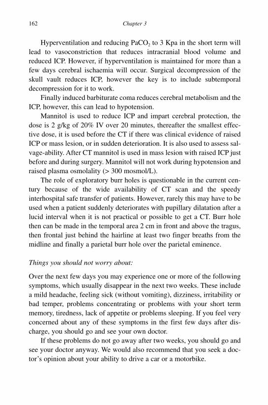
Hyperventilation and reducing PaCO2 to 3 Kpa in the short term willlead to vasoconstriction that reduces intracranial blood volume andreduced ICP. However, if hyperventilation is maintained for more than afew days cerebral ischaemia will occur. Surgical decompression of theskull vault reduces ICP, however the key is to include subtemporaldecompression for it to work.
Finally induced barbiturate coma reduces cerebral metabolism and theICP, however, this can lead to hypotension.
Mannitol is used to reduce ICP and impart cerebral protection, thedose is 2 g/kg of 20% IV over 20 minutes, thereafter the smallest effec-tive dose, it is used before the CT if there was clinical evidence of raisedICP or mass lesion, or in sudden deterioration. It is also used to assess sal-vage-ability. After CT mannitol is used in mass lesion with raised ICP justbefore and during surgery. Mannitol will not work during hypotension andraised plasma osmolality (> 300 mosmol/L).
The role of exploratory burr holes is questionable in the current cen-tury because of the wide availability of CT scan and the speedyinterhospital safe transfer of patients. However, rarely this may have to beused when a patient suddenly deteriorates with pupillary dilatation after alucid interval when it is not practical or possible to get a CT. Burr holethen can be made in the temporal area 2 cm in front and above the tragus,then frontal just behind the hairline at least two finger breaths from themidline and finally a parietal burr hole over the parietal eminence.
Things you should not worry about:
Over the next few days you may experience one or more of the followingsymptoms, which usually disappear in the next two weeks. These includea mild headache, feeling sick (without vomiting), dizziness, irritability orbad temper, problems concentrating or problems with your short termmemory, tiredness, lack of appetite or problems sleeping. If you feel veryconcerned about any of these symptoms in the first few days after dis-charge, you should go and see your own doctor.
If these problems do not go away after two weeks, you should go andsee your doctor anyway. We would also recommend that you seek a doc-tor’s opinion about your ability to drive a car or a motorbike.
162 Chapter 3
b1009 Problem Based Neurosurgery
b1009_Chapter-03.qxd 11/12/2010 10:49 AM Page 162

Problem 3-2: Spinal trauma and traumatic spinal cord syndromes.How to manage a patient following spinal trauma?
To manage spinal trauma correctlyyou need to keep a high index of sus-picion as the main objective in spinaltrauma management is to prevent sec-ondary damage and maximise thechances of recovery from the primaryinjury.
PCS3-2-1:
A 61-year-old, right-handed advocacy female worker presented to theAccident and Emergency department following a road traffic accident(RTA) in which she, the pedestrian, was hit at around 30 mph by a car. Shewas hit on the left side, fell to the ground and sustained head and neckinjuries. Immediately after the impact she lost consciousness for less thanone minute and awoke to find herself lying on the ground unable to moveor feel sensation in any of her limbs. After a few minutes she was able tomove her left arm and leg but continued to be unable to move or feel herright limbs. She was experiencing pain in the occipital region of the headwhich was later found to be a small left-sided laceration. She did not haveany other pain. She did not feel nauseous or experienced visual, hearing,swallowing or speech difficulties. She did not have any further loss ofconsciousness. She had hypothyroidism, depression, gastro-oesophagealreflux and left hip replacement in 2009. She was on Levothyroxine 75 µgOD, Omeprazole 20 mg OD, Fluoxetine 20 mg OD, Paracetamol 1 g PO,Movicol 1 sachet oral OD, Fragmin 500 units SC, Codeine Sulphate60 mg PO, Cyclizine 50 mg PRN, Chlorepheriamine 4 mg PO, Diazepam2 mg PO, Oromorph 20 mg PO, and Prochlorperazine 12 mg PO/IM.
On examination she was fully conscious and respiratory rate was21, heart rate was 60 bpm, blood pressure was 126/46 mmHg, and O2-saturation of 100% on room air. Cranial nerves I to XII had no abnormality.Examination of the limbs revealed the signs summarised in Table 3-3.
Trauma (Head and Spinal Injured Patients) 163
b1009 Problem Based Neurosurgery
Problem based tool box:Spinal trauma/fractures/shockAnterior/Central Cord
syndromeBrown-seqaurd syndromeJefferson’s/Hangman’s/
Odontoid fractures.Traction/Braces/Collars
b1009_Chapter-03.qxd 11/12/2010 10:49 AM Page 163
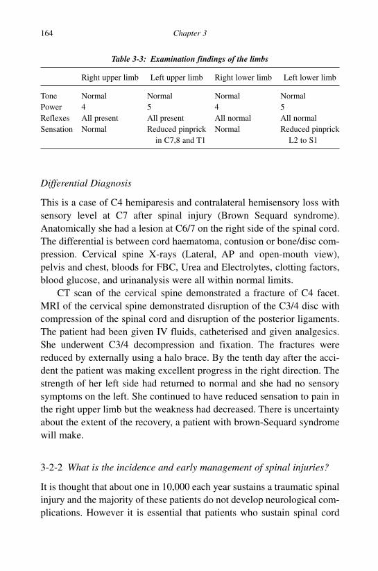
Differential Diagnosis
This is a case of C4 hemiparesis and contralateral hemisensory loss withsensory level at C7 after spinal injury (Brown Sequard syndrome).Anatomically she had a lesion at C6/7 on the right side of the spinal cord.The differential is between cord haematoma, contusion or bone/disc com-pression. Cervical spine X-rays (Lateral, AP and open-mouth view),pelvis and chest, bloods for FBC, Urea and Electrolytes, clotting factors,blood glucose, and urinanalysis were all within normal limits.
CT scan of the cervical spine demonstrated a fracture of C4 facet.MRI of the cervical spine demonstrated disruption of the C3/4 disc withcompression of the spinal cord and disruption of the posterior ligaments.The patient had been given IV fluids, catheterised and given analgesics.She underwent C3/4 decompression and fixation. The fractures werereduced by externally using a halo brace. By the tenth day after the acci-dent the patient was making excellent progress in the right direction. Thestrength of her left side had returned to normal and she had no sensorysymptoms on the left. She continued to have reduced sensation to pain inthe right upper limb but the weakness had decreased. There is uncertaintyabout the extent of the recovery, a patient with brown-Sequard syndromewill make.
3-2-2 What is the incidence and early management of spinal injuries?
It is thought that about one in 10,000 each year sustains a traumatic spinalinjury and the majority of these patients do not develop neurological com-plications. However it is essential that patients who sustain spinal cord
164 Chapter 3
b1009 Problem Based Neurosurgery
Table 3-3: Examination findings of the limbs
Right upper limb Left upper limb Right lower limb Left lower limb
Tone Normal Normal Normal NormalPower 4 5 4 5Reflexes All present All present All normal All normalSensation Normal Reduced pinprick Normal Reduced pinprick
in C7,8 and T1 L2 to S1
b1009_Chapter-03.qxd 11/12/2010 10:49 AM Page 164

injury are identified and managed appropriately in order to avoid furtherneurological injury. It is thought that 85% of cord injuries occur at thetime of the trauma but correct management can ensure that the other 15%occurring thereafter can be prevented. Spinal injuries are caused by fallsin 41.7%, road traffic accidents in 36.8%, sports injuries in 11.6%, indus-trial accidents in 4.2%, and assaults in 2.7%. About half of all cervicalspinal injuries result in spinal cord injury. These injuries are more com-mon among males at a mean age of 30 years. A high index of suspicion isneeded to detect spinal injury. Spinal injury is suspected in any patientwho had significant trauma, unresponsive patients and presence of spinalpain. At the scene patients should have their airways, breathing and cir-culation maintained and their spine immobilised. Immobilisation can beachieved with sandbags and straps, hard cervical collar, and spinal board.This is maintained in hospital, in addition to maintaining oxygenation,passing a nasogastric tube, urinary catheter, and maintaining temperature.The cervical spine must be stabilised in hospital using sandbags andstraps, a hard collar such as a Philadelphia collar, skull traction or halovest. Soft collars are not recommended because they do not prevent spinalmovements. To examine the spine “logrolling” techniques have to be usedto avoid secondary spinal cord injuries. When dealing with spinal traumamusculoskeletal and neurological injuries need to be considered.
3-2-3 What are the types of spinal cord injuries?
1- Complete lesion: Complete cord or nerve injury occurs when thespinal cord, nerve root or peripheral nerve is completely transactedwith complete loss of function of the injured neurological structure.There has been no study so far to show that early surgery for completespinal cord lesion makes any difference to recovery.
2- Incomplete lesions: These lesions include partial section, neuro-praxia or compression. These lesions present with incomplete loss offunction:
a. Brown Sequard Syndrome (BSS).b. Central Cord Syndrome (CCS).c. Anterior Cord Syndrome (ACS).
Trauma (Head and Spinal Injured Patients) 165
b1009 Problem Based Neurosurgery
b1009_Chapter-03.qxd 11/12/2010 10:49 AM Page 165

The American Spinal Injury Association categorised traumatic spinal cordinjuries into five types:
A) Complete spinal cord injury where no motor or sensory function ispreserved.
B) Incomplete spinal cord injury where sensory but no motor function ispreserved below the neurological level and includes the sacral seg-ments S4-S5. This is typically a transient phase and if the personrecovers any motor function below the neurological level, that personessentially becomes motor incomplete.
C) Incomplete spinal cord injury where motor function is preservedbelow the neurological level and more than half of key muscles belowthe neurological level have a muscle grade of less than 3.
D) Incomplete spinal cord injury where motor function is preservedbelow the neurological level and at least half of the key musclesbelow the neurological level have a muscle grade of 3 or more.
E) Normal where motor and sensory scores are normal.
3-2-4 What is spinal shock in trauma?
Shock in spinal injuries is not uncommon; shock could arise because ofexcessive blood loss related to trauma (hypovolaemic shock) or transientcomplete loss of reflexes (spinal shock). Hypovolaemic shock occursbecause of hypotonia, and blood loss and it is treated as part of ABC andresuscitation except in spinal trauma a systolic blood pressure >90 mmHgwould be acceptable because of the vasodilatation associated with loss ofautonomic vasomotor responses. Pneumatic boots are useful to increasevenous return in these patients. If these measures are insufficient vaso-pressers can be used. Spinal shock on the other hand can last from fewhours to months and is characterised by flaccidity and loss of reflexes.
3-2-5 How to evaluate the spine radiologically?
1) Lateral X-ray of cervical spine:
This needs to be complete and able to visualise C1 to T1 (Figure 3-9). Ifonly top of C6 is visible (Figure 3-10), caudal traction might be sufficient
166 Chapter 3
b1009 Problem Based Neurosurgery
b1009_Chapter-03.qxd 11/12/2010 10:49 AM Page 166

Trauma (Head and Spinal Injured Patients) 167
b1009 Problem Based Neurosurgery
Figure 3-9: Adequate lateral C-spine X-ray demonstrating C1 to T1.
Figure 3-10: Incomplete lateral C-spine X-ray demonstrating C5/6 subluxation but C7is not seen.
b1009_Chapter-03.qxd 11/12/2010 10:49 AM Page 167

to reveal the rest and the right thing to do is to repeat these X-rays withcaudal traction. Otherwise perform fine cuts CT scan with 3D reconstruc-tion (Figure 3-11).
2) Anterio-posterior C-spine X-ray.3) Open mouth view to X-ray the odontoid process (Figure 3-12).
The American Association of Neurological Surgeons (AANS) reviewedaround 40,000 trauma patients who were asymptomatic for cervical spineinjury and suggested that no radiographic assessment was required as theprocedure exposes patients to unnecessary radiation and discomfort whenin a collar. If a fracture or abnormality of the cervical spine was foundafter trauma a CT scan is required to gain better understanding of the frac-ture’s orientation, extent and mechanism of injury. Radiographs areinadequate because they are less sensitive in detecting fractures, with asensitivity of 19–31% and as high as 96% of the lateral C-spine X-rayscould be incomplete.8 However, CT scan of the spine exposes that patientto a higher radiation dose than C-spine X-ray (2.5 mSv for CT compared
168 Chapter 3
b1009 Problem Based Neurosurgery
Figure 3-11: CT reconstruction of C-spine demonstrating that C7 and T1 were fine inthe same patient shown in Figure 3-10.
b1009_Chapter-03.qxd 11/12/2010 10:49 AM Page 168

to 0.04 mSv from a single radiograph of the cervical spine). MRI is animportant investigation that can be employed to study acute neurologicalsymptoms and ligamentous injury. MRI demonstrates spinal cord com-pression, haemorrhage or contusion. MRI has 100% sensitivity for spinalcord injury and ligamentous damage but around 55% sensitivity for detec-tion of spinal fractures. MRI also offers information about the prognosisof the injury. Increased amounts of oedema and haemorrhage are associ-ated with poorer recovery. MRI is contraindicated in patients withpacemakers, aneurysm clips, ocular implants, middle ear implants andneurostimulators.
3-2-6 How to manage patients with suspected cervical spinal injuryafter radiological evaluation?
After the X-rays were taken, and the X-rays were complete, patient hadno focal neurology and (s)he had no spinal pain:
a. If the X-rays were radiographically normal, no further action isrequired and the C-spine can be cleared.
b. If the X-rays demonstrated < 3 mm subluxation, perform flexion/extension X-rays. If they were normal, no further action is required.If the subluxation reduces or exacerbates, immobilise the spine.
Trauma (Head and Spinal Injured Patients) 169
b1009 Problem Based Neurosurgery
Figure 3-12: Open mouth view of the odontoid process — C1 == Atlas, C2 == Axis.
b1009_Chapter-03.qxd 11/12/2010 10:49 AM Page 169
3-2-7 When are whole spine X-rays required?
Because a number of patients suffer multiple spinal injuries, a high indexof suspicion is required. Here are some of the indicators associated withmultiple spinal injuries:
1- Unconscious patient.2- Thrown out of a vehicle.3- No adequate history available.4- History of fall from a height (greater than six feet).5- Complaining of spinal pain in multiple locations.
3-2-8 When will you perform an emergency MRI scan?
Emergency MRI will be indicated to demonstrate soft tissue injury includ-ing spinal cord injury. If the MRI scan will change the management of thepatient then it should be done;
1- Incomplete spinal cord lesion to rule out compressive lesions.2- Deteriorating neurological deficit.3- If the radiological abnormality differs from patient’s signs and symp-
toms location.4- Suspected acute disc.
3-2-9 When will emergency surgery be required?
No scientific study had demonstrated any benefit from emergency surgeryin patients who had complete cord lesions. Therefore indications foremergency spinal surgery in trauma include:
1- Progressive neurological deficit.2- Retro-pulsed bone fragment.3- Acute anterior disc syndrome.4- Penetrating injury.5- Irreducible locked facets.6- Vital nerve compression.
170 Chapter 3
b1009 Problem Based Neurosurgery
b1009_Chapter-03.qxd 11/12/2010 10:49 AM Page 170
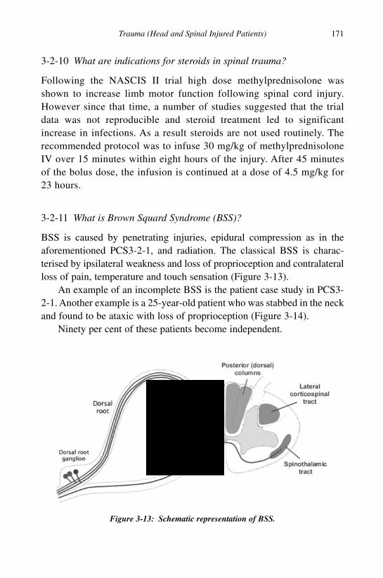
3-2-10 What are indications for steroids in spinal trauma?
Following the NASCIS II trial high dose methylprednisolone wasshown to increase limb motor function following spinal cord injury.However since that time, a number of studies suggested that the trialdata was not reproducible and steroid treatment led to significantincrease in infections. As a result steroids are not used routinely. Therecommended protocol was to infuse 30 mg/kg of methylprednisoloneIV over 15 minutes within eight hours of the injury. After 45 minutesof the bolus dose, the infusion is continued at a dose of 4.5 mg/kg for23 hours.
3-2-11 What is Brown Squard Syndrome (BSS)?
BSS is caused by penetrating injuries, epidural compression as in theaforementioned PCS3-2-1, and radiation. The classical BSS is charac-terised by ipsilateral weakness and loss of proprioception and contralateralloss of pain, temperature and touch sensation (Figure 3-13).
An example of an incomplete BSS is the patient case study in PCS3-2-1. Another example is a 25-year-old patient who was stabbed in the neckand found to be ataxic with loss of proprioception (Figure 3-14).
Ninety per cent of these patients become independent.
Trauma (Head and Spinal Injured Patients) 171
b1009 Problem Based Neurosurgery
Figure 3-13: Schematic representation of BSS.
b1009_Chapter-03.qxd 11/12/2010 10:49 AM Page 171

3-2-12 What is central cord syndrome (CCS)?
CCS is often found after hyperextension injury in a narrow spinal canal.Elderly patients with cervical spondylosis who had narrow spinal canal,go out drinking, walking back home, tripping over and falling face down,coming to hospital with bruised face and tetraparesis worse in the upperlimbs and affecting more the extensor muscles. Patients may exhibit vari-able sensory changes and urinary retention (Figure 3-15). Only 50% ofpatients may walk independently after CCS.
172 Chapter 3
b1009 Problem Based Neurosurgery
Figure 3-14: MRI scan demonstrating stabbing and severance of the posterior column.C1 == post-arch of atlas, and C2 == spinous process of C2.
Figure 3-15: Schematic presentation of CCS.
b1009_Chapter-03.qxd 11/12/2010 10:49 AM Page 172

3-2-13 What is Anterior Cord Syndrome (ACS)?
The causes of ACS are anterior cord compression, such as that followingacute disc prolapse or retropulsed bone fragment, and occlusion of theanterior spinal artery. It results in tetraplegia or paraplegia and dissociateanaesthesia (loss of pain sensation, preserved proprioception) (Figure 3-16).Surgery is often performed but only 10–20% recovers the lower limbfunctions after ACS.
3-2-14 What are the different types of C-spine fractures?
1- Atlanto-occipital subluxation (AOS):
AOS represents 1% of all cervical spinal traumas. Patients often do nothave any neurological deficit, the fracture however is unstable andpatients should be immobilised in a halo brace for three to four months. Ifit did not heal spontaneously, it should be internally fixed using occipito-cervical fixation.
2- C1 fractures:
They represent 3–13% of all cervical spinal injuries, 56% of C1 fractureswere isolated and 44% were associated with C2 fractures. Blow out fracturesof C1 were called Jefferson’s fractures (Figure 3-17). They are caused by
Trauma (Head and Spinal Injured Patients) 173
b1009 Problem Based Neurosurgery
Figure 3-16: Schematic presentation of ACS.
b1009_Chapter-03.qxd 11/12/2010 10:49 AM Page 173

axial loading and are associated with normal neurology. However, thesefractures are unstable and require immobilisation. The diagnosis is madeon the open mouth view when the overhang of C1 condyles exceeds 6 mm(Figure 3-18). Fractures of C1 have an excellent prognosis.
3- Atlanto-Axial Rotary Subluxation (AARS):
AARS is characterised by torticollis of 20 degrees lateral flexion and 20degrees rotation. This deformity was described as cock-robin torticollis.The treatment is reduction followed by immobilisation in a halo brace.Reduction is performed by gradual skull traction (Figure 3-19).
4- Hangman’s fractures:
It is traumatic bilateral fracture of the pedicles of C2 and often associatedwith disruption of C2/3 disc (Figure 3-20).
174 Chapter 3
b1009 Problem Based Neurosurgery
Figure 3-17: CT scan of Jefferson’s fracture.
Figure 3-18: Schematic representation of diagnosis of C1 fracture on open mouth viewby adding the overhang A ++ B (rule of Spence).
b1009_Chapter-03.qxd 11/12/2010 10:49 AM Page 174

These fractures occur because of hyperextension injury associatedwith axial loading and can be classified according to the fracture dis-placement and the angle between the odontoid process and the verticalaxis of the cervical spine (Figure 3-21 and Table 3-4).
Trauma (Head and Spinal Injured Patients) 175
b1009 Problem Based Neurosurgery
Figure 3-19: Three-dimensional CT reconstruction of AARS.
Figure 3-20: Hangman’s fracture associated with C2/3 disc disruption and posteriorangle.
b1009_Chapter-03.qxd 11/12/2010 10:49 AM Page 175

For stable type I fractures cervical collar might be sufficient for paincontrol. For the other types (unstable) reduce and immobilise in a halobrace for 12 weeks. Surgical reduction and fixation is reserved for failedreduction and non-union or associated incomplete spinal cord injury dueto associated C2/3 disc prolapse (Figure 3-22).
5- Odontoid fractures:
Odontoid fractures occur in 10–15% of cervical spinal injuries. In youngadults they are commonly seen after road traffic accidents, falls and skiaccidents, whilst in the elderly are common after minor falls. Hence whenyou are faced with an elderly patient after a fall, look out for three com-mon things that you do not want to miss: fracture of the odontoid peg,
176 Chapter 3
b1009 Problem Based Neurosurgery
Figure 3-21: Schematic representation of Hangman’s fractures.
Table 3-4: Effendi’s classification modified by Levine and Edwards
Type (%) # displacement Angle Mechanism Stability
Type I 3 mm or less Zero Hyperextension with axial Stable(29%) loading and intact ligaments
Type II > 3 mm 10 degree Hyperextension + axial loading Unstable(56%) or less followed by hyperflexion +
axial loadingType IIA 3 mm or less > 10 Hyperflexion with axial loading Unstable
(6%) degreesType III > 3 mm > 10 Hyperflexion and axial loading Unstable
(9%) degrees
b1009_Chapter-03.qxd 11/12/2010 10:49 AM Page 176

fracture of neck of femur and chronic subdural haematoma. The exactmechanism of odontoid fractures is not completely understood but italmost always involves an element of hyperflexion that may be associatedwith extensions and lateral rotation. The classical presentation is withneck pain and occipital neuralgia associated with neck spasm. Patientswith odontoid peg fractures tend to hold their head in their hands to reducethe pain and may complain of pins and needles in the tips of the fingers.
Odontoid fractures are classified according to their location along theodontoid peg into three types (Table 3-5 and Figure 3-23).
The majority of fractures of C1/2 are associated with no focal neuro-logical deficit due to the fact that the size of C1 ring is wide enough toaccommodate the odontoid peg (1/3 of the space), and the spinal cord (1/3of the space) and sufficient spare space remains to accommodate CSF, andblood vessels. (This rule of thirds is called the rule of Steele.) Unstable
Trauma (Head and Spinal Injured Patients) 177
b1009 Problem Based Neurosurgery
Figure 3-22: Type II Hangman’s fracture associated with C2/3 disc prolapse in a35-year-old who had somersaulted in a racing car and had hyperextension with axialloading followed by hyperflexion with axial loading.
Table 3-5: Classification of odontoid peg fractures
Type (Incidence) Description Stability
Type I (5%) Avulsion fracture of the odontoid tip Usually unstableType II (60%) Fracture at the base of the odontoid UnstableType III (30%) Fracture at the base and body Usually stableC2 body # (5%) This is pure fracture of the body of C2 Usually stable
b1009_Chapter-03.qxd 11/12/2010 10:49 AM Page 177

type 1 odontoid fractures are best treated with spinal fixation while othertypes of fractures are best treated with reduction and halo vest immobili-sation for 12 weeks. Failed reduction and non-union are indications forsurgical fixation. Factors associated with non-union include widely dis-placed fracture (6 mm or more), age > 65 years and poor healingenvironment such as osteoporosis and chronic steroids. Non-union canoccur in up to 30% of odontoid fractures. Surgical fusion/fixation was ini-tially performed using Gallie’s technique where the posterior arch of C1is fixed to the posterior elements of C2 using a wire and bone graft. Thiswas later modified by Brooks by passing the wires behind the laminae ofC2 to reduce the high failure of Gallie’s fusion (10–28%). Further modi-fications were also reported by Sonttag and others including posterior archplate and hooks (Figure 3-24). Other techniques include posterior C1/2transarticular screws, anterior odontoid screws and occipito-cervical fixa-tion in patients who suffered associated C1 fractures.
6- Subaxial spinal injuries:
These injuries include the following types:
1- Anterior subluxation associated with hyperflexion (Figure 3-25).2- Locked facets due to hyperflexion associated with rotation.3- Burst vertebral body fractures, also called teardrop fracture of the ver-
tebral body. This type of serious fracture needs to be distinguishedfrom teardrop sign often seen in hyperextension injuries seen on lat-eral C-spine X-rays where the edge of the vertebral body avulsed.
178 Chapter 3
b1009 Problem Based Neurosurgery
Figure 3-23: Schematic representation of odontoid fractures.
b1009_Chapter-03.qxd 11/12/2010 10:49 AM Page 178

4- Clay shoveler’s fracture of the spinous process of C7 seen in individ-uals who shovel the clay above their shoulders.
The treatment of these fractures follows the same general principles ofreduction and immobilisation. Surgery is preserved for failed reduction,
Trauma (Head and Spinal Injured Patients) 179
b1009 Problem Based Neurosurgery
Figure 3-24: Posterior C1/2 fusion (1 == Gallie, 2 == Brooks and 3 == Sonttag).
Figure 3-25: Subaxial subluxation of C4/5 in a 50-year-old man involved in a road traf-fic accident, reduced by 23 pounds of skull traction followed by internal fixation.
b1009_Chapter-03.qxd 11/12/2010 10:49 AM Page 179

non-union or decompression to preserve neurological function and pro-vide the best chances of neurological recovery.
Subaxial vertebral body fractures represent about 5% of all spinal frac-tures. They mostly occur in the cervical spine and Tl1/L2 area (Figure 3-26).They are often associated with retropulsed bone fragment and they areoften unstable (Figures 3-27 and 3-28).
The indications for surgery in patients with incomplete spinal cord/nerve lesions is to decompress the nerves/spinal cord in patients who haddeteriorating neurology, acute disc or > 50% spinal canal compromise. Inpatients with complete cord lesion early surgery may be indicated forearly mobilisation or to prevent spinal deformity. The approach of surgeryis dependent on the answer to three specific questions:
1- Where was the compression, if one of the goals of surgery was todecompress the nerves/spinal cord?
180 Chapter 3
b1009 Problem Based Neurosurgery
Figure 3-26: Burst fracture of L2 associated with retropulsed fragment after a fall froma horse.
b1009_Chapter-03.qxd 11/12/2010 10:49 AM Page 180

2- What was the mechanism of injury causing spinal instability?3- What were the risks of each approach compared to the potential
benefits?
For example if the mechanism of injury was hyperflexion and there wasno decompression necessary, a posterior approach to fix the spine from
Trauma (Head and Spinal Injured Patients) 181
b1009 Problem Based Neurosurgery
Figure 3-27: Axial CT scan of the same patient demonstrating the retropulsed bonefragment and note the split fracture of L2 body.
Figure 3-28: Sagittal CT scan of the same patient demonstrating the retropulsed bonefragment (F).
b1009_Chapter-03.qxd 11/12/2010 10:49 AM Page 181
the back would make sense. If the cause of neurological deficit was ante-rior compression it would make more sense to decompress the spine fromthe front to achieve maximum decompression.
Occasionally patients develop cord injury with normal radiology. Inthese circumstances patients need to be admitted to hospital for observa-tion and immobilisation of the spine. This kind of injury is common inchildren under 16 years, their diagnosis is often delayed in over 50% andthey are prone to repeated injuries.
3-2-15 How to use skull traction?
Skull traction is used to reduce cervical fractures or subluxations and toprovide immobilisation (Figure 3-29). The location of the pins is 4 cmabove the ear pinnas at the level of the external auditory meatus.However move 3 cm posterior to achieve skull traction in slight flexionif the mechanism of injury was hyperextension and 3 cm anterior forslight extension if the mechanism of injury was hyperflexion. It wouldbe better to make small nick incisions in the scalp at the pin sites. Toprevent infection of the pin sites, they must be cleaned carefully andregularly. The average starting weight of skull traction is 10 lb + the level,e.g. for C5 fracture start with 15 lb and for C6 fracture start with 16 lband so on. Perform lateral cervical spine X-rays after two to three hoursof traction. Increase the traction by 5 lb increments till the fracture/subluxation is reduced. Remove the cervical collar after the skull trac-tion is placed. Skull traction pins are spring loaded and the pins need tobe tightened after three days. The classical set for skull traction is theGardner-Wells tongs. However, it would make more sense to apply thehead ring of the halo vest for skull traction so that the halo vest couldbe applied for immobilisation once the fracture or subluxation wasreduced.
3-2-16 What are the mechanisms of spinal injuries?
Several mechanisms of injury can potentially lead to spinal injury. Hyper-flexion hinge-like injury occurs more commonly in the lumbar spine butcan occur in the lower cervical spine as well. The movement forward is
182 Chapter 3
b1009 Problem Based Neurosurgery
b1009_Chapter-03.qxd 11/12/2010 10:49 AM Page 182

usually limited by the chin coming into contact with the chest in the cer-vical spine. Hyperextension hinge injuries on the other hand, such as thatfrom a blow to the front of the head, e.g. falling face down, can causeinjury to the upper cervical vertebrae. This can result in stable fracture ofatlas or axis, or an unstable fracture of C2 pedicle “hangman fracture”. Afracture to the odontoid peg of C2 can occur from severe falls or road traf-fic accidents. Many spinal injuries are not straight forward flexion orextension injury and a degree of rotation may occur during the traumaticevent. This commonly happens during falls when the patient impacts ontothe head and can result in facet joint dislocation in the cervical spine.Rotation and extension are more likely to result in fracture of the pedi-cles.9 A burst fracture of the cervical vertebrae can occur if there is verticalcompression, such as a heavy object falling from above onto the head orin a diving accident in a shallow or empty swimming pool. Retropulsionof bone fragments into the spinal canal causing spinal cord damage mayoccur and the energy from the impact is transferred to the cord.10 Avulsioninjuries may occur as in the clay shovler’s fracture of C7 when the tip ofthe spinous process fractures and remains attached to the soft tissues.
References
1. Head injury — Triage, assessment, investigation and early management of
head injury in infants, children and adults. NICE, 2007. http://www.nice.org.
uk/guidance/index.jsp?action=download&o=36260. http://www.nice.org.uk/
Guidance/CG56.
2. SIGN Publication No. 46 (2000) — Early management of patients with a
head injury. http://www.sign.ac.uk/guidelines/fulltext/46/.
Trauma (Head and Spinal Injured Patients) 183
b1009 Problem Based Neurosurgery
Figure 3-29: Skull traction in cervical spine injuries.
b1009_Chapter-03.qxd 11/12/2010 10:49 AM Page 183
3. Eljamel MS, Waring DJ. The paragon immunofixation for CSF identifica-
tion. Biomed Sci 1993; 4(2): 43–45.
4. Eljamel MS, Foy PM. Acute cerebrospinal fluid fistulae, the risk of intra-
cranial infections. Br J Neurosurg 1990; 4(5): 381–385.
5. Eljamel MS. The role of surgery and B2-transferrin in the management of
CSF fistulae. MD thesis, Liverpool University, 1991.
6. Eljamel MS. Antibiotic prophylaxis in CSF fistulae. Br J Neurosurg 1993;
7(5): 501–506.
7. Eljamel MS, Pidgeon CN, Toland J et al. MRI-cisternography in CSF fistu-
lae localisation. Br J Neurosurg 1994; 8(4): 433–437.
8. Duane TM et al. Is the lateral cervical spine plain film obsolete? J Surg Res
2008; 147: 267–269.
9. Vaccaro AR, Madigan L et al. Magnetic resonance imaging analysis of soft
tissue disruption after flexion-distraction injuries of the subaxial cervical
spine. Spine 2001; 26: 1866–1872.
10. Hall RM, Oakland RJ et al. Spinal cord–fragment interactions following
burst fracture: an in vitro model. J Neurosurg Spine 2006; 5: 243–250.
Your personal notes:
.....................................................................................................................
.....................................................................................................................
.....................................................................................................................
.....................................................................................................................
.....................................................................................................................
.....................................................................................................................
.....................................................................................................................
.....................................................................................................................
.....................................................................................................................
.....................................................................................................................
184 Chapter 3
b1009 Problem Based Neurosurgery
b1009_Chapter-03.qxd 11/12/2010 10:49 AM Page 184

Chapter 4: Sudden Headache or Collapse(SAH, ICH, Seizures)
Problem 4-1: Sudden headache and subarachnoid haemorrhage.How to manage a patient presenting with sudden headache?
Any patient presenting with suddenheadache should be managed as sub-arachnoid haemorrhage (SAH) unlessand until proven otherwise.
Although many patients presentingwith sudden headache do not haveSAH, missing a SAH can have seriousconsequences to the patient becauseof the high rebleeding rate and death.
PCS4-1-1:
A 67-year-old woman presented acutely with sudden severe headache ofan eight-hour duration. She described the headache as a “sudden blow tothe head and the worst experience so far”. The headache was occipital tostart with but now is all over the head. She vomited once and complainedof neck stiffness. Apart from hiatus hernia and Omeprazole she had noother past medical history. She was a smoker. Examination revealed thatshe was fully conscious, with no focal neurological deficit. Fundoscopywas normal, BP and pulse were normal.
4-1-2 What is the differential diagnosis of sudden (acute) headache?
1- SAH — patient should be managed as SAH till proven otherwise.2- Thunderclap headache.3- Orgasmic cephalgia.
185
b1009 Problem Based Neurosurgery
Problem based toolbox:Aneurysm AVM CollapseOrgasmic cephalgiaSubarachnoid haemorrhageSudden headacheThunder clap headache
b1009_Chapter-04.qxd 11/12/2010 10:49 AM Page 185

The classical presentation of SAH is sudden severe headache associatedwith nausea, vomiting, photophobia, and neck stiffness (meningitic pres-entation). Some patients however may present in coma, with focalneurological deficit or after seizures.
The headache of SAH is usually severe, often described as “the worstheadache of my life” or “like an axe struck at the back of my head” or asan “explosion in my head”. The initial headache may be mild and clearsoff and the patient may not seek medical attention (referred to as sentinelheadache or warning headache). The warning headache occurs in 10–40%of patients presenting with SAH.1 About 10–25% of patients with suddensevere headache have a SAH.2,3 When sudden severe headache is the onlysymptom, one in ten patients turned out to have SAH, therefore theabsence of other symptoms cannot be used to rule out SAH. Conversely,other symptoms of SAH may accompany other causes of sudden severeheadache, so they cannot reliably distinguish SAH from migraine.3
4-1-3 What are the signs associated with SAH?
Some patients with SAH present in coma after the headache, while othercauses of sudden headache hardly ever cause coma. Coma in SAH hap-pens as a result of raised intracranial pressure. cerebral ischaemic damageto brain tissue from intraparenchymal haemorrhage, development ofhydrocephalus, or seizures. Assessment of conscious level (GCS) is there-fore crucial in the management of these patients. Some patients loseconsciousness after SAH and recover fully shortly after. In these patientsthe mechanism of brief coma is caused by the sudden rise of ICP duringthe SAH leading to transient cerebral hypoperfusion. Some patients maypresent after minor head injury as a result of a fall secondary to SAH orroad traffic accident secondary to SAH. In these patients it is very diffi-cult to disentangle trauma and spontaneous SAH. These patients shouldtherefore be treated and investigated thoroughly to rule out a cause ofSAH and you can only avoid missing SAH in patients who appear on thesurface to have a head injury is by keeping a high index of suspicion andthinking outside the box.
Focal cranial nerve deficits may also occur in SAH, e.g. third nervepalsy with diplopia, ptosis or pupillary dilatation from aneurysmal SAH
186 Chapter 4
b1009 Problem Based Neurosurgery
b1009_Chapter-04.qxd 11/12/2010 10:49 AM Page 186

following expanding PComA aneurysm, but could also be due to supe-rior cerebellar artery (SCA) aneurysm. Sixth nerve palsy may occur dueto raised ICP and often is a false localising sign. Partial or complete oph-thalmoplegia may occur in aneurysms located in the cavernous sinus.Focal neurological deficits due to intracerebral haemorrhage orischaemia may also follow SAH, e.g. dysphasia in dominant MCAaneurysms.
Fundoscopy is essential in SAH as it may reveal haemorrhage andarterial disease. Three types of intraocular haemorrhages may occur inassociation with SAH. These may occur alone or in various combinationsin 20–40% of patients with SAH.4
1. Subhyaloid haemorrhage: This bleed is in the preretinal layer andtakes the shape of half circle. It is detected in 11–33% of cases asbright red blood near the optic disc that obscures the underlying reti-nal vessels. It may be associated with a higher mortality rate.
2. Intraretinal haemorrhages: Occur in small number of patients.3. Vitreous haemorrhage: Occurs in 4–27% of patients with SAH. It is
often missed on initial examination. Most vitreous haemorrhagesclear spontaneously within six to 12 months, but patients should befollowed up for complications of elevated intraocular pressure. If thehaemorrhage was bilateral and obscured the red reflex it may lead toblindness (Terson’s syndrome).
4-1-4 What is the epidemiology of SAH?
The incidence of SAH is approximately eight per 100,000 of the popula-tion.5 It is therefore estimated that there will be 4800 new cases of SAHdiagnosed in the UK alone per year. The mean age at presentation is 61years with a female preponderance (64% females); 85% of the patients areover 45 years of age.6 SAH accounts for 3% of patients presenting toemergency departments with headaches and represent around 20 admis-sions per year to a general hospital covering 300,000 population.7 Asfamily doctors will encounter only a few cases of SAH during a lifetimecareer, a high index of suspicion is paramount to early and referral ofpatients with suspected SAH.
Sudden Headache or Collapse (SAH, ICH, Seizures) 187
b1009 Problem Based Neurosurgery
b1009_Chapter-04.qxd 11/12/2010 10:49 AM Page 187
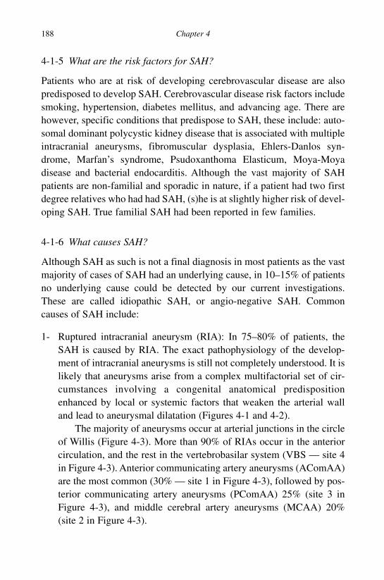
4-1-5 What are the risk factors for SAH?
Patients who are at risk of developing cerebrovascular disease are alsopredisposed to develop SAH. Cerebrovascular disease risk factors includesmoking, hypertension, diabetes mellitus, and advancing age. There arehowever, specific conditions that predispose to SAH, these include: auto-somal dominant polycystic kidney disease that is associated with multipleintracranial aneurysms, fibromuscular dysplasia, Ehlers-Danlos syn-drome, Marfan’s syndrome, Psudoxanthoma Elasticum, Moya-Moyadisease and bacterial endocarditis. Although the vast majority of SAHpatients are non-familial and sporadic in nature, if a patient had two firstdegree relatives who had had SAH, (s)he is at slightly higher risk of devel-oping SAH. True familial SAH had been reported in few families.
4-1-6 What causes SAH?
Although SAH as such is not a final diagnosis in most patients as the vastmajority of cases of SAH had an underlying cause, in 10–15% of patientsno underlying cause could be detected by our current investigations.These are called idiopathic SAH, or angio-negative SAH. Commoncauses of SAH include:
1- Ruptured intracranial aneurysm (RIA): In 75–80% of patients, theSAH is caused by RIA. The exact pathophysiology of the develop-ment of intracranial aneurysms is still not completely understood. It islikely that aneurysms arise from a complex multifactorial set of cir-cumstances involving a congenital anatomical predispositionenhanced by local or systemic factors that weaken the arterial walland lead to aneurysmal dilatation (Figures 4-1 and 4-2).
The majority of aneurysms occur at arterial junctions in the circleof Willis (Figure 4-3). More than 90% of RIAs occur in the anteriorcirculation, and the rest in the vertebrobasilar system (VBS — site 4in Figure 4-3). Anterior communicating artery aneurysms (AComAA)are the most common (30% — site 1 in Figure 4-3), followed by pos-terior communicating artery aneurysms (PComAA) 25% (site 3 inFigure 4-3), and middle cerebral artery aneurysms (MCAA) 20%(site 2 in Figure 4-3).
188 Chapter 4
b1009 Problem Based Neurosurgery
b1009_Chapter-04.qxd 11/12/2010 10:49 AM Page 188

2- Ruptured arteriovenous malformation (AVM): In about 5% ofpatients, SAH is caused by an AVM, a condition where a leach ofblood vessels cluster together and form abnormal connections that areweak and prone to bleeding (Figures 4-4 and 4-5).
Sudden Headache or Collapse (SAH, ICH, Seizures) 189
b1009 Problem Based Neurosurgery
Figure 4-1 (A) and Figure 4-2 (B): Intracranial aneurysm — real angiographic picture(A) and schematic presentation (B). ICA == Internal carotid artery, PComA == posteriorcommunicating artery, ACo == anterior communicating artery, M1 == middle cerebralartery (MCA) segment between ICA bifurcation and MCA bifurcation, M2 == MCA seg-ments after its bifurcation, A1 == anterior cerebral artery (ACA) segment between ICAbifurcation to Aco, and A2 == ACA segments distal to ACo.
Figure 4-1
Figure 4-2
b1009_Chapter-04.qxd 11/12/2010 10:49 AM Page 189

4-1-7 What is the natural history of SAH?
The natural history of SAH is dependent on the underlying cause. In themost common case scenario the underlying cause is RIA. In these patients10–15% die before reaching a hospital, a further 5% die within the first24 hours of SAH.6 Of the survivors 25–30% of patients rebleed within thefirst four weeks from the SAH, of these; approximately 70% die after therebleed.8 By the 30th day of SAH, the overall case fatality rate increases
190 Chapter 4
b1009 Problem Based Neurosurgery
Figure 4-3: CT angiographic picture of the circle of Willis (BA == basilar artery, PCA ==posterior cerebral artery).
Figure 4-4: AVM appearance on DSA.
b1009_Chapter-04.qxd 11/12/2010 10:50 AM Page 190

to nearly 50%.6,9 After the first six weeks of SAH, the rebleeding rate isabout 4% per year. In contrast patients who had an AVM underlying theSAH, the rebleeding rate is about 12% in the first year and 4% per yearthereafter. In patients with idiopathic SAH, where a good quality cerebralangiogram was negative, the rebleeding rate is negligible. SAH patientsirrespective of the underlying cause may develop delayed cerebralischaemia or hydrocephalus, e.g. 10% of patients with aneurysmal SAHdevelop delayed cerebral ischaemia and one in four develop hydrocephalus.
4-1-8 What is the management of a patient suspected of having SAH?
Any patient suspected to have had SAH, should have the following:
1- Ensure that the respiratory airways are patent and clear.2- Ensure that the patient has adequate spontaneous or assisted breathing.3- Ensure that the patient has adequate circulation.4- Admit the patient to hospital for:
a. Bed rest.b. Neurological observation.
Sudden Headache or Collapse (SAH, ICH, Seizures) 191
b1009 Problem Based Neurosurgery
Figure 4-5: Sketch of an AVM.
b1009_Chapter-04.qxd 11/12/2010 10:50 AM Page 191

c. Fluid balance (keep the patient normovolaemic).d. Adequate analgesia.e. Investigationsf. Further management.
4-1-9 What investigations a SAH-suspected patient should have? Whatis the interpretation? How to confirm SAH?
Patients suspected to have SAH should have the following investigationsdesigned to detect the underlying cause, and to detect and manage SAHcomplications:
1- Full blood count (FBC): FBC in SAH may reveal slightly raised whitecell count (WBC). Red cell count (RBC), and platelet count should benormal unless there was concomitant illness such as anaemia, andthrombocytopenia.
2- Blood chemistry: Blood glucose (BGL) may be slightly elevated dueto endogenous glucocorticoids release due to the stress of SAH, serumsodium (Na), potassium (K), and blood urea should be normal unlessthere was a concomitant illness such as renal failure.
3- Urine analysis: There may be slight glucose due to the stress-relatedhyperglycaemia, or slight protein related to reactive hypertension.Otherwise the urine analysis should be normal unless there is a con-comitant illness such as urinary tract infection.
4- Electrocardiogram (ECG): The ECG should be normal unless there is aconcomitant heart disease. However, due to the associated reactivehypertension at the onset of SAH, the ECG may demonstrate ST eleva-tion or inversion of T-waves due to subendocardial ischaemia. In a smallnumber of patients the ECG may reveal cardiac arrhythmia or infarction.
5- Clotting factors: Should be normal unless the patient is on anticoagu-lants (that should be stopped in SAH) or has a concomitant blooddiathesis.
6- Chest X-ray: Should be normal unless there is a concomitant lung orheart disease.
7- Computerised Tomography (CT) scan of the head: CT scan of thehead is mandatory in patients suspected to have SAH. It should be
192 Chapter 4
b1009 Problem Based Neurosurgery
b1009_Chapter-04.qxd 11/12/2010 10:50 AM Page 192

done urgently to confirm the SAH. The longer the scan is delayedthe more likely it will be negative. The sensitivity of CT brainexceeds 95% if performed within 48 hours of the SAH.10 SAH onCT appears as high density in the subarachanoid space (Figure 4-6).Delays in scanning allow the blood in the subarachnoid space todegrade and increase the possibility of a CT appearing as normal.The sensitivity of CT to demonstrate SAH falls sharply after48 hours to 85% in five days, 50% after one week, 30% after twoweeks and almost to nil after three weeks from the haemorrhage.11
The CT appearances can be very subtle and can be easily missed inparticular if the blood is confined to one fissure.12 A good place todetect bleed is the occipital horns where blood usually producesfluid level (Figure 4-7).
The distribution of the blood on the CT scan may help in localis-ing the location of the aneurysm that bled in patients who harbourmultiple aneurysms; 20% of patients with SAH have multipleintracranial aneurysms. For example if the SAH is concentrated in theleft Sylvain fissure, a MCAA is most likely. If it is concentrated in the
Sudden Headache or Collapse (SAH, ICH, Seizures) 193
b1009 Problem Based Neurosurgery
Figure 4-6: Diffuse SAH.
b1009_Chapter-04.qxd 11/12/2010 10:50 AM Page 193

interhemispheric fissure anteriorly, an aneurysm in the ACoA is mostlikely; If the blood is mainly in the interpenduncular cistern, a basilartip aneurysm will be more likely. If the blood is in the third ventricle theaneurysm may be in the ACoA, and if it is in the fourth ventriclethe aneurysm will be in the vertebrobasilar system. However, whenthe SAH is diffuse (Figure 4-6) the aneurysm can be anywhere. TheCT scan may also show the actual aneurysm (Figure 4-8) or an
194 Chapter 4
b1009 Problem Based Neurosurgery
Figure 4-7: Blood in the occipital horn.
Figure 4-8: SAH with MCAA.
b1009_Chapter-04.qxd 11/12/2010 10:50 AM Page 194
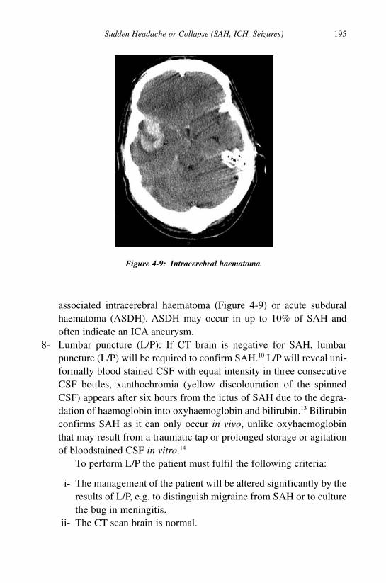
associated intracerebral haematoma (Figure 4-9) or acute subduralhaematoma (ASDH). ASDH may occur in up to 10% of SAH andoften indicate an ICA aneurysm.
8- Lumbar puncture (L/P): If CT brain is negative for SAH, lumbarpuncture (L/P) will be required to confirm SAH.10 L/P will reveal uni-formally blood stained CSF with equal intensity in three consecutiveCSF bottles, xanthochromia (yellow discolouration of the spinnedCSF) appears after six hours from the ictus of SAH due to the degra-dation of haemoglobin into oxyhaemoglobin and bilirubin.13 Bilirubinconfirms SAH as it can only occur in vivo, unlike oxyhaemoglobinthat may result from a traumatic tap or prolonged storage or agitationof bloodstained CSF in vitro.14
To perform L/P the patient must fulfil the following criteria:
i- The management of the patient will be altered significantly by theresults of L/P, e.g. to distinguish migraine from SAH or to culturethe bug in meningitis.
ii- The CT scan brain is normal.
Sudden Headache or Collapse (SAH, ICH, Seizures) 195
b1009 Problem Based Neurosurgery
Figure 4-9: Intracerebral haematoma.
b1009_Chapter-04.qxd 11/12/2010 10:50 AM Page 195

iii- In the absence of CT brain:
a. The patient must be fully conscious (GCS = 15).b. There is no papilloedema.c. There is no focal neurological deficit.
The opening pressure of CSF must be recorded at the time of L/P andthree samples of CSF are taken and observed for colour. If all thethree samples are similar in colour (blood stained), SAH should besuspected and the last sample should be spinned and protected fromlight and analysed for bilirubin (xanthochromia). The other samplesare also analysed for protein, cell-counts, and glucose (remember tosend blood for blood sugar estimation at the same time as in menin-gitis the CSF glucose will be markedly diminished — haemorrhagicmeningitis is rare but it can occur in Haemophylis influenza bacterialmeningitis). The CSF fluid should be protected from light to preventdegradation of the bilirubin. The estimation of red cell counts in threeconsecutive samples of CSF does not reliably distinguish SAH fromtraumatic tap.13 However, if you are in doubt, err on the side of SAHand investigate the patient with angiography if the history is sugges-tive of SAH.
9- Cerebral angiography: This can be achieved using CTA, DSA orMRA.
a- CTA:
CTA can be used to reveal the underlying cause of SAH nowadays dueto its speed, tolerability, convenience, and its ability to provide three-dimensional reconstructions of the cerebral circulation. Essentially ifSAH is suspected CT is performed to confirm the diagnosis. If the CTdemonstrated SAH, proceed to CTA immediately if the patient is stableenough. The sensitivity of CTA is about 95% in detecting intracranialaneurysms compared to conventional DSA and decisions about treat-ment can be based on CTA alone in most cases (Figure 4-10).
b- DSA:
Digital subtraction angiography (Figure 4-11) becomes necessary inpatients who have confirmed SAH with negative CTA or during
196 Chapter 4
b1009 Problem Based Neurosurgery
b1009_Chapter-04.qxd 11/12/2010 10:50 AM Page 196

coiling. 3D-DSA (Figure 4-12) may also become necessary to deter-mine the configuration of the aneurysm before deciding on the besttherapy. Repeated cerebral DSA, spinal catheter DSA, or magneticresonance imaging (MRI) may be necessary to identify alternativecauses of SAH in some patients. For patients with purely perimesen-cephalic bleed (blood around the brain stem), a normal, good quality
Sudden Headache or Collapse (SAH, ICH, Seizures) 197
b1009 Problem Based Neurosurgery
Figure 4-10: CTA of pericallosal artery aneurysm (arrow).
Figure 4-11: DSA left ICA PComAA (arrow).
b1009_Chapter-04.qxd 11/12/2010 10:50 AM Page 197

cerebral DSA allows a diagnosis of idiopathic perimesencephalicSAH without the need for further investigation.13
10- MRI and MRA: MRI with proton density and fluid attenuatedinversion recovery (FLAIR) have been shown to be as sensitive asCT for the detection of SAH.15 However, MRI is impractical inmost acute cases of SAH due to its lack of availability on one handand the extra safety concerns in the MRI setting. MRI would bevery useful in those who present late when CT scan and L/P werenegative or undiagnostic. MRI-FLAIR and T2-weighted sequenceswere shown to be very sensitive in detecting the byproduct of SAH,haemosiderin. An MRI sensitivity of 90% was reported in delayedpresentation of SAH compared to 46% with CT at that stage.16
MRA can also be used to detect an underlying cause of SAH suchas aneurysms (Figure 4-13). However its use is limited due to itslow sensitivity, e.g. as low as 35% detection rate was reported inaneurysms of < 5 mm.17
The following flow chart depicts the investigations of suspectedSAH patients.
198 Chapter 4
b1009 Problem Based Neurosurgery
Figure 4-12: 3d-DSA (ACoAA-arrow).
b1009_Chapter-04.qxd 11/12/2010 10:50 AM Page 198

There are advantages and disadvantages of CTA and DSA in SAHwhich is given in Table 4-1.
4-1-10 How to manage a patient with confirmed SAH?
1- Medical management:
Patients with SAH should be managed initially as follows:
a- Admit to hospital for neurological observation.b- Bed rest.
Sudden Headache or Collapse (SAH, ICH, Seizures) 199
b1009 Problem Based Neurosurgery
Figure 4-13: MRA demonstrating a large ICA aneurysm (arrow).
?SAH
CT +ve
CT - ve
CTA+ve
CT
CTA
CTA-ve
Treat theaneurysm
LPLP-ve
LP+veTreat as no SAH except in delayed
presentation where FLAIR MRI
DSA
DSA+ve
DSA-ve
Treat angio-veSAH
b1009_Chapter-04.qxd 11/12/2010 10:50 AM Page 199

c- Intravenous fluids using crystalloids. The patient should be kept nor-movolaemic with 3 L/24 hours.
d- Analgesia using moderate strength medications, e.g. Tramadol,Codeine, Tylex, Dihydrocodeine or similar analgesics. Avoid strongopiates as they may interfere with neurological observations or lead todepression of respiration. Avoid non-steroidal anti-inflammatorydrugs as they interfere with platelet functions.
e- Laxative to counteract and treat constipation, e.g. Lactulose.f- Antiemetics to treat nausea and vomiting.g- Oxygen therapy by face mask.h- Nimodipine orally or intravenously to reduce the risk of ischaemia.
Nimodipine is a selective calcium channel antagonist. There isenough scientific evidence that supports the use of Nimodipine toreduce the incidence of a poor outcome from delayed cerebralischaemia.18
i- Referral to specialist centre for urgent investigations and further man-agement of the underlying cause of SAH.
2- Prevention of rebleeding:
The risk of rebleeding is more or less evenly distributed during the firstfour weeks after the aneurysmal rupture, with a cumulative risk of 30%without intervention. Following rebleeding, the prognosis is poor: 70% ofpatients die or remain severely disabled.19
The treatment options for securing the aneurysm is either to coil theaneurysm by packing it with platinum coils that lead to thrombosis of the
200 Chapter 4
b1009 Problem Based Neurosurgery
Table 4-1: Comparison of CTA and DSA in SAH
CTA DSA
Access Readily available In specialist centres onlyRisk Almost zero 1.8% risk of stroke (CVA)Invasiveness Non-invasive InvasiveSensitivity 95% >95%Timing Can be obtained straightaway after Requires special arrangement
SAH diagnosis3D views Possible Needs special equipment
b1009_Chapter-04.qxd 11/12/2010 10:50 AM Page 200

aneurysm or by clipping the neck of the aneurysm. To coil an aneurysm ithas to be accessible to endovascular approaches such as aneurysmslocated at the bifurcation of the major vessels at the circle of Willis, theneck of the aneurysm preferably narrower than its fundus, no importantbranches coming off the aneurysm and the aneurysm of adequate size tohold the coils, otherwise the coils may prolapse back into the parent arteryand cause complications (Figures 4-14 and 4-15).
Sudden Headache or Collapse (SAH, ICH, Seizures) 201
b1009 Problem Based Neurosurgery
Figure 4-14: Coiled aneurysm-BTAA.
Figure 4-15: Incomplete coiling and prolapse of coil.
b1009_Chapter-04.qxd 11/12/2010 10:50 AM Page 201
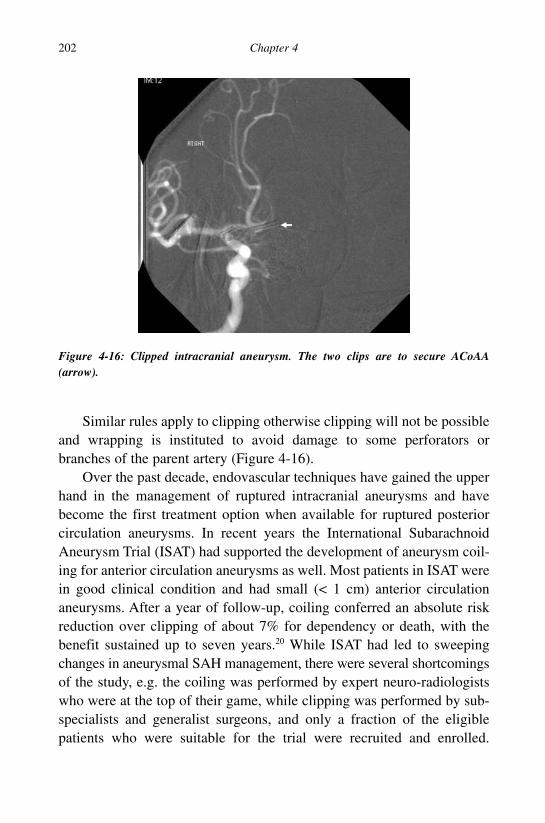
Similar rules apply to clipping otherwise clipping will not be possibleand wrapping is instituted to avoid damage to some perforators orbranches of the parent artery (Figure 4-16).
Over the past decade, endovascular techniques have gained the upperhand in the management of ruptured intracranial aneurysms and havebecome the first treatment option when available for ruptured posteriorcirculation aneurysms. In recent years the International SubarachnoidAneurysm Trial (ISAT) had supported the development of aneurysm coil-ing for anterior circulation aneurysms as well. Most patients in ISAT werein good clinical condition and had small (< 1 cm) anterior circulationaneurysms. After a year of follow-up, coiling conferred an absolute riskreduction over clipping of about 7% for dependency or death, with thebenefit sustained up to seven years.20 While ISAT had led to sweepingchanges in aneurysmal SAH management, there were several shortcomingsof the study, e.g. the coiling was performed by expert neuro-radiologistswho were at the top of their game, while clipping was performed by sub-specialists and generalist surgeons, and only a fraction of the eligiblepatients who were suitable for the trial were recruited and enrolled.
202 Chapter 4
b1009 Problem Based Neurosurgery
Figure 4-16: Clipped intracranial aneurysm. The two clips are to secure ACoAA(arrow).
b1009_Chapter-04.qxd 11/12/2010 10:50 AM Page 202

However, complete aneurysm occlusion was more likely to be achievedwith clipping. The durability of coiling beyond seven years is stillawaited. If coiling is not appropriate, clipping of the aneurysm is under-taken. Clipping is usually performed within 72 hours of SAH to minimisethe rebleed risk and enable aggressive treatment of any subsequentdelayed cerebral ischaemia.
3- Post coiling/clipping management:
The main complications of aneurysmal SAH are:
a- Rebleeding can be prevented by clipping or coiling. However, if thepatient has incomplete occlusion of the ruptured aneurysm or multi-ple aneurysms, the patient is still at risk of rebleeding (Figure 4-17).
b- Delayed cerebral ischaemia: one in ten of patients with aneurysmal SAHmay develop vasospasm and delayed cerebral ischaemia (Figure 4-18).
c- Hydrocephalus due to blockage of CSF absorption at the arachnoidvilli (Figure 4-19) or by intraventricular blood clot blocking the III or
Sudden Headache or Collapse (SAH, ICH, Seizures) 203
b1009 Problem Based Neurosurgery
Figure 4-17: Rebleeding following coiled ICA bifurcation aneurysm (notice the artefactfrom the coils in the left Sylvain fissure, blood in the subarachnoid cisterns and IVventricle).
b1009_Chapter-04.qxd 11/12/2010 10:50 AM Page 203

204 Chapter 4
b1009 Problem Based Neurosurgery
Figure 4-18: Vasospasm after aneurysmal SAH. The basilar and its branchesvasospastic.
Figure 4-19: Post-SAH hydrocephalus.
b1009_Chapter-04.qxd 11/12/2010 10:50 AM Page 204

IV ventricles. One in four patients with SAH had acute hydrocephalusat presentation (Figure 4-20).
The principal causes of post-treatment deterioration are distal vessel com-promise as a result of treatment, delayed cerebral ischaemia, hydrocephalus,cerebral salt wasting syndrome, infective or thromboembolic complica-tions. The former is minimised by careful attention during the procedure.Aspirin or heparin may be beneficial to minimise the risk of an early intra-luminal thrombosis in patients who have undergone coiling. Delayedcerebral ischaemia (peak incidence day 4–10 post-SAH) due to vasospasmshould be managed prophylactically (normal or hypervolaemia andNimodipine) and therapeutically by HHH- therapy (HHH-therapy standsfor hypertension, hypervolaemia, haemodilution). A target systolic bloodpressure of around 160 mmHg and CVP of 8–10 cm are desirable in HHH-therapy, using fluids and ionotropes as required. Should clinical features ofvasospasm become evident, the blood pressure is further augmented to eval-uate whether improvement occurs or not. Transcranial doppler sonographyis used in some centres to detect impending cerebral ischaemia by means ofincreased blood flow velocity, but the positive and negative predictive value
Sudden Headache or Collapse (SAH, ICH, Seizures) 205
b1009 Problem Based Neurosurgery
Figure 4-20: Blood clot in the fourth ventricle.
b1009_Chapter-04.qxd 11/12/2010 10:50 AM Page 205
of these tests are disappointing.21 The value of angioplasty or papaverineinjection in patients with symptomatic vasospasm is yet to be proven andwould require further randomised controlled clinical trials.
Repeat CT brain is required to diagnose or exclude hydrocephalus.Patients with intraventricular blood or with extensive blood clot in thebasal cistern will be at risk of developing acute hydrocephalus, and shouldbe treated with ventricular or lumbar drainage of CSF. A small percentageof patients will need long term CSF diversion with a shunt.
Careful monitoring of fluid balance and electrolytes is also mandatory.Hyponatraemia, mostly due to cerebral salt wasting syndrome, should notbe managed with fluid restriction since it can lead to worsening of cerebralischaemia and subsequent infarction. Other medical complications shouldbe dealt with according to local departmental and hospital protocols.
4-1-11 What is the prognosis after aneurysmal SAH?
Almost all SAH-deaths that occur after hospital admission happen withinthe first three weeks after SAH, mostly due to rebleeding. Around one-third of survivors become dependent, often with cognitive impairment,and two-thirds have a reduced quality of life.22 The three strongest pre-dictors of poor outcome of aneursmal SAH are impaired conscious levelon admission,23 increasing age and large volume of blood on the initial CTbrain (Fisher grades):
1- The level of consciousness is the main factor in determining the gradeof SAH in the World Federation of Neurological Surgeons (WFNS)grading system of SAH and is a strong predictor of outcome afterSAH (Table 4-2).
2- The Fisher grade of SAH on the CT scan also predicts delayed cere-bral ischaemia and the outcome of SAH (Table 4-3).24
The first clinical grading system used in SAH to predict prognosis is theHunt and Hess scale in 196825 (Table 4-4).
In good grade SAH patients, a favourable outcome can be achieved in90% of patients. However, the prognosis in poor grade SAH patients isgenerally poor (67% mortality and 25% with favourable outcome).25,26
206 Chapter 4
b1009 Problem Based Neurosurgery
b1009_Chapter-04.qxd 11/12/2010 10:50 AM Page 206

A recent national SAH audit in the UK showed that the six monthsoutcome of 2168 patients was 62% had favourable outcome (good recov-ery or moderate disability). The percentage of patients with good recoverydid not differ between patients who were clipped or coiled. Only 19% ofpatients who underwent no repair had favourable outcome.27
Late rebleeding can occur in patients with successfully occludedaneurysms from de novo aneurysms, or from regrowth of the aneurysm
Sudden Headache or Collapse (SAH, ICH, Seizures) 207
b1009 Problem Based Neurosurgery
Table 4-2: The WFNS grades of SAH and the outcome of SAH23
Hemi/mono-paresis or Prognosis odd ratio forGrade GCS dysphasia poor outcome (95% CI)
I 15 AbsentII 13–14 Absent 2.3 (1.3 to 4.1)III 13–14 Present 6.1 (2.9 to 12.8)IV 7–12 With or without 7.7 (4.3 to 13.7)V 3–6 With or without 69.2 (30.6 to 156.3)
Table 4-3: Fisher grades of SAH on CT
Grade Description Signs of vasospasm
I No blood detected on CT 0II Diffuse or vertical layers < 1 mm thick 0III Localised clot or vertical layer of 1 mm or thicker 96%IV Intraventricular or intracerebral clot and diffuse SAH 0
Table 4-4: Hunt and Hess grades of SAH
Grade Description Survival rate
I Asymptomatic; or minimal headache and slight nuchal rigidity. 70%II Moderate to severe headache; nuchal rigidity; no neurologic 60%
deficit except cranial nerve palsy.III Drowsy; minimal neurologic deficit. 50%IV Stuporous; moderate to severe hemiparesis; possibly early 20%
decerebrate rigidity and vegetative disturbances.V Deep coma; decerebrate rigidity; moribund. 10%
b1009_Chapter-04.qxd 11/12/2010 10:50 AM Page 207

that caused the first bleed. The risk of late rebleeding is a concern aftercoiling. The ISAT trial showed 0.7% risk of rebleeding between onemonth and one year after coiling. This risk of late rebleeding is also notedto be higher when compared to clipping20 which is around 2–3% in tenyears after clipping.28
Epilepsy develops after discharge in 14–20% of patients followingSAH. The risk factors for epilepsy after SAH include the presence ofacute subdural haematoma, cerebral infarction, or disability at discharge.The ISAT trial had shown that the risk of epilepsy is slightly higher forpatients treated by clipping when compared to those treated by coiling.20
Cognitive deficits and psychosocial dysfunction in the first year afterSAH are common in patients who make good recovery. Althoughimprovement occurs up to 18 months after SAH, many patients and theirpartners experience reduced quality of life. In a survey of 610 patientswho were interviewed after a mean period of 8.9 years after SAH andtreated by surgical clipping, 26% of the employed patients had stoppedworking, and another 24% worked shorter hours or had a position withless responsibility, 60% of the patients reported changes in personality,and only 25% reported complete recovery without psychosocial or neuro-logical problems.29
Your personal notes:
.....................................................................................................................
.....................................................................................................................
.....................................................................................................................
.....................................................................................................................
.....................................................................................................................
.....................................................................................................................
.....................................................................................................................
.....................................................................................................................
.....................................................................................................................
.....................................................................................................................
208 Chapter 4
b1009 Problem Based Neurosurgery
b1009_Chapter-04.qxd 11/12/2010 10:50 AM Page 208

Problem 4-2: Collapse and sudden focal neurological deficits. Howto manage a patient presenting with sudden focal neurologicaldeficit or collapse?
Focal neurological deficit (FND) is amanifestation of a wide range of neu-rological disorders and any patientpresenting with FND needs urgentinvestigation to confirm the underlyingaetiology.
PCS4-2-1:
A 65-year-old man presented with acute speech impairment that lasted forfew hours. Few days later he developed sudden flaccid right hemiplegiaand drowsiness. His hemiplegia was flaccid and associated with weaknessof the lower half of the face. He was hypertensive on antihypertensivetherapy. He had a CT scan of the brain (Figure 4-21).
Sudden Headache or Collapse (SAH, ICH, Seizures) 209
b1009 Problem Based Neurosurgery
Problem based tool box:Stroke TIAFocal neurological deficitBlood supply of the brain
Figure 4-21: CT scan of the brain demonstrating a large low density area (1) of the lefthemisphere. This was infarct in the middle cerebral artery (MCA) territory.
b1009_Chapter-04.qxd 11/12/2010 10:50 AM Page 209

4-2-2 What is the differential diagnosis?
A patient presenting with sudden onset FND may had suffered from:
1- Transient ischaemic attack (TIA).2- Cerebrovascular infarct.3- Spontaneous intracranial haemorrhage.4- Spontaneous arterial dissection.5- Cerebral venous sinus thrombosis.6- Todd’s paresis after focal seizure.7- Hemiplegic migraine.8- Haemorrhage secondary to other underlying pathology.
4-2-3 What is CVA (cerebrovascular accidents: strokes, infarcts, TIAs)?
CVAs occur as a result of an occlusion of blood supply to part of the brain.The brain is supplied by two internal carotid arteries (ICA) and two verte-bral arteries (VA). The ICA on each side divides into anterior (ACA) andmiddle cerebral arteries (MCA). Branches of the ICA include meningohy-pophysial artery (MHA) supplying the meninges and the pituitary gland,the ophthalmic artery (OA), posterior communicating artery (PComA) andanterior choroidal artery (AChA). The VA contributes to the anterior spinalartery (ASA), gives rise to the posterior inferior cerebellar artery (PICA)before joining its counterpart VA to form the basilar artery (BA). The BAgives rise to the anterior inferior cerebellar arteries (AICA), the superiorcerebellar arteries (SCA) and the posterior cerebral arteries (PCA). Anyblockage of these arteries leads to infarction. Infarctions can be due to:
1- Thrombosis or embolism of large arteries due to atherosclerosis.2- Embolism of cardiac or carotid origin.3- Occlusion of small blood vessels.4- Other determined cause.5- Undetermined cause.
Investigations include brain imaging and blood biochemistry and haema-tology looking for diabetes mellitus, high cholesterol, etc. CT scan is of
210 Chapter 4
b1009 Problem Based Neurosurgery
b1009_Chapter-04.qxd 11/12/2010 10:50 AM Page 210

no value in TIAs after the first week of presentation. CT scan is often nor-mal in the first 24 hours. MRI using diffusion protocols may demonstrateinfarction in the first 24 hours (Figure 4-22).
On imaging infarcts are labelled according to their vascular territory,e.g. left MCA infarct (Figure 4-21), ACA infarct, lacunar infarct or cer-bellar infarct (Figure 4-23).
4-2-4 What are the risk factors for cerebral infarct?
CVA is the second commonest cause of death worldwide. Risk factorsinclude hypertension, previous stroke or TIA, diabetes mellitus, hyperc-holesterolaemia, cigarette smoking, atrial fibrillation and advanced age.Control of hypertension protects against stroke (odds ratio 0.63, CI0.55–0.72)30 Smoking cessation is also advisable in patients who sufferedTIAs to prevent stroke, high cholesterol can be reduced by statins, atrialfibrillation with anticoagnulants, and antiplatelets to prevent further strokes(aspirin or clopidegrol).31 Patients who present within three hours withthrombotic or embolic stroke benefit from thrombolysis. Endarterectomyis used to treat ICA stenosis in patients with TIAs and decompressive
Sudden Headache or Collapse (SAH, ICH, Seizures) 211
b1009 Problem Based Neurosurgery
Figure 4-22: Diffusion MRI demonstrating an infarct.
b1009_Chapter-04.qxd 11/12/2010 10:50 AM Page 211

craniotomy (Figure 4-24) is used to salvage patients who develop intracra-nial hypertension secondary to ICA stroke or those who develophydrocephalus secondary to cerebellar infarct (Figure 4-23). Sometimesdrainage of the ventricles is required to drain hydrocephalus using exter-nal ventricular drain.
4-2-5 How to manage spontaneous intracerebral haematoma (SICH)?
Haemorrhagic stroke leads to spontaneous intracerebral haematoma.SICH forms 20% of all strokes and has the highest morbidity and mortal-ity. CT scan is the most common modality of investigation (Figures 4-25and 4-26).
Causes of SICH include AVMs, aneurysms and tumours (Figures 4-27and 4-28).
Treatment should be directed towards the underlying pathology.However, significant haematomas that cause neurological deteriorationrespond to surgical decompression particularly in the cerebellum and
212 Chapter 4
b1009 Problem Based Neurosurgery
Figure 4-23: CT scan demonstrating left cerebellar infarct (1). Note the hyperdenseobject in the right frontal horn is the tip of an external drain to divert CSF and reliefhydrocephalus.
b1009_Chapter-04.qxd 11/12/2010 10:50 AM Page 212

cerebral lobes. Adverse prognostic factors include advanced age, reducedconscious level, co-morbidity, intraventricular bleed and hydrocephalus. Ifthe patient was stable a multicentre randomised controlled trial found a pol-icy of early surgical intervention was not indicated in patients with SICH.32
Sudden Headache or Collapse (SAH, ICH, Seizures) 213
b1009 Problem Based Neurosurgery
Figure 4-24: Decompressive craniotomy (2) after MCA infarct (1).
Figure 4-25: CT scan of brain demonstrating SICH in the right striatum (1).
b1009_Chapter-04.qxd 11/12/2010 10:50 AM Page 213

4-2-6 How to manage spontaneous arterial dissection?
Spontaneous carotid (SCD) and vertebral (SVD) dissections are morecommon than reported in the literature. They present with ipsilateral
214 Chapter 4
b1009 Problem Based Neurosurgery
Figure 4-26: CT scan of brain demonstrating SICH in the cerebellum and brainstem (1).
Figure 4-27: CT scan demonstrating SICH (1) associated with SAH due to MCAaneurysm.
b1009_Chapter-04.qxd 11/12/2010 10:50 AM Page 214

headache associated with sudden onset focal neurological deficit. SCDalso associated with ipsilateral Horner’s syndrome (partial ptosis, miosisand lack of sweating) and carotid bruits. The diagnosis could be made byDSA (string sign, Figure 4-29), carotid duplex doppler or MRI/MRA.
Once considered uncommon, SCD is an increasingly recognisedcause of stroke that preferentially affects individuals between 20–40years.33,34 The incidence of SCD has been reported to be 2.6 to 2.9 per100,000. Patients with SCD may have hereditary connective tissue disor-ders or family history of stroke. These include Marfan’s syndrome,vascular Ehlers-Danlos syndrome, autosomal dominant polycystic kidneydisease, pseudoxanthoma elasticum, fibromuscular dysplasia, and osteo-genesis imperfecta type I. Carotid dissection is more common after severeviolent trauma to the head or neck. An estimated 0.67% of patients admit-ted to hospital after major accidents were found to have blunt carotidinjury; 76% had intimal dissections, pseudoaneurysms, or both. Themechanism of injury for most internal carotid injuries is rapid decelera-tion, with resultant hyperextension and rotation of the neck, which
Sudden Headache or Collapse (SAH, ICH, Seizures) 215
b1009 Problem Based Neurosurgery
Figure 4-28: Small SICH in the right frontal region (1) surrounded with low density(2) on axial CT of patient. The underlying pathology was metastatic malignantmelanoma.
b1009_Chapter-04.qxd 11/12/2010 10:50 AM Page 215

stretches the ICA over the upper cervical vertebrae, producing an intimaltear. After such an injury, the patient may remain asymptomatic, have ahemispheric TIA, or suffer a stroke. The goal of treatment is to prevent thedevelopment or continuation of neurological deficits. Treatments includeobservation, anticoagulation, stent implantation and carotid artery liga-tion. There is no controlled study for the best treatment or management ofSCAD. Empiric treatment in acute SCAD to prevent secondary embolismis partial thromboplastin time (PTT)-guided anticoagulation by intra-venous heparin followed by anticoagulation with Warfarin. Carotidsurgery for treatment of SCAD is not recommended with the possibleexception of persisting severe stenosis of the proximal ICA. Carotidangioplasty by balloon dilatation and stenting is used in selected patientsof severe cerebral haemodynamic impairment by bilateral SCAD. Theduration of secondary prophylaxis by anticoagulation is best guided byDoppler sonography follow-up, and should be continued until normalisa-tion of blood flow or until at least one year after the vessel is occluded.There is no evidence that pseudoaneurysms increase the risk for emboliccomplications, and there is no evidence for surgery or continuation of
216 Chapter 4
b1009 Problem Based Neurosurgery
Figure 4-29: DSA of the right carotid artery (1 == internal carotid artery with stringsign, 2 == external carotid artery (normal), and 3 == common carotid artery).
b1009_Chapter-04.qxd 11/12/2010 10:50 AM Page 216

anticoagulation in patients with pseudoaneurysms makes much differ-ence. Patients should be advised to avoid exercises that involve excessivehead movements (bungee jumping, trampoline jumping, and chiropracticmanoeuvres). The patient should be informed that recurrent rate is low innon-familial cases. Doppler sonography is a low-cost and highly sensitivemethod for patients at risk.
4-2-7 How to manage cerebral venous sinus thrombosis (CVST)?
Another example of haemorrhagic stroke is infarction secondary toCVST. It is a rare form of stroke that results from thrombosis of duralvenous sinuses. The incidence is about four to five per million and 75%of the affected patients are females. However, CVST is difficult to diag-nose and its incidence is likely to be higher than that reported in theliterature. Symptoms include headache, visual disturbance, motor weak-ness or seizures. The diagnosis is based on the demonstration ofobstructed venous sinuses by thrombus. Sometimes a thrombus is seen inthe confluence of dural sinuses (Torcula) as a triangular filling defect(delta sign). CT and CT venogram (CTV) (Figure 4-30) is more sensitivethan MRI and MR venogram (MRV) (Figure 4-31). CTV’s sensitivity isabout 75–100%. D-dimer may be positive in some cases. Cerebral venous
Sudden Headache or Collapse (SAH, ICH, Seizures) 217
b1009 Problem Based Neurosurgery
Figure 4-30: Image of normal CTA and CTV demonstrating: 1 == internal cerebralveins, 2 == veins of Rosenthal, 3 == straight sinus, 4 == Torcula, and 5 == superior sagittalsinus.
b1009_Chapter-04.qxd 11/12/2010 10:50 AM Page 217

thrombosis can occur post-operatively when dural sinus or cerebral veinis occluded or thrombosed, e.g. vein of Labe after subtemporal surgery orsuperior sagittal sinus after surgery of parasagittal meningioma.
Cavernous sinus thrombosis can occur secondary to infection andmanifests by proptosis. Treatment is with anticoagulants and rarely throm-bolysis. It is important to look for an underlying cause as 85% of patientshad one or more risk factors (Table 4-5). CVST is often complicated byraised intracranial pressure, which may warrant surgical intervention suchas CSF diversion.
218 Chapter 4
b1009 Problem Based Neurosurgery
Figure 4-31: MRV demonstrating: 1 == superior sagittal sinus, 2 == straight sinus, 3 ==internal cerebral veins, 4 == lateral sinus, and 5 == sigmoid sinus.
Table 4-5: Risk factors for CVST
Risk factor Comment/Tests
Thrombophilia Proteins C, S or antithrombin deficiency Nephrotic syndrome Chronic renal failureChronic inflammation Inflammatory bowel disease or SLEBlood disorder PolycythaemiaHormones Pregnancy, OCPTrauma Direct injury to sinusSurgery Operations near venous sinuses
b1009_Chapter-04.qxd 11/12/2010 10:50 AM Page 218

Your personal notes:
.....................................................................................................................
.....................................................................................................................
.....................................................................................................................
.....................................................................................................................
.....................................................................................................................
.....................................................................................................................
.....................................................................................................................
.....................................................................................................................
.....................................................................................................................
.....................................................................................................................
Sudden Headache or Collapse (SAH, ICH, Seizures) 219
b1009 Problem Based Neurosurgery
b1009_Chapter-04.qxd 11/12/2010 10:50 AM Page 219

Problem 4-3: Collapse, seizures, fits and funny turns. How tomanage a patient presenting with seizure or funny turn?
Epilepsy is not a diagnosis unless anunderlying aetiology was excluded.Any patient presenting with seizuresneeds careful history and examinationto establish the semiology and under-lying pathology.
PCS4-3-1:
A four-year-old child presented with seizures, otherwise the child wasnormal on examination. His blood tests were normal except his blood glu-cose was low. This was corrected and had an MRI (Figure 4-32) thatdemonstrated mushroom gryri. These gyri occurred after perinatal asphyxiaand was associated with epilepsy.
PCS4-3-2:
A 50-year-old woman presented with three successive tonic-clonicseizures associated with drowsiness. No other relevant findings. She had
Figure 4-32: T2-weighted axial image demonstrating mushroom gyri (1).
220 Chapter 4
b1009 Problem Based Neurosurgery
Problem based tool box:Epilepsy DysplasiaAnticonvulsants VNSMesial temporal sclerosis
b1009_Chapter-04.qxd 11/12/2010 10:50 AM Page 220
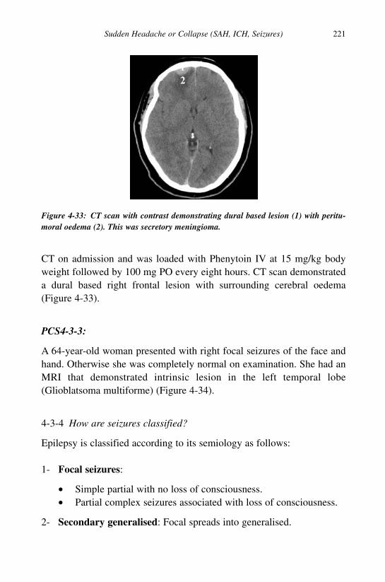
CT on admission and was loaded with Phenytoin IV at 15 mg/kg bodyweight followed by 100 mg PO every eight hours. CT scan demonstrateda dural based right frontal lesion with surrounding cerebral oedema(Figure 4-33).
PCS4-3-3:
A 64-year-old woman presented with right focal seizures of the face andhand. Otherwise she was completely normal on examination. She had anMRI that demonstrated intrinsic lesion in the left temporal lobe(Glioblatsoma multiforme) (Figure 4-34).
4-3-4 How are seizures classified?
Epilepsy is classified according to its semiology as follows:
1- Focal seizures:
• Simple partial with no loss of consciousness.• Partial complex seizures associated with loss of consciousness.
2- Secondary generalised: Focal spreads into generalised.
Sudden Headache or Collapse (SAH, ICH, Seizures) 221
b1009 Problem Based Neurosurgery
Figure 4-33: CT scan with contrast demonstrating dural based lesion (1) with peritu-moral oedema (2). This was secretory meningioma.
b1009_Chapter-04.qxd 11/12/2010 10:50 AM Page 221

3- Generalised seizures (associated with loss of consciousness):
• Absence attacks (petit mal).• Tonic-clonic (grand mal).
The incidence of epilepsy is about 50 per 100,000 population; 80% ofepilepsies are controlled by anticonvulsants (AEDs) but are not cured byAEDs. About 20% however fail medical therapy.
4-3-5 How do AEDs work and what are the principles of AED therapy?
Seizures occur secondary to sudden and excessive depolarisation of neu-rons. AEDs control but not cure seizures by inhibiting the release orfunction of excitatory neurotransmitters, by stimulating the release orfunction of inhibitory neurotransmitters or by direct stabilisation of thecell membranes. Aim for monotherapy with a drug with proven efficacyand long-term safety data. Start with low dose and gradually escalate overabout a month (enzyme induction). Increase dose up to maximum toler-ated dose (if fits persist). Assess dose selection by therapeutic drug
222 Chapter 4
b1009 Problem Based Neurosurgery
Figure 4-34: Axial T1-weighted image with gadolinium demonstrating intrinsic lesion(1) in the left temporal lobe.
b1009_Chapter-04.qxd 11/12/2010 10:50 AM Page 222

monitoring using blood levels. Around 80% otherwise healthy patients arefit free at one year, i.e. provided they have no structural lesion or con-comitant metabolic illness that caused the epilepsy. It is desirable towithdraw therapy in such patients because of undesirable side effects ofany long term AED therapy, particularly unwanted effects on cognition/behaviour (especially in children). Therefore consider withdrawal ofAEDs after three to four years if fit freedom was achieved but shouldreduce the dose gradually over several months and expect 20% relapse inthe first year after withdrawal of AEDs, and a further 20% relapse over thenext five years. However, subsequent relapse is rare after six years.
4-3-6 What are the rules regarding epilepsy and driving?
The government agency that issued the driving license should be informedby the patient about any development of seizures. The UK rules that nohigh goods vehicle (HGV) license will be issued to someone with historyof seizures. However driving a car is possible on or off treatment as longas there have been no seizures for 12 months or no daytime seizures forthe previous three years. These rules may change from one jurisdiction toanother and you need to check your local jurisdiction rules before advis-ing the patient.
4-3-7 What are the common AEDs used?
Valproate, carbamazepine and phenytoin are the most commonly usedAEDs in primary generalised tonic-clonic seizures, partial seizures, andsecondary generalised seizures. Valproate and ethosuximide are used inabsence attacks and valproate in atyoical absences, myoclonic and atonicseizures (Table 4-6).
4-3-8 How to investigate a patient presenting with seizures?
Patients presenting with seizures should have the following investigations:
1- Brain imaging is essential to exclude structural lesion. MRI would bethe best investigation. Almost all intracranial pathologies causeepilepsy (Table 4-7).
Sudden Headache or Collapse (SAH, ICH, Seizures) 223
b1009 Problem Based Neurosurgery
b1009_Chapter-04.qxd 11/12/2010 10:50 AM Page 223

224 Chapter 4
b1009 Problem Based Neurosurgery
Table 4-6: AEDs commonly used in epilepsy
AED Class of AED Half life Important side effect
Carbamazepine Tricyclic 10–15 h Hyponatraemia, rash, ataxiaPhenytoin Hydantoin 6–24 h Gum hyperplasia, rash, sedation
(22 h) neuropathy, ataxia, hirsutismValproate Carboxylic acid 5–20 h Hepatic dysfunction, hair loss,
coagulopathy, weight gainPhenobarbitone Barbiturates 5 days Sedation, anaemiaClonazepam Benzodiazepines 35 h Sedation, hypotoniaEthosuximide Succinimide 53 h Ataxia, sedation, SLE, headacheVigabartin GABA inhibitor 20–30 h Ataxia, weight gainLamotrigine Membrane stabiliser 24–34 h Skin rashGabapentin GABA analogue 5–7 h Dizziness, drowsinessTopiramate Membrane stabiliser 19–23 h Fatigue, confusions, nausea
Table 4-7: Structural causes of epilepsy
Pathology Risk of epilepsy
All types of head injuries 2–2.5%Mild head trauma 1.5%Moderate head injury 2.9%Severe head injury 17%Missile head injury 53%Traumatic haematomas 25%Traumatic cerebral contusions 26–31%Chronic subdural haematoma 7%Depressed skull fracture 10–15%Cerebral abscess 37%Glioblastoma multiforme 30–60%Oligodendrogliomas 70–90%Astrocytomas 86%Cerebral metastases 21%Arteriovenois malformation 47%Subarachnoid haemorrhage 7%Spontaneous haematoma/stroke 10–15%Meningitis 2.4–13%Encephalitis 10–22%
b1009_Chapter-04.qxd 11/12/2010 10:50 AM Page 224

2- EEG — electroencephalogram:
EEG is essential to distinguish seizures from pseudoseizures and tolocalise the focus of the epilepsy (Figure 4-35).
4-3-9 What are the indications for surgery in epilepsy?
The first line of treatment of epilepsy is AEDs. Surgery is indicated in thefollowing conditions:
1- Focal lesions such as tumour or vascular malformation: Treatmentshould be directed at treating the underlying lesion, e.g. meningiomascan be resected. AVM can be either resected or treated with stereotac-tic radiosurgery.
2- Medically refractory epilepsy is another indication for surgery:Epilepsy is considered medically refractory if it was not controlled by
Sudden Headache or Collapse (SAH, ICH, Seizures) 225
b1009 Problem Based Neurosurgery
Figure 4-35: EEG localisation of epileptic focus between lead 1 and 2, the inverteddepolarisation at time (E) pointing to the focus of the seizure.
b1009_Chapter-04.qxd 11/12/2010 10:50 AM Page 225

two attempts of high dose AED monotherapy and one attempt at poly-therapy. The proposed procedure should have low risk and the patienthad good understanding and desire to become seizure free.
a. If the epilepsy focus can be localised then resective surgery wouldbe best, e.g. Amygdalo-hippo-campectomy.
b. If the focus could not be determined then non-resective surgery.
4-3-10 How effective is epilepsy surgery?
The most common resective epilepsy surgery is Amygdalo-hippo-campec-tomy (AHC). The main indication for AHC is mesial temporal sclerosis(MTS). AHC is effective in 80% of patients with MTS (Figure 4-36) (Egelgrade I = no seizures) and it is better than prolonged medical therapyusing AEDs,35 58% of patients had no seizures after AHC compared to 8%in the AED arm (p < 0.001) and the surgical group had better quality oflife compared to the AED group (p < 0.001). There were no deaths in thesurgical arm compared to one in the AED arm. AHC is more cost effec-tive than prolonged AEDs after 8.5 years and quality adjusted life yearsimproved from 0.65 to 0.89 after AHC. Lesionectomy controls epilepsy in50–70% of patients. These may include cavernoma, AVM, cortical dys-plasia, Tuberous sclerosis, cerebral tumours and gliotic scars. The first
226 Chapter 4
b1009 Problem Based Neurosurgery
Figure 4-36: Images of left mesial temporal sclerosis (2) and normal right mesialtemporal lobe.
b1009_Chapter-04.qxd 11/12/2010 10:50 AM Page 226

epilepsy surgery was performed by Sir Victor Horsley in 1886. AHC wasfirst performed by Baldwin 1952, Hemispherectomy by McKenzie in1938 and corpus callostomy by Erickson in 1940.
4-3-11 What other therapeutic options are available apart fromresective surgery for epilepsy?
Medically refractory epilepsy can also be treated by neurostimulation:vagus nerve stimulation (VNS), deep brain stimulation (DBS) and stereo-tactic radiosurgery (SRS).
1- VNS in epilepsy:The left vagus nerve is mainly sensory and its stimulation had been usedto treat epilepsy and treatment refractory depression. Two double-blindedstudies [E03 (1994) and E05 (1998)] were conducted in patients withepilepsy, with a total of 313 treatment-resistant completers. In this diffi-cult to treat group, the mean decline of overall seizure frequency wasabout 25–30% compared to baseline.36 VNS is indicated adjunctive ther-apy in reducing the frequency of seizures in adults and adolescents over12 years of age with partial onset seizures, which are refractory to AEDs.Data from uncontrolled observations suggest that, contrary to a toleranceeffect, improvement in seizure control is maintained or may improve overtime. Infection leading to device removal was noted in 2%, left vocal cordparalysis in 1%, lower facial muscle paresis in 1%, and pain and accumu-lation of fluid over the pulse generator requiring aspiration in 0.5%.
2- DBS for epilepsy:High-frequency electrical stimulation of the anterior nucleus of the thala-mus (ANT) and of brain structures that project to the ANT was shown toreduce or inhibit seizure activity in animal models of epilepsy. TheSANTE® (Stimulation of the Anterior Nucleus of the Thalamus inEpilepsy) study is a prospective, randomised, double-blind pivotal studyto evaluate the use of DBS therapy for patients with medically refractoryepilepsy with partial-onset seizures, a form of epilepsy that does notrespond well to AEDs. The results of the study showed improvement overtime with median (mid-point) reduction in seizure frequency of 41% at
Sudden Headache or Collapse (SAH, ICH, Seizures) 227
b1009 Problem Based Neurosurgery
b1009_Chapter-04.qxd 11/12/2010 10:50 AM Page 227

one year, 56% at two years, and 68% at three years of DBS therapy, inconjunction with AEDs, compared to baseline. Of the original 110patients who received DBS implants in the trial, 91 remain active in thestudy, including some who have received DBS therapy for more than fiveyears.37
3- SRS for epilepsy:SRS is an established treatment for many lesions that cause epilepsy, e.g.AVMs. Its use in other forms of epilepsy such as MTS had been reported.The beneficial effects of radiosurgery are not displayed immediately.Most patients achieve seizure reduction at nine to 12 months and completecessation of seizures between 18 and 24 months after SRS. Typically, atransient increase in partial seizures (auras) is noted at approximately thesame time as complex seizure-decrease. SRS should be reserved to non-surgical candidates who are not fit for resective surgery and are medicallyrefractory, e.g. hypothalamic hamartomas. These are rare lesions with aprevalence of one to two in 100,000 individuals; they are commonly asso-ciated with precocious puberty, developmental cognitive delay, andgelastic (laughing) seizures. Recent reports suggest that SRS may be anexcellent treatment option for patients with hypothalamic hamartomas.However, you should be mindful of the long-term complications of SRS.The true incidence of long-term complications following SRS is not yetknown. There are however reported cases of “radiation-induced” malig-nancies associated with SRS.
References
1. de Falco FA. Sentinel headache. Neurol Sci 2004; 25(suppl 3): s215–217.
2. Landtblom AM, Fridriksson S, Boivie J et al. Sudden onset headache: a
prospective study of features, incidence and causes. Cephalgia 2002; 22:
354–360.
3. Linn FH, Wijdicks EF, van der Graaf Y et al. Prospective study of sentinel
headache in aneurysmal subarachnoid haemorrhage. Lancet 1994; 344:
590–593.
4. Manschot WA. Subarachnoid haemorrhage. Intraocular symptoms and their
pathogenesis. Am J Ophthalmol 1954; 38: 501–503.
228 Chapter 4
b1009 Problem Based Neurosurgery
b1009_Chapter-04.qxd 11/12/2010 10:50 AM Page 228

5. Linn FH, Rinkel GJ, Algra A et al. Incidence of subarachnoid haemorrhage:
role of region, year and rate of computer tomography: a meta-analysis. Stroke
1996; 27: 625–629.
6. Pobereskin LH. Incidence and outcome of subarachnoid haemorrhage: a ret-
rospective population based study. J Neurol Neurosurg Psychiatry 2001; 70:
340–343.
7. Al-Shahi R, White PM, Davenport RJ et al. Subarachnoid haemorrhage. Br
Med J 2006; 333: 235–240.
8. Rosenorn J, Eskesen V, Schmidt K et al. The risk of rebleeding from ruptured
intracranial aneurysms. J Neurosurg 1987; 67: 329–332.
9. Hop JW, Rinkel GJ, Algra A et al. Case-fatality rates and functional outcome
after subarachnoid haemorrhage: a systematic review. Stroke 1997; 28: 660–664.
10. Van der Wee N, Rinkel GJE et al. Detection of subarachnoid haemorrhage on
early CT: is lumbar puncture still needed after a negative scan? J Neurol
Neurosurg Psychiatry 1995; 58: 357–359.
11. Boesiger BM, Shiber JR. Subarachnoid haemorrhage diagnosis by computed
tomography and lumbar puncture are fifth generation CT scanners better at
identifying subarachnoid haemorrhage? J Emerg Med 2005; 29: 23–27.
12. van Gijn J, van Dongen KJ. The time course of aneurysmal haemorrhage on
computed tomograms. Neuroradiology 1982; 23: 153–156.
13. van Gijn J, Rinkel GJ. Subarachnoid haemorrhage: diagnosis, causes and
management. Brain 2001; 124: 249–278.
14. Williams A. Xanthochromia in the cerebrospinal fluid. Pract Neurol 2004; 4:
174–175.
15. Noguchi K, Ogawa T, Inugami A et al. MR of acute subarachnoid haemor-
rhage: a preliminary report of fluid-attenuated inversion-recovery pulse
sequences. Am J Neuroradiol 1994; 15: 1940–1943.
16. Ogawa T, Inugami A, Fujita H et al. MR diagnosis of subacute and chronic
subarachnoid haemorrhage comparison with CT. Am J Roentgenol 1995;
165: 1257–1262.
17. White PM et al. Intracranial aneurysms: CT angiography and MR angiogra-
phy for detection prospective blinded comparison in a large patient cohort.
Radiology 2001; 219: 739–749.
18. Pickard JD, Murray GD, Illingworth R et al. Effect of oral nimodipine on
cerebral infarction and outcome after subarachnoid haemorrhage: British
aneurysm nimodipine trial. Br Med J 1989; 298: 636–642.
Sudden Headache or Collapse (SAH, ICH, Seizures) 229
b1009 Problem Based Neurosurgery
b1009_Chapter-04.qxd 11/12/2010 10:50 AM Page 229

19. van Gijin J, Kerr RS, Rinkel GJ. Subarachnoid haemorrhage. Lancet 2007;
369: 306–318.
20. Molyneux AJ, Kerr RS, Yu LM et al. International subarachnoid aneurysm
trial (ISAT) of neurosurgical clipping versus endovascular coiling in 2143
patients with ruptured intracranial aneurysms: a randomised comparison of
effects on survival, dependency, seizures, rebleeding, subgroups, and
aneurysm occlusion. Lancet 2005; 366: 809–817.
21. Rabinstein AA, Friedman JA, Weigand SD et al. Predictors of cerebral infarc-
tion in aneurysmal subarachnoid haemorrhage. Stroke 2004; 35: 1862–1866.
22. Hackett ML, Anderson CS, for the Australasian Cooperative Research on
Subarachnoid Haemorrhage Study (ACROSS) Group. Health outcomes 1
year after subarachnoid haemorrhage: an international population-based
study. The Australian Cooperative Research on Subarachnoid Haemorrhage
Study Group. Neurology 2000; 55: 658–662.
23. Annemarie W, van Heuven, MD, Sanne M et al. Validation of a prognostic
subarachnoid haemorrhage grading scale derived directly from the Glasgow
coma scale. Stroke 2008; 39: 1347–1348.
24. Fisher CM et al. Relation of cerebral vasospasm to subarachnoid haemor-
rhage visualized by computerized tomographic scanning. Neurosurgery
1980; 6: 1–9.
25. Whitfield PC, Moss H, O’Hare D et al. An audit of aneurysmal subarachnoid
haemorrhage: earlier resuscitation and surgery reduces inpatient stay and
deaths from rebleeding. J Neurol Neurosurg Psychiatry 1996; 60: 301–306.
26. Hutchinson PJ, Power DM, Tripathi P et al. Outcome from poor grade
aneurysmal subarachnoid haemorrhage — which poor grade subarachnoid
haemorrhage patients benefit from aneurysm clipping? Br J Neurosurg 2000;
14: 105–109.
27. National Study of Subarachnoid Haemorrhage — final report. The Royal
College of Surgeons of England, February 2006.
28. Wermer MJH, Greebe P, Algra A et al. Incidence of recurrent subarachnoid
haemorrhage after clipping for ruptured intracranial aneurysms. Stroke 2005;
36: 2394–2399.
29. Wermer MJH, Kool H, Albrecht KW et al. Aneurysm screening after treat-
ment for ruptured aneurysms study group. Subarachnoid haemorrhage
treated with clipping: long-term effects on employment, relationships,
personality and mood. Neurosurgery 2007; 60: 91–98.
230 Chapter 4
b1009 Problem Based Neurosurgery
b1009_Chapter-04.qxd 11/12/2010 10:50 AM Page 230
30. He J et al. Active treatment better than placebo. Active treatment worse than
placebo. Am Heart J. 1999; 138: 211–219.
31. Diener HC et al. Aspirin and clopidogrel compared with clopidogrel alone
after recent ischaemic stroke or transient ischaemic attack in high-risk
patients (MATCH): randomised, double-blind, placebo-controlled trial.
Lancet 2004; 364: 331–337.
32. Mendelow AD, Gregson BA, Fernandes HM et al. STICH investigators.
Early surgery versus initial conservative treatment in patients with sponta-
neous supratentorial intracerebral haematomas in the International Surgical
Trial in Intracerebral Haemorrhage (STICH): a randomised trial. Lancet
2005; 365(9457): 387–397.
33. Lee VH et al. Incidence and outcome of cervical dissection; a population-
based study. Neurology 2006; 67: 1809–1812.
34. Eljamel MS, Humphrey PRD, Shaw MD. Spontaneous dissection of the cer-
vical internal carotid artery, the role of Doppler/Duplex scanning. J Neurol
Neurosurg Psychiatry 1989; 52: 1461–1462.
35. Wiebe S, Blume WT, Girvin JP et al. A randomized, controlled trial of sur-
gery for temporal-lobe epilepsy. N Engl J Med 2001; 345: 311–318.
36. Ben-Menachem E, Hellstrom K, Waldton C et al. Evaluation of refractory
epilepsy treated with vagus nerve stimulation for up to 5 years. Neurology
1999; 52: 1265–1267.
37. Nguyen DK, Spencer SS. Recent advances in the treatment of epilepsy. Arch
Neurol 2003; 60: 929–935.
Your personal notes:
.....................................................................................................................
.....................................................................................................................
.....................................................................................................................
.....................................................................................................................
.....................................................................................................................
.....................................................................................................................
.....................................................................................................................
.....................................................................................................................
.....................................................................................................................
.....................................................................................................................
Sudden Headache or Collapse (SAH, ICH, Seizures) 231
b1009 Problem Based Neurosurgery
b1009_Chapter-04.qxd 11/12/2010 10:50 AM Page 231

b1009 Problem Based Neurosurgery
b1009_Chapter-04.qxd 11/12/2010 10:50 AM Page 232
This page intentionally left blankThis page intentionally left blank

Chapter 5: Raised ICP (Tumours, Abscessand Hydrocephalus)
Problem 5-1: Raised ICP and primary malignant brain tumours.How to manage a patient presenting with raised ICP due to primarymalignant brain tumours (PMBT)?
The main goal in any patient present-ing with subacute headache is toexclude causes of raised intracranialpressure that may require urgenttreatment. Although many patientspresenting with subacute headache donot have raised ICP, missing a patientwith raised ICP could have seriousconsequences to the patient.
PCS5-1-1:
A 52-year-old woman presented with four weeks history of headacheassociated with nausea and vomiting. Over the last week she developedphotophobia and slurred speech. She felt fatigued and lost her appetite butno weight loss. She had previous history of trauma. She was hypothyroid.She smoked when she was in her teens and drank alcohol socially.Examination revealed she was overweight. She had no finger clubbing,cyanosis or anaemia. She was fully conscious and orientated with MMSEof 30/30. She had no focal neurological deficit and no papilloedema. Restof clinical examination was normal.
Differential diagnosis:
1- High grade glioma.2- Metastatic brain tumour.
233
b1009 Problem Based Neurosurgery
Problem based toolkit:Chronic headache ICPGliomas GBMOligodendrogliomaPrimary cerebral lymphomaChoroid plexus papilloedema
b1009_Chapter-05.qxd 11/12/2010 10:50 AM Page 233

3- Hydrocephalus.4- Brain abscess.5- Infection — meningitis.6- Chronic subdural haematoma.
Investigations:
• Full blood count, urea and electrolytes were normal.• Chest X-ray was clear.• Brain imaging demonstrated intrinsic mass lesion in the right tempo-
rofrontal region. The lesion enhanced with contrast in irregularmanner (Figure 5-1).
A diagnosis of malignant brain tumour was made and she was treated withsteroids (Dexamethasone 4 mg QID, orally with gastric protection) forthree days. The patient was told that the MRI scan confirmed an abnormallesion in the right side of the brain that caused these symptoms. Theoptions of treatment were discussed with the patient and the patient’sfamily.
234 Chapter 5
b1009 Problem Based Neurosurgery
Figure 5-1: MRI scan image (T1) with contrast demonstrating the lesion (G).
b1009_Chapter-05.qxd 11/12/2010 10:50 AM Page 234
Options of management in this case scenario were as follows:
• Stereotactic biopsy to ascertain the histology of the tumour with a riskof bleeding or brain swelling leading to permanent neurologicaldeficit of about 1% and 2% risk of infection.
• Cytoreductive therapy to obtain histological diagnosis and debulkthe tumour. Craniotomy to debulk the tumour carries risks includ-ing up to 5% risk of permanent neurological deficit and 2% risk ofinfection.
• Both these options require pretreatment with dexamethazone 4 mgQID.
She underwent debulking of the tumour using fluorescence guided resec-tion technology and the enhancing tumour was removed completely. Thepathology confirmed glioblastoma multiforme (GBM) and the patient wastreated with 60 Grays external beam radiotherapy.
PCS5-1-2:
A 56-year-old right-handed woman noticed that she bumped into thingson the left hand side for four weeks; first event was when she hit herhead against a water pipe when bending down. On questioning shereported a one week history of intermittent headaches, with a feeling ofpressure in forehead; gradual onset, getting more frequent but not muchworse in severity. She had been diagnosed ten years ago with MyastheniaGravis and she was taking Piridostigmine, Azothioprine, and Diazide.She lived alone and never smoked. On examination she was disorien-tated with MMSE of 17 out of 30 and had an obvious left homonymousvisual field defect. There was no papilloedema and no other focal neu-rological signs.
Differential diagnosis:
The combination of age, headaches and homonymous visual field defectmakes her likely to have a lesion in the right parietal-occipital lobe. Theinsidious onset makes likely to be a mass lesion rather than a vascular
Raised ICP (Tumours, Abscess and Hydrocephalus) 235
b1009 Problem Based Neurosurgery
b1009_Chapter-05.qxd 11/12/2010 10:50 AM Page 235

cause. The lack of papilloedema and short history makes likely to be fastgrowing mass. Therefore high grade glioma would top the list, followedby metastatic lesion, abscess and other rare lesions.
This patient had initially CT scan followed by an MRI scan. Bothscans demonstrated a single lesion similar in appearance as that shown inFigure 5-1 but located in the right parietal lobe.
Management:
She was treated with steroids and her neurological status improvedmarkedly. Options of therapy were discussed as in PCS5-1-1 and under-went gross total resection using fluorescence guided techniques followedby temazolamide — radiotherapy as the intra-operative diagnosis wasGBM.
5-1-3 What are the features of raised ICP headaches?
Raised ICP headache is a generalised ache over the cranium, worst onawakening, may awaken the patient from sleep, aggravated by bending,stooping, or straining and its severity gradually progresses. Initially theheadache might be mild and controllable by simple paracetamol, as it pro-gresses even stronger painkillers might have little effect. The reason thatraised ICP headache is worse upon a wakening up or may awaken thepatient in the middle of the night is due to hypoventilation occurring dur-ing sleep, in particular during rapid eye movement (REM) sleep, thatleads to accumulation of CO2 in the blood. Increased CO2 in the bloodleads to cerebral vasodilatation and increased intracranial pressure. Theheadache is associated with: nausea and vomiting, obscuration (transientloss) of vision with sudden change in posture. (This feature is an indica-tion of markedly elevated ICP and the patient might be at risk ofdeveloping impaired conscious level.)
5-1-4 What are the characteristic features of raised ICP?
• Headache — typical of raised intracranial pressure.• Nausea and vomiting.
236 Chapter 5
b1009 Problem Based Neurosurgery
b1009_Chapter-05.qxd 11/12/2010 10:50 AM Page 236

• Visual disturbances:
� Blurring of vision.� Visual obscuration — transient blindness.� Papilloedema in some patients.� Retinal haemorrhages if the rise in ICP has been rapid.
• Brain shifts — often, with depression of conscious level.• Sixth cranial nerve palsy leading to lateral rectus weakness, as the
sixth nerve had a very long intracranial course it is susceptible toinjury in raised ICP — false localising sign.
• Third nerve palsy due to ipsilateral uncal herniation — true localisingsign, the lesion usually on the same side as the third nerve palsy.
• Hemiparesis due to contralateral shift of the cerebral peduncle lead-ing to pressure against the edge of tentorium — false localising sign,the lesion most often on the same side as the hemiparesis.
• Peptic ulceration.• Cushing’s response: when transtentorial herniation occurs, hyperten-
sion and bradycardia develop.• In infants, slowly increasing intracranial pressure may present as a
slowly increasing head size.
5-1-5 What are the causes of raised ICP?
• Congenital:
� Hydrocephalus.� Dandy Walker Syndrome.
• Acquired:
� Tumours:Gliomas, primary cerebral lymphoma, choroid plexus papilloma,brain metastases, meningiomas, haemangioblastoma, vestibularschwannoma, colloid cysts, pineal tumours, craniopharyngioma, andepidermoid cyst.
� Trauma:EDH, ASDH, CSDH, DAI, cerebral contusion, and brain oedema.
Raised ICP (Tumours, Abscess and Hydrocephalus) 237
b1009 Problem Based Neurosurgery
b1009_Chapter-05.qxd 11/12/2010 10:50 AM Page 237

� Infection/inflammation:Meningitis, encephalitis, brain abscess, tuberculoma, TB-meningitis,sarcoidosis, and other chronic infections.
� Vascular:Spontaneous SAH, SICH, intracranial aneurysm, AVM, infarct,malignant hypertension, and sinus venous thrombosis.
� Metabolic:Respiratory failure, hepatic failure, renal failure, intoxication, andsome drugs.
5-1-6 Epidemiology of primary malignant brain tumours:
Primary malignant tumours of the central nervous system occur in 5–7 per100,000 of the general population.1 It is important to note that metastaticbrain tumours are more common in the adult population. Metastasisoccurs either by haematogenous or direct spread — the commonest pri-mary site being lung, bowel, kidney, breast and malignant melanoma.Studies have shown that one in four cancer patients will have brain metas-tasis on autopsy2 and it is not uncommon that many patients present withbrain metastasis without ever finding a primary site.3 Glioblastoma multi-forme (GBM) peaks around the age of 65 to 74 years whileoligodendroglioma peaks earlier at the age of 35 to 44 years. The averageage-at-onset of all primary tumours is around 54 years.
In children the main diagnosis is astrocytoma and medulloblastoma,and 70% of childhood cancers occur in the posterior cranial fossa. Inadults 70% of tumours occur above the tentorium with a wider variety ofcell types. The WHO in 1993, issued a global classification system for pri-mary brain tumours,4 which was later modified in 2000 (Table 5-1); 65%of primary CNS tumours are intrinsic in nature.
The aetiology of primary brain tumours had been studied over theyears and a number of potential causative factors had been investigatedover the years as follows;
• There is some evidence suggesting some inherited genes mightincrease the risk of developing primary brain tumours. Hereditary
238 Chapter 5
b1009 Problem Based Neurosurgery
b1009_Chapter-05.qxd 11/12/2010 10:50 AM Page 238

Raised ICP (Tumours, Abscess and Hydrocephalus) 239
b1009 Problem Based Neurosurgery
Table 5-1: Modified WHO classification of brain tumours
1 Neuroepithelial (Neuroectodermal)
1.1 Astrocytoma
1.2 Oligodendroglioma
1.3 Ependymoma
1.4 Mixed gliomas
1.5 Choroid plexus tumours
1.6 Uncertain origin
1.7 Neuronal and mixed neuronal/glial
1.8 Pineal parenchymal
1.9 Embryonal
2 Tumours of Nerve Sheath
2.1 Schwannoma
2.2 Neurofibroma
2.3 Malignant (peripheral) nerve sheath tumour
3 Tumours of the Meninges
3.1 Meningioma
3.2 Mesenchymal (not meningothelial)
3.2.1-4 Benign
3.2.5-10 Malignant
3.2.5 Haemangiopericytoma
3.3 Primary melanocytic lesions
4 Lymphoma
5 Germ Cell Tumours
5.1 Germinoma
6 Cysts and Tumour-like Lesions
6.1 Rathke cleft cyst
6.2 Epidermoid cyst
6.3 Dermoid cyst
6.4 Colloid cyst
7 Tumours of the Sellar Region
7.1 Pituitary adenoma
7.3 Craniopharyngioma
8 Local Extension of Regional Tumours
9 Metastatic
10 Unclassified
b1009_Chapter-05.qxd 11/12/2010 10:50 AM Page 239
syndromes including tuberous sclerosis, types 1 and 2 neurofibro-matosis, syndromes involving adenomatous polyps and nevoid basalcell carcinoma are known to have a predisposition to brain tumours.Although these genes are likely to increase the chance of developingbrain tumours, they are only responsible for the development of a lim-ited proportion of primary brain tumours. It is estimated that they onlycontribute to about 2% of tumours diagnosed in children and less than1% in adults. The majority of primary brain tumours are thereforesporadic.
• Head injury and trauma have been investigated as a potential risk fac-tor for several types of brain tumours. A number of studies wereconducted in this area and their findings were not consistent. A largecohort study (n = 228,055) carried out in Denmark, showed noincreased risk of glioma or meningioma.
• Some authors reported higher numbers of patients with gliomassuffering from epilepsy or seizures compared to controls. However,seizure is a common symptom of brain tumours and it would be diffi-cult to determine the causality.
• Smoking and alcohol consumption are major risk factors in lung,breast and liver cancers. However there is no evidence to prove thatsmoking and excess alcohol consumption are responsible forincreased risk of developing brain tumours. Most carcinogenic com-pounds in tobacco do not cross the BBB except N-nitroso compounds.Studies found neither clear association nor significant contributionfrom smoking tobacco. Review of alcohol brain tumour associationfound four of a total of eight studies reported a relative risk of lessthan one for any alcohol use compared to no alcohol use.
• Ionizing radiation is a strong risk factor for brain tumours. In children,therapeutic ionizing radiation is thought to play a role in increasingthe risk even though the radiation is relatively low. In adults, whohave undergone irradiation for acute lymphoblastic leukaemia duringchildhood have been reported to have an increased risk of glioma.
• Association between mobile phone use and brain tumour have beenunder investigation because of its political interest. Most studiesshowed no association between the use of mobile phones, the durationof mobile phones-use and brain tumours.
240 Chapter 5
b1009 Problem Based Neurosurgery
b1009_Chapter-05.qxd 11/12/2010 10:50 AM Page 240

The relative incidence of brain tumours is summarised in Table 5-2.
5-1-7 What are the different types of primary malignant brain tumours?
1- Astrocytomas — These can be divided into several categories asfollows:
a. Pilocystic Astrocytomas (PAs) WHO Grade I: PAs usually locatedin the cerebellum, diencephalon (especially the optic nerves andhypothalamus). Children five to 15 years of age are the most affected,with a peak incidence around ten years. Histologically: alternatingdense and loose areas, with fusiform “piloid” bipolar astrocytes, andmicrocysts in loose areas that may coalesce to form large cysts. Thepresence of nuclear atypia (without mitotic activity) does not conveya worse prognosis. Vascular changes are usually limited to capillaryproliferation that may include glomeruloid capillaries and endothelialproliferation. Eosinophilic “Rosenthal fibres” are a characteristic fea-ture. Calcification is possible and always positive on GFAP (GlialFibrillary Acidic Protein) stain. Prognosis is good and the tumourremains stable but rarely disseminate via CSF pathways and malig-nant transformation had been reported. On MRI scan these tumoursclassically look cystic with a tumour nodule. The wall of the cyst isoften composed of non-neoplastic tissue, with the tumour limited to a“mural nodule”. Enhancement is almost universal, and outlines the
Raised ICP (Tumours, Abscess and Hydrocephalus) 241
b1009 Problem Based Neurosurgery
Table 5-2: Relative incidence of primary brain tumours comparedto other intracranial tumours
Category Frequency (%)
Glioma (primary malignant brain tumours) 38Meningiomas 16Pituitary adenomas 8Schwannomas 6Craniopharyngiomas 3Haemangioblastomas 2Metastasis 12Others 15
b1009_Chapter-05.qxd 11/12/2010 10:50 AM Page 241

neoplastic tissue — i.e. part of the cyst wall may not enhance — thisis the “classic” cyst-with-nodule appearance (Figure 5-2).
The main treatment is complete surgical excision. Although theyare benign tumours, some clinicopathological factors, such as partialresection, optic chiasmatic location, invasion of surrounding struc-tures, and the pilomyxoid variant, have a worse prognosis.5
b. Subependymal Giant Cell Astrocytoma (SGCA) WHO Grade I:SGCA commonly located in the lateral ventricle, attached to caudatenucleus head. Occurring in 5% to 15% of patients with tuberous scle-rosis, nearly all subependymal giant cell astrocytomas are associatedwith this disorder. Mostly occur in childhood or adolescence; and fewtumours are congenital. Due to their anterior location in the lateralventricle and proximity to the foramen of Monro, symptoms of raisedICP due to obstruction of CSF flow are not uncommon. CT and MRIscans show the demarcated lesion to be contrast-enhancing and with-out oedema (Figure 5-3).
Calcium is often evident on CT. SGCA show considerable histo-logical variation. Although considered an astrocytic tumour, it oftenshows a glio-neuronal phenotype. This includes ganglion-like cellsand immunoreactivities for glial (S-100 protein, GFAP) and neuronalmarkers (neurofilament protein, class III beta-tubulin, and neuropep-tide staining). Mixed differentiation may also be present onultrastructure, in addition to glial filament accumulation, micro-tubules, secretory granules, and even synapses may be present. If
242 Chapter 5
b1009 Problem Based Neurosurgery
Figure 5-2: MRI of pilocystic astrocytoma.
b1009_Chapter-05.qxd 11/12/2010 10:50 AM Page 242

SGCA is partly resected it may progress later. Rare examples withpleomorphism, mitotic activity, neovascular proliferation or necrosisdo not behave in a malignant fashion in this type of tumour. Thebehaviour of SGCA corresponds to that of WHO grade I tumours.Treatment mainly gross total resection, recurrence is very rare.6
c. Pleomorphic Xanthoastrocytoma (PXA) WHO Grade II–III: PXAarises from subpial astrocytes and commonly located in the cerebralhemispheres, usually superficial, and often temporal. Children andyoung adults are the most commonly affected age group. Histologically,PXA is a mixture of unusually pleomorphic cells, ranging from fibril-lary to bizarre giant multinucleated cells with intracellular lipidvacuoles (“xanthoma” cells). These xanthoma cells are GFAP positive.PXA may progress in some cases to Grade III (anaplastic astrocytoma)or even Grade IV (GBM). Radiologically, PXA appears as a large hemi-spheric mass, closely related to the cerebral surface and may have aheterogeneous appearance with cyst formation, variable calcification,and prominent enhancement. The superficial location (Figure 5-4) andpresentation in childhood are the most helpful diagnostic features.Dural involvement and a “dural tail” may be seen.7
d. Infiltrating Diffuse Astrocytomas (IDA) WHO Grade II: IDAoften progresses to higher grades and originates from astrocytes. IDAoccurs most commonly in cerebral hemispheres in adults and in the
Raised ICP (Tumours, Abscess and Hydrocephalus) 243
b1009 Problem Based Neurosurgery
Figure 5-3: Subependymal giant cell astrocytomas.
b1009_Chapter-05.qxd 11/12/2010 10:50 AM Page 243

pons (“brainstem glioma”) in children. They affect adults in theirfourth and fifth decades of life (30’s and 40’s). They appear under themicroscope as ill-defined and diffusely infiltrating, with overrun(trap) neurons, and cause enlargement but not destruction of theinvaded structures, mitoses, neovascular proliferation, and necrosisare not present in IDA and they stain positive for GFAP. They natu-rally progress to anaplastic astrocytomas (AA), and then to GBM. OnMRI, IDA is usually recognised by causing expansion of the infil-trated portion of the cerebrum or brainstem. Mass effect may beminimal for the overall size of the signal/attenuation abnormality, andthe lesion may be surprisingly large at the time of presentation withminimal symptoms (Figure 5-5). Regions of signal and attenuationchange usually represent the neoplasm itself — since the intact BBBdoes not allow enhancement or vasogenic oedema. Haemorrhage israre, however, calcification can occur. The tumour tends to followwhite-matter tracts and may cross the corpus callosum, or follow thepeduncles to or from the brainstem.8
e. Anaplastic Astrocytoma (AA) WHO Grade III: AA originates fromastrocytes. AA occurs in the cerebral hemisphere or the brain stem.The histological features of AA are increased cellularity, pleomor-phism, mitotic activity, and nuclear atypia but no necrosis. AA stainspositive to GFAP and most likely to progress to GBM. Radiologically
244 Chapter 5
b1009 Problem Based Neurosurgery
Figure 5-4: PXA of the left posterior frontal cortical region.
b1009_Chapter-05.qxd 11/12/2010 10:50 AM Page 244

AA is variable in appearance of enhancement and surroundingoedema (Figure 5-6). Necrosis (ring enhancement) does not occur,and cyst formation is extremely rare.
AA is considered the least common stage and may represent ashort-term intermediate lesion during the transition from WHO grade
Raised ICP (Tumours, Abscess and Hydrocephalus) 245
b1009 Problem Based Neurosurgery
Figure 5-5: Infiltrating diffuse astrocytomas of the insula. The high signal area caus-ing enlargement of the insular cortex and the mass effect is relatively small comparedto the size of the tumour with no vasogenic oedema.
Figure 5-6: AA of right cerebral hemisphere.
b1009_Chapter-05.qxd 11/12/2010 10:50 AM Page 245

II to GBM. The 12-month survival of AA varies from 60% to 73% andthe two-year survival from 36% to 50%. Adverse prognostic factorsinclude age > 65 years, Karnofsky performance score of < 70, loca-tion in eloquent brain area and partial resection.9
f. Glioblastoma Multiforme (GBM) WHO Grade IV: GBM originatesfrom astrocytes. It occurs in the cerebral hemispheres, occasionallyelsewhere (brainstem, cerebellum, or spinal cord). The mean age atpresentation is 45 to 60 years. Histologically, GBM is grossly hetero-geneous, with degeneration, necrosis and haemorrhage. GBM hasvariable GFAP stain, and often present in areas of better differentia-tion. Radiologically GBM is usually seen as a grossly heterogeneousmass. Ring enhancement surrounding a necrotic centre is the mostcommon appearance (Figure 5-7), but there may be multiple ringswith surrounding vasogenic oedema that adds significantly to themass effect.
Signs of recent (methaemoglobin) and remote (haemosiderin) haem-orrhage are common. Despite its apparent demarcation on enhanced
246 Chapter 5
b1009 Problem Based Neurosurgery
Figure 5-7: MRI scan of GBM of the right frontotemporal region with irregularlyenhancing ring.
b1009_Chapter-05.qxd 11/12/2010 10:50 AM Page 246

scans, the lesion may diffusely infiltrate into the brain, crossing thecorpus callosum in 50–75% of cases (butterfly glioma Figure 5-8).
GBM is locally malignant brain cancer that leads to inevitable deathwithin two years of diagnosis in almost all but very few patients.10,11 Itsability to invade the brain renders it beyond the reach of the surgicalmicroscope and by the time it manifests clinically, tumour cells havealready migrated a long way from what the surgeon can see at operationor what the MRI scan can demonstrate pre- or intra-operatively.Maximal or complete tumour resection of AA or GBM is paramount asthe wealth of recent evidence suggests that more extensive resectionmeans longer survival compared to partial resection or biopsy.12–14
Recent technology had made very little impact on the survival of GBMexcept for fluorescence guided resection (FGR) that was subjected torandomised controlled trials and had led to 65% complete resection ofthe enhancing lesion on MRI compared to 35% using standard tech-niques.15,16 with significant prolongation of time to tumour recurrence.
Systemic chemotherapy had limited effect because of toxicity andfailure to cross the BBB in adequate concentration.17 Local carmus-tine implants had been demonstrated to prolong survival in newlydiagnosed GBM18 with median survival of 13.5 months compared to11.4 months with placebo. However, serious adverse events had beenreported with carmustine wafers.
Raised ICP (Tumours, Abscess and Hydrocephalus) 247
b1009 Problem Based Neurosurgery
Figure 5-8: Butterfly glioma.
b1009_Chapter-05.qxd 11/12/2010 10:50 AM Page 247

2- Oligodendrogliomas (ODG):They arise from oligodendrocytes. ODG are rarer than astrocy-tomas and are slower growing tumours characteristically seen with a“fried egg” round centred haloed appearance often with calcification(Figure 5-9).
3- Choroid plexus papilloma (CPP) (WHO Grade I) and ChoroidPlexus Carcinoma (WHO Grade III–IV): They arise from the choroidplexus epithelium. The most common location in adults is the fourthventricle and in children the left lateral ventricle.19 It affects childrenmore than adults and 40–50% of papillomas are seen in the first yearof life, and 85% in the under five years of age. Carcinomas usuallyseen only in paediatric age group. They often present with hydro-cephalus. CPP have characteristic lobulated gross appearance. Mostare well-differentiated and may resemble normal choroid plexus.However, anaplastic transformation can occur. Parenchymal invasionsuggests carcinoma, but can be seen with benign tumours. Cytokeratindistinguishes CPP from ependymoma; Prealbumin (transthyretin)may be helpful (although metastases may also stain positive). It is oneof CNS tumours that metastasise via CSF seeding (others includeependymomas, and medulloblastoma). On neuroimaging CPP appearswell-demarcated intraventricular (or cerebellopontine angle) mass
248 Chapter 5
b1009 Problem Based Neurosurgery
Figure 5-9: Oligodendroglioma on MRI.
b1009_Chapter-05.qxd 11/12/2010 10:50 AM Page 248

with hydrocephalus. Calcification is frequent in fourth ventriculartumours and often attached to the choroid plexus. Hydrocephalus mayreflect multiple factors, including CSF over-production, ventricularobstruction, and impaired CSF reabsorption (Figure 5-10).
4- Neuronal and mixed glial tumours:
Ganglion Cell Tumours:
i. Gangliocytoma (WHO Grade I).
ii. Ganglioglioma (WHO Grade I–II).These tumours arise from large, mature neurons +/− glial component.They may occur anywhere in the neural axis, but especially common inthe temporal lobe. They affect any age but most common in the first twodecades of life. They present with seizures. Gangliocytomas are com-posed only of neuronal elements while gangliogliomas also have glial
Raised ICP (Tumours, Abscess and Hydrocephalus) 249
b1009 Problem Based Neurosurgery
Figure 5-10: CPP of fourth ventricle with calcification and CS dissemination.
b1009_Chapter-05.qxd 11/12/2010 10:50 AM Page 249

component. They have varied histological appearance and histologypoorly correlated with prognosis. The neuronal component can be veri-fied by synaptophysin, neuron-specific enolase stain and the glialcomponent by GFAP positivity. These tumours are slow-growing lesions.However they have low malignant potential which is restricted to theglial component of ganglioglioma. They appear as well-defined solid ormixed cystic/solid mass with minimal or no mass effect. Calcification iscommon. Enhancement pattern is variable, but often is in the periph-ery. They are rarely seen in association with congenital malformationssuch as Down’s syndrome, callosal dysgenesis, and neuronal migrationdisorders.
iii. Desmoplastic infantile ganglioglioma (WHO Grade I).These tumours originate from large, mature neurons with glial compo-nents and together with superficial cerebral astrocytoma may be referredto as desmoplastic supratentorial neuroepithelial tumours of infancy. Theypresent with macrocephaly. They are composed of glial and neuronal dif-ferentiation with moderate pleomorphism, infrequent mitoses andabundant desmoplasia characteristic. The neuronal component confirmedby synaptophysin or electron microscopy and GFAP positivity. Treatmentis surgical excision with good prognosis. On neuroimaging these tumoursappear as very large mass with cystic and solid components. The solidportion tends to be superficial and enhances intensely with contrast. Theseare related to superficial cerebral astrocytoma, which is distinguished onlyby its lack of neuronal elements.
iv. Lhermitte-Duclos (Dysplastic Gangliocytoma WHO Grade I).These tumours arise from cerebellar neurons and are consideredhamartomas in origin. They arise in the cerebellum in early childhood.They often present with hydrocephalus but may be found as incidentalfinding on neuro-imaging. Under the microscope they are characterisedby derangement of normal laminar cellular organisation of the cerebel-lum: thickening of outer molecular cell layer, loss of middle Purkinje celllayer, and infiltration of inner granular cell layer with dysplastic ganglioncells. They are synaptophysin-positive. They carry good prognosis withsurgical excision. Recurrence is very rare. On neuro-imaging they appearas non-enhancing cerebellar hemispheric mass with characteristic striated
250 Chapter 5
b1009 Problem Based Neurosurgery
b1009_Chapter-05.qxd 11/12/2010 10:50 AM Page 250

MR appearance and occasionally contained calcification. They havean association with Cowden’s disease (mucosal neuromas and breastcancer).
v. Dysembryoplastic neuroepithelial tumour (WHO Grade I).They are thought to arise from external granular layer of the cortex but thehistogenesis is uncertain. They are called DNET or DNT and are locatedin the cortex of the temporal lobe. They can occur at any age but mostcommonly in children and young adults. They present with partial com-plex seizures. The diagnosis is based on the presence of: a specificglioneuronal element, consisting of oligodendrocytes in a mucinousmatrix in which neurons appear to “float”, or glial nodules associated withcortical dysplasia. Neuronal markers (synaptophysin, neuronal specificenolase) and glial markers (GFAP, S-100) are all positive. Despite theirbenign behaviour, they may have a high MIB-1 labelling index. Theyremain stable over long periods of time. On neuroimaging they appear asnodular cortical lesions without oedema or mass effect. They may havemegagyric or multicystic appearance. Occasionally they may enhance orcontain calcification and CT may show calvarial remodelling. They areone of the surgically curable causes of seizures.
vi. Central neurocytoma (WHO Grade I).They arise from neurons and arise within the ventricles (lateral morethan the third) often attached to septum pellucidum. They affect youngadults (mean age: 25–30 years). They present with hydrocephalus andraised ICP. They have uniform appearance of small, round cells mimick-ing oligodendroglioma. Electron microscopy may demonstrate neuronalfeatures. Purely neuronal origin is demonstrated by neuronal markers(synaptophysin, neuronal specific enolase) and negativity to glial markers(GAFP). They are slow growing, benign tumours with no extraventricu-lar extension. Surgical resection is usually curative. On CT and MRI theyappear as well-circumscribed, lobulated intraventricular mass. About50% have gross calcification, which is best demonstrated on CT. Theyare isodense or slightly hyderdense on CT and may be isointense to graymatter on both T1- and T2-weighted MR images. MR often demonstratessmall “cysts”. In the past they were misdiagnosed as intraventricularoligodendroglioma.
Raised ICP (Tumours, Abscess and Hydrocephalus) 251
b1009 Problem Based Neurosurgery
b1009_Chapter-05.qxd 11/12/2010 10:50 AM Page 251

5- Primary cerebral lymphoma (PCL) WHO Grade IV:
PCL arises from microglial cells, usually to B-cell lineage. They are com-monly found in the basal ganglia, periventricular, and gray-white junctionand may involve the corpus callosum and rarely may arise in leptomeninges(Figure 5-11). The fifth and sixth decades in non-AIDS cases are the mostaffected but can occur at any age group in relation to AIDS. Some seriesshow male predominance of up to 2:1. PCL may express Epstein-Barr virussurface markers. Their presentations are variable including focal neurolog-ical deficit, increased intracranial pressure, cognitive dysfunction, andseizures in common with other intracranial tumours. Under the microscopethey appear infiltrative in nature with patchy cellularity characterised byangiocentricity and angioinvasion and round lymphocytes with large roundnuclei with prominent nucleoli. They are positive for Epstein-Barr virusPCR in immunocompromised patients. PCL had rapid growth, especially inassociation with AIDS, they may however disappear temporarily withsteroids but recurrence is certain and CSF dissemination is common. OnCT/MRI scan they appear as iso- to slightly hyperdense on CT (Figure 5-11); may be iso- or hypointense on T2-weighted MR images. Single ormultiple, homogeneously or ring-enhancing lesions may be seen andsubependymal, callosal, or CSF spread can be best demonstrated by MR.
252 Chapter 5
b1009 Problem Based Neurosurgery
Figure 5-11: CT scan of PCL (left without contrast, right with contrast) close to corpuscallosum and slightly hyperdense.
b1009_Chapter-05.qxd 11/12/2010 10:50 AM Page 252

5-1-8 What are the other neuroimaging techniques used in braintumours?
1- Functional MRI (fMRI): The disadvantages of anatomical diagnos-tic CT and MRI are: their limited prognostic value, poor indication ofthe true extent of tumour particularly in malignant types, and post-treatment changes (gliosis post-surgery and radiation necrosis) limitsdetection of true recurrence, and cannot distinguish tumours fromnon-tumours due to similar appearance. fMRI would be helpful inlocalising eloquent brain areas adjacent to tumours and is useful dur-ing surgical planning and surgical excision (Figure 5-12).
2- MRI — spectroscopy: (MRS): MRS measures relative chemicalcomposition of metabolically relevant compounds in a region of brain.It is very useful in distinguishing destructive lesions from neoplasticprocesses. For example it can distinguish glioma from tumour-likelesions: big infarct or tumefactive demyelination (destructive lesions).In destructive lesions (infarct, demyelination) MRS demonstratesclassical spectra with large lactate peak (Figure 5-13) while neoplas-tic lesions demonstrate atypical spectra with large choline peak(Figure 5-14). The main disadvantage of MRS is voxel size may belarge and does not conform with the lesion.
3- Positron emission tomography (PET): PET measures blood flow,glucose metabolism, receptor binding, DNA and protein syntheticprocesses. Most commonly used tracers in PET are FDG and methio-nine. PET can distinguish recurrent high grade tumours from radiation
Raised ICP (Tumours, Abscess and Hydrocephalus) 253
b1009 Problem Based Neurosurgery
Figure 5-12: fMRI scan of the left cerebral hemisphere demonstrating Broca’s area (a).
b1009_Chapter-05.qxd 11/12/2010 10:50 AM Page 253

necrosis and lymphoma from infective lesions in AIDS. It is a non-invasive alternative to biopsy. However it has limited resolution(detection limited in lesions below 1 cm in size).
4- Single photon emission tomography (SPECT): SPECT also meas-ures: blood flow, sodium-potassium pump activity, and receptorbinding. Tracers used in SPECT include: Thallium and HMPAO. Itcan distinguish recurrent high grade tumours from radiation necrosisand lymphoma from infective lesions in AIDS and therefore it can
254 Chapter 5
b1009 Problem Based Neurosurgery
Figure 5-13: Normal MRS (Ch == choline, Cr == creatinine, Na == N-acetyl aspartate).
Figure 5-14: MRS-tumour: increased Ch and decreased Na.
b1009_Chapter-05.qxd 11/12/2010 10:50 AM Page 254

avoid biopsy. It has poorer resolution and decreased sensitivity rela-tive to PET.
5-1-9 How to manage gliomas?
The management of gliomas is dependent on tumour grade, location, KPSof patient and age. The grading system of gliomas is based upon four fea-tures: mitotic index, microvascular proliferation, nuclear polymorphismand necrosis. Necrosis is a hallmark of a Grade IV (GBM) and associatedwith the worst outcome. Grade I is seen almost exclusively in childhood.Grade II is slow-growing and Grade III has two features and classified asmalignant. The higher the grade the poorer the outcome. Grading of braintumours in this way is important for treatment, prognosis, and research.The presumption that GBM could arise “de novo” or “progressively” hasled the way for multiple factor aetiology of brain tumours. Furthermore,in 2002 a literature review of the epidemiological concepts in primarybrain tumours, noted that there was consistently sex and ethnic differencesin glioma.20
It could be argued that the future of glioma treatment lies not in surgerybut within the laboratory. Current management of primary malignant braintumours at their first presentation is often limited due to their delayed pres-entation. By the time they present with generalised symptoms from masseffect and increased ICP, the compensation mechanisms of the Monro-Kellie doctrine and cerebral autoregulation had failed. Pre-symptomatictreatment and diagnosis may be possible in the future as scientific advance-ment in fields of gene therapy and insertion of tumour suppressor genes,retinoblastoma gene transfer, regulated toxin gene therapy, oncolyticviruses, and genetically modified bacteria are a workable reality.
As stated previously, survival rates for GBM are incredibly grim —with one-year survival of 29.3%, two-year survival of 8.7% and five-yearsurvival of 3.3%. The clinical and molecular factors that contribute to long-term survival are still unknown. Current treatment is essentially palliative,and limited. Surgery is the first line of treatment for a number of reasons:
1- Debulking the tumour to decrease ICP and mass effect thus relievingsymptoms.
Raised ICP (Tumours, Abscess and Hydrocephalus) 255
b1009 Problem Based Neurosurgery
b1009_Chapter-05.qxd 11/12/2010 10:50 AM Page 255

2- Pathological diagnosis for tumour staging, prognosis, treatmentoptions and epidemiological data collection.
3- Decreasing the mitotic index and tumour size helps adjunctivechemo- and radiotherapy.
The surgical management decision between biopsy and resection is acontentious issue. A Cochrane review21 documented that resection is morerisky but relieves symptoms and that there is no evidence to suggestimproved survival rates over biopsy. Biopsy itself is a lower risk proce-dure, but will not reduce symptoms or improve survival. Several studieshave shown that resection of 98% of tumour is associated with anincreased survival. These studies had sufficient numbers and the extent ofresection was confirmed by post-operative MRI scan, while few studieshave reported the opposite. The latter studies however, have not had suf-ficient numbers and were not powered enough or did not evaluate theextent of resection post-operatively. There are clearly times when resectionis indicated, such as in acute situations of severe raised ICP, and biopsy isclearly indicated when resection is not possible. The use of fluorescenceguided resection (FGR) was reported recently. FGR uses 5-aminolevulinicacid, a non-fluorescent compound in haeme synthesis pathway whichleads to the intracellular accumulation of fluorescent porphyrin-IX inmalignant gliomas (and other neoplastic tissue). Under blue light in theoperating theatre the surgeon is able to visualise fluorescent malignant tis-sue and therefore be able to perform a more complete resection of thecontrast-enhanced tumour — ultimately leading to improved progression-free survival in patients with malignant glioma.14–16
Radiotherapy is seen as a useful intervention in the treatment ofmalignant gliomas (Grades III and IV), with radiation therapy clearlyimproving survival rates when compared to chemotherapy alone or withsupportive care.22 Radiation after surgical resection in most studiesshowed a median survival of GBM of 12 months or less.22 Systemicchemotherapy has been shown to be less beneficial than radiotherapy witha small benefit if any at all.23 Amongst the most promising agents,temozolamide, a randomised controlled trial comparing radiation aloneagainst radiation with concurrent temazolamide, the temozolamide armhad increased median survival by two months.24
256 Chapter 5
b1009 Problem Based Neurosurgery
b1009_Chapter-05.qxd 11/12/2010 10:50 AM Page 256

Side effects of radiotherapy include hair loss, tiredness, fatigue,drowsiness, headache, nausea, anorexia and skin changes and the risk ofradiation necrosis. Side effects of chemotherapy include marrow suppres-sion and its consequences, hair loss, nausea and vomiting and loss ofappetite. Moreover, brain tumours themselves affect cognition, behaviourand personality, decrease mobility and loss of independence.25
Your personal notes:
.....................................................................................................................
.....................................................................................................................
.....................................................................................................................
.....................................................................................................................
.....................................................................................................................
.....................................................................................................................
.....................................................................................................................
.....................................................................................................................
.....................................................................................................................
.....................................................................................................................
Raised ICP (Tumours, Abscess and Hydrocephalus) 257
b1009 Problem Based Neurosurgery
b1009_Chapter-05.qxd 11/12/2010 10:50 AM Page 257

Problem 5-2: Raised ICP and secondary brain tumours. How tomanage a patient presenting with raised ICP due to secondary braintumour (SBT)?
Any patient suspected of harbouringintracranial mass lesion (metastases)should have systemic review andsystemic examination hunting forprimary, chest radiography and stag-ing CT of chest, abdomen and pelvisprior to undertaking cranial biopsy orresection.
PCS5-2-1:
A 56-year-old right-handed woman presented with headache, in-coordination, nausea, and vomiting of two months duration. She tended tofall to the left. She gradually got worse over the last two months and shesuddenly became confused with twitching of the left hand. She had a diag-nosis of migraine in 2000, tinnitus in 1998, fibroadenoma of the rightbreast in 1994, and atopic eczema in 1993. She had left leg varicose veinligation in 1987, and hiatus hernia in 1979. She had no history of hyper-tension, diabetes mellitus or seizures. She was on Citalopram 20 mg oncedaily, Cyclizine 50 mg, and Propranolol 80 mg. She had no known drugallergies and systemic enquiry revealed no additional symptoms. Herfather died at the age of 96 of MI and mother died with bronchiectasis,whilst her sister died of ovarian cancer aged 41 years. She was married,never smoked and drank socially. She was fully conscious (GCS 15),speech was normal, and her MMSE was 29/30. Her right pupil was 3 mmreactive and left pupil 4 mm and reactive. Both disc margins were blurred.Rest of cranial nerve examination was normal. Examination of the limbsrevealed left side spastic hemiparesis with increased tone, weakness of 4,brisk reflexes and upgoing planter response. Sensation was normal. Therest of general and systemic examination was normal with body tempera-ture of 37.2°C, BP 137/79 mmHg, pulse 68 bpm, RR 16 rpm, and O2
saturation of 96% on air.
258 Chapter 5
b1009 Problem Based Neurosurgery
Problem based toolkit:Brain metastasesSymptoms SignsInvestigations TreatmentPrognosis
b1009_Chapter-05.qxd 11/12/2010 10:50 AM Page 258
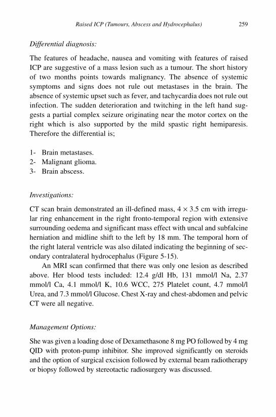
Differential diagnosis:
The features of headache, nausea and vomiting with features of raisedICP are suggestive of a mass lesion such as a tumour. The short historyof two months points towards malignancy. The absence of systemicsymptoms and signs does not rule out metastases in the brain. Theabsence of systemic upset such as fever, and tachycardia does not rule outinfection. The sudden deterioration and twitching in the left hand sug-gests a partial complex seizure originating near the motor cortex on theright which is also supported by the mild spastic right hemiparesis.Therefore the differential is;
1- Brain metastases.2- Malignant glioma.3- Brain abscess.
Investigations:
CT scan brain demonstrated an ill-defined mass, 4 × 3.5 cm with irregu-lar ring enhancement in the right fronto-temporal region with extensivesurrounding oedema and significant mass effect with uncal and subfalcineherniation and midline shift to the left by 18 mm. The temporal horn ofthe right lateral ventricle was also dilated indicating the beginning of sec-ondary contralateral hydrocephalus (Figure 5-15).
An MRI scan confirmed that there was only one lesion as describedabove. Her blood tests included: 12.4 g/dl Hb, 131 mmol/l Na, 2.37mmol/l Ca, 4.1 mmol/l K, 10.6 WCC, 275 Platelet count, 4.7 mmol/lUrea, and 7.3 mmol/l Glucose. Chest X-ray and chest-abdomen and pelvicCT were all negative.
Management Options:
She was given a loading dose of Dexamethasone 8 mg PO followed by 4 mgQID with proton-pump inhibitor. She improved significantly on steroidsand the option of surgical excision followed by external beam radiotherapyor biopsy followed by stereotactic radiosurgery was discussed.
Raised ICP (Tumours, Abscess and Hydrocephalus) 259
b1009 Problem Based Neurosurgery
b1009_Chapter-05.qxd 11/12/2010 10:50 AM Page 259

Outcome:
She underwent successful resection of her tumour aided by image guidedtechnology and she made an excellent post-operative recovery followedby external beam radiotherapy. The histological diagnosis was consistentwith metastatic lesion from lung cancer.
5-2-2 What is the differential of a single intrinsic cerebral lesion?
The differential diagnosis of a patient presenting with a possible cerebralmetastases is brain abscess as the scan appearances are almost the same.However, a patient with brain abscess may have in addition a source ofinfection (dental, mastoid, middle-ear, endocarditis, sinusitis, etc.) andsystemic symptoms such as pyrexia, whilst a patient with brain metastasismay have a systemic primary in the lung (Figure 5-15), breast (Figure 5-16),
260 Chapter 5
b1009 Problem Based Neurosurgery
Figure 5-15: CT of the brain demonstrating a rounded lesion (1) surrounded by exten-sive oedema (2), producing shift of the midline (3) and pineal body (5) to the left andcontralateral ventricular dilatation (4). This appearance is consistent with cerebralmetastasis (lesion is in the grey-white matter interface and the amount of oedema isgreater than the size of the lesion.)
b1009_Chapter-05.qxd 11/12/2010 10:50 AM Page 260

colon (Figure 5-17), renal, or skin (Figure 5-18). Table 5-3 summarisessources of brain metastases.
5-2-3 What is the epidemiology and presentation of brain metastases?
Brain metastases are more common than primary brain tumours and10–40% of patients with systemic cancer develop brain metastases. Forty
Raised ICP (Tumours, Abscess and Hydrocephalus) 261
b1009 Problem Based Neurosurgery
Figure 5-16: CT demonstrating slightly hyperdense lesion (M) in the left cerebellarhemisphere causing fourth ventricle deviation and oedema. Patient presented withsymptoms of raised ICP and ataxia. Pathology confirmed breast cancer as a source ofthe metastasis.
Figure 5-17: CT showing multiple intracerebral lesions (1–3) in a 65-year-old womanwho had bowel cancer resected 18 months ago.
b1009_Chapter-05.qxd 11/12/2010 10:50 AM Page 261

to fifty per cent of patients present with headaches, 10–20% with seizures,5–10% with stroke-like symptoms, and some present with cognitivedecline. On examination 35% will have altered level of consciousness,44% hemiparesis, 9% hemisensory disturbance, 9% papilloedema, and13% gait ataxia.26 Single metastasis occurs in 53%, most commonly fromcolon, breast, lung and kidney, whilst multiple in 47% most commonly
262 Chapter 5
b1009 Problem Based Neurosurgery
Figure 5-18: Metastatic malignant melanoma (1) on CT presented with haemorrhagein a 45-year-old patient.
Table 5-3: Sources of cerebral metastases 26
Metastasis source % of total Single % Multiple %
Non-small cell lung cancer 24 50 50Breast cancer 17 49 51Small cell lung cancer 15 43 57Malignant melanoma 11 49 51Renal cell cancer 6 56 44Bowel cancer 6 67 33Uterine cancer 5 53 47Unknown source 5 70 30Ovarian cancer 2 57 43Bladder cancer 2 64 36Prostate cancer 2 82 18Testicular cancer 2 55 45Total metastases 100 53 47
b1009_Chapter-05.qxd 11/12/2010 10:50 AM Page 262

from malignant melanoma and small cell cancer of lung. Brain metastaseshave poor prognosis: the overall one-year survival is about 10%, ifuntreated the median survival is one month, with steroids two months,with external beam radiotherapy three to six months, and with surgicalexcision followed by radiotherapy ten to 16 months. The prognosis ismuch better in patients who had good KPS (70 or better), controlled pri-mary, absence of other metastases and age under 65 years.27 The goals oftreatment include relief of symptoms, and improvement and extension ofgood quality of life.
5-2-4 How to manage brain metastases?
1) Symptomatic treatment
— Analgesia (Dihydrocodeine) for headache.— Steroids: dexamethazone to reduce oedema.— Metoclopramide for nausea and vomiting.
2) Treatment of underlying cancer including hunting for the primary byperforming chest X-ray (Figure 5-19), CT chest, abdomen and pelvis(Figures 5-20 and 5-21).
3) Surgical excision when there is good prospect of extending goodquality of life or relieving symptoms.
Raised ICP (Tumours, Abscess and Hydrocephalus) 263
b1009 Problem Based Neurosurgery
Figure 5-19: Chest X-ray in a man suspected to have brain metastasis confirming asource in the lung (1).
b1009_Chapter-05.qxd 11/12/2010 10:50 AM Page 263

4) Radiotherapy following surgery or stereotactic radiosurgery fortumours 3 cm or less in diameter.
5) Supportive care for poor prognosis patients and in the presence ofwidespread systemic disease.
264 Chapter 5
b1009 Problem Based Neurosurgery
Figure 5-20: CT chest demonstrating primary lung cancer (1).
Figure 5-21: CT abdomen demonstrating multiple liver metastases in a patient withmalignant melanoma and cerebral metastasis.
b1009_Chapter-05.qxd 11/12/2010 10:50 AM Page 264

Problem 5-3: Raised ICP, brain abscess and CNS infections. Howto manage a patient presenting with raised ICP due to CNSinfection?
Any patient suspected of harbouringCNS infection requires immediateadmission, investigations and antibi-otic treatment to prevent death andserious morbidity.
PCS5-3-1:
A 34-year-old male, right-handed, shop fitter, presented with short historyof headache and nausea. The headaches were acute and sharp all over forthe last 24 hours and are made worse on bending. The nausea was of sim-ilar onset and duration, was constant and associated with vomiting threetimes. Three days earlier he had been complaining of earache. He had sorethroat and fever associated with dysphagia five days earlier. By the timehe presented to hospital, he was on Paracetamol (PO) 1 g, four times daily,Ibuprofen (PO) 400 mg, three times daily, Dihydrocodeine (PO) 50 mg,every four to six hours, Metoclopramide (PO) 10 mg, every eight hours,as required for nausea. He had no allergies. He was married with two chil-dren, smoked 15 cigarettes per day and drank alcohol occasionally. Hewas fully conscious (GCS 15), his temperature was 38.1°C, pulse was86/min, regular, respiration was 15/min, BP 118/68 mmHg and was notanaemic, jaundiced or cyanosed. Cranial nerve examination was normal,and he had no abnormal focal neurological signs. Rest of examination wasnormal.
Differential diagnosis:
A patient presenting with acute headache associated with nausea and vom-iting with preceding history of infection (sore throat or ear infection in thiscase scenario) should be suspected of harbouring CNS infection tillproved otherwise, the differential diagnosis would be;
Raised ICP (Tumours, Abscess and Hydrocephalus) 265
b1009 Problem Based Neurosurgery
Problem based tool box:Brain abscess AntibioticsCSF fistulae CSF leaksEmpyema EncephalitisMeningitis Tuberculosis
b1009_Chapter-05.qxd 11/12/2010 10:50 AM Page 265

1- Cerebral abscess.2- Bacterial meningitis.3- Viral encephalitis.4- Other types of meningitis.
Investigations:
Full blood count Urea and electrolytes Others
Hb 13.4 Na 142 CRP 92*MCV 98.7 K 4.8WCC 16.4* Urea 4.0Platelet 421 Creatinine 79
* Raised.
Blood culture: No growth after five days for both aerobic and anaerobicmicrobes.
Left middle ear aspirate culture: No growth after two days of incubation.
CT scan: There was low attenuation lesion in left temporal lobe with ringenhancement. This is likely to be a temporal lobe abscess. There was evi-dence of bilateral otitis media with extension of infection into the lefttemporal lobe. There was extensive surrounding oedema.
Management:
• Symptomatic treatment: analgesia (Dihydrocodeine) for headache,Paracetamol for pyrexia and Metoclopramide for nausea and vomiting.
• Treatment of underlying cause (otitis media): antibiotics (Ceftriaxoneand Metronidazole) and drainage of middle ear effusion and insertionof grommet.
• Antibiotics for cerebral abscess: empirical therapy (Ceftriaxone andMetronidazole).
• Surgery: burr hole aspiration of the abscess.• Steroid for reducing oedema once antibiotics have been started IV.
266 Chapter 5
b1009 Problem Based Neurosurgery
b1009_Chapter-05.qxd 11/12/2010 10:50 AM Page 266

Patient’s outcome: After drainage of the abscess and mastoidectomy, heimproved gradually and fully recovered.
5-3-2 What is the epidemiology of CNS infections?
The central nervous system (CNS) is vulnerable to infection and differentorganisms cause meningitis, encephalitis and brain abscess. Knowledgeand prompt recognition of CNS infections are crucial because many ofthese conditions are likely to result in death or severe morbidity if notdiagnosed and treated promptly. The reasons are that the CNS is protectedfrom the body immune system (the body defence mechanisms againstinfections) by the blood brain barrier (BBB). Therefore when bugs gainaccess to the CNS there are no defence mechanisms to counteract theirattack. The advent of antibiotics and improved treatment of ear and sinusinfections has led to a reduction in intracranial abscess formation but theincidence is still about two to three per million per year. However, cere-bral abscess is still common in the developing world. The combined effectof improved diagnostic imaging techniques (hence the earlier discovery ofcerebral abscesses) and antibiotic treatments have improved the prognosisof patients with cerebral abscess. Mortality rates have decreased from40–50% in the 50s to less than 5% recently. Cerebral abscess may occurat any age, with no age or sex predilection.
5-3-3 What are the causes and sources of CNS infections?
The mode of spread of CNS infections can be divided into haematogenousspread and local direct spread.
1- Brain infection via haematogenous spread usually originates from aknown septic site or occult focus such as skin pustule particularly inthe danger zone of the face (the face area around the mouth and nosebecause its venous drainage goes directly into the cavernous sinus),chronic pulmonary infection (e.g. bronchiectasis), diverticulitis,osteomyelitis and bacterial endocarditis. Congenital septal defects ofthe heart with right to left shunt are a risk factor for developing mul-tiple brain abscesses because the blood does not filter through the
Raised ICP (Tumours, Abscess and Hydrocephalus) 267
b1009 Problem Based Neurosurgery
b1009_Chapter-05.qxd 11/12/2010 10:50 AM Page 267

capillary beds within the lungs before reaching the brain. Therefore,it is important to notice any other focus of infection during historytaking and examination of a patient suspected to have cerebralabscess.
2- Local spread occurs as a result of infection of adjacent structures suchas frontal sinusitis, otitis media, mastoiditis or infected dental caries.The spread of infection can be due to direct penetration of the dura orindirectly via the extension of an infected thrombus or embolus alonga vein (emissary veins).
3- Direct implantation of infection occurs following craniocerebraltrauma (e.g. compound depressed skull fracture and basal skull frac-ture) or following cranial surgery.
The best way to predict the causative organism(s) is by looking at theprimary source of infection. Middle ear infection is often caused by mixedorganisms such as Strep. milleri, Bacteroides fragilis, E. coli, Proteus andStrep. pneumonia. Infection from sinus or blood is usually by Strep. pneu-monia, Strep milleri and Staph aureus. In circumstances where trauma is theaetiology, Staph aureus is the most likely organism responsible for cerebralabscess. In basal skull fractures Strep. pneumonia is the most common.Following surgery Staph aureus is often seen. Opportunistic infections ofthe brain are also common in the immuno-compromised patients includingToxoplasma, Aspergillus, Candida, Nocardia and Listeria.
5-3-4 What types of CNS infections occur?
1- Cerebral abscess:Cerebral abscess is a localised bacterial infection of the brain. The mostcommon sites are: the temporal lobe or the cerebellar hemisphere whenthe source of infection is located in the ear and the frontal lobe for infec-tions arising from the frontal sinus or teeth (Figure 5-22), parietal lobe orscattered in the brain commonly in the left hemipshere (the left carotidoriginates from the aortic arch directly) in case of haematogenous spread(Figure 5-23). Diagnosis is made on neuroimaging and the underlyingorganism is cultured from blood or abscess aspirate. Lumbar puncture(L/P) is absolutely contraindicated.
268 Chapter 5
b1009 Problem Based Neurosurgery
b1009_Chapter-05.qxd 11/12/2010 10:50 AM Page 268

It is sometimes difficult to distinguish between single or multipleintracranial brain abscesses and single or multiple malignant brain tumourson neuroimaging alone. Table 5-4 compares the two types of pathologies.It is important to distinguish the two pathologies as their treatment andmanagement is different. Abscesses require intravenous antibiotics and
Raised ICP (Tumours, Abscess and Hydrocephalus) 269
b1009 Problem Based Neurosurgery
Figure 5-22: CT with contrast showing ring enhancing lesion (a) in the left frontal lobe(brain abscess).
Figure 5-23: CT scan demonstrating multiple intracerebral enhancing lesions withoedema (a case of multiple intracranial abscesses).
b1009_Chapter-05.qxd 11/12/2010 10:50 AM Page 269

drainage whilst malignant brain tumours require steroids, biopsy, resec-tion or radiotherapy.
2- Meningitis:Bacterial meningitis is a life-threatening acute infection of the meningesand subarachnoid space with a rapidly progressive course. Its pathologybegins with the multiplication of pathogenic organisms within the sub-arachnoid space after penetrating the BBB thus making it difficult for hostdefence mechanisms to reach the infection. An inflammatory process isinitiated and neutrophils migrate into the subarachnoid space producing apurulent exudate which gathers in the basal cisterns at the base of thebrain before spreading throughout the subarachnoid space. Although theunderlying brain is not directly infected by the bacteria it becomes con-gested, oedematous and ischaemic; at this stage only the pia matterprotects the brain from abscess formation. The inflammatory exudatesmay also affect vascular structures in the subarachnoid space resulting in
270 Chapter 5
b1009 Problem Based Neurosurgery
Table 5-4: Differences between brain abscess and malignant brain tumour
Diagnosis Brain abscess(es) Malignant brain tumour (s)
Location (epicentre) Grey-white matter Grey-white matter interface forinterface metastases and subcortical white
matter for high grade glioma (HGG)Enhancement Smooth ring-shaped Irregular ring-shaped with large solid
partSurrounding oedema Large Large in metastases, less in HGGSystemic symptoms Often present Often absent: no fever, or leucocytosisPrimary source Infection Primary cancer in metastasesC-reactive protein Very high Normal or slightly elevatedCompare image
b1009_Chapter-05.qxd 11/12/2010 10:50 AM Page 270

arteritis or venous thrombophlebitis causing infarction. The pus can insome cases obstruct the flow of CSF in the ventricles and subarachnoidspace causing hydrocephalus.
5-3-5 What are the causes and epidemiology of bacterial meningitis?
Although only few bacterial organisms are responsible for most cases ofbacterial meningitis there is a wide range of causal organisms which varywith age. Worldwide, the three major bacteria accounting for 80% ofmeningitis are Haemophylis influenzae (Hi), Nisseria meningitidis (Nm)and Streptococcus pneumonia (Sp). Nearly all cases of Hi meningitisoccur in children under the age of five years with most presenting betweenfour months and two years of age. It is also more common in boys thangirls. It is thought to be rare in those under two months due to the placen-tal transfer of maternal antibodies during pregnancy. Meningitis in anindividual older than six years caused by Hi is suggestive of an underly-ing pathology or immunodeficiency such as otitis media, sinusitis,epiglotitis, diabetes or alcoholism. Meningitis resulting from Nm is pri-marily a disease of children and young adults with fewer than 10% ofcases occurring in over 45 years and is also more prevalent in boys andmen. Adults over 24 years of age, who develop Nm, commonly haveunderlying conditions such as congestive cardiac failure or HIV. WhilstSp meningitis can affect all ages, Sp is the commonest cause of bacterialmeningitis in adults with the highest number of cases reported at theextremes of age. Certain factors have a strong predisposition to causingSp meningitis: pneumonia present in 15% to 25% of patients and acuteotitis media occurring in 30%. In addition, alcoholism and liver cirrhosisare associated features in approximately 20% to 30% of patients thatdevelop Sp meningitis. The circumstances in which meningitis developscan also be indicative of particular bacteria. Meningitis, that developsafter a penetrating injury or neurosurgical operation is usually caused bystaphylococcal, streptococcal or Gram-negative organisms. However,meningitis following a closed head trauma accompanied by a skull frac-ture or CSF leak is more commonly due to Sp. Post-traumatic meningitismay occur after basal skull fractures and CSF leaks (Figure 5-24).28 Inparticular fractures of the middle third of the face (Le Forte) predispose to
Raised ICP (Tumours, Abscess and Hydrocephalus) 271
b1009 Problem Based Neurosurgery
b1009_Chapter-05.qxd 11/12/2010 10:50 AM Page 271

CSF rhinorrhoea, or otorrhoea and subsequent meningitis. Depressedskull fractures (Figure 5-24) associated with dural breach predispose tointracranial infection and require antibiotic treatment and surgical toilet ofthe overlying wound within six hours.
Basal skull fractures with or without CSF leaks (Figure 5-25) aremanaged with vigilant neurological observation without antibiotic pro-phylaxis29 to avoid replacing the normal bacterial flora with more resistantmicrobes.
5-3-6 What are the symptoms and signs of meningitis?
The classic triad of symptoms in bacterial meningitis are high fever,headache and neck stiffness. The fever is generic to systemic infectionsbut the meningitic symptoms of a severe frontal/occipital headache, a stiff“board-like” neck on gentle flexion, photophobia, vomiting and associ-ated neurological signs are all suggestive of meningitis. Often patientsmay have recently suffered from an upper respiratory tract or ear infectionpreceding the onset of meningitis. During the initial stages of the illness
272 Chapter 5
b1009 Problem Based Neurosurgery
Figure 5-24: Compound depressed skull fracture with skin and dural breach associatedwith intracranial air (dark bubbles in the subarachnoid space).
b1009_Chapter-05.qxd 11/12/2010 10:50 AM Page 272

patients will usually be alert but as many as 90% will go on to experiencean impaired conscious level. Without prompt treatment further deteriora-tion of consciousness may result due to hydrocephalus, septic effects onthe underlying brain parenchyma or septic thrombosis of cerebral arteriesand veins which can cause secondary infarction. Focal or generalisedseizures occur in 30% of meningitis, cranial nerve signs in 15% and focalneurological signs such as hemiparesis, dysphasia and hemianopia in10%. Kernig’s sign in which the lumbar roots are stretched by hip flexionproducing pain, may also be present suggesting inflammation of themeninges.
5-3-7 How to investigate a patient with suspected meningitis?
Patients in which bacterial meningitis is suspected can undergo investiga-tions to confirm the diagnosis. CT brain is essential before CSFexamination to rule out mass lesions. CSF is then obtained via lumbarpuncture (L/P) to identify the causal organism. In an individual withmeningitis the CSF may appear cloudy because of the raised white cellcount usually in excess of 500 cells/mm3. In bacterial meningitis poly-morphonuclear leucocytes contributes 80–90% of the white blood cells
Raised ICP (Tumours, Abscess and Hydrocephalus) 273
b1009 Problem Based Neurosurgery
Figure 5-25: Right temporal basal skull fracture, note the bubble of air epidurally.
b1009_Chapter-05.qxd 11/12/2010 10:50 AM Page 273

found in the CSF unlike viral meningitis where neutrophils are far moreabundant. The CSF protein level is usually substantially high and areduced glucose concentration is present in 58% of bacterial meningitis.Identification of the pathogen may be possible through Gram stain in upto 90% of bacterial meningitis when common bacteria are involved. CSFshould also undergo specific tests to check for Mycobacterium tuberculo-sis (Ziehl-Neelsen stain for acid fast bacilli). Other investigations that areuseful in the diagnosis include blood culture which succeeds in isolatingan organism in 80% of Haemophilus influenzae but only in 50% of thetime in pneumococcal and meningococcal meningitis. Imaging such aschest X-ray and CT scan of sinuses can also be employed to detect theoriginal source of infection. The value of CT scanning in a suspected bac-terial meningitis patient is primarily to exclude other possible pathologyin the CNS but can be used to detect complications such as signs of raisedICP and causes of focal neurological deficits. Treatment of bacterialmeningitis includes immediate high dose intravenous antibiotics until allbacteria have been eradicated from the CSF. In addition, if a patient ini-tially presents in a critical condition or deteriorating intensive care supporttherapy may be required until the individual improves — this is particu-larly true in meningococcal meningitis.
Post-traumatic CSF leaks that did not resolve spontaneously withina few days, recurs, or complicated by meningitis require further inves-tigations to localise the underlying CSF fistula. This can be achievedin most cases by fine cuts CT scan (Figure 5-26) or MRI cisternogra-phy (Figure 5-27).30,31 Subsequent repair of the fistula is essential toprevent recurrent meningitis using an endoscope transnasally and duralpatch or intradurally using the microscope, The choice of surgicalrepair is dependent on the location, size and accessibility of the duraldefect.32–34
A number of tests were developed to distinguish between CSF leak,other body fluids and blood. For example blood mixed with CSF will pro-duce a dark red centre and lighter halo around it while blood alone willproduce a spot of red without a halo, all body fluids may contain glucoseand protein and estimation of glucose and protein in the fluid leak is notreliable to confirm CSF leak.35 B2-tranferrin is a byproduct of B1-transferrinand only produced in the CSF and aqueous humour of the eye and therefore
274 Chapter 5
b1009 Problem Based Neurosurgery
b1009_Chapter-05.qxd 11/12/2010 10:50 AM Page 274

can be used to reliably confirm the presence or absence of CSF in the fluidleak.35
5-3-8 How to treat bacterial meningitis?
Effective antibiotic treatment relies on achieving an adequate concentra-tion of the antibiotic within the CSF to have a bactericidal effect. Severalfactors contribute to this concentration: the ability of the antibiotic to pen-etrate the BBB, the bactericidal activity of the antibiotic once it reaches
Raised ICP (Tumours, Abscess and Hydrocephalus) 275
b1009 Problem Based Neurosurgery
Figure 5-26: Fine cut CT of a patient with paradoxical rhinorrhoea (CSF leaking fromthe middle cranial fossa into the middle ear, via the Eustachian tube into the nostril)demonstrating a bone defect in the right temporal bone and opacification of the rightmiddle ear and mastoid air cells (B).
Figure 5-27: MRI cisternography demonstrating CSF in the left mastoid air cells (A) ina patient with paradoxical rhinorrhoea.
b1009_Chapter-05.qxd 11/12/2010 10:50 AM Page 275

the infected CSF, and the antibiotic’s rate of metabolism and clearancefrom the CSF. The capacity for an antibiotic to penetrate the BBB isdependent on its molecule size and structure but most importantly its lipidsolubility. As the BBB behaves like a lipid bilayer the greater the lipid sol-ubility of the antibiotic the greater its ability to penetrate into the CSF. Thephysiological condition of the BBB can affect its permeability to antibi-otics, e.g. it is dramatically increased when the meninges are inflamed.Selection of an antibiotic will also depend on the most likely causativeorganism, the age of the patient and the original source of the infection. Ifthere is no obvious site of infection and taking into account the patient’sage initial antibiotic therapy prior to identification of the pathogen shouldinvolve: in neonates (under three months) — ampicillin, aminoglycosideand cephalosporin; in children (under five years) — ampicillin andcephalosporin; and in adults — penicillin G or cephalosporin. The mosteffective cephalosporin is kefatazidine IV. In the immunocompromised —ampicillin and cephalosporin. Following organism identification theantibiotic therapy may have to change accordingly to ensure the greatestsensitivity and penetration into the CSF. In general Haemophilus infectionreponds best to chloramphenicol with cefotaxime to which ampicillin orfurther cephalosporins can be added. Benzylpenicillin is effective inpneumococcal and meningococcal meningitis with Chloramphenicol andcephalosporins a possible alternative. Antibiotic treatment should be con-tinued for at least one week after the patient becomes afebrile inHaemophilus and Meningococcus infection and ten to 14 days followingPneumococcus meningitis. Recent research has also indicated that chil-dren treated with antibiotics and steroids such as dexamethasoneimproves illness outcome but that benefit in adults was not as significant.
5-3-9 What is the prognosis of meningitis?
Prompt diagnosis and treatment with an appropriate antibiotic(s) is nor-mally enough to ensure recovery and protect against the potentially fatalcomplications of bacterial meningitis. As mentioned previously majorcomplications include cerebral oedema, seizures, hydrocephalus, subduraleffusion (particularly in children) and more rarely subdural empyema orbrain abscess. Mortality rates from bacterial meningitis vary depending on
276 Chapter 5
b1009 Problem Based Neurosurgery
b1009_Chapter-05.qxd 11/12/2010 10:50 AM Page 276

the causal organism. Haemophilus meningitis occurring in younger chil-dren carries a good outcome with death in less than 5%. However,pneumococcal meningitis commonly presenting in adults has a worseprognosis with a mortality rate of 20%. Meningococcal meningitis has avariable outcome; a sudden onset with systemic involvement has a poorprognosis where as a gradual onset has an improved prognosis. Overallmortality occurs in 10%. Bacterial meningitis is just one of numerousways CNS infection may present. Unlike viral meningitis which is usuallyself-limiting and fungal infection which tends to present in immunocom-promised individuals, bacterial meningitis is a potentially life-threateningillness affecting all age groups with serious complications.
5-3-10 How to manage subdural empyema?
3- Subdural empyema:Subdural empyema depicts pus in the subdural space. The aetiology andpathogenesis is similar to bacterial meningitis. It can follow surgery(Figure 5-28) or spontaneously secondary to haematogenous spread(Figure 5-29).
Raised ICP (Tumours, Abscess and Hydrocephalus) 277
b1009 Problem Based Neurosurgery
Figure 5-28: Post-craniotomy subdural empyema.
b1009_Chapter-05.qxd 11/12/2010 10:50 AM Page 277

It may be difficult to distinguish on neuroimaging between subduralempyema, hypodense subdural haematoma or subdural hygroma as bothcan follow surgery or trauma. Table 5-5 lists some of the distinguishingfeatures but if you are in doubt treat as infection in the same way as youwould treat a ring enhancing lesion as an abscess.
The treatment of subdural empyema is immediate antibiotics intra-venously and either burr hole drainage and washout or craniotomydepending on the consistency of the pus and how easily it comes out.
278 Chapter 5
b1009 Problem Based Neurosurgery
Figure 5-29: Interhemispheric subdural empyema (a).
Table 5-5: Distinguishing features of chronic subdural haematoma and empyema
Diagnosis Subdural empyema Chronic subdural haematoma
Location Lateral/interhemispheric Lateral or bilateralEnhancement Almost always of the dura Uncommon/rareCerebral oedema Common RareSystemic symptoms Fever, leucocytosis NoneC-reactive protein Very high NormalSource of infection Yes No
b1009_Chapter-05.qxd 11/12/2010 10:50 AM Page 278

5-3-11 How to manage encephalitis?
4- Encephalitis:Encephalitis is an inflammatory condition of the brain, usually caused bya viral infection, although it can also be bacterial in origin. Cases canrange from mild to severe and can lead to serious neurological conse-quences if missed or the diagnosis was delayed, e.g. in herpes simplexencephalitis (HSE). Mild cases present as flu-like symptoms, whilst seri-ous cases can cause severe headache, sudden fever, drowsiness, loss ofconsciousness, vomiting, confusion, seizures and coma. Mild cases,respond to rest, fluids and a pain reliever, whilst severe cases, might needto be hospitalised and even ventilated. The incidence of HSE is rare (0.2per 100,000) but is important to detect and treat. West Nile encephalitis(WNE) is rare and also requires early detection and treatment. HSE istreated with acyclovir. CT and MRI are essential diagnostic tests to beperformed prior to CSF examination in these patients primarily to ruleout space occupying lesions. MRI in particular often demonstrates highsignal on T2-weighted images in the medial temporal structures, sub-frontal cortex and the insula. CSF is examined for increased lymphocytesand protein and polymerase chain reaction (PCR). A PCR for DNAHerpes Simplex Virus (HSV) is 100% specific and 75–98% sensitivewithin the first 25–45 hours. Types 1 and 2 HSV cross-react, but nocross-reactivity with other herpes viruses occurs. Viral serology may alsohelp as complement fixation antibodies are useful in identifyingarbovirus. Cross-reactivity exists among one subgroup of arboviruses,the flaviviruses (e.g. St. Louis encephalitis, WNE), and with antibodiesraised in persons inoculated with the yellow fever vaccine. EEG may alsohelp in identifying HSE as the EEG may show characteristic paroxysmallateral epileptiform discharges (PLEDs) (Figure 5-30) are oftenobserved, even before neuroradiographic changes and are found in 80%of cases.
5- Tunberculoma:Tuberculosis (TB) is uncommon in the west and much reduced in devel-oping countries because of active vaccination programmes. However, due
Raised ICP (Tumours, Abscess and Hydrocephalus) 279
b1009 Problem Based Neurosurgery
b1009_Chapter-05.qxd 11/12/2010 10:50 AM Page 279

to worldwide travel and globalisation TB can pop in any country aroundthe world and vigilance is important to clinch the diagnosis and provideappropriate treatment. Tuberculomas are at the top of the list of differen-tial diagnosis of any space occupying lesion in the Indian subcontinent(Figure 5-31).
280 Chapter 5
b1009 Problem Based Neurosurgery
Figure 5-30: EEG demonstrating PLED from the temporal lobe in a patient with HSE.
Figure 5-31: Tuberculoma in a young man from the Indian subcontinent.
b1009_Chapter-05.qxd 11/12/2010 10:50 AM Page 280

Problem 5-4: Raised ICP and hydrocephalus. How to manage apatient presenting with raised ICP due to hydrocephalus?
In patients presenting with symptoms,signs and imaging features suggestiveof hydrocephalus, look for a cause ashydrocephalus alone is not sufficientdiagnosis.
PCS5-4-1:
A 74-year-old right-handed male presented with deteriorating memoryand mobility for nine months. He used to walk one mile a day but unableto mobilise independently at all and rapidly declined in the last threeweeks. His decline in memory was stepwise rather than gradual with poorrecall and difficulty concentrating. His wife reported increased fatigueand somnolence over the same time period. Recently he was in low spir-its. He had an MI and stable angina. He had CABG 1986, hypertension,colonic carcinoma resection three years ago, and diverticular disease. Hewas alert with good eye contact, normal affect, and MMSE 17/30.
Differential diagnosis:
Vascular dementia, normal pressure hydrocephalus (NPH), Parkinson’sdisease (PD) with dementia, Alzheimer’s disease, and Dementia withLewy bodies are the main differential in this patient. His blood biochem-istry and blood counts showed: Na 131(↓), K 4.9 (normal), Urea 4.4(normal), Creatinine 73 (normal), Hb 14.0 (normal), WBC 11.8 (slightlyelevated), and PLT 272 (normal). MRI scan demonstrated dilated ventri-cles with some cerebral atrophy (Figure 5-32).
Because of the MRI appearances and the history NPH was thoughtto be possible than probable and DaTSCAN was performed to rule outPD. The scan revealed symmetrical and normal pattern of tracer uptake inthe basal ganglia bilaterally. No evidence of a pre-synaptic dopamine
Raised ICP (Tumours, Abscess and Hydrocephalus) 281
b1009 Problem Based Neurosurgery
Problem based toolkit:Aqueduct stenosisHydrocephalusNormal pressure hydrocephalusObstructive hydrocephalusShunts and CSF diversions
b1009_Chapter-05.qxd 11/12/2010 10:50 AM Page 281

receptor deficiency ruling out PD. His cognitive assessment at this pointdemonstrated Adenbroke’s Cognitive Examination (ACE) 47/100, MMSE15/30, Attention and Orientation 8/18, Memory 9/26, Fluency 1/14,Language 24/26 and Visuospatial 5/16. He had lumbar puncture (L/P):opening CSF pressure was 150 mmHO2 (normal 70–180), WCC none,RBC 8 and no organisms seen, glucose 3.9 mmol/L, protein 422 mg/L(normal 150–450). All CSF parameters were normal. He had CSFdrainage of 20 ml on three consecutive days. His cognitive assessmentsafter this showed: ACE 56/100 (better), MMSE 17/30 ((better), Attentionand Orientation 9/18 ((better), Memory 11/26 (better), Fluency 4/14 (bet-ter), Language 26/26 (better), and Visuospatial 6/16 (better).
He had a shunt inserted and improved following the shunt gradually.
PCS5-4-2:
A 46-year-old right-handed woman presented with a severe circumferen-tial headache that was worse first thing in the morning, and associatedwith double vision. The symptoms had been ongoing for six days andwere present throughout the day. The headache was a dull ache rated 7/10
282 Chapter 5
b1009 Problem Based Neurosurgery
Figure 5-32: NPH presented with dementia and ataxia in a 74-year-old.
b1009_Chapter-05.qxd 11/12/2010 10:50 AM Page 282

on VAS. There was little relief with pain medication, and there was noassociated aura. The double vision was worse when looking to the left.The patient had been nauseous but had not vomited. There was no associ-ated loss of consciousness or seizures. The patient had complained ofbeing off balance, although this was present for many years and sherequired the use of a walking stick. She had posterior fossa surgery toremove a dermoid cyst 13 years ago and had a ventriculoperitoneal shuntplaced 11 years ago. She was fully conscious with GCS of 15, MMSE30/30 and had difficulty with upward gaze and partial left sixth nervepalsy.
Differential diagnosis:
A history of headache of insidious onset associated with diplopia of sixthnerve paresis indicates raised ICP. The history of previous PCF surgeryand shunt and loss of upward gaze would indicate hydrocephalus due toblocked shunt. The shunt valve could be checked to find out if the reser-voir was filling and emptying.
Investigations:
Urea and electrolytes, full blood count, blood glucose, and C-reactive pro-tein were normal. CT head demonstrated obstructive hydrocephalusindicating shunt blockage.
Management options:
This patient needed shunt revision as a matter of urgency. One can buytime by measures to decrease ICP, e.g. aspiration of the shunt reservoirand draining 10–20 ml. However, this will only work if the ventricularcatheter was patented.
5-4-3 How to classify hydrocephalus (differential diagnosis)?
Hydrocephalus can be divided into congenital and acquired. Congenital ispresent from birth and can be a manifestation of something else, e.g. aqueduct
Raised ICP (Tumours, Abscess and Hydrocephalus) 283
b1009 Problem Based Neurosurgery
b1009_Chapter-05.qxd 11/12/2010 10:50 AM Page 283

stenosis. Acquired hydrocephalus is caused by another pathology, e.g.tumour, bleed, or infection. To learn the different types of hydrocephalusyou need to understand CSF pathways first.
CSF circulates throughout the central nervous system. It protects thebrain and spinal cord as a shock absorber, and metabolically supportsneurones and glial cells by delivering nutrients derived from the blood,as well as maintaining homeostasis by detoxifying the environment byremoving the cellular waste products. Sixty to seventy per cent of CSFis produced in the ventricles, by choroid plexuses in the lateral andfourth ventricles. It is secreted by a combination of diffusion, pinocyto-sis and active transfer. The choroid plexus consists of tufts of capillarieswith thin fenestrated endothelial cells, covered by modified ependymalcells with bulbous microvilli. The remainder of the CSF is produced byependymal cells found around blood vessels and along ventricular walls.CSF then circulates from the lateral ventricle (LV) via the interventric-ular foramina [foramena of Monro (FM)] into the III ventricle (3V) theninto the fourth ventricle (4V) via cerebral aqueduct [aqueduct of Sylvius(AS)]. CSF then exits 4V into the subarachnoid space (SAS) throughtwo lateral apertures [foramina of Luschka (FL)], and one median aper-ture [foramen of Magendie (FM)]. Most of the CSF passes through FMand enters the cisterna magna (CM), which is located between themedulla and cerebellum. Remaining CSF exits through the FL to enterthe subarachnoid space in the region of the cerebellopontine angle(CPA). From CM and CPA, the majority of CSF flows superiorly upthrough the perimesencephalic cistern (PNC) and around the cerebralhemispheres. A small proportion of CSF exiting the fourth ventricleflows inferiorly down into the thecal sac surrounding the spinal cord(Figure 5-33).
CSF is reabsorbed back into the venous sinuses, principally at thesuperior sagittal sinus (SSS) via numerous arachnoid villi (AV), whichconsist of invaginations of arachnoid matter through the dural wall andinto the lumen of the sinus. AV act as one-way valves between the sub-arachnoid space and the dural sinuses. Reabsorption occurs at AVbecause of greater hydrostatic pressure in the subarachnoid space. Abouthalf a litre of CSF is produced every 24 hours and any blockage or dis-turbance of CSF circulation can lead to rapid increase of ICP. From
284 Chapter 5
b1009 Problem Based Neurosurgery
b1009_Chapter-05.qxd 11/12/2010 10:50 AM Page 284

clinical management point of view hydrocephalus could be divided intothe following.
1- Communicating hydrocephalus (CHC):
CHC means that there is no obstruction of CSF circulation between theventricular system and the SAS. This can be further divided into twocategories:
a. Normal pressure hydrocephalus (NPH):
As the name implies the ICP is within normal limits. It is often seen in theelderly and presents with a triad of symptoms: memory deficit, ataxia andurinary incontinence. Diagnosis is made on clinical grounds and imagingfeatures (Figure 5-32).
The management of NPH is complex and sometimes difficult to dis-tinguish from vascular dementia associated with cerebral atrophy. Up to10% of those diagnosed with dementia are believed to actually have NPH.In the US alone it is estimated that 750,000 suffer from NPH, and fewerthan 20% are appropriately diagnosed and treated. NPH is often mistaken
Raised ICP (Tumours, Abscess and Hydrocephalus) 285
b1009 Problem Based Neurosurgery
Figure 5-33: CSF circulation. a == Frontal horn of the lateral ventricle, b == FM, c == 3V,e == ADS, f == trigone of the lateral ventricle (LV), g == occipital horn of LV, d == temporalhorn of LV, h == 4V, i == FM, and j == FL.
b1009_Chapter-05.qxd 11/12/2010 10:50 AM Page 285

for Alzheimer’s disease, Parkinson’s disease, or merely accepted as partof old age. There is a greater difficulty in determining whether a patientwith NPH is an appropriate candidate for CSF diversion or drainagebecause no single diagnostic study had emerged as highly reliable.36
5-4-4 How to diagnose normal pressure hydrocephalus?
Guidelines in diagnosis and treatment of NPH have been developed andare based on the presence or absence of symptoms and signs of NPH asfollows:
I. Probable NPH:
Probable NPH patients should fulfil the following criteria: The NPHsymptoms are insidious in onset, take origin after the age of 40 years, havea duration of three months or greater, are not attributable to other docu-mented neurological, psychiatric or general medical conditions, and areprogressive in nature. Gait disturbance is a mandatory symptom forpatients to be categorised as probable NPH. In addition, an impairment ofeither urinary function or cognition must be present. All three classicsymptoms need not be present. Gait disturbance may manifest itself in avariety of ways. At least two of the following signs of gait must beobserved for probable NPH: decreased step height, decreased step length,decreased cadence/speed of stride, increased trunk sway during ambula-tion, widened standing base, toes turned outward on ambulation,spontaneous or provoked retropulsion, and en bloc turning (requiringthree or more steps for a 180-degree turn). Urinary impairment in proba-ble NPH should include one of the following symptoms not attributable toprimary urologic disorders or other causes: episodic or persistent urinaryincontinence, urinary urgency as defined by frequent perception of apressing need to void, urinary frequency as defined by greater than sixvoiding episodes in an average 12-hour period despite normal fluid intake,and nocturia as defined by the need to urinate more than two times in anaverage night. Cognition impairment in probable NPH patients mustinvolve at least two of the following that cannot be fully attributableto other conditions: psychomotor slowing (increased response latency),
286 Chapter 5
b1009 Problem Based Neurosurgery
b1009_Chapter-05.qxd 11/12/2010 10:50 AM Page 286

difficulty dividing or maintaining attention, memory lapses (especiallyshort-term), executive dysfunction (e.g. impairment in multi-step proce-dures, working memory, formulation of abstractions/similarities, insight),and behavioural or personality changes.
II. Possible NPH:
If patients do not meet all of the above criteria but are still suspected tohave NPH, certain exceptions are permissible for a designation of possi-ble NPH. The clinical presentation and history may include any of thefollowing six exceptions: subacute or indeterminate mode of onset, onsetat any age following childhood, have less than a three-month or indeter-minate duration, if symptoms remotely follow events such as mild headtrauma, intracerebral haemorrhage, childhood and adolescent meningitis,or other conditions that the clinician judges unlikely to be immediatelycausally related, if symptoms co-exist with other neurological, psychiatricor general medical disorders that the clinician judges not to be entirelyattributable to these conditions, and if symptoms are non-progressive ornot clearly progressive. For a “possible NPH” designation, observablegait or balance disturbance is not a mandatory symptom as long asincontinence or cognitive impairment are determined. Similarly, gait dis-turbance or dementia alone can stand as an acceptable sign of NPHpossibility. In brain imaging, evidence of cerebral atrophy of sufficientseverity to potentially explain ventricular size is acceptable, as well as thediscovery of any structural lesions that might influence ventricular size.
III. Unlikely NPH:
Patients should be classified as unlikely to have NPH if they do not fulfilthe criteria for either probable or possible NPH or if they exhibit any ofthe following eight items: acute presentation of symptoms, recent historyof subarachnoid haemorrhage, meningitis, or brain injury, signs ofincreased intracranial pressure such as papilloedema, no component of thesymptom triad (gait, dementia and incontinence), other causes clearlyexplain findings, no evidence of ventriculomegaly through brain imaging,obstructive hydrocephalus revealed through brain imaging, or congenitalhydrocephalus revealed through brain imaging.
Raised ICP (Tumours, Abscess and Hydrocephalus) 287
b1009 Problem Based Neurosurgery
b1009_Chapter-05.qxd 11/12/2010 10:50 AM Page 287

5-4-5 How to treat normal pressure hydrocephalus?
The best treatment for NPH is still controversial because of the difficultiesin diagnosis. However, CSF drainage trial via lumbar puncture (L/P) eitherthrough three-day continuous drainage or repeated L/P with assessment ofcognition, and gait before and after CSF drainage is reasonable and simple.Those whose symptoms improve have a better chance of response afterpermanent CSF diversion. CSF diversion using a shunt from either theSAS in the lumbar region (LPS) or ventricle (VPS) to the peritoneum oftenprovides adequate solution. The shunt pressure should be either in the lowrange or programmable. Immediate complications of shunting includeinfection, and bleeding. Long term complications include infection orblockage. Only 50–70% of patients respond to shunting and the youngerthe patient the better the response. The average ICP of NPH is about 150mmHO2 compared to the normal mean of 122 mmHO2. During prolongedICP monitoring plateau waves of raised ICP can be observed.
b. High pressure CHC:
This occurs after diffuse SAH, basal meningitis or excessive CSF pro-duction: diffuse SAH blocks the arachnoid villi preventing CSFabsorption. In the early stages this may respond to repeated L/Ps. In thelater stages may require CSF shunt (Figure 5-34). In the acute periodwhile CSF protein and RBC counts are high, repeat LPs will keep CSFpressure down, however, if there was any doubt about the presence of dif-ferential pressure between the different intracranial and spinal CSFcompartments an external ventricular drain would be recommended toavoid brain shift and herniation. CSF protein had to drop to under 1 g/lbefore the shunt is inserted.
Basal meningitis blocks the arachnoid villi and prevents CSF absorp-tion because of inflammation. The underling infection needs to be treatedand eradicated completely before insertion of a shunt in these patients. Inthe initial period while the infection is being eradicated repeat L/Ps willkeep CSF pressure down, if there was any doubt about the presence of dif-ferential pressure between the different intracranial and spinal CSFcompartments an external ventricular drain (EVD) would be recom-mended to avoid brain shift and herniation.
288 Chapter 5
b1009 Problem Based Neurosurgery
b1009_Chapter-05.qxd 11/12/2010 10:50 AM Page 288

Increased CSF production is very rare but can occur in secretorychoroid plexus papilloma (CPP). The diagnosis would be evident on brainimaging and the treatment is that of the underlying lesion. If the treatmentof the underlying CPP did not resolve the hydrocephalus and the CSF pro-tein is low enough, CSF shunt would be the treatment.
2- Non-communicating hydrocephalus (NCHC):
This can be divided into:
5-4-6 What is congenital hydrocephalus and Chiari malformations?
a) Congenital OHC (COHC):
It affects about one in every 1000 live births and more common when pre-natal screening for neural tube defect is not performed. The most commontype of NCHC is aqueduct atresia (ADA) and aqueduct stenosis (ADS)followed closely by Arnold Chiari malformation (ACM). ADA occursin utero or post-natally, and may be caused by clots from intraventricu-lar bleeding, infection, or any pathology that causes gliosis that leads to
Raised ICP (Tumours, Abscess and Hydrocephalus) 289
b1009 Problem Based Neurosurgery
Figure 5-34: CT scan demonstrating CHC after SAH. Note that both the LVs (b) and 4V(a) are dilated. c == Marks the temporal horns that often dilate in acute hydrocephalus.
b1009_Chapter-05.qxd 11/12/2010 10:50 AM Page 289
obliteration of the aqueduct. ADS is narrowing of the aqueduct withoutgliosis. It tends to be a component of a set of complex malformationswhich may be inherited in autosomal recessive or X-linked patterns. CMFis often associated with Spina bifida and hydrocephalus. The malforma-tion involves abnormalities of the posterior cranial fossa and its contents.These abnormalities consist of a shallow posterior fossa and a low inser-tion of the tentorium. As a direct result of these deformities, thecerebellum and brainstem are crowded together, forcing the cerebellarvermis, tonsils and the medulla down through an enlarged foramen mag-num into the cervical spinal canal. The medulla becomes elongated andfolded dorsally while the cerebral aqueduct and the fourth ventricle areforced to collapse. The FL and FM are forced to lie in the spinal canal, andare surrounded by subarachnoid space, which is collapsed and fibrotic.The blockage of CSF pathway from these lesions causes hydrocephalus.
• ADA and ADS:It leads to obstruction of the aqueduct of Sylvius. This manifests ininfancy and is recognised by enlarging head circumference, bulging tenseanterior fontanel or sunset sign where the infant’s sclera is visible supe-rior to the cornea due to defects in upward gaze because of pressure on thetectal plate of the midbrain (Figure 5-35). The treatment is by creating anopening in the floor of the 3V (third ventriculostomy), this would avoidthe long term risks of permanent shunt placement. However, if this pro-cedure failed a shunt would be necessary. Patients who had a shuntinserted at childhood need to be kept under surveillance for elective exten-sion of the peritoneal catheter during growth spurt if the child was shuntdependent.
The best way to recognise ADS is finding that LV and 3V are dilatedwhile 4V is normal (Figure 5-35). However, MRI scan using sagittal planewould be the best way to visualise the aqueduct directly (Figure 5-36).
• 4V exit obstruction:This is caused by obstruction of the 4V foramena (FM and FL). Thiswould lead to balloon dilatation of the roof of the 4V and can be mistakenfor large cisterna magna. This condition is known as Dandy WalkerSyndrome (DWS). The treatment is CSF diversion.
290 Chapter 5
b1009 Problem Based Neurosurgery
b1009_Chapter-05.qxd 11/12/2010 10:50 AM Page 290

b) Acquired OHC (AOHC):
This is a common finding in clinical practice as obstruction from tumours,blood clot, abscess or giant aneurysms can block the CSF pathways inseveral locations, particularly the FM, 3V, aqueduct and 4V.
Raised ICP (Tumours, Abscess and Hydrocephalus) 291
b1009 Problem Based Neurosurgery
Figure 5-35: Congenital hydrocephalus shunted at birth and required several shuntrevisions over the years note: LV and 3V dilated, 4V is normal and previous shuntcatheters in the LV.
Figure 5-36: Sagittal MRI showing aqueduct atresia (a).
b1009_Chapter-05.qxd 11/12/2010 10:50 AM Page 291

5-4-7 What is colloid cyst and how to manage it?
• AOHC at the FM may be caused by colloid cyst:Colloid cysts are uncommon and may present with features of chronichydrocephalus, or drop attacks. The mechanisms of chronic hydrocephalicsymptoms: cognitive decline, dementia and ataxia, are as those describedunder NPH. Drop attacks occur when the cyst obstructs the FM leading toacute rise in ICP. Acute rise in ICP leads to sudden loss of consciousness andcollapse. Once the patient drops on the ground the cyst dislodges from theFM causing the CSF to drain normally and the patient regains consciousnessas quickly as he had lost it. The differential diagnosis in patients with dropattacks would include colloid cyst, Stoke Adam’s Syndrome due to completeheart block or rapidly developing generalised seizures that spread so fast inthe two hemispheres of the brain across the corpus callosum. An example ofcolloid cyst is shown in Figure 5-37. The treatment of symptomatic colloidcysts is surgical removal transcortically, transcallosally or endoscopically.Some surgeons aspirate colloid cysts stereotactically but the thick capsuleand the cyst mobility makes it less than ideal target, particularly if the con-tents of the cyst spills into the ventricles causing chemical meningitis andhydrocephalus. Shunting of the LV has also been used as primary treatmentparticularly in the elderly and as a rescue procedure after resection.
292 Chapter 5
b1009 Problem Based Neurosurgery
Figure 5-37: A 35-year-old male presented with drop attacks and headaches. MRI scandemonstrating a cyst (a) in the FM.
b1009_Chapter-05.qxd 11/12/2010 10:50 AM Page 292

The commonest location of colloid cysts is in the anterior third of the3V and they appear as high signal on T1 and T2 and hyperdense on CT(Figure 5-38). Isodense cysts are not uncommon and may be missed onCT scan, therefore great care should be taken not to perform L/P in thesepatients without thorough examination of the MRI scan. Colloid cysts canbe removed without problems, but be mindful as potential complicationsof surgery in particular injury to the fornix leading to complete loss ofrecent memory is a real risk.
• AOHC at the anterior third of 3V:This could be due to colloid cyst as discussed above, invasive pituitaryadenoma (Figure 5-39), craniopharyngioma (Figure 5-40), or a giant ante-rior communicating artery aneurysm. The treatment of these patients isdirected at treating the cause with peri-operative external ventriculardrainage (EVD) and Ventriculoperitoneal shunt (VPS) as a rescue proce-dure to treat persistent hydrocephalus.
• AOHC due to obstruction of post-3V:This could be due to pineal body tumours or cysts (Figure 5-41) or basi-lar tip aneurysm (BTA) (Figure 5-42). Blood clot in 3V (Figure 5-43) may
Raised ICP (Tumours, Abscess and Hydrocephalus) 293
b1009 Problem Based Neurosurgery
Figure 5-38: CT showing hyperdense colloid cyst in a 35-year-old woman presentedwith headaches and drop attacks. She had this removed with excellent results.
b1009_Chapter-05.qxd 11/12/2010 10:50 AM Page 293

294 Chapter 5
b1009 Problem Based Neurosurgery
Figure 5-39: OHC due to invasion of 3V with pituitary adenoma (c). This patient wasa 46-year-old male who presented with bitemporal visual defects in addition to hydro-cephalus. He underwent transsphenoidal resection of non-functioning pituitaryadenoma.
Figure 5-40: AOHC due to craniopharyngioma. This 30-year-old patient presentedwith fatigue and tiredness with bitemporal visual field defects. He underwent transsphe-noidal drainage of the cyst.
b1009_Chapter-05.qxd 11/12/2010 10:50 AM Page 294

also lead to AOHC in some patients if the clot did not resolve in time orthe ADS is blocked.
• AOHC due to blockage of ADS:The aqueduct of Sylvius is vulnerable and can be blocked by blood clot asshown in the previous example but can also be blocked by tumour or any
Raised ICP (Tumours, Abscess and Hydrocephalus) 295
b1009 Problem Based Neurosurgery
Figure 5-41: AOHC due to pineal cyst (a) in a 27-year-old man presented with chronicheadaches. This was drained endoscopically without the need for a shunt.
Figure 5-42: BTAA (hyperdense lesion in the middle of the CT scan image) blockingthe post-3V and may lead to hydrocephalus.
b1009_Chapter-05.qxd 11/12/2010 10:50 AM Page 295

mass lesion in the midbrain, e.g. haemorrhage, midbrain glioma, abscessor cyst (Figure 5-44).
• AOHC due to blockage or compression of 4V:This is a common clinical entity associated with:
1. Intrinsic cerebellar tumours [glioma, medulloblastoma (Figure 5-45),ependymomas, haemangioblastoma, juvenile astrocytomas or metas-tases].
2. Extrinsic tumours such as giant CPA schwannoma, meningioma(Figure 5-46), clival meningioma, or epidemoid cyst.
3. Cerebellar haematoma or infarct (Figure 5-47).4. Clot in the 4V (Figure 5-48).
All the aforementioned lesions can present with hydrocephalus in additionto their cardinal symptoms and signs.
296 Chapter 5
b1009 Problem Based Neurosurgery
Figure 5-43: AOHC due to blood clot in 3V (hyperdense lesion c). Note that the clotextended from the lentiform nucleus on the left and in the left LV. This 60-year-oldpatient presented with sudden right hemiplegia including right UMN of VII nerve andpatient was hypertensive. The hydrocephalus was managed by EVD till the CSF wasclear and the clot resolved. Patient did not require a shunt.
b1009_Chapter-05.qxd 11/12/2010 10:50 AM Page 296
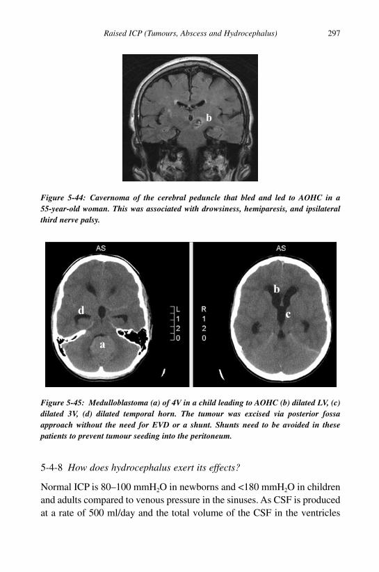
5-4-8 How does hydrocephalus exert its effects?
Normal ICP is 80–100 mmH2O in newborns and <180 mmH2O in childrenand adults compared to venous pressure in the sinuses. As CSF is producedat a rate of 500 ml/day and the total volume of the CSF in the ventricles
Raised ICP (Tumours, Abscess and Hydrocephalus) 297
b1009 Problem Based Neurosurgery
Figure 5-44: Cavernoma of the cerebral peduncle that bled and led to AOHC in a55-year-old woman. This was associated with drowsiness, hemiparesis, and ipsilateralthird nerve palsy.
Figure 5-45: Medulloblastoma (a) of 4V in a child leading to AOHC (b) dilated LV, (c)dilated 3V, (d) dilated temporal horn. The tumour was excised via posterior fossaapproach without the need for EVD or a shunt. Shunts need to be avoided in thesepatients to prevent tumour seeding into the peritoneum.
b1009_Chapter-05.qxd 11/12/2010 10:50 AM Page 297

and subarachnoid space is around 135–150 ml, this creates a turnover ofabout 3.7 times a day and a pressure gradient leading to CSF reabsorption.This reabsorption is facilitated by the aforementioned hydrostatic pressuregradient and the difference in osmotic pressure between CSF and venous
298 Chapter 5
b1009 Problem Based Neurosurgery
Figure 5-46: CPA meningioma (a) causing obstruction and shift of the 4V and hydro-cephalus. This 60-year-old man presented with dysphonia and swallowing difficulties.He had this tumour removed without EVD or shunt. He made an excellent recovery.
Figure 5-47: Cerebellar infarct (a) in a 70-year-old woman leading to hydrocephalus (b)required an EVD. Some of these patients benefit from sub-occipital decompression.
b1009_Chapter-05.qxd 11/12/2010 10:50 AM Page 298

blood. The CSF osmotic pressure is lower than plasma because of thelower protein and glucose content of the CSF. Hydrocephalus exerts itseffects by increasing ICP. According to the Monro-Kellie hypothesis thecranial cavity is a ridged incompressible box, with a fixed volume. Its con-tents; brain tissue (80%/1400 ml), CSF (10%/150 ml), and blood (10%/150ml) create a state of volume equilibrium, such that any increase in the vol-ume of one of the cranial contents must be compensated by a decrease inthe volume of another otherwise the ICP will rise.37 To maintain constantICP, there are a number of mechanisms which are able to compensate forsmall increases in ICP. These mechanisms include movement of CSF intothe thecal sac, increased reuptake of CSF, and compression of venoussinuses. Once compensatory mechanisms are overcome, any increase inCSF volume will increase the ICP dramatically.38 Increased ICP may causebrain tissue ischaemia and uncal hernation where the temporal lobe herni-ates beneath the tentorium and cerebellar tonsils herniation where thecerebellar tonsils are forced through the foramen magnum. Both are clini-cal emergencies as they can compress the brainstem and lead to death.Hence obstructive hydrocephalus is a neurosurgical emergency even whenthe patient is apparently fully conscious. Symptoms of acute hydrocephalusvary with age, the rate of disease progression, and underlying cause of thehydrocephalus. In adults and older children the most common presentation
Raised ICP (Tumours, Abscess and Hydrocephalus) 299
b1009 Problem Based Neurosurgery
Figure 5-48: Clot in 4V (b) leading to hydrocephalus and requiring an EVD.
b1009_Chapter-05.qxd 11/12/2010 10:50 AM Page 299

is raised ICP; headaches, nausea, vomiting and papilloedema. Impairedupward gaze and impaired conscious level and visual disturbances are alsocommon symptoms due to compression of the brainstem. A late sign ofraised ICP is bradycardia and hypertension followed by pupillary dilatationand coma. In infants the signs are enlarged head circumference, dysjunc-tion of sutures, bulging and tense anterior fontanel, prominent scalp veinsand sunset sign.
5-4-9 How to investigate a suspected patient with hydrocephalus?
The investigation of choice for hydrocephalus is MRI scan becauseMRI scan can demonstrate the extent of the hydrocephalus and theunderlying cause such as PCF lesions and brain stem lesions. While CTscan can diagnose hydrocephalus quite well, its resolution is not goodenough for aqueduct and PCF pathology. The key diagnostic signs onbrain imaging include; prominence of the temporal horns of LV(Figures 5-35, 5-45 and 5-47). The temporal horns are barely visible innormal brain without hydrocephalus. The ratio between the width of thefrontal horn and the biparietal diameter (Evans ratio) would be > 0.4if hydrocephalus is present and periventricular oedema due to seepingof CSF into the brain parenchyma (Figures 5-43 and 5-45). By com-paring and contrasting the dilatation of ventricles, it is possible toidentify the obstruction location. Generalised dilatation of all ventri-cles suggests communicating hydrocephalus (Figure 5-34), while thedilatation of frontal horns of lateral ventricles and third ventricle(“Mickey mouse” ventricles) indicates a non-communicating (Figures 5-35,5-45 and 5-47). Ultrasound might be useful in neonates before fontanelclosure.
5-4-10 How to manage hydrocephalus?
The basic principles of hydrocephalus management are:
1- To treat the underlying causes (Figure 5-49).2- Third ventricolostomy for aqueduct obstruction.
300 Chapter 5
b1009 Problem Based Neurosurgery
b1009_Chapter-05.qxd 11/12/2010 10:50 AM Page 300

3- Ventriculo-peritoneal shunt (VPS).
The choice of shunt is dependent on the patient’s age, type of hydro-cephalus and surgical preferences. Each shunt consists of:
1- Ventricular catheter inserted into the LV to drain CSF (Figure 5-50).The ventricular catheter is often connected to a small subcutaneousreservoir to facilitate management of shunt malfunction by measuringthe CSF pressure within the reservoir (Figure 5-51).
2- Regulating valve (low, medium or high pressure or programmablevalves) that regulates CSF pressure to the predetermined pressure.
3- Distal catheter that carries excess CSF to its final location forre-absorption.
Raised ICP (Tumours, Abscess and Hydrocephalus) 301
b1009 Problem Based Neurosurgery
Figure 5-49: Paradigm of managing a patient with suspected hydrocephalus.
b1009_Chapter-05.qxd 11/12/2010 10:50 AM Page 301

5-4-11 What are the types of shunts?
Shunts are divided according to the location of the distal catheter place-ment into:
a- VPS: Ventriculoperitoneal shunt draining the ventricle to the peri-toneum. VPS is the most commonly used shunt at the moment.
b- VAS: Ventriculo-atrial shunt draining the CSF into the right atrium.VAS had gone out of fashion because of venous thrombosis and bac-terial endocarditis risks.
c- LPS: Drains CSF from the lumbar thecal sac into the peritoneum, itshould only be used in communicating hydrocephalus.
302 Chapter 5
b1009 Problem Based Neurosurgery
Figure 5-50: Shunt types: 1 == ventricular catheter, 2 == valve, 3 == Torkildson’s shunt, 4 ==peritoneal catheter in VPS, 5 == atrial catheter in VAS, 6 == intrathecal catheter for LPS,7 == peritoneal catheter in LPS.
b1009_Chapter-05.qxd 11/12/2010 10:50 AM Page 302

d- Torkildson’s shunt is a simple tube draining CSF from the occipitalhorn of LV to cisterna magna bypassing the aqueduct. It had gone outof fashion when modern CSF valves came into being.
References
1. Stupp R. Malignant gliomas: ESMO clinical recommendations for diagnosis,
treatment and follow up. Ann Oncol 2007; 18(Suppl 2): 60–70.
2. Lassman AB, DeAngelis LM. Brain metastases. Neurol Clin 2003; 21: 1–23.
3. D’Ambrosio AL, Agazzi S. Prognosis in patients presenting with brain
metastasis from an undiagnosed primary tumour. Neurosurg Focus 2007; 15:
22(3): E7.
4. Kleihues P, Burger PC, Scheithauer BW. Histological classification of CNS
tumours. In: Sobin LH, editor. Histological Typing of Tumours of the Central
Nervous System, 2nd Edn. Berlin: Springer-Verlag, 1993: pp. 1–105.
Raised ICP (Tumours, Abscess and Hydrocephalus) 303
b1009 Problem Based Neurosurgery
Figure 5-51: Management chart of suspected shunt malfunction.
b1009_Chapter-05.qxd 11/12/2010 10:50 AM Page 303

5. Fernandez C, Figarella-Branger D, Girard N et al. Pilocytic astrocytomas in
children: prognostic factors — a retrospective study of 80 cases. Neurosurgery
2003; 53: 544–555.
6. Hirose T, Scheithauer BW, Lopes MB et al. Tuber and subependymal giant
cell astrocytoma associated with tuberous sclerosis. An immunohistochemi-
cal, ultrastructural, and immunoelectron microscopic study. Acta Neuropathol
(Berl) 1995; 90: 387–399.
7. Crespo-Rodríguez AM, Smirniotopoulos JG, Rushing EJ. MR and CT imag-
ing of 24 pleomorphic xanthoastrocytomas (PXA) and a review of the
literature. Neuroradiology 2007; 49: 307–315.
8. Steiger HJ, Markwalder RV, Seiler RW et al. Early prognosis of supratento-
rial grade 2 astrocytomas in adult patients after resection or stereotactic
biopsy. An analysis of 50 cases operated on between 1984 and 1988. Acta
Neurochir 1990; 106: 99–105.
9. Kleihues P, Cavanee WK (editors). World Health Organization classification
of tumours. Pathology and genetics-tumours of the nervous system. Lyon:
IARC Press, 2000.
10. Devaux BC, O’Fallon JR, Kelly PJ. Resection, biopsy and survival in malig-
nant glial neoplasms, a retrospective study of clinical parameters, therapy
and outcome. J Neurosurg 1993; 78: 767–775.
11. Obwegeser A, Ortler M, Seiwald M et al. Therapy of glioblastoma multiforme,
a cumulative experience of 10 years. Acta Neurochir (Wien) 1995; 137: 29–33.
12. Van den Bent MJ, Stupp R, Mason W et al. Impact of the extent of resection
on overall survival in newly-diagnosed glioblastoma after chemo-irradiation
with temozolamide: further analysis of EORTC study 26981. Eur J Cancer
Suppl. 2005; 3: 134.
13. Keles GE, Chang EF, Lamborn KR et al. Volumetric extent of resection and
residual contrast enhancement on initial surgery as predictors of outcome in
adult patients with hemispheric anaplastic astrocytomas. J Neurosurg 2006;
105: 34–50.
14. Stummer W, Reulen HJ, Meinel T et al. ALA-Glioma Study Group. Extent
of resection and survival in glioblastoma multiforme: identification of and
adjustment for bias. Neurosurgery 2008; 62: 564–576.
15. Stummer W, Oitchimeier U, Meinel T et al. Fluorescence-guided surgery
with 5-aminolevulinic acid for resection of malignant glioma: a randomized
controlled multicentre phase III trial. Lancet Oncol 2008; 7: 392–401.
304 Chapter 5
b1009 Problem Based Neurosurgery
b1009_Chapter-05.qxd 11/12/2010 10:50 AM Page 304

16. Eljamel MS, Goodman C, Moseley H. ALA and photofrin fluorescence-
guided resection and repetitive PDT in glioblastoma multiforme: a single
centre Phase III randomised controlled trial. Lasers Med Sci 2008; 23:
561–567.
17. Ashby LS, Ryken TC. Management of malignant glioma: steady progress
with multimodal approaches. Neurosurg Focus 2006; 20(4): E3.
18. Westphal M, Ram Z, Riddle V et al. Executive Committee of the Gliadel
Study Group. Gliadel wafer in initial surgery for malignant glioma: long-
term follow-up of a multicenter controlled trial. Acta Neurochir (Wien) 2006;
148: 269–275.
19. Eljamel MS, Jeffreys RV. Mucous-secreting choroid plexus adenoma — case
report and review of the literature. Neuropediatrics 1990; 21: 55–56.
20. Wrensch M, Minn Y, Chew T et al. Epidemiology of primary brain tumors:
current concepts and review of the literature. Neuro-Oncology 2002; 4(4):
278–299.
21. Hart MG, Grant R, Metcalfe SE. Biopsy versus resection for high grade
glioma. Cochrane Database Syst Rev 2000; (2): CD002034. DOI: 10.1002/
14651858.CD002034.
22. Walker MD, Alexander E Jr, Hunt WE et al. Evaluation of BCNU and/or radio-
therapy in the treatment of anaplastic gliomas. J Neurosurg 1978; 49: 333–343.
23. Gilbert MA. New treatments for malignant gliomas: careful evaluation and
cautious optimism required. Ann Intern Med 2006; 144: 337–343.
24. Stupp R, Mason WP, van den Bent MJ et al. Radiotherapy plus concomitant
and adjuvant temozolomide for glioblastoma. N Engl J Med 2005; 352:
987–996.
25. Krex D, Klink B, Hartmann C et al. Long-term survival with glioblastoma
multiforme. Brain 2007; 130: 2596–2606.
26. Klos K, O’Neill B. Brain metastases. Neurologist 2004; 10: 31–46.
27. Gaspar L, Scott C, Rotman M, Asbell S et al. Recursive partitioning analysis
(RPA) of prognostic factors in three Radiation Therapy Oncology Group
(RTOG) brain metastases trials. Int J Radiat Oncol Biol Phys 1997; 37:
745–751.
28. Eljamel MS. Fractures of the middle third of the face and CSF fistulae. Br J
Neurosurg 1994; 8(7): 289–294.
29. Eljamel MS. Antibiotic prophylaxis in CSF fistulae. Br J Neurosurg 1993;
7(5): 501–506.
Raised ICP (Tumours, Abscess and Hydrocephalus) 305
b1009 Problem Based Neurosurgery
b1009_Chapter-05.qxd 11/12/2010 10:50 AM Page 305

30. Eljamel MS, Pidgeon CN. Localisation of inactive cerebrospinal fluid
fitulas. J Neurosurg 1995; 83(5): 795–798.
31. Eljamel MS, Pidgeon CN, Toland J et al. MRI-cisternography in CSF fistu-
lae localisation. Br J Neurosurg 1994; 8(4): 433–437.
32. Eljamel MS, Foy PM. Non-traumatic CSF fistulae: clinical history and man-
agement. Br J Neurosurg 1991; 5(3): 275–279.
33. Eljamel MS, Foy PM. Acute cerebrospinal fluid fistulae, the risk of intra-
cranial infections. Br J Neurosurg 1990; 4(5): 381–385.
34. Eljamel MS, Foy PM. Post-traumatic CSF fistulae, the case for surgical
repair. Br J Neurosurg 1990; 4(6): 479–483.
35. Eljamel MS, Waring DJ. The paragon immunofixation for CSF identifica-
tion. Biomed Sci 1993; 4(2): 43–45.
36. Tsakanikas D, Relkin N. Normal pressure hydrocephalus. Semin Neurol
2007; 27: 58–65.
37. Mokri B. The Monro-Kellie hypothesis: applications in CSF volume deple-
tion. Neurology 2001; 56: 1746–1748.
38. Steiner LA, Andrews PJ. Monitoring the injured brain: ICP and CBF. Br J
Anaesth 2006; 97: 26–38.
Your personal notes:
.....................................................................................................................
.....................................................................................................................
.....................................................................................................................
.....................................................................................................................
.....................................................................................................................
.....................................................................................................................
.....................................................................................................................
.....................................................................................................................
.....................................................................................................................
.....................................................................................................................
306 Chapter 5
b1009 Problem Based Neurosurgery
b1009_Chapter-05.qxd 11/12/2010 10:50 AM Page 306

Chapter 6: Visual Symptoms (Meningiomas,Pituitary Adenomas)
Problem 6-1: Visual failure and intracranial meningiomas. How tomanage a patient presenting with compressive optic neuropathy?
Any patient presenting with gradualslowly progressive unilateral visualfailure should be investigated toexclude compressive optic neuropathy.
PCS6-1-1:
A 35-year-old female, right-handed, presented with two years historygradual slowly progressive visual failure in the left eye. More recently shenoted headache. The headaches were right-sided and were getting worse.The headaches improved on becoming pregnant but in the last three weeksher vision deteriorated further in the left eye. She had no diplopia and shewas not on any medications. On examination her visual acuity wasreduced in the left eye to 6/60 and had lost colour vision. The left opticdisc was pale and the right was normal.
Differential diagnosis:
A patient presenting with gradual slowly progressive visual failure in oneeye associated with optic disc pallor means compressive optic neuropathy(CON) till proven otherwise. The causes of CON include:
• Medial sphenoid wing meningiomas.• Optic nerve meningiomas.
307
b1009 Problem Based Neurosurgery
Problem based tool box:Compressive optic neuropathyFoster Kennedy SyndromeIntracranial meningiomasMultiple meningiomas and
NFII
b1009_Chapter-06.qxd 11/12/2010 10:50 AM Page 307

• Ophthalmic artery aneurysm.• Any other lesion compressing the optic nerve: tumour or granuloma.
Investigations:
This patient needed urgent brain imaging in the form of an MRI. Becauseshe was pregnant CT scan was avoided at this stage. The MRI scandemonstrated a lesion around the left optic foramen compressing the leftoptic nerve. The lesion was iso-intense on T1-weighted MRI andenhanced well on gadolinium. This appearance was consistent with opticnerve meningioma. The lesion was also closely related to the left intracra-nial carotid artery and the apex of the left orbit. It had not crossed themidline and there was no oedema or midline shift (Figure 6-1).
PCS6-1-2:
A right-handed 49-year-old female presented with visual disturbance forseveral months. She had also developed amenorrhea in 1986 and was sub-sequently diagnosed with hyperprolactinoma. This was treated withBromocriptine and later with Cabergoline. The patient had no history ofangina, MI, hypertension, asthma, TIA, diabetes, epilepsy, rheumaticfever, jaundice/hepatitis or peptic ulcer disease. Her temperature was
308 Chapter 6
b1009 Problem Based Neurosurgery
Figure 6-1: T1 MRI with contrast demonstrating intracranial meningioma around theleft optic foramen (1) compressing the left optic nerve.
b1009_Chapter-06.qxd 11/12/2010 10:50 AM Page 308
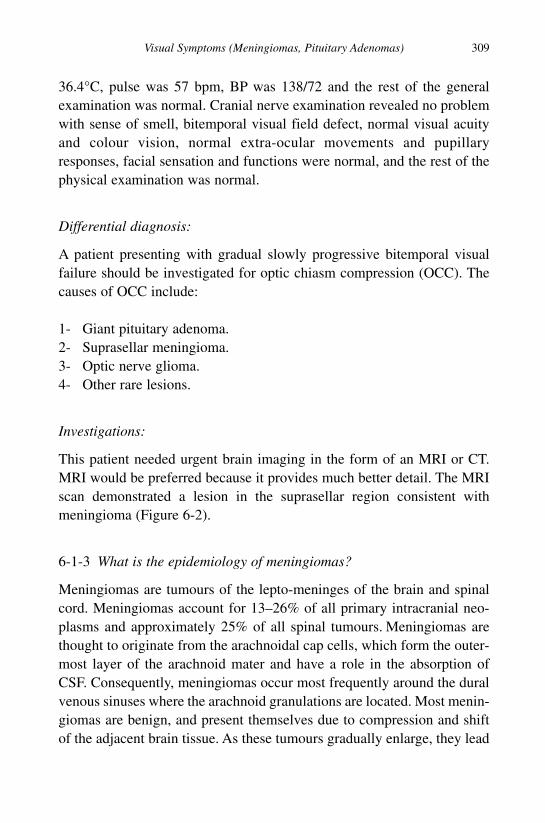
36.4°C, pulse was 57 bpm, BP was 138/72 and the rest of the generalexamination was normal. Cranial nerve examination revealed no problemwith sense of smell, bitemporal visual field defect, normal visual acuityand colour vision, normal extra-ocular movements and pupillaryresponses, facial sensation and functions were normal, and the rest of thephysical examination was normal.
Differential diagnosis:
A patient presenting with gradual slowly progressive bitemporal visualfailure should be investigated for optic chiasm compression (OCC). Thecauses of OCC include:
1- Giant pituitary adenoma.2- Suprasellar meningioma.3- Optic nerve glioma.4- Other rare lesions.
Investigations:
This patient needed urgent brain imaging in the form of an MRI or CT.MRI would be preferred because it provides much better detail. The MRIscan demonstrated a lesion in the suprasellar region consistent withmeningioma (Figure 6-2).
6-1-3 What is the epidemiology of meningiomas?
Meningiomas are tumours of the lepto-meninges of the brain and spinalcord. Meningiomas account for 13–26% of all primary intracranial neo-plasms and approximately 25% of all spinal tumours. Meningiomas arethought to originate from the arachnoidal cap cells, which form the outer-most layer of the arachnoid mater and have a role in the absorption ofCSF. Consequently, meningiomas occur most frequently around the duralvenous sinuses where the arachnoid granulations are located. Most menin-giomas are benign, and present themselves due to compression and shiftof the adjacent brain tissue. As these tumours gradually enlarge, they lead
Visual Symptoms (Meningiomas, Pituitary Adenomas) 309
b1009 Problem Based Neurosurgery
b1009_Chapter-06.qxd 11/12/2010 10:50 AM Page 309

to a slowly progressive pattern of focal neurological deficit, seizures orraised intracranial pressure. With recent advances in neuro-imaging moreand more of incidental asymptomatic meningiomas are discovered.Autopsy studies have estimated that as many as 2–3% of the populationhave an incidental asymptomatic meningioma. Females are twice as com-monly affected compared to males.1 Meningiomas are increasing withage, usually occurring after the seventh decade of life.
6-1-4 What are the causes of meningiomas?
Whilst most meningiomas are idiopathic and have unknown aetiology,there are several recognised risk factors for the development of thesetumours, including genetic factors2 such as neurofibromatosis type 2(NFII) and exposure to ionising radiation. NFII is an autosomal dominantmulti-system genetic disorder, with an incidence of one per 37,000 peryear. The disease is characterised by the presence of multiple menin-giomas3,4 as well as vestibular schwannomas (Figure 6-3), spinal cordschwannomas, gliomas and juvenile cataracts.
310 Chapter 6
b1009 Problem Based Neurosurgery
Figure 6-2: Sagittal T1 MRI with contrast demonstrating suprasellar lesion (S) withwide dural base that enhances well, consistent with suprasellar meningioma.
b1009_Chapter-06.qxd 11/12/2010 10:50 AM Page 310

Approximately 40% of children presenting with meningiomas sufferfrom NFII, and this genetic syndrome must be ruled out in any child knownto have such a tumour. NFII results from mutations in the NFII gene locatedon the long arm of chromosome 22. This gene produces a protein known asmerlin (schwannomin), which serves as a tumour-suppressor gene and pro-tects against the formation of various tumours within the central andperipheral nervous systems. Of interest, up to 60% of all idiopathic menin-giomas also show mutations in the NFII gene. Of the remaining 40%without mutations in NFII, there is often evidence for additional mecha-nisms of merlin inactivation, such as calpain-mediated proteolysis ofmerlin. It is also well established that the risk of developing meningiomas isincreased following exposure to ionising radiation.5 Local low dose irradia-tion with 8 Gy, used for the treatment of tinea capitis in the 1950s, has beenfound to carry a 2.3% lifetime risk for the development of meningiomas. Anassociation has also been found between the development of meningiomasand previous radiotherapy to the skull for the treatment of intracerebraltumours. In the Hiroshima and Nagasaki tumour registries, 88 meningiomas
Visual Symptoms (Meningiomas, Pituitary Adenomas) 311
b1009 Problem Based Neurosurgery
Figure 6-3: A case of multiple meningiomas (1,2,3,4) and vestibular schwannoma in a30-year-old woman who presented with right sensori-neural hearing loss.
b1009_Chapter-06.qxd 11/12/2010 10:50 AM Page 311

were discovered within 80,160 atomic bomb survivors, and there is evenevidence that very small doses of radiation, such as those used in dentalradiography, could lead to an increased risk of meningioma development.A less well established risk factor for the development of meningiomas isprevious head trauma. The possible mechanism for this relationship is thelocal alteration of the BBB due to the head injury, associated with a largeinflux of cytokines, histamine and bradykinin into the extravascular space.Some studies have shown that patients with meningiomas have a higherincidence of head trauma than controls, usually with a long delay betweentrauma and presentation. However, the significance of this link is question-able, with meningiomas having a higher incidence in females despite malessuffering from head trauma two to three times more frequently.6 Anotheraetiological factor which has been related to an increased risk of meningiomadevelopment is the presence of high levels of sex hormones. Approximatelytwo-thirds of all meningiomas express progesterone receptors on their cellmembranes. Expression of oestrogen and androgen receptors by menin-giomas is also commonly demonstrated. The precise role of sex hormonesin the development of meningiomas is as yet unclear. However, it is nowbecoming obvious that progesterone must at least be a factor in the growthof some meningiomas. This follows the observation that reversible aggra-vation of symptoms caused by meningiomas occurs during both the lutealphase of the menstrual cycle and pregnancy, when levels of circulatingprogesterone are increased. In addition, there has also been some successwith in vitro inhibition of meningioma cell lines using progesterone recep-tor (PR) antagonists. Another interesting discovery is that the level of PRexpression of a meningioma approximates roughly with both tumour pro-liferation and histological grade, with higher expression equating to a morebenign growth.7 A study in Sweden identified a significant correlationbetween patient meningioma diagnosis and familial history by using datafrom the Swedish Family-Cancer Database and a family history of menin-giomas increases the risk by 2.5.8
6-1-5 How do meningiomas look like and where are they located?
Macroscopically, meningiomas appear smooth and lobular, with a finevascular pattern on their surface. They most commonly exhibit a globular
312 Chapter 6
b1009 Problem Based Neurosurgery
b1009_Chapter-06.qxd 11/12/2010 10:50 AM Page 312

shape (Figure 6-4), although this may differ depending on the location ofthe tumour within the cranium. For example, meningiomas arising fromthe optic sheath often have a characteristic tubular form, while those lyingnext to the sphenoid ridge may grow in a flat and diffuse pattern over thedura, termed meningioma en plaque (Figure 6-5).
Visual Symptoms (Meningiomas, Pituitary Adenomas) 313
b1009 Problem Based Neurosurgery
Figure 6-4: Globular meningioma on the left frontal region (1) seen on MRIT1-weighted with gadolinium presenting with seizures or found incidentally.
Figure 6-5: Meningioma of the lateral wall of the left cavernous sinus (1) taking the shapeof en plaque on CT with contrast presenting with diplopia due to partial third nerve palsy.
b1009_Chapter-06.qxd 11/12/2010 10:50 AM Page 313
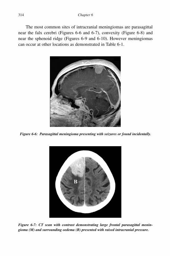
The most common sites of intracranial meningiomas are parasagittalnear the falx cerebri (Figures 6-6 and 6-7), convexity (Figure 6-8) andnear the sphenoid ridge (Figures 6-9 and 6-10). However meningiomascan occur at other locations as demonstrated in Table 6-1.
314 Chapter 6
b1009 Problem Based Neurosurgery
Figure 6-6: Parasagittal meningioma presenting with seizures or found incidentally.
Figure 6-7: CT scan with contrast demonstrating large frontal parasagittal menin-gioma (M) and surrounding oedema (B) presented with raised intracranial pressure.
b1009_Chapter-06.qxd 11/12/2010 10:50 AM Page 314

6-1-6 What are the pathological classifications of meningiomas?
Meningiomas are graded according to the WHO classification into threecategories: benign (WHO grade I), atypical (WHO grade II) or anaplastic
Visual Symptoms (Meningiomas, Pituitary Adenomas) 315
b1009 Problem Based Neurosurgery
Figure 6-8: MRI scan T1 with gadolinium demonstrating left temporal convexitymeningioma presented with seizures and focal neurological deficit (dysphasia).
Figure 6-9: MRI scan T1 with gadolinium demonstrating medial sphenoid wing menin-gioma close to the optic nerve. This may present with unilateral visual failure oripsilateral optic atrophy and contralateral papilloedema (Foster-Kennedy Syndrome).
b1009_Chapter-06.qxd 11/12/2010 10:50 AM Page 315

316 Chapter 6
b1009 Problem Based Neurosurgery
Figure 6-10: MRI scan T1 with gadolinium demonstrating lateral sphenoid wingmeningioma (1) surrounded with oedema and midline shift. This patient presented withraised intracranial pressure.
Table 6-1: Locations of meningiomas
Location Incidence Location Incidence
Parafalcine 25% (Figure 6-6) Convexity 19% (Figure 6-8)Sphenoid ridge 17% (Figure 6-9) Suprasellar 9% (Figure 6-2)Posterior fossa 8% (Figure 6-11) Olfactory groove 8% (Figure 6-12)Cavernous sinus 4% (Figure 6-5) Peritorcular 3% (Figure 6-13)Tentorial 3% (Figure 6-14) Lateral ventricle 1.5% (Figure 6-15)Foramen magnum 1.5% (Figure 6-16) Optic nerve 1.5%Clivus 1% (Figure 6-17)
Figure 6-11: MRI scan T1 with gadolinium demonstrating large meningioma (1) in theleft posterior cranial fossa causing midline shift and compression of the fourth ventri-cle presented with raised intracranial pressure due to hydrocephalus.
b1009_Chapter-06.qxd 11/12/2010 10:50 AM Page 316

(WHO grade III). Table 6-2 summarises the WHO classification of menin-giomas.
WHO grade II is characterised by mitotic index greater or equal tofour mitoses per ten high-power fields (HPF), and at least three of the fol-lowing five parameters: increased cellularity, high nuclear/cytoplasmatic
Visual Symptoms (Meningiomas, Pituitary Adenomas) 317
b1009 Problem Based Neurosurgery
Figure 6-12: MRI scan T2 demonstrating a large mass in the anterior cranial fossa(olfactory groove meningioma 1) causing raised intracranial pressure and anosmia in a45-year-old woman. These tumours can extend into the nose.
Figure 6-13: MRI scan T1-weighted with gadolinium demonstrating a small menin-gioma near the torcula (1).
b1009_Chapter-06.qxd 11/12/2010 10:50 AM Page 317

ratio (small cells), prominent nucleoli, uninterrupted patternless or sheet-like growth, foci of spontaneous necrosis, or brain invasion. WHO gradeIII on the other hand is characterised by mitotic index greater or equal to20 mitoses per ten HPF, and anaplasia (sarcoma, carcinoma, ormelanoma-like histology). Eighty per cent of meningiomas are benign,WHO grade I and approximately 15–20% are atypical and the remaining1–5% are considered anaplastic.
318 Chapter 6
b1009 Problem Based Neurosurgery
Figure 6-14: MRI scan T1 with gadolinium demonstrating a lesion at the tentoriumbefore gadolinium (2) and after gadolinium (1).
Figure 6-15: MRI scan T1 with gadolinium demonstrating intraventricular menin-gioma (1) in the trigone of the left lateral ventricle.
b1009_Chapter-06.qxd 11/12/2010 10:50 AM Page 318

Visual Symptoms (Meningiomas, Pituitary Adenomas) 319
b1009 Problem Based Neurosurgery
Figure 6-16: MRI scan demonstrating a meningioma at the foramen magnum.
Figure 6-17: CT scan with contrast demonstrating clival meningioma (1).
Table 6-2: WHO classification of meningiomas
Grade Tumour types
I Syncytial, Transitional, Fiberous, Psammomatous, Angiomatous, Microcystic,Secretory, Clear cell, Choroid, Lymphoplasmocyte-rich, Xanthomatous,Myxoid, Osseous and Cartilagenous.
II Atypical meningioma.III Anaplastic meningioma (malignant).
b1009_Chapter-06.qxd 11/12/2010 10:50 AM Page 319

6-1-7 How do meningiomas present?
Meningiomas may present in several ways (Table 6-3):
1- Seizures: Epileptic seizures in a previously healthy individual are acommon phenomenon with meningiomas, and approximately a quar-ter of all patients will present in this way. These seizures often have afocal component, closely related to the location of the tumour. Thepathophysiology of tumour-induced epilepsy remains a poorly under-stood topic, although focal cortical hypoxia, direct mass effect,perilesional oedema and altered levels of excitatory amino acids arethought to contribute.
2- Raised ICP: In the remainder of patients, the onset is more insidiouswith pressure effects such as headache and vomiting often developingbefore focal neurological signs become evident.
3- Focal neurological deficits: When focal signs do occur, they can beused to predict the location of the tumour. For example; parasagit-tal/parafalcine tumours close to the vertex may affect the “foot” and“leg” area of the motor or sensory cortex, with partial seizures orweakness affecting foot dorsiflexion, then knee and hip flexion.Parasagittal tumours situated posteriorly may present with a homony-mous hemianopia and tumours arising anteriorly may produceimpairment of memory, intellect and personality if they grow to a
320 Chapter 6
b1009 Problem Based Neurosurgery
Table 6-3: Presentation of intracranial meningiomas
History % Physical signs %
Headaches 36 Paresis 30Personality change 22 No signs 26Weakness 19 Memory impairment 15Generalised seizures 19 Cranial nerve signs 12Visual disturbance 16 Visual field deficit 10Focal seizures 15 Paraesthesia 9Imbalance 15 Speech impairment 9Speech difficulty 10 Papilloedema 8Decreased conscious 7 Reduced visual activity 6Numbness 6 Reduced conscious 5Vertigo 1 Nystagmus 3
b1009_Chapter-06.qxd 11/12/2010 10:50 AM Page 320

large enough size. Medial sphenoidal wing and parasellar menin-giomas may compress the optic nerve and produce visual impairment,often with a central scotoma or other field defect. Suprasellar menin-giomas often present early, once compression of the optic chiasm hasled to a visual field defect, usually a bitemporal hemianopia. Opticnerve sheath tumours may produce a progressive unilateral visualloss. Olfactory groove meningiomas, may destroy the olfactory bulbor tract causing unilateral anosmia. This can often pass unnoticed bypatients, until tumour expansion leads to a bilateral defect of smell.
4- Asymptomatic: With the ever-increasing availability and image qual-ity of CT and MRI, the diagnosis of meningioma is usually quitestraightforward. These tumours appear as well defined extra-axialmasses, which displace the adjacent brain.
6-1-8 How do meningiomas appear on neuroimaging?
Meningiomas tend to be hyperdense compared with normal brain on CT(Figure 6-18), and following intravenous contrast injection, there is stronguniform enhancement of the lesion (Figure 6-7). MRI is the preferred inves-tigation to show the dural origin of the tumour. Meningiomas usually appearisointense (Figure 6-14) or slightly hypointense to brain on T1-weightedMR imaging and hyperintense on T2-weighted imaging (Figure 6-19).Meningiomas show strong uniform enhancement after intravenous injec-tion of a paramagnetic contrast such as gadolinium (Figure 6-12). Oncerebral angiography meningiomas demonstrate a “tumour blush”.
Meningiomas may cause midline shift, oedema, calcification or cys-tic formation (Table 6-4).
6-1-9 How to manage symptomatic meningiomas?
The primary management of symptomatic meningioma is surgical exci-sion of the tumour including its dural (Simpson grade 1). A totalexcision may not be possible in some locations, particularly whenimportant neural or vascular structures are enveloped by the tumour.Gross total excision with diathermy of the dural origin (Simpson grade 2)without dural diathermy (Simpson grade 3) or subtotal excision
Visual Symptoms (Meningiomas, Pituitary Adenomas) 321
b1009 Problem Based Neurosurgery
b1009_Chapter-06.qxd 11/12/2010 10:50 AM Page 321

(Simpson grade 4) may therefore lead to post-surgical recurrence. Ratesof recurrence are directly related to the extent of surgical excision.Based on this system, recurrence rates at five years were estimated at9% for Simpson’s grade 1 excision, 19% for a grade 2 and 29% for a
322 Chapter 6
b1009 Problem Based Neurosurgery
Figure 6-18: CT scan demonstrating hyperdense lesion in the left frontal region (1).
Figure 6-19: T2-weighted MRI scan demonstrating a large bifrontal lesion (1) slightlyhyperintense with surrounding oedema (2) and brain shift.
b1009_Chapter-06.qxd 11/12/2010 10:50 AM Page 322

grade 3. Simpson grade 5 is merely tumour biopsy. Pre-operative steroidtherapy is desirable to prevent intra- and post-operative brain swelling.Emobilisation of meningiomas before surgery is rapidly gaining supportto minimise intra-operative blood loss. Stereotactic radiotherapy is alsocommonly employed in the management of meningiomas, either as anadjuvant to surgery for example in skull base or cavernous sinus menin-giomas or as a primary treatment in patients who refuse or unfit forsurgery.
Visual Symptoms (Meningiomas, Pituitary Adenomas) 323
b1009 Problem Based Neurosurgery
Figure 6-20: MRI scan demonstrating bone invasion (2) of meningioma (1) in the rightoccipital region.
Table 6-4: Neuro-imaging characteristics of meningiomas
Features % Features %
Midline shift 78 Homogenous enhancement 72No oedema 48 Mixed enhancement 23Mild oedema 31 Severe oedema 16Hyperostosis (Figure 6-20) 18 Calcification 27Fringing or dural tail* 1 Mushroom appearance* 0
*These appearances are more common in WHO grade III meningiomas.
b1009_Chapter-06.qxd 11/12/2010 10:50 AM Page 323

Problem 6-2: Visual symptoms and pituitary adenomas. How tomanage a patient presenting with visual field defect?
Any patient presenting with progres-sive bitemporal visual field defect(BTVD) should be treated as pitu-itary adenoma unless and until provenotherwise.
PCS6-2-1:
A 60-year-old woman presented with six months history of difficulty inreading where the right side of each word was missing. Vision hasremained unchanged for the past six months. She had a number of fallsin the past as a result of her poor vision. Past medical history includedcoronary angioplasty (1990), bilateral cataract removal, and age-relatedmacular degeneration. Neurological examination showed bitemporalhemianopia and visual acuity of 6/12 bilaterally. Systemic reviewrevealed no additional useful information.
Differential diagnosis:
This patient presented with visual problems of insidious onset of sixmonths duration. She had a history of previous eye problems; maculardegeneration and cataracts therefore primary eye disease should beconsidered as a cause; other local eye diseases that should be consid-ered include retinopathy, retinal detachment, iridocyclitis, glaucomaand optic neuritis. However other extra-ocular causes of visual failureshould be considered, particularly intracranial compressive lesionssuch as meningiomas and pituitary adenomas. The absence of local eyedisease to explain the history and the finding of bitemporal visual fielddefects pointed to a chiasmatic compression most likely from giant
324 Chapter 6
b1009 Problem Based Neurosurgery
Problem based toolkit:Acromegaly Cushing’s
CraniopharyngiomasPituitary abscess/adenomaPituitary apoplexyRathke’s cleft cystsVisual field defects
b1009_Chapter-06.qxd 11/12/2010 10:50 AM Page 324
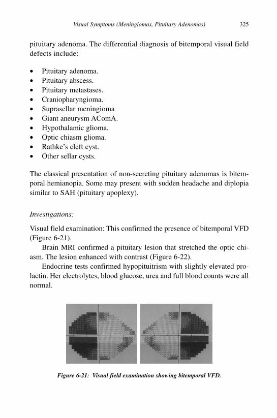
pituitary adenoma. The differential diagnosis of bitemporal visual fielddefects include:
• Pituitary adenoma.• Pituitary abscess.• Pituitary metastases.• Craniopharyngioma.• Suprasellar meningioma• Giant aneurysm AComA.• Hypothalamic glioma.• Optic chiasm glioma.• Rathke’s cleft cyst.• Other sellar cysts.
The classical presentation of non-secreting pituitary adenomas is bitem-poral hemianopia. Some may present with sudden headache and diplopiasimilar to SAH (pituitary apoplexy).
Investigations:
Visual field examination: This confirmed the presence of bitemporal VFD(Figure 6-21).
Brain MRI confirmed a pituitary lesion that stretched the optic chi-asm. The lesion enhanced with contrast (Figure 6-22).
Endocrine tests confirmed hypopituitrism with slightly elevated pro-lactin. Her electrolytes, blood glucose, urea and full blood counts were allnormal.
Visual Symptoms (Meningiomas, Pituitary Adenomas) 325
b1009 Problem Based Neurosurgery
Figure 6-21: Visual field examination showing bitemporal VFD.
b1009_Chapter-06.qxd 11/12/2010 10:50 AM Page 325

Management: She was pretreated with hydrocortisone 100 mg QID andunderwent transsphenoidal excision of the pituitary adenoma.
PCS6-2-2:
A 75-year-old right-handed woman presented with sudden severeheadache. On questioning she had visual loss of insidious onset for sev-eral months and felt fatigued. No other significant history. Examinationrevealed that she was fully conscious, with bitemporal visual field defectand no other signs.
Differential diagnosis:
Sudden severe headache raised the possibility of subarachnoid haemor-rhage (SAH) and thunderclap headache syndrome, but the presence ofvisual symptoms and visual field defect in the temporal VF points to opticchiasm compression. Putting the two together a diagnosis of pituitaryapoplexy will be most likely.
326 Chapter 6
b1009 Problem Based Neurosurgery
Figure 6-22: MRI demonstrating large pituitary adenoma (C) compressing the opticchiasm (OC), invading the cavernous sinus encircling the internal carotid arteries (CA).
b1009_Chapter-06.qxd 11/12/2010 10:50 AM Page 326

Investigations:
Full blood counts, urea, electrolytes and blood glucose were all normal.VF examination demonstrated bitemporal VF defects similar to thoseshown in Figure 6-21.
CT brain demonstrated hyperdense lesion in the sella turcica regionconsistent with a bleed in a giant pituitary adenoma (Figure 6-23).
The patient was treated with hydrocortisone 100 mg QID and under-went transsphenoidal excision of the adenoma. At surgery there was clearevidence of haemorrhage.
6-2-3 What is the epidemiology of pituitary adenomas?
Pituitary adenomas (PA) are benign tumours of the pituitary gland and rep-resent about 12% of all intracranial tumours.9,10 They are classified accordingto their function into secreting or non-secreting PA. Secreting adenomas tendto be small (microadenomas) that present with syndromes of hypersecretionsuch as Cushing’s disease, acromegaly or hyperprolactinaemia. Whereasnon-secreting adenomas tend to present late and by the time they revealthemselves clinically they have often already compressed the optic chi-asm11–15 or invaded the cavernous sinus and surrounding structures.15,16
Visual Symptoms (Meningiomas, Pituitary Adenomas) 327
b1009 Problem Based Neurosurgery
Figure 6-23: Axial CT scan demonstrated a large lesion in the sella turcica (c). Thelesion is hyperdense.
b1009_Chapter-06.qxd 11/12/2010 10:50 AM Page 327

6-2-4 How do pituitary adenomas present?
The clinical presentation of PA can be due to local mass effect orendocrine effects. The local mass effect is due to the close position of thePA to the optic chiasm and cavernous sinuses. Large tumours (greater than10 mm), otherwise known as macroadenomas can cause compression ofadjacent neural structures in the wall of the cavernous sinus, e.g. III cra-nial nerve causing partial third nerve palsy and periorbital pain andheadaches due to enlargement of the pituitary fossa. When the PA expandsup the way it compresses the optic chiasm and causes visual field defects.This usually affects the superior temporal quadrants (Figure 6-24) pro-gressing to bitemporal hemianopia (Figure 6-21).
The PA can also compress normal pituitary gland, leading to reducedhormone secretion and eventually panhypopituitarism. Compression ofthe pituitary stalk leads to slight elevation of prolactin levels.
The endocrine presentation of PA can be due to either hypo- or hyper-secretion. As the pituitary gland controls many other glands in the bodythe symptoms can be variable. However, only one or two of the pituitaryhormones are affected at any one time. The cause of hyposecretion, is aresult of large PA compressing the pituitary gland. This can cause weightgain, loss of libido, fatigue, secondary amenorrhoea, sterility, glucocorti-coid and androgen deficiency, muscle weakness and secondaryhypothyroidism. Panhypopituitarism is when all of the pituitary hormonesare diminished. Hypersecretion on the other hand presents in differentways dependent upon the hormone secreted in excess: growth hormone-secreting PA causes acromegally in adults and gigantism in children;
328 Chapter 6
b1009 Problem Based Neurosurgery
Figure 6-24: Bitemporal upper quadrantanopia.
b1009_Chapter-06.qxd 11/12/2010 10:50 AM Page 328

prolactinomas cause galactorrhoea, impotence and secondary amenor-rhoea; and ACTH-secreting PA causes Cushing’s disease. TSH andFSH/LH-secreting tumours are very rare. Rarely, pituitary tumours canpresent with severe sudden headache similar to that of SAH, rapidly pro-gressive visual defects and acute pituitary insufficiency (pituitaryapoplexy), where there is infarction followed by haemorrhage into the PA.
6-2-5 How to investigate a patient with possible PA?
Investigation of a suspected PA involves visual fields, neuro-imaging andendocrine tests.
1- Visual flied (VF) assessment: VFs are assessed in patients with PAbecause of the aforementioned reasons. VF compromise in any waydictates the type of therapy and the urgency of therapy. Generally ifthere are significant VF defects, surgery will be necessary to decom-press the optic chiasm and preserve vision. The commonest VF defectin PA is bitemporal VF defect, initially affecting the upper quadrants(Figure 6-24) and then bitemporal hemianopia (Figure 6-21).However other types of VF defects can be found if the optic chiasmis pre- or post-fixed (Figure 6-25).
2- Neuro-imaging: The scan of choice is an MRI scan for obvious reasons;it details the anatomy well and can show the chiasm and cavernoussinuses (Figures 6-22). CT however, can be useful (Figure 6-23).
Visual Symptoms (Meningiomas, Pituitary Adenomas) 329
b1009 Problem Based Neurosurgery
Figure 6-25: VF defect in post-fixed chiasm and large PA affecting the left VF superiortemporal and nasal fields.
b1009_Chapter-06.qxd 11/12/2010 10:50 AM Page 329

3- Endocrine tests:
a. Prolactin: This hormone is kept under control via the pituitaryhypothalamic axis where dopamine secreted in the hypothalamuskeeps the prolactin level within normal range. Prolactin is ele-vated by stress, pregnancy, beast feeding and during sleep. This isa physiological response and returns to normal when the physio-logical stimulus ceases. A number of drugs can elevate prolactin:dopaminergic inhibitors (oestrogen, metoclopramide, verapamil,domperidone, cocaine, and opiates), antipsychotics (phenoth-iazines, benzamides; sulpiride, butyrophenones; haloperidol,benperidol, risperidone, molindone, quetiapine), antidepressants(selective serotonin reuptake inhibitors (SSRI), tricyclics;chlomipramine, desipramine and mono amine oxidase inhibitors;pargyline, clorgyline), and adrenergic inhibitors (reserpine,methyldopa). Hyperprolactinaemia can occur also due to stalkcompression or irritation such as in non-functioning PA, headinjuries, and following cranial surgery, in hypothyroidism, and inprolactinomas. Prolactinomas present in females as secondaryamenorrhea or irregularity of menstruation, infertility and galact-orrhoea in 30–80% of patients. In males they present late withgalactorrhoea in < 30%, VF defects, headaches, impotence andhypopituitrism.
b. Growth hormone (GH): GH over-secretion causes acromegalyin adults and gigantism in children. GH over-secretion may pres-ent in many forms and to different specialists, e.g. it may presentto a paediatrician with gigantism, to a general physician withlethargy or sleep apnoea, to a neurologist with sleepiness,headaches, and paraesthesia, to an ophthalmologist with visualfailure and field defects, to a dentist with malocclusion of teeth,or clicking jaw, to an ENT surgeon with snoring, sinusitis, largetongue, voice change, to a dermatologist with greasy thick skin,and sweating, to an orthopaedic/rheumatologist with largehands/feet (Figure 6-26), joint pains, or carpal tunnel syndrome,to a cardiologist with hypertension, or heart failure symptoms, toa gastroenterologist with large spleen and liver, to a diabetologist
330 Chapter 6
b1009 Problem Based Neurosurgery
b1009_Chapter-06.qxd 11/12/2010 10:50 AM Page 330

with diabetes mellitus and to a urologist with polyuria, or urinecalculi. The diagnosis is made by measuring IGF1 which wouldbe markedly elevated compared to matched age and sex levels.The diagnosis is confirmed by glucose-tolerance-test (GTT). Innormal individuals GH suppresses to < 5 mu/l after glucose inges-tion whilst in acromegaly the GH is unchanged and shows nosuppression or paradoxically rises. GH remains > 5 mu/l after glu-cose ingestion.
c. ACTH and cortisol measurements: Excess ACTH producesCushing’s disease if the source of the excess ACTH is PA orCushing’s syndrome if the source of excess ACTH is outside thepituitary. Excess cortisol leads to protein loss, myopathy andwasting of muscles, osteoporosis and bone fractures, thin skin,striae, and bruising; altered carbohydrate metabolism leading todiabetes mellitus; altered psyche leading to psychosis and depres-sion; mineralocorticoid effects leading to hypertension andoedema, obesity, buffalo hump and round moon face; and excessandrogens leading to virilism, hirsutism, acne and oligo/amenor-rhoea in women. To distinguish Cushing’s from obesity Cushing’sis characterised by thin skin, proximal myopathy, frontal baldingin women, conjunctival oedema (chemosis) and osteoporosis.To confirm the diagnosis of Cushing’s disease dexamethasonesuppression tests are performed:
i. Overnight 1 mg dexamethasone suppression test: If cortisol levelwas < 50 nmol/l the next morning = normal, not Cushing’s.
Visual Symptoms (Meningiomas, Pituitary Adenomas) 331
b1009 Problem Based Neurosurgery
Figure 6-26: Photograph of acromegalic hands with other features listed.
b1009_Chapter-06.qxd 11/12/2010 10:50 AM Page 331

ii. Urine free cortisol: Total < 250 units is normal and cortisol/creatinine ratio of < 25 is normal.
iii. Two-day 2 mg/day dexamethasone suppression test: Cortisol< 50 nmol/l six hours after last dose indicates that there is noCushing’s.
d. Complete pituitary function tests: Required in patients with PAafter treatment, these include — Thyroid: free T4 and TRH test.Gonadotrophins and in females oestradiol and LHRH test; in malestestosterone. Adrenal; insulin tolerance test (ITT) and short synac-then test (SST) and glucagon test. GH: IGF1, ITT or glucagon andanti-diuretic hormone (ADH) water deprivation test (WDT).
6-2-6 What is the differential diagnosis of PA?
Lesions in the region of the sella are varied in nature:
1- PA is the most common (Figure 6-22).2- Rathke’s cleft cyst: Rathke’s cleft cyst is a benign cystic lesion that
affects the pituitary gland. It is the most common incidental finding inthe pituitary and comprises about 3.7% of all the incidental findingsthat were greater than 2 mm and they tend to be medially located.Symptomatic Rathke’s cleft cyst is relatively uncommon. When thesecysts are large enough (Figure 6-26) they may cause headaches, visualdisturbances, nausea, decreased energy, vomiting, seizures, pituitarydysfunction, and diabetes insipidus because of mass effect on thepituitary stalk, the third ventricles and the surrounding structures.Haemorrhage occasionally precipitates symptoms. The mean age atpresentation is 34 years with female predominance. Relief of symp-toms is common with operative management with resolution ofsymptoms in 78% of cases. The recurrence rate varies from 19–28%.During the third week of gestation an invagination of the floor of thethird ventricle (the infundibulum) starts to coalesce to form the pitu-itary gland and another from the stomodeum (Rathke’s pouch). Thelatter requires sequential induction from the neighbouring dien-cephalon to attain the morphology required to reach the
332 Chapter 6
b1009 Problem Based Neurosurgery
b1009_Chapter-06.qxd 11/12/2010 10:50 AM Page 332

infundibulum. Both of these entities are from the ectoderm; Rathke’spouch develops into the anterior and middle lobes, and the infundibu-lum develops into the posterior lobe and stalk of the pituitary gland.The lumen that connects Rathke’s pouch to the oral cavity begins toobliterate around the sixth week of gestation by the development ofthe sphenoid bone. The lumen of Rathke’s pouch is usually obliteratedby proliferation of the anterior and posterior walls of the pouch thatwill become the anterior and middle lobes. The apical extremity of theRathke’s pouch will remain as a small remnant into post-natal life andadulthood as a small cleft between the anterior pituitary (pars distalis)and the posterior pituitary (pars nervosa). The cleft is lined by colum-nar to cuboidal epithelium that is often ciliated and occasionallyassociated with mucous-secreting goblet cells. Accumulation ofsecretory products forms a cyst in this space. It should be noted thatsome authors have stated that one cannot differentiate a Rathke’s cleftcyst from a neuroepithelial cyst, such as a colloid cyst of the thirdventricle, based on histology or histochemistry. They propose thatsome Rathke’s cleft cysts have neuroepithelial origin due to pinchingoff cells from the infundibulum into the cells that make up the dorsalwall of Rathke’s pouch. On MRI they have several distinctive fea-tures. They are most often intrasellar cysts with or without suprasellarextension. Radiologically, it occurs as a single, well demarcated cystwithout calcification (Figure 6-27). The content is homogenous.Depending on the content of the cyst, the MRI signal intensities canvary greatly. The radiological features overlap with that of cranio-pharyngioma particularly those with extensive cystic changes andwithout calcification. Histologically, they are lined by a single layerof cuboidal to columnar cells that are often ciliated. Occasionalmucous cells may be present. Squamous metaplasia may occur andraises the possibility of a craniopharyngioma. Occasional cells ofanterior pituitary gland may be present. In Rathke’s cleft cysts thathave substantial prior haemorrhage, there may be cholesterol cleft andforeign body giant cell reaction.
3- Arachnoid cyst: Arachnoid cysts in the sella are uncommon andcan easily be confused both clinically and radiologically with intra-sellar cystic pituitary neoplasms such as Rathke’s cleft cysts,
Visual Symptoms (Meningiomas, Pituitary Adenomas) 333
b1009 Problem Based Neurosurgery
b1009_Chapter-06.qxd 11/12/2010 10:50 AM Page 333

craniopharyngiomas or even empty sella. Adequate diagnosis is quiteimportant because of different treatment modalities and differentprognosis. Cystic lesions of the sellar and parasellar regions have afluid-filled compartment and a connective tissue wall and/or epithe-lial lining but no communication with the subarachnoid space thatdistinguishes them from an empty sella (Figure 6-28). These lesionsare classified according to the site of origin, tissue of origin and
334 Chapter 6
b1009 Problem Based Neurosurgery
Figure 6-27: MRI scan demonstrating a Rathke’s cleft cyst (c) in a 35-year-old manwho presented with headaches and diplopia.
Figure 6-28: Sellar arachnoid cyst (c).
b1009_Chapter-06.qxd 11/12/2010 10:50 AM Page 334

pathological features. They are broadly classified into primary andsecondary cysts. The primary lesions arise within the sella and areconfined to the pituitary fossa including true intrasellar arachnoidcysts (Figure 6-28), pituitary cysts, Rathke’s cleft cysts and other raretypes. Secondary non-neoplastic cystic lesions of the sella may arisefrom the sphenoid sinus or the parasellar region and extend into thefossa, as sphenoid sinus mucocele, arachnoid cysts, orLeptomeningeal cysts. Arachnoid cysts are lined by a single layer ofmesothelial cells, surrounded by collagenous layer.
4- Sellar and suprasellar meningioma: Meningiomas constitute about15% of all intracranial tumours and are the most common primarynon-glial lesions. Suprasellar location is the fifth most common site oforigin with 5%. Suprasellar meningioma arising from tubereculumsellea clinoid processes and diaphragma sellae may grow downwardinto the sella turcica. But pure intrasellar meningiomas are well-known lesions. These tumours seem to be exceptional and are thoughtto arise within the sella turcica and extend superiorly into the chias-matic cistern, causing visual and endocrine dysfunction bycompressing optic pathways and pituitary stalk. CT and MRI demon-strate contrast-enhancing intrasellar or suprasellar lesion (Figure 6-29).Serum prolactin levels may be slightly elevated and visual fields may
Visual Symptoms (Meningiomas, Pituitary Adenomas) 335
b1009 Problem Based Neurosurgery
Figure 6-29: a == MRI showing suprasellar meningioma (T), CA == carotid artery. b ==Before contrast and c == with contrast. This was a woman who presented with bitempo-ral VF defects and lethargy.
b1009_Chapter-06.qxd 11/12/2010 10:50 AM Page 335
show bitemporal VF defect similar to that in Figure 6-21. Surgery isthe main treatment with the aim of total resection.
5- Pituitary abscess: Pituitary abscess is a rare but potentially life threat-ening condition if not adequately diagnosed and treated. Since thefirst description in 1941, over 121 cases have been reported in the lit-erature.17 Pituitary abscess implies pituitary gland involvement by aninfectious process within the sella turcica characterised by the pres-ence of an acute or chronic inflammatory reaction. This process mayderive from a localised or generalised infection source (meningitis,sepsis), facilitated or not by a previous existing sellar lesion such asadenoma, craniopharyngioma or Rathke’s cleft cyst. Pituitary abscessis treated as an emergency by drainage and IV antibiotics.
6- Craniopharyngioma: Craniopharyngiomas are classified into twohistopathologically and clinically distinct subtypes — adamantinousand squamous-papillary variants.18 The adamantinous type consists of apredominantly cystic lobulated tumour, which is often observed in anintrasellar/suprasellar location in children. These cysts contain variousamounts of cholesterol, triglycerides, methaemoglobin, protein,desquamated epithelium, and watery fluid content. Squamous-papillarycraniopharyngioma, on the other hand, consists of a predominantlysolid or mixed solid-cystic spherical tumour in a suprasellar location inadults. The solid tumour parts have an inhomogeneous but intenseenhancement with small necrotic areas and calcifications. The combi-nation of papillary and adamantinous tumour parts within the sameneoplasm has been described in 15% of these tumours.Craniopharyngiomas arise anywhere along the migration of Rathke’spouch, which extends from the vomer and the roof of the nasopharynx,through the midline sphenoid bone to the floor of the sella turcica. Thus,craniopharyngiomas can potentially arise in unusual locations such asthe nasopharynx,18 sphenoid bone,19 third ventricle,20 and posteriorfossa. Craniopharyngiomas are rare, they make up 6–10% of all child-hood brain tumours with the majority of children being diagnosedbetween the ages of five and ten years old. However, they can growinto, and cause problems with, the pituitary and hypothalamus, and thethird ventricle, and can damage the optic chiasm. Adverse prognosticfactors include; young age at diagnosis, hydrocephalus, hypothalamic
336 Chapter 6
b1009 Problem Based Neurosurgery
b1009_Chapter-06.qxd 11/12/2010 10:50 AM Page 336

invasion, large size and optic chiasm compression. In children, cranio-pharyngiomas present with either raised ICP from hydrocephalus orslowly forming symptoms of pressure on adjacent structures. RaisedICP with hydrocephalus is found in 50%. Visual symptoms and hor-monal deficiency, e.g. dwarfism and possibly diabetes insipidus andhypothalamic problems, for example obesity, represent other forms ofpresentations. MRI scan will be the best investigation to determine thediagnosis, location and extent of craniopharyngioma and provide theroadmap for treatment (Figure 6-30). The appearances of adamantinousand papillary subtypes are not dissimilar (Table 6-5).
CT brain can also be helpful in detecting craniopharyngiomas asthey can demonstrate a cystic lesion in the suprasellar region with cal-cification (Figure 6-31).
Visual Symptoms (Meningiomas, Pituitary Adenomas) 337
b1009 Problem Based Neurosurgery
Figure 6-30: MRI of adamantinous craniopharyngioma (C) presenting in an adult withvisual failure.
Table 6-5: MRI appearances of craniopharyngiomas
Craniopharyngioma Adamantinous Papillary
Site Often suprasellar Often third ventricleAppearance Predominantly cystic Predominantly solidT1 signal Often hyperintense Often isointenseT2 signal Often hyperintense Often hyperintenseEnhancement Yes Yes
b1009_Chapter-06.qxd 11/12/2010 10:50 AM Page 337

The main objectives of treatment are: lowering raised ICP,decompression of the optic apparatus, and preservation of pituitaryfunctions. Complete surgical resection should be the aim of surgeryfollowed by radiotherapy if a remnant was left after surgery.Hormonal replacement of any deficient hormones is instituted.
7. Metastases: Metastasis to the pituitary gland is a recognised compli-cation in almost all extracranial cancers such as breast, lung, kidney,gut, thyroid, prostate and other cancers. The incidence of pituitarymetastases varies from 0.14% to 28.1% of all brain metastases and ishigher in autopsy series.21 They most frequently originate from lungcancer in males and breast carcinoma in females. Pituitary metastasesmore commonly affect the posterior lobe and the infundibulum thanthe anterior lobe. A review of 201 reported cases showed that the fre-quency of involvement within the pituitary gland was 50.8% in theposterior lobe alone, 33.8% in both lobes, and 15.4% in the anteriorlobe alone.21 The predilection of metastases for the neurohypophysismay reflect the fact that the posterior lobe receives its blood supplydirectly from the inferior hypophyseal arteries, whereas the anteriorlobe is nourished indirectly by portal vessels. On MRI scan pituitarymetastases can be differentiated by their intrasellar and suprasellar
338 Chapter 6
b1009 Problem Based Neurosurgery
Figure 6-31: CT scan demonstrating calcified remnant of craniopharyngioma (C).Patient had bilateral pterional craniotomy (A and B) and VP shunt.
b1009_Chapter-06.qxd 11/12/2010 10:50 AM Page 338

dumbbell-shaped appearance that indent the diaphragma sellae, whichis morphologically different in pituitary adenoma that usuallyexpands the diaphragma sellae.
8- Chordoma: Clival chordoma is a locally aggressive and relativelyrare tumour of the skull base that is thought to originate fromembryonic remnants of the notochord. Chordomas account for 1%of all intracranial tumours and 4% of all primary bone tumours.22
Chordoma may occur at any age but it is usually seen in adults, withpeak prevalence in the fourth decade of life. Chordomas have a 2:1male predilection. Clival chordomas constitute one-third of allchordomas. Although clival chordomas are generally slow-grow-ing, their intimate relation to critical structures and their extremelyhigh local recurrence rate have often resulted in high mortality.MRI and CT allow precise delineation of chordomas with respectto volume and relation to adjacent neural structures, thereby help-ing achieve their eradication. Generally, chordomas grow slowlyand produce symptoms insidiously. Symptoms of intracranial chor-domas vary with lesion location and proximity to critical structures,reflecting the specific sites of extension from the clivus; sellar,parasellar, and retroclival areas and, occasionally, the sphenoidsinus. The most common initial complaint is diplopia related to cra-nial nerve palsy and headaches. Among cranial nerves, the abducentnerve is the most commonly affected. Headache is usually reportedin an occipital or retro-orbital location. The classic appearance ofintracranial chordoma on CT is well-circumscribed, expansile soft-tissue mass that arises from the clivus with associated extensivelytic bone destruction. The bulk of the tumour is usually hyper-dense relative to the adjacent brain. Intratumoral calcificationsappear irregularly on CT and are usually thought to representsequestra from bone destruction rather than dystrophic calcifica-tions in the tumour itself. On conventional T1-weighted MRimages, intracranial chordoma has intermediate to low signal inten-sity and is easily recognised within the high signal intensity of thefat of the clivus (Figure 6-32).
Small foci of hyperintensity can sometimes be visualised in thetumour on T1-weighted images, a finding that represents intratumoral
Visual Symptoms (Meningiomas, Pituitary Adenomas) 339
b1009 Problem Based Neurosurgery
b1009_Chapter-06.qxd 11/12/2010 10:50 AM Page 339

haemorrhage or a mucus pool. The presence of haemorrhagic focican be confirmed with gradient-echo imaging that is susceptible toblood, at which the foci appear as dark areas. Classic intracranialchordoma has high signal intensity on T2-weighted images, a find-ing that likely reflects the high fluid content of vacuolated cellularcomposition. The intratumoral areas of calcification, haemorrhage,and highly proteinaceous mucus usually demonstrate heterogeneoushypointensity on T2-weighted imaging. Radical total resection withsparing of vital structures is the main treatment followed by radia-tion is the current therapy. The use of fractionated proton beamradiation therapy permits delivery of tumour doses 15–35% higherthan those associated with standard external beam radiotherapy,allowing improved local control and higher survival rates.22 Therecurrence-free five-year survival rate for patients with skull basechordoma who undergo combined treatment with surgery and radia-tion therapy is 60–70%.
9- Mucocoele: Sphenoid sinus mucocele is rare and often misdiag-nosed.23 Since its first description, less than 150 cases have beenreported in the literature. This benign, readily treatable lesion ispotentially fatal if misdiagnosed. To make the correct diagnosis pre-operatively careful radiological examination and interpretation isessential. The aetiology of paranasal mucoceles is controversial andusually occurs when drainage of a sinus is obstructed by inflammation,
340 Chapter 6
b1009 Problem Based Neurosurgery
Figure 6-32: MRI scan demonstrating chordoma in a 35-year-old man who presentedwith diplopia due to sixth nerve palsy.
b1009_Chapter-06.qxd 11/12/2010 10:50 AM Page 340

fibrosis, trauma, previous surgery or anatomical abnormality.23
Mucoceles occur most commonly in the frontal and ethmoid sinusesbut are quite infrequent in the sphenoid sinus.24 Out of 63 reportedcases, 29 patients (46%) had previous diagnosis of sinusitis,intranasal polyps, nasal mass or nasal discharge. The clinical andradiographic features of sphenoid sinus mucoceles are usuallyrelated to sinus expansion and extension of the lesion beyond theconfines of the sinus. This usually follows the path of least resist-ance, namely anteriorly to involve ethmoid air cells and orbits, butoccasionally posteriorly into the clivus and superiorly into the sella.Clinically the symptoms and signs are of orbital apex syndrome,superior orbital fissure syndrome and anterior cavernous sinus syn-drome. The most common complaint was headache occurring in 55out of 63 cases (87%). The headache is typically frontal or retro-orbital and tends to be worse towards evening. It may be due todural stretching if the lesion breaks through the sella turcica. Visualsymptoms often alert the patient and his physician to the problem;these occur in 62% with diplopia in 30%. Plain skull X-rays showopacification of the sphenoid sinus and any erosion of the sellarfloor. Ballooning of the sella turcica characteristic of pituitary ade-noma is unusual with sphenoid sinus mucocele. Very infrequentlythe petrous apex may be eroded. The sella was eroded in 29 out of63 cases reviewed (46%). CT and bone window studies are essentialfor evaluating the cribriform plate, the roof of the ethmoid sinuses,the involvement of the supraorbital ethmoid extension and extensioninto the superior orbital fissure. Mucoceles appear isodense, hypo-or hyperdense and lack contrast enhancement but may show rimenhancement. MRI is better than CT in delineating sphenoid muco-celes and their relationship to sellar and parasellar anatomy. Theyhave higher protein content, resulting in a shorter T1 and longer T2that produce high signals on both T1- and T2-weighted images.Surgical drainage is the primary treatment leading to cure in mostcases.
10- Giant aneurysms: Aneurysms in the sellar and parasellar regions canarise from the internal carotid artery and the anterior communicatingartery (Figure 6-33).
Visual Symptoms (Meningiomas, Pituitary Adenomas) 341
b1009 Problem Based Neurosurgery
b1009_Chapter-06.qxd 11/12/2010 10:50 AM Page 341

References
1. Claus EB. Epidemiology of intracranial meningiomas. Neurosurgery 2005;
57: 1088–1095.
2. Black P. Meningiomas. Neurosurgery 1993; 32: 643–658.
3. Eljamel MS. Multiple meningiomas. J Neurosurg 1990; 72(5): 834–835.
4. Eljamel MS, Foy PM. Multiple meningiomas and their relation to neurofi-
bromatosis. Review of the literature and report of seven cases. Surg Neurol
1989; 32(2): 131–136.
5. Musa BS, Pople IK. Intracranial meningiomas following irradiation — a
growing problem? Br J Neurosurg 1995; 9: 629–637.
6. Brenner AV, Linet MS, Fine HA et al. History of allergies among adults with
glioma and controls. Int J Cancer 2002; 99: 252–259.
7. Hsu DW, Efird JT, Hedley-Whyte ET. Progesterone and oestrogen receptors
in meningiomas: prognostic considerations. J Neurosurg 1997; 86: 113–120.
8. Hemminki K, Li X, Collins VP. Parental cancer as a risk factor for brain
tumours (Sweden). Cancer Causes Control 2001; 12: 195–199.
9. Oruckaptan HH, Senmevsim O, Ozcan OE et al. Pituitary adenomas: results
of 684 surgically treated patients and review of the literature. Surg Neurol
2000; 53: 211–219.
10. Yildiz F, Zorlu F, Erbas T et al. Radiotherapy in the management of giant
pituitary adenomas. Radiother Oncol 1999; 52: 233–237.
11. Amar AP, Hinton DR, Krieger MD et al. Invasive pituitary adenomas:
significance of proliferation parameters. Pituitary 1999; 2: 117–122.
342 Chapter 6
b1009 Problem Based Neurosurgery
Figure 6-33: Giant aneurysm from the internal carotid artery.
b1009_Chapter-06.qxd 11/12/2010 10:50 AM Page 342
12. Anson JA, Segal MN, Baldwin NG et al. Resection of giant invasive pitu-
itary tumors through a transfacial approach: technical case report.
Neurosurgery 1995; 37: 545–546.
13. Ebersold MJ, Quast LM, Laws ER Jr et al. Long-term results in transsphe-
noidal removal of nonfunctioning pituitary adenomas. J Neurosurg 1984; 64:
713–719.
14. Majos C, Coll S, Aguilera C et al. Imaging of giant pituitary adenomas.
Neuroradiology 1998; 40: 651–655.
15. Meij BP, Lopes MB, Ellegala DB et al. The long-term significance of micro-
scopic dural invasion in 354 patients with pituitary adenomas treated with
transsphenoidal surgery. J Neurosurg 2002; 96: 195–208.
16. Scheithauer BW, Kovacs KT, Laws ER Jr et al. Pathology of invasive pitu-
itary tumors with special reference to functional classification. J Neurosurg
1986; 65: 733–744.
17. Vates GE, Berger MS, Wilson CB. Diagnosis and management of pitu-
itary abscess: a review of twenty-four cases. J Neurosurg 2001: 95;
233–241.
18. Kanungo N, Just N, Black M, et al. Nasopharyngeal craniopharyngioma in
an unusual location. Am J Neuroradiol 1995; 16: 1372–1374.
19. Graziani N, Donnet A, Bugha TN et al. Ectopic basisphenoidal cranio-
pharyngioma: case report and review of the literature. Neurosurgery 1994;
34: 346–349.
20. Linden CN, Martinez CR, Gonzalvo AA et al. Intrinsic third ventricle cran-
iopharyngioma: CT and MR findings. J Comput Assist Tomogr 1989; 13:
362–368.
21. Kucharczyk W, Lenkinski RE, Kucharczyk J et al. The effect of phospho-
lipid vesicles on the NMR relaxation of water: an explanation for the
MR appearance of the neurohypophysis? Am J Neuroradiol 1990; 11:
693–700.
22. Dahlin DC, MacCharty CS. Chordoma: a study of 59 cases. Cancer 1952; 5:
1170–1178.
23. Chui MC, Briant TD, Gray T et al. Computed tomography of sphenoid sinus
mucocele. J Otolaryngol 1983; 12: 263–269.
24. Hesselink JR, Weber AL, New PFJ et al. Evaluation of mucoceles of the
paranasal sinuses with computed tomography. Radiology 1979; 133:
397–400.
Visual Symptoms (Meningiomas, Pituitary Adenomas) 343
b1009 Problem Based Neurosurgery
b1009_Chapter-06.qxd 11/12/2010 10:50 AM Page 343

Your personal notes:
.....................................................................................................................
.....................................................................................................................
.....................................................................................................................
.....................................................................................................................
.....................................................................................................................
.....................................................................................................................
.....................................................................................................................
.....................................................................................................................
.....................................................................................................................
.....................................................................................................................
344 Chapter 6
b1009 Problem Based Neurosurgery
b1009_Chapter-06.qxd 11/12/2010 10:50 AM Page 344

Chapter 7: Hearing Loss, Ataxia, Vertigoand Facial Pain (CPA Lesions)
Problem 7-1: Hearing loss and cerebellopontine angle(CPA) lesions. How to manage a patient presentingwith hearing loss?
Any patient presenting with unilateralhearing loss with or without vertigoand tinnitus could have CPA lesion.
PCS7-1-1:
A 68-year-old man presented with slowly worsening left hearing lossfirst noticed 18 months ago when he did not seem to hear the alarm clockwhile a sleep on his right side. He tried his phone on both ears and wasnot able to hear as clearly in the left ear compared to his right. He went tohis family practitioner who examined his ears and did not find any earwaxor any other external ear problem and observed normal eardrums. He wasreferred to the local otolaryngology. Examination revealed that his hear-ing was reduced on the left. Air conduction (AC) was better than boneconduction (BC) in both ears and Weber’s test lateralised to the good ear(right). He went on to have pure tone audiogram that confirmed sensoryneural deafness on the left and MRI scan that confirmed a CPA lesion con-sistent with vestibular schwannoma.
7-1-2 What causes unilateral sensory neural deafness?
1- Vestibular schwannoma (VS) (most common 92%).2- CPA meningiomas (second most common 3–7%).
345
b1009 Problem Based Neurosurgery
Problem based toolkit:Deafness DizzinessTinnitus VertigoCPA meningiomaCPA epidermoid cystVestibular schwannoma
b1009_Chapter-07.qxd 11/12/2010 10:50 AM Page 345

3- Epidermoid cyst (2–6%).4- Trigeminal schwannoma.5- Facial schwannoma.
The main presentation of VS is unilateral sensory neural hearing loss withhigh frequency and speech discrimination being the most affected.Tinnitus and dizziness are common but true vertigo is rare. As the VSexpands into the CPA it stretches the adjacent cranial nerves. The facialnerve is very resistant to stretch and facial weakness is very rare in VS. (Iffacial weakness is a feature of CPA lesion think of facial schwannoma.) IfVS expands superiorly the trigeminal nerve will be affected with reducedcorneal reflex or facial numbness. Trigeminal neuralgia is rare in VS. VSis the diagnosis until proven otherwise by imaging in any patient present-ing with unilateral sensory neural hearing loss and reduced ipsilateralcorneal reflex or ipsilateral facial numbness. With further VS enlarge-ment the cerebellum and brain stem will be affected and ataxia or longtract signs may be detected clinically. Downward growth of the VS mayresult into compression of the lower cranial nerves leading to difficulty inswallowing, aspiration or hoarseness. In very large VS the fourth ventri-cle can be obstructed leading to hydrocephalus, papilloedema and reducedlevel of consciousness.
CPA meningioma presents slightly different in that they often presentwith facial pain similar to trigeminal neuralgia, facial weakness and thenhearing loss. Trigeminal schwannoma presents with trigeminal nerve dys-function such as facial numbness and impaired sensation rather thantrigeminal neuralgia. CPA lesions presenting with multiple lower cranialnerve dysfunctions are unlikely to be one of the above lesions and maywell be malignant in nature such as metastases.
7-1-3 What is the epidemiology of VS?
VS represent about 6% of all intracranial tumours. They are benign, slow-growing tumours, which arise from Schwann-cells in the nerve sheath thatsurrounds the vestibular nerves. Their growth is slow and difficult to pre-dict in individual patients. The advent of MRI had resulted in thediagnosis of asymptomatic VS in patients who were scanned for other
346 Chapter 7
b1009 Problem Based Neurosurgery
b1009_Chapter-07.qxd 11/12/2010 10:50 AM Page 346

unrelated conditions.1–3 The incidence of VS is around 13 cases/million/year.4 However, the incidence was reported to have increased overthe last 20 years.5 This increased incidence is probably a reflection of bet-ter diagnostic methods, ageing population and availability of MRI, ratherthan a true increase in VS incidence. The median age at diagnosis isapproximately 50 years.2 The tumours are unilateral in more than 90%,6
affecting the right and left sides with equal frequency.
7-1-4 What are the risk factors for VS?
The vast majority of VS are sporadic and unilateral in more than 90%.6
Bilateral vestibular schwannomas are primarily limited to patients withautosomal dominant neurofibromatosis (NFII).7
7-1-5 What is the natural history of VS?
VS are slow growing tumours. The growth rate of individual VS can-not be predicted accurately. However, many studies had demonstratedthat these tumours are slow growing and may shrink in size sponta-neously.8–10 Some authors recommended an initial one year radiologicalsurveillance in all small VS and if they grew in the first year theywere treated.8 Others recommended that surveillance should be aban-doned in favour of intervention if the tumour grew by 2 mm in thefirst year.9,10
7-1-6 What investigations should a patient with unilateral hearing losshave? What is the interpretation of these tests?
All patients presenting with unilateral hearing loss should have thefollowing examinations:
• Thorough neurological examination.• Extra-ocular movements.• Funduscopic examination.• Facial motor and sensory functions.• Auroscopy/Weber/Rinne tests.
Hearing Loss, Ataxia, Vertigo and Facial Pain (CPA Lesions) 347
b1009 Problem Based Neurosurgery
b1009_Chapter-07.qxd 11/12/2010 10:50 AM Page 347

• Hitselberger’s sign (loss of sensation in the ear canal supplied byArnold’s nerve (branch of Vagus nerve to the ear).
• Gag reflex and Sternocliedomastoid and trapezium muscles.• Pure tone and speech discrimination audiometry (Figure 7-1)• Impedance audiometry (acoustic reflex and tone decay).• Auditory brainstem evoked response (ABR) (Figure 7-2).• Vestibular testing (ENG).• Neuro-imaging:
1- MRI: MRI is the investigation of choice in patients suspected tohave VS. On a FLAIR (fluid attenuated inversion recovery) orCISS (constructive interference in steady state) sequence whereliquid signals are suppressed by inversion-recovery at an adaptedTI, VS appear as dark signal (Figure 7-3).
On T1-weighted MRI sequence VS appears as isointense orslightly hypointense that enhances with gadolinium homogenously
348 Chapter 7
b1009 Problem Based Neurosurgery
Figure 7-1: Pure tone audiogram. In VS hearing loss usually in the high frequency andif the hearing loss is more than 50 decibels the hearing is not useful.
b1009_Chapter-07.qxd 11/12/2010 10:50 AM Page 348
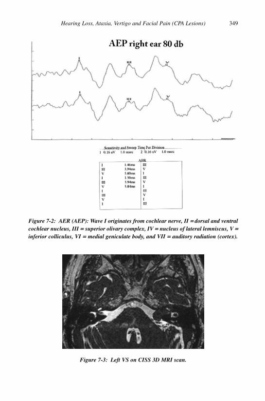
Hearing Loss, Ataxia, Vertigo and Facial Pain (CPA Lesions) 349
Figure 7-2: AER (AEP): Wave I originates from cochlear nerve, II == dorsal and ventralcochlear nucleus, III == superior olivary complex, IV == nucleus of lateral lemniscus, V ==inferior colliculus, VI == medial geniculate body, and VII == auditory radiation (cortex).
Figure 7-3: Left VS on CISS 3D MRI scan.
b1009_Chapter-07.qxd 11/12/2010 10:50 AM Page 349

350 Chapter 7
Figure 7-4: Right VS on T1-weighted MRI before contrast (a). The VS is slightlyhypointense and globular in shape.
Figure 7-5: Same right VS after IV gadolinium, note VS enhances homogenously.
Figure 7-6: VS with gadolinium demonstrating cystic degeneration.
unless there was cystic degeneration or haemorrhage (Figures 7-4to 7-7).
2- CT scan: Fine slice CT can reveal large VS or enlargement of theinternal auditory meatus (IAM) (Figures 7-8 and 7-9). However
b1009_Chapter-07.qxd 11/12/2010 10:50 AM Page 350

Hearing Loss, Ataxia, Vertigo and Facial Pain (CPA Lesions) 351
Figure 7-7: Large right VS slightly hypointense with high signal posteriorly due tohaemorrhage.
Figure 7-8: CT scan demonstrating enlargement of the left IAM (arrow).
Figure 7-9: CT scan demonstrating large right VS.
the sensitivity of CT is very low and CT nowadays only used toidentify the jugular bulb to avoid a high one during surgery.
7-1-7 How to recognise VS, CPA meningioma and epidermoid of CPA?
Table 7-1 summarises the difference between VS and CPA meningiomaand CPA epidermoid cyst.
b1009_Chapter-07.qxd 11/12/2010 10:50 AM Page 351

352 Chapter 7
Table 7-1: The difference between VS, CPA meningioma and CPA epidermoid
Diagnosis VS CPA meningioma CPA epidermoid
Location Centred to IAM Eccentric to IAM Anterior/posterior tobrain stem
Bone changes Enlarges IAM Hyperostosis Bone erosionoccasionally
Shape Spherical or ice Hemispherical or en Irregular shape andcream shaped plaque (Figure 7-10) dumbbell(Figure 7-4) (Figure 7-11)
Appearance Isodense on CT and Hyperdense and more Hypodenselower signal on likely to showT1 (Figure 7-4) calcification
Enhancement Strongly enhance Moderate None(Figure 7-5) enhancement
Dural tail None Often present None
Figure 7-10: MRI with contrast of CPA meningioma, note location eccentric to IAM,hemispherical shape, wide dural base with small tail.
Figure 7-11: MRI scan (CISS) of an epidermoid cyst in the right CPA region.
b1009_Chapter-07.qxd 11/12/2010 10:50 AM Page 352

7-1-8 How to manage VS?
The management of VS is close surveillance, microsurgical removal orstereotactic radiosurgery.
1- Conservative radiological surveillance is justified at least in the first12 months after diagnosis in small VS and has become more popularas many studies have demonstrated that VS are slow growing and mayshrink in size spontaneously.8–10 Surveillance should be abandoned infavour of intervention if the tumour grew by 2 mm in the first year.9,10
Another group recommended intervention for tumours of 15 mm orbigger and radiological surveillance could be stopped if tumours wereunchanged after five years.11
2- Microsurgery is recommended in tumours over 3 cm in diameter andin the presence of brain stem compression and hydrocephalus.Surgical approaches of CPA lesions vary depending on the pathologicentity as well as the size and involvement of adjacent structures.While complete excision is planned for most cases, the intimateinvolvement of surrounding structures may impose unwarranted mor-bidity if complete excision is attempted. Meningiomas are excisedcompletely more readily than epidermoids, whereas adequate therapyfor arachnoid cysts is drainage. Standard approaches to the CPAinclude the translabyrinthine, suboccipital (retrosigmoid = RS), ormiddle fossa (MCF) craniotomies. The choice of approach is based onspecific location and hearing status. Occasionally, these craniotomiescan be combined or performed in addition to an infratemporal fossadissection for larger tumours. In a large series of 1000 VS, a group ofmultidisciplinary team achieved total excision in 97.9%, with serv-iceable hearing in 47%, good eye closure in 79%, facial weakness in48% and mortality of 1.1%.12 In small tumours < 3 cm in diameternormal facial function had been preserved in more than 90%, hearingwas preserved in 57% and mortality was 0.16%. If one of the goals ofsurgery was to preserve hearing then the retrosigmoid (RS) or middlecranial fossa (MCF) approaches would be preferred. If the VS wasmainly in the CPA, the RS approach would be preferable and for intra-canalicular VS the MCF approach would be preferred. Otherwise the
Hearing Loss, Ataxia, Vertigo and Facial Pain (CPA Lesions) 353
b1009 Problem Based Neurosurgery
b1009_Chapter-07.qxd 11/12/2010 10:50 AM Page 353

translabrynthine approach is preferred by some teams althoughdrilling the pertrous bone can take significantly longer time than theRS approach. The largest reported series of 1000 surgically treated VSat the time of discharge from the hospital, 73% of the patients in thisseries had satisfactory facial nerve function with complete eye clo-sure; 59% showed good function (House-Brackmann (HB) Grades 1and 2), and 14% with HB Grade 3 were expected to recover withinweeks to months. In cases of nerve discontinuity or loss of the facialnerve stump at the brain stem, it is recommended that reconstructionbe performed immediately by transplantation or by nerve reanimationwith a donor nerve, respectively. Cochlear nerve deafness can be pre-vented in increasing numbers by increasing the rates of anatomic(68%) and functional (39%) nerve preservation. The RS approach isthe only one that enables hearing preservation regardless of tumoursize. Good pre-operative hearing and small tumour sizes arefavourable factors. The mortality rate was 1.1% in this study. It maybe lowered if patients with severe pre-operative morbidity do notundergo surgery or if they are not transferred from other centres at alast desperate moment but at a time when surgery still offers a realis-tic lifesaving chance. Cystic schwannomas require special surgicaland post-surgical attention, because they are more dangerous toremove with regard to brain stem and facial nerve integrity and aremore likely to lead to haemorrhage in the acute and subacute post-operative periods.
3- Stereotactic radiosurgery (SRS): SRS had become a universallyaccepted treatment option for small VS (< 3 cm in diameter). Ninety-four per cent tumour control rate, 77% normal facial preservation rateand 51% hearing preservation rate was reported with SRS.13 In viewof the natural history of VS not changing or shrinking in size in themajority of cases8–10 the reported tumour control rate may be mis-leading. However, the lack of mortality remains attractive to patientsand the small risk of malignant transformation may be a persuadingfactor to other patients to favour other options.
354 Chapter 7
b1009 Problem Based Neurosurgery
b1009_Chapter-07.qxd 11/12/2010 10:50 AM Page 354

Your personal notes:
.....................................................................................................................
.....................................................................................................................
.....................................................................................................................
.....................................................................................................................
.....................................................................................................................
.....................................................................................................................
.....................................................................................................................
.....................................................................................................................
.....................................................................................................................
.....................................................................................................................
Hearing Loss, Ataxia, Vertigo and Facial Pain (CPA Lesions) 355
b1009 Problem Based Neurosurgery
b1009_Chapter-07.qxd 11/12/2010 10:50 AM Page 355

Problem 7-2: Facial pain and trigeminal neuralgia. How tomanage a patient presenting with facial pain?
Not every patient presenting withfacial pain has trigeminal neuralgia(TN). Facial pain can be the onlypresentation of invasive skull basetumours and you need to keep this inmind. As a result in current medicalpractice it would be advisable thatthese patients have at least one MRIscan of the head and the skull basebefore merely diagnosing TN.
PCS7-2-1:
A 49-year-old female presented with lancinating pain in the right side ofher face for two weeks. She was unable to sleep, eat, laugh or talk becauseof the pain. There were no other features. She had several episodes in thepast since 2004. Her pain was sharp and stabbing in nature and scored 10on VAS (Visual Analogue Scale). There were no relieving factors and noobvious triggers apart from laughing, eating and talking. She was receiv-ing carbamazepine 100 mg tid. The family doctor tried paracetamol,codeine, diazepam, and Ibuprofen without any lasting effect. She had nodrug allergies. Neurological and systemic examinations were normal.
7-2-2 What is the differential diagnosis of facial pain?
1- Classical or idiopathic trigeminal neuralgia (ITN).2- Symptomatic or Atypical TN (ATN) which is trigeminal neuralgia
with unusual features, e.g. facial numbness.3- Tempero-mandibular pain arising from TM Joint.4- Jaw claudication as a result of temporal (giant cell) arteritis.5- Toothache arising from tooth related infections, e.g. abscess.6- Maxillary sinusitis.
356 Chapter 7
b1009 Problem Based Neurosurgery
Problem based toolkit:Atypical Trigeminal neuralgiaClassical Trigeminal neuralgiaFacial painMicrovascular decompressionPercutaneous rhizotomy
b1009_Chapter-07.qxd 11/12/2010 10:50 AM Page 356

When considering an underlying pathology, think of:
i- CPA lesions: Vestibular schwannoma, epidermoid, facial schwan-noma and meningioma.
ii- MCF tumours: Trigeminal neuroma, and cavernous sinus meningioma.iii- Skull base tumours: Meningioma, chordoma, invasive pituitary
adenoma, metastasis, and nasopharyngeal carcinoma.iv- Demyelination.v- TMJ arthritis.
vi- Temporal giant cell arteritis.vii- Maxillary conditions: Sinusitis and tumours.
viii- Tooth abscess.
7-2-3 How to manage a patient with facial pain?
This patient had already been investigated with MRI scan to rule out anunderlying pathology and vascular compression (Figure 7-12). The MRIscan ruled out MS, CPA lesion (Figures 7-13 and 7-14), middle cranialfossa lesions (Figures 7-15 and 7-16) and skull base lesion (Figures 7-17to 7-19). It was confirmed vascular compression at surgery.
Hearing Loss, Ataxia, Vertigo and Facial Pain (CPA Lesions) 357
b1009 Problem Based Neurosurgery
Figure 7-12: MRI inversion recovery (CISS) revealing vascular compression (arrow) oftrigeminal nerve presenting with ITN.
b1009_Chapter-07.qxd 11/12/2010 10:50 AM Page 357

358 Chapter 7
Figure 7-13: MRI (CISS) demonstrating PCF epidermoid cyst (A) in a 31-year-oldmale presenting with ATN.
Figure 7-14: CT scan with contrast demonstrating clival meningioma (F) in a 35-year-old presenting with mild hemiparesis and ATN.
b1009_Chapter-07.qxd 11/12/2010 10:50 AM Page 358

Hearing Loss, Ataxia, Vertigo and Facial Pain (CPA Lesions) 359
Figure 7-15: MRI (T2) demonstrating MCF epidermoid cyst (B) in a 40-year-oldfemale presenting with ATN.
Figure 7-16: MRI (T1 with contrast) demonstrating MCF meningioma (E) in a65-year-old female presenting with ATN and partial III nerve palsy.
b1009_Chapter-07.qxd 11/12/2010 10:50 AM Page 359

360 Chapter 7
Figure 7-17: MRI with contrast demonstrating skull base invasive chordoma (C) in a45-year-old man presenting with ATN and partial III nerve palsy.
Figure 7-18: MRI (T2) demonstrating fibrous dysplasia of the skull base (D) in a30-year-old male presenting with ATN and headaches.
b1009_Chapter-07.qxd 11/12/2010 10:50 AM Page 360

7-2-4 What is the epidemiology of ITN?
ITN or tic douloureux is a neuropathic disorder of one or both of thetrigeminal nerves. It is relatively rare and affects around four to five per100,000 of the population and is twice as common in females.14 Its inci-dence increases with age and is rare among people below the age of40 years. It is characterised by sudden, lancinating, severe facial pain thatis often described as “stabbing” or “shooting”, which lasts for minutes ata time. It remits and relapses and often worsens over time. It is often madeworse by chewing, laughing, shaving, and exposure to cold wind.
7-2-5 What is TN?
Trigeminal neuralgia (also known as tic douloureux) has been defined bythe International Association for the Study of Pain as “sudden, usually uni-lateral, severe brief stabbing recurrent pains in the distribution of one ormore branches of the fifth cranial nerve”.15 Usually the second (V2 maxil-lary) and third (V3 mandibular) divisions are more affected than the first
Hearing Loss, Ataxia, Vertigo and Facial Pain (CPA Lesions) 361
b1009 Problem Based Neurosurgery
Figure 7-19: MRI (T2) demonstrating cholesterol cyst (G) in the petrous apex in apatient presenting with ATN.
b1009_Chapter-07.qxd 11/12/2010 10:50 AM Page 361

(V1 ophthalmic), with only around 3% of cases occurring bilaterally. Othersymptoms include tingling or numbness in the face before the developmentof pain and spasms that can last from a few seconds to minutes each time.
7-2-6 What are the classification of TN?
The International Headache Society has classified TN into classical (ITN)and symptomatic trigeminal neuralgia (ATN).16 Included in ITN are all thecases without an established aetiology such as idiopathic and those with vas-cular compression. The majority of cases within this group are classified asidiopathic. However there is increasing evidence that 80–90% of these casesare caused by an aberrant loop of artery or vein compressing the trigeminalnerve close to its root entry into the pons.14 Such evidence includes: an aber-rant loop of the artery or vein being found compressing the trigeminal nerveof patients at surgery; demyelination of the trigeminal nerve; after surgery fordecompression most patients experience long term relief and improvementof any sensory defects.17,18 A diagnosis of ATN is made secondary to a num-ber of different causes, including tumours, multiple sclerosis and structuralabnormalities of the skull base as described above.19 Approximately 1–5% ofpatients suffering from MS will develop trigeminal neuralgia.17
7-2-7 What is the diagnostic criteria and natural history of TN?
Attacks occur infrequently and can last from several days to weeks at atime, however when these attacks settle the patient can become pain freefor months at a time. During severe attacks the patient can experiencespasms of pain hundreds of times a day. For the majority of trigeminalcases the diagnosis is clinical. The International Headache Society has putforward the following diagnostic criteria for trigeminal neuralgia:
A- Paroxysmal attacks of pain lasting from a fraction of a second to twominutes, affecting one or more division of the trigeminal nerve andfulfilling criteria B and C.
B- Pain has at least one of the following characteristics:
a- Intense, sharp, superficial or stabbing.b- Precipitated from trigger areas or by trigger factors.
362 Chapter 7
b1009 Problem Based Neurosurgery
b1009_Chapter-07.qxd 11/12/2010 10:50 AM Page 362

C- Attacks are stereotyped in the individual patient.D- There is no clinically evident neurological deficit.E- Not attributed to another disorder.
Trigger factors stated in the above criteria include certain actions ormovements such as: chewing, speaking, washing the face, tooth-brushing,cold winds or even touching a specific “trigger spot” on the face.Frequently the pain described in many cases does not exactly fit these cri-teria, for example, a constant ache between the paroxysms or mild sensoryloss in the face. These cases have been named “atypical” or mixed trigem-inal neuralgia and are usually more likely to be due to symptomaticdisease rather than classical trigeminal neuralgia.
A review in the European Journal of Neurology18 recommended thatfor patients with non-trigeminal neurological symptoms, routine imagingshould be considered to identify ATN. Electrophysiological studies mayalso prove useful for distinguishing ATN from ITN. Five studies looked atthe accuracy of using electrophysiological testing. This was found to berelatively high, especially when using trigeminal reflexes. Of particularnote from the review was the lack of evidence to either support or refutethe usefulness of MRI in identifying ITN patient.
7-2-8 How to manage ITN?
The main aim of treating ITN is to control the pain. Drug therapy is thefirst line treatment, commonly using anticonvulsants. From the limitedevidence available, carbamazepine is the drug of choice20,21 and proveseffective in most patients (and also may confirm the diagnosis).However many patients develop side effects including drowsiness,ataxia, nausea and constipation. If the patient responds and goes intoremission, the dosage can be gradually lowered. If carbamazepine wasintolerable or ineffective there are a number of second line drugs,although the evidence is weaker for their usage. Oxcarbazepine is foundto be better tolerated (fewer side effects) and being a pro-drug of carba-mazepine, is often the chosen alternative. Gabapentin is also generallyeffective in dealing with neuropathic pain; however the evidence is lack-ing of its specific use in treating trigeminal neuralgia. Other drugs which
Hearing Loss, Ataxia, Vertigo and Facial Pain (CPA Lesions) 363
b1009 Problem Based Neurosurgery
b1009_Chapter-07.qxd 11/12/2010 10:50 AM Page 363

may benefit when pain control is limited include baclofen, lamotrigineand phenytoin.
If medical therapy does not work then a review of the diagnosisshould be made. Surgical options should then be considered if there is per-sistence of the pain. Surgical interventions for trigeminal neuralgia can beclassified according to the principal treatment target, splitting the treat-ment options into four groups: peripheral techniques targeting portions ofthe trigeminal nerve distally to the ganglion; percutaneous ganglion tech-niques targeting specifically the ganglion; gamma knife radiosurgerytargeting the trigeminal root and posterior fossa vascular decompressiontechniques. Peripheral techniques involve blocking or destroying portionsof the trigeminal nerve, using substances such as alcohol or phenol.Studies have shown that these techniques only provide temporary relief,with 50% of patients having recurrence of pain after one year. The mor-bidity of these procedures is low. Percutaneous procedures on theganglion itself entail penetrating the foramen ovale with a cannula andthen controlled compression, chemical or thermocoagulation of thetrigeminal ganglion or root.22 The methods include: thermal (radiofre-quency thermocoagulation), chemical (injection of glycerol, which iscorrosive to the nerve fibres thus mildly injuring the nerve enough to hin-der the pain signals) or mechanical (compression by a balloon inflatedinto Merkel’s cave). Percutaneous techniques have been particularly help-ful in treating elderly patients for whom surgery is contraindicated due toco-existing health conditions. These procedures are usually very effectiveat providing pain relief (around 90%). This figure drops though to 72%after three years for patients who underwent the thermal method.22 Severalside effects have been reported with this group with almost some patientsexperiencing sensory loss. Around 4–6% developed dysaesthesias/anaesthesia dolorosa (troublesome sensory disturbance) as a result of thedestructive technique. Post-operatively around 12% complain of discom-fort, which dramatically increases with the balloon technique, with aroundhalf of all patients suffering temporary masticator problems.23 The mor-tality of these procedures, however, is extremely low. Gamma kniferadiosurgery is the only technique which involves aiming a focused beamof radiation at the point where the trigeminal nerve leaves the brain (pos-terior fossa), in order to destroy it. Initially there was quite a high
364 Chapter 7
b1009 Problem Based Neurosurgery
b1009_Chapter-07.qxd 11/12/2010 10:50 AM Page 364
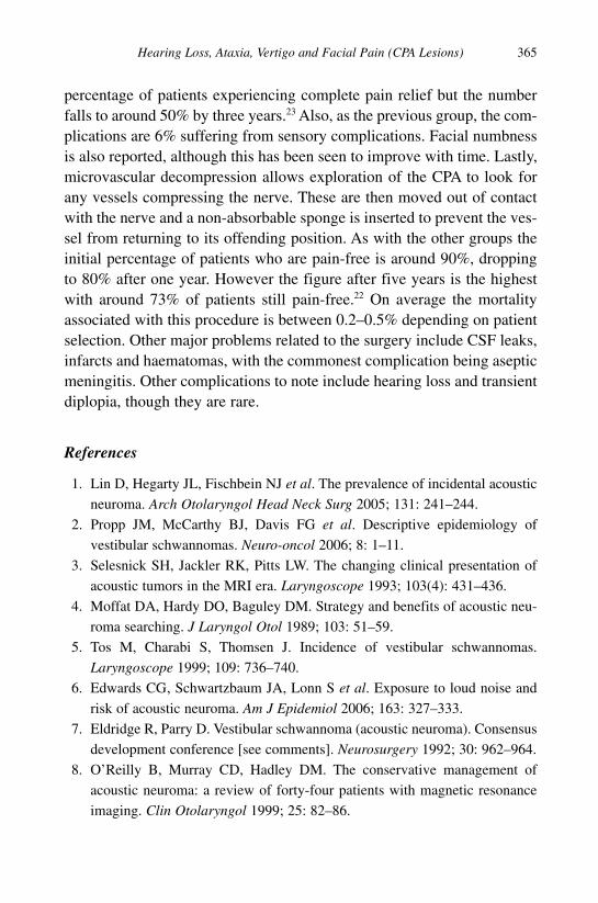
percentage of patients experiencing complete pain relief but the numberfalls to around 50% by three years.23 Also, as the previous group, the com-plications are 6% suffering from sensory complications. Facial numbnessis also reported, although this has been seen to improve with time. Lastly,microvascular decompression allows exploration of the CPA to look forany vessels compressing the nerve. These are then moved out of contactwith the nerve and a non-absorbable sponge is inserted to prevent the ves-sel from returning to its offending position. As with the other groups theinitial percentage of patients who are pain-free is around 90%, droppingto 80% after one year. However the figure after five years is the highestwith around 73% of patients still pain-free.22 On average the mortalityassociated with this procedure is between 0.2–0.5% depending on patientselection. Other major problems related to the surgery include CSF leaks,infarcts and haematomas, with the commonest complication being asepticmeningitis. Other complications to note include hearing loss and transientdiplopia, though they are rare.
References
1. Lin D, Hegarty JL, Fischbein NJ et al. The prevalence of incidental acoustic
neuroma. Arch Otolaryngol Head Neck Surg 2005; 131: 241–244.
2. Propp JM, McCarthy BJ, Davis FG et al. Descriptive epidemiology of
vestibular schwannomas. Neuro-oncol 2006; 8: 1–11.
3. Selesnick SH, Jackler RK, Pitts LW. The changing clinical presentation of
acoustic tumors in the MRI era. Laryngoscope 1993; 103(4): 431–436.
4. Moffat DA, Hardy DO, Baguley DM. Strategy and benefits of acoustic neu-
roma searching. J Laryngol Otol 1989; 103: 51–59.
5. Tos M, Charabi S, Thomsen J. Incidence of vestibular schwannomas.
Laryngoscope 1999; 109: 736–740.
6. Edwards CG, Schwartzbaum JA, Lonn S et al. Exposure to loud noise and
risk of acoustic neuroma. Am J Epidemiol 2006; 163: 327–333.
7. Eldridge R, Parry D. Vestibular schwannoma (acoustic neuroma). Consensus
development conference [see comments]. Neurosurgery 1992; 30: 962–964.
8. O’Reilly B, Murray CD, Hadley DM. The conservative management of
acoustic neuroma: a review of forty-four patients with magnetic resonance
imaging. Clin Otolaryngol 1999; 25: 82–86.
Hearing Loss, Ataxia, Vertigo and Facial Pain (CPA Lesions) 365
b1009 Problem Based Neurosurgery
b1009_Chapter-07.qxd 11/12/2010 10:50 AM Page 365

9. Smouha EE, Yoo M, Mohr K et al. Conservative management of acoustic
neuroma: a meta analysis and proposed treatment algorithm. Laryngoscope
2005; 115(3): 450–454.
10. Nedzelski JM, Schessel DA, Pfleiderer A et al. Conservative management of
acoustic neuromas. Otolaryngol Clin N Am 1992; 25(3): 691–705.
11. Stangerup SE, Caye-Thomasen P, Tos M et al. The natural history of vestibu-
lar schwannoma. Otol Neurotol 2006; 27(4): 547–552.
12. Samii M, Matthies C. Management of 1000 vestibular schwannomas
(acoustic neuromas): surgical management and results with an emphasis on
complications and how to avoid them. Neurosurgery 1997; 40: 248–262
http://www.c3.hu/~mavideg/ns/samii1-97.html.
13. Kondziolka D, Lunsford LD, McLaughlin MR et al. Long-term outcomes
after radiosurgery for acoustic neuromas. N Engl J Med 1998; 339:
1426–1433.
14. Bennetto L, Patel NK, Fuller G. Trigeminal neuralgia and its management.
Br Med J 2007; 332: 201–205.
15. Zakrzewska JJM, Linskey ME. Neurosurgical interventions for the treatment
of classical trigeminal neuralgia (Protocol). Cochrane Database Syst Rev
2008, Issue 3.
16. Headache Classification Subcommittee of the International Headache
Society. The International Classification of Headache Disorders, 2nd edition.
Cephalalgia 2004; 24(Suppl 1): 9–160.
17. Nurmikko TJ, Eldridge PR. Trigeminal neuralgia — pathophysiology, diag-
nosis and current treatment. Br J Anaesth 2001; 87: 117–132.
18. Love S, Coakham HB. Trigeminal neuralgia: pathology and pathogenesis.
Brain 2001; 124: 2347–2360.
19. Nurmikko TJ, Eldridge PR. Trigeminal neuralgia — pathophysiology, diag-
nosis and current treatment. Br J Anaesth 2001; 87: 117–132.
20. Wiffen P, Collins S, McQuay H et al. Anticonvulsant drugs for acute and
chronic pain. Cochrane Database Syst Rev 2005, Issue 3.
21. Wiffen PJ, McQuay HJ, Edwards JE et al. Gabapentin for acute and chronic
pain. Cochrane Database Syst Rev 2005, Issue 3.
22. Haridas A, Mathewson C, Eljamel S. Long-term results of 405 refractory
trigeminal neuralgia surgeries in 256 patients. Zentralbl Neurochir 2008;
69(4): 170–174.
366 Chapter 7
b1009 Problem Based Neurosurgery
b1009_Chapter-07.qxd 11/12/2010 10:50 AM Page 366

23. Gronseth G, Cruccu G, Alksne J et al. Practice parameter: the diagnostic
evaluation and treatment of trigeminal neuralgia (an evidence-based review):
report of the Quality Standards Subcommittee of the American Academy of
Neurology and the European Federation of Neurological Societies.
Neurology 2008; 71: 1183–1190.
Your personal notes:
.....................................................................................................................
.....................................................................................................................
.....................................................................................................................
.....................................................................................................................
.....................................................................................................................
.....................................................................................................................
.....................................................................................................................
.....................................................................................................................
.....................................................................................................................
.....................................................................................................................
Hearing Loss, Ataxia, Vertigo and Facial Pain (CPA Lesions) 367
b1009 Problem Based Neurosurgery
b1009_Chapter-07.qxd 11/12/2010 10:50 AM Page 367

b1009_Chapter-07.qxd 11/12/2010 10:50 AM Page 368
This page intentionally left blankThis page intentionally left blank

Chapter 8: Tremor (Parkinson’s Diseaseand Dystonia)
Problem 8-1: Tremor and Parkinson’s disease. How to manage apatient presenting with tremor?
Tremor and movement disorders aresymptoms of other neurological disor-ders. The underlying pathology isoften progressive and treatment isoften symptomatic. Some of these dis-orders may respond to surgery.
PCS8-1-1:
A 75-year-old man presented with a two-year history of right-sided handtremor and freezing of speech. His gait was satisfactory at that time andhe was given Levodopa and benserazide hydrochloride. His symptomsgradually deteriorated and two and a half years after his initial presenta-tion he developed right side leg tremor and bradykinesia (slowness ofmovement). Selegiline and propranolol were added to his medications.
He deteriorated gradually and three years after his initial presentationhe underwent a left-sided deep brain stimulation (DBS) procedure. Post-operatively he developed dysphasia which was very brief and resolvedspontaneously. One month after his DBS implantation he developed amoderate degree left sided tremor which was not apparent previously. Hisright sided tremor was under excellent control by the left sided DBS. Oneyear after his DBS surgery his Parkinson’s disease had progressed and hismovements became slower although he still had very good control of theright sided tremor. One and a half years after his left side DBS, the tremor,rigidity and bradykinesia on the left had become worse so a right sideDBS was inserted (Figure 8-1).
369
b1009 Problem Based Neurosurgery
Problem based tool box:TremorEssential tremorRubral tremorParkinson’s diseaseParkinsonism
b1009_Chapter-08.qxd 11/12/2010 10:50 AM Page 369

8-1-2 What is the differential diagnosis of tremor?
The differential diagnosis in a patient presenting with tremor includes:
• Tremor of Parkinson’s disease (PD).• Essential tremor (ET).• Rubral tremor (RT).• Dystonia.• Other movement disorders.
8-1-3 What is Parkinson’s disease (PD) and Parkinsonism?
PD is a movement disorder characterised by muscle rigidity, tremor, andbradykinesia and, in extreme cases, a loss of physical movement (akine-sia). The primary symptoms are the results of decreased stimulation of themotor cortex by the basal ganglia, normally caused by insufficient forma-tion and action of dopamine. Secondary symptoms may include high levelcognitive dysfunction and subtle language problems. PD is the most com-mon cause of chronic progressive Parkinsonism, a syndrome of tremor,rigidity, bradykinesia and postural instability. While many forms ofparkinsonism are “idiopathic”, “secondary” cases may result from drugside effects (metclopromide, chlorpromazine and haloperidol), head
370 Chapter 8
b1009 Problem Based Neurosurgery
Figure 8-1: Plain radiograph of the skull demonstrating bilateral DBS (1). Note theDBS leads are connected at the scalp level to lead extenders tunneled subcutaneously inthe neck to reach an implantable pulse generator (IPG) located in the left subclavicularregion.
b1009_Chapter-08.qxd 11/12/2010 10:50 AM Page 370

trauma, or other medical disorders. The disease is named after JamesParkinson, who made a detailed description of PD in 1817.
PD prevalence is estimated at being from 120–180 per 100,000.
8-1-4 How to treat PD?
At present, there is no cure for PD, but medications or surgery can pro-vide relief from symptoms.
The most widely used form of treatment is L-dopa in various forms.L-dopa is transformed into dopamine in the dopaminergic neurons byL-aromatic amino acid decarboxylase. However, only 1–5% of L-DOPAenters the dopaminergic neurons. The remaining L-DOPA is oftenmetabolised to dopamine elsewhere, causing a wide variety of sideeffects. Due to feedback inhibition, L-dopa results in a reduction in theendogenous formation of L-dopa, and eventually becomes counterpro-ductive. Carbidopa and benserazide are dopa decarboxylase inhibitors.They help prevent the metabolism of L-dopa before it reaches thedopaminergic neurons and are generally given as combination prepara-tions of carbideopa/levodopa (Sinemet) and benserazide/levodopa(Madopar). There are also controlled release versions of Sinemet andMadopar that spread out the effect of the L-dopa. The second type ofdrug treatment includes COMT inhibitors: (Tolcapone) prolonging theeffects of L-dopa, and so has been used to complement L-dopa.However, it can cause liver failure. Dopamine agonists are the secondline anti-PD drug treatment. Bromocriptine, pergolide, pramipexole,ropinirole, piribedil, cabergoline, apomorphine, and lisuride are moder-ately effective. These have their own side effects including somnolence,hallucinations or insomnia. Several forms of dopamine agonists havebeen linked with a markedly increased risk of addictive gambling.Dopamine agonists initially act by stimulating some of the dopaminereceptors. However, they cause the dopamine receptors to becomeprogressively less sensitive; thereby increasing the symptoms of PD.Dopamine agonists can be useful for patients experiencing on-off fluctu-ations and dyskinesias as a result of high doses of L-dopa. Apomorphinecan be administered via subcutaneous injection using a small pumpwhich is carried by the patient. A low dose is automatically administered
Tremor (Parkinson’s Disease and Dystonia) 371
b1009 Problem Based Neurosurgery
b1009_Chapter-08.qxd 11/12/2010 10:50 AM Page 371
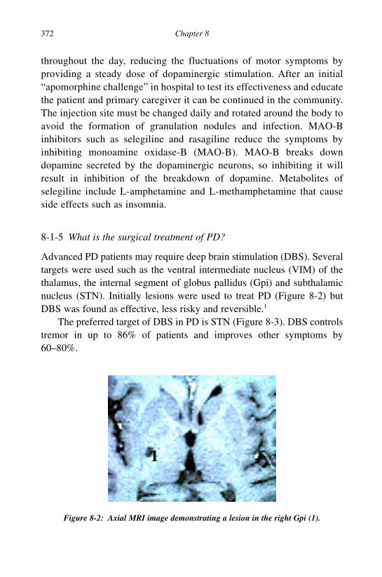
throughout the day, reducing the fluctuations of motor symptoms byproviding a steady dose of dopaminergic stimulation. After an initial“apomorphine challenge” in hospital to test its effectiveness and educatethe patient and primary caregiver it can be continued in the community.The injection site must be changed daily and rotated around the body toavoid the formation of granulation nodules and infection. MAO-Binhibitors such as selegiline and rasagiline reduce the symptoms byinhibiting monoamine oxidase-B (MAO-B). MAO-B breaks downdopamine secreted by the dopaminergic neurons, so inhibiting it willresult in inhibition of the breakdown of dopamine. Metabolites ofselegiline include L-amphetamine and L-methamphetamine that causeside effects such as insomnia.
8-1-5 What is the surgical treatment of PD?
Advanced PD patients may require deep brain stimulation (DBS). Severaltargets were used such as the ventral intermediate nucleus (VIM) of thethalamus, the internal segment of globus pallidus (Gpi) and subthalamicnucleus (STN). Initially lesions were used to treat PD (Figure 8-2) butDBS was found as effective, less risky and reversible.1
The preferred target of DBS in PD is STN (Figure 8-3). DBS controlstremor in up to 86% of patients and improves other symptoms by60–80%.
372 Chapter 8
b1009 Problem Based Neurosurgery
Figure 8-2: Axial MRI image demonstrating a lesion in the right Gpi (1).
b1009_Chapter-08.qxd 11/12/2010 10:50 AM Page 372

8-1-6 What are the indications for surgery in PD?
The indications for surgery in PD are: advanced PD that failed medicaltreatment either because of side effects or lack of sustainable response, PDmust be L-dopa responsive and the patient does not suffer from severe cog-nitive dysfunction, severe depression or uncorrectable bleeding disorder.
8-1-7 What is essential tremor (ET)?
ET is familial progressive neurological disorder manifesting with tremorof arms that is apparent during voluntary movements such as eating andwriting. This type of tremor is often referred to as “kinetic tremor”. Thetremor may affect the head (neck), jaw, speech and other body regions.Women are more likely to develop head tremor than men. Other types oftremor: postural tremor of the outstretched arms, intentional tremor of thearms and rest tremor in the arms may also occur. Some patients may haveunsteadiness and problems with gait and balance. Recent studies havedemonstrated that old-onset ET may be associated with an increased riskof developing dementia.2 ET is one of the most common neurological dis-eases and the second most common tremor after physiological tremor in
Tremor (Parkinson’s Disease and Dystonia) 373
b1009 Problem Based Neurosurgery
Figure 8-3: Coronal MRI image of basal ganglia with the stereotactic atlas superim-posed demonstrating the STN (1), Gpi (2) and VIM (3).
b1009_Chapter-08.qxd 11/12/2010 10:50 AM Page 373

the population, with a prevalence of approximately 4% of those over40 years of age. Although ET is often mild, patients with severe tremorhave difficulty performing many of their routine activities of daily living.
8-1-8 How to treat ET?
There are no effective drugs against ET, however patients often observedthat the tremor is significantly reduced with drinking alcohol, and it canalso be reduced by propranolol. Propranolol improves tremor in 50–60%of patients and 15–20% of responders develop tolerance. Primidone hasalso been used in ET with similar response as propranolol. However, aswith propranolol, primidone is most beneficial for essential hand-tremor,and efficacy against head and speech tremor is variable. The duration ofeffect after a single dose is approximately 24 hours. As with propranolol,after 12 months of therapy approximately 10–15% of responders maydevelop tolerance to primidone.
8-1-9 What is the surgical treatment of ET?
Vim-DBS is effective against ET in 80–90% of patients (Figure 8-4).Patients should be considered for surgery if their tremor was severe enoughto interfere with activities of daily living, and they do not suffer from severecognitive decline, severe depression or uncorrectable bleeding disorders.
374 Chapter 8
b1009 Problem Based Neurosurgery
Figure 8-4: Photograph of Archimedes spiral in a patient with ET: (A) before the DBSwas switched on and (B) after the DBS was switched on.
b1009_Chapter-08.qxd 11/12/2010 10:50 AM Page 374

8-1-10 What is rubral tremor (RT)?
RT is characterised by a slow coarse tremor at rest that is exacerbated bypostural adjustments and by guided voluntary movements. Lesions of thesuperior cerebellar peduncle, midbrain tegmentum or posterior part of thethalamus may cause this peculiar tremor, and it is probable that lesions ofthe red nucleus itself are not crucial for its production. RT often compli-cates MS and there is no effective drug treatment for it. The samemedications used for ET can be used and VIM-DBS can also be used withsome success.
Your personal notes:
.....................................................................................................................
.....................................................................................................................
.....................................................................................................................
.....................................................................................................................
.....................................................................................................................
.....................................................................................................................
.....................................................................................................................
.....................................................................................................................
.....................................................................................................................
.....................................................................................................................
Tremor (Parkinson’s Disease and Dystonia) 375
b1009 Problem Based Neurosurgery
b1009_Chapter-08.qxd 11/12/2010 10:50 AM Page 375

Problem 8-2: Movement disorders and dystonia. How to manage apatient with dystonia?
Dystonia should be distinguished fromother movement disorders such astremor, and dyskinesia as their manage-ment is different.
8-2-1 What is dystonia?
Dystonia is characterised by sustained muscle contractions causing twist-ing and repetitive movements or abnormal postures. The disorder may behereditary or caused by other factors such as birth-related or other physi-cal trauma, infection, poisoning (lead) or reaction to drugs.
8-2-2 How to classify dystonia?
Dystonia can be generalised (GD), focal (FD) or segmental (SD) in dis-tribution. The incidence of GD is about one in 2,000,000. Examples of FDinclude:
1- Cervical dystonia causes the head to rotate to one side (spasmodic tor-ticollis (ST)), to pull down towards the chest, or back (retrocollis), ora combination of these postures.
2- Blepharospasm causes rapid blinking of the eyes or even their forcedclosure.
3- Focal hand dystonia (also known as musician’s or writer’s cramp)interferes with writing or playing a musical instrument by causinginvoluntary muscular contractions.
8-2-3 What causes dystonia?
The causes of dystonia are not yet known or understood; however, theyare categorised as follows:
• Primary dystonia is caused by pathology in the basal ganglia,and the GABA producing neurons. The precise cause of primary
376 Chapter 8
b1009 Problem Based Neurosurgery
Problem based tool box:DystoniaPrimary dystoniaSecondary dystonia
b1009_Chapter-08.qxd 11/12/2010 10:50 AM Page 376

dystonia is unknown. In many cases it may involve geneticpredisposition.
• Secondary dystonia is brought on by identified cause, usually involv-ing brain injury, or chemical imbalance. Some cases of (focal)dystonia are brought on after trauma, are induced by certain drugs(tardive dystonia), or the result of (Wilson’s disease).
Environmental and task-related factors are suspected to trigger the devel-opment of focal dystonias because they appear disproportionately inindividuals who perform high precision hand movements such as musi-cians, engineers, architects and artists.
8-2-4 How to treat dystonia?
Treatment of dystonia has been limited to minimising the symptoms ofthe disorder as there is yet no successful treatment for its cause.Reducing movements that trigger or worsen dystonic symptoms pro-vides some relief, as does reducing stress, getting plenty of rest,moderate exercise, and relaxation techniques. Various treatments focuson sedating brain functions or blocking nerve communications with themuscles by drugs, neuro-suppression or denervation. Drug treatment isoften ineffective. Medications that have had positive results in somepatients include: diphenhydramine, benzatropine, trihexyphenidyl,diazepam, benzatropine, and acetylcholine. Botulinum toxin injectionsinto affected muscles have proved quite successful in providing somerelief for around three to six months, depending on the kind of dysto-nia. Botox injections have the advantage of ready availability and theeffects are not permanent. There is a risk of temporary paralysis of themuscles being injected or the leaking of the toxin into adjacent musclegroups causing weakness or paralysis. The injections have to berepeated as the effects wear off and around 15% of recipients willdevelop immunity to the toxin. There is a Type A and Type B toxinapproved for treatment of dystonia. Dopamine-responsive dystonia, canbe completely treated with regular doses of L-DOPA in a form such asSinemet.
Tremor (Parkinson’s Disease and Dystonia) 377
b1009 Problem Based Neurosurgery
b1009_Chapter-08.qxd 11/12/2010 10:50 AM Page 377

8-2-5 What is the surgical treatment of dystonia?
Baclofen intrathecal infusion pump has been used to treat patients of allages exhibiting muscle spasticity along with dystonia. The pump deliversbaclofen via a catheter into the spinal thecal sac. Surgical denervation ofselected muscles may provide some relief; however, the destruction ofnerves in the limbs or neck is not reversible and should only be consid-ered in the most extreme cases. DBS has proven successful in a numberof cases of severe generalised dystonia. The target in dystonia is bilateralGpi DBS stimulation.
8-2-6 What other surgical treatments for functional disorders?
There are a number of established and emerging surgical techniques for thetreatment of functional disorders. These interventions are not curative andshould only be performed within specialist centres with appropriate infra-structure of multidisciplinary teams. They should only be performed inpatients with treatment refractory disorders who are able to consent and haverealistic understanding of the goals of therapy. These techniques include:
1- Vagus nerve stimulation (VNS) for treatment refractory epilepsy.2- VNS for treatment refractory depression.3- Subgenual cingulate DBS for treatment refractory depression.4- Anterior capsular DBS for treatment refractory obsessive compulsive
disorders (OCD).5- Anterior capsulotomy for treatment refractory OCD.6- Anterior cingulotomy for treatment refractory depression.7- Anterior thalamic DBS for treatment refractory epilepsy.
References
1. Schuurman PR, Bosch DA, Bossuty PMM et al. A comparison of continuous
thalamic stimulation and thalamotomy for suppression of severe tremor.
N Engl J Med 2000; 342: 461–468.
2. Bermejo-Pareja F, Louis ED, Benito-Leon J. Risk of incident dementia in
essential tremor: a population-based study. Mov Disord 2007; 22: 1573–1580.
378 Chapter 8
b1009 Problem Based Neurosurgery
b1009_Chapter-08.qxd 11/12/2010 10:50 AM Page 378

Your personal notes:
.....................................................................................................................
.....................................................................................................................
.....................................................................................................................
.....................................................................................................................
.....................................................................................................................
.....................................................................................................................
.....................................................................................................................
.....................................................................................................................
.....................................................................................................................
.....................................................................................................................
Tremor (Parkinson’s Disease and Dystonia) 379
b1009 Problem Based Neurosurgery
b1009_Chapter-08.qxd 11/12/2010 10:50 AM Page 379

b1009 Problem Based Neurosurgery
b1009_Chapter-08.qxd 11/12/2010 10:50 AM Page 380
This page intentionally left blankThis page intentionally left blank

Chapter 9: Para-/Tetraparesis(Spinal Compression)
Problem 9-1: Bilateral limb paresis (malignant spinalcompression). How to manage suspected malignant spinalcompression?
Patients suspected of malignant cordcompression require immediate adm-ission, investigations and appropri-ate management to prevent seriousmorbidity.
PCS9-1-1:
A 64-year-old right-handed man presented with four days history ofreduced mobility. The immobility began gradually and slowly progressed.He also complained of weakness in the right leg and reduced sensation onthe left side of his body up to the waist. He had dull aching pain betweenthe shoulder plates. He was able to feel when he wanted to pass urine andwas able to control it until it was convenient. Physical examinationrevealed that he had weakness of all muscle groups in the left lowerextremity grade 4, and 4+ on the right. His ankle and knee jerks were briskcompared to his biceps and triceps jerks and his planters were upgoing.He had impaired sensation to pain from T6 downwards on the right spar-ing the saddle area. There was no other significant history.
9-1-2 What is the differential diagnosis of bilateral leg weakness?
A patient presenting with acute gradual onset paraparesis with sensorylevel has spinal compression until proven otherwise. Abnormal lowerlimbs and normal upper limbs locate the lesion below T1 spinal cord seg-ment. The presence of brisk reflexes and upgoing planters locate the
381
b1009 Problem Based Neurosurgery
Problem based tool box:Malignant spinal compressionParaparesis Sensory levelTetraparesis
b1009_Chapter-09.qxd 11/12/2010 10:50 AM Page 381

lesion above the conus and a sensory level at T6 puts the lesion around theT6 segment. A unilateral sensory level on the right and more weakness onthe left put the lesion on the left side of the spinal cord because the painis transmitted in spinothalamic tracts that cross over to the contralateralside few segments above the level in the spinal cord and the descendingmotor fibres cross high up in the medulla oblongata. The pathological dif-ferential diagnosis would be;
• Malignant extradural spinal cord compression (MSC).• Benign spinal cord compression (BSC).• Other non-compressive causes, e.g. traverse myelitis.
9-1-3 What investigations would you do in suspected MSC?
1- Full blood count looking for anaemia or polycythemia that may beassociated with malignancy. This was normal in this patient.
2- Chest X-ray looking for primary or secondary in the lungs. The chestX-ray was clear in PCS9-1-1 above.
3- C-reactive protein (CRP) and ESR/Plasma Viscosity (PV) to see if itwas elevated indicating malignancy or infection. The CRP and PVwere elevated in the patient (PCS91-1).
4- MRI spine covering the thoracic spine to confirm or rule out the sus-pected diagnosis (Figures 9-1 and 9-2) and the rest of the spine to ruleout other metastases.
5- CT spine of the involved level to assess the integrity of the spine ifinternal fixation of the spine was planned.
PCS9-1-4:
A 78-year-old woman presented with low back pain radiating into both legsassociated with urinary incontinence of seven days duration. When she wasexamined she had bilateral leg weakness with reduced reflexes andimpaired sensation from S2 downward. The saddle area was also involved.
The differential diagnosis in this patient is the same as in problemcase scenario PCS9-1-1. However, the fact that she had reduced reflexesbilaterally means the lesion’s location is in the cauda equina rather than
382 Chapter 9
b1009 Problem Based Neurosurgery
b1009_Chapter-09.qxd 11/12/2010 10:50 AM Page 382
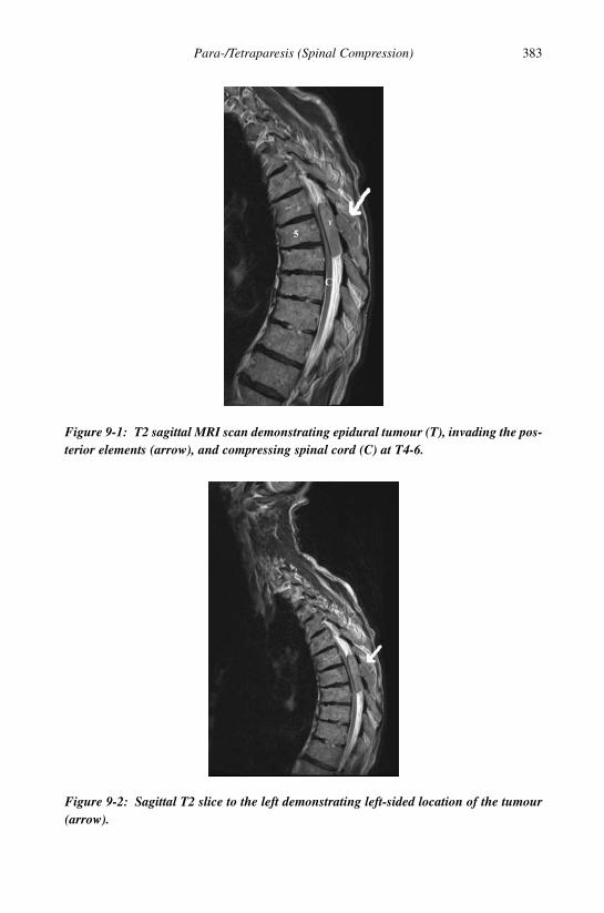
Para-/Tetraparesis (Spinal Compression) 383
b1009 Problem Based Neurosurgery
Figure 9-1: T2 sagittal MRI scan demonstrating epidural tumour (T), invading the pos-terior elements (arrow), and compressing spinal cord (C) at T4-6.
Figure 9-2: Sagittal T2 slice to the left demonstrating left-sided location of the tumour(arrow).
b1009_Chapter-09.qxd 11/12/2010 10:50 AM Page 383

affecting the spinal cord. Similarly the sensory level and pain levels sup-port this location.
Investigations required in this patient were the same as those inPCS9-1-1. The MRI of this patient (Figures 9-3 and 9-4) demonstratedmalignant cord compression in the lumbo-sacral region.
384 Chapter 9
b1009 Problem Based Neurosurgery
Figure 9-3: T2-MRI demonstrating a mass (1) destroying the sacral vertebrae andinvading the spinal canal and posterior elements.
Figure 9-4: The same patient’s T2-MRI demonstrating a second lesion (2).
b1009_Chapter-09.qxd 11/12/2010 10:50 AM Page 384

9-1-5 What is the incidence of malignant spinal compression?
Malignant spinal compression (MSC) is a surgical emergency. Improvedlife expectancy of cancer patients increased the prevalence of spinalmetastatic disease and the morbidity and mortality due to MSC had alsoincreased. Five percent of all cancer patients develop spinal extraduralmetastases, and of these 20% develop symptoms of MSC. MSC requiresearly diagnosis and urgent treatment to avoid severe neurological distur-bances. Therefore keeping a high index of suspicion and recognising earlysymptoms of MSC are paramount if patients are to have any chance ofretaining the ability to walk and sphincter control. Urgent diagnosticinvestigations, access to MRI and immediate treatment are all part ofmaintaining good quality of life of these patients.
9-1-6 How does MSC present?
MSC presents with spinal pain associated with progressive neurologicaldeficit. Neurological deficits often include progressive motor weakness ofthe limbs, progressive sensory impairment with a sensory level corre-sponding to the level of MSC and impaired bladder and bowel functions.Disorders of bladder and bowel control, and sexual function indicatedecompensation and rapid deterioration of cord function. Decompensationoccurs in the spine when the spinal cord is no longer able to deal with thepresence of tumour to maintain function. There is dysfunction of the auto-nomic nervous system. Bladder function is particularly affected when thecauda equina or S2, 3, 4 nerve roots are involved, causing urgency andhesitancy as in PCS9-1-4. Bowel dysfunction also occurs with S2, 3, 4nerve root involvement, but more often causes constipation rather thanincontinence of faeces.
T12 and T6 are the most often affected spinal levels. The mode ofonset can be insidious or acute depending upon whether bony collapse hadoccurred and occasionally MSC presents after minor trauma. Back pain isthe commonest initial symptom and is present in virtually all patients atthe time of diagnosis. Neurological symptoms usually present about six toeight weeks after the onset of spinal pain. Therefore a recent onset of spinalpain in a patient known to have malignant disease, e.g. breast cancer or a
Para-/Tetraparesis (Spinal Compression) 385
b1009 Problem Based Neurosurgery
b1009_Chapter-09.qxd 11/12/2010 10:50 AM Page 385

patient in the high risk for malignancy should be taken seriously to keepthe patient ambulant. Spinal compression can be the initial presenting fea-ture of malignant disease, e.g. lung cancer, prostate cancer, multiplemyeloma, lymphoma or any other systemic malignancy.
9-1-7 What are the stages of MSC?
MSC can be divided into three stages:
a- Neuralgic stage: Characterised by pain and paraesthesia around thetrunk at the level of compression and tight band-like sensationsaround the trunk (girdle pain), extensor plantar response, exaggera-tion of lower limb reflexes, and urinary urgency or hesitancy.
b- Hemicord syndrome: The clinical picture is atypical of Brown-Sequard Syndrome. Characterised by pain and temperature loss overaffected dermatome and also the opposite side of the body below thelesion, dorsal column dysfunction below and ipsilateral to the lesion(dissociate anaesthesia), upper motor neuron picture on the side of thelesion and urinary urgency, incontinence and hesitancy.
c- Transection stage: Characterised by the loss of all motor and sensoryfunctions below the level of the lesion and acute retention of urine.
These stages occur if the lesion is situated lateral to the spinal cord. Mostmetastatic tumours are situated laterally or anteriorly.
At the time of presentation, 80% of patients with metastases have weak-ness, 50% have sensory symptoms and 50% sphincter dysfunction. Themain sites from which metastases occur are from lung, breast and prostate.Most of these cancers spread to the spine by haematogenous routes due toa system of valveless vertebral veins which communicate freely with theintercostal and lumbar veins. Involvement of posterior spinal elements bycancers is uncommon because the vascular networks are anteriorly located.
9-1-8 How do metastases cause MSC?
Tumours can cause MSC in a number of ways. The most importantis by mechanical compression due to the expanding mass (Figure 9-4).
386 Chapter 9
b1009 Problem Based Neurosurgery
b1009_Chapter-09.qxd 11/12/2010 10:50 AM Page 386

A malignancy can also cause vertebral collapse which allows fragments ofbone to access the spinal canal causing MSC (Figure 9-5).
Compression of arterial blood supply can also lead to neuralischaemia, and venous occlusion can lead to congestion and infarction.Venous occlusion may be more relevant as venous drainage occurs via theextradural space, and this is where most malignant lesions occur. Slowgrowing tumours may allow for neural and vascular adaptation (compen-sation) and so may not present until further on in the disease process.
9-1-9 How to investigate suspected MSC?
Suspected MSC should be investigated on an urgent basis. These investi-gations include:
i) A spinal X-ray may reveal pathological collapse, lytic lesion or ero-sion of the pedicle. The hallmark of metastatic disease to the spine on
Para-/Tetraparesis (Spinal Compression) 387
b1009 Problem Based Neurosurgery
Figure 9-5: Malignant tumour caused collapse of L3 vertebra (T1) causing MSC atthat level, there is also disease at L1 (T2).
b1009_Chapter-09.qxd 11/12/2010 10:50 AM Page 387

X-ray is erosion of the pedicles (winking owl sign when one pedicleis eroded, Figures 9-6 and 9-7). Other non-specific features includemultiple lytic, osseous deposits and angled end-plates. However, plainradiographs will pick up MSC only after 50% of trabecular bone isdestroyed.
388 Chapter 9
b1009 Problem Based Neurosurgery
Figure 9-6: Anterior-posterior plain radiographs of the lumbar spine demonstratingnormal pedicles and spinous process (A).
Figure 9-7: AP radiograph demonstrating eroded pedicle on the right (winking owlsign) (B).
b1009_Chapter-09.qxd 11/12/2010 10:50 AM Page 388

Metastatic lesions in the spine can be osteolytic or osteoplastic innature. Different tumours produce different types of lesions (Table 9-1).A chest X-ray is also essential when dealing with metastatic disease.
ii) MRI is the gold standard for imaging the spine in a patient suspectedto have MSC as it gives accurate anatomical details of anatomy, exactlocation of the tumour, state of the bone marrow and extent of disease(Figures 9-8 to 9-11).
Para-/Tetraparesis (Spinal Compression) 389
b1009 Problem Based Neurosurgery
Table 9-1: Lytic and plastic metastases
Lytic PlasticLung BreastBreast ProstateKidney LungThyroidLarge bowel
Figure 9-8: Sagittal T2 MRI scan of cervical spine of a 60-year-old woman with pasthistory of breast cancer diagnosed five years ago who presented with neck pain andweakness of all four limbs of three weeks duration. The MRI demonstrated collapse ofC6 vertebra and spinal cord compression at that level (T).
b1009_Chapter-09.qxd 11/12/2010 10:50 AM Page 389

iii) Myelography shows block of contrast and is especially useful whenused with CT scan (CT-myelogram). The pattern of contrast displace-ment will indicate the position of the tumour. The disadvantages ofmyelography include invasiveness and disturbance of the intraspinal
390 Chapter 9
b1009 Problem Based Neurosurgery
Figure 9-9: MRI scan demonstrates large tumour posteriorly causing MSC at T12 (T).
Figure 9-10: MRI demonstrating MSC at T4 (T) the tumour completely destroyed T4leading to kyphotic deformity.
b1009_Chapter-09.qxd 11/12/2010 10:50 AM Page 390

CSF pressure that may lead to neurological deterioration in the pres-ence of complete spinal block and may not show the upper or lowerlimits of the MSC if the contrast was injected from one side of a com-plete spinal block.
iv) CT scan of the chest, abdomen and pelvis are required for staging(Figures 9-12 and 9-13).
v) Other useful investigations include ESR which, when raised, indicatesan inflammatory process or disseminated malignancy, serum protein
Para-/Tetraparesis (Spinal Compression) 391
b1009 Problem Based Neurosurgery
Figure 9-11: MRI of the cervical and thoracic spine showing multiple metastatic lesions(1, 2, 3, and 4) with collapse at locations 1 and 4 and marrow infiltration at othervertebrae.
Figure 9-12: Axial CT demonstrating spinal compression with a large tumour (2) scal-loping the vertebra, 1 is the liver.
b1009_Chapter-09.qxd 11/12/2010 10:50 AM Page 391

electrophoresis used to diagnose multiple myeloma, and serum acidphosophatase may be raised.
9-1-10 How to manage patients diagnosed with MSC?
The management of these patients is dependent on the following factors:Is the underlying cancer known? What is the state of cancer control at theprimary site? What is the underlying pathology? What is the state of thepatient? What is the state of the spine? Where is the MSC?
Management of each patient should be individualised as no jacket fitsall. The decision about the best management plan should be agreed withina multidisciplinary team involving a surgeon with the appropriate expert-ise in spinal compression, an oncologist with the appropriate expertise inthe underlying cancer and others as necessary, e.g. pain specialist and pal-liative care physicians. Histological diagnosis and biopsy may have to beobtained in patients where the primary was unknown. The technique ofbiopsy will vary from patient to patient, e.g. a patient with predominantlybony disease with good neurology may benefit from CT-guided corebiopsy whilst another fit patient with spinal canal compromise and moresevere neurology may benefit from surgical decompression at the sametime as the biopsy. If the spine was unstable the patient will benefit frominternal fixation at the same time as the decompression. Fixation may also
392 Chapter 9
b1009 Problem Based Neurosurgery
Figure 9-13: CT-myelogram demonstrating how useful the CT after myelogram indemonstrating the exact location of compression.
b1009_Chapter-09.qxd 11/12/2010 10:51 AM Page 392

be indicated in patients with a lot of spinal instability pain. If the diagno-sis is confirmed or known and the neurology is minor and stable mostpatients will go for primary radiotherapy treatment with surgery as abackup for future deterioration. Fit patients with good prognosis,for example breast cancer, multiple myeloma and prostate cancer MSCoften receive aggressive spinal surgery while unfit patients with a lot ofco-morbidity and poor prognosis will receive symptomatic relief withoutsurgery.
All patients with MSC should be discussed with a neurosurgeon forassessment as emergency neurosurgery may be needed. Diagnostic erroris the main cause of delay of treatment, as well as poor neurologicalassessment. All physicians should be aware of the symptoms of MSCand know the importance of immediate referral. Back pain in a patientwho is known to have cancer must always be urgently investigated. Thepatient must first be stabilised and pain should be controlled adequately.Pain is particularly severe in patients with extradural metastases andopiates may be required. Bed rest is important to avoid instability pro-voking symptoms or pathological fractures. If there is acute urineretention or dribbling incontinence, a urinary catheter should beinserted. Dexamethasone is used to give neurological stability untildefinitive treatment is carried out. It is used to reduce local oedema ofthe spinal cord which can cause further compression. Furthermore, veryhigh doses of steroids increase the blood flow to the spine, reducing theeffects of ischaemia.
The aims of treatment are to relieve pain, preserve function andrestore mobility and normal sphincter function. The expected survivaltime is often short and will depend on the primary tumour. It is moreimportant to maintain good quality of life rather than prolonging life.The best and most effective treatment of a spinal tumour is completeexcision although this often cannot be obtained. The amount of exci-sion depends on the location of the tumour and its relationship toadjacent neurological structures. How the surgery is approached alsodepends on these factors. Removing metastases involving the spinalcord is not possible. Posterior and lateral tumours can be approached byremoval of the overlying spinal elements and the extradural componentof the tumour. In principle if the tumour lies posterior causing posterior
Para-/Tetraparesis (Spinal Compression) 393
b1009 Problem Based Neurosurgery
b1009_Chapter-09.qxd 11/12/2010 10:51 AM Page 393

MSC, the approach is via laminectomy, laterally located MSC isapproached through the posterior-lateral approach, e.g. costo-transver-sectomy, while anteriorly located MSC is approached anteriorly viavertebrectomy.
Following surgical decompression, spinal stabilisation is essential.This can be done with pedicle screws posteriorly, vertebral replacementcage anteriorly and lateral plate when necessary or a combination oftwo of these techniques. Not all patients are suitable for surgery. Theage and general medical condition of the patient are important whendeciding on treatment. If a patient has complete cord transection, mul-tiple levels of disease and those with advanced disseminated diseasemay benefit more from conservative symptomatic treatment. In thesepatients, pain control is of utmost importance. All treatment options forpatients with MSC are palliative in nature and aimed at maintaininghigh quality of life. Apart from surgery, the other options are radio-therapy and chemotherapy.
Although surgery is the first option for treatment, several studieshave shown that radiotherapy combined with steroids has a similar ben-eficial effect when the neurology is good and the MSC was diagnosedearly.
In general, surgical decompression should be considered when thetumour is radio-resistant, if deterioration occurred during radiotherapy, ifvertebral collapse caused cord compression, if compression recurred aftermaximum radiotherapy, and if single level disease with anterior cord com-pression. On the other hand, radiotherapy is indicated when the tumour isradiosensitive, in stable patients with minor neurological deficits,advanced generalised disease, refusal of, or unfit for surgery.Rehabilitation techniques can greatly improve the patient’s health andquality of life by helping them to use their remaining abilities. Thisinvolves working closely with physiotherapists, occupational therapistsand a palliative care team as required to optimise the effects of treatmentand to allow the patient to return to as much normality as possible. Thefollowing flow diagram (Figure 9-14) explains how a patient with sus-pected MSC should be managed.1,2
394 Chapter 9
b1009 Problem Based Neurosurgery
b1009_Chapter-09.qxd 11/12/2010 10:51 AM Page 394

Your personal notes:
.....................................................................................................................
.....................................................................................................................
.....................................................................................................................
.....................................................................................................................
.....................................................................................................................
.....................................................................................................................
.....................................................................................................................
.....................................................................................................................
.....................................................................................................................
.....................................................................................................................
Para-/Tetraparesis (Spinal Compression) 395
b1009 Problem Based Neurosurgery
Figure 9-14: Flow chart of MSC management.
b1009_Chapter-09.qxd 11/12/2010 10:51 AM Page 395

Problem 9-2: Bilateral limb paresis (benign spinal compression).How to manage suspected benign spinal compression?
Any patient suspected of harbouringspinal tumour should be investigatedurgently using MRI scan.
Extramedullary tumours mostoften originate from the dura (menin-gioma) or the nerve root sheet(neurofibroma), while intradural intra-medullary tumours often have nightspinal pain as a feature.
PCS9-2-1:
A 40-year-old marathon runner presented with bilateral leg pain in the sci-atic nerves distribution associated with difficulty in passing urine for thelast three months. It started gradually and was associated with back painat night time. Previously he had been very well. Examination revealedbilateral leg weakness of 4 associated with sensory level at L1 includingthe saddle area.
9-2-2 What is the differential diagnosis of cauda equina?
A patient presenting with bilateral leg pain associated with urinary prob-lems, paraparesis and sensory level at L1 means cauda equinacompression, the differential diagnosis would be;
• Acute disc prolapse at L1/2 level, but this would be unlikely based oninsidious onset and its rarity.
• Spinal infection: the lack of tenderness and normal temperature andCRP and ESR would be against this diagnosis.
• Malignant spinal compression (MSC).• Benign spinal compression (BSC) particularly with the night pain due
to spinal meningioma, neurofibroma, ependymoma or epidermoidcyst.
396 Chapter 9
b1009 Problem Based Neurosurgery
Problem based tool box:Spinal tumours NeurofibromaSpinal meningiomaSpinal ependymomasSpinal astrocytomasCauda equina syndrome
b1009_Chapter-09.qxd 11/12/2010 10:51 AM Page 396

9-2-3 How to investigate suspected cauda equina?
This patient needed blood counts, urea and electrolytes, blood sugar andMRI of the spine. All results were normal except the MRI which demon-strated an intradural mass arising from the conus and compressing thecauda equina (Figures 9-15 to 9-17).
Para-/Tetraparesis (Spinal Compression) 397
b1009 Problem Based Neurosurgery
Figure 9-15: T2-weighted sagittal image demonstrating intradural tumour arising fromthe conus (1) from T12 to L3 and associated with distal cystic component (2).
Figure 9-16: T1-weighted sagittal MRI scan demonstrating the tumour (1) enhancedwith gadolinium.
b1009_Chapter-09.qxd 11/12/2010 10:51 AM Page 397

9-2-4 How to manage a patient with intradural spinal tumour?
This patient was given dexamethasone 4 mg four times daily then under-went posterior decompression and near-total excision of the tumour,which turned to be myxopapillary ependymoma WHO grade I. He under-went whole spine and cranial MRI looking for other lesions. There wereno other lesions. He underwent adjuvant radiotherapy.
PCS9-2-5:
A 58-year-old woman presented with gradual slowly progressive para-paresis with no sphincter disturbances. Her muscle tone in the legs wasincreased, power of hip flexors was 4+, knee extenders were 4 and ankledorsiflexion was 3. The ankle and knee jerks were exacerbated withclonus. She had reduced sensation to L2 sparing the saddle area.
9-2-6 What is the differential of paraparesis?
A patient presenting with insidious progressive spastic paraparesis andsensory level at L1 means spinal cord compression in the lower thoracicregion. The differential diagnosis would be:
• Thoracic disc prolapse.• Spinal infection: The lack of tenderness and normal temperature and
CRP and ESR would be against this diagnosis.
398 Chapter 9
b1009 Problem Based Neurosurgery
Figure 9-17: T1-weighted MRI of the tumour (1) axial (A) and sagittal with contrast (B).
b1009_Chapter-09.qxd 11/12/2010 10:51 AM Page 398

• Malignant spinal cord compression (MSC).• Spinal AVM.• Benign spinal cord compression (BSC) particularly with the night
pain meningioma, neurofibroma, ependymoma or epidermoid cyst.
9-2-7 How to investigate paraparesis?
This patient needed blood counts, urea and electrolytes, blood sugar andMRI of the spine. All results were normal except the MRI which demon-strated an intradural extramedullary mass (Figures 9-18 to 9-21).
9-2-8 How to manage intradural extramedullary spinal tumours?
This patient was given dexamethasone 4 mg four times daily then under-went posterior decompression and total excision of the tumour, whichturned out to be meningioma WHO grade I.
Para-/Tetraparesis (Spinal Compression) 399
b1009 Problem Based Neurosurgery
Figure 9-18: Sagittal T2-weighted MRI demonstrating intradural extramedullarylesion (1) at T12.
b1009_Chapter-09.qxd 11/12/2010 10:51 AM Page 399

Figure 9-21: Axial T1 with contrast demonstrating the tumour (c) was located anteri-orly and the spinal cord was compressed and pushed back and to the right.
400 Chapter 9
b1009 Problem Based Neurosurgery
Figure 9-19: Magnified view of the tumour (c).
Figure 9-20: The tumour (c) enhances with gadolinium.
b1009_Chapter-09.qxd 11/12/2010 10:51 AM Page 400

9-2-9 What are the different types of dural/intradual spinal tumours?
1- Spinal meningiomas:
Epidemiology:
Spinal meningiomas are the second most common benign spinal tumoursand account for 25% of all spinal tumours. Their incidence is about threeper 100,000. The most common location is in the thoracic spine (60%)followed by the cervical spine (15%). Only 3% occur in the lumbar regionand 2% around the foramen magnum. They are most common in womenand in the fifth and sixth decades of life, with less than 6% occuring inchildren.3,4
Presentation:
Spinal meningiomas present with insidious onset of motor and sensorydeficits depending on the location: spastic tetraparesis in cervical location,spastic paraparesis in thoracic location or Brown Sequard syndrome in lat-erally located tumour.
Diagnostic features:
On MRI scan spinal meningiomas appear as intradural extramedullarylesion with dural base and enhances with gadolinium (Figures 9-22and 9-23).5
Management:
Surgery is the treatment of choice. Surgical excision is curative and theprognosis is excellent. After surgical resection patients are followed up todetect any recurrences.
2- Spinal neurofibromas:
Epidemiology:
Spinal neurofibromas or schwannomas are the commonest benign spinaltumour (30%). They affect men and women equally and are commonestduring the fourth and fifth decades of life. They are mostly benign and
Para-/Tetraparesis (Spinal Compression) 401
b1009 Problem Based Neurosurgery
b1009_Chapter-09.qxd 11/12/2010 10:51 AM Page 401

only 2.5% are malignant. One third occur in association with neurofibro-matosis, a hereditary genetic disorder divided into: neurofibromatosistype I (NFI) characterised by subcutaneous neurofibromas and café-au-laitspots, and NFII characterised by multiple meningiomas and centralschwannomas.
402 Chapter 9
b1009 Problem Based Neurosurgery
Figure 9-22: Sagittal T1-weighted MRI demonstrating spinal cord compression at T3due to meningioma (1).
Figure 9-23: Axial T1-weighted MRI image of the same tumour (1) in Fig. 8 —3 == disc, 2 == spinal cord, and 4 == inter-vertebral foramen.
b1009_Chapter-09.qxd 11/12/2010 10:51 AM Page 402

Presentation:
Spinal schwannomas present with either radiculopathy with radicularpain, dermatomal sensory impairment, and myotomal weakness ormyelopathy due to spinal cord compression or cauda equina.
Diagnostic features:
Spinal schwannoma often extends intradural and extradural in a dumbbellshape eroding and enlarging the intervertebral foramen (Figures 9-24 to9-28). The tumour enhances with gadolinium and may be cystic in nature.
Para-/Tetraparesis (Spinal Compression) 403
b1009 Problem Based Neurosurgery
Figure 9-24: Axial T1 with gadolinium demonstrating a dumbbell lesion (N) expand-ing the foramen. V == vertebral body, L == lamina, and C == spinal cord compressed andpushed to the right.
Figure 9-25: Large neurofibroma (N) arising in the lumbar intervertebral foramen andextending into the abdomen.
b1009_Chapter-09.qxd 11/12/2010 10:51 AM Page 403

Management:
Surgical excision is the treatment of choice with good prognosis.
3- Spinal ependymomas:
Epidemiology:
Ependymomas represent 60% of all intramedullary spinal cord tumours.They are divided into intramedullary (IME), myxopapillary (MPE) and
404 Chapter 9
b1009 Problem Based Neurosurgery
Figure 9-26: Large neurofibroma (N) demonstrated on axial T2-weighted MRI in thelumbar spine.
Figure 9-27: Sagittal T1-weighted MRI image demonstrating a neurofibroma (3) at theforamen magnum.
b1009_Chapter-09.qxd 11/12/2010 10:51 AM Page 404

drop metastases from intracranial ependymomas. They are more commonthan intracranial location and MPE are more common in the young and inmales whilst IME are more common in females. MPE almost exclusivelyoccurs in the conus as in PCS9-1-1 above and represent 90% of tumoursin this location compared to IME. IME are more common in the cervicaland crevice-thoracic region.
Presentation:
They present with LBP especially at night time associated with spinalcord compression symptoms and signs, e.g. numbness, weakness andsphincter disturbance.
Diagnostic features:
MRI findings are of intramedullary or conal lesion which diffuselyexpands the spinal cord that enhances with contrast and with cystic for-mation (Figures 9-15 to 9-17).
Management:
Surgery is the treatment of choice and gross total resection is curative.However, adjuvant radiotherapy reduces recurrence rates in those incom-pletely resected.6
Para-/Tetraparesis (Spinal Compression) 405
b1009 Problem Based Neurosurgery
Figure 9-28: Axial T1-weighted MRI image of the same patient from in Figure 9-27.The neurofibroma (1) enhances with contrast and 2 is the spinal cord.
b1009_Chapter-09.qxd 11/12/2010 10:51 AM Page 405

4- Spinal astrocytomas:
Epidemiology:
Eighty per cent of spinal cord astrocytomas are low grade and repre-sent one-third of all intramedullary tumours. The incidence is about1.1 per 100,000 and accounts for 4–10% of all CNS tumours. Theyoccur at any age but peak between 30–60 years with slight malepreponderance.6
Presentation:
These tumours present with back pain (70%) especially at night associatedwith numbness, weakness, sphincter disturbance or ataxia below the loca-tion of the tumour and can be unilateral or bilateral.
Diagnostic features:
On MRI these tumours are shown as intramedullary diffuse lesions asso-ciated with tumour cyst or syrinx. Enhancement is variable and dependentupon the grade of the tumour (Figure 9-29).
406 Chapter 9
b1009 Problem Based Neurosurgery
Figure 9-29: Sagittal T2-weighted MRI demonstrating large multiloculatedintramedullary astrocytomas (2) arising in upper thoracic cord (1).
b1009_Chapter-09.qxd 11/12/2010 10:51 AM Page 406

Management:
Gross total resection increases disease-free progression to 50% at fiveyears in low grade astrocytomas. However, neurological deficit is notuncommon post-operatively. Adjuvant radiotherapy is recommended inhigher grade tumours.
5- Spinal haemangioblastoma:
Epidemiology:
They represent 3% of all intramedullary tumours with 75% sporadic and25% in association with Hipplet-Lindau disease. They affect the young,30–40 years of age, with slight male predominance.7
Presentation:
Their presentation is similar to spinal ependymomas and astrocytomas.
Diagnostic features:
MRI is the investigation of choice and demonstrates the tumour as a cys-tic lesion with solid enhancing nodule or an enhancing nodule. Digitalsubtraction angiography demonstrates tumour blush.
Management:
Surgical excision is the treatment of choice and total surgical excision iscurative.
6- Spinal dermoid/epidermoid:
Epidemiology:
These are inclusion cysts rather than tumours and account for 0.3% of allCNS tumours. They can be congenital associated with dermal sinus, tuftof hair or subcutaneous lump. Acquired dermoid/epidermoid cysts can beas a result of trauma or iatrogenic. They are most common in males andbetween 40–50 years of age. Their location is 50% intramedullary and50% intradural extramedullary.
Para-/Tetraparesis (Spinal Compression) 407
b1009 Problem Based Neurosurgery
b1009_Chapter-09.qxd 11/12/2010 10:51 AM Page 407

Presentation:
Their presentation is similar to that of other spinal intradural tumours.
Diagnostic features:
MRI is the best investigation; it demonstrates the exact location of thelesion in multiplanes with high spatial resolution. Fat droplets withinthese cysts appear high signal on T1 and T2 (Figure 9-30).
Management:
Symptomatic lesions are surgically treated to evacuate the cyst and removethe cystic capsule that can be removed safely without compromising neu-rological structures. The capsule is quite often adherent to the surroundingstructures making its removal dangerous, risky and almost impossible.
408 Chapter 9
b1009 Problem Based Neurosurgery
Figure 9-30: Sagittal T1-weighted MRI image demonstrating epidermoid cyst (1) asso-ciated with congenital vertebral anomaly (2).
b1009_Chapter-09.qxd 11/12/2010 10:51 AM Page 408

Yours personal notes:
.....................................................................................................................
.....................................................................................................................
.....................................................................................................................
.....................................................................................................................
.....................................................................................................................
.....................................................................................................................
.....................................................................................................................
.....................................................................................................................
.....................................................................................................................
.....................................................................................................................
Para-/Tetraparesis (Spinal Compression) 409
b1009 Problem Based Neurosurgery
b1009_Chapter-09.qxd 11/12/2010 10:51 AM Page 409

Problem 9-3: Bilateral limb paresis (spinal infections). How tomanage suspected spinal infection?
Any patient presenting with acuteback pain and muscle weaknessinvolving the limbs bilaterally shouldbe considered to be suffering fromspinal cord compression until provenotherwise. If there was fever, andsevere bony tenderness spinal sepsisshould be suspected.
Although some patients presenting with acute paraparesis or tetraparesisdo not in fact have spinal cord compression, any patient presenting in thisfashion should be managed as such to exclude spinal cord compression.
PCS9-3-1:
A 27-year-old man presented with three weeks history of gradually wors-ening lower cervical/upper thoracic back pain. The pain was constant andaching in character, radiating bilaterally to his shoulders. Increasingseverity over the previous four days confining patient to bed, was fol-lowed by a 24-hour history of sudden bilateral lower limb numbness andnumbness up to the level of the umbilicus. Back pain radiated to the baseof the back. Patient described legs as “feeling heavy” on attempts to walkand was unsteady on his feet. Neither leg suffered from a feeling oftingling, temperature change or burning sensation. Patient remainedcontinent — bladder and bowel were not affected. No complaint ofheadache, dizziness or changes in speech or vision. No history of trauma.No significant past medical history and was on Paracetamol and Ibuprofenas required. He had no allergies and works in a fast food chain whilestudying at college. He smoked four to five cigarettes per day and drankone to four units of alcohol per week. Examination revealed that his tem-perature was 36.1°C, BP 169/95 mmHg, pulse 96/min — regular,respiratory rate 14/min and he was alert and oriented. Muscle tone wasnormal in all limbs. Reflexes: SJ ++, BJ ++, TJ ++, KJ +/(present with
410 Chapter 9
b1009 Problem Based Neurosurgery
Problem based toolkit:Acute back pain DiscitisOsteomyelitisPsoas abscessSpinal tuberculosisSpinal epidural abscess
b1009_Chapter-09.qxd 11/12/2010 10:51 AM Page 410

reinforcement), AJ and plantar response was normal. Muscle power was 5in all muscle groups in the upper limbs, hip flexion and extension was 4,knee flexion and extension was 4 and plantar and dorsi-flexion of the footwas also 4 bilaterally.
Sensory examination demonstrated normal light touch and pinprick(PP) in upper limbs and reduced from T10 downwards. Joint position andmovement as well as vibration sensation were abnormal in the feet.
9-3-2 What is the differential of acute backache with paraparesis?
1) Malignant cord compression (MSC) due to extradural tumour arisingfrom the vertebra, e.g. metastases from lung, prostate, or kidney, ormyeloma or lymphoma. However the patient is only 27 years of ageand MSC is unlikely.
2) Degenerative spinal pathology due to prolapsed intravertebral disc orspinal stenosis. Again the patient’s young age is against this diagnosis.
3) Benign spinal tumour such as meningioma or neurofibroma is a consid-eration. Thoracic region is a common place for either, but meningiomais more common in females and neurofibromas in neurofibromatosis.
4) Spinal AVM but this is uncommon.5) Guillain-Barré syndrome should also be considered but only made by
excluding compressive lesion.6) Infection due to discitis, osteomyelitis or epidural abscess.
9-3-3 Where is the lesion in PCS9-3-1?
The patient had paraparesis so the lesion is most likely to affect spinalcord segment T1 downwards (thoracic cord, conus, cauda equina, orperipheral nerves). The sensory level at T10 points to a lesion at T10 level,although one would expect to find hyperreflexia in the lower limbs andup-going planters (which was not the case in this patient).
9-3-4 What investigations should this patient have?
1- Plain radiographs of the thoracic spine may reveal a collapse or oste-olytic lesion, but in this patient it was normal.
Para-/Tetraparesis (Spinal Compression) 411
b1009 Problem Based Neurosurgery
b1009_Chapter-09.qxd 11/12/2010 10:51 AM Page 411

2- Blood counts and blood chemistry demonstrated that he had a veryhigh ESR, plasma viscosity and CRP.
3- MRI spine revealed an epidural lesion at T10 compressing the spinalcord posteriorly (Figure 9-31). It was thought initially to be alymphoma.
This patient underwent decompressive laminectomy and the epiduralmass was noted to have caseation material and intra-operative diagnosisof tuberculosis (TB) was therefore suspected and the patient was startedon triple anti-TB treatment. TB was confirmed both on culture andhistopathology. Although the patient was not infective, the public healthdepartment was informed and all his contacts were traced and screened forTB. Following decompression, the patient had made an excellent post-operative full recovery.
Anti-TB drug therapy is required for a long time and it carries signif-icant side effects that need to be discussed with the patient and monitoringis required to detect these side effects. Liver function tests should beobtained regularly to ensure no lasting damage is being done to the liverwhen the drugs are being taken, particularly as the drug course can last atleast six months and in some cases up to 18 months.
412 Chapter 9
b1009 Problem Based Neurosurgery
Figure 9-31: MRI thoracic spine demonstrating an epidural lesion (E).
b1009_Chapter-09.qxd 11/12/2010 10:51 AM Page 412

• Rifampicin — although its adverse effects are infrequent, they can beserious as it causes hepatotoxicity and certain “toxic syndromes”.Orange discolouration of the urine is also a common side effect. Italso has several drug interactions.
• Isoniazid — adverse effects occur in 5% of patients and includeperipheral neuropathy, hepatotoxicity and may induce autoimmuneconditions.
• Pyrazinamide — causes hepatotoxicity but also raises plasma uratelevels that can lead to gout. Resistance to the drug unfortunatelydevelops rapidly.
• Ethambutol — rarely causes adverse effects but is known to inducereversible optic neuritis.
Only 2% of cases involving TB affect the spine.8 The risk of developingspinal TB is associated with certain risk factors including the patient’scountry of origin, socioeconomic status, HIV infection, alcoholism andintravenous drug abuse.8 Spinal TB occurs most frequently in the thoracicspine9–11 but may occur anywhere along the dura.10 Most patients present-ing with spinal TB are under the age of 30.10 In comparison to other CNSinfections, spinal cord compression resulting from an extradural abscessis more frequently seen in TB.10 Diagnosis is regarded as difficult due tospinal TB’s insidious onset with chronic progression culminating in a sud-den deterioration to cause neurological deficits.9,11 Most agree that earlysurgical intervention, particularly when focal neurological signs are pres-ent, reduces the risk of permanent spinal cord damage.9 However, whenfocal neurological signs are not present it is regarded as safer to maintainpharmacological therapy and avoid surgery.12
9-3-5 What are the types of spinal infections?
The spine can be infected by numerous organisms including bacteria andfungi. The incidence of these pathogens varies and can present acutely,sub-acutely or chronically with an array of clinical features related to theanatomical site infected and the pathogen involved. Classification of CNSinfections is made according to the main site infected, e.g. infection ofmeninges results in meningitis, spinal cord infection causes myelitis
Para-/Tetraparesis (Spinal Compression) 413
b1009 Problem Based Neurosurgery
b1009_Chapter-09.qxd 11/12/2010 10:51 AM Page 413

whilst infection of the disc space is called discitis and infection of the ver-tebrae is called osteomyelitis. An abscess may form in the epidural spaceor in front of the spine particularly in the region of the psoas muscle.
9-3-6 How does the spine become infected?
Bacterial infections enter the spine by the following routes:
1- Haematogenous spread from distal infection such as bacterial endo-carditis or bronchiectasis that may result in septicaemia or produceseptic emboli.
2- Iatrogenic infection may occur following procedures, e.g. aftermicrodiscectomy, decompression or fixation of the spine — notablyStaphylococcus aureus is the most common organism.
3- Direct spread can occur from an external source with an infectiongaining access following trauma such as an open fracture.
PCS9-3-7:
A 35-year-old woman presented with severe low back pain (LBP) sevendays after lumbar microdiscectomy. She had no root tension signs onexamination and her previous severe sciatica resolved. She was slightlytender over the small of her back. Her bladder and bowel functions werenormal. Her straight leg raising was restricted to ten degrees because ofsevere back pain. Her C-reactive protein (CRP), plasma viscosity (PV)and ESR were very elevated (CRP > 100).
9-3-8 How to manage discitis?
The differential diagnosis is either pure mechanical LBP or post-operativeinfection. As the inflammatory markers were elevated (CRP > 100) infec-tion should be the working diagnosis in this patient. If the surgical woundhealed with no obvious infection and she was not very tender, then disci-tis would be the most likely diagnosis.
Discitis is a low-grade infection that affects the disc space betweentwo vertebrae. Although discitis is uncommon, post-disc surgery (all types
414 Chapter 9
b1009 Problem Based Neurosurgery
b1009_Chapter-09.qxd 11/12/2010 10:51 AM Page 414

of discectomy: open, micro, endoscopic, percutaneous, laser and evendiscography) can occur in <1% of patients. Post-operative discitis can beminimised by pre-operative antibiotic prophylaxis.13 Spontaneous discitisis much rarer and children under ten are usually the ones affected. Inadults, discitis is most common in those predisposed to infections, e.g.diabetics or IV drug users. The most common organism is Staphylococcusaureus. Discitis is characterised by the slow onset of severe back pain andmay or may not be associated with fever, chills, sweats, feeling tired, lossof appetite or other systemic symptoms. The diagnosis is usually madeby seeing narrowing of the disc space between two vertebrae on plainradiographs (Figure 9-32) and an isotope bone scan that shows the discand adjacent vertebrae as “hot” spot. MRI scan shows the discitis well(Figure 9-33). Inflammatory markers (CRP, ESR and PV) are markedlyelevated.
Discitis can be very painful and is often aggravated by any movementof the spine. The pain often radiates to other areas including the abdomen,hip, leg, or groin. It usually occurs in the lumbar and thoracic spine. Theadjacent endplates of the infected disc become eroded, and the degree oferosion depends on the amount of destruction resulting from the infection.The areas of erosion become recalcified as the healing process occurs andeventually an interbody fusion is evidence of a successful resolution of thediscitis.
Para-/Tetraparesis (Spinal Compression) 415
b1009 Problem Based Neurosurgery
Figure 9-32: Plain radiographs demonstrating narrowing of disc space (L3/4) (A).
b1009_Chapter-09.qxd 11/12/2010 10:51 AM Page 415

Young children with this condition are usually irritable and uncom-fortable and refuse to sit up, stand or walk. The treatment of discitisgenerally involves intravenous antibiotics, bed rest, and a brace andmobilisation when the infection is on the road to recovery. Surgery israrely needed except in cases where there was doubt about the diagnosiswhere CT-guided aspiration may be required. Response to treatment ismonitored by daily CRP measurements.
PCS9-3-9:
A 60-year-old diabetic patient was admitted with severe LPB in the mid-thoracic area of the spine. He quickly developed bilateral leg weaknessand sensory level at the umbilicus area. This ascended very quicklyupwards. He was extremely tender and would not allow anyone to touch
416 Chapter 9
b1009 Problem Based Neurosurgery
Figure 9-33: MRI demonstrating discitis at L1/2 disc (C). This patient was 88 years oldwho had recently had nephrostomy and urinary tract infection and came in with severelow back pain and elevated CRP and ESR.
b1009_Chapter-09.qxd 11/12/2010 10:51 AM Page 416

his back. His diabetes was way out of control with blood sugars all overthe place. The CRP, PV and ESR were all markedly elevated (CRP > 150).
9-3-10 How to manage spinal epidural abscess?
This clinical picture is classical for spinal epidural abscess. The key fea-tures are the ascending neurological deficit and the severe spinaltenderness. The neurological deficit ascends upwards and is only limitedby the dural attachment to the foramen magnum. The other differentialdiagnosis of rapidly ascending neurological deficit is Guillain-Barrésyndrome (GBS).
If an epidural abscess in the spine was suspected an emergency MRIscan is needed to clinch the diagnosis. In spinal vertebral infections theplain radiographs may be normal early on and involvement of the discspace helps to differentiate osteomyelitis from MSC (Figure 9-34).
Para-/Tetraparesis (Spinal Compression) 417
b1009 Problem Based Neurosurgery
Figure 9-34: Osteomyelitis on MRI (B) note that the disc between the involved boneshad been involved. There was also an epidural abscess (A) with enhancing edges.
b1009_Chapter-09.qxd 11/12/2010 10:51 AM Page 417

Treatment with empirical antibiotics must include anti-Staphylococcus aureus antibiotics intravenously. Blood culture is used togrow the microorganism. The effectiveness of antibiotic therapy is moni-tored by measuring CRP and ESR. Bed rest is also implemented tominimise pain and promote resolution. If the patient presented withincomplete neurological deficit or deteriorating neurology, then emer-gency surgical decompression is the usual treatment of choice to drain thepus.
References
1. Levack P et al. A prospective audit of the diagnosis, management and out-
come of malignant cord compression (CRAG 97/08). Edinburgh: CRAG,
2001.
2. Loblaw DA, Laperriere NJ, Mackillop WJ. A population-based study of
malignant spinal cord compression in Ontario. Clin Oncol 2003; 15(4):
211–217.
3. Solero CL, Fornari M, Giombini S et al. Spinal meningiomas: review of 174
operated cases. Neurosurgery 1989; 25: 153–160.
4. Dillon WP, Norman D, Newton TH et al. Intradural spinal cord lesions:
Gd-DTPA-enhanced MR imaging. Radiology 1989; 170: 229–237.
5. Matsumoto S, Hasuo K, Uchino A et al. MRI of intradural-extramedullary
spinal neurinomas and meningiomas. Clin Imaging 1993; 17: 46–52.
6. Epstein FJ, Farmer JP, Freed D. Adult intramedullary astrocytomas of the
spinal cord. J Neurosurg 1992; 77: 355–359.
7. Neumann HP, Eggert HR, Weigel K et al. Hemangioblastomas of the central
nervous system. A 10-year study with special reference to von Hippel-Lindau
syndrome. J Neurosurg 1989; 70: 24–30.
8. Mulleman D, Mammou S, Griffoul I et al. Characteristics of patients with
spinal tuberculosis in a French Teaching Hospital. Joint Bone Spine 2006;
73: 424–427.
9. Nas K, Kemaloglu MS, Cevik R et al. The results of rehabilitation on motor
and functional improvement on spinal tuberculosis. Joint Bone Spine 2004;
71: 312–316.
10. Almeida A. Tuberculosis of the spine and spinal cord. Eur J Radiol 2005;
55: 193–201.
418 Chapter 9
b1009 Problem Based Neurosurgery
b1009_Chapter-09.qxd 11/12/2010 10:51 AM Page 418
11. Baleriaux DL, Neugroschl C. Spinal and spinal cord infection. Eur Radiol
2004; 14: 72–83.
12. Moon MS. Tuberculosis of spine — contemporary thoughts on current issues
and perspective views. Curr Orthop 2007; 21: 364–379.
13. Mastronardi L, Rychlicki F, Tatta C et al. Spondylodiscitis after lumbar
microdiscectomy: effectiveness of two protocols of intraoperative antibiotic
prophylaxis in 1167 cases. Neurosurg Rev 2005; 28: 303–307.
Your personal notes:
.....................................................................................................................
.....................................................................................................................
.....................................................................................................................
.....................................................................................................................
.....................................................................................................................
.....................................................................................................................
.....................................................................................................................
.....................................................................................................................
.....................................................................................................................
.....................................................................................................................
Para-/Tetraparesis (Spinal Compression) 419
b1009 Problem Based Neurosurgery
b1009_Chapter-09.qxd 11/12/2010 10:51 AM Page 419

b1009 Problem Based Neurosurgery
b1009_Chapter-09.qxd 11/12/2010 10:51 AM Page 420
This page intentionally left blankThis page intentionally left blank

Chapter 10: Pain, Weakness or Numbnessin a Limb (Radiculopathy, Myelopathy andPeripheral Nerve Pathologies)
Problem 10-1: Brachalgia, myelopathy and cervical disc prolapse.How to manage a patient presenting with arm pain or myelopathy?
Any patient presenting with neck painradiating into the arm should be man-aged urgently if there are associatedred flag signs such as neurologicaldeficit, sphincter disturbance, bonytenderness, night spinal pain or his-tory of or high risk of malignancy.
PCS10-1-1:
A 51-year-old right-handed man presented with two years history of neckpain associated with right shoulder and arm pain of two weeks duration.The pain in the right arm spreads to the thumb and index finger. It startedovernight and did not get better. Over the last few days he began to feelpins and needles in the right thumb and index. He had no bladder or bowelproblems and can walk without difficulties. When he coughed or strainedthe right arm pain was unbearable and he had to sleep in an upright posi-tion most of last week. In the past he was involved in a motorcycleaccident and took painkillers (dihydrocodiene and diclofenac) for the lasttwo years. He also had a history of hypertension and was on ben-droflumethiazide and perindopril to control it. He had no other relevantpast history and had no known allergies. He was married and lived withhis wife and two children. He did not smoke and drank alcohol socially.He worked in a heavy manual job.
421
b1009 Problem Based Neurosurgery
Problem based tool box:Brachalgia Neck painRadiculopathyCervical disc prolapseCervical spondylotic
myelopathy
b1009_Chapter-10.qxd 11/12/2010 10:51 AM Page 421

He generally looked well with an average built. His neck movementswere restricted to the right and had an associated muscle spasm of theright trapezium. His right shoulder movements were normal. Right lateralflexion of the neck reproduced the paraesthesia and exacerbated his pain,whilst putting his right palm behind his occipital region relieved the pain(root tension signs). The power was reduced in his right biceps 4 and wristextension 4 and had reduced sensation in the right thumb, index and radialaspect of the right forearm (C6). His biceps and brachioradialis reflexeswere reduced. He had no signs of myelopathy.
10-1-2 What is the differential diagnosis of brachalgia?
A patient presenting with neck pain radiating in C6 distribution and asso-ciated with reduced biceps and brachioradialis reflexes and C6 sensorychanges is consistent with right C6 radiculopathy. The absence ofmyelopathy indicates that the C6 nerve root on the right is compressedand the spinal cord is unaffected. The acute nature of the symptoms andthe long history of neck pain make the diagnosis of acute cervical discprolapse at C5/6 most likely. However, the differential diagnosis of cervi-cal radiculopathy includes:
• Cervical disc prolapse.• Cervical stenosis.• Cervical subluxation.• Cervical extradural malignant spinal compression (MSC).• Cervical spinal infection.• Cervical spinal neurofibroma.• Cervical meningioma.
10-1-3 How to investigate cervical radiculopathy?
The most important investigation in this patient is MRI scan of the cervi-cal spine. If the patient cannot have or did not tolerate an MRI scan thenCT-myelography can be performed. If there was any suspicion that theneck was not aligned, flexion/extension radiographs of the cervical spineunder doctor’s supervision should be performed to assess stability. This
422 Chapter 10
b1009 Problem Based Neurosurgery
b1009_Chapter-10.qxd 11/12/2010 10:51 AM Page 422

patient had an MRI of the cervical spine that demonstrated acute disc pro-lapse at C5/6 (Figure 10-1).
PCS10-1-4:
A 65-year-old right-handed woman presented with three months history ofgradual slowly progressive immobility and stiffness of the lower limbsassociated with clumsiness of the hands. She was no longer able to buttonand unbutton her shirts and her mobility was very poor. She had no painand no sensory or sphincter problems. In the past she had no significantpast medical or surgical history. She was not on any medications and hadno known allergies. She smoked 20 cigarettes per day and drank alcoholsocially. She generally looked well. Her neck movements were normal.Muscle tone was increased in all four limbs with brisk reflexes and herplanter responses were going up. She could not walk because of the stiff-ness but the muscle power was normal. She had no sensory deficit.
10-1-5 What the differential diagnosis of cervical myelopathy?
A patient presenting with spastic quadri-paresis without sensory lossmeans there was compression of the cervical spinal cord above C5 seg-ment. The commonest cause of this clinical picture is cervical spondylotic
Pain, Weakness or Numbness in a Limb 423
b1009 Problem Based Neurosurgery
Figure 10-1: Sagittal and axial MRI scan T2-weighted demonstrating C5/6 disc (D).
b1009_Chapter-10.qxd 11/12/2010 10:51 AM Page 423

myelopathy. However, the differential diagnosis of cervical myelopathyincludes cervical:
1- Central disc prolapse.2- Stenosis.3- Subluxation.4- Extradural malignant spinal cord compression (MSC).5- Spinal infection.6- Spinal neurofibroma.7- Meningioma.
10-1-6 How to investigate cervical myelopathy?
The most important investigation in this patient is MRI scan of the cervi-cal spine. This patient had an MRI of the cervical spine that demonstratedsevere spinal canal stenosis at C4/5 (Figure 10-2).
10-1-7 What is the pathophysiology of the cervical spondylosis?
The vertebral bodies of C3 to C7 articulate via facet joints posteriorly andthe lateral aspects of vertebral bodies articulate via uncovertebral joints. Theuncovertebral joints can develop osteophytic spurs which can narrow theintervertebral foramina (Figure 10-3).
424 Chapter 10
b1009 Problem Based Neurosurgery
Figure 10-2: Sagittal and axial MRI scan T2-weighted demonstrating C4/5 stenosis (D).
b1009_Chapter-10.qxd 11/12/2010 10:51 AM Page 424

The intervertebral discs (IVD) exist between adjacent vertebrae fromC2/3 to C6/7. The IVD are composed of an outer annular fibrosis and aninner nucleus pulposus and serve as shock absorbers. The foramina arelargest at C2/3 and progressively decrease in size to the C6/7. The nerveroot occupies approximately 25% of the foraminal space. The neural fora-men is bordered anteromedially by the uncovertebral joints,posterolaterally by the facet joints, superiorly by the pedicle of the verte-bra above and inferiorly by the pedicle of the lower vertebra. Medially theforamina are formed by the edge of the end plates and the IVD. The nerveroots exit above their corresponding numbered vertebral body from C2 toC7. C1 exits between the occiput and the atlas and C8 exits below thepedicle of C7. C1 had no dermatomal supply, C2 supplies the skin of theocciput, C3 supplies the skin of the neck including a small area aroundthe angle of lower jaw, C4 supplies the shoulder, and C5 to C8 supplies theupper limb. Degeneration of the structures that form the foramina cancause nerve root compression (radiculopathy). This compression canoccur from osteophytes formation, disc herniation or a combination of the
Pain, Weakness or Numbness in a Limb 425
b1009 Problem Based Neurosurgery
Figure 10-3: Plain X-ray of the neck demonstrating anterior osteophytes at C4, C5, C6and C7.
b1009_Chapter-10.qxd 11/12/2010 10:51 AM Page 425

two. Cervical radiculopathy is more common in men than women and hasa peak incidence in the fifth to sixth decades of life.
Cervical spondylotic myelopathy (CSM) occurs as a result of threeimportant pathophysiologic factors: static-mechanical, dynamic-mechanical,and spinal cord ischaemia. A congenitally narrowed spinal canal (10–13 mm)is an important predisposing factor to CSM. As ventral osteophytosisoccurs in a person with a congenitally narrowed canal, the space availableto the spinal cord becomes further reduced. Age-related hypertrophy ofthe ligamentum flavum and thickening of bone may restrict the cord spacefurther and cause buckling of these elements into the canal. Dynamic fac-tors may also be important in that normal flexion and extension of thespine may aggravate spinal cord damage, initiated by static compressionof the cord. During flexion, the spinal cord lengthens, which stretches itover osteophytic bars. During extension, the ligamentum flavum maybuckle into the spinal canal, pinching the cord between the ligaments andanterior osteophytes. Spinal cord ischaemia is also involved in CSM.Histopathologic changes that are observed in CSM frequently involvegray matter with minimal white matter involvement (a pattern consistentwith ischaemic insult). On T2 MRI the cord may appear high signal(Figure 10-4) and if this signal was low on T1 MRI sequences it indicatesthat irreversible spinal cord damage had occurred.
CSM is the most common cause of non-traumatic spastic paraparesisand quadriparesis. CSM represents 23.6% of patients presenting with non-traumatic myelopathic symptoms.1 The prevalence of CSM in males was13% in the third decade, rising to nearly 100% by the age of 70 years. Infemales, the prevalence ranged from 5% in the fourth decade to 96% inthose older than 70 years.2
10-1-8 How does cervical nerve root compression present?
Clinically cervical root compression presents with pain, described as sharp,achy or burning in the neck, shoulder, arm or chest depending on the root orroots involved. Pain is displayed in a myotomal pattern whereas other sen-sory symptoms such as tingling follow a dermatomal pattern. Weakness ofthe arm and hand is reported less often than symptoms of pain and paraes-thesia. Provocative tests may reproduce symptoms of radiculopathy. Holding
426 Chapter 10
b1009 Problem Based Neurosurgery
b1009_Chapter-10.qxd 11/12/2010 10:51 AM Page 426

the affected arm on top of the head or moving the head to look down andaway from the symptomatic side often improves the pain, whereas rotationof the head or bending it towards the symptomatic side increases the pain(Spurling’s test-observing a positive result from doing this test is helpful indifferentiating cervical radiculopathy from other neurological disorders pre-senting with upper limb pain). Findings on physical examination varydepending on the level of the radiculopathy (Table 10-1). The nerve-root thatis most frequently affected is the C7, followed by the C6.
10-1-9 What are the indications for surgery?
1- Failure of conservative therapy.2- Patients who do not have the time to wait.3- Progressive pain.4- Progressive motor deficit.
The most widely used surgical operations to relieve symptoms and signsof cervical disc disease are anterior microdiscectomy, anterior cervicaldiscectomy and fusion (ACDF) and formenotomy. The introduction of the
Pain, Weakness or Numbness in a Limb 427
b1009 Problem Based Neurosurgery
Figure 10-4: T2-weighted MRI sagittal of a patient presented with CSM demonstratinghigh signal at C6/7 level in the spinal cord.
b1009_Chapter-10.qxd 11/12/2010 10:51 AM Page 427

surgical microscope provided better illumination and magnification of thesurgical field leading to better teaching and less blood loss. Several tech-niques are used after disc removal to stabilise the level, e.g. artificial cages,plates and screws, and more recently disc replacement devices. The choiceof procedure is governed by the pathology treated, the stability of the spineand the expertise and preferences of the surgeon. Recently, cervical IVDreplacement has been instituted. However, its long-term results are stillawaited and if the benefits prove to be excellent and reduces adjacent discdisease, it may become the procedure of choice in the future in somepatients. The main reasons for performing disc replacement in the spine areto avoid adjacent disc disease and slow down spinal degeneration.
Your personal notes:
.....................................................................................................................
.....................................................................................................................
.....................................................................................................................
.....................................................................................................................
.....................................................................................................................
.....................................................................................................................
.....................................................................................................................
.....................................................................................................................
.....................................................................................................................
.....................................................................................................................
428 Chapter 10
b1009 Problem Based Neurosurgery
Table 10-1: Symptoms and signs of cervical nerve root compressions
Nerve Muscle Sensoryroot (%) Disc Site of pain affected (reflex) change
C4 C3/4 Shoulder Shoulder elevators Shoulder areaC5 (2) C4/5 Lateral arm Deltoid (BJ) Lateral armC6 (19) C5/6 Lateral forearm Biceps/wrist Thumb and
extension (SJ) IndexC7 (69) C6/7 Back of arm/forearm Triceps (TJ) Middle fingerC8 (10) C7T1 Ulnar side of Intrinsic hand Little finger
arm/forearm muscles
b1009_Chapter-10.qxd 11/12/2010 10:51 AM Page 428

Problem 10-2: Sciatica, cauda equina and lumbar disc prolapse.How to manage a patient presenting with leg pain or cauda equina?
Any patient presenting with suddenlow back pain (LBP) should be exam-ined carefully to establish if (s)he hasany of the red flags associated withLBP. Any patient who has LBP andany of the red flags should be investi-gated to rule out sinister aetiologythat may cause irreversible nerveinjury such as cauda equina syndrome(CES).
PCS10-2-1:
A 41-year-old labourer presented with sudden onset sharp pain in hislower back and right leg, radiating down to his foot for six days. This fol-lowed lifting a sofa at home. The pain radiated from his back down theposterior aspect of his right thigh and calf to the foot. The pain had grad-ually decreased over the last five days, but was still bad and scored 80%on VAS and was not alleviated by analgesia. The pain was aggravated bylying on his left side and walking down stairs. He felt no noticeable weak-ness but he had episodes where the right leg could not support his weightand he tripped over a few times. He had constant numbness in the rightfoot. He knew when he wanted to pass urine and was able to hold it untilit was convenient. He had left carpel tunnel decompression in 2008 andL2/3 lumbar microdiscectomy in 2007. He smoked 20 cigarettes a day forthe last 25 years, and drank ten units of alcohol a week. He had no knownallergies. Physical examination was normal apart from reduced straightleg raising (SLR) of 45 degrees on the right because of leg pain that wasmade worse by dorsiflexion (Lasègue’s sign positive) and made better bybending the right hip and knee (bow-string sign). SLR on the left was60 degrees because of pain in the right leg (cross-leg sign) He hadimpaired sensation in the lateral border of the right foot, absent right anklejerk and globally reduced power in the right leg because of pain.
Pain, Weakness or Numbness in a Limb 429
b1009 Problem Based Neurosurgery
Problem based toolkit:Backache DiscectomyRadiculopathy Laseque’sRoot tension SLRSciatica StenosisSpinal claudication
b1009_Chapter-10.qxd 11/12/2010 10:51 AM Page 429

10-2-2 What is the differential diagnosis of sciatica?
1- Acute prolapsed lumbar disc most likely to be a right L5/S1 due to thesensory changes, reflex change and distribution of pain.
2- Extradural radicular nerve compression due to metastatic or primarybone lesion.
3- Extradural spinal infection, e.g. osteomyelitis.4- Extradural benign radicular nerve compression due to neurofibroma,
or epidural cyst.
10-2-3 How to investigate a patient with sciatica?
Full blood count, ESR and C-reactive protein were all normal excludingmalignancy and spinal infections.
MRI scan of the spine demonstrated acute sequestrated disc at L5S1level (Figures 10-5 and 10-6).
10-2-4 How to manage a patient with lumbar radiculopathy?
Management of patients with a prolapsed lumbar disc is initially conser-vative. If this fails surgery would be indicated.
430 Chapter 10
b1009 Problem Based Neurosurgery
Figure 10-5: Sagittal MRI (T2) demonstrating disc prolapse at L5/S1. Note also thatL4/5 and L3/4 discs are also degenerative, while discs above L3 look normal inappearance.
b1009_Chapter-10.qxd 11/12/2010 10:51 AM Page 430

• Conservative management: This involves rest for one week withadequate analgesia and muscle relaxant if there was spasm of the backmuscles. Once the pain is better, gentle back exercises would be rec-ommended under the supervision of a physiotherapist. A second weekof rest could be instituted if the pain was not getting worse and therewas no significant neurological deficit. However, if the pain did notget better after two weeks of conservative therapy, or there was sig-nificant neurological deficit, or the pain was getting worse than better,then further investigation and surgery might become necessary. Thereason for initial conservative therapy in all patients who do not haveserious neurological deficit is that most patients in this category getbetter spontaneously (80% of patients in eight weeks from acute exac-erbation) making surgery unnecessary in most patients. Historicallypatients with acute back pain were advised bed rest to avoid excessivestrain on the spine. The initial thinking of the benefit of resting supinewas based on the observation of symptom alleviation in this position.Further study of the intradiscal pressure showed significant reductionof intradiscal pressure in the horizontal position.3 However morerecent studies showed that staying active had a beneficial effect onpain and reduced sick leave in the long run. Thus, current advice forpatients with acute LBP is to avoid complete bed rest and remainactive within the limits of pain.4
Pain, Weakness or Numbness in a Limb 431
b1009 Problem Based Neurosurgery
Figure 10-6: Axial MRI (T2) at the same level showing right-sided disc prolapse(arrow).
b1009_Chapter-10.qxd 11/12/2010 10:51 AM Page 431

• Surgical management: The indications for surgery include failure ofconservative therapy, as in this case scenario PCS10-1-1, focal neuro-logical deficit such as foot drop or sphincter disturbance or in somecases patient’s preference. Discectomy including microdisectomy andpercutaneous techniques are effective in 80% of patients. The risks ofthis procedure include infection in 2% of patients, discitis in < 1%,CSF leakage due to unintended durotomy, worsening neurologicaldeficit in 8% of those who present with a neurological deficit and newneurological deficit such as cauda equina in one in 400.
10-2-5 What is the pathophysiology of lumbar disc prolapse?
Lumbar disc prolapse is a common presentation in middle-aged patients.Intervertebral discs consist of an annulus fibrosis, a tough outer layeranalogous to onion skin, and a central soft nucleus pulposus, analogous tocrab meat (Figure 10-7).
The disc acts as a shock absorber between the adjacent vertebral bod-ies. However, over time the disc is susceptible to degenerative changeswith the annulus fibrosis weakening due to multiple tears occurring as aresult of the frequent flexion/extension manoeuvres of activities of dailyliving and the nucleus pulposus drying out due to pressure. Figure 10-5
432 Chapter 10
b1009 Problem Based Neurosurgery
Figure 10-7: Schematic representation of intervertebral disc: PLL == posterior longitu-dinal ligament.
b1009_Chapter-10.qxd 11/12/2010 10:51 AM Page 432

demonstrates disc degeneration in L3/4, L4/5 and L5/S1. These changesoccur because the human spine was designed to be in the horizontal posi-tion and as humans adopted the upright posture with frequent flexion, thespine undergoes spinal degeneration of the discs, hypertrophy of the pos-terior elements (ligamentum flavum and facet joints) and development ofosteophytes (spur of bones from the edges of the vertebrae) (Figure 10-8).
10-2-6 What are the types of disc prolapse?
Spinal degeneration follows the following steps:3,4
1- Disc degeneration: The discs become dehydrated resulting in weak-ening and reduction of the disc height, without herniation (L3/4 andL4/5 in Figure 10-5).
2- Disc prolapse: The annulus might stretch out like a balloon andthe disc bulges causing slight narrowing of the spinal canal. This is
Pain, Weakness or Numbness in a Limb 433
b1009 Problem Based Neurosurgery
Figure 10-8: X-ray lumbar spine demonstrating osteophytes (O).
b1009_Chapter-10.qxd 11/12/2010 10:51 AM Page 433

called disc protrusion, diffuse disc bulge or contained disc herniation(Figure 10-9).
3- Disc extrusion: The soft nucleus pulposus ruptures through the annu-lus fibrosus but remains in continuity with the disc space (L5/S1 discprolapse in Figure 10-5).
4- Disc sequestration (sequestered disc): The nucleus pulposus is squeezedout and is separated from the main part of the disc (Figure 10-10).A fragment of the nucleus pulposus therefore lies outside the disc andwithin the spinal canal. In some instances the fragment is sequestratedinto the foramen and at a distance from the actual rupture, referred toas a free disc fragment (Figure 10-11).
The vast majority of intervertebral disc prolapses (IVDP) occur at L4/5and L5/S1 levels and posterior lateral (Figure 10-5) because the centralpart of the annulus is supported by the posterior longitudinal ligamentwhich is less formed laterally (Figure 10-7).
434 Chapter 10
b1009 Problem Based Neurosurgery
Figure 10-9: Contained disc hernia at L4/5.
b1009_Chapter-10.qxd 11/12/2010 10:51 AM Page 434

10-2-7 How does lumbar IVDP present and what are the signs?
Lumbar IVDP often presents with pain radiating down the nerve root:L5 and S1 at the back of the thigh and leg down to the foot in a sciatic
Pain, Weakness or Numbness in a Limb 435
b1009 Problem Based Neurosurgery
Figure 10-10: Sequestrated disc at L5/S1.
Figure 10-11: Free disc fragment.
b1009_Chapter-10.qxd 11/12/2010 10:51 AM Page 435

distribution, L3 and L4 down the lateral side of the thigh or sometimesanteriorly in femoral nerve distribution, while L1 and L2 radiate into thegroin. Absence of sciatica in lumbar IVDP is found in less than 0.1%.Motor weakness occurs in 28%; sensory disturbance in the form ofparaesthesia or impaired sensation or pins and needles in a dermatomaldistribution occurs in 45%; reflex changes occurs in 51%; root tensionsigns are found in the majority of cases; positive cough effect occurs in87% and bladder symptoms occur in a very small number of patients.
In high lumbar region L1/2, L2/3 and L3/4, 40% of patients will havepositive root tension on SLR, 50% will have reduced knee jerk and in 51%of cases, trauma was a major factor in causation. Although each discprolapse in the lumbar region compresses the descending nerve root,e.g. L5/S1 compresses S1 and L4/5 compresses L5, 3–10% of discs are morelaterally located and compress the upper nerve root, e.g. a lateral L5/S1disc will compress the L5. In these cases, 90% will have positive SLR roottension, 75% will have root tension on lateral spinal bending, in 60% willhave free fragment (Figure 10-11) and double IVDP in 15% of patients.
10-2-8 What are the root tension signs in lumbar spine?
1- Lasègue’s sign: SLR is restricted because of pain along the sciaticnerve between 30 and 60 degrees and made worse by dorsiflexion ofthe foot. When the leg is raised in a straight line without bending theknee from the supine position: during the first 30 degrees the lumbarlordosis is reversed and if the SLR was restricted at this range, it indi-cates mechanical LBP or an overlay sign due to secondary gain.When the SLR is 30–60 degrees, the sciatic nerve is stretched andany restriction due to leg pain at this range, indicates nerve root ten-sion sign and is found in 83% of IVDP at L3/4, L4/5 and L5/S1.When the SLR is beyond 60 degrees, the pelvis is tilted and if restric-tion at this range then the sacro-iliac joints (SIJ) might be responsible(Figure 10-12).
2- Cram test: Some patients, particularly those who have free fragmentand those who have high lumbar IVDP affecting the femoral nerve,patients are most comfortable with their knees and hip flexed; if theleg is straightened down, severe leg pain ensues.
436 Chapter 10
b1009 Problem Based Neurosurgery
b1009_Chapter-10.qxd 11/12/2010 10:51 AM Page 436

3- Fajersztajn’s sign: Present in free disc fragment and sequestrateddiscs, it is elicited by doing the SLR on the opposite leg and trigger-ing leg pain in the symptomatic leg.
4- Femoral stretch test: Elicited by extending the hip in the prone posi-tion and would be positive if the L2, L3, or L4 are affected. Most ofthese patients are comfortable sitting up with their painful leg ele-vated with the knee extended and the hip flexed to 90 degrees.
5- Bowstring sign: Elicited by flexing the knee and hip from the SLR torelief root tension in the sciatic nerve.
6- FABER test: Femoral abduction and external rotation is a useful testto differentiate between hip joint problems and sciatica. In lumbarIVDP FABER is negative.
10-2-9 How to clinically identify which nerve roots are affected?
The most common locations for lumbar IVDP is L5/S1 representing 50%,the L4/5 is the second most common at 45%, the L3/4 represents 3.6%,the L2/3 1.8% and L1/2 0.28%. The following table lists the signs for eachof the common locations (Table 10-2).
Pain, Weakness or Numbness in a Limb 437
b1009 Problem Based Neurosurgery
Figure 10-12: SLR and its interpretations; 0–30 degrees reversal of lumbar lordosis,30–60 degrees stretching of the sciatic nerve and beyond 60 degrees pelvic tilt occurs.
b1009_Chapter-10.qxd 11/12/2010 10:51 AM Page 437

10-2-10 What imaging can be performed in lumbar IVDP?
The investigation of choice is MRI scan as demonstrated in the exam-ple patient in this section, because MRI provides soft tissue highresolution images in multiple planes (Figures 10-5 and 10-6) and itdoes not expose the patient to ionising radiation. However, certainpatients cannot have an MRI scan, these include: patients with cardiacpacemakers, metallic heart valves, intracranial aneurysm clips, neu-rostimulators and claustrophobics. In these circumstances it might bepossible to reach the diagnosis by performing myelography or CT scanof the spine (Figure 10-13).
438 Chapter 10
b1009 Problem Based Neurosurgery
Figure 10-13: Myelogram demonstrating thumb impression sign indicating posterior-lateral lumbar IVDP.
Table 10-2: Signs of lumbar IVDP at the most common locations
IVDP level L5/S1 L4/5 L3/4
Nerve root S1 L5 L4Reflex affected Ankle jerk None Knee jerkMuscle affected Gastrocnemius Extensor HL QuadricepsSensory change Lateral border of foot First inter-toes web Medial malleolus area Location of pain Posterior thigh and Posterior thigh and Anterior lateral thigh
calf calf
b1009_Chapter-10.qxd 11/12/2010 10:51 AM Page 438

10-2-11 What are the indications for surgery?
1- Failure of conservative therapy.2- Patients who do not have the time to wait.3- Progressive pain.4- Progressive motor deficit.5- Cauda equina syndrome.
The most widely used surgical operation to relieve symptoms and signs oflumbar IVDP is microdiscectomy. It differs from naked eye discectomy inthat it can be performed through a much smaller incision, the microscopeprovides better illumination and magnification of the surgical field lead-ing to better teaching, less blood loss and less post-operative pain andearlier hospital discharge.5 Several percutaneous procedures have beendeveloped over the years including nucleotomy, endoscopy, laser, andchemonucleolysis. All these techniques achieve pain relief in the rightpatient: the right patient would be a patient with contained disc herniawith favourable trajectory, e.g. L4/5. Chemonucleolysis for example, wasfirst used in 1963 by injecting the enzyme chymopapain derived frompapaya skin, directly into the intervertebral disc. The enzyme degrades thenucleus pulposus relieving symptoms of IVDP. Studies comparing its effi-cacy with microdiscectomy showed conflicting results and highlightedhow important patients’ selection was.
Recently, lumbar IVD replacement has been instituted. However, itslong-term results are still awaited and if the benefits prove to be excel-lent compared to the magnitude and risks of the procedure, it maybecome the procedure of choice in the future in some patients. The mainreasons for performing disc replacement in the spine are to avoid adja-cent disc disease and slow down spinal degeneration. In a recentrandomised controlled trial (SPORT) comparing surgery to non-opera-tive management of lumbar IVDP, of the 743 patients enrolled in theobservational cohort, 528 patients received surgery and 191 receivedusual non-operative care. At three months, patients who chose surgeryhad greater improvement in the primary outcome measures of bodilypain (mean change: surgery, 40.9 vs. non-operative care, 26.0; treatmenteffect, 14.8; 95% confidence interval (CI), 10.8–18.9), physical function
Pain, Weakness or Numbness in a Limb 439
b1009 Problem Based Neurosurgery
b1009_Chapter-10.qxd 11/12/2010 10:51 AM Page 439

(mean change: surgery, 40.7 vs. non-operative care, 25.3; treatmenteffect, 15.4; 95% CI, 11.6–19.2), and Oswestry Disability Index (meanchange: surgery, –36.1 vs. non-operative care, –20.9; treatment effect,–15.2; 95% CI, –18.5 to –11.8). These differences narrowed somewhat attwo years: bodily pain (mean change: surgery, 42.6 vs. non-operativecare, 32.4; treatment effect, 10.2; 95% CI, 5.9–14.5), physical function(mean change: surgery, 43.9 vs. non-operative care, 31.9; treatmenteffect, 12.0; 95% CI, 7.9–16.1), and Oswestry Disability Index (meanchange: surgery, –37.6 vs. non-operative care, –24.2; treatment effect,–13.4; 95% CI, –17.0 to –9.7). In conclusion, the SPORT trial had shownthat patients with persistent sciatica from lumbar IVDP improved in bothoperated and usual care groups. Those who chose operative interventionreported greater improvements than patients who elected non-operativecare.6
10-2-11 What is cauda equina syndrome (CES)?
CES is compression of the nerve roots of the cauda equina in the lumbarregion, it manifests with urinary retention in over 90% of patients, asso-ciated with reduced anal tone in 70% and impaired sensation of the saddlearea in over 98% with or without motor weakness and bilateral leg pain.
10-2-12 What is the outcome and risks of lumbar IVDP surgery?
The leg pain relief rate at one year is 73% and for LBP is 63%. At tenyears, 86% of patients are better and 5% develop failed back surgery syn-drome (FBSS). In randomised controlled studies, patients who went forsurgery upfront were better in the first year, but at four and ten years therewas no difference and about 30% of patients developed neck problems byten years. If patients presented with reflex changes, the reflex remainsabnormal in 35–43%, motor deficit improved in 80% and worsened in3%, sensory impairment improved in 69% and worsened in 15%. Therisks of surgery include 2% risk of superficial wound infection, <1% riskof discitis, motor deficit in 1–8%, dural tear in 5% and very rarely otherinjuries.
440 Chapter 10
b1009 Problem Based Neurosurgery
b1009_Chapter-10.qxd 11/12/2010 10:51 AM Page 440

10-2-13 How do red flags work in patients with LBP?
Red flags were established by LBP guidelines development groups to helpdoctors and other health care workers to identify those patients at risk ofsinister pathologies as LBP is very common in the community with morethan 80% of the population experienced LBP sometime in their lives. Redflags associated with LBP can help identify which patients should beinvestigated (Figure 10-14). However, if you are in doubt, it is better to
Pain, Weakness or Numbness in a Limb 441
b1009 Problem Based Neurosurgery
Patient with LBP
Has the patient CANCER related red flags?History of cancer
Unexplained weight loss >10 kg within 6 monthsAge over 50 years or under 17 years
Failure to improve with therapyPain persists for more than 4 to 6 weeks
Night pain or pain at rest
Has the patient any spinal infection red flags?
Persistent feverHistory of intravenous drug abuse
Recent bacterial infectionImmunocompromised states
Rest pain
INVESTIGATE
Has the patient any red flags for major nerve root
compression?Major muscle weakness (strength 3 of 5 or less)
Foot drop
Has the patient red flags for Cauda Equina Syndrome?
Urinary incontinence or retentionSaddle anaesthesia
Anal sphincter tone decreasedBilateral lower extremity weakness
Progressive neurological deficit
Has the patient any red flags for vertebral fracture?
Prolonged use of corticosteroidsMild trauma over age of 50 years
Age greater than 70 yearsHistory of osteoporosis
Recent significant trauma at any age
Has the patient any red flags for Abdominal Aortic Artery
Aneurysm Leak?Abdominal pulsating mass
Atherosclerotic vascular diseasePain at rest or nocturnal pain
Age greater than 60 years
NO
NO
NO
NO
NO
YES
YES
YES
YES
YES
Figure 10-14: LBP and red flags.
b1009_Chapter-10.qxd 11/12/2010 10:51 AM Page 441

investigate the patient with MRI scan of the spine to make sure that youdo not miss a critical diagnosis.
Your personal notes:
.....................................................................................................................
.....................................................................................................................
.....................................................................................................................
.....................................................................................................................
.....................................................................................................................
.....................................................................................................................
.....................................................................................................................
.....................................................................................................................
.....................................................................................................................
.....................................................................................................................
442 Chapter 10
b1009 Problem Based Neurosurgery
b1009_Chapter-10.qxd 11/12/2010 10:51 AM Page 442

Problem 10-3: Hands and feet numbness and peripheral nerves.How to manage a patient presenting with hand or foot numbness?
Patients presenting with sensorysymptoms in the arms and legs mostlikely to be suffering from peripheralnerve or nerve root disorder. Commonproblems of peripheral nerves includecarpal tunnel syndrome (CTS), ulnarnerve compression (UNC), peripheral neuropathy (PN) and Guillain-Barre syndrome (GBS).
PCS10-3-1:
A 45-year-old woman presented with several weeks history of pins andneedles in the radial three fingers. She tended to wake up in the middle ofthe night because of these symptoms. She often gets rid of the symptomsby shaking her hand, but lately she wakes up with difficulty grippingobjects first thing in the morning. Examination revealed that she hadaltered sensation in the radial three and the half fingers and palm and shehad some difficulty in thumb opposition. Her sensation was normal at thewrist and in the forearms.
10-3-2 What is the differential diagnosis of numb index and thumb?
You should suspect the following in any patient presenting with sensorysymptoms in the radial aspect of the hand:
1- Carpal Tunnel Syndrome (CTS).2- C6–C7 radiculopathy.3- Peripheral neuropathy (PN).
PCS10-3-3:
A 56-year-old man presented with several months history of pins and nee-dles in the little finger. He tended to wake up in the morning with
Pain, Weakness or Numbness in a Limb 443
b1009 Problem Based Neurosurgery
Problem based tool box:Paraesthesia NumbnessCTS UNCNeuropathy GBS
b1009_Chapter-10.qxd 11/12/2010 10:51 AM Page 443

difficulty in straightening his little and ring fingers. Examination revealedthat he had altered sensation in the little and ring fingers and the ulnar sideof his palm and had normal sensation at the wrist and in the forearms.
10-3-4 What is the differential diagnosis of the numb little finger?
You should suspect the following in any patient presenting with sensorysymptoms in the ulnar aspect of the hand:
1- Ulnar nerve compression at the elbow (UNC).2- C8 radiculopathy.3- Peripheral neuropathy (PN).
PCS10-3-5:
A 60-year-old diabetic woman presented with several months history ofpins and needles in both hands and feet. She was an insulin-dependentdiabetic for 20 years. Examination revealed that she had altered sensationin both hands up to 4 cm above the wrists and in both feet up to the levelof the knees (gloves and stocking).
10-3-6 What is the differential diagnosis of numb hands and feet?
You should suspect the following in any patient presenting with bilateralsensory symptoms in hands and feet:
1- Peripheral polyneuropathy (PPN).2- Chronic inflammatory demylinating polyneuropathy (CIDP).3- Guillain-Barre Syndrome (GBS).
10-3-7 How to manage carpal tunnel syndrome (CTS)?
CTS is median neuropathy at the wrist, in which the median nerve is com-pressed at the wrist, leading to paraesthesia, numbness and weaknessin the hand. Night symptoms and waking up at night is a characteristicof established CTS. CTS symptoms can be managed effectively with
444 Chapter 10
b1009 Problem Based Neurosurgery
b1009_Chapter-10.qxd 11/12/2010 10:51 AM Page 444

night-time wrist splint in most patients. However, some patients mayrequire carpal tunnel release surgery. This is effective in relieving symp-toms and preventing further median nerve damage, but established nervedysfunction in the form of static (constant) numbness, atrophy, or weak-ness are usually permanent and do not respond to surgery. Compressionof the median nerve as it runs deep to the transverse carpal ligament(TCL) causes wasting of the thenar eminence, weakness of the flexorpollicis brevis (FPB), opponens pollicis (OP), abductor pollicis brevis(APB), as well as sensory loss in the distribution of the median nervedistal to the TCL. There is a superficial sensory branch of the mediannerve, which branches proximal to the TCL and travels superficial to it.This branch is therefore spared, and it innervates the palm towards thethumb.
The most important risk factor for CTS is structural and biologicalrather than environmental or activity-related. The strongest risk factor isgenetic predisposition.7 However, CTS is sometimes associated with otherdiseases; Myxoedema, Edema, Diabetes mellitus, Idiopathic, Acromegaly,Neoplasm, Trauma, Rheumatoid arthritis, Amyloidosis and Pregnancy(MEDIAN TRAP).
10-3-8 How to diagnose CTS?
If a patient presented with numbness in the median nerve include CTS inthe differential. However, if pain was the predominant feature it would beunlikely that CTS is the cause of pain. You can also carry out the follow-ing tests:
1- Phalen’s manoeuvre is performed by flexing the wrist gently as far aspossible, then holding this position and awaiting symptoms. If numb-ness starts within one minute of holding the wrist in this position,Phalen’s test is considered positive for CTS. The quicker the numb-ness starts, the more advanced the condition.
2- Tinel’s sign is a way to detect irritated nerves. Tinel’s is performed bylightly tapping the skin over the TCL to elicit a sensation of tinglingor “pins and needles” in the median nerve. Tinel’s sign is less specificin CTS.
Pain, Weakness or Numbness in a Limb 445
b1009 Problem Based Neurosurgery
b1009_Chapter-10.qxd 11/12/2010 10:51 AM Page 445

3- Durkan test, carpal compression test, or applying firm pressure to thepalm over the nerve for up to 30 seconds to elicit symptoms has alsobeen proposed.
4- NCS is the main diagnostic test to confirm CTS by demonstratingslowness of conduction velocity across the wrist joint. Normal sen-sory conduction velocity (SCV) across the wrist to index finger is lessthan 3.7 m/sec and motor conduction velocity (MCV) across the wristto APB muscle is less than 4.5 m/sec. Mild CTS is diagnosed whenSCV and MCV were 3.7–4 and 4.4–6.9 m/sec respectively. ModerateCTS is diagnosed when SCV and MCV were 4.1–5 and 7–9.9 m/secrespectively. Severe CTS is diagnosed when SCV and MCV were >5and >10 m/sec respectively.
10-3-9 How to manage cubital tunnel syndrome (UNC)?
UNC is the effect of pressure on the ulnar nerve, as it passes behind themedial epicondyle at the elbow. It can result in a variety of problems,including pain, swelling, weakness or clumsiness of the hand and tinglingor numbness of the ring and little fingers. It also often results in elbowpain on the ulnar side of the arm. The ulnar nerve is positioned right nextto the bone and has very little padding over it, so pressure on this can putpressure on the nerve, e.g. leaning the elbow against a table. In somepatients, the ulnar nerve at the elbow clicks back and forth over the medialepicondyle as the elbow is bent and extended. If this occurs repetitively,the nerve may be significantly irritated. Additionally, pressure on the ulnarnerve can occur from holding the elbow in a bent position for a long time,which stretches the nerve across the medial epicondyle. Such sustainedbending of the elbow tends to occur during sleep. Sometimes the connec-tive tissue over the nerve becomes thicker, or there may be variations ofthe muscle structure over the nerve at the elbow that causes pressure onthe nerve. UNC occurs when the pressure on the nerve is significantenough, and sustained enough, to disturb the way the ulnar nerve works.
UNC symptoms usually include pain, numbness, and tingling. Thenumbness or tingling most often occurs in the ring and little fingers. Thesymptoms are usually felt when there is pressure on the nerve, such as sit-ting with the elbow on an arm rest, or with repetitive elbow bending and
446 Chapter 10
b1009 Problem Based Neurosurgery
b1009_Chapter-10.qxd 11/12/2010 10:51 AM Page 446

straightening. Often symptoms will be felt when the elbow is held in abent position for a period of time, such as when holding the phone, orwhile sleeping. Some patients may notice weakness while pinching, occa-sional clumsiness, or a tendency to drop things. In severe cases, sensationmay be lost and the muscles in the hand may lose bulk and strength, par-ticularly the first dorsal interosseous muscle.
The majority of UNC cases are idiopathic and a small number may beassociated with trauma, pregnancy, rheumatoid arthritis or hypothyroidism.
10-3-10 How to diagnose UNC?
The diagnosis of UNC should be suspected in any patient presenting withnumbness, paraesthesia and pain in the little and ring fingers, wasting ofthe interosseous muscles of the hand or clawing of the ulnar fingers. Othertests that may help in the diagnosis include:
1- Sensory loss restricted to the ulnar side of the palm and little and ringfingers with no sensory loss above the wrist joint.
2- Wartenberg’s sign is abducted little finger due to weakness of the thirdpalmer interosseous muscle.
3- Froment’s prehensile thumb sign is elicited by asking the patient tohold on to a sheet of paper between the thumb and index fingers.A positive Froment’s sign when the patient flexes the proximal pha-lanx of the thumb and flexes the distal phalanx as he uses the flexorpollicus longus to hold the paper instead of the weak first dorsalinterosseous muscle.
4- Claw deformity of the hand.5- NCS confirms the diagnosis by demonstrating slowness of SCV and
MCV of the ulnar nerve across the elbow joint slower than 48 m/secor more than 10 m/sec slower at the elbow than above or below theelbow.
Non-operative therapy such as painkillers and splints might help mildsymptoms, but the majority of patients with troublesome symptomsrequire surgical treatment to decompress the ulnar nerve or transpose itaway from the cubital tunnel.
Pain, Weakness or Numbness in a Limb 447
b1009 Problem Based Neurosurgery
b1009_Chapter-10.qxd 11/12/2010 10:51 AM Page 447

10-3-11 How to manage peripheral neuropathy (PN)?
PN is dysfunction or damage of peripheral nerves as a result of disease orside effect of systemic illness or therapy. There are four main types;polyneuropathy (PPN), mononeuropathy (PMN), mononeuritis multiplex(MNM) and autonomic neuropathy (AN). The most common form is PPNthat can be divided into small or large fibre PPN. In the majority of casesno cause could be identified (idiopathic).
Sensory symptoms include tingling, numbness, burning sensation orpain. Motor symptoms include weakness, twitching, cramps and spasms.Autonomic symptoms include abnormal blood pressure, abnormal heartrate, constipation, incontinence and impotence.
Although in many patients the cause of PPN cannot be ascertained thecauses of PPN can be divided into:
1- Genetic diseases: Friedreich’s ataxia, Charcot-Marie-Toothsyndrome.
2- Metabolic/endocrine: Diabetes mellitus, chronic renal failure, por-phyria, amyloidosis, liver failure, hypothyroidism.
3- Toxic causes: Alcoholism, drugs [vincristine, phenytoin, nitrofuran-toin, isoniazid, organic metals, heavy metals, excess intake ofVitamin B6 (Pyridoxine)].
4- Fluoroquinolone toxicity: Irreversible neuropathy is a serious adversereaction of Fluoroquinolone drugs.
5- Inflammatory diseases: Guillain-Barré syndrome, systemic lupus ery-thematosis (SLE), leprosy, Sjögren’s syndrome.
6- Vitamin deficiency states: Vitamin B12 (Cyanocobalamin), Vitamin A,Vitamin E, Vitamin B1 (Thiamin).
7- Physical trauma: Compression, pinching, cutting, projectile injuries(gunshot wound), strokes including prolonged occlusion of bloodflow.
8- Others: Shingles, malignant disease, HIV, radiation, chemotherapy.
MNM is characterised by multifocal sensory motor axonal neuropathy onNCS and often associated with diabetes mellitus, polyarteritis nodosa,Wagner’s granulomatosis, Chrug-Srauss syndrome, rheumatoid arthritis,
448 Chapter 10
b1009 Problem Based Neurosurgery
b1009_Chapter-10.qxd 11/12/2010 10:51 AM Page 448

SLE, sarcoidosis, leprosy, HIV, Lyme disease, amyloidosis, ceyoglobuli-naemia, trichloroethylene and dapsone.
PPN can be divided into:
1- Distal axonopathy where the cell bodies remain intact while the axonsare damaged commonly seen in diabetes mellitus.
2- Demyelinating neuropathy where the axons remain intact while themyelin sheath is damaged, e.g. in GBS and CIDP.
3- Ganglionopathy, the least common type.
Negative (loss of function) symptoms of PN include impaired sensation,muscle weakness, imbalance, tiredness and gait disturbance. Positive(gain) symptoms include tingling, itchiness, pain, burning sensation, pinsand needles, cramps, tremor and fasciculations.
References
1. Moore AP, Blumhardt LD. A prospective survey of the causes of non-
traumatic spastic paraparesis and tetraparesis in 585 patients. Spinal Cord
1997; 35: 361–367.
2. Irvine D, Foster J et al. Prevalence of cervical spondylosis in a general prac-
tice. Lancet 1965; 14: 1089–1092.
3. Scannell J, McGull S. Disc prolapse: evidence of reversal with repeated
extension. Spine 2009; 34: 344–350.
4. Hofstee DJ, Gijtenbeek JM, Hoogland PH et al. Westeinde sciatica trial:
randomized controlled study of bed rest and physiotherapy for acute sciatica.
J Neurosurg Spine 2002; 96: 45–49.
5. Gibson JNA, Waddell G. Surgical interventions for lumbar disc prolapse.
Cochrane Database Syst Rev 2007; (2): CD001350. DOI: 10.1002/
14651858.CD001350.pub4.
6. Weinstein JN, Tosteson TD, Lurie JD et al. Surgical vs nonoperative treat-
ment for lumbar disk herniation: the Spine Patient Outcomes Research Trial
(SPORT): a randomized trial. J Am Med Assoc 2006; 296: 2451–2459.
7. Hakim AJ, Cherkas L, El Zayat S et al. The genetic contribution to carpal
tunnel syndrome in women: a twin study. Arthr Rheum 2002; 47(3):
275–279.
Pain, Weakness or Numbness in a Limb 449
b1009 Problem Based Neurosurgery
b1009_Chapter-10.qxd 11/12/2010 10:51 AM Page 449

Your personal notes:
.....................................................................................................................
.....................................................................................................................
.....................................................................................................................
.....................................................................................................................
.....................................................................................................................
.....................................................................................................................
.....................................................................................................................
.....................................................................................................................
.....................................................................................................................
.....................................................................................................................
450 Chapter 10
b1009 Problem Based Neurosurgery
b1009_Chapter-10.qxd 11/12/2010 10:51 AM Page 450

Appendices
Appendix I: Scales and classifications in neurosurgery. How bestto use these scales and grades?
Scales and grades used in neurosurgery to assist decision making, predictprognosis and compare results.
1-1 The Glasgow coma scale (GCS): used to assess levelsof consciousness:
Score EOR BVR BMR Definitions
1 None None None Any patient within2 To speech Sounds Extension to pain this shaded area3 To pain Words Abnormal flexion to pain is in COMA4 Spontaneous Confused Flexion to pain5 Orientated Localising pain6 Obeys simple commands Not in coma
1-2 The WFNS grades of SAH and the outcome of SAH:
Hemi-/monoparesis Prognosis odds ratio forGrade GCS or dysphasia poor outcome (95% CI)
I 15 AbsentII 13–14 Absent 2.3 (1.3 to 4.1)III 13–14 Present 6.1 (2.9 to 12.8)IV 7–12 With or without 7.7 (4.3 to 13.7)V 3–6 With or without 69.2 (30.6 to 156.3)
451
b1009 Problem Based Neurosurgery
b1009_Appendices.qxd 11/12/2010 10:51 AM Page 451

1-3 Fisher grades of SAH on CT:
Grade Description Signs of vasospasm
I No blood detected on CT 0II Diffuse or vertical layers <1 mm thick 0III Localised clot or vertical layer of 1 mm or thicker 96%IV Intra-ventricular or intra-cerebral clot and diffuse SAH 0
1-4 Spetzler-Martin AVM classifications:
Grade Size of nidus Site Venous drainage
I <3 cm Non-eloquent SuperficialII <3 cm Eloquent location Superficial
<3 cm Non-eloquent Deep3–6 cm Non-eloquent Superficial
III <3 cm Eloquent Deep3–6 cm Eloquent Superficial3–6 cm Non-eloquent Deep>6 cm Non-eloquent Superficial
IV 3–6 cm Eloquent Deep>6 cm Eloquent Superficial>6 cm Non-eloquent Deep
V >6 cm Eloquent Deep
1-5 Engel grades of epilepsy surgery outcome:
Grade Descriptor
I Seizure freeII Occasional seizures controlled on medications III Worthwhile improvementIV No improvement
452 Appendices
b1009 Problem Based Neurosurgery
b1009_Appendices.qxd 11/12/2010 10:51 AM Page 452

1-6 Gardner Robertson hearing grades (outcome of hearingpreservation):
Grade Description Audiogram Speech discrimination
I Good hearing 0–30 dB 70–100%II Serviceable 31–50 dB 50–69%III Non-serviceable 51–90 dB 5–49%IV Poor 91–max 1–4%V None None 0
1-7 House Brackmann grades of facial nerve function outcome:
Grade Facial nerve function Description
1 Normal Normal function in all areas2 Slight dysfunction Weakness noticeable on close examination3 Moderate dysfunction Noticeable but not disfiguring4 Moderate to severe Disfiguring facial asymmetry5 Severe dysfunction Barely facial movement6 Total paralysis No movement in the face
1-8 Modified MRC grades of muscle power:
Grade Description
5 Full power against maximum resistance — difficult to overcome4+ Movement against strong resistance — can be overcomed4 Movement against moderate resistance that can be easily overcome 4- Movement against mild resistance — easily overcomed3 Movement against gravity2 Movement with gravity eliminated1 Flicker of movement0 No movement — total paralysis
Appendices 453
b1009 Problem Based Neurosurgery
b1009_Appendices.qxd 11/12/2010 10:51 AM Page 453

1-9 Karnofsky performance status score (KPS) for physical function:
Score Description
100 Normal, no symptoms or signs of disease90 Able to carry on normal activities, minor complaints80 Able to carry on normal activities with effort, some symptoms70 Self caring but unable to carry on with normal activities60 Caters for most needs, requires occasional assistance50 Requires frequent assistance and frequent care40 Disabled requires special care and assistance 30 Severely disabled, requires hospitalisation20 Very ill, requires active supportive care10 Moribund progressing to death rapidly
1-10 Glasgow outcome scale (GOS):
Score Description
5 Good recovery, resumption of normal activities despite minor disability 4 Moderate disability but independent, able to use public transport 3 Severe disability, dependent for daily support2 Persistent vegetative state, unresponsive and speechless 1 Death
1-11 Ashworth grades of spasticity:
Score Description
1 Normal tone2 Slight increase “a catch”3 More marked increase in tone, passive movement easy4 Considerable increase in tone, passive movement with effort5 Rigid in flexion or extension
454 Appendices
b1009 Problem Based Neurosurgery
b1009_Appendices.qxd 11/12/2010 10:51 AM Page 454

1-12 ASIA spinal injury scale: each function graded 0–5:
Right Segment Muscles Tested function Left
C5 Deltoid/Biceps Shoulder abduction elbow flexC6 Wrist extenders Wrist extensionC7 Triceps Elbow extensionC8 Finger flexors Finger flexionT1 Hand intrinsic Finger abductionL2 Iliopsoas Hip flexionL3 Quadriceps Knee extensionL4 Tibialis anterior Dorsiflexion footL5 EHL Dorsiflexion big toeS1 Gastrocnemius Plantar flexion50 Maximum score on each side 50
1-13 Astrocytoma grading system:
Grade Kernohan’s WHO Description
Low I I Minimal hypercellularity and pleomorphism Low II II Moderate hypercellularity and pleomorphismHigh III III (AA) Marked hypercellularity and pleomorphismHigh IV IV (GBM) Same as III plus neovascularization + necrosis
1-14 Modified Brice and MacKissock spinal cord functionwith spinal metastasis:
Grade Weakness Description
0 None Normal power and sensation1 Mild Able to walk unaided2 Moderate Can mobilise against gravity for transfer3 Severe Unable to transfer but some movement and sensation4 Very severe Very slight residual or sensory function5 Profound No motor or sensory function
Appendices 455
b1009 Problem Based Neurosurgery
b1009_Appendices.qxd 11/12/2010 10:51 AM Page 455

1-15 Simpson grading for meningioma excision:
Grade Description Risk of recurrence
I Complete removal including dural origin 9% in 5 yearsII Complete excision cauterisation of origin 19% in 5 yearsIII Gross excision only 29% in 5 yearsIV Partial removal 100%V Biopsy or decompression 100%
1-16 NIH stroke score (NIHSS):
Score Function Descriptions (Score 0 = Normal)
0–3 LOC 1 = easy to arouse, 2 = difficult to arouse, 3 = coma0–2 Orientation 1 = partially oriented, 2 = disoriented0–2 Commands 1 = intermittently obeys, 2 = not obeying commands0–2 Gaze 1 = partial gaze palsy, 2 = forced gaze palsy0–3 Visual 1 = scotoma, 2 = hemianopsia, 3 = bilateral hemianopsia0–3 Face 1 = minor, 2 = partial, 3 = complete facial palsy0–4* Motor arm 1 = drift, 2 = against gravity, 3 = gravity eliminated,
4 = none0–4* Motor leg 1 = drift, 2 = against gravity, 3 = gravity eliminated,
4 = none0–2* Ataxia 1 = one limb, 2 = present in two limbs0–2 Sensory 1 = mild to moderate, 2 = severe sensory loss0–3 Speech 1 = mild to moderate, 2 = severe dysphasia, 3 = mute0–2** Dysarthria 1 = mild to moderate, 2 = severe0–2 Attention 1 = neglect in one area, 2 = hemi neglect
1-17 Smith grading of oligodendrogliomas:
Descriptor Grade A B C D
Nucleus: cytoplasm ratio Decreased * Increased IncreasedCell density Decreased * Increased IncreasedPleomorphism Absent * Present PresentEndothelial proliferation Absent Absent Present PresentNecrosis Absent Absent Absent Present
* If one of these present, oligo is classified as B.
456 Appendices
b1009 Problem Based Neurosurgery
b1009_Appendices.qxd 11/12/2010 10:51 AM Page 456

1-18 ECOG/WHO/Zubrod score
The ECOG score, also called the WHO or Zubrod score (after C. GordonZubrod) runs from 0 to 5, with 0 denoting perfect health and 5 death
• 0 — Asymptomatic (fully active, able to carry on all pre-disease activ-ities without restriction).
• 1 — Symptomatic but completely ambulatory (restricted in physicallystrenuous activity but ambulatory and able to carry out work of a lightor sedentary nature. For example, light housework, office work).
• 2 — Symptomatic, <50% in bed during the day (ambulatory and capa-ble of all self care but unable to carry out any work activities. Up andabout more than 50% of waking hours).
• 3 — Symptomatic, >50% in bed, but not bedbound (capable of onlylimited self care, confined to bed or chair 50% or more of wakinghours).
• 4 — Bedbound (completely disabled. Cannot carry on any self care.Totally confined to bed or chair).
• 5 — Death.
1-19 Lansky score
Children, who might have more trouble expressing their experiencedquality of life, require a somewhat more observational scoring systemsuggested and validated by Lansky et al. in 1987.
• 100 — fully active, normal.• 90 — minor restrictions in strenuous physical activity.• 80 — active, but tired more quickly.• 70 — greater restriction of play and less time spent in play activity.• 60 — up and around, but active play minimal; keeps busy by being
involved in quieter activities.• 50 — lying around much of the day, but gets dressed; no active play-
ing participates in all quiet play and activities.• 40 — mainly in bed; participates in quiet activities.• 30 — bedbound; needing assistance even for quiet play.
Appendices 457
b1009 Problem Based Neurosurgery
b1009_Appendices.qxd 11/12/2010 10:51 AM Page 457

• 20 — sleeping often; play entirely limited to very passive activities.• 10 — doesn’t play; does not get out of bed.• 0 — unresponsive.
Comparison
A comparison between the Zubrod and Karnofsky scales has been vali-dated in a large sample of patients
• Zubrod 0 equals Karnofsky 100; 90–100.• Zubrod 1 equals Karnofsky 80–90; 70–80.• Zubrod 2 equals Karnofsky 60–70; 50–60.• Zubrod 3 equals Karnofsky 40–50; 30–40.• Zubrod 4 equals Karnofsky 20–30; 10–20.
1-20 Modified Rankin Scale
SCORE DESCRIPTION
0 No symptoms at all.1 No significant disability despite symptoms; able to carry out all usual
duties and activities.2 Slight disability; unable to carry out all previous activities, but able to
look after own affairs without assistance.3 Moderate disability; requiring some help, but able to walk without
assistance.4 Moderately severe disability; unable to walk without assistance and
unable to attend to own bodily needs without assistance.5 Severe disability; bedridden, incontinent and requiring constant nursing
care and attention.6 Dead.
• TOTAL (0–6): ________
458 Appendices
b1009 Problem Based Neurosurgery
b1009_Appendices.qxd 11/12/2010 10:51 AM Page 458

1-21 Menimental state examination (MMSE):
Orientation:
What is the year, season, month, day, date? 1 point eachWhere are we (state, country, city, hospital floor)? 1 point each
Registration:
Name 3 objects taking one second to say each. Ask pt to repeat all 1 point each3 immediately after you say them. Repeat until he/she learnsall three.
Attention & Calculation:
Serial 7’s (stop after 5 correct), or spell “world” backwards. 1 point each up to 5
Recall:
Ask pt to name the three objects named above. 1 point each
Language:
Name 2 objects that you show (i.e. pencil, pen, cup). 1 point eachRepeat “no ifs, ands or buts”. 1 pointHave pt read sentence “Close your eyes” and have them do what 1 point
it says.Follow a three-step command (i.e. take the piece of paper, fold 1 point each step
it in half, and toss it on the floor).Write a sentence. 1 pointCopy a complex polygon. 1 point
Appendices 459
b1009 Problem Based Neurosurgery
b1009_Appendices.qxd 11/12/2010 10:51 AM Page 459
Appendix II: Syndromes in neurosurgery
2-1 Adie’s tonic pupil:
Pathology with unknown aetiology, which determines pupil alter-ations probably secondary to damage at the level of post-ganglionfibres. As the fibres innervating both the pupillary sphincter muscleand the ciliary muscle are involved, both pupil reflexes and accom-modation are compromised. The pathology is characterised by anirregular and dilated pupil, with scarce or absent reaction to light. It isoften associated with hypo- or areflexia.
2-2 Argyll Robertson’s pupil:
Found in neurosyphilis, small-sized (< 2 mm) and often irregularpupils. Near dissociation is present, and pupils show scarce dilationafter instillation of mydriatic eye drops. Similar features, for the pres-ence of near dissociation, are present in diabetes (probably due to aperipheral autonomic neuropathy), chronic alcoholism, encephalitisand some degenerative diseases.
2-3 Benedikt’s syndrome:
Ipsilateral third nerve palsy and contralateral hemiparesis and tremordue to a lesion involving the third nerve fibres, the red nucleus, cere-bellothalamic fibres and the corticospinal tract.
2-4 Cavernous sinus syndrome (CSS):
Will result into complete ophthalmoplegia associated with trigeminalnerve dysfunction in V1 and V2 and often associated with propotosisdue to obstruction of venous drainage of the orbit. The eye may bepulsatile and the eye is red in carotid-cavernous fistula (CCF).
2-5 False localising sign (FLS):
A sign that cannot be relied on to localise the neurological lesion, e.g.hemiparesis and sixth nerve palsy in raised intracranial pressure andC8 radiculopthy in cervical spinal cord compression.
460 Appendices
b1009 Problem Based Neurosurgery
b1009_Appendices.qxd 11/12/2010 10:51 AM Page 460
2-6 Foville’s syndrome:
Ipsilateral sixth and seventh nerve palsies and contralateral hemipare-sis due to a lesion involving the corticospinal tract, the sixth nucleusand the fibres of the facial nerve.
2-7 Gerstman’s syndrome:
Means dyscalculia, dysgraphia and finger agnosia found in lesions inthe dominant parietal lobe.
2-8 Gradenigo’s syndrome:
Gradenigo’s syndrome, also called Gradenigo-Lannois syndrome andpetrous apicitis, is a complication of otitis media and mastoiditisinvolving the apex of the petrous temporal bone. Symptoms of thesyndrome include: otalgia due to pain in the area supplied by the oph-thalmic branch of the trigeminal nerve (fifth cranial nerve), ipsilateralparalysis of the abducens nerve (sixth cranial nerve), and otitis media.
2-9 Millard-Gubler’s syndrome:
Ipsilateral lower motor neuron facial weakness and contralateralhemiparesis due to a lesion in the pons involving the facial nucleusand corticospinal tract.
2-10 Parinaud’s syndrome:
Or dorsal mesencephalus syndrome. Pupils in medium mydriasis(4–5 mm), round and regular. Dissociation between light reflex,which is scarce or absent, and near reflex, which is normal. A con-sequence of: involvement of afferent pupillary fibres at the pretectallevel, that is the fibres which, once leaving the visual pathways,direct towards the pretectal nuclei. This syndrome can be associatedwith proptosis or compressive optic neuropathy. The signs are:paralysis of the upwards gaze and nystagmus (convergence-retraction) eyelid retraction (Collier’s sign). The most frequentcauses of Parinaud’s syndrome are: tumours of the pineal gland
Appendices 461
b1009 Problem Based Neurosurgery
b1009_Appendices.qxd 11/12/2010 10:51 AM Page 461

region, multiple sclerosis, ischaemic lesions and hydrocephalus withventricular dilation.
2-11 Raymond’s syndrome:
Ipsilateral sixth nerve palsy and contralateral hemiparesis due to alesion involving the sixth nerve nucleus and the corticospinal tract.
2-12 Superior orbital fissure syndrome (SOFS):
Means complete ophthalmoplegia and ophthalmic trigeminal neu-ropathy as the third, fourth, sixth and the ophthalmic division of thetrigeminal nerve (V1) enter the orbit at the superior orbital fissure.Tumours, either primary or secondary, are the primary causes ofSOFS and depending on the size and extent of the lesion.
2-13 True localising sign (TLS):
A sign indicating the location of an expanding intracranial lesionsuch as the third nerve palsy indicating that the side of the lesion is onthe same side of the third nerve palsy.
2-14 Uncal herniation syndrome (UHS):
Third nerve palsy due to uncal herniation that may be associated withipsilateral hemiparesis.
2-15 Weber’s syndrome:
Iipsilateral third nerve palsy and contralateral hemiparesis due to alesion involving the third nerve fibres and the corticospinal tract.
462 Appendices
b1009 Problem Based Neurosurgery
b1009_Appendices.qxd 11/12/2010 10:51 AM Page 462

Appendix III: A–Z symptoms of body systems
• Abdominal Cramps, Abdominal Discomfort, Abdominal Pain• Abnormal Taste (Loss of Appetite)• Abnormal Vaginal Bleeding, Discharge (Vaginal Discharge)• Abnormally Rapid Breathing (Hyperventilation)• Absent Periods (Missed Menstrual Period), Amenorrhea• Ache, Back (Back Pain), Ache, Ear (Earache), Ache, Tooth
(Toothache)• Acid Indigestion (Heartburn), Acid Reflux (Heartburn)• Agitation (Anxiety)• Alopecia (Hair Loss)• Amnesia (Memory Loss)• Anal Itching• Anaemia• Anorexia (Loss of Appetite)• Anxiety• Appetite, Loss of (Loss of Appetite)• Apprehension (Anxiety)• Arm Weakness (Weakness)• Arthralgia (Joint Pain)• Arthralgia, Elbow (Elbow Pain)• Arthralgia, Knee (Knee Pain)• Arthralgia, Shoulder (Shoulder Pain)
• Back Pain, Backache (Back Pain)• Balance (Dizziness)• Baldness (Hair Loss)• Beat (Fatigue and Tiredness)• Belching (Gas)• Belly Ache (Abdominal Pain), Belly Pain (Abdominal Pain)• Black Nails (Nail Discolouration)• Black Stools (Stool Colour and Texture Changes)• Blackout (Fainting)• Bladder Incontinence (Incontinence, Urine)• Bloating (Gas)
Appendices 463
b1009 Problem Based Neurosurgery
b1009_Appendices.qxd 11/12/2010 10:51 AM Page 463

• Blood in Semen• Blood in Spit (Bloody Sputum)• Blood in Stool (Rectal Bleeding)• Blood in Urine• Blood in Vomit (Vomiting Blood)• Bloodshot Eye (Pink Eye)• Bloody Mucus (Bloody Sputum)• Bloody Nose• Blurred Thinking (Confusion)• Bottom Itch (Anal Itching)• Bottom Pain (Buttock Pain)• Breast Discharge• Breast Lumps• Breast Mass (Breast Lumps)• Breast Pain• Breathing Shortness• Breathlessness (Hyperventilation)• Brown Vaginal Discharge (Vaginal Discharge)• Bruising, Easy (Easy Bruising)• Bumps on Skin• Burning Eyes (Eye Pain)• Burning in Throat (Sore Throat)• Burning Urination• Butt Pain (Buttock Pain)• Buttock Pain
• Cachexia (Weight Loss)• Can’t Sleep (Insomnia)• Cervical Pain (Neck Pain)• Chest Pain• Chest Pain with Breathing (Pleurisy)• Chronic Cough (Cough)• Chronic Pain• Cloudy Thoughts (Confusion)• Cold Feet• Cold Fingers
464 Appendices
b1009 Problem Based Neurosurgery
b1009_Appendices.qxd 11/12/2010 10:51 AM Page 464

• Confusion• Constipation• Convulsions (Seizures)• Cotton Mouth (Dry Mouth)• Cough• Coughing Up Blood (Bloody Sputum)• Cramps, Abdominal (Abdominal Pain)• Cramps, Menstrual (Menstrual Cramps)• Cramps, Muscle (Muscle Cramps)
• Dandruff (Flaky Scalp)• Dark Stools (Stool Colour and Texture Changes)• Deafness (Hearing Loss)• Dental Pain (Toothache)• Depigmentation of Skin• Depression• Diarrhea• Difficulty Breathing (Shortness of Breath)• Difficulty Sleeping (Insomnia)• Difficulty Swallowing (Sore Throat)• Discharge, Breast (Breast Discharge)• Disorientation (Confusion)• Dizziness• Drowsiness (Fatigue and Tiredness)• Dry Eye• Dry Flaky Scalp (Flaky Scalp)• Dry Heaves (Nausea)• Dry Mouth• Dry Vagina (Vaginal Dryness)• Dyspepsia• Dyspnea (Shortness of Breath)• Dysuria (Burning Urination)
• Ear Ache (Earache)• Ear Ringing (Ringing in Ears)• Earache
Appendices 465
b1009 Problem Based Neurosurgery
b1009_Appendices.qxd 11/12/2010 10:51 AM Page 465

• Easy Bruising• Ecchymosis (Easy Bruising)• Eczema (Rash)• Edema (Leg Swelling)• Elbow Pain• Epilepsy (Seizures)• Epistaxis (Bloody Nose)• Erectile Dysfunction (Impotence)• Erythema (Rash)• Exhaustion (Fatigue and Tiredness)• Eye Pain• Eye Redness (Pink Eye)• Eye, Watery (Watery Eye)
• Fainting• Farting (Gas)• Fatigue and Tiredness• Fear Syndrome (Anxiety)• Faeces Colour Changes (Stool Colour and Texture Changes)• Feeling Tired (Fatigue and Tiredness)• Feeling Uptight (Anxiety)• Fever• Finger Numbness (Numbness Fingers)• Finger Tingling (Numbness Fingers)• Fingers, Cold (Cold Fingers)• Flaky Scalp• Flatulence (Gas)• Fluid Retention (Weight Gain)• Food Aversion (Loss of Appetite)• Foot Pain• Forgetfulness (Memory Loss)• Frequent Bowel Movements (Diarrhea)• Frequent Urination• Fuzzy Thinking (Confusion)
466 Appendices
b1009 Problem Based Neurosurgery
b1009_Appendices.qxd 11/12/2010 10:51 AM Page 466
• Gas• Genital Itching (Female) (Vaginal Itching)• Gland Swelling (Swollen Lymph Nodes)• Green Nails (Nail Discolouration)• Green Stools (Stool Colour and Texture Changes)• Green Vaginal Discharge (Vaginal Discharge)• Gut Pain (Abdominal Pain)
• Hair Loss• Headache• Hearing Loss• Heart Palpitations (Palpitations)• Heartbeat Sensations (Palpitations)• Heartburn• Heat Rash (Rash)• Heavy, Prolonged, Irregular Periods (Vaginal Bleeding)• Haematemesis (Vomiting Blood)• Haematochezia (Rectal Bleeding)• Haematospaemia (Blood in Semen)• Haematuria (Blood in Urine)• Haemoptysis (Bloody Sputum)• Hives (Rash)• Hoarseness• Hot Flashes• Hyperventilation
• Icterus (Jaundice)• Impotence• Inability to Think Clearly (Confusion)• Inablity to Think Quickly (Confusion)• Incontinence, Urine• Increased Respiratory Rate (Hyperventilation)• Indigestion (Dyspepsia)• Indigestion, Acid (Heartburn)
Appendices 467
b1009 Problem Based Neurosurgery
b1009_Appendices.qxd 11/12/2010 10:51 AM Page 467

• Infertility• Ingrown Fingernail (Nail Discolouration)• Ingrown Nail (Nail Discolouration)• Ingrown Toenail (Nail Discolouration)• Insomnia• Intention Tremor (Tremor)• Itch
• Jaundice• Joint Aches (Joint Pain)• Joint Pain
• Knee Pain
• Lack of Energy (Fatigue and Tiredness)• Lack of Sleep (Insomnia)• Lacrimation (Watery Eye)• Laryngeal Voice (Hoarseness)• Leg Cramps (Muscle Cramps)• Leg Swelling• Leg Weakness (Weakness)• Leg, Restless (Restless Leg Syndrome)• Lethargy (Fatigue and Tiredness)• Lightening of Skin (Depigmentation of Skin)• Lightheadedness (Fainting)• Long-Term Memory Loss (Memory Loss)• Loose Bowel Movements (Diarrhea)• Loose Stool (Diarrhea)• Loss of Appetite• Loss of Balance (Dizziness)• Loss of Bladder Control (Incontinence, Urine)• Loss of Hair (Hair Loss)• Loss of Hearing (Hearing Loss)• Loss of Memory (Confusion)• Loss of Memory (Memory Loss)
468 Appendices
b1009 Problem Based Neurosurgery
b1009_Appendices.qxd 11/12/2010 10:51 AM Page 468

• Loss of Orientation (Confusion)• Loss of Skin Pigment (Depigmentation of Skin)• Loss of Sleep (Insomnia)• Loss of Strength (Weakness)• Loss of Weight (Weight Loss)• Lower (Low) Back Pain (Back Pain)• Lumbar Pain (Back Pain)• Lump in Breast (Breast Lumps)• Lupus Rash (Rash)
• Melena (Stool Colour and Texture Changes)• Memory Loss• Memory Loss (Confusion)• Men’s Snoring (Snoring)• Mennorrhagia (Vaginal Bleeding)• Menorrhea (Vaginal Bleeding)• Menstrual Cramps• Missed Menstrual Period• Muscle Cramps• Muscle Pain and Weakness (Weakness)• Muscle Weakness (Weakness)
• Nail Discolouration• Nausea• Neck Pain• Nipple Discharge (Breast Discharge)• Noise, Ear (Ringing in Ears)• Non-Acid Dyspepsia (Dyspepsia)• Non-Cardiac Chest Pain (Heartburn)• Nosebleed (Bloody Nose)• Numbness Fingers• Numbness Toes
• Obesity (Weight Gain)• Odour, Vagina (Vaginal Odour)
Appendices 469
b1009 Problem Based Neurosurgery
b1009_Appendices.qxd 11/12/2010 10:51 AM Page 469

• Overactive Bladder (Incontinence, Urine)• Overbreathing (Hyperventilation)• Overweight (Weight Gain)
• Pain (Chronic Pain)• Pain in the Butt (Buttock Pain)• Palpitations• Paresthesia, Fingers (Numbness Fingers)• Paresthesia, Toes (Numbness Toes)• Pharyngitis (Sore Throat)• Pink Eye• Pleurisy• Poor Appetite (Loss of Appetite)• Problems Sleeping (Insomnia)• Pruritic (Itch)
• Rapid Breathing (Hyperventilation)• Rash• Rectal Bleeding• Red Eye (Pink Eye)• Red Stools (Stool Colour and Texture Changes)• Reflux, Acid (Heartburn)• Restless Leg Syndrome• Ringing in Ears• Ringworm of the Nails (Nail Discolouration)• Runny Nose
• Seizures• Shakes (Tremor)• Shaky Feet (Tremor)• Shaky Hands (Tremor)• Short-Term Memory Loss (Memory Loss)• Shortness of Breath• Sick to Stomach (Nausea)• Skin Depigmentation (Depigmentation of Skin)• Skin Eruption (Rash)
470 Appendices
b1009 Problem Based Neurosurgery
b1009_Appendices.qxd 11/12/2010 10:51 AM Page 470

• Skin Rash (Rash)• Skin Redness (Rash)• Sleep Difficulty (Insomnia)• Sleep Disturbances (Snoring)• Sleeplessness (Insomnia)• Sleepy (Fatigue and Tiredness)• Smelly Vagina (Vaginal Odour)• Snoring• Sore Throat• Spitting up Blood (Bloody Sputum)• Sterility (Infertility)• Stomach Ache (Abdominal Pain)• Stomach Cramps (Abdominal Pain)• Stomach Pain (Abdominal Pain)• Stomach Upset (Dyspepsia)• Stress (Anxiety)• Sudden Memory Loss (Memory Loss)• Suicide• Swollen Ankles and/or Swollen Feet• Swollen Legs (Leg Swelling)• Swollen Lymph Nodes• Syncope (Fainting)
• Tachypnea (Hyperventilation)• Tailbone Pain (Coccydynia)• Teary Eye (Watery Eye)• Tennis Elbow (Elbow Pain)• Throat Pain (Sore Throat)• Tingling Fingers (Numbness Fingers)• Tingling Toes (Numbness Toes)• Tinnitus (Ringing in Ears)• Tiredness (Fatigue and Tiredness)• Toothache• Trembling (Tremor)• Tremor
Appendices 471
b1009 Problem Based Neurosurgery
b1009_Appendices.qxd 11/12/2010 10:51 AM Page 471

• Unclear Thinking (Confusion)• Uncontrollable Bladder (Incontinence, Urine)• Unsteadiness (Dizziness)• Upset Stomach (Dyspepsia)• Urinary Frequency (Frequent Urination)• Urinary Incontinence (Incontinence, Urine)
• Vaginal Bleeding• Vaginal Discharge• Vaginal Dryness• Vaginal Itching• Vaginal Odour• Vaginal Pain• Vertigo (Dizziness)• Vomiting (Nausea)• Vomiting Blood
• Watery Eye• Watery Stool (Diarrhea)• Weakness• Weakness in Arms (Weakness)• Weakness in Legs (Weakness)• Weariness (Fatigue and Tiredness)• Weight Gain• Weight Loss• Wheezing (Shortness of Breath)• White Nails (Nail Discolouration)• White Spots on the Nails (Nail Discolouration)• White Vaginal Discharge (Vaginal Discharge)• Woozy (Dizziness)
472 Appendices
b1009 Problem Based Neurosurgery
b1009_Appendices.qxd 11/12/2010 10:51 AM Page 472

Index
473
b1009 Problem Based Neurosurgery
Abscess 233, 234, 236, 238, 259,260, 265–270, 276, 278, 291, 296
Acromegaly 324, 327, 330, 331Adie’s tonic pupil 460Anitepileptic drug (AED)
222–228Anterior cord syndrome (ACS) 165,
173Argyll Robertson’s pupil 460Ashworth grades of spasticity 454ASIA spinal injury scale 455Astrocytoma grading system 455Ataxia 345, 346, 363
Bell’s phenomenon 55Benedict’s syndrome 460Bilateral arm or leg weakness (MSC)
382, 385, 396, 399, 411Differential of 381
Brachalgia 421, 422Brain metastases 237, 258, 259,
261, 263Brown Squard Syndrome (BSS)
165, 171
Carpal tunnel syndrome (CTS) 443,444
Cauda equina syndrome 396Cavernous sinus syndrome 460Central cord syndrome (CCS) 163,
165, 172
Central nervous system (CNS)infection 265, 267, 268
Sources of 267Cerebellopontine angle (CPA)
Lesion 345Meningioma 345, 346, 351, 352
Cerebral perfusions pressure (CPP)161
Cerebral venous sinus thrombosis210, 217
Cerebrospinal fluid (CSF)Analysis 134, 136, 137Examination 134Leak 156, 157
Cerebrovascular accident (CVA)(cerebral infarct) 200, 208, 210,211
Cervical cord injury 165Cervical spondylosis
Pathophysiology of 424Chiari malformation 289Chordoma 339, 340Collapse 185, 209, 220Colloid cyst 237, 239, 292, 293Concussion 153, 154Confrontation visual field 29Conscious level 14, 18, 49Contusion 145, 147, 148, 153, 154,
157, 164, 169Coordination 49, 96, 103Cortical sensation 96, 97, 102Cover-uncover test 36, 37
b1009_Index.qxd 11/12/2010 11:44 AM Page 473

Craniopharyngioma 324, 325, 333,334, 336–338
CT image 113Cubital tunnel syndrome (UNC)
443, 444, 446Cushing’s disease 327, 329, 331
Differential of numb hands and feet443, 444
Discitis 410, 411, 414–416Dural fistulae 156Dystonia 369, 370, 376–378
Surgical treatment of 378
ECOG/WHO/Zubrod score 457Electroencephalography (EEG)
134, 137–140Encephalitis 238, 265–267, 279Engel grades of epilepsy surgery
outcome 452Epidermoid 345, 346, 351–353,
357–359Epilepsy
and driving 223Surgery 226, 227
Facial nerve lesion 55, 56Facial pain 2, 3, 345, 346
Differential diagnosis of 356False localising sign 460Fisher grades of SAH on CT 452Focal neurological deficit 185–187,
196, 209, 215Foville’s syndrome 461Fracture
C-spine 173Hangman’s 174, 176Odontoid 163, 176–178Subaxial spinal 178–180
Fundoscopy 23, 27, 28, 30Funny turn 220F-wave study 140–142
Gait 73, 75, 96, 105, 106Gardner Robertson hearing grades
453Gerstman’s syndrome 461Glasgow coma scale (GCS) 12, 14,
15, 451Glasgow outcome scale (GOS)
454Glioma 233, 237, 239, 240, 248,
249, 255, 256Gradenigo’s syndrome 461
Haematoma 154, 158Handedness 14, 22Hearing loss 345–348, 365Higher mental function 6, 13, 18History 1, 2, 5, 7–10, 12–14, 18, 42House Brackmann grades of facial
nerve function outcome 453Hydrocephalus 233, 234, 237,
248–251, 259, 271, 273, 276, 281,283–302
Classification of 238, 239
Inattention 29, 105, 107, 108Intelligence 14, 21Intracranial pressure (ICP) 145,
155, 160–162Features of raised ICP 236,
259Raised 233, 236, 237, 242,
251, 256, 258, 259, 261, 265,274, 281, 283, 288, 300
Intradural extramedullary spinaltumour 399
474 Index
b1009 Problem Based Neurosurgery
b1009_Index.qxd 11/12/2010 11:44 AM Page 474
Intradural spinal tumour 398
Karnofsky performance status score(KPS) 454
Lansky score 457Lumbar disc prolapse
Pathophysiology of 432Lumbar intervertebral disc prolapse
(LVDP) surgeryOutcome and risks of 440
Lumbar puncture 134, 136
Memory 6, 13, 14, 20, 21Menimental state examination
(MMSE) 459Meningioma 307, 309–321, 323,
324, 335Classification of 315
Meningitis 234, 238, 265–267,270–277, 287, 288, 292
Bacterial 266, 270–277Millard-Gubler’s syndrome 461Modified Brice and MacKissock
MSC 455Modified MRC grades of muscle
power 453Modified Rankin Scale 458Motor pathway 75, 76Movement disorder 369, 370, 376MRI image 120, 121, 126, 129,
130Mucocoele 340Muscle power 73, 77, 78Myelopathy 421–424, 426
Nerve conduction study (NCS) 134,139–142
NerveCranial 1, 6, 12, 13, 23, 24,
32, 42, 43, 45, 48, 49, 57, 65,66, 69, 85
Eighth 51, 57, 58Eleventh 69–71Fifth 45, 47–50, 55Fourth 33, 41, 42Ninth 64, 65Optic 24, 25, 27, 29, 38, 40,
51Seventh 51, 53, 55Sixth 42, 43, 46, 51Tenth 65, 66, 69Third 32–35, 38, 55Twelfth 71
NeurologicalExamination 24, 70Investigation 109
Neuropathy 443, 444, 448, 449NIH stroke score (NIHSS) 456Non-radiological neuro-investigation
134Normal pressure hydrocephalus
(NPH) 281, 282, 285–288, 292Nystagmus 61–64
Painful III nerve palsy 4, 10Paraparesis 381, 396, 398, 399,
401, 410, 411Differential of 398Investigation of 399
Parinaud’s syndrome 461Parkinson’s disease (PD) and
Parkinsonism 370Indications for surgery 373
Peripheral neuropathy (PN) 443,444, 448
Index 475
b1009 Problem Based Neurosurgery
b1009_Index.qxd 11/12/2010 11:44 AM Page 475

Physical 1, 10, 12–14, 23, 24, 32,45, 57, 69, 73, 96
Pituitary 307, 309, 324–333, 335,336, 338, 339, 341
Abscess 324, 325, 336Adenoma 307, 324, 325, 327,
328Posterior column 96, 100Primary malignant brain tumour
233, 238, 241, 255Pupil 30, 32, 35–40
Radiculopathy 421, 422, 425–427,429, 430, 443, 444
Cervical 421–424, 426, 427Lumbar 430
Rathke’s cleft cyst 324, 325,332–336
Raymond’s syndrome 462Reflex
Corneal 50, 54Gag 57, 65, 67Glabellar 55Snout 55
Relative afferent papillary defect(RAPD) 38–40
ResponseEye opening 14, 15Motor 14, 15, 18Speech and language 14, 18Verbal 14, 18
Rinne’s test 57, 59
Sciatica 429, 430, 436, 437, 440Secondary brain tumour 258Seizure 185, 186, 217, 220–223,
225–228Sensory pathway 97, 98Shunt types 302
Simpson grading for meningiomaexcision 456
Single parenchymal cerebral lesion260
Skull traction 165, 174, 179, 182,183
Smith grading of oligodendroglioma456
Speech discrimination 60Spetzler-Martin AVM classification
452Spinal compression 381, 385, 386,
391, 392, 396Benign 396Malignant 381, 385, 396
Spinal injury 145, 164–166, 170,173, 176, 178, 182, 183
Mechanism of 182Spinal
Astrocytoma 396, 406Dermoid 407Ependymoma 396, 404,
407Epidural abscess 410, 417Haemangioblastoma 407Infection 410, 413Meningioma 396, 401Metastases 385Neurofiboma 401Shock 166
Spine X-ray 164, 168, 170, 178, 182
Spinothalamic pathway (STT) 47,96, 98
Spontaneous carotid dissection 210,214
Spontaneous intracerebralhaematoma 212
Steroids in spinal trauma 171
476 Index
b1009 Problem Based Neurosurgery
b1009_Index.qxd 11/12/2010 11:44 AM Page 476

Subarachnoid haemorrhage (SAH)185, 224
Aneurysmal 186, 191,202–204, 206
Subdural empyema 276–278Sudden headache 185, 186Superior orbital fissure syndrome 462Symptoms of body systems 463
Tetraparesis 381, 401, 410Transient ischaemic attack (TIA)
210, 211Traumatic brain injury 145, 147,
154, 159, 174, 183Tremor 369, 370, 372–376
Differential diagnosis of 370Essential 369, 370, 373Rubral tremor (RT) 369, 370,
375Trigeminal neuralgia (TN) 346,
356, 361–364Classification of 362
True localising sign 462Tumour 233, 237–243, 248–255,
257, 258, 261, 264, 269, 270, 291,293, 296
Uncal herniation syndrome 462Unilateral sensory neural deafness
345
Vertigo 345, 346Vestibular schwannoma (VS) 345,
347, 357Management of 353
VestibularCaloric test 63Function (balance) 61
Visual pathway 25–27, 40
Weber’s syndrome 462Weber’s test 57, 60WFNS grades of SAH and the
outcome of SAH 451
Index 477
b1009 Problem Based Neurosurgery
b1009_Index.qxd 11/12/2010 11:44 AM Page 477


