Fast, Simple and Easy Renal Protocol for All Indications 99m Tc- MAG 3 with zero time injection of Furosemide (MAG 3 -F 0 ) : A Seventeen Year Experience G. N. Sfakianakis University of Miami School of Medicine 2009
Transcript
Fast, Simple and Easy Renal Protocol for All Indications
99mTc- MAG3 with zero time injection of Furosemide (MAG3-F0) :A Seventeen Year Experience
G. N. SfakianakisUniversity of Miami School of Medicine
2009
Diuretic Renal Scintigraphy
Indications for the Old (Sophisticated) Protocol
a) Rules Out Obstruction
b) Evaluates Renal Function
The New Fast, Simple and Easy Protocol
a) Helps establish the correct Diagnosisfor Vascular, Parenchymal, or Drainage Disorders
b) Provides Prognostic Information
c) Evaluates more accurately Renal Function
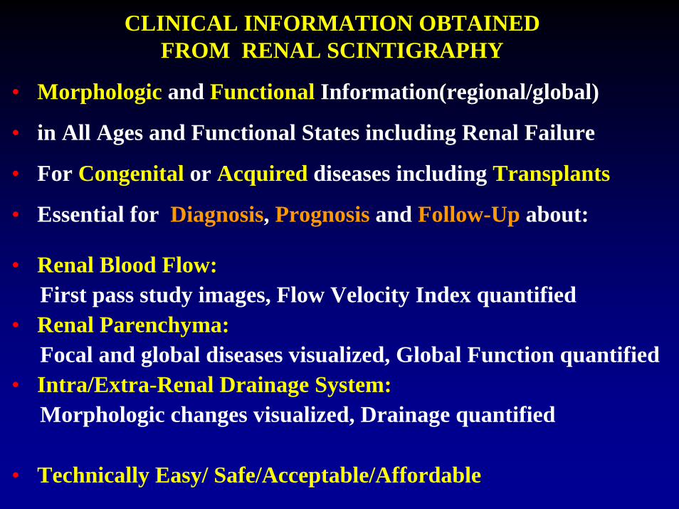
CLINICAL INFORMATION OBTAINED FROM RENAL SCINTIGRAPHY
• Morphologic and Functional Information(regional/global)
• in All Ages and Functional States including Renal Failure
• For Congenital or Acquired diseases including Transplants
• Essential for Diagnosis, Prognosis and Follow-Up about:
• Renal Blood Flow:First pass study images, Flow Velocity Index quantified
• Renal Parenchyma:Focal and global diseases visualized, Global Function quantified
Old “Sophisticated” Protocols for Renal Scintigraphy
We call old “Sophisticated” Renal Protocols the traditional methods of Diuretic Renal Scintigraphy
with Bladder Catheterizationand injection of Diuretic 20-30+ min post radiopharmaceutical
These traditional methods were inspired before U/S, CT, MRI were introduced
They were proposed by the experts in the field of renal imaging at the time,
were endorsed by the Societies ( SNM, Pedi Fetal etc ) but never revisited
and are still followed by many laboratories (due to lack of communication)
Considering the current standards of practice we believe that these protocols
Serve the Exceptions rather than the Patient Population we currently evaluate
Problems with theOld “Sophisticated” Protocols for Renal Scintigraphy
• Patient Preparation is Painful and potentially Dangerous(Bladder Catheterization and IV hydration)
• Studies are Too Lengthy (40min-60min)• The radiation Exposure to the gonads is high• Diagnosis for Cortical (parenchymal) problems is not obtained• Prognosis is not usually possible• Quantification of renal function is not adequately obtained
As a result renal scintigraphy has always been and still is underutilized
Problems with theOld “Sophisticated” Protocols for Renal Scintigraphy
Father: “Better operate on my child than ask for a nuclear study”
(Testimony of Dr Ricardo Gonzalez, Pedi-Urologist)
Those who practice pediatric nuclear medicine had these concerns
since the seventies and eighties
The New Protocol for Renal Scintigraphy
There has always been
a need for a fast, easy, and simple protocol
but changes in patient population
improvements in other renal imaging methods
and economic issues
have made this change mandatory
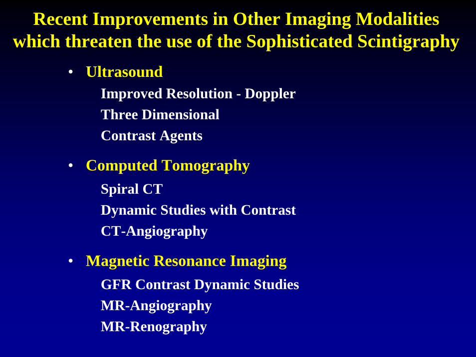
Recent Improvements in Other Imaging Modalitieswhich threaten the use of the Sophisticated Scintigraphy
123/131I-o-Hippurate:..GF and TE (EE = 80%)Dynamic ERPF Imaging
99mTc-DTPA:……….Glomerular Filtration (EE = 20%) Dynamic Imaging of GFR
99mTc-DMSA:………Cortical Fixation(GF and reabsorption)(EE = 5%)Parenchymal Function Imaging
99mTc-GH:…………..Combined GF and Cortical Fixation (EE = 20%) Dynamic GF and Parenchymal Imaging
ADVANTAGES of MAG3 for RENAL SCINTIGRAPHY
• High Renal Extraction Efficiency 60%
Fast parenchymal accumulation
Fast background clearance
Effective Early Parenchymal Imaging
Accurate evaluation of Intra/Extra Renal Drainage
• Small Distribution Space
Steep (sensitive) Renograms
• Technetium-99m-Chemical Labeling
High dose, Good statistics, Low radiation exposure
• Safe (experience 17 years)
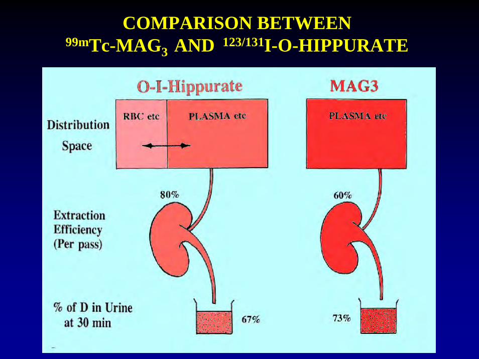
COMPARISON BETWEEN99mTc-MAG3 AND 123/131I-O-HIPPURATE
MAG3 ADVANTAGES (v/s DTPA)
MAG3: TUBULAR EXCRETION IMAGING AGENT WITH HIGH RENAL EXTRACTION EFFICIENCY
(60% as compared to DTPA which is 20%)
Renal imaging studies with MAG3 are:- Usually diagnostic- Highly sensitive to function changes- Substantially disease specific- Easily quantifiable- Fast, with low radiation exposure- Provide information about the cortexProvide Prognostic Infdormation
MAG3 comparison to DTPAMAG3 DTPA
1) FUNCTION TUBULAR GLOMERULARVisualize kidney even with no GF(ATN, total obstruction, toxicity): Yes NoVisualize Regional Dysfunction(APN, Focal Nephritic/otic, RVH): Yes No
2) EXTRACTION EFFICIENCY 60% 20%Parenchymal Evaluation(Rejection, Nephrotic, Nephritic etc) Yes ?Study of Drainage(Diuretic Renography etc): Sensitive Mediocre Patient Radiation (Radiation exposure of the patient): Low High
COMPARISON OF MAG3 WITH DTPA
The New Protocol for Renal Scintigraphy
To obtain best results with the New Protocol
we must use
the Best FDA approved Radiopharmaceutical
Finally it was Recognized to be 99mTc - MAG3
and apply the Best Protocol
The New Protocol for Renal Scintigraphy
17 years Experience at the UM,
indicates that MAG3-F0 is the protocol, which covers
most of the current renal imaging needs
for the majority of cases
99mTc- MAG3 with zero time injection of Furosemide
and apply the Best Protocol:99mTc- MAG3 with zero time injection of Furosemide
the Best FDA approved Radiopharmaceutical:
Finally it was Recognized to be 99mTc - MAG3
To obtain best results with the New Protocol
we must use
The New Protocol for Renal Scintigraphy
Basic Principle for the New Protocol: (MAG3-F0)
When Lasix is injected IV:
its Diuretic Effect begins between 3 - 5 min
(the maximum effect at 9 min)
Therefore with the F0 injection there is enough time to study the drainage (4 to 22 = 18 min)
and in addition the parenchyma can be studied
THE FAST PROTOCOL:It began at OSU and continued at UM first as Hippuran-F3
and when MAG3 became available (1990) as 99mTc-MAG3-F0
Diuretic Renal Study: Protocol at UM/JMH99mTc-MAG3-F0 (or MAG3-F0 )
No difficult Patient Preparation is needed
No Sedation (only restriction of motion)
Hydration 5-10 ml/kg water orally at –30 min
No Bladder Catheterization (except in selected cases)
Study either supine or upright
Renal Scintigraphy at UM/JMMC: (MAG3-F0) Method applied the last 17 years:
FOR NATIVE KIDNEY STUDIES FOR RENAL TRANSPLANT STUDIES
A) Dynamic Renal Scintigraphy UM/JMMC:(MAG3-F0) Method applied the last 17 years:
Injection iv 1-10 mCi MAG3 + 40-80 mg LASIX (Furosemide)
SIMULTANEOUS INJECTION OF MAG3 AND LASIX = F0
ACQUISITION: FLOW: 1 min ( 1 frame per 1 sec)FUNCTION: 22 min ( 1 frame per 30 sec)POST VOID: 2 min static image (at 25-30 min) DELAYED 2 min static images (at 1 hr)
GROUPING IMAGES : -FLOW: in 3 sec images-FUNCTION: in 2 min images
A method was developed to calculate from the MAG3-F0 study
THE GLOBAL RENAL FUNCTION (GRF) using:
a) The Kidney Uptake/Body Background Activity (at 2 min)
and b) The Residual Cortical Activity (at 20 min)
(Both these parameters are related with the creatinine levels)
QUANTIFICATION OF RENAL FUNCTION
A = la+ra-bBKG
la ra
BKG
The Kidney Uptake/Body Background Activity (at 2 min)
MAGMAG33--FF00
QUANTIFICATION OF RENAL FUNCTION
Basic Calculations MAGMAG33--FF00
Global Renal Function Calculations
ℓn Creatinine = 1.22 – 0.55 * ℓn MADRE
e 1.22
Creatinine =(MADRE) 0.55
3Creatinine ≈
MADRE
ℓn: natural logarithmMAGMAG33--FF00
MAG3 Accumulation and Discharge Rate Equivalent
-2
-1
0
1
2
-2 -1 0 1 2 3 4 5
ln RGF
ln C
reat
inin
e
ln Creatinine = 1.22 – 0.55 * ln MADRE
ℓn MADREMAGMAG33--FF00
ℓn: natural logarithm
Results: Prediction of Creatinine levels by the Formula GRFComparison with real Creatinine values
0
1
2
3
4
5
6
7
0 1 2 3 4 5
Predicted Creatinine
Cre
atin
ine
6 MAGMAG33--FF00
NORMAL STUDIES
what is a normal study?
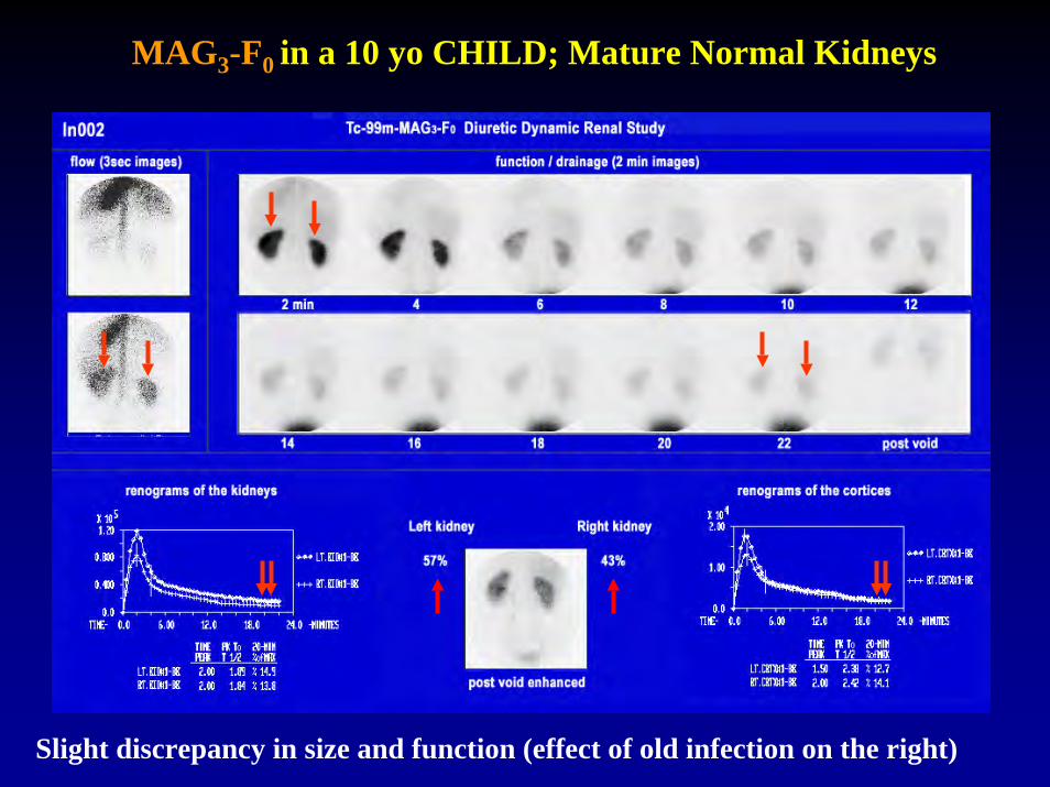
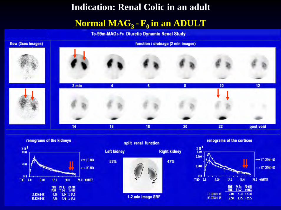
Findings: L and R kidneys orthotopic, normal size, normal flow and function, , normal cortical activity, normal drainage
Right kiney thiner with split renal function 56%/44% L/R
Adult with renal colic
NORMAL STUDIES
A variation of Normal
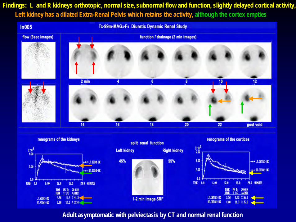
Findings: L and R kidneys orthotopic, normal size, Left kidney has a dilated Extra-Renal Pelvis which retains the activity, although the cortex empties
Adult asymptomatic with pelviectasis by CT and normal renal function
subnormal flow and function, slightly delayed cortical activity,
CLINICAL EXPERIENCE
Many misconceptions were clarifiedNew horizons were discovered
NORMAL STUDIES
Misconceptions:You cannot study the newbornYou need to catheterize the bladder
FactsYou can study the newbornyou don’t need to catheterize the bladder
Evaluate Pelviectasis found by Ultrasound
Typical MAG3-F0 study in a Newborn; No bladder catheter
Kidneys empty. No Obstruction; Slight Immaturity; Study Normal for Age
The infant urinated twice during the study = No need for catheter
Evaluate Pelviectasis found by Ultrasound
Typical MAG3-F0 study in a Newborn; No bladder catheter
What happens if the infant does not empty the Bladder?
Kidneys empty. No Obstruction; Slight Immaturity; Study Normal for Age
A 7 yo boy without bladder catheter has a MAG3-F0 study
Indication: Evaluate effects of urinary tract infection.
MAG3-F0 in a 10 yo CHILD; Mature Normal Kidneys
Slight discrepancy in size and function (effect of old infection on the right)
Evaluate Renal Colic. Is there obstruction?
Indication: Renal Colic in an adult
Normal MAG3 - F0 in an ADULT
Misunderstanding
Ye have heard that it was said by them of old time:You cannot do tomography with MAG3
But we say unto youMAG3 can do tomography within 4 minutes
And we can use SPECT/CT to localize and identify lesions
NORMAL MAG3-F0 SPECT (tomogram)tomograms axial/coronal/sagittal
35yo female with history of pyelonephritis
Axial
Coronal
Sagittal
Evaluate Obstruction
INDICATIONS
1) To differentiate between Dilatation without Obstruction
and Partial Obstruction
2) To confirm Complete Obstruction
PRINCIPLE
Diuresis (completely) washes out the activity from collecting
system when there is no obstruction
but fails to do so in obstructive uropathy
DIURECTIC RENOGRAPHY:to make the diagnosis of Kidney Obstruction
WHITAKER TEST
At 10 ml/min infusiona renal pelvis pressure>10 cm water from the pressure in the bladder defines obstruction
Problem 1:It is an invasive method
Problem 2: Many kidneys cannot produce 10 ml/min urine (hypofunctioning) = FP
OBSTRUCTION
MisconceptionsFor Diuretic Renography you need to inject the diureticeither 20-30 min after MAG3 (old O’Reily=F+20/30 )
or 15 min before MAG3 (new O’Reily =F-15 )
Fact You can inject MAG3 and Lasix Simultaneously (F0 )
DIURETIC RENOGRAPHY
TIMING OF DIURETIC INJECTION
F+20 : 20-30 min POST MAG3 (old O’Reily)(40-50 min study time)
F-15 : 15 min PRE MAG3 ……(new O’Reily)(35-40 min study time)
F0 : SIMULTANEOUSLY with MAG3 (UM)
(22 min study time)
COMPARATIVE STUDY OFTHE TIMING OF DIURETIC INJECTION
UM/JMH PRESENTED SNM AM 2000
20 min
15 min
(0 min)
(20 min)
(35 min)
COMPARATIVE STUDY OFTHE TIMING OF DIURETIC INJECTION
UM/JMH PRESENTED SNM AM 2000
MAG3 F+20 MAG3 F-15 HIP F0
DIURETIC RENOGRAPHY with the THREE METHODS: Results: Equivalent
Radiation Exposure: Higher with F+20
40+ min 40 min 22 minDuration of the Examination
Lasix
ZERO TIME INJECTION DIURETIC RENOGRAPHY (F0):
• It is at least as Accurate as F+20/30 and F-15
• Better Tolerated (Shorter – One Injection)
• Fewer interruptions in adults for voiding
• Cost Effective as it is Concluded in 25 min
• Reduces the Radiation Exposure of the Patient
• It Allows The Evaluation of the Parenchyma
These two studies prove the opportunity to study the cortexOn the left the study is F+20
On the right it is F0
Which one can evaluate the parenchyma?
NORMAL MAG3Diuretic given at F+20
Cortex full of urinecannot be studied effectively
Diuretic F0
NORMAL MAG3 Diuretic given at F0Cortex is empty and
can be studied effectively
Diuretic F+20
RENAL SCINTIGRAPHY AT UM/JMMC: (MAG3-F0) Method applied the last 17 years:
This protocol was originally applied in the evaluation of drainage
Soon it was realized that it allowed the evaluation of the parenchyma
Then it was applied in all parenchymal indications (including APN)
It was also utilized for the study of Renovascular Hypertension
It allowed the study of HIV and other Acquired Nephropathies
In patients with renal colic unraveled the Stunned (decompressed) kidney
It was finally successful in the study of complications of renal transplants
NEW HORIZONS
• Obstruction
• Focal Parenchymal Disease
• Diffuse Parenchymal Disease
• RVH
• Renal Colic
• Complications of renal Transplants
OBSTRUCTION
Misconception:To make the Diagnosis of Obstruction you need to Study the Collecting System of the Kidney
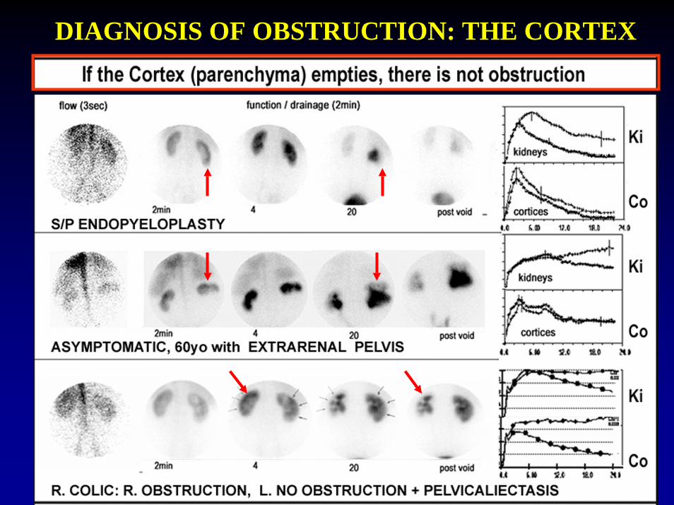
New Horizons: To make the Diagnosis of Obstruction you better study the behavior of the Renal Cortex:
If the cortex empties, there is no obstruction! (even when the drainage system is dilated
and it does not empty appropriately)
Study presented SNM 2003
OBSTRUCTION Bilateral, Right Complete, Old, Left Partial
L kidney: Collecting System Retention and Cortical Retention
DIAGNOSIS OF OBSTRUCTION: THE CORTEX
An adult had endopyeloplasty 6 months ago to relieve obstruction.He is now evaluated for the results of the operation
There is retention within an enlarged remaining pelvisbut the cortex empties
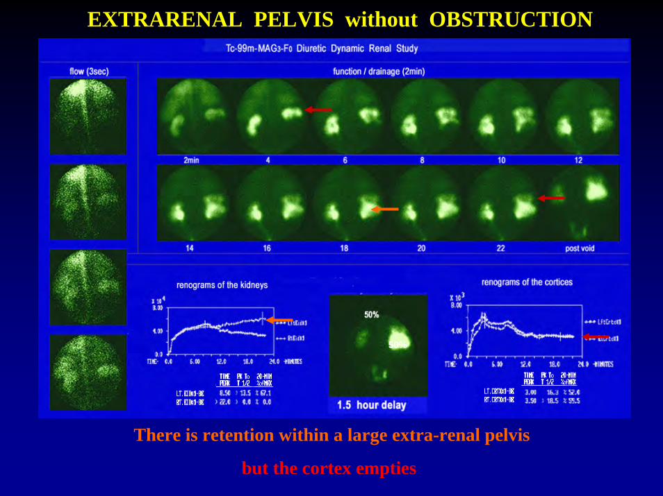
Asymptomatic 55 yo man with incidental finding of “hydronephrosis” on CT
EXTRARENAL PELVIS without OBSTRUCTION
50%
There is retention within a large extra-renal pelvis
but the cortex empties
A patient with a history of bilateral nepholithiasis is studied because of a recent Right Colic
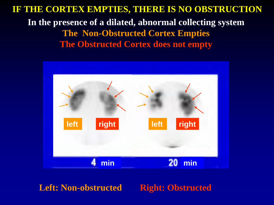
In the presence of a dilated, abnormal collecting systemThe Non-Obstructed Cortex Empties
The Obstructed Cortex does not empty
min min
left leftright right
IF THE CORTEX EMPTIES, THERE IS NO OBSTRUCTION
Left: Non-obstructed Right: Obstructed
A 12 year old child is evaluated because the U/S suggested bilateral “hydronephrosis”
MAG3 - F0 in Pelviectasis without Obstruction
There is retention within dilated renal pelvices bilaterally
but the cortices empty
Evaluate “hydronephrosis” found by ultrasound in a newborn
EXTRARENAL PELVIS without OBSTRUCTIONNewborn
17 month old
THE VALUE OF MAG3 -F0 DIURETIC RENOGRAPHY IN PREDICTING THE NEED FOR SURGERY IN THE NEONATE WITH
URETEROPELVIC JUNCTION OBSTRUCTION
Sfakianakis G, Vensel E, Tapia M, Policaro F, Gosalbez R, Labbie A, Zilleruelo G, Abitbol C, Montane B, Strauss J
Abstract: SNM 2000
CONGENITAL RENAL OBSTRUCTION THE NEED FOR SURGICAL CORRECTION
PELVIECTASIS: MILD OBSTRUCTION
A down-sloping MAG3-F0 renography
in the neonate predicts
Spontaneous Compensation
PELVIECTASIS: SEVERE OBSTRUCTION
An Up-sloping MAG3-F0 renography
in the neonate predicts
The need of Surgical Correction
A Horizontal Renogramrequires follow up studies
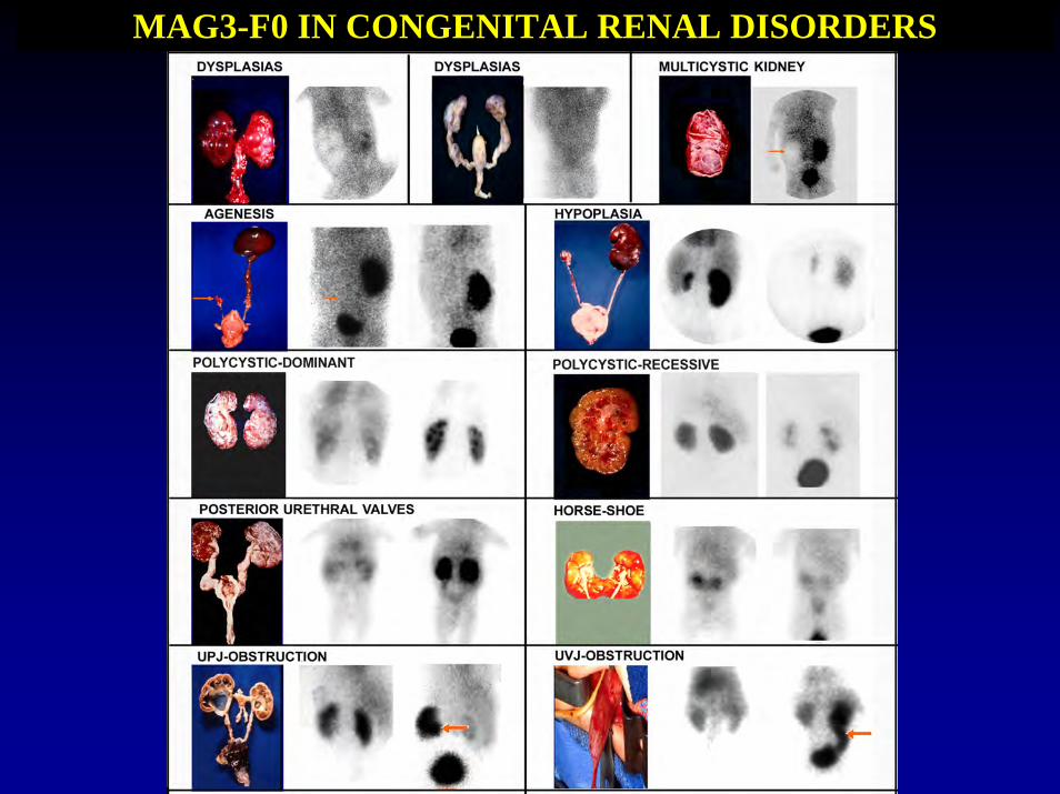
MAG3-F0 IN CONGENITAL RENAL DISORDERS
CONGENITAL RENAL DISORDERS
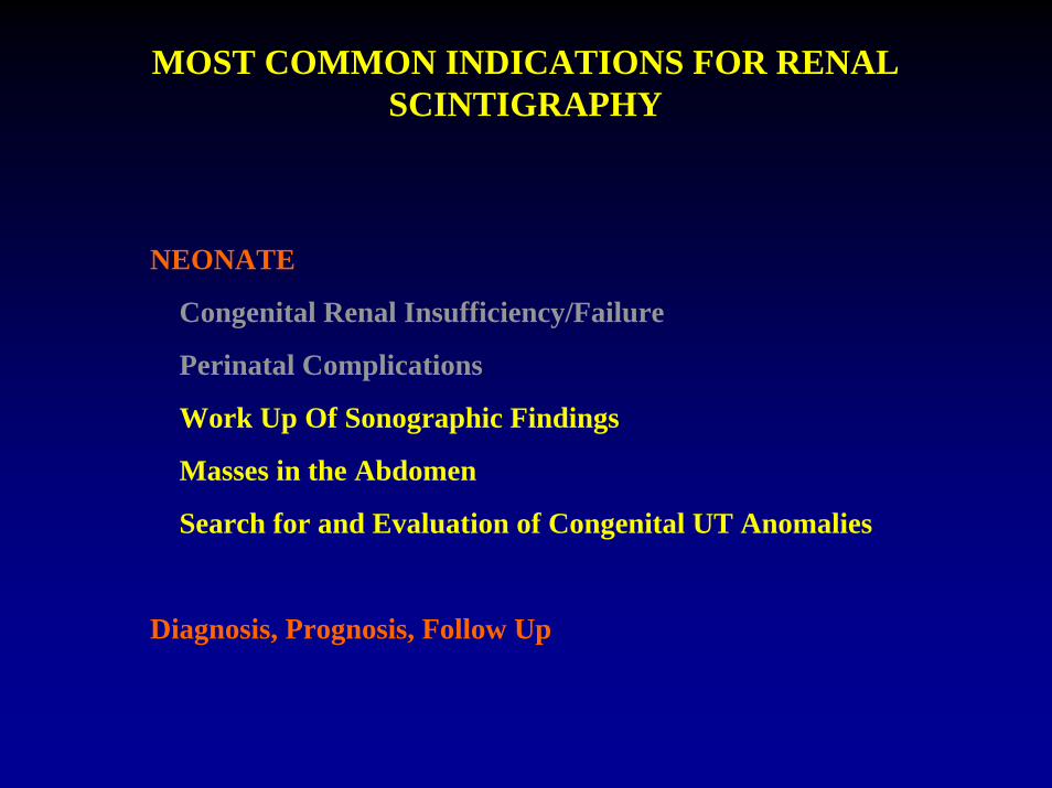
MOST COMMON INDICATIONS FOR RENAL SCINTIGRAPHY
NEONATE
Congenital Renal Insufficiency/Failure
Perinatal Complications
Work Up of Sonographic Findings
Masses in the Abdomen
Search for and Evaluation of Congenital UT Anomalies
Diagnosis, Prognosis, Follow Up
A Newborn child with no urination for 24 hr
CONGENITAL RENAL INSUFFICIENCY/FAILURE
Bilateral Dysplasias
L kidney: No function with lucences
R kidney: Barely functioning
• Bilateral Multicystic Dysplasias, left worse than right• No functioning (left) or barely functioning (right) renal parenchyma• No Intervention indicated, no recovery expected
A Newborn child with no urination for 24 hrUS showed bilateral hydronephrosis
CONGENITAL RENAL INSUFFICIENCY/FAILURE
Posterior Urethral Valves
2 min 4 min
Presence of functioning renal parenchyma
Bilateral Hydronephrosis = Obstruction:Intervention indicated, some recovery expected
A newborn with congenital renal insufficiencyU/S: Bilateral Hydronephrosis
CONGENITAL RENAL INSUFFICIENCY/FAILURE
Bilateral obstruction (UPJ)Worse on the Left
Early Correction of UPJO (the first week of life)
may Prevent Loss of Function and may Promote
Recovery of Function
There is substantial improvement of the left kidney
The right remains unchanged
MOST COMMON INDICATIONS FOR RENAL SCINTIGRAPHY
NEONATE
Congenital Renal Insufficiency/Failure
Perinatal Complications
Work Up of Sonographic Findings
Masses in the Abdomen
Search for and Evaluation of Congenital UT Anomalies
Diagnosis, Prognosis, Follow Up
A newborn with Ischemia during birth from a diabetic mother, is in Renal Failure
U/S normal
MAG3 - F0 in Acute Tubular Necrosis
Kidneys orthotopic, with preservation of flow and accumulation.No drainage problem. Cortical retention of activity
No Intervention indicated, full recovery expected
PERINATAL RENAL INSUFFICIENCY/FAILURE
Acute Tubular Necrosis
•Ischemia during birth from a diabetic mother•No Intervention indicated, full recovery expected
Neonates with hypertension from renal ischemiadue to thrombus in the umbilical cathetershall not be treated with ACE-Inhibitors
A newborn with umbilical artery catheterization for therapydeveloped hypertension
A baseline and an ACE-Inhibition renal study was performed
Left Kidney not visualized = infarctedRight Kidney normal at BSL, but with no excretion after ACE-Inhibition
Right Kidney normal renogram at BSL, but with cortical retention after ACE-Inhibition
MOST COMMON INDICATIONS FOR RENAL SCINTIGRAPHY
NEONATE
Congenital Renal Insufficiency/Failure
Perinatal Complications
Work Up Of Sonographic Findings
Masses in the Abdomen
Search for and Evaluation of Congenital UT Anomalies
Diagnosis, Prognosis, Follow Up
The children in the following studies had abnormal U/S studies as defined in each case individually
PELVIC ECTOPIC NORMAL RIGHT KIDNEYLess active than the right because it is deeper (more attenuation)
U/S: Non-visualization of the right kidney
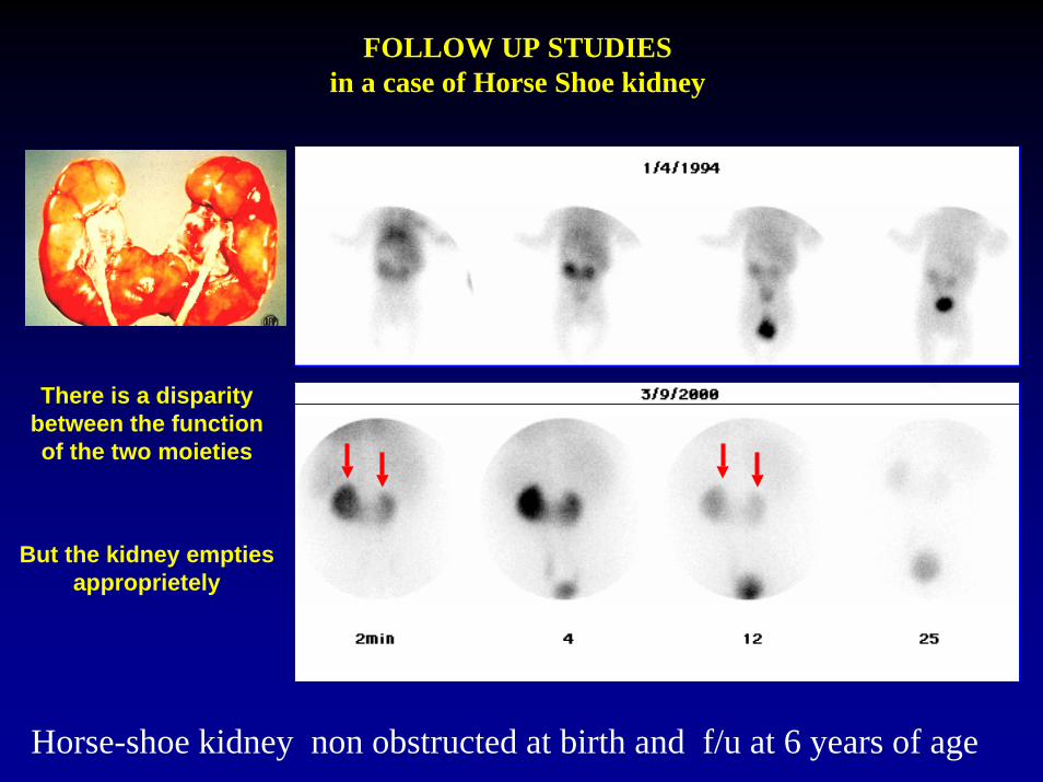
NORMAL HORSE-SHOE KIDNEY
Newborn 2do with questionable U/S
Horse-shoe kidney withNormal Function and Drainage
Immaturity with Increased Residual Cortical Activity
HYPOPLASIA with contralateral compensatory HYPERTROPHY
kc
2 min
20 min
5 yo child with an U/S indicating a small left
kidney
Hypoplastic, but normally functioning
left kidney and compensatory
Hypertrophy of the R
AGENESIS
U/S: Non-visualization of the left kidney
Two congenital malformations deal with cysts
a) The Multicystic KidneyUnilateral/Non-functioning/US detectable
b) Polycystic Kidney DiseaseBilateral/Functioning/US undetectable
LEFT MULTICYSTIC DYSPLASTIC KIDNEYRIGHT NORMAL
U/S: Cysts in the left kidney
Non-Functioning left kidney with background defect =
Multicystic dysplastic kidney
Differentiate a MULTICYSTIC kidneyfrom AGENESIS
No BKG Defect: AGENESIS
BKG Defect: MULTICYSTIC
A correct U/S study shows Cysts in Multicystic kidney
Duplicated Left Kidney withMulticystic Dysplastic Upper Moiety
POLYCYSTIC KIDNEY DISEASE
Autosomal Recessive
In Children
Asymptomatic
Difficult to diagnose
Autosomal Dominant
In Adults
May have symptoms
Cysts on US/CT
POLYCYSTIC KIDNEY DISEASEAUTOSOMAL RECESSIVE
Mild Infantile TypeLarge Kidneys with good drainage
POLYCYSTIC KIDNEY DISEASEAUTOSOMAL DOMINANT
GH
Fragmented parenchyma around the cysts / multifocality of the collecting system /satisfactory drainage (unless there are complications=obstructions)
POLYCYSTIC KIDNEY DISEASEAUTOSOMAL DOMINANT
Fragmented parenchyma around the cysts / multifocality of the collecting system /satisfactory drainage (unless there are complications=obstructions)
MAG3 - F0 in Congenital Renal Obstruction
UPJO
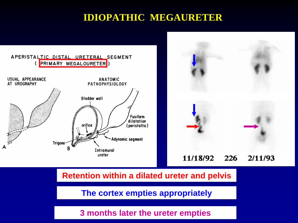
UVJO
Can we prevent the loss?OBSTRUCTION
Neonate 5 year old
The following three patients were sent for MAG3-F0because they were thought obstructed
Pelviectasis: No Obstruction
Urine bag
This patient had a MAG3 study at an outside lab, which was read as UPJO.The referring physician had asked to use a bladder catheter
Both kidneys drained adequately: No obstruction
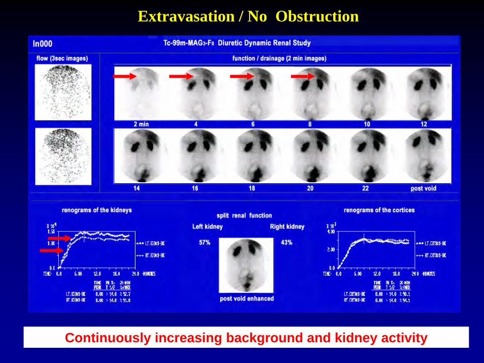
Extravasation / No Obstruction
Continuously increasing background and kidney activity
Pelviectasis: No Obstruction
Infant with an U/S indicating left “hydronephrosis
The kidneys empty: No obstruction Pelviectasis of the left kidney
The next two infants are evaluated because the U/S showed Hydronephrosis on the left in the first and on the Right in the
second
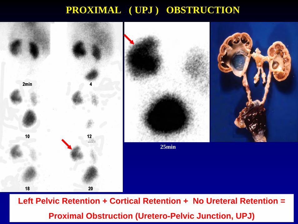
PROXIMAL ( UPJ ) OBSTRUCTION
25min
Left Pelvic Retention + Cortical Retention + No Ureteral Retention =


![Kaken 99Mo- Tc ProcessA4x4] TcMM Brochure (160907).pdf · Kaken 99Mo-99mTc Process@ Generator of highly concentrated pure 99mTc from low specific activity 99Mo produced by reactor](https://static.documents.pub/doc/80x56/602aa12686e8fd284b2e33f6/kaken-99mo-tc-a4x4-tcmm-brochure-160907pdf-kaken-99mo-99mtc-process-generator.jpg)


