109
9 th International Copper Meeting Copper 2014 October 5 to October 10, 2014 Vico Equense, Italy Organizers: Jonathan D. Gitlin, Dennis J. Thiele and Simone Ciofi-Baffoni
9th International Copper Meeting Copper 2014
October 5 to October 10, 2014 Vico Equense, Italy
Organizers: Jonathan D. Gitlin, Dennis J. Thiele and Simone Ciofi-Baffoni


Contents
Welcome to Copper 2014 …………………………………………………………...………...2
Organizers ………………………………………………………………………………...…….3
Local Organizer …………………………………………………………………………………3
Scientific Organizing Committee ……………………………………………………...………3
Poster Session and Poster Talk Chair …………………………………………………...…..3
List of Supporters ……………………………………………………………………………….4
Scientific Program …………………………………………………………………………..….5
Oral Presentations …………………………………………………………………………….10
Posters ………………………………………………………………………………………....45
Poster Abstract Index …………………………………………………………………….…105
List of Attendees ………….………………………………………………………………....108
1
Welcome to Copper 2014
The International Copper Meetings have been a premier venue for scientific discovery
and dialogue amongst the many investigators in our field in biology, chemistry, medicine
and the environment and an important attraction for new and young investigators. This
9th International Copper Meeting (Copper 2014) takes place from October 5th –
October 10th 2014 at the Aequa Hotel in Vico Equense, a beautiful coastal town in the
province of Campania, outside of Naples on the Sorrentine peninsula. This tradition of
holding the International Copper Meetings in Italy provides a wonderful venue for
fostering cutting edge scientific presentations, collegial discussions and collaborations
that all serve to advance the field.
Welcome to Vico Equense.
Warm wishes,
Jonathan D. Gitlin, Dennis J. Thiele and Simone Ciofi-Baffoni Organizers, Copper 2014
2

Organizers
Simone Ciofi Baffoni Magnetic Resonance Center and Department of Chemistry University of Florence [email protected]
Jonathan D. Gitlin Marine Biological Laboratory [email protected]
Dennis J. Thiele Department of Pharmacology and
Cancer Biology Duke University School of Medicine [email protected]
Local Organizer
Roman Polishchuk Telethon Institute of Genetics and Medicine [email protected]
Scientific Organizing Committee
Jennifer Cavet, University of Manchester, Manchester Betty Eipper, University of Connecticut Health Ctr, Farmington Mauricio Gonzalez, University of Chile, Santiago Ute Kraemer, Ruhr University Bochum, Bochum Scot Leary, University of Saskatchewan, Saskatoon Alistair McEwan, University of Queensland, Queensland Roman Polishchuk, Telethon Institute of Genetics and Medicine, Naples Bing Zhou, Tsinghua University, Beijing
Poster Session and Poster Talk Chairs
Julian Mercer, Deakin University, Melbourne Joseph Prohaska, University of Minnesota, Duluth
3

Copper Meeting gratefully acknowledges the following Supporters
4

Scientific Program
Monday, October 6
07:45-08:00: Introduction by the Organizers 8:00-11:00 Session 1 - Human Disease (Chair: Scot Leary) 08:00-08:30 1. Stephen Kaler – National Institutes of Health – Bethesda, USA Translational neuroscience approaches to inherited disorders of
copper transport 08:30-09:00 2. Svetlana Lutsenko – Johns Hopkins University – Baltimore, USA Activation of nuclear receptors improves liver morphology and
function in Wilson disease 09:00-09:30 3. Rosanna Squitti - Fatebenefratelli Hospital - Rome, Italy Copper metabolism abnormalities as a risk factor for Alzheimer’s
disease 09:30-10:00 Coffee Break 10:00-10:30 4. Bing Zhou – Tsinghua University – Beijing, China Researching roles of metal ions in neurodegenerative diseases
with a Drosophila model 10:30-11:00 5. Stefan Kins – Kaiserslautern University of Technology –
Kaiserslautern, Germany Copper promotes Amyloid Precursor Protein dimerization and
synaptogenic function 11:30-13:30 Lunch /Free Time 14:00-17:00 Session 2 - Neuroscience (Chair: Roman Polishchuk) 14:00-14:30 1. Christopher Chang – University of California – Berkeley, USA Enabling discovery and study of copper signaling with
fluorescent probes 14:30-15:00 2. Richard Burke – Monash University – Melbourne, Australia Novel regulators of neuronal copper homeostasis in Drosophila
5
15:00-15:30 3. Michael Petris – University of Missouri – Columbia, USA Copper, Cancer, and Chemotherapy: Lessons from ATP7A 15:30-16:00 Coffee Break 16:00-16:30 4. Gerald Zamponi – University of Calgary – Calgary, Canada Copper dependent regulation of NMDA receptors: the role of
cellular prion protein 16:30-17:00 5. Betty Eipper – University of Connecticut – Farmington, USA Getting Copper to the Peptide Amidating Enzyme Evening Free Tuesday, October 7
8:30-11:00 Session 3 - Microbiome/Microbial Pathogenesis (Chair: Betty Eipper) 08:30-09:00 1. Simone Ciofi-Baffoni – CERM, University of Florence –Florence,
Italy Arturo Leone Young Investigator Award Copper and [2Fe-2S] cluster trafficking: two sides of the same
coin 09:00-09:30 2. Heran Darwin – New York University – New York, USA Tuberculosis and Copper: friends or foes 09:30-10:00 3. Alastair McEwan – University of Queensland – Brisbane, Australia The effect of aqueous copper and copper(II)-
bis(thiosemicarbazonato) complexes on Neisseria gonorrhoeae 10:00-10:30 Coffee Break 10:30-11:00 4. Simon Labbé – University of Sherbrooke – Sherbrooke, Canada Copper metabolism proteins in meiotic differentiation 11:00-11:15 5. Marc Solioz - Tomsk State University - Tomsk, Russian Federation
(Short talk) Characterization of a copper-induced quinone degradation
pathway in Lactococcus lactis 11:15-11:30 6. Marianne Ilbert - BIP01, CNRS, Marseille, France (Short talk) Unusual features for a single-domain cupredoxin with a green
copper site
6
11:30-13:15 Lunch & Poster Session (Mercer and Prohaska) 13:15- 13:45 Poster Talk Session – Posters selected during meeting 14:00-17:00 Session 4 - Evolution, Ecology & Comparative Biology (Chair: Mick
Petris) 14:00-14:30 1. Mak Saito – Woods Hole Oceanographic Institute – Woods Hole,
USA Targeted Metaproteomic Analyses of Metalloproteins in the
Pacific Ocean and Estimates of Microbial Metal Use 14:30-15:00 2. Sílvia Atrian – University of Barcelona – Barcelona, Spain Cu-thioneins: where, how, why? 15:00-15:30 3. Olena Vatamaniuk – Cornell University – Ithaca, USA Transcriptional regulatory networks that coordinate copper
homeostasis and crosstalk with cadmium resistance in Arabidopsis thaliana
15:30-16:00 Coffee Break 16:00-16:30 4. Ute Krämer – Ruhr University – Bochum, Germany Copper homeostasis interacts with iron homeostasis and
reproductive development of vascular plants 16:30-16:45 5. Alejandro Vila - National University of Rosario - Rosario, Argentina
(Short talk) Mechanism of Biogenesis of the CuA Center in Human
Cytochrome c Oxidase 16:45-17:00 6. Pernilla Wittung-Stafshede - Umeå University – Umeå Sweden
(short talk) Mechanisms of metal transfer to and from the human cytoplasmic
copper chaperone, Atox1
Evening Free Wednesday, October 8 8:00-11:00 Session 5 - Cell Biology & Metabolism (Chair: Ute Krämer)
7

08:00-08:30 1. Valeria Culotta – Johns Hopkins University – Baltimore, USA David Danks Award Copper and fungal SOD enzymes at the host-pathogen interface 08:30-09:00 2. Scot Leary - University of Saskatchewan – Saskatoon, Canada Sco1 transgenic mouse models: advancing our understanding of
human disease 09:00-09:30 Coffee Break 09:30-10:00 3. Heinz Osiewacz – Goethe University – Frankfurt, Germany Systematic analysis of the molecular basis of aging and lifespan
control in P. anserina. 10:00-10:30 4. Paul Cobine - Auburn University – Alabama, USA Recruitment and transport of mitochondrial copper for assembly
of cytochrome c oxidase 10:30-11:00 5. Roman Polishchuk – Telethon Institute of Genetics and Medicine –
Naples, Italy New insights in the ATP7B trafficking in health and disease 11:30-13:15 Lunch & Poster Session (Mercer and Prohaska) 13:15- 13:45 Poster Talk Session – Posters selected during meeting 14:00-17:00 Session 6 - Proteins & Chemistry (Chair: Nigel Robinson) 14:00-14:30 1. Amy Rosenzweig – Northwestern University – Chicago, USA Ivano Bertini Award Deconstructing the copper switch in methanotrophs 14:30-15:00 2. Shinobu Itoh – Osaka University – Osaka, Japan Molecular Oxygen Activation by Copper Proteins and Models 15:00-15:30 Coffee Break 15:30-16:00 3. Vinzenz Unger – Northwestern University – Chicago, USA First One, then Two – Emergence of a Unifying Concept in
Intracellular Copper Distribution 16:00-16:30 4. Lucia Banci – CERM, University of Florence – Florence, Italy The unique contribution of NMR to copper systems biology
8
16:30-17:00 5. Martina Ralle – Oregon Health Sciences University – Portland, USA Revising the intracellular 'copper thermometer': examples of
intracellular copper stores
Evening Free Thursday, October 9 8:00-11:00 Session 7 - Systems Biology (Chair: Alastair McEwan) 08:00-08:30 1. Nigel Robinson – Durham University – Durham, England A chemical potentiator of copper-accumulation used to
investigate the iron-regulons of Saccharomyces cerevisiae 08:30-09:00 2. David Giedroc – Indiana University – Bloomington, USA Copper resistance in Streptococcus pneumoniae 09:00-09:30 Coffee Break 09:30-10:00 3. Byung-Eun Kim – University of Maryland – College Park, USA Caenorhabditis elegans as a model to identify novel copper-
regulated pathways 10:00-10:30 4. José Argüello – Worcester Polytechnic Institute – Worcester, USA From the cytoplasm to the periplasm and beyond, passing the
Cu+ along 10:30-11:00 5. Marinus Pilon – Colorado State University – Fort Collins, USA Regulation of Cu delivery for photosynthesis 13:00 – 17:00 Excursion to Herculaneum or Free time for interactions 19:00 – 22:00 Closing Session, Dinner & Award Presentations Arturo Leone Young Investigator Award Presentation to Simone Ciofi-Baffoni, University of Florence Ivano Bertini Award Presentation to Amy Rosenzweig, Northwestern University David Danks Award Presentation to Valeria Culotta, Johns Hopkins University Celebration of the Careers and Contributions of Professor Joseph Prohaska, University of Minnesota Medical School and Professor Julian Mercer, Deakin University
9

Oral Presentations
10
Translational neuroscience approaches to inherited disorders of copper transport Stephen G. Kaler, Section on Translational Neuroscience, Metal Biology and Molecular Medicine Group, Eunice Kennedy Shriver National Institute of Child Health and Human Development, National Institutes of Health. Bethesda, MD, USA While Menkes and Wilson diseases were recognized 50 and 100 years ago, respectively, recent advances in genomics and neuroscience have ushered in a new research era for inherited disorders of copper transport. ATP7A-related distal motor neuropathy, Huppke-Brendel and MEDNIK syndromes are inherited conditions all identified within the past five years, and which reveal novel and important aspects of human copper metabolism. These include the requirement for ATP7A in proper motor neuron function, acetylation as a crucial post-translational modification of certain copper proteins, and the mechanistic role of adaptor protein (AP) complexes in copper ATPase trafficking. In addition to triggering such advances in our basic understanding, inherited disorders of copper transport represent ripe targets for experimental therapeutic approaches to cure or ameliorate the associated human illnesses. Based on preclinical studies in mouse models, brain-directed viral gene therapy combined with subcutaneous copper injections holds great promise for Menkes disease, even when caused by severe mutations in ATP7A. Newborn screening utilizing whole exome sequencing of DNA from dried blood spots, currently under assessment in pilot studies, will enhance early diagnosis of this condition, a requirement for successful neurodevelopmental outcomes. The study and investigation of inherited disorders of copper transport involves the intersection of biochemistry, cell biology, genetics and gene regulation, metabolism neurology, and structural biology. While many uncertainties remain, the pace of discovery across multiple disciplines has quickened noticeably in recent years, auguring well for substantial further advances in translational neuroscience relevant to these conditions.
11
Activation of Nuclear Receptors Improves Liver Morphology and Function in Wilson disease James P. Hamilton1, Lahari Koganti1, Susrut Pendyala2, Dominik Huster3, Abigael Muchenditsi2, Svetlana Lutsenko2 1 Department of Medicine and 2Department of Physiology, Johns Hopkins University School of Medicine, Baltimore, MD, USA, and Deakoness Hospital, Leipzig, Germany Wilson disease (WD) is a potentially lethal disorder caused by mutations in the copper-transporter ATP7B and accumulation of copper in tissues. Current treatment of WD is based on chelation of copper. The treatment ameliorates many manifestations of the disease, but often worsens neurologic symptoms and causes other side effects. Our previous studies have identified lipid metabolism as a major target of copper toxicity in WD liver. We hypothesized that upregulation of lipid metabolism may decrease the disease manifestations in the absence of copper chelation. To test this hypothesis, we treated control and Atp7b-/- mice with an agonist of LXR receptor, T0901317, for 8 weeks, with treatments starting prior to pathology onset. We then characterized copper levels, liver histology, serum metabolites, and liver function. We found that treatment with T0901317 had no effect on hepatic copper, which was 38-fold higher in Atp7b-/- livers compared to control. However, liver histology and function were significantly improved. The improvements were associated with a decrease in genetic markers of liver fibrosis and a decrease in inflammatory cytokines. AST was reduced by 65% and ALT was reduced by 47%. Total cholesterol, LDL, and HDL more than doubled in the drug treated mice compared to the vehicle-treated controls. Serum triglycerides were significantly reduced in the Atp7b-/- mice, but normal in drug-treated animals. These findings suggest that disregulation of nuclear receptors is an important contributor to WD pathology and that modulation of nuclear receptor activity could be a supplementary approach to improving liver function in WD.
12

Non-ceruloplasmin-copper involvement in Alzheimer’s disease and Mild Cognitive Impairment
Rosanna Squitti, PhD
Department. of Neuroscience, Fondazione Fatebenefratelli, AFaR Division - Osp.
Fatebenefratelli, Rome, Italy.
A large number of studies indicate a specific role of copper in aging and in Alzheimer’s disease mechanisms. Specifically, it has been proposed that the hypermetallation of the Abeta peptide can be at the basis of redox cycles of oxidative stress, toxicity, Abeta oligomer formation and precipitation (reviewed in Squitti and Polimanti, 2013). We have contributed to this topic demonstrating in living AD patients that an increase in the serum copper fraction that does not bind to ceruloplasmin (Non-Cp-Cu), correlates with the AD typical deficits (Squitti, et al., Neurol 2002, 2005), cerebrospinal fluid (CSF) markers (Squitti et al., Neurol 2006), and with a worse prognosis for AD (Squitti et al., Neurol 2009). Furthermore, we have shown that loci of susceptibility for AD lie in genes pertinent to copper metabolism (Squitti et al., 2013; Bucossi et al., 2012; Squitti and Polimanti 2013), in particular in the ATP7B gene, which codes for the ATP7B-pump controlling copper excretion through the bile and ceruloplasmin biosynthesis. Moreover, we have demonstrated that copper and non-Cp-cu levels are higher in AD patients vs. healthy controls by means of meta-analyses (Squitti et al., J Alzh Dis 2013). Non-Cp-Cu helps in properly classifying subjects with Mild Cognitive Impairment (MCI) from healthy ones (Squitti et al., J Alzh Dis 2011) and provides prognostic information about the conversion to full AD (Squitti et al., Ann Neurol 2014). We have recently developed a patented devise to directly measure serum Non-Cp-Cu [Colabufo, N. and R. Squitti, P.E. European Patent Office (EPO) (RO/EP), 2012], highly reliable.
13
Studying the roles of metal ions in neurodegenerative diseases with Drosophila models
Bing Zhou, PhD School of Life Sciences, Tsinghua University, Beijing 100084, China
[email protected] It has long been known that metal ions often accumulate in pathologically affected regions in neurodegenerative patients. However, the cause or result of this effect is largely not established. We used Drosophila melanogaster as models to study how metal homeostasis could impact the development of neurodegenerative diseases. Several metals are implicated in the etiology of Alzheimer’s disease (Abeta model), whereas copper likely participates in Huntingtin aggregation and zinc in the toxicity of Tau protein. In general, metals exacerbate the affected phenotypes. By substituting metal binding residues in some of these proteins, we were able to show that the altered proteins are associated with greatly reduced toxicity and importantly, the degree of toxicities no longer responds to metal changes. We conclude that metal dyshomeostasis is likely important in at least some of the common neurodegenerative diseases, and direct metal binding to Huntingtin and Tau proteins might be a key contributing factor to the relevant neurodegenerative diseases (Huntington and Tauopathy). References 1. Xiao G, Fan Q, Wang X, Zhou B. (2013). Huntington disease arises from a combinatory toxicity of
polyglutamine and copper binding. Proc Natl Acad Sci U S A. 110(37):14995-5000
2. Huang Y, Wu Z, Cao Y, Lang M, Lu B, Zhou B. (2014). Direct Zinc Binding Is Critical for Tau Toxicity
Independent of Hyperphosphorylation. Cell Reports 8(3):831-42.
3. Lang M, Wang L, Fan Q, Xiao G, Wang X, Zhong Y, Zhou B (2012). Genetic Inhibition of SLC 39 Family
Transporter 1 Ameliorates Abeta Pathology in a Drosophila model of Alzheimer's Disease. PLoS Genet
8(4):e1002683.
4. Lang M, Fan Q, Wang L, Zheng Y, Xiao G, Wang X, Wang W, Zhong Y, Zhou B. (2013). Inhibition of human
high-affinity copper importer Ctr1 orthologous in the nervous system of Drosophila ameliorates Aβ42-induced
Alzheimer's disease-like symptoms. Neurobiology of Aging 34(11):2604-12.
14
Copper promotes Amyloid Precursor Protein dimerization and synaptogenic function Frederik Baumkötter1, Nadine Schmidt1, Yannick Finger1, Carolyn Vargas2, Sandra
Schilling1, Rebecca Weber1, Katja Wagner1, Sebastian Fiedler2, Wilfried Klug4, Jens
Radzimanowski5, Sebastian Nickolaus3, Sandro Keller2, Simone Eggert1, Klemens
Wild4 and Stefan Kins1
1Division of Human Biology and Human Genetics, 2Molecular Biophysics and 3Division of Plant
Physiology, University of Kaiserslautern, Kaiserslautern, Germany, 4Heidelberg University
Biochemistry Center, University of Heidelberg, Heidelberg, Germany, and 5Unit of Virus Host Cell
Interactions, International Coeducational Unit 3265, Joseph Fourier University–European Molecular
Biology Laboratory–National Center of Scientific Research, F-38042 Grenoble, France
Accumulating evidence suggests that the Amyloid Precursor Protein (APP), which
plays a key role in Alzheimer’s Disease, has an essential synaptic function. APP
synaptogenic function depends on trans-directed dimerization of the extracellular E1
domain encompassing a growth factor-like domain (GFLD) and a copper-binding
domain (CuBD).
Here we report the 1.75 Å crystal structure of the GFLD in complex with a copper ion
bound with high affinity (KD = 28 nM) to an extended hairpin-loop at the dimerization
interface. In co-immunoprecipitation assays increased intracellular copper levels
promotes APP interaction, whereas mutations in the copper binding sites of either the
GFLD or CuBD result in a drastic reduction in APP cis-orientated dimerization.
Further, we provide evidence that copper is essential and sufficient to induce
reversible trans-directed dimerization of highly purified APP extracellular domain
coupled to agarose beads. Finally we show that expression of APP in HEK293 cells
promotes presynaptic differentiation of contacting axons of primary neurons and that
this synaptogenic activity depends on dimerization and copper binding.
15
Together, these findings demonstrate that copper binding to the GFLD of APP is
required for APP dimerization and APP synaptogenic function in a mixed co-culture
assay. Thus neuronal activity or disease-associated changes in copper homeostasis
likely go along with altered synaptic function of APP.
16
Enabling discovery and study of copper signaling with fluorescent probes Christopher J. Chang University of California, Berkeley, United States Copper is an emerging new type of chemical signal in biological settings, but aberrant alterations in its homeostasis are connected to a variety of aging and disease states. Fluorescent probes and related chemical tools offer a developing technology for screening studies in live specimens, which can then be complemented by traditional measurements for bulk metal analysis in fixed counterparts, as well as appropriate spectroscopic and genetic controls to enable new biological discoveries. This presentation will describe our latest results on the synthesis and characterization of fluorescent probes for enabling molecular imaging of dynamic, mobile pools of copper in living cells and animals and their application in brain, fat, and immune system models, where these tools have enabled discovery and study of new copper-dependent signaling pathways.
17

Novel Regulators of Neuronal Copper Homeostasis in Drosophila
Joab E.C. Hwang, Stephen Mercer, Jianbin Wang, Tim Binks and Richard Burke
School of Biological Sciences, Monash University
Cellular copper homeostasis in the vinegar fly Drosophila is regulated by the co-ordinated activity of the copper uptake genes Ctr1A and Ctr1B and the copper efflux gene ATP7. From genetic screens we identified several modifiers of copper transport activity in the fly. We found that the neurogenic protein big brain and several subunits of the vacuolar H+-ATPase complex are required for optimal copper accumulation in Drosophila tissues. However loss of these proteins also results in disruptions to zinc homeostasis and we show that general membrane protein mislocalization is the most probable cause of these defects. We will also present data indicating that the homeodomain-interacting protein kinase (HipK) and one of its targets, the E3 Ubiquitin ligase Slimb, may be regulating copper transporter activity as well.
Next, we investigated the specific role of copper in the Drosophila central nervous system using targeted manipulation of the copper transporter genes. We found that both copper deficiency (ATP7 over expression) and copper excess (Ctr1B over expression) cause developmental lethality when targeted to all neurons and that this lethality can be ameliorated by altering dietary copper levels. Furthermore, surviving flies exhibit wing expansion defects that can be attributed to deficiency of neuropeptides that require amidation by the cupro-enzyme PAM/PHM. We were able to recapitulate these wing expansion defects by restricting copper dyshomeostasis to a small subset of neuropeptidergic cells. Finally, we provide preliminary evidence that glutathione production is critical in maintaining appropriate copper levels in these neuropetidergic cells.
18

Copper, Cancer, and Chemotherapy: Lessons from ATP7A Sha Zhu1,3 Yanfang Wang2,3, Jaekwon Lee4, Michael J. Petris1,2,3
1Departments of Biochemistry, 2Nutrition and Exercise Physiology, and 3the Christopher S. Bond Life Science Center, University of Missouri, Columbia, MO, 65211; 4Redox Biology Center, Department of Biochemistry, University of Nebraska, Lincoln, Nebraska, 68588 The Menkes protein (ATP7A) is a ubiquitously expressed copper-translocating P-type ATPase that controls cytoplasmic copper concentrations by mediating cellular copper egress. It also serves to metallate copper-requiring enzymes within secretory compartments by transporting copper into the Golgi complex. Previous studies have correlated ATP7A expression to the sensitivity of cultured cells to cisplatin, a widely used chemotherapy agent. New findings will be presented that demonstrate deletion of the Atp7a gene in isogenic mouse embryonic fibroblast cells confers sensitivity to cisplatin in a xenograft tumor model. Surprisingly, Atp7a silencing markedly prevented tumor formation in the absence of chemotherapy agent. Using a metastatic breast cancer cell line, we show that silencing of Atp7a suppressed tumorigenesis and markedly reduced secondary lung metastasis. This was was associated with a transition from mesenchymal to epithelial-like properties. The underlying mechanisms and potential clinical implications of these findings will be discussed.
19
Copper dependent regulation of NMDA receptors: the role of cellular prion protein
Gerald W. Zamponi
University of Calgary
Cellular prion protein (PrPC) is a ubiquitously expressed protein that is highly conserved throughout evolution. PrPC contains up to five different copper binding sites whose occupancy affects its structural conformation the conformation, however, the physiological significance of these copper interaction sites has proven elusive. We have shown that PrPC physically associates with NMDA receptors to inhibit glutamate mediated calcium entry, thereby mediating neuroprotection (Khosravani at al., J. Cell Biol., 2008), as well as protection from conditions such as depression and pain. Remarkably, the association of PrPC with the NMDAS receptor complex is copper dependent such that it is weakened by copper chelation via bathocuproine (BCS). This in turn results in augmented NMDA receptor currents. Importantly, application of pathological concentrations of Aβ (which also binds copper with high affinity) produces similar effects on NMDA currents as BCS, and increased both calcium entry into neurons and CNS white matter und contribute to neurodegeneration and myelin damage. Hence, copper binding sites on PrPC are critical determinants of NMDA receptor function. Furthermore, our work underscores the role of copper ions as key modulators of brain function by virtue of their ability to alter glutamate receptor-dependent calcium dynamics.
20

Getting Copper to the Peptide Amidating Enzyme
Betty Eipper1
1Neuroscience Department, University of Connecticut Health Center, Farmington, CT, USA
The biosynthesis of amidated peptides like vasopressin and oxytocin is dependent on peptidylglycine alpha-amidating monooxygenase (PAM). The two copper ions bound to PAM both play essential roles in the stereospecific alpha-hydroxylation of each peptidylglycine substrate that is converted into an amidated product. Peptide amidation, one of the final steps in the conversion of inactive preproproteins into their bioactive products, generally occurs only after endo- and exoproteases have done their jobs. PAM, a type 1 integral membrane enzyme, has been identified in Chlamydomonas, a unicellular flagellate. Regulated intramembrane proteolysis of PAM generates a cytosolic fragment that accumulates in the nucleus. The linker regions that connect the catalytic core regions of PAM play essential roles in the ability of PAM to respond to changes in pH, to bind copper and to return to the regulated secretory pathway after endocytic retrieval from the plasma membrane. The adaptor protein 1 complex, which links specific cargo proteins to clathrin coats for vesicular transport, plays an essential role in the trafficking of both PAM and Atp7a. A neuroendocrine cell line in which AP-1 function was reduced by targeting the cargo-binding mu1A subunit of AP-1 was used to determine whether altered peptide amidation could contribute to the altered copper metabolism observed in MEDNIK syndrome, a disease caused by diminished AP-1 function due to the loss of the sigma 1A subunit of AP-1A.
21
Copper and [2Fe-2S] cluster trafficking: two sides of the same coin
Julia Winkelmann1, Maciej Mikolajczyk1, Angelo Gallo1, Riccardo Muzzioli1, Mario Piccioli1,2, Simone Ciofi-Baffoni1,2
1 CERM University of Florence, Via Luigi Sacconi 6, 50019, Sesto Fiorentino, Florence, Italy 2 Department of Chemistry "Ugo Schiff", University of Florence, Via della Lastruccia 3-13, 50019, Sesto Fiorentino, Florence, Italy. In a living cell the maturation of copper and Fe/S proteins is a catalyzed process involving the participation of a surprisingly large number of proteins. Both copper and Fe/S clusters need to reach vital destinations without inflicting damage or becoming trapped in adventitious binding sites. [2Fe-2S] clusters are first assembled on scaffold proteins and then transported, as it occurs for copper ions, where they are required at cellular level. Metallochaperones assist this trafficking function specifically releasing metal cargo upon contact with protein partners. Copper and [2Fe-2S] trafficking pathways will be described and compared at the molecular level by an integrated structural biology approach. Specifically, the molecular mechanisms of mitochondrial and cytoplasmic trafficking pathways involving monothiol [2Fe-2S]-binding glutaredoxins and copper(I) chaperones will be reported. The presented data show that monothiol glutaredoxins act as cluster transfer proteins following a specific and cluster-mediated protein-protein recognition mechanism, as it occurs in copper chaperones. This mechanism guarantees a safe transfer at the cellular level of the potentially harmful copper ions and [2Fe-2S] clusters from one protein to another, up to its final target protein.
22
Copper Control of Mycobacterium tuberculosis
K. Heran Darwin New York University School of Medicine, Department of Microbiology, 550 First Avenue MSB 236, New York, NY, 10016, USA Mycobacterium tuberculosis (Mtb) is the causative agent of tuberculosis, killing millions of people every year. Therefore, understanding the biology of Mtb is crucial for the development of new therapies to treat this devastating disease. Our studies reveal that although host-supplied Cu can suppress bacterial growth, Mtb has a unique pathway, the RicR regulon, to defend against Cu toxicity. These findings suggest that Cu homeostasis pathways in both the host and the pathogen could be exploited for the treatment of tuberculosis. I will discuss recent developments in the understanding the function of the Cu responsive RicR regulon in Mtb pathogenesis, and data interrogating host factors that may be important for controlling bacterial growth.
23

The effect of aqueous copper and copper(II)-bis(thiosemicarbazonato) complexes on Neisseria gonorrhoeae Alastair G. McEwan and Karrera Y. Djoko
School of Chemistry and Molecular Biosciences, The University of Queensland, QLD, Australia Copper can exert a toxic effect by inhibiting metalloproteins, particularly those containing solvent-exposed iron-sulfur clusters. Bacterial pathogens differ greatly in their biochemical properties and thus copper may exert its toxicity in different ways depending on the bacterium. In Neisseria gonorrhoeae, exposure to aqueous copper led to an increased sensitivity to hydrogen peroxide. However, this sensitivity was not due to promotion of Fenton chemistry but rather to a decrease in catalase activity. This was a result of a reduction in heme biosynthesis as a consequence of the inhibition of coproporphyrinogen III oxidase (HemN) by copper. The global loss of heme had a systemic effect on cellular biochemistry, since it reduced aerobic respiratory activity as well as defense against hydrogen peroxide (reduction of catalase activity) and nitric oxide (reduction of nitric oxide reductase activity). Aqueous copper lacks the potency to have any therapeutic potential against bacterial pathogens. However, copper(II) complexes of bis-thiosemicarbazones [(Cu(btsc)s] exerted a bactericidal effect towards wild-type N. gonorrhoeae under conditions where aqueous copper exerts no effect. These compounds also prevented formation of gonococcal biofilms. Although Cu(btsc)s are generally considered to act as “copper boosting” agents that release CuI following reduction, the primary effect of Cu(btsc)s in gonococcus is inhibition of respiration via NADH dehydrogenases (Nuo and Nqr) and succinate dehydrogenase. This suggests that the lipophilic Cu(btsc)s can access target enzymes that are not exposed to aqueous copper ions. We recently extended our study to include other pathogens including Haemophilus influenzae, uropathogenic Escherichia coli and Streptococcus pneumoniae. a Djoko, & McEwan. ACS Chemical Biology, 2013, 8, 2217–2223. b Djoko, Paterson, Donnelly & McEwan. Metallomics, 2014, 6, 854-863.
24
Copper metabolism proteins in meiotic differentiation.
Simon Labbé, Samuel Plante, Vincent Normant, Raphaël Ioannoni, and Jude Beaudoin
Département de Biochimie, Faculté de médecine et des sciences de la santé, Université de Sherbrooke, Sherbrooke, QC, J1E 4K8, Canada.
Despite a requirement for copper during meiosis, the role of copper transporters (Ctrs) in this process remains poorly understood. Investigation of copper starvation response during meiosis has revealed that the expression of ctr4+ and ctr5+ was induced shortly after meiotic induction, followed by their repression during middle meiosis. At that stage, a meiosis-specific gene, mfc1+, was induced in response to copper starvation. Mfc1-Cherry-associated fluorescence localized at the forespore membrane. As opposed to ctr4+ and ctr5+ that were primarily expressed under low copper conditions, ctr6+ mRNA levels were detected in meiotic cells under standard, copper-depleted and copper-replete conditions. Consistent with their gene expression profiles, Ctr4 and Ctr5 proteins co-localized at the plasma membrane shortly after induction of meiosis and then disappeared. In the case of Ctr6, the protein localized to vacuolar membranes in early meiosis and then underwent a re-distribution in a time-dependent manner to reach forespore membranes. Under copper starvation conditions, meiotic ctr4 ctr6 mutant cells were defective in SOD1 activity. Although these cells exhibited a lower level of CAO activity in early meiosis, their CAO activity significantly increased after 6 h of meiotic induction. The increase of CAO activity was due to the presence of Mfc1 since a triple ctr4 ctr6 mfc1 mutant strain was unable to exhibit an increase of CAO activity. Together, the results revealed that during the meiotic program, copper transporters are expressed and localized in a time-dependent manner, exhibiting distinct contributions in delivering copper to two meiotic copper-dependent enzymes, SOD1 and Cao1.
25
Targeted Metaproteomic Analyses of Metalloproteins in the Pacific Ocean and Estimates of Microbial Metal Use
Mak Saito, Alyson Santoro, Chris Dupont, Matthew McIlvin, Dawn Moran, Tyler Goepfert, Giacomo Ditullio, Anton Post, Carl Lamborg
Marine Chemistry and Geochemistry Department
Woods Hole Oceanographic Institution Metals have a key role in ocean biogeochemistry due their required use within metalloenzymes, yet
they can be extremely scarce in seawater -‐ typically found in nanomolar or picomolar concentrations. Recent advances in mass spectrometry-‐based proteomics have enabled the direct measurement of metalloenzymes in the oceanic water column. We present the development and deployment of a
quantitative targeted metaproteomic method to determine the abundances of biomarkers related metals and major nutrients in several microbial populations across the a meridional transect in the Central Pacific Ocean. Comparison of the concentrations of metalloenzymes and the total particulate
metals allows estimations of the contribution of individual enzymes to the overall microbial metal. Specific enzyme systems quantified include, nickel superoxide dismutase, ammonia monooxygenase, nitrite reductase, nitrate reductase, flavodoxin, as well as (micro)nutrient transporters and sensors.
Protein measurements from the marine cyanobacteria as well as the recently isolated marine ammonia oxidizing archaeal isolate Nitrosopelagicus brevis will be discussed in the context of their metalloenzyme
production and influence on the microbial biogeochemistry of the open oceans.
26
Cu-thioneins: where, how, why? Silvia Atrian1 and Mercè Capdevila2
1Department of Genetics, Universitat de Barcelona, Spain 2Department of Chemistry, Universitat Autònoma de Barcelona, Spain Cu-thioneins are a subset of non-homologous Metallothioneins (MTs), occuring in non-taxonomically linked organisms. They are able to yield well-folded, stable homonuclear complexes upon Cu(I) coordination. Fungal MTs were the first Cu-thioneins reported, and thus, for a long time Cu-thioneins were considered primitive MTs (S. cerevisiae, N. crassa, A. bisporus), serving Cu detoxification purposes. Thereafter, Cu-thioneins were isolated in animal organisms, and they were related with more specialized functions, such as Cu storage/supply to Cu-respiratory pigments (hemocyanins) in Arthropods (Crustacea, Insecta) and Molluscs. No specific role has been hypothesized for the ciliate Tetrahymena Cu-thioneins. Most recently, Cu-thioneins of pathogenic microbes (both prokaryotic -M. tuberculosis- and eukaryotic -C. neoformans-) were identified as virulence factors responsible of their capacity to survive the high Cu concentrations secreted by infection-fighting macrophages. Analysis of all the known Cu-thionein sequences reveals their extreme variability, ranging from the smallest MTs reported up to now (26 aa-7 Cys, N.crassa), to the longest ones (183 aa-37 Cys, C. neoformans MT2). Therefore, selection pressure may have caused a tandem amplification of a basic Cu-binding block, which is surprisingly similar to the N. crassa and A. bisporus peptides. We are now analyzing how convergent evolution of MTs yielded protein sequences highly optimized for Cu-binding in such different taxa, with the final purpose of identifying the sequence signature patterns determining a Cu-thionein character for a MT polypeptide. Work supported by the Spanish Ministerio de Economía y Competitividad (MINECO) grants BIO2012-39682-C02-01 (SA) and 02 (MC). These grants are partially supported by FEDER funding.
27

Transcriptional Regulatory Networks that Coordinate Copper Homeostasis and Crosstalk with Cadmium Desistance in Arabidopsis Thaliana.
Jiapei Yan1, Ha-il Jung1, Sheena Gayomba1, Michael Rutske1,2, Leon V. Kochian2, Zhangjun Fei3, Olena K. Vatamaniuk1
1Department of Crop and Soil Sciences, Cornell University, Ithaca, NY, USA; 2Robert W. Holley Center for Agriculture and Health, USDA-ARS, Ithaca, NY, USA; 3Boyce Thompson Institute for Plant Research, Ithaca, NY, USA Crop development and productivity depend on copper (Cu) bioavailability in agricultural soils. While Cu deficiency in alkaline and organic soils can be remedied by the application of Cu-based fertilizers, this strategy is not environmentally friendly, and the repeated use of fertilizers and Cu-containing pesticides have led to the build-up of toxic levels of Cu in soils. Plants cope with the fluctuations in Cu concentrations in the environment using mechanisms that include transcriptional control of genes encoding proteins involved in Cu uptake, trafficking, tissue partitioning and reallocation. However, knowledge of transcription factors (TFs) and the hierarchy of their regulatory pathways in plants is surprisingly limited. The only identified TF with a role in Cu homeostasis in higher plants is Arabidopsis thaliana SPL7, an orthologue of the Cu response regulator in Chlamydomonas reinhardtii, CRR1. We have discovered recently that a previously uncharacterized member of the basic helix-loop-helix family of TFs, CCIT1, is essential for Cu homeostasis in A. thaliana too. Although CCIT1 has been considered to act immediately downstream of SPL7, our genetic studies using spl7-1 and ccit1-1 single and double mutants pointed to the existence of a complex transcriptional regulatory network, disruption of which is detrimental to plant viability. A separate study has established a crosstalk between SPL7 and CCIT1-dependant Cu homeostasis and the resistance of A. thaliana to a highly toxic element, cadmium (Cd). The components of the CCIT1 regulatory pathway, including their crosstalk with the SPL7-dependent regulatory pathway and Cd resistance in A. thaliana, will be presented.
28
Copper homeostasis interacts with iron homeostasis and reproductive development of vascular plants
Ute Kraemer1, María Bernal1, Vasantika Singh1, Grandon T. Wilson2, Huijun Yang2, David Casero Diaz-Cano3, Arne Grande4, Sheel C. Dodani5, Chris J. Chang5, Matteo Pellegrini3, Peter Huijser4, Erin L. Connolly2, Sabeeha Merchant6 1 Department of Plant Physiology, Ruhr University Bochum, Germany 2 Department of Biology, University of South Carolina, Columbia, USA 3 Department of Molecular, Cell and Developmental Biology, University of California Los Angeles, USA 4 Department of Comparative Development and Genetics, Max Planck Institute for Plant Breeding Research, Cologne, Germany 5 Department of Chemistry, University of California Berkeley, USA 6 Department of Chemistry and Biochemistry, University of California Los Angeles, USA Copper (Cu) is required as an essential cofactor of approximately 200 metalloproteins in vascular plants, with the most abundant use in chloroplast-localized proteins. About one fifth of all soils are poor in available Cu, thus causing Cu deficiency symptoms, followed by markedly reduced crop or wood yields and quality, unless plants are fertilized with Cu. Our objective is to understand the molecular basis of acclimation to low Cu supply and of Cu deficiency symptoms. The Arabidopsis SQUAMOSA PROMOTER BINDING PROTEIN-LIKE 7 (SPL7) transcription factor activates the transcription of a subset of Cu deficiency-responsive genes, encompassing predominantly Cu acquisition and mobilization functions as well as miRNA precursors acting to economize on Cu. Our transcriptome sequencing-based comparison of responses to Cu deficiency in roots, shoots and inflorescences of hydroponically cultivated wild-type and spl7-2 mutant plants identified a novel component of the root Cu uptake system. FERRIC REDUCTASE OXIDASEs 5 and 4 (FRO5/4) function in the extracellular reduction specifically of Cu(II) to Cu(I) prior to root uptake of Cu(I). Furthermore, the differential abundance of marker transcripts identified Cu-deficiency responsive physiological changes and transcriptional remodeling that are independent of SPL7. Importantly, Cu deficiency caused secondary iron deficiency, associated with reduced ferroxidase activity in vitro. We are presently examining possible roles for candidate multi-Cu oxidases in Fe homeostasis of Arabidopsis, in analogy to multi-Cu oxidase functions observed in yeast, mammals, insects and green algae. We will also discuss the developmental and regulatory basis underlying reduced fertility of flowers in Cu-deficient plants and spl7 mutants.
29
Copper and fungal SOD enzymes at the host-pathogen interface Val Culotta1, Julie Gleason1, Ryan Peterson1, Cissy Li1, Brendan Cormack1, Vincent Bruno2, P. John Hart3 1Johns Hopkins Medical Institutions, Baltimore, MD USA 2University of Maryland School of Medicine, Baltimore, MD USA 3University of Texas, San Antonio, Texas, USA Candida albicans is the most prevalent human fungal pathogen. The organism is highly adaptive and capable of thriving in many diverse niches of the animal host. One such adaptive measure involves differential expression of a large family of superoxide dismutase enzymes. While most eukaryotes express only 2 or 3 SODs, C. albicans has 6, including four (SOD1, SOD4, SOD5 and SOD6) that use copper as co-factor. SOD4, SOD5 and SOD6 are extracellular GPI-anchored SODs and the robust expression of SOD5 during infection provides the first line of defense against the oxidative burst of innate immunity. These extracellular fungal SODs represent a highly unusual class of SOD enzymes where the copper co-factor is surface-exposed rather than buried, and the enzymes do not require zinc. These special metal co-factor attributes make these SODs well suited to deal with challenges in metal availability at the host pathogen interface. The intracellular copper and zinc containing SOD1 of C. albicans is a biomarker of fungal copper status. The enzyme is only expressed when copper is abundantly available as occurs during early stages of kidney infection. During later stages of infection and conditions of copper limitation, C. albicans replaces copper SOD1 with a cytosolic manganese containing SOD3. This swapping of SOD enzymes is paralleled by an induction of the CTR1 copper transporter and of AOX2, a copper independent form of mitochondrial respiration. C. albicans can face low copper stress during infection and responds accordingly through changes in utilization of copper as enzymatic co-factor.
30
Characterizing transgenic Sco1 mouse models to advance our understanding of human mitochondrial copper handling disorders Scot C. Leary, Department of Biochemistry, University of Saskatchewan Mitochondrial diseases are among the most common genetic disorders, with a minimum estimated birth prevalence of 1 in 5,000. Almost without exception, however, viable treatment options for affected individuals are lacking. The development of effective therapeutics is currently challenged by an acute need for robust animal models of the associated diseases. To begin to address these issues, we developed a transgenic mouse in which loxP sites flank the second exon of Sco1, a nuclear-encoded mitochondrial metallochaperone. Because the patient from the original SCO1 pedigree succumbed from a severe hepatopathy, we first generated a liver-specific Sco1 knockout mouse. Rewardingly, this mouse model fully recapitulates the critical clinical hallmarks of disease observed in humans, dying at a median age of 69 days because of severe homeostatic defects in cellular energy and copper metabolism. Since morbidity in each of the 3 SCO1 pedigrees is caused by loss of function of a distinct tissue (liver, heart or brain), we are also characterizing a heart-specific Sco1 knockout mouse. My talk will focus on 1) our extensive molecular genetic and biochemical characterization of these two tissue-specific Sco1 knockout mouse models, and 2) our efforts to further understand the residual function of the pathogenic Sco1 alleles in vivo.
31
Systematic analysis of the molecular basis of aging and lifespan control in Podospora anserina
Heinz D. Osiewacz Molecular Developmental Biology; Institute of Molecular Biosciences; Faculty of Biosciences & Cluster of Excellence ‘Macromolecular Complexes’, Goethe University, Frankfurt am Main, Germany Podospora anserina is a filamentous ascomycete with a limited short lifespan that is controlled by environmental and genetic traits. Various molecular pathways have been identified which influence aging and lifespan. In particular, mitochondria turned out to be of key relevance. These organelles are best known for their essential role in energy transduction and the generation of adenosine triphosphate. Due to the dependency of complex IV of the respiratory chain on copper as a cofactor, this process requires the controlled delivery of copper to mitochondria and the respiratory chain (RC). In previous work, we analyzed this link and demonstrated that copper-dependent respiration and the generation of superoxide anion during electron transfer at the RC is of key relevance for aging. We also observed age-dependent changes of copper abundance in the cytoplasm leading to changes in gene expression and enzyme activity. Similar changes were found to occur during senescence of human fibroblasts. The applied reductionistic approaches, addressing specific candidate processes, identified multiple responses and changes in molecular pathways and let to the conclusion that unravelling complex processes like aging requires more holistic analyses and the integration of various components and pathways. To obtain the necessary data, we performed non-biased, age-related high-throughput studies. The analysis of these data revealed new insides into the complex network involved in the control of aging and verified the relevance of copper in this biological process.
32

Recruitment and transport of mitochondrial copper for assembly of cytochrome c oxidase Paul A. Cobine, Katherine E. Vest, Maegan Neufedlt and Rita Graze Department of Biological Sciences, Auburn University, Auburn, AL, USA. Copper is a cofactor in the mitochondrial enzyme cytochrome c oxidase. The delivery and insertion of this cofactor is mediated by the metallochaperones Cox17, Sco1, and Cox11 that transiently bind copper in the intermembrane space to deliver it to cytochrome c oxidase. However this enzyme only accounts for 15-20% of the total copper present in mitochondria with the majority of found in a labile pool within the matrix. Matrix copper is bound to an anionic, fluorescent molecule known as the copper ligand (CuL). The CuL is a component of the polydisperse buffer of the cytoplasm that is required to enhance uptake into mitochondria by stabilizing cuprous copper for presentation to transporters in the inner membrane. We have shown that the mitochondrial carrier family protein, Pic2, acts as a matrix copper importer in Saccharomyces cerevisiae. Using the knowledge gained about mechanisms of Pic2 copper transport, our current state of knowledge for storage, binding and utilization of mitochondrial copper, our advances in identifying functional homologs of Pic2 in other eukaryotes and the potential insights into human copper-related disease states will be presented.
33

Wilson disease protein ATP7B utilizes lysosomal exocytosis to maintain copper homeostasis Elena V. Polishchuk1, Mafalda Concilli1, Simona Iacobacci1, Giancarlo Chesi1, Nunzia Pastore1, Pasquale Piccolo1, Simona Paladino2, Daniela Baldantoni3, Sven C. D. van IJzendoorn4, Jefferson Chan5, Christopher J. Chang5, Angela Amoresano6, Francesca Pane6, Piero Pucci6, Antonietta Tarallo1, Giancarlo Parenti1,7, Nicola Brunetti-Pierri1,7, Carmine Settembre1,7,8,9, Andrea Ballabio1,7,8,9, Roman S. Polishchuk1 1Telethon Institute of Genetics and Medicine (TIGEM), Naples, Italy 2Department of Molecular Medicine and Medical Biotechnology, Federico II University, Naples,
Italy 3University of Salerno, Fisciano (SA), Italy 4Dept. of Cell Biology, University Medical Center, Groningen, The Netherlands 5Dept. of Chemistry and Molecular and Cell Biology and the Howard Hughes Medical Institute,
University of California, Berkeley, CA, USA 6Dept. of Chemical Sciences, University of Naples Federico II, Napoli, Italy. 7Medical Genetics, Dept. of Translational and Medical Sciences, Federico II University, Naples,
Italy 8Dept. of Molecular and human Genetics, Baylor College of Medicine, Houston, Texas, USA 9Jan and Dan Duncan Neurological Research Institute, Texas Children Hospital, Houston, Texas,
USA ABSTRACT Copper is an essential yet toxic metal as its overload causes Wilson disease, a genetic liver disorder due to mutations in copper transporter ATP7B. To remove excess copper into the bile ATP7B traffics towards canalicular/apical area of hepatocytes. However the trafficking mechanisms of ATP7B remain poorly understood. Here we show that in response to elevated copper ATP7B moves from the Golgi to lysosomes and imports metal into their lumen. ATP7B also enables lysosomes to undergo apical exocytosis and therefore to release stored copper into bile. This exocytic process is triggered by the copper-dependent interaction between ATP7B and p62 subunit of dynactin that allows lysosomes to move along the microtubule tracks towards the canalicular pole of hepatocytes. Transcriptional activation of lysosomal exocytosis significantly increases ATP7B delivery to the canalicular membrane and copper clearance from the hepatocytes and allows rescue of the most frequent Wilson disease-causing ATP7B mutant to appropriate functional site. Our findings indicate that lysosomes serve as an important intermediate in ATP7B trafficking, whereas lysosomal exocytosis operates as an integral process in copper excretion and hence can be targeted for novel therapeutic approaches to combat Wilson disease.
34
Deconstructing the copper switch in methanotrophs Grace E. Kenney, Laura M. K. Dassama, Joseph D. Hurley, Thomas J. Lawton, and Amy C. Rosenzweig Departments of Molecular Biosciences and of Chemistry, Northwestern University, Evanston, IL USA Methanotrophic bacteria, organisms that oxidize methane to methanol in the first step of their metabolism, have become increasingly important in the quest for efficient bioconversion of natural gas to fuels and chemicals. The primary metabolic enzyme in methanotrophs is particulate methane monooxygenase (pMMO), a copper-containing integral membrane enzyme. Methanotrophs thus have high requirement for copper. Some methanotrophs can also produce an iron-containing soluble methane monooxygenase (sMMO) under copper starvation conditions, and in these strains, copper availability regulates which type of MMO is produced. The mechanism of this “copper switch” has been a mystery in the methanotroph field for 30 years. Methanotrophs acquire copper from the environment via a family of copper-chelating compounds known as methanobactins (Mbns). Mbns are ribosomally produced, post-translationally modified natural products that are secreted under low-copper conditions and re-internalized by an active transport process. Genome mining has led to the identification of operons containing potential Mbn biosynthesis, transport, and regulatory genes. The relationship between these operons, the sMMO and pMMO operons, and copper availability has been probed by gene expression and genomic analyses, and several key protein players have been biochemically characterized. Taken together, these data provide new models for the copper switch mechanism and copper homeostasis in methanotrophic bacteria.
35
Molecular Oxygen Activation by Copper Proteins and Models Shinobu Itoh Department of Material and Life Science, Division of Advanced Science and Biotechnology, Graduate School of Engineering, Osaka University, Suita, Osaka 565-0871, Japan
Molecular mechanism of the monooxygenase (phenolase) activity of type-3 copper proteins has been examined in detail both in a simple model system and an enzymatic system. The reaction of phenolates (deprotonated form of phenols) with a side-on peroxide dicopper(II) complex proceeds via an electrophilic aromatic substitution mechanism to give the oxygenated products (catechols). Mechanistic details of the monooxygenase activity of Mushroom tyrosinase have also been examined using a simplified enzymatic reaction system to demonstrate that the enzymatic reaction mechanism is virtually the same as that of the model reaction, that is, an electrophilic aromatic substitution mechanism. Comparison of the crystal structure of the active form of Mushroom tyrosinase and
that of the pro-form (inactive form) of Rice Koji tyrosinase has revealed that the pro-form of Rice Koji tyrosinase has a C-terminal domain that inhibits substrate access to the active site of enzyme. We have found that the proteolytic cleavage of the C-terminal domain converted the pro-form to the active form of the enzyme. We have also found that the overall structure of the pro-form of Rice Koji tyrosinase is very close to that of the functional unit g of Octopus hemocyanin (oxygen carriers protein). Thus, the similar proteolytic treatment of the native hemocyanin induced the monooxygenase (phenolase) activity as in the case of Rice Koji tyrosinase. In this case as well, the o-hydroxylation of phenols to catechols has been demonstrated to involve an electrophilic aromatic substitution mechanism.
36

First One, then Two – Emergence of a Unifying Concept in Intracellular Copper Distribution Adrian G Flores1, Christopher J Pope1, Samuel Jayakanthan2, Svetlana Lutsenko2, and Vinzenz M Unger1
1Dept of Molecular Biosciences, Northwestern University, Evanston, IL 60208, USA 2Dept of Physiology, Johns Hopkins University School of Medicine, MD 21205, USA Contrasting with the relative wealth of structural data, the molecular mechanisms that govern intracellular copper fluxes are poorly understood. Two particularly vexing questions relating to copper movements within cells are how copper chaperones receive their copper cargo, and how the loaded chaperones find their intracellular targets. If intracellular copper distribution were to occur by random 3D-walks, two things need to happen for copper to reach a final target: after uptake and release from CTR1 a copper ion would first have to find a copper binding site on a chaperone which could be anywhere in the protein accessible cytosol and second, the copper bound chaperone would have to conduct a random 3D-search to find the appropriate downstream interaction partner. This does not sound too unreasonable until it is cast in terms of volumes where the problem presents itself as a challenge in which a 3Å3 copper ion would need to find a ~1000Å3 copper binding site on a chaperone in 30,000,000,000,000Å3 of accessible cytosolic volume, followed by the second search that is as challenging. Beating these odds, we discovered that the two principal chaperones, CCS and Atox1, can exist as peripheral membrane proteins. This property allows chaperones to obtain and deliver their cargo through a membrane-assisted process that involves direct molecular interactions between chaperones, transporters and their downstream targets. I will summarize our findings and present mechanistic models that are inspired by our discoveries.
37

The unique contribution of NMR to copper systems biology.
Lucia Banci,
CERM and Department of Chemistry, University of Florence, Sesto Fiorentino (Italy) [email protected]
Metals transport in cells and their insertion in the final recipient proteins involve weak, transient protein-protein interactions1. Metal transfer is determined by metal affinity gradients among the various proteins in the transfer process, with kinetic factors contributing to the selectivity of the processes2 The characterization of these functional processes requires their description both at system (e.g. a cell) and at molecular level, (e.g. atomic-resolution characterization of biomolecules). NMR spectroscopy constitutes a unique tool for describing functional biological processes at atomic level and in a cellular context. NMR is indeed suitable not only to characterize the structure and dynamics of individual biomolecules, but also can describe weak and transient protein-protein complexes. More importantly, NMR can analyze processes in living cells at atomic resolution. Among processes involving transient interactions are the copper transfer processes, in which copper, from metal transporters to the final recipient proteins, is transferred through a series of protein-protein interactions3. The power of NMR in describing cellular pathways at atomic resolution in a cellular environment will be presented for a few pathways responsible for copper trafficking in the cell and for its alterations in some pathological mutants. New major advancements in in-cell NMR3 will be also discussed within an integrated approach where, from single structures to protein complexes, the processes are described in their cellular context within a molecular perspective.
1 Banci L, Bertini I, Cantini F and Ciofi-Baffoni S. Cellular copper distribution: a mechanistic systems biology approach. Cell Mol Life Sci: 67, 2563-2589, 2010. 2 Banci L, Bertini I, Ciofi-Baffoni S, Kozyreva T, Zovo K and Palumaa P. Affinity gradients drive copper to cellular destinations. Nature 465: 645-648, 2010 3 Banci, L., Barbieri, L., Bertini, I., Luchinat, E., Secci, E., Zhao, Y., and Aricescu, A. R. Atomic-resolution monitoring of protein maturation in live human cells. Nat.Chem.Biol. 9, 297-299, 2013.
38
Revising the Intracellular 'Copper Thermometer': Examples of Intracellular Copper Stores. Martina Ralle1, Tony R. Capps1, Megan E. Duffy1, Charlotte S Gleber2, David Vine2, and Stefan Vogt2 1 Department of Molecular and Medical Genetics, Oregon Health and Sciences University, Portland, OR 2 X-ray Science Division, Advanced Photon Source, Argonne National Laboratory, Argonne, IL Copper is a trace element that is essential to virtually all six kingdoms of life. To prevent damaging redox reactions of free copper, cells have developed a sophisticated network of pathways for copper import, transport, export, and storage. It is assumed that cells take up copper on a loosely regulated, per-need basis and all extra copper is promptly excreted. This image of a cellular ‘copper thermometer’ with a small dynamic range for
‘healthy’ cellular copper concentrations is increasingly being challenged. We and others recently discovered highly concentrated, intracellular copper foci in the ventricular and subventricular zone of rodent brains with concentrations exceeding those of the surroundings by > 500 fold and surpassing those typically observed in Wilson’s disease
patients by > 10-fold. Here we present our latest results to further characterize these copper foci using state of the art synchrotron x-ray fluorescence techniques. We also investigated copper homeostasis in primary astrocytes which are known for their ability to accumulate large amounts of copper. We will discuss new findings towards elucidating their unique copper homeostasis. X-ray microscopic as well as spectroscopic analysis suggests similarities as well as differences in the copper accumulation pattern between astrocytes and cells in the ventricular and subventricular zone in mice brains.
39
A potentiator of Cu-accumulation used to explore Cu-toxicity and Fe-sensing Andrew W. Foster1, Carl J. Patterson1, Samantha Dainty1, Julian Rutherford2, Andy Corran3 and Nigel Robinson1 1 Department of Chemistry, School of Biological and Biomedical Sciences, Durham University, Durham, DH1 3LE, UK. 2 Institute for Cell and Molecular Biosciences, Newcastle University, Newcastle, UK 3 Fungicide Biochemistry, Syngenta Ltd, Jealott's Hill Research Station, Bracknell, Berkshire, UK Commonly used strains of Saccharomyces cerevisiae show remarkable resistance to copper which it has been suggested result from the use of copper as a fungicide on vines, and possibly from the use of copper containing fermentation vessels. We have recently shown that a candidate anti-fungal agrochemical, 2-(6-benzyl-2-pyridyl)quinazoline (BPQ), overcomes the extreme copper resistance of S. cerevisiae providing a chemical-biology tool which has been exploited in two lines of discovery (Molecular Microbiology 2014 93: 317-330). First, BPQ is shown to form a red (BPQ)2 Cu(I) complex and promote Ctr1-independent copper-accumulation in whole cells and in mitochondria isolated from treated cells. Multiple phenotypes, including loss of aconitase activity, are consistent with copper-BPQ mediated damage to mitochondrial iron-sulphur clusters. These phenotypes will be described which thus indicate that a biochemical basis of copper-toxicity in S. cerevisiae is analogous to that reported by others in a variety of other organisms. Second, iron regulons controlled by Aft1/2, Cth2 and Yap5 that respond to mitochondrial iron-sulphur cluster status are modulated by copper-BPQ causing iron hyper-accumulation via up-regulated iron-import. Comparison of copper-BPQ treated, untreated and copper-only treated wild-type and fra2Δ by RNA-seq has uncovered a new candidate Aft1 target-gene (LSO1) and paralogous non-target (LSO2), plus nine putative Cth2 target-transcripts. The respective contributions of different iron-responsive pathways have been evaluated.
40
Copper resistance in Streptococcus pneumoniae Yue Fu1, Kevin Bruce1,2, Malcolm E. Winkler2 and David P. Giedroc1 Departments of 1Chemistry and of 2Biology, Indiana University, Bloomington, IN USA
The ability of the successful microbial pathogen to respond to the challenge of host-manipulated transition metal deprivation and excess is an emerging aspect of the host-pathogen interface. Copper is particularly toxic due to its ability to disassemble Fe-S clusters and a potential to mediate oxidative stress. Here, recent studies of novel Cu(I) metallochaperone, CupA, in Streptococcus pneumoniae in copper resistance will be discussed. CupA is widely distributed among the genus Lactobacillus and Streptococcus, facultative anaerobes that are aerotolerant despite the lack of a respiratory chain, and is so doing, generate copious hydrogen peroxide to which they are resistant. CupA is plasma membrane-anchored and adopts a cupredoxin fold harboring two Cu(I) sites, S1 and S2, that share a bridging thiolate ligand (C74) in a binuclear Cu(I) cluster. Here we show that the high-affinity bis-thiolate (C111, C74) S1 site (log KCu 17.9) is dispensable for cellular Cu(I) resistance in a ∆copA:CupAC111S strain and appears to play a primary role in Cu(I) buffering at low Cu. In contrast, a Spn strain harboring substitutions of the Met pair (M113A, M115A; ∆cupA:CupA2MA) that coordinates the low affinity S2 Cu (log KCu 14.8), is as Cu(I)-sensitive as a ∆cupA strain. Both ∆cupA and ∆cupA:CupA2MA strains accumulate significant total Cu relative to the wild-type strain. Substitution of the soluble Cu(I)-binding domain of CupA with a canonical Cu-chaperone fold (CopZ) fails to restore Cu-resistance, despite its high expression in the membrane and fully-reduced Cys pair competent to bind Cu(I). Finally, the NMR solution structure of apo-sCupA reveals a fold virtually identical to that of Cu2-sCupA, with the exception of the S2 Cu-binding loop (residues 112-116), which is highly mobile on a range of timescales, both on the backbone and in the Met side chain methyl groups. These studies collectively suggest that the architecture of the CupA S2 site enables a rapid Cu(I) on-rate and facile ligand exchange with the CopA Cu transporter, and a Cu(I) affinity that is tuned to significantly higher free Cu(I) than observed in other bacteria. Supported by a grant from the US National Institutes of Health (GM042569).
41

Copper homeostasis in C. elegans Alexandria Richart1, Sai Yuan1, Haarin Chun1,Sijung Yun2, Tetsunari Fukushige2, Michael Krause2 and Byung-Eun Kim1 1Department of Animal and Avian Sciences, University of Maryland, College Park, MD, USA 2Laboratory of Molecular Biology, NIDDK, National Institutes of Health, Bethesda, MD, USA Copper (Cu) plays key catalytic and regulatory functions in biochemical reactions that are essential for normal growth, development and health. It is critical to understand the mechanisms by which organisms control the acquisition, distribution and utilization of Cu. While significant advances have been made in our understanding of Cu transport and utilization in cellular models, little is known about Cu homeostasis at the organ level. C. elegans is a highly tractable animal model that has proved very useful in studying the metabolism of other metals, however, little has been done to study Cu metabolism in this system. To explore Cu metabolism in C. elegans, we carried out RNAseq experiments in worms grown in basal, Cu-depleted or Cu-replete conditions with a goal of identifying novel genes required for Cu homeostasis in this animal. The analysis revealed ~1800 potential Cu-responsive genes, more than 300 of which have human homologs, including Ctr1. Furthermore, we conducted an RNAi screen of the human homologs identified by RNAseq for Cu-dependent growth phenotypes, and found a number of new candidates genes required for normal growth and development of C. elegans under Cu-deficient or Cu-overloaded conditions. Our studies revealed that fine-tuned Cu homeostasis regulation is required for normal growth in C. elegans. Currently, studies are underway for functional analysis of the top-listed candidate genes to identify novel components of the Cu homeostasis in C. elegans. Elucidation of these homeostatic pathways may make significant progress in our understanding of how defects in Cu metabolism impact human health.
42

From the cytoplasm to the periplasm and beyond, passing the Cu+ along
José M. Argüello and Teresita Padilla-Benavides
Department of Chemistry and Biochemistry, Worcester Polytechnic Institute, Worcester, MA 01609
Copper homeostasis requires transmembrane transport and compartmental trafficking while maintaining the cell free of uncomplexed Cu+. In bacteria, soluble cytoplasmic and periplasmic chaperones bind Cu+ with high affinity. We have showed that cytoplasmic chaperones (CopZ) deliver Cu+ to the transmembrane transport sites of Cu+-ATPases (CopA). We have also established that CopZ interact with a structural platform in CopA and metal delivery occurs via ligand exchange with invariant residues at the entrance of the ion permeation path. Following the transient interaction with entrance sites, Cu+ is stably bound to transmembrane sites. Upon ATP hydrolysis, conformational changes expose Cu+ to an exit pathway. In vivo, a chaperone is likely to receive the metal. CusF is a periplasmic Cu+-chaperone that supplies Cu+ to the CusCBA system for efflux to the extracellular milieu. Using Escherichia coli CopA and CusF, we observed the Cu+ transfer from the ATPase to the periplasmic chaperone. This required the specific interaction of the Cu+ bound form of CopA with apo-CusF. In the absence of ATP, this interaction is stable and the ATPase/chaperone complex can be isolated. Mutation of CopA extracellular loops or the electropositive surface of CusF led to a decrease in Cu+ transfer efficiency. These interactions between chaperones and transporters explain the movement of Cu+ from the cytoplasmic pool to the extracellular milieu. Moreover, the data suggest a mechanism by which cytoplasmic Cu+ can be precisely directed to periplasmic targets via specific transporter-chaperone interactions.
43

Regulation of Cu delivery for photosynthesis Marinus Pilon1, Karl Ravet1, Muhammad Shahbaz1, Wiebke Tapken1 1 Biology Department, Colorado State University, Fort Collins, CO USA In plants copper (Cu) is a cofactor for enzymes that are involved in housekeeping processes such as respiration (cytochrome-c oxidase) and photosynthesis (plastocyanin). Under Cu limiting conditions, several mRNAs encoding seemingly non-essential cuproproteins such as Cu/ZnSOD are subject to Cu-microRNA-mediated down-regulation. The concerted expression of these Cu-microRNAs is mediated by the Cu-responsive transcription factor SPL7. We have proposed that the Cu-microRNAs serve in order to economize the available cellular Cu for use in plastocyanin (PC), thus allowing to maintain photosynthesis under mild Cu deficiency. Indeed, PC is a preferred target for Cu delivery when previously Cu-depleted plants are re-supplied with Cu. Studies in Poplar confirmed and extended this Cu economy model. How is the prioritization of Cu delivery to PC achieved? PC mRNA is not a target of a microRNA. In order for Cu to reach PC in the thylakoid lumen, cytosolic Cu is first transported over the inner chloroplast envelope and subsequently over the thylakoid membrane by two P1B-type ATPases, PAA1/HMA6 (inner chloroplast envelope) and PAA2/HMA8 (thylakoid membrane). We confirmed the subcellular localization of these proteins by direct biochemical approaches and analyzed their targeting to the correct chloroplast membrane systems. We investigated if PAA1 or PAA2 are involved in the regulation of sub-cellular Cu distribution. We found that PAA2 protein is most stable at low Cu concentrations and its abundance decreases significantly with Cu addition. This regulation occurs post-translationally, via turnover mediated by the CLP protease system. Most likely, PAA2 becomes a CLP substrate when the transporter binds Cu.
44

Posters Poster abstracts are ordered alphabetically by the names of submitting authors.
45
How Periplasmic Thioredoxin TlpA Reduces Bacterial Copper Chaperone ScoI and Cytochrome Oxidase Subunit II (CoxB) Prior to Metallation* Helge K. Abicht1,2, Martin A. Schärer1,3, Nick Quade1, Raphael Ledermann2, Elisabeth Mohorko1, Guido Capitani3, Hauke Hennecke2, and Rudi Glockshuber1 1ETH, Institute of Molecular Biology and Biophysics, CH-8093 Zürich, Switzerland 2ETH, Institute of Microbiology, CH-8093 Zürich, Switzerland 3Laboratory of Biomolecular Research, Paul Scherrer Institute, CH-5232 Villigen-PSI, Switzerland ABSTRACT Two critical cysteine residues in the copper-A site (CuA) on subunit II (CoxB) of bacterial cytochrome c oxidase lie on the periplasmic side of the cytoplasmic membrane. As the periplasm is an oxidizing environment compared with the reducing cytoplasm, the prediction was that a disulfide bond formed between these cysteines must be eliminated by reduction prior to copper insertion. We show here that a periplasmic thioredoxin (TlpA) acts as a specific reductant not only for the Cu2+-transfer chaperone ScoI but also for CoxB. The dual role of TlpA was documented best with high-resolution crystal structures of the kinetically trapped TlpA-ScoI and TlpA-CoxB mixed-disulfide intermediates. They uncovered surprisingly disparate contact sites on TlpA for each of the two protein substrates. The equilibrium of CoxB reduction by TlpA revealed a thermodynamically favorable reaction, with a less negative redox potential of CoxB (E0' = –231 mV) compared with that of TlpA (E0' = –256 mV). The reduction of CoxB by TlpA via disulfide exchange proved to be very fast, with a rate constant of 8.4 x 10⁴ M–1s–1 that is similar to that found previously for ScoI reduction. Hence, TlpA is a physiologically relevant reductase for both, ScoI and CoxB. While the requirement of ScoI for assembly of the CuA-CoxB complex may be bypassed in vivo by high environmental Cu2+ concentrations, TlpA is essential in this process because only reduced CoxB can bind copper ions.
46

Updated data and anatomical region differences on copper levels in human brain Patrícia Ramos1, Nair Rosas Pinto2, Ricardo Mendes2, Agostinho Santos2-5 and Agostinho Almeida1 1REQUIMTE, Department of Chemical Sciences, Laboratory of Applied Chemistry, Faculty of Pharmacy, Porto University, Porto, Portugal 2National Institute of Legal Medicine and Forensic Sciences, North Branch, Porto, Portugal 3CENCIFOR – Forensic Science Center, Coimbra, Portugal 4Faculty of Medicine, Porto University, Porto, Portugal 5School of Health Sciences, Minho University, Braga, Portugal Disturbances in brain copper (Cu) homeostasis have been identified as potentially responsible for the cognitive decline associated with normal ageing and development of some neurodegenerative diseases but the evidence is still fragmentary and its definite role remains unclear. The main goals of this work were to establish the “normal” (reference) levels for Cu in the human brain and to evaluate the anatomical region differences and the age-related changes, a prior and essential step in order to enlighten its role in human brain physiology and its involvement in ageing and neurodegenerative processes. From neurologically and psychiatrically healthy individuals submitted to autopsy (n=42; 71±12, range: 50–101 years old) the following 14 brain areas were sampled: frontal cortex, superior and middle temporal, caudate nucleus, putamen, globus pallidus, cingulated gyrus, hippocampus, inferior parietal lobule, occipital lobe, midbrain, pons, medulla and cerebellum. After samples microwave-assisted acid digestion, Cu levels were determined by inductively coupled plasma-mass spectrometry. Considering the whole data set (n=588; 42 individuals x 14 brain areas), Cu levels (on a dry weight basis) ranged from 10 to 37 μg/g, with a mean±SD content of 22±5 μg/g. Copper levels across the different brain areas showed a quite heterogeneous distribution: the highest levels (mean±SD; range) were found in the putamen (36±13; 21–76 μg/g) and the lowest in the medulla (11±6; 2–30 μg/g). A tendency for an age-related decrease in Cu levels was found in the most of the brain regions studied, namely in the cerebellum.
47

Cardiac-specific Sco1 knockout mice succumb from a hypertrophic
cardiomyopathy associated with severe cytochrome c oxidase and total copper
deficiencies
Zakery N. Baker1 and Scot C. Leary1
1Department of Biochemistry, University of Saskatchewan, Saskatoon, SK, Canada, S7N 5E5
Two copper prosthetic groups are required for the catalytic competence of cytochrome c oxidase (COX), the terminal enzyme of the mitochondrial respiratory chain. The biogenesis, delivery and insertion of these copper prosthetic groups into the assembling holoenzyme is facilitated by a large number of accessory proteins termed COX assembly factors. Pathogenic mutations in two of these copper metallochaperones, have been described in humans, and SCO1 and SCO2 patients present with heterogeneous, tissue-specific forms of fatal disease that primarily affect heart, liver and/or brain function. Because the etiologic progression of these diseases remains poorly understood, we used the Cre-lox system to generate a heart-specific Sco1 knockout mouse. We observed Mendelian inheritance of the alleles in question, and found that SCO1 was undetectable in the hearts of heart-specific Sco1 knockout mice as early as postnatal day 18. Surprisingly, however, mice survived in the absence of immunologically detectable SCO1 protein until roughly postnatal day 100, and cardiac hypertrophy was only reproducibly detectable from postnatal day 90 onward. Disease progression was accompanied by a progressive loss of COX activity in the heart, and a gradual reduction in total cardiac copper content. Molecular genetic and cell biological studies are on-going to establish how the deletion of Sco1 in the heart produces a global perturbation in cardiac copper homeostasis.
48
A Copper Chaperone for the Chloroplast
Crysten E. Blaby-Haas
1, Teresita Padilla-Benavides2,3, Roland Stüebe1, José M. Argüello2 and Sabeeha Merchant1.
1 Department of Chemistry and Biochemistry, UCLA, Los Angeles, CA 90095‐1569 2 Department of Chemistry and Biochemistry, Worcester Polytechnic Institute, Worcester, MA 01609 3 Present affiliation: University of Massachusetts Medical School, Worcester, MA
To avoid toxicity, metallochaperones traffic copper from its point of entry at the plasma membrane to its destination. In plants, one destination is the chloroplast, which houses plastocyanin, a copper-dependent electron-transfer protein involved in photosynthesis in the lumen of the internal thylakoid membrane. We show that a new copper chaperone evolved early in the plant lineage by an alternate-splicing event of the mRNA encoding the chloroplast envelope copper transporter, which creates a truncated transcript that encodes the soluble copper-binding domain but lacks the transporter domain. In a few land plants, recent duplication events have led to a separate chaperone-encoding gene coincident with loss of alternate splicing. The new copper chaperone delivers copper with specificity for the plastid envelope Cu(I)-ATPase, whose orientation in the membrane is flipped relative to the bacterial prototype so that the molecule functions as an importer rather than exporter. The ubiquity of the new chaperone suggests conservation of this copper-delivery mechanism and provides a unique snapshot into the evolution of a copper transport pathway.
49
Copper is required for oncogenic BRAF signaling and tumorigenesis Donita C. Brady1, Matthew S. Crowe1, Michelle L. Turski1, G. Aaron Hobbs2, Xiaojie Yao3, Apirat Chaikuad4, Stefan Knapp4, Kunhong Xiao3, Sharon L. Campbell2, Dennis J. Thiele1 and Christopher M. Counter1,5 1Department of Pharmacology and Cancer Biology, 3Department of Medicine, 5Department of Radiation Oncology, Duke University Medical Center, Durham, NC 27710, USA, 2Department of Biochemistry and Biophysics, University of North Carolina at Chapel Hill, Chapel Hill, NC 27599, USA, 4Nuffield Department of Clinical Medicine, Target Discovery Institute and Structural Genomics Consortium, University of Oxford, Oxford OX3 7DQ, UK. The BRAF kinase is mutated, typically V600E, to induce an active oncogenic state in a large fraction of melanoma, thyroid, hairy cell leukemia, and to a lesser extent, a wide spectrum of other cancers. BRAFV600E phosphorylates and activates the kinases MEK1 and MEK2, which in turn phosphorylate and activate the kinases ERK1 and ERK2, stimulating the MAPK pathway to promote cancer. Targeting MEK1/2 is proving to be an important therapeutic strategy, as a MEK1/2 inhibitor provides a survival advantage in metastatic melanoma, which is increased when co-administered with a BRAFV600E inhibitor. In this regard, we previously found that copper (Cu) influx enhances MEK1 phosphorylation of ERK1/2 through a Cu-MEK1 interaction. We now show that genetic loss of the high affinity Cu transporter Ctr1 or mutations in MEK1 that disrupt Cu binding reduced BRAFV600E-driven signaling and tumorigenesis. Conversely, a MEK1-MEK5 chimera that phosphorylates ERK1/2 independent of Cu or an active ERK2 restored tumor growth to cells lacking Ctr1. Importantly, Cu chelators used in the treatment of Wilson disease reduced tumor growth of both BRAFV600E-transformed cells and cells resistant to BRAF inhibition. Taken together, these results suggest that Cu-chelation therapy could be repurposed to treat BRAFV600E mutation-positive cancers.
50
Title: Dietary sucrose and low copper singularly and synergistically promote inflammation
and fibrosis in a mature rat model of non-alcoholic fatty-liver disease
ABSTRACT
Nonalcoholic fatty-liver disease (NAFLD) prevalence is increasing worldwide, with the affected US population estimated near 30%. However, the nutrient-associated molecular etiology of NAFLD spectrum including progression to non-alcoholic steatohepatitis (NASH) and fibrosis are not well defined. Negative correlation between copper (Cu) status and clinical NAFLD/NASH severity has been reported, though the relationship between Cu status and NAFLD is influenced by consumption of simple sugars such as sucrose. This study investigated the molecular responses to dietary Cu deficiency independently and in combination with a relevant level of sucrose. A mature rat model was used and fed control or experimental purified chow with deficient Cu, 30% (w/w) sucrose, or both factors in combination. Histological, biochemical and transcriptomic analyses were performed after 12 weeks feeding the diets. Low Cu significantly decreased hepatic and serum Cu (p ≤0.05), and induced NAFLD-like histopathology, mild steatosis, up-regulated transcripts in inflammation and hepatic stellate cell activation (Fold change >2, p <0.02), and significantly increased oxidative stress. Rats fed low Cu together with 30% sucrose also developed insulin resistance, increased ATP citrate lyase and FASN expression, and greater oxidative stress. High sucrose with adequate Cu also promoted inflammation and fibrosis, but not steatosis. This study indicates that low dietary Cu and sucrose consumption are singular and synergistic dietary factors in promotion of NAFLD and NASH that act independently of obesity or severe steatosis. The data indicate a mechanism where these diet factors promote oxidative stress and infiltration of immune cells with upregulated inflammation and fibrosis pathways.
51

COMMD1 T174M mutation identified in an atypical Wilson’s Disease manifestation disrupts ATP7B localization in HepG2 cells. Kristopher Short1, Jessica Schwartz1, Elena Buglo1 and Jason Burkhead1 1Department of Biological Sciences, University of Alaska Anchorage, Anchorage, AK 99508 Since its identification as mutated in Bedlington Terriers with Canine Copper Toxicosis, the influence of COMMD1 has been documented in a number of processes including NFκB signaling, ubuiquitin-proteasome pathways, and regulation of integral membrane proteins delta epithelial sodium channel (δENaC) and cystic fibrosis transmembrane conductance regulator (CFTR). Mechanistic studies indicate a biochemical function for COMMD1 as a ubiquitin E3 ligase adaptor, either as a positive or negative regulator of target protein ubiquitination. The influence of COMMD1 on cellular copper levels has been confirmed, while the mode of action for this regulation is still unclear. Roles for COMMD1 in both ATP7B trafficking and stability have been explored, though a definitive model of the relationship remains elusive. Further, in vitro work defined phosphatidylinositol (4,5)-bisphosphate (PtdIns(4,5)P2) recruitment of COMMD1, suggesting a potential role for this signaling lipid in COMMD1 activity. Our experimental goal in this study was to understand how COMMD1 might influence ATP7B trafficking in hepatocytes. We used quantitative immunofluorescence microscopy in the HepG2 hepatoma cell line to analyze the subcellular locations of ATP7B in response to COMMD1 and PtdIns(4,5)P2. COMMD1 knockdown by siRNA had limited, but measurable influence on the steady state trafficking of ATP7B. However, overexpression of the COMMD1 T174M identified in an atypical Wilson’s Disease patient significantly decreased the Golgi-associated fraction of ATP7B, regardless of copper treatment, indicating a trafficking defect. Finally, the colocalization of COMMD1 and ATP7B is modulated by PtdIns(4,5)P2 levels in the cell, suggesting a model where PtdIns(4,5)P2 enhances the likelihood of ATP7B-COMMD1 interaction.
52

ATP7B Activity is Stimulated by PKCε in Porcine Liver Luiza H.D. Cardoso1, Thiago Britto-Borges1,2, Adalberto Vieyra1, Jennifer Lowe1
1Instituto de Biofísica Carlos Chagas Filho, Universidade Federal do Rio de Janeiro; Instituto Nacional de Ciência e Tecnologia de Biologia Estrutural e Bioimagem, Brazil 2Division of Computational Biology and Division of Biological Chemistry and Drug Discovery, College of Life Sciences, University of Dundee, UK (current adress) Different studies demonstrated phosphorylation of Cu(I)-ATPases by protein kinases, however, little is known about how this process regulates ATPase activity. We have investigated whether PKC regulates ATP7B activity. Golgi-enriched membrane preparations were obtained from pig liver, in which ATP7B and PKC isoforms were detected by immunoblotting. Cu(I)-ATPase activity was measured using a colorimetric assay to detect inorganic phosphate. PKC activator PMA (10-8 M) stimulated Cu(I)-ATPase activity by 55%, while PKC inhibitor calphostin C (10-8 M) and phospholipase C inhibitor U73122 (10-10 M) decreased Cu(I)-ATPase activity by 40% and 55%, respectively. Protein phosphatase lambda induced a decrease in ATP7B activity, even with PMA, indicating that a phosphorylation step is necessary for its activation. Analysis of ATP7B sequence by neural networks indicated the presence of putative PKC-target sites. A kinase-mediated phosphorylation assay using Pro-Q-Diamond, a specific dye for phosphorylated protein residues, confirmed that ATP7B was phosphorylated by PKC. PMA combined with Ca2+ chelator EGTA increased ATP7B activity, indicating the involvement of a novel PKC (Ca2+-independent isoform). PKCε specific inhibitor decreased Cu(I)-ATPase activity, showing that this PKC isoform is responsible for ATP7B stimulation. Enzyme kinetics assays demonstrated that PKC modulation changed Vmax, but not the affinity for ATP and copper. Catalytic phosphorylation assays did not present any changes in ATP7B phosphorylation or dephosphorylation steps, indicating that the change occurs in a specific step after ATP7B dephosphorylation. In conclusion, signaling pathways that activate PKCε may stimulate ATP7B activity, leading to alterations in active copper transport, which could change the hepatic copper metabolism.
53

Identification and functional study of Wilson disease protein interactors Mafalda Concilli1, Giancarlo Chesi1, Simona Iacobacci1, Diana Canetti2, Maria Monti2, Piero Pucci2,
Roman Polishchuk1
1 Telethon Institute of Genetics and Medicine (TIGEM), Via P. Castellino 111, 80131 Naples, Italy 2 CEINGE Advanced Biotechnology and Department of Organic Chemistry and Biochemistry,
Federico II University, Via Pansini 5, 80131 Naples, Italy
Wilson's disease (WD) is an inherited autosomal recessive disorder caused by mutations in ATP7B gene encoding the copper-transporting ATPase (ATP7B). ATP7B traffics from the trans-Golgi network to the canalicular domain of hepatocytes, where it facilitates excretion of excess Cu into the bile. The large cohort of ATP7B mutations results in protein products which still transports Cu but undergo retention in the endoplasmic reticulum (ER). Thus they fail to reach Cu excretion sites, inducing toxic accumulation of Cu in the liver. Therefore correcting these mutants to appropriate functional site, would be beneficial for significant part of WD patients. In this study we identified and compared interactomes of wild type ATP7B (ATP7B-WT) and of its H1069Q mutant using an immunoprecipitation approach combined with mass spectrometry analysis. Apart from common binding partners both ATP7B-WT and ATP7B-H1069Q exhibites specific interactors. Gene Onthology analysis of such specific interactors revealed enrichment in membrane trafficking proteins for WT protein while H1069Q interactome was enriched in components of ER quality control/degradation machinery. Therefore we decided to silence promising candidates from both interactomes to see which interactions lost by mutant could contribute to development WD and which mutant specific interactors could be targeted for its rescue from ER retention and, therefore, to serve as a basis of novel therapeutic approach. Using this strategy we detected several proteins which play role in either ATP7B trafficking or in retention of the mutant in ER. Thus we expect this approach to reveal new components of ATP7B trafficking machinery and new molecular mechanisms of Wilson disease pathogenesis.
54
Novel Innate Immune Function Mimetics with Potent Copper-Dependent Antibacterial Properties. Alex G Dalecki1,3, Mehri Haeili2, James B Cochran3, Santosh Shah3 and Frank Wolschendorf3
1Department of Microbiology, University of Alabama at Birmingham, Birmingham, USA 2Department of Microbiology, University of Tehran, Tehran, Iran 3Department of Medicine, University of Alabama at Birmingham, Birmingham, USA Macrophages are crucial for responding to bacterial infections as they engulf bacteria and destroy them in their phagosomes. Mycobacterium tuberculosis (Mtb) has evolved strategies to inhibit phagosome maturation thereby exploiting macrophages as sanctuary for replication and immune escape. As Mtb adaptation strategies are centered on halting phagosome maturation, rather than combating the actively destructive environment of matured phagosomes, compounds that mimic conditions within matured phagosomes provide a promising opportunity to therapeutically target Mtb. In pursuance of this idea, we have capitalized upon copper intoxication, a recently described bactericidal mechanism within macrophage phagosomes, as a viable drug blueprint for novel innate immune function mimetic drugs. Through development of a high-throughput screening assay we have identified molecules exhibiting antimicrobial properties in a copper-dependent fashion. Crucially, these copper boosting compounds (CBCs) are membrane permeable, even bypassing the outer membrane of Mtb, a significant contributor to copper resistance. CBCs allow movement of copper ions into the cell and likely cloak copper ions from intracellular metal defense mechanisms, which exacerbates the antimicrobial properties of copper. Here we describe the copper-dependent antimicrobial properties of an FDA-approved drug - disulfiram. We demonstrate its copper-dependent and copper-specific anti-mycobacterial properties as well as its ability to effectively cross the outer membrane of Mycobacteria, which is notoriously difficult to penetrate. Additionally, the effective dose of 0.3 μM is within normal clinical serum levels, indicating disulfiram as a promising antimycobacterial compound that could be repurposed for the treatment of Tuberculosis.
55
Nanobodies Link Molecular Dynamics of Metal Binding Domains and Intracellular Localization of ATP7B. Sergiy Nokhrin1, Corey H. Yu1, Yiping Huang2, Gholamreza Hassanzadeh-Ghassabeh3,4, Marco Tonelli5, John L. Markley5, Serge Muyldermans3 , Svetlana Lutsenko2 , and Oleg Y. Dmitriev1
1Department of Biochemistry, University of Saskatchewan, Saskatoon, SK, Canada, 2Department of Physiology, Johns Hopkins University School of Medicine, Baltimore, MD, USA, 3Vrije Universiteit Brussel, Structural Biology Research Center, and 4Nanobody Service Facility, VIB, 1050 Brussel, Belgium, 5National Magnetic Resonance Facility at Madison, Department of Biochemistry, University of Wisconsin-Madison, WI, USA
The six metal binding domains (MBDs), which comprise the N-terminal cytoplasmic module of the human copper transporter ATP7B, are connected by long flexible loops and appear to be highly mobile. All six MBDs can bind copper, and copper binding to MBDs is believed to regulate activity and intracellular localization of ATP7B. However, neither mechanism of such regulation nor specific roles of individual domains are known, although MBD5-6, located closest to the lipid membrane, and hence to the transmembrane Cu-binding site, were shown to form a structurally and functionally distinct unit.
In this work, we used single-domain antibodies, or nanobodies, to analyze molecular dynamics of the chain of six metal-binding domains of ATP7B by NMR and link it to the regulation of intracellular localization of ATP7B. We characterized a panel of 16 nanobodies generated against MBD1-6. Most nanobodies bound to MBD4, and some to MBD3. Nanobody binding to MBD3 caused anomalous changes in the dynamics of MBD1 and MBD2 suggesting that MBD1-3 form transient interactions with each other, which are disrupted by the nanobody. In contrast dynamics of MBD1-6 with nanobody bound to MBD4 indicated that molecular tumbling of MBD4 is largely independent of the other domains. Thus MBD1-6 appears to consist of two dynamically correlated domain groups, MBD1-3 and MBD5-6, with MBD4 serving as a linker between them. In the cell, modulation of the interactions between the metal-binding domains with nanobodies enhanced localization of ATP7B to the plasma membrane linking molecular dynamics of metal-binding domains and intracellular trafficking of ATP7B.
56
Characterizing Copper Resistance in Primary Astrocytes
Megan E. Duffy1, Tony R. Capps1, Amelia Munson1, Charlotte S. Gleber2, David Vine2, Stefan Vogt2, and Martina Ralle1 1Department of Molecular and Medical Genetics, Oregon Health and Science University, Portland, OR 2X-ray Science Division, Advanced Photon Source, Argonne National Laboratory, Argonne, IL
Astrocytes are specialized glial cells in the central nervous system that contribute to a wide range of essential processes. Astrocytes have also been shown to exhibit an exceptional copper tolerance: for example, after exposure to 100 µM Cu over 48 hrs their Cu concentration is 2-3-fold higher than that of ATP7A-/- fibroblasts without any changes in viability. Here, we present our latest results to further characterize the accumulation pattern of Cu in astrocytes to better understand the differences of Cu handling in these cells. We investigated the subcellular copper distribution and changes thereof in astrocytes that were exposed to copper using synchrotron-based X-ray fluorescence microscopy, a technique that allows for 2-dimensional imaging of elements with high sensitivity at high resolution. We furthermore conducted cellular Cu-uptake and export experiments using Cu-65 in combination with inductively coupled plasma mass spectrometry (ICPMS). We found that the accumulated copper preferably resides peri-nuclear, but also that it remains bioavailable as it easily gets exported when the copper containing culture medium is removed. We also discuss results of our sulfur X-ray absorption near edge structure (XANES) experiments as well as cellular homogenate separation by HPLC-ICPMS coupled with MS/MS studies to identify the copper binding partner in copper loaded astrocytes. These experiments complement other assays from our lab such as gene expression and provide further insights into the mechanism of high copper tolerance in astrocytes.
57

Targeting Copper Homeostasis at the Host-Pathogen Interface During Cryptococcus neoformans Infection Richard A. Festa and Dennis J. Thiele Department of Pharmacology and Cancer Biology, Duke University School of Medicine, Durham, NC, USA To successfully treat infectious disease, we must fully understand the interplay between pathogens and their hosts, what weapons they use, and how they resist such measures. In the work presented here, we focus on the role of copper during Cryptococcus neoformans infection. C. neoformans is a fungal pathogen that primarily infects the lungs of immunocompromised individuals, but eventually leads to cryptococcal meningitis with a high mortality rate. At the fulcrum of the copper balance in C. neoformans is a single transcription factor, Cuf1, that targets the promoters of and activates genes in high and low copper conditions, ensuring that copper homeostasis is maintained. Work in our lab identified key Cuf1-dependent genes required for both copper uptake as well as copper resistance, and determined that copper resistance provided by metallothioneins is crucial for lung infection. This data is in agreement with the notion that hosts use the antimicrobial properties of copper bombardment, while successful infectious microbes have developed mechanisms to deal with this insult. To counteract the copper defenses of C. neoformans, we employed a small copper ionophore that is conditionally activated by inflammatory conditions, and showed that perturbing the balance of Cu during infection can reduce fungal burden in the lung. Current work focuses on understanding the constellation of Cuf1 target genes and how they interface with the mechanism of action of anti-fungal Cu ionophores. The modulation of copper at the host-pathogen axis during infection and understanding the antifungal mechanisms of Cu ionophores may lead to promising new antifungal therapies.
58
Metal-specificity of cyanobacterial nickel-responsive repressor InrS: Cells maintain zinc and copper below the detection-threshold for InrS
Andrew W. Foster1, Rafael Pernil1, Carl J. Patterson1, Ehmke Pohl1, Carolyn Carr2, Heidi Hu2, Michael Maroney2 and Nigel J. Robinson1
1Department of Chemistry, School of Biological and Biomedical Sciences, Durham University, DH1 3LE, UK. 2Department of Chemistry, University of Massachusetts, Amherst, MA 01003, USA.
InrS is a Ni(II)-responsive, CsoR/RcnR-like, DNA-binding transcriptional-repressor of the nrsD nickel efflux gene in the cyanobacterium Synechocystis. Unlike copper sensing CsoR, InrS does not respond to copper in vivo. Why doesn’t InrS respond to copper in an analogous manner to the related copper sensor? We show that copper and also Zn(II) bind tightly to InrS and in vitro these ions can impair InrS-binding to the nrsD operator-promoter. The allosteric mechanism of InrS is distinct from Cu(I)-CsoR, with conservation of deduced second shell residues better predicting metal-specificity than do the metal-ligands, and permits greater promiscuity in vitro than CsoR. The factors dictating metal-selectivity in vivo are that KNi(II) and ΔGC
Ni(II)-InrS·DNA are sufficiently high, relative to other metal-sensors, for InrS to detect Ni(II), while the equivalent parameters for copper may be insufficient for copper-sensing in Synechocystis. InrS KZn(II) (5.6 x10⁻¹³ M) is comparable to the sensory-sites of the zinc sensors ZiaR (and Zur), but ΔGC
Zn(II)-InrS·DNA is less than ΔGCZn(II)-ZiaR·DNA implying
that relative to other sensors, ΔGCZn(II)-Sensor·DNA rather than KZn(II) determines the final
detection threshold for Zn(II). XAS and site directed mutagenesis have been employed to characterise the novel nickel binding site of InrS.
59
A new twist on SOD1: the Cu-only SOD domains in pathogenic fungi and animals Julie Gleason1, P. John Hart2, Jonathan Gitlin3, and Valeria Culotta1 1Department of Biochemistry and Molecular Biology, Johns Hopkins Bloomberg School of Public Health, Baltimore, Maryland, USA 2Department of Biochemistry, University of Texas Health Science Center, San Antonio, Texas, USA 3Cellular Dynamics Program, Marine Biological Laboratory, Woods Hole, MA, USA Cu/Zn SOD1 enzymes are key intracellular anti-oxidants that function in oxidative stress defense and signaling in eukaryotes. New classes of proteins have arisen from modifications of the SOD1 module. Among these are the CCS copper chaperones, which have lost SOD1 metal binding sites and catalytic residues and have gained metal ion transfer domains. We recently reported a new deviation from the SOD1 module, the Cu-only SOD domain, which is found in fungi and animals. The prototype for this Cu-only SOD is SOD5 from the pathogenic yeast Candida albicans. SOD5 deviates from the canonical Cu/Zn SOD structure. It is a monomeric copper-only protein that lacks both a zinc binding site and an electrostatic loop. In spite of these anomalies, SOD5 is truly an SOD, as it disproportionate superoxide at rates similar to Cu/Zn SOD. Recently, we have found that C. albicans is capable of producing superoxide that can be eliminated by its Cu-only SODs, suggesting a role in signaling. SOD5-like proteins are not unique to C. albicans. GPI anchored Cu-only SODs are found in at least 60 species of fungi. Additionally, we find examples of SOD5-like proteins dispersed among animals. Like SOD5, these proteins lack both the zinc binding site and electrostatic loop and are predicted to be GPI-anchored extracellular proteins. Unlike SOD5, all of these proteins are predicted to be expressed as one large (approximately 100 kDa) protein with four tandem repeats of SOD5 like modules. We are currently working toward determining the function of this protein during zebrafish development.
60

Copper Supplementation Restores Cytochrome c Oxidase Assembly Defect in a Mitochondrial Disease Model of COA6 Deficiency Alok Ghosh1, Prachi P. Trivedi1, Shrishiv A. Timbalia1, Aaron T. Griffin1, Jennifer J. Rahn2, Sherine S. L. Chan2, Vishal M. Gohil1
1Department of Biochemistry and Biophysics, Texas A&M University, College Station, TX 77843, USA 2Department of Drug Discovery and Biomedical Sciences, Medical University of South Carolina, Charleston, SC 29425, USA Mitochondrial respiratory chain biogenesis is orchestrated by hundreds of assembly factors, many of which are yet to be discovered. Using an integrative approach based on clues from evolutionary history, protein localization and human genetics, we have identified a conserved mitochondrial protein, C1orf31/COA6, and shown its requirement for respiratory complex IV biogenesis in yeast, zebrafish and human cells. A recent next-generation sequencing study reported potential pathogenic mutations within the evolutionarily conserved Cx9CxnCx10C motif of COA6, implicating it in mitochondrial disease biology. Using yeast coa6Δ cells, we show that conserved residues in the motif, including the residue mutated in a patient with mitochondrial disease, are essential for COA6 function, thus confirming the pathogenicity of the patient mutation. Furthermore, we show that zebrafish embryos with zfcoa6 knockdown display reduced heart rate and cardiac developmental defects, recapitulating the observed pathology in the human mitochondrial disease patient who died of neonatal hypertrophic cardiomyopathy. The specific requirement of Coa6 for respiratory complex IV biogenesis, its intra-mitochondrial localization, and the presence of the Cx9CxnCx10C motif suggested a role in mitochondrial copper metabolism. In support of this, we show that exogenous copper supplementation completely rescues respiratory and complex IV assembly defects in yeast coa6Δ cells. Taken together, our results establish an evolutionarily conserved role of Coa6 in complex IV assembly and support a causal role of the COA6 mutation in the human mitochondrial disease patient.
61

A Novel Metal Transporter Gene ABCA12 is Identified in Non-COMMD1 del/del
Bedlington Terriers affected with Copper Toxicosis
Susan Haywood 1, Michael Boursnell 2, Cathryn Mellersh 2, Michael J. Loughran 1, James Trafford 1, Diana Isherwood 1, Sophie Major1, Xuan Liu3, Lisa Olohan3, Oliver Forman2 and Stuart Carter1.
1. School of Veterinary Science, University of Liverpool, Liverpool L69 3GB, UK 2. Canine Genetics Animal Health Trust, Lanwades Park, Kentford, Newmarket,
Suffolk, CB8 7UU, UK 3. Centre for Genomics Research, Institute of Integrative Biology,University of
Liverpool L69 7ZJ, UK
Copper plays an essential part in many biological processes but is toxic in excess. Complex systems of homeostasis have evolved in which copper chaperones and transporters deliver copper safely in requisite amounts to different biological sites. Genetic mutations in any one of these processes could result in copper overload and toxicity. Currently Wilson disease, caused by a mutation in the ATP-ase 7B gene is the only genetically characterised disease in human patients in which inhibited biliary excretion of copper results in toxic accumulation in the liver. In animals a similar copper toxicosis occurs in Bedlington terriers (CT). Although CT has long been associated with a defect in the COMMD1 gene, Bedlington terriers with the disease and lacking this mutation (non-COMMD1 del/del) have been recognised.
A study was designed to identify other gene polymorphisms associated with CT. Blood for DNA analysis and liver for confirmation of the diagnosis by copper analysis, histopathology/histochemistry was obtained from 30 non-COMMD1 del/del Bedlington terriers comprising equal numbers (15) of CT cases and controls. DNA was initially subjected to genome wide association screening (GWAS) followed by deep sequencing of the candidate region and gene expression studies.
The study has identified a significant genetic association with a region on chromosome 37 and further identified SNP variants (ABCA12 gene) which are highly significantly associated with non-COMMD1 del/del CT. The ABCA12 gene encodes an ATP-binding cassette, a divalent metal transporter protein which bears a close functional relationship to ATP-ase 7B responsible for Wilson disease.
62
Characterization of the porcine ATP7B activity and its modulation by PKA Elaine Hilário-Souza1, Rafael H. Valverde1, Thiago Britto-Borges1,2, Adalberto Vieyra1 and Jennifer Lowe1. 1Instituto de Biofísica Carlos Chagas Filho, UFRJ, IBCCF and Instituto Nacional de Ciência e Tecnologia de Biologia Estrutural e Bioimagem (INBEB). 2Division of Computational Biology and Division of Biological Chemistry and Drug Discovery, College of Life Sciences, University of Dundee, UK (actual address) The main objective of this study was to analyze the modulation of the ATP7B activity in porcine liver by cyclic AMP-dependent protein kinase (PKA) and the direct effects of this regulatory phosphorylation on the catalytic cycle. After obtaining Golgi-enriched membrane fractions from porcine liver, the formation of the acyl-phosphate intermediate during the catalytic cycle was confirmed. The enzymatic assays were conducted to determine the optimum conditions for ATP7B function. The optimum pH was within the range of 7 to 7.5, the final protein concentration was 0.25 mg/mL, ATP concentration was 5 mM and the ATP hydrolysis rate was linear during 20 minutes. This Cu(I)-ATPase has a high affinity for copper, since the K0,5 for free copper was 2.5 х 10-17 M. ATP7B activity was inhibited by 50% when the PKA pathway was stimulated using 1 nM forskolin, 100 nM cAMP, 1 nM cholera toxin or 500 U exogenous PKA α-catalytic subunit. When incubated with 10 nM PKA inhibitor (PKAi5-24 peptide) ATP7B activity was increased by 50%. Addition of purified PKA α-catalytic subunit increases K0.5 for free copper (6.2 × 10⁻¹⁷ M) without modification in the affinity for ATP in the low-affinity range of the substrate curve (∼1 mM). The Hill coefficient for free copper activation also remains unchanged if exogenous PKA is added. The results demonstrate that this high-affinity copper pump in its natural environment is a target of the liver PKA pathway, being regulatory phosphorylation able to influence the ion affinity and consequently ATP7B turnover rate.
63
Cisplatin inhibits MEK1/2 Tetsu Yamamoto and Stephen B. Howell Moores UCSD Cancer Center, University of California, San Diego, La Jolla, CA
Cisplatin (cDDP) is known to interact with several types of Cu-binding proteins including ATOX1 and ATP7B. MEK1 has recently been identified as a Cu-dependent enzyme. Using wild type and mutant H-Ras-expressing cells, we found that cDDP inhibited signaling in the MAPK pathway as measured by a concentration-dependent reduction in ERK1/2 phosphorylation in a manner similar to the effect of the Cu chelating agent tetrathiomolybdate. The inhibition of ERK1/2 phosphorylation by cDDP was offset by supplementary Cu in a concentration-dependent manner. Further investigation demonstrated that both Cu and cDDP bind to recombinant MEK1 as detected by their ability to stabilize the protein against thermal denaturation. cDDP inhibited the activity of recombinant MEK at micromolar concentrations when ERK1/2 was used as a substrate. Using the cellular thermal stabilization assay, both Cu and cDDP were found to bind to both MEK1/2 and phospho-MEK1/2 in whole cells. cDDP had no effect on the level of either phospho- or total MEK1/2 indicating that it was not inhibiting the upstream pathways responsible for the activation of MEK1/2. As quantified by ICP-MS cDDP did not cause an acute depletion of intracellular Cu that could account for the reduction in MEK1/2 activity due to limitation of exchangeable intracellular Cu. We conclude that, at clinically relevant concentrations, cDDP binds to and inhibits MEK1/2. This observation provides the basis for potentially useful interactions with other drugs that inhibit MAPK pathway signaling.
64
Apparent lack of cooperativity among the metal-binding domains of human wilson protein and the high chemical stability of metal-binding domain four
Alia Hinz1, Joshua Muia1, Wilson Okumu1, Ibtesam Alja’afreh1, Javan Kisaka1, and David L. Huffman1 1. Western Michigan University, Kalamazoo, USA
Wilson disease protein (WLNP) is a P1b-type ATPase crucial for maintaining
copper homeostasis in humans. Mutations in this protein result in the autosomal recessive disorder Wilson disease. There are six metal-binding domains (MBDs) in WLNP, found within the first 650 amino acids of this 1,465 amino acid protein. The manner in which the six MBDs communicate with each other and how they affect other cytosolic-facing domains of WLNP is not understood.
To better understand how the first four MBDs function, a detailed biophysical characterization of these domains was pursued. Strikingly, when MBD4 is expressed by itself, it is highly resistant to both chemical and thermal denaturation: 50% of its structure is retained in 5.9 M guanidine hydrochloride (GuHCl) and it has a melting temperature of 78˚C. In contrast, when MBDs1-3 are expressed as expressed together as a single protein, 50% of its structure is retained at 2.3 M GuHCl and the melting temperature is 58˚C. Furthermore, the unusual stability of MBD4 is preserved when it is expressed in a protein construct that contains all four MBDs (MBDs1-4). Steady state and time-resolved fluorescence was used to study the effect of disease-causing mutations in MBD5-6. In the picosecond time regime, both Y532H and V536A relaxed much faster than native protein. This highlights the importance role of residues near the interfacial region between domains 5 and 6. Generally, our results suggest a lack of cooperativity between MBDs.
65
Impact of Atp7b deficiency on hepatic cholesterol metabolism and athero-sclerotic plaque formation in Atp7b-/-mice, an animal model for Wilson Disease.
Wiebke Schirrmeister1,2 Daniel Teupser3, Dominik Huster1,4
1University of Leipzig, Institute of Biophysics 2Otto-von-Guericke-University Magdeburg, Department for Gastroenterology, Hepatology and Infectious Diseases 3Ludwig-Maximilians-University Munich, Institute of Laboratory Medicine 4Deaconess Hospital Leipzig Wilson disease (WD) is caused by mutations in the copper transporting P1-type ATPase ATP7B gene and results in copper accumulation and toxicity in liver and brain tissues. In Atp7b-/-mice copper accumulation in the liver leads to nekro-inflammation followed by regeneration and neoplastic proliferation. Recently an unexpected link between hepatic copper overload and cholesterol metabolism was uncovered. Copper accumulation alters gene expression and cholesterol biosynthesis in hepatocytes resulting in reduced liver and serum cholesterol. To further analyze these findings, Ldlr/Atp7b-DKO (double knockout) mice were generated and characterized. The Ldlr-/-(low density lipoprotein receptor) mouse is a model for hypercholesterolemia and atherosclerotic plaque formation. After weaning at 4 weeks Ldlr/Atp7b-DKO and control mice were fed a cholesterol-enriched diet for 16 weeks. At 20 weeks mice were euthanized and serum lipids were quantified, histological analysis and microarray-based gene expression analysis were carried out in liver tissue. Atherosclerotic lesions were quantified in brachiocephalic artery and aortic root. In 20-week-old Ldlr/Atp7b-DKO mice expression of nine genes involved in cholesterol biosynthesis was reduced. Ldlr/Atp7b-DKO mice had significantly reduced serum cholesterol levels compared to Ldlr-/-Atp7b+/+control mice. Ldlr/Atp7b-DKO mice showed significant lower hepatic steatosis compared to controls. Atherosclerotic plaque formation was significantly reduced in female Ldlr/Atp7b-DKO mice (71400±36500μm2) compared to female controls (143300±79000μm2). In conclusion, these new findings in Ldlr/Atp7b-DKO mice further underline the relevance of the new link between copper and lipid metabolism and improve our understanding of the pathomechanisms of hepatic WD. This model may be important for the development of specific therapies to ameliorate WD progression.
66
Setting the cellular system for a high content screening of correctors for the most frequent Wilson disease mutant of ATP7B Simona Iacobacci1, Elena Polishchuk1, Mafalda Concilli1, Sandro Montefusco1, Diego
Medina1, Roman Polishchuk1
1 Telethon Institute of Genetics and Medicine (TIGEM), Via P. Castellino 111, 80131
Naples, Italy
Wilson disease (WD) is an autosomal recessive disorder that is caused by toxic accumulation of copper (Cu) in the liver. The ATP7B gene, which is mutated in WD, encodes a multi-transmembrane domain ATPase that traffics from the trans-Golgi network (TGN) to the canalicular area of hepatocytes, where it facilitates excretion of excess Cu into the bile. Several ATP7B mutations, including H1069Q, the most frequent variants, result in protein products, which although still function remain in the endoplasmic reticulum (ER). Thus they fail to reach Cu excretion sites, causing toxic build-up of Cu in the liver of WD patients. Therefore correcting the location of these mutants, by leading them to appropriate functional sites in the cell, should restore Cu excretion and would be beneficial for help large cohorts of WD patients. However, molecular targets for correction of ER-retained ATP7B mutants remain elusive. In our study we set up a cellular system for a high-content microscopy screening experiments to find siRNAs or small chemicals that may correct ATP7B-H1069Q localization. The expression of GFP-tagged ATP7B and the mutant H1069Q were obtained using adenoviral vector whih carries GFP protein. The difference in localization of ATP7B and its mutant in steady state condition was analyzed using different cell line with Opera system with the goal to define the algorithm that authomatically distinguishes WT and H1069Q intracellular patterns. The experimental approaches proposed in this work should provide a solid basis for the construction of high-content microscopy screening experiments through which the assay will be validate and optimize to be robust in order to identify suitable correctors and drug targets for WD.
67

Unusual features for a single-domain cupredoxin with a green copper site Magali Roger1, Frédéric Biaso1, Cindy Castelle2, Elisabeth Lojou1, Marie-Thérèse Giudici-Orticoni1, Giuliano Sciara1 and Marianne Ilbert1
1Unité de Bioénergétique et Ingénierie des Protéines, Institut de Microbiologie de la Méditerranée, CNRS-UMR7281, Aix-Marseille Université, Marseille, France 2Department of Earth and Planetary Science, University of California, Berkeley, California, USA Cupredoxins are widespread copper-binding proteins, mainly involved in electron transfer pathways. They display a typical rigid greek key motif consisting of an eight stranded β-sandwich. We report herein the spectrocopic characterization of AcoP, a novel kind of single domain cupredoxin of green color, involved in the iron respiratory pathway of the acidophilic organism Acidithiobacillus ferrooxidans (1). Biochemical, spectroscopic and structural characterization, coupled to bioinformatics analysis, reveal the existence of some unusual features for this novel member of the green cupredoxin sub-family (2). AcoP is the first single domain, green type cupredoxin found in respiratory pathway, it has the highest redox potential reported to date for a green-type cupredoxin and it has a constrained green copper site insensitive to pH or temperature variations. These unique properties might be explained by a region of unknown function never found in other cupredoxins, and by an unusual length of the loop between the second and the fourth copper ligands. Recent mutagenesis studies also reveal intriguing features, confirming AcoP as an interesting model to define the determinants responsible for geometry, redox potential and function among the large family of cupredoxins. (1) Castelle C., Ilbert M., Infossi P., Leroy G., Nitschke W., Guiral M. and Giudici-Orticoni M.T. (2010) Journal of Biological Chemistry, 285(28), 21519-25.
(2) Roger M., Biaso F., Castelle C.J., Bauzan M., Chaspoul F., Lojou E., Sciara G., Caffarri S., Giudici-Orticoni M.T. and Ilbert M. (2014) Plos One. Jun 16;9(6):e98941. doi: 10.1371
68

Influence of Silver Ions on Copper Metabolism in Rats Ekaterina Y. Ilyechova1, Alexey N. Skvortsov2, Andrew N. Saveliev2, Polina S. Babich3, Michail G. Pliss4, Nadezhda V. Tsymbalenko2 and Ludmila V. Puchkova1,2. 1Research Institute of Experimental Medicine, St. Petersburg, Russia 2Institute of Physics, Nanotechnology, and Telecommunications, St. Petersburg State Polytechnical University, St. Petersburg, Russia 3Herzen State Pedagogical University, St. Petersburg, Russia 4Institute of Experimental Cardiology and Pharmacology, St. Petersburg, Russia
Ag(I) is isoelectronic to Cu(I), therefore it can be handled by copper-transporting proteins and incorporated into cuproenzymes. In this study the changes of copper metabolism were assessed in the rats that received Ag-diet (50 mg AgCl/kg daily) for 30 days in adulthood (Ag-A1) or for 6 months from birth (Ag-N6). In the rats of both groups, silver was absorbed in GIT, delivered to the liver, included metabolically to ceruloplasmin (Cp), and secreted to bloodstream as Ag-Cp. In newborns, silver was excreted via urine. In the liver of Ag-A1, silver was accumulated in mitochondria, while in Ag-N6 silver was found in cytosol associated with metallothionein and an unidentified low molecular weight complex. The activity of genes, associated with copper metabolism, was lower in the liver of Ag-N6. Oxidase activity of Cp was reduced to practical null-level in Ag-A1, while in Ag-N6, it decreased only to ~50% of physiological level. Comparative analysis of partially purified Cp fractions has shown that (Ag-N6)-Cp is closer to control holo-Cp by specific enzymatic activities and tertiary structure than (Ag-A1)-Cp. Moreover (Ag-N6)-Cp differed from control holo-Cp and (Ag-A1)-Cp by affinity to DEAE-Sepharose and by binding properties with lectins. In pulse-chase experiments it was shown that two Cp forms were present in bloodstream of Ag-N6. The pulse-experiments on the rats with liver isolated from circulation demonstrated that one of these Cp’s had extra-hepatic origin. Probably it was synthesized by the cells of adipose tissue. The mechanisms of mobilization in copper homeodynamics during chronic extracellular copper deficit are discussed. The work was supported by RFBR grants 14-04-01640 and 12-04-01530
69
Copper-mediated Oxidation of Amino Acids as Functional ACC Oxidase Models
Dóra Lakk-Bogáth, Gábor Speier and József Kaizer
University of Pannonia, Veszprém, Hungary
(1-Amino(cyclo)alkane-1-carboxylato-2N,O)copper(II) complexes containing
2,2'-bipyridine (bpy) as supporting ligand were investigated in relation with enzymatic
oxidation of amino acids (AAs). X-Ray structural analysis of six new bpy-based
complexes revealed SPY-5 copper(II) centres that form carboxylato-bridged polymer
chain in the solid state. Similar complexes with two aminophosphonic acids (APAs):
1-aminocyclopropane-1-phosphonic acid (ACP) and (1-amino-1-
methyl)ethylphosphonic acid (AMEP) were also investigated in relation with their
known inhibiting effect on 1-aminocyclopropane-1-carboxylic acid oxidase (ACCO).
The copper(II)-AMEP complex has also been structurally characterised to
demonstrate the coordination of α-aminophosphonates to copper. The complexes
react with H2O2 and give degradation products of the AAs or APAs that were
identified by gas chromatography. These products indicate the dominance of
decarboxylation for acyclic vs. homolytic ring opening for cyclic AAs. Trends in the
Cu(II) to Cu(I) reduction potentials from cyclic voltammetry and the rates of the amino
acid oxidations imply identical initial step for the peroxide/copper activation.1,2
[1] J. S. Pap, N. El Bakkali-Tahéri, A. Fadel, S. Góger, D. Bogáth, M. Molnár, M. Giorgi, G. Speier, A. J. Simaan, J. Kaizer, Eur. J. Inorg. Chem., 2014, 2829-2838.
[2] S. Góger, J. S. Pap, D. Bogáth, A. J. Simaan, G. Speier, M. Giorgi, J. Kaizer, Polyhedron, 2014, 73, 37-44.
70

Understanding Cu and Cd, Pb interactions during uptake in gills of zebrafish
Danio rerio
Irina Komjarova1 and Nicolas Bury1
1King’s College London
Understanding of environmental toxicology of metals is essential for setting adequate site specific water quality criteria.This study investigates accumulation of essential (Cu) and toxic (Cd, Pb) metals in zebrafish Danio rerio gills during a mixed-metal exposure using stable isotopes and molecular approaches. Zebrafish were exposed to 106Cd, 204Pb and 65Cu as single metals and their mixtures in the medium hard OECD water at environmentally relevant concentrations. The metal burdens of zebrafish gills were followed in time for 48h to obtain metal uptake rates. The transcript levels of genes involved in transport of Cu (CTR1, ATP7a), divalent metals (DMT1), Zn (ZIP8), Ca (ECaC), and Na (NHE-2) determined by quantitative RT-PCR technique at 24 and 48h of exposure were compared with obtained metal uptake profiles. The results showed a strong Cu dependent decrease in Cd uptake rates in Cd-Cu exposures accompanied by an increase in ECAC and decrease in ZIP8 transcript levels at 24h. Exposure to Cd also induced transcription of DMT1 transporter at 48h, but the effect was less pronounced in the presence of Cu. The transcript levels patterns of CTR1 and ATP7a transporters closely followed each other and there were no significant changes in NHE-2. Addition of Pb stimulated Cu uptake, however, no apparent changes in the transcript levels of studied transporters was observed. The observed effects indicate presence of common uptake pathways that may explain alteration of the metal uptake rates in combined exposures, although a particular mechanism of the interaction has to be further investigated.
71

Ancient roles of a copper-dependent peptide processing enzyme in algae
Dhivya Kumar1, Daniela Strenkert3, Crysten Blaby-Haas3, Sabeeha Merchant3, Stephen King1, Richard Mains2 and Betty Eipper1,2
Departments of 1Molecular Biology and Biophysics and 2Neuroscience, University of Connecticut Health Center, Farmington, CT
3Department of Chemistry and Biochemistry, University of California, Los Angeles
A C-terminal amide moiety is required by many neuropeptides to gain full activity. Immature glycine-extended substrates are converted into amidated peptides by peptidylglycine α-amidating monooxygenase (PAM), a copper and ascorbate-dependent enzyme that functions in the secretory pathway lumen. A phylogenetic study revealed the presence of a PAM-like gene in several green algal genomes (Attenborough et al, Mol Biol Evol 2012 29:3095-3109), suggesting that it preceded multicellularity. Our studies using Chlamydomonas reinhardtii, a unicellular ciliated green alga, demonstrated the presence of monooxygenase and lyase activities resembling those of bifunctional mammalian PAM. When expressed in mammalian cells, the soluble PAL domain of CrPAM, which as a bifunctional molecule is predicted to be a 97 kDa type 1 integral membrane protein, yielded active enzyme. Affinity-purified polyclonal antibodies generated against the C-terminal domain of CrPAM immunoprecipitated a 120kDa protein and most of the PAM activity from cell lysates. In immunofluorescence studies, CrPAM was identified both in the Golgi region of Chlamydomonas and in punctate structures distributed along the length of the cilia. Immunostaining experiments with mammalian cells confirmed localization of PAM in motile and primary cilia. We are currently investigating the role of PAM in Chlamydomonas using gene knockdown and overexpression systems. Using PAM knockout mice, we are exploring the possible role of PAM in ciliary function. Since peptide based signaling has not been reported in green algae and PAM is present in organisms with sensory cilia (such as C. elegans), we hypothesize that PAM plays a novel signaling role in cilia.
72

The Role of the Copper Transporter ATP7A in Defending Against Bacterial
Infection
Erik Ladomersky1, Jennifer S. Cavet2, Michael J. Petris1, 3, 4
Depts. of Biochemistry1,
Nutrition and Exercise Physiology2,
Christopher S. Bond Life Science Center3
Although Copper is an essential trace nutrient necessary for a multitude of cellular processes, free copper can be extremely toxic to the cell due primarily to its redox activity. Recent studies suggest that this toxic property of copper may be harnessed by mammalian host cells against invading pathogens. ATP7A is a copper transporting P-type ATPase which imports copper into the trans-Golgi network to be incorporated into cuproenzymes, and also traffics to post Golgi vesicles in response to high copper concentrations to export potentially toxic copper across the plasma membrane. However in cultured macrophage cell lines, previous studies have shown that a fraction of ATP7A traffics to the phagolysosome in response to a number of pro-inflammatory stimuli and that silencing of ATP7A is associated with reduced bactericidal activity. Other studies have suggested that proinflammatory agents promote an increase in phagosomal copper levels, suggesting that ATP7A might be essential for bacterial killing by generating high copper levels in the phagolysosome. To test this hypothesis in vivo, our lab has developed a LysMcre Atp7a knockout mouse model to delete the Atp7a gene in the myeloid lineage of cells. It is the goal of my studies to elucidate the requirement for myeloid cell Atp7a in the resistance of mice to infection from the pathogenic bacteria, S. typhimurium. Such studies will shed light on the role of copper in adaptive immunity and provide a biochemical model for understanding the relationships between copper malnutrition and susceptibility to infection.
73

Mechanisms of Purkinje Cell Degeneration in Menkes Disease Andrew J. Latimer1, Victoria Hodgkinson2, Michael J. Petris2, Jonathan D. Gitlin1
1Marine Biological Laboratory, Woods Hole, MA 2Department of Biochemistry, University of Missouri, Columbia, MO Copper is an essential nutrient that plays a critical role in neuronal development, as evidenced by Menkes disease, a neurodegenerative disorder resulting from inherited loss-of-function mutations in the gene encoding the copper-transporter Atp7a. calamity, a mutant defective in zebrafish atp7a, demonstrates neurodegeneration of the midbrain-hindbrain region, analogous to the gray matter degeneration and neuronal loss in the cerebellum of patients. calamity embryos exhibit normal midbrain-hindbrain development up to 36 hours post-fertilization, after which the cerebellar primordium becomes progressively smaller compared to wild-type siblings. Molecular markers for cerebellar neuronal progenitors reveal no differences in cell number or patterning, suggesting that copper deficiency directly affects the survival of differentiated neurons. Consistent with this idea, live cell confocal imaging utilizing an aldoca:mGFP transgene specific for Purkinje cells reveals decreased numbers of differentiated Purkinje cells within the cerebellum of calamity embryos. Identical results are observed with copper chelation, highlighting the unique sensitivity of Purkinje cell survival to copper limitation. To determine the mechanisms of Purkinje cell neurodegeneration, transplantation experiments were performed utilizing wild type and calamity aldoca:mGFP transgenic fish as donors. Wild type Purkinje cells transplanted into calamity hosts demonstrate progressive degeneration. In contrast, calamity Purkinje cells transplanted into wild type hosts survive and develop normally. Consistent with these results, mice with a Purkinje-cell specific deletion of Atp7a reveal normal cerebellar development with no apparent Purkinje cell loss or abnormalities. These studies demonstrate a non-cell autonomous mechanism of Purkinje cell degeneration in Menkes disease and reveal a unique mechanism for copper in neuronal development.
74
Identification of common and specific cellular processes transcriptionally activated during copper treatments in four biological models Mauricio Latorre1,2, Alejandro Maass2,3 and Mauricio González1,2,3
1 Laboratorio de Bioinformática y Expresión Génica, INTA-Universidad de Chile, Chile. 2 Center for Genome Regulation (Fondap 15090007), University of Chile, Chile. 3 Department of Mathematical Engineering and Center for Mathematical Modeling (UMI2807CNRS), Chile. [email protected] Background. To prevent the consequences of copper deficiency and overload, living organisms not only coordinate mechanisms involved in the metal homeostasis, also activate a complex set of metabolic reactions. Here, using a system biology approach we identify common and specific cellular processes transcriptionally activated during copper treatments in four different biological models. Methods. Copper exposure (CuSO4) microarray raw data were obtained from GeoDataSet: Human/HelpG2 (600µM/24h/GDS3640); Caenorhabditis elegans (1mM/1h/GSE42703); Mouse/Mus musculus (Atp7b-/-liver/6weeks/GSE5348); Arabidopsis thaliana (50µM/6h/GSE13114). Differential gene-expression was obtaining by RobiNA. Gene classification/clustering was performed by Cytoscape/MeV using Gene Ontology (GO) assignation. Results. The clustering classification showed six common metabolic processes activated in the four models, related to basal metabolism (542 genes), cell division/growth (411 genes), signaling (283 genes), transport (100 genes), stress response (95 genes) and copper homeostasis (30 genes). The elevated activation of basal metabolism suggest a strong conserved requirement to synthetize new molecules, probably induce by stress damage generated by copper. Proportion of activated processes between human HepG2 exposed to copper and mouse Atp7b mutant was very similar, denoting a similar metabolic response in these two scenarios. Interestingly, Atp7b knockout showed an unique induction of DNA repair systems, indicating that the lack of this protein directly affects the cellular integrity even during copper normal concentrations. Conclusions. Our System Biology approach allowed to identify conserved activated processes in four different species. This strategy could potentially be used to group other microarrays, aiding in the identification of biologically relevant markers to monitor copper status in humans. Financing. FONDECYT-1110427 and FONDAP-15090007.
75
Blood Plasma Ceruloplasmin (Cp) is Not Just an Enzyme But Also a Direct Source of Circulating Copper for Cells Maria C. Linder1, Danny Ramos1, David Mar1, Michael Ishida1, Michaella Gaite1, Theodros Z. Kidane1 1Department of Chemistry and Biochemistry, California State University, Fullerton, CA, USA Blood plasma ceruloplasmin probably accounts for 50-70% of total plasma copper in most mammals. Holo-Cp has six non-dialysable (buried) copper atoms, but about half is actually in the apo form. Recent research on Cp has mainly focused on its role in cellular iron efflux (ferroxidase activity); but it has other functions. We showed in several studies that i.v. infusion of rats with purified 67Cu-labeled rat Cp resulted in rapid uptake of Cp-copper by most tissues, whereas uptake of 125I-Cp was much slower. Certain organs took up Cp-copper more avidly than that given as copper ions. To investigate whether and how Cp may deliver copper to cells, we incubated cultured cells with purified 64Cu/67Cu-labeled Cp. Human mammary epithelial cells (PMC42), grown as monolayers with tight junctions in bicameral chambers, took up radiotracer from human Cp on the basal side and released it into apical (milk) secretions. Mouse embryonic fibroblasts (with/without Ctr1 expression) accumulated copper from mouse Cp in linear fashion over time. This copper could not be washed away (even at pH 3) and was located mainly in the cell cytosol. Uptake was inhibited by an excess of non-radioactive Cu(II) and Cu(I) but not by inhibitors of endocytosis. During uptake, holo-Cp was lost from the medium leaving apo-Cp. The only known copper reductase expressed in the fibroblasts was Steap 2. We conclude that uptake of Cp-copper occurs at the cell surface and is mediated by a reductase that provides Cu(I) to uptake transporters, forming apo-Cp in the process.
76

A Small Copper Component/Carrier (SCC) Detected in the Urine of Atp7b-/- Mice is Present in Human, Rodent and Canine Blood Plasma Maria C. Linder1, Matthew Dalphin1, Lawrence W. Gray2, Svetlana Lutsenko2, Stephen Flynn1, Miguel Tellez1, Hille Fieten3 1Department of Chemistry and Biochemistry, California State University, Fullerton, CA, USA 2Department of Physiology, Johns Hopkins University School of Medicine, Baltimore, MD, USA 3Department of Clinical Sciences of Companion Animals, Faculty of Veterinary Medicine, University of Utrecht, Utrecht, The Netherlands Most mammals use the bile as their main route for copper excretion. This process requires an active copper pump (ATP7B), as well as some other hepatic proteins (like COMMD1) the functions of which are not yet fully understood. Urinary excretion of copper increases in humans with Wilson disease, where the activity of ATP7B is impaired resulting in massive accumulation of copper in the liver and liver toxicosis. In the mouse model of this disease (Atp7b-/-), liver copper also accumulates. By 14-20 weeks of age, these animals exhibit a large increase in urinary copper concentration and excretion. This change appears to mitigate against further liver copper accumulation by providing an alternative mechanism for excretion. In Atp7b-/- mice and humans, urinary copper is mainly in the form of a small copper component/carrier (SCC). We show that very similar SCC is present in the blood plasma of humans, mice, and dogs, where it accounts for a significant proportion of total plasma copper (8-10% in humans, much more in some dogs). In normal humans, most of the SCC is released from larger proteins by EDTA, but is not in the form of Cu-EDTA. In some dogs (which have much higher levels of liver copper than other mammals), most of the SCC is not bound to other proteins. We propose that high liver copper can – in some species and conditions – induce release of SCC from larger plasma proteins so it may be filtered into the urine to accelerate copper excretion.
77
Interaction of copper(II) ions with histone amino acid - lysine in nucleotide-containing systems Romualda Bregier-Jarzebowska1, Anna Gasowska1 and Lechoslaw Lomozik1 1 Faculty of Chemistry, A. Mickiewicz University, Poznan, Poland
Interactions between copper(II) ions and small organic molecules affect the character of many biological processes. Metal binding centres of bioligands are also the sites of non-covalent interactions with compounds such as polyamines, nucleotides or amino acids. For example, results obtained for Cu(II)/ATP/1,11-diamino-4,8,diazaundecane/ /uridine system are an interesting example of the role of metal ions. Introduction of copper(II) ions into the system substantially changes the mode of interaction between complementary base pairs relative to that proposed in the Watson-Crick model. The influence of metal ions on non-covalent interactions is also observed in systems containing nucleotides and amino acids. Lysine, basic amino acid – a component of histones (acting as spools around which DNA winds; non-covalent interaction nucleotide/amino acid) plays an essential role in gene regulation.
In this study, results of the observation of reactions proceeding in binary metal-free Lys/ATP and Cu(II)/Lys systems, as well as in ternary Cu(II)/ATP/Lys system are presented. In the above systems, LL´ and ML type of species and complexes of MLL´Hx type (see the example presented in the Figure) are formed. It was found that the introduction of Cu(II) ions into the system
ATP/Lys results in the disappearance of non-covalent interactions between the ligands. The endocyclic nitrogen atoms from the nucleotide are unblocked and can become potential centers for reactions with other biomolecules. Moreover, the introduction of ATP into the Cu(II)/Lys system changes the mode of lysine coordination in the Cu(ATP)(Lys) species.
N
NN
N
NH2
O
OHOH
HH
HH
O
P
O
O O
PO2-
O
PO3-2
CuNH2
O
NH2
O
78
Investigating how cellular transformation changes intracellular copper Shashank Masaldan1, Delphine Denoyer1 and Michael Cater1,2 1Centre for Cellular and Molecular Biology, School of Life and Environmental Sciences, Deakin University, Burwood, Victoria, Australia. 2Department of Pathology, University of Melbourne, Parkville, Victoria, Australia Copper aberrations in both malignant tissue and serum are universal features of many cancer types. We are building upon our discovery that copper-ionophores can selectively target and eliminate cancerous prostate cells in vitro and significantly reduce prostate cancer burden in an orthotopic mouse model. Ionophores transport specific metals into cells often allowing them to become bioavailable. In addition, to further develop copper-ionophores as therapeutics, we aim to understand the cellular mechanisms and pathways leading to metal aberrations and ionophore sensitivity in cancer cells. Metal aberrations are somehow coupled with cellular transformation, but it is unknown when, or how, this occurs during the transformation process. We have recapitulated different stages of cancer progression (immortalization and transformation) by manipulating primary cells (fibroblast and epithelial) with SV40 Large-T antigen, HRasV12 and cMyc, singularly, or in different combinations. Although a simplistic model of a highly complex process, these cell lines are helping to elucidate when, and how, metal aberrations occur during cellular transformation. We have demonstrated significant changes in intracellular metals as a consequence of cellular transformation and have correlated these changes with ionophore sensitivities. Intracellular metal levels changed differently across the stages of cancer progression. Together with metal analyses of human patient prostate tissues (benign and malignant), we now have a better understanding of copper aberrations in cancer.
79

Investigating the role of Glutathione in neuronal copper homeostasis in Drosophila
Stephen Mercer1, Richard Burke1 1School of Biological Sciences, Monash University, Clayton, Vic, Australia
Glutathione (GSH) is a tripeptide (γ-glutamyl-cysteinyl-glycine) now very well known as being the cell’s master antioxidant. GSH plays a central role in intracellular redox regulation, mainly through its interaction with protein thiol groups. GSH is also known to have a key role in mammalian copper regulation. Currently there is evidence to suggest that GSH is involved in copper uptake, sequestration and efflux. This project is designed to further investigate the roles that GSH plays in neuronal copper homeostasis in vivo, using the model organism Drosophila melanogaster. RNAi-mediated knockdown of the gene that encodes the catalytic subunit of glutamate-cysteine ligase (Gclc), which is the rate-limiting enzyme in GSH biosynthesis, was utilised to genetically deplete GSH levels. Neuronal-specific knockdown of Gclc resulted in copper linked phenotypes. When Gclc was knocked down in all neurons, this caused a fully penetrant lethality, which was partially rescued with copper supplementation. Furthermore, when Gclc was knocked down in a subset of neurons that produce the neuropeptide Crustacean cardioactive peptide (CCAP), this resulted in progeny with 100% unexpanded wings, a phenotype that has been shown to be associated with copper dyshomeostasis in these neurons. Interestingly, copper supplementation was unable to rescue the unexpanded wings, however when raised on copper supplemented food, there was a significantly increased survival rate. This work has shown that GSH may have an important role in regulating neuronal copper levels that could be linked to copper related neurological diseases.
80
Sub-cellular metal imaging identifies dynamic sites of Cu
accumulation in Chlamydomonas
Marcus Miethke,
1 Anne Hong-Hermesdorf,
1 Sean D. Gallaher,
1 Janette Kropat,
1
Sheel C. Dodani,3 Jefferson Chan,
3 Dulmini Barupala,
4 Dylan W. Domaille,
3 Dyna I.
Shirasaki,2 Joseph A. Loo,
1,2 Peter K. Weber,
5 Jennifer Pett-Ridge,
5 Timothy L.
Stemmler,4 Christopher J. Chang,3 and Sabeeha S. Merchant
1,2
1Department of Chemistry and Biochemistry, University of California, Los Angeles,
USA 2Institute for Genomics and Proteomics, University of California, Los Angeles, USA
3Department of Chemistry and Howard Hughes Medical Institute, University of
California, Berkeley, USA 4Department of Pharmaceutical Sciences, Wayne State University, Detroit, USA
5Chemical Sciences Division, Lawrence Livermore National Laboratory, Livermore,
USA We have identified a Cu accumulating structure in the green algal model Chlamydomonas reinhardtii, which has a dynamic role in intracellular Cu homeostasis. During Zn limitation, C. reinhardtii hyperaccumulates Cu, dependent on the nutritional Cu sensor CRR1, however the cells are functionally Cu-deficient. Visualization of intracellular Cu(I) by fluorescent staining revealed major Cu accumulation sites coincident with electron-dense structures that stained positive for low pH and polyphosphate, suggesting that they are lysosome-related organelles. NanoSIMS analysis showed colocalization of Ca and Cu, and X-ray absorption spectroscopy (XAS) was consistent with Cu(I) accumulation in a specific ligand environment. Zn resupply restored Cu homeostasis concomitant with reduced abundance of the electron-dense structures. Further, Cu isotope labeling during addition of Zn demonstrated that sequestered Cu(I) became bioavailable for the synthesis of plastocyanin, and transcriptome profiling indicated that mobilized Cu was recognized by CRR1. In conclusion, Cu trafficking to intracellular accumulation sites may be a strategy for preventing protein mis-metallation during Zn deficiency and enabling efficient cuproprotein (re)-metallation upon Zn resupply.
81
Contribution of copper tolerance for emergence and success of multidrug
resistant Salmonella enterica serotype 4,[5],12:i:- clones
Joana Mourão1, Carla Novais1, Jorge Machado2, Luísa Peixe1 and Patrícia Antunes1,3
1 REQUIMTE, Departamento de Ciências Biológicas, Laboratório de Microbiologia,
Faculdade de Farmácia, Universidade do Porto, Porto, Portugal 2 Laboratório Nacional de Referência de Infeções Gastrintestinais, Departamento de
Doenças Infeciosas, Instituto Nacional de Saúde Dr. Ricardo Jorge, Lisboa, Portugal
3 Faculdade de Ciências da Nutrição e Alimentação, Universidade do Porto, Porto, Portugal Objectives: Unveil copper tolerance (CuT) contribution for the emergence of the multidrug resistant (MDR) Salmonella enterica serotype 4,[5],12:i:- clones, increasingly associated with human infections. Methods: S. 4,[5],12:i:- (n=131) from human, animal, food and environmental sources previously identified within the European, Spanish or Southern-European clones were studied. Screening of copper (pcoA-pcoD), silver/copper (silA-silE) tolerance genes and characterization of other pco/sil operon genes, in representative isolates, was performed by PCR/sequencing. MICs of CuSO4 were determined in aerobic/anaerobic atmospheres by agar dilution method. Conjugation assays, genomic location of copper and antibiotic resistance (ABR) genes (I-CeuI/S1-PFGE/hybridization) and plasmid analysis (PCR-based replicon typing/sequencing) were performed by standard procedures. Results: A high frequency of silA-silE were found in isolates from European (98%) and Spanish (74%) clones, contrasting with Southern-European one (26%). All silA-silE positive isolates from European clone also carried pcoA-pcoD. Detection of pco/sil full operons was achieved for all representative isolates (n=24). In anoxic conditions, higher MICCuSO4 were detected for pcoA-pcoD/silA-silE+ carrying isolates (MIC50=24mM) contrasting with 2mM from pcoA-pcoD/silA-silE-. The pco+sil and ABR genes [blaTEM-strA-strB-sul2-tet(B)] were chromosomally located in European clone. In Spanish and Southern-European clones sil was within non-transferable IncA/C or IncR plasmids, respectively, with sul3-atypical integrons and variable ABR genes. Conclusions: Increased CuT detected among S. 4,[5],12:i:-, particularly belonging to the two major MDR European and Spanish clonal lineages, might confer a selective advantage in environments contaminated with this metal. Fixation of particular chromosomal or plasmidic genetic platforms carrying both metals and ABR genes contribute for the success of this clinically-relevant foodborne zoonotic pathogen.
82
The sensor domain of the copper-responsive histidine kinase CorS from Myxococcus xanthus
José Muñoz-Dorado, Francisco Javier Marcos-Torres, María Ruiz, Elena García Bravo, Nuria Gómez-Santos, Marina Martínez-Cayuela, Aurelio Moraleda-Muñoz and Juana Pérez
Departamento de Microbiología. Facultad de Ciencias. Universidad de Granada. Granada. Spain.
Copper response in the bacteria Myxococcus xanthus includes a cluster of nine genes regulated by the two component system CorSR, where CorS is the histidine kinase (HK) and CorR its cognate response regulator (Sánchez-Sutil et al. 2013). CorS consists of an extracytoplasmic input region which is very different in sequence from all the described copper and other metals sensor domains. Secondary structure prediction of this domain indicates that 87.3% of the residues are folded into 4 α helices. Accordingly, CorS must be included in the "all helical fold" sensor class.
By using systematic in-frame deletion mutations, it has been determined that the ability of CorS to detect copper resides in both the N-terminal (28 residues) and the C-terminal (9 residues) regions. Site-directed mutagenesis of two His residues located in these two regions indicate that both amino acids are essential for copper recognition. Moreover, it has been probed that the in vivo dimerization of the complete HK is favored by the addition of copper and that the response-impaired CorSHis38R and CorSHis171R mutants form less solid dimeric associations. Our analysis suggests that copper binding to the His38 and His171 induces conformational changes in the N- and C-terminal regions of the periplasmic domain to help to transmit the signal in the presence of the metal.
Sánchez-Sutil et al. (2013) PLoS One: e68240
83
Mechanism of copper transfer from human chaperone Atox1 to the fourth metal binding domain of Wilson disease protein (WD4)
Moritz S. Niemiec1, Artur Dingeldein1, Pernilla Wittung-Stafshede1 1Chemistry Department, Chemical Biological Center, Umeå University, 90187 Umeå, Sweden Human cytoplasmic secretory transport of Cu(I) involves the chaperone Atox1, that delivers Cu to the Cu transporters ATP7A and ATP7B in the Golgi, also called Menkes and Wilson disease protein, respectively. Despite structural work on the proteins involved in this pathway, the mechanism of copper transfer between Atox1 and the transporters’ six metal binding domains are unclear, in part as the involved proteins have the same, ferrodoxin-like, folds and Cu-binding sites, a conserved C1XXC2 motif. Metal transfer is believed to occur via direct protein-protein interactions in which the Cu is temporarily coordinated by cysteines in both proteins’ metal sites. Here, we dissect the mechanism and thermodynamic parameters of Cu transfer from Atox1 to the fourth metal binding domain (WD4) of ATP7B by analyses of single Cys-to-Ala variants . Using size-exclusion chromatography and calorimetry, we demonstrate that Cu-dependent hetero-protein complexes involving three Cys residues only form when the Cys2 in either protein is exchanged for Ala. Comparison of hetero-complex thermodynamic parameters suggests that the wild-type Atox1-WD4 reaction involves entropy-enthalpy compensation. This can be explained by a rapid, dynamic inter-conversion of the two identified tricoordinate Cu sites. Transient switching of Cys-Cu ligands may be a mechanism that protects against Cu misligation and avoids deleterious thermodynamic traps.
84
A novel gene is regulated in a copper- and Cuf1-dependent manner in meiotic
cells.
Vincent Normant1, Jude Beaudoin1 and Simon Labbé1
1 Department of Biochemistry, Université de Sherbrooke, Qc, Canada
Meiosis is a cell division process by which precursor diploid cells produce the haploid gametes (or spores) required for sexual reproduction. In this study, we have identified a novel meiosis-specific gene, denoted cum1+, which is induced at the transcriptional level in response to copper starvation. cum1+ induction requires the copper-responsive transcription factor Cuf1. Consistently, conserved copper-signaling elements (CuSEs) are present in the cum1+ promoter region. When expressed, cum1+ steady-state mRNA levels accumulate mostly during late meiosis. During the meiotic program, several specific genes are transcribed, but then selectively removed during mitotic growth. In the case of cum1+, its transcript is eliminated in cells undergoing vegetative growth. The elimination of cum1+ transcripts is likely due to the presence of an antisense transcript that prevents its accumulation in dividing (mitotic) yeast cells.
85

A Role for Copper in Mast Cells:
Copper regulates mast cell maturation by affecting the storage and expression of tryptase and proteoglycans
Helena Öhrvik1, Dennis J. Thiele2 and Gunnar Pejler1 1) Department of Anatomy, Physiology and Biochemistry, Swedish University of Agricultural Sciences, Uppsala, Sweden. 2) Department of Pharmacology and Cancer Biology, Duke University School of Medicine, Durham, North Carolina, USA
To meet the need for precisely controlled Cu uptake, expression of the high affinity Cu transporter (Ctr1) can be modified in several ways. Recently, we showed that the structurally related Cu transporter 2 (Ctr2) is important for the Ctr1-dependent acquisition and mobilization of Cu. Cu is known be critical for the immune system, e.g. for the function and number of neutrophils and macrophages. Mast cells (MCs) are immune cells belonging to the myeloid linage, containing both degrading and secretory lysosomes that are filled with a variety of compounds, including amines, cytokines, various MC-specific proteases, lysosomal enzymes and serglycin proteoglycans. Serglycin is critical for storage of MC-specific proteases and for activation of MC tryptase. In bone marrow-derived mast cells (BMMCs) the predominant form of serglycin has chondroitin sulfate (CS) chains attached. By contrast, heparin is present in low amounts in BMMCs but is the dominant form in fully differentiated connective tissue-type MCs. To explore the function of Cu in MCs, we isolated BMMCs from mice with a targeted deletion of the Ctr2 gene. BMMCs lacking Ctr2 hyper-accumulate Cu and have increased numbers of electron dense granules as assessed by electron microscopy. A skewed ratio between heparin and CS was demonstrated in the Ctr2-/- BMMCs, with increased amounts of heparin accompanied by a reduction of CS, indicating a higher state of differentiation. In line with this, the expression and enzymatic activity of tryptase was increased in Ctr2-/- MCs. Taken together, these findings support a role for Cu in regulating MC maturation.
86
Production of stable copper nanoparticles using a biological approach
Nikolaos Pantidos1, David. E. Arnot1 and Louise Horsfall1
1University of Edinburgh, School of Biological Sciences, United Kingdom
Many nonferrous industries such as mining and surface treatment plants produce co-products that are high in heavy metals and therefore toxic to the environment. A less obvious producer of heavy metal containing co-products is the whisky industry. Current methods of copper removal from such co-products include electrolysis and membrane filtration which are reported to be impractical and costly. Alternatively, when copper is found as a salt, current methods of removal include settlement, filtration and precipitation. Biological copper ion removal from effluents has been shown to be quite effective.
There are two biological methods to remove copper from effluent which involve biosorption and reduction. Biosorption involves bacteria binding to copper via the cysteine-rich transport proteins that are associated with the cell membrane to precipitate it. Some bacteria are also able to reduce higher valency insoluble copper ions into zero valency insoluble forms of the metal.
Here we present a metal-reducing bacterium Morganella psychrotolerans which is able to reduce Cu2+ to insoluble Cu0 nanoparticles. The copper nanoparticles are produced as part of the bacterium’s defence mechanism against the toxic effects of metal ions. M. psychrotolerans grows at low a temperature which makes it ideal for industrial applications as energy for heating is not required; hence it can be potentially cheaper than current methods of copper removal. The copper nanoparticles produced during the distillery co-product retreatment can be isolated and subsequently used for purposes such as optics, catalysts, antimicrobials or recycled in order to make new copper stills for whisky distilleries.
87

Deepening into the mechanism of action of the CorE-like metal regulators
Juana Pérez, Francisco Javier Marcos-Torres and José Muñoz-Dorado
Departamento de Microbiología. Facultad de Ciencias. Universidad de Granada. Granada. Spain.
Copper homeostasis in the soil bacterium Myxococcus xanthus is very complex and its study is uncovering some unique responses and very interesting regulatory elements.
One of the most outstanding metal regulators is CorE, the founding member of the new CorE-like family of metal-dependent extracytoplasmic function (ECF) sigma factors (Mascher, 2013). The mechanism of action of this regulator depends on a C-terminal extension containing several cysteine residues, named as CRD (Cysteine Rich Domain), which is necessary for the metal ion-dependent gene regulation mediated by CorE (Gómez-Santos et al 2011).
M. xanthus genome holds a CorE paralog, named CorE2, which also exhibits the typical CRD, suggesting a similar mechanism of action. However, CorE2 active form it is not achieved by copper binding, but by cadmium. CorE2 is regulating a cation efflux pump and a protein that belongs to the glyoxal oxidase family. Site directed mutagenesis of several residues of the CRD of CorE2 has confirmed the implication of several of these Cys in cadmium regulation. Furthermore, we have found that changing only one residue of the CRD of this sigma factor change its metal specificity, turning CorE2 response from cadmium to copper.
In addition to the CRD we have demonstrated that there is a CxC domain located between the σ2 and σ4.2 regions, conserved in all the corE-like ECF sigma factors, which is essential for their activity.
Mascher T (2013) Curr Opin Microbiol 6:148-55 Gómez-Santos et al (2011) PLoS Genet 7: e1002106
88
Cytoplasmic copper chaperones Atox1 and CCS exhibit cross-reactivity and can be loaded with copper by the Ctr1 C-terminus in vitro.
Svenja Petzoldt1, Dana Kahra1, Michael Kovermann1, Artur P.G. Dingeldein1, Moritz S. Niemiec1, Jörgen Åden1, and Pernilla Wittung-Stafshede1
1Department of Chemistry, Umeå University, 901 87 Umeå, Sweden
After copper (Cu) has entered the cell via the membrane-spanning Ctr1 protein, the human cytoplasmic Cu chaperones Atox1 and CCS transfer Cu to the P1B-type ATPases ATP7A/B in the Golgi and to cytoplasmic superoxide dismutase, respectively. As Atox1 and the first domain of CCS share the same ferredoxin-like fold and a Cu-binding CXXC motif, we hypothesized that cross-reactivity between the chaperones was possible. We showed by NMR that the first domain of CCS (CCS1) is monomeric in solution in apo and holo form, but upon the addition of two or more equivalents of Cu CCS1 oligomerized. Using size exclusion chromatography (SEC) and the 254/280 nm ratio, we observed Cu transfer between the proteins in mixtures of Cu-Atox1+apo-CCS1 and apo-Atox1+Cu-CCS1, with a bias of Cu transfer towards Atox1. No stable Atox1-Cu-CCS1 ternary complex was detected. Metal analysis via ICP-MS confirmed the SEC results of Cu transfer. Cu-loaded full-length CCS was also able to transfer Cu to Atox1. In support of a Cu sites-involving mechanism, the ability of CCS1 to transfer Cu was lost when one Cu-binding Cys was exchanged for Ala in Atox1. To assess if the cytoplasmic Cu chaperones receive Cu from Ctr1 via direct protein-protein interactions, we investigated the C-terminal cytoplasmic part of Ctr1 (13 amino acid peptide: KKAVVVDITEHCH) which has a HCH metal-binding motif. The peptide oligomerized in solution but could bind Cu in roughly 1 to 1 ratio. Analyzed by SEC, Cu was found to have been transferred completely from the Cu-peptide complex to CCS1 or Atox1.
89
Wilson disease protein ATP7B utilizes lysosomal exocytosis to maintain copper homeostasis Elena V. Polishchuk1, Mafalda Concilli1, Simona Iacobacci1, Giancarlo Chesi1, Nunzia Pastore1, Pasquale Piccolo1, Simona Paladino2, Daniela Baldantoni3, Sven C. D. van IJzendoorn4, Jefferson Chan5, Christopher J. Chang5, Angela Amoresano6, Francesca Pane6, Piero Pucci6, Antonietta Tarallo1, Giancarlo Parenti1,7, Nicola Brunetti-Pierri1,7, Carmine Settembre1,7,8,9, Andrea Ballabio1,7,8,9, Roman S. Polishchuk1 1Telethon Institute of Genetics and Medicine (TIGEM), Naples, Italy 2Department of Molecular Medicine and Medical Biotechnology, Federico II University, Naples,
Italy 3University of Salerno, Fisciano (SA), Italy 4Dept. of Cell Biology, University Medical Center, Groningen, The Netherlands 5Dept. of Chemistry and Molecular and Cell Biology and the Howard Hughes Medical Institute,
University of California, Berkeley, CA, USA 6Dept. of Chemical Sciences, University of Naples Federico II, Napoli, Italy. 7Medical Genetics, Dept. of Translational and Medical Sciences, Federico II University, Naples,
Italy 8Dept. of Molecular and human Genetics, Baylor College of Medicine, Houston, Texas, USA 9Jan and Dan Duncan Neurological Research Institute, Texas Children Hospital, Houston, Texas,
USA ABSTRACT Copper is an essential yet toxic metal as its overload causes Wilson disease, a genetic liver disorder due to mutations in copper transporter ATP7B. To remove excess copper into the bile ATP7B traffics towards canalicular/apical area of hepatocytes. However the trafficking mechanisms of ATP7B remain poorly understood. Here we show that in response to elevated copper ATP7B moves from the Golgi to lysosomes and imports metal into their lumen. ATP7B also enables lysosomes to undergo apical exocytosis and therefore to release stored copper into bile. This exocytic process is triggered by the copper-dependent interaction between ATP7B and p62 subunit of dynactin that allows lysosomes to move along the microtubule tracks towards the canalicular pole of hepatocytes. Transcriptional activation of lysosomal exocytosis significantly increases ATP7B delivery to the canalicular membrane and copper clearance from the hepatocytes and allows rescue of the most frequent Wilson disease-causing ATP7B mutant to appropriate functional site. Our findings indicate that lysosomes serve as an important intermediate in ATP7B trafficking, whereas lysosomal exocytosis operates as an integral process in copper excretion and hence can be targeted for novel therapeutic approaches to combat Wilson disease.
90

The 9th International Copper Meeting, Vico Equense, Italy
Biochemical characterization of AtHMA8/PAA2, a chloroplast-thylakoid Cu(I)-ATPase
Emeline Sautron1, Patrice Catty2, Daniel Pro1, Norbert Rolland1, and Daphné Seigneurin-Berny1 1Laboratoire Physiologie Cellulaire et Végétale, Univ. Grenoble Alpes, CNRS, INRA, CEA, Grenoble, France 2Laboratoire Chimie et Biologie des Métaux, Univ. Grenoble Alpes, CNRS, CEA, Grenoble, France Copper is a transition metal essential for plant development which acts as structural or catalytic element in proteins. Thanks to his redox properties copper is involved in many biological processes. In Arabidopsis chloroplast, copper is an essential cofactor required for superoxide radicals’ detoxification (via Cu/Zn superoxide dismutase) and photosynthetic electron transfer (via plastocyanin). However, its chemical properties make copper toxic when present in excess in the cell. To prevent copper toxicity, cells throughout evolution have evolved a finely tuned homeostasis specific of this metal ion. Membrane transport proteins, among them PIB-type ATPases, play crucial role in the regulation of copper concentration, In Arabidopsis thaliana, three PIB-type ATPases are involved in chloroplast copper homeostasis: HMA1and HMA6 at the envelope membrane and HMA8 at the thylakoids membrane. So far, only HMA6 has been biochemically characterized. The role of HMA8 has only been deduced from reverse genetic analyses in planta. We have carried out the biochemical study of HMA8. Native and mutated forms of the transporter have been produced in Lactoccoccus lactis. The enzymatic properties have been determined by phosphorylation assays in vitro, and confirmed by phenotypic analysis in yeast. This study shows that HMA8 is a high affinity copper(I) transporter, with enzymatic properties distinct from those of HMA6.
91
Structural properties of the Human Hetero-Complex BolA3-Glrx5
Maria Rosaria Saviello1, Veronica Nasta1, Simone Ciofi Baffoni1,2 and Lucia Banci1
1 CERM University of Florence, Via Luigi Sacconi 6, 50019, Sesto Fiorentino, Florence, Italy. 2 Department of Chemistry "Ugo Schiff", University of Florence, Via della Lastruccia 3-13, 50019, Sesto Fiorentino, Florence, Italy. Mitochondria contain an Iron-Sulfur-Cluster Assembly (ISC) machinery responsible of the maturation of both mitochondrial and cytosolic iron-sulfur (Fe/S) proteins in eukaryotes. Recent studies, in E. coli cells, have shown that proteins involved in Fe/S protein biogenesis, such as IscA protein, have a strong and specific binding activity for Cu(I) and that the copper(I) ion competes with iron for the same metal binding site. Here we determined the 3D structure of the mitochondrial BolA3 protein and investigate the interaction with its protein partners. BolA3 have recently emerged as novel players involved in human ISC machinery, but its functional relevance in the Fe/S protein biogenesis is not yet defined. Previous studies have shown a physical interaction between monothiol glutaredoxins and BolA-like proteins in Saccharomyces cerevisiae and Drosophila melanogaster. Therefore, it can be postulated that BolA3 interacts with human glutaredoxin 5 (Glrx5), which is involved in the transfer of [2Fe-2S] clusters to human protein partners Isca1/Isca2 for the maturation of mitochondrial [4Fe-4S] proteins. Protein-protein interaction between BolA3 and Glrx5 was investigated by NMR. The data showed that the apo form of BolA3 interacts with [2Fe-2S] form of Glrx5 forming a stable [2Fe-2S] cluster hetero-complex. The functional role of this complex and its interaction with Isca1/Isca2 protein partners is now under investigation with the final aim of defining its role in Fe/S protein biogenesis, and understanding the copper binding properties of the complex, in order to define a potential role of copper in inhibiting IscA1/IscA2-mediated [4Fe-4S] cluster assembly in human cells.
92
Iron copper interactions in the marine phytoplankton Ostreococcus tauri
Ivo F. Scheiber1, Jana Pilátová1, François-Yves Bouget2, Emmanuel Lesuisse3 and Robert Sutak1
1Department of Parasitology, Faculty of Science, Charles University in Prague, Czech Republic 2LOMIC, UMR7621, Centre National de la Recherche Scientifique, Universite Pierre et Marie Curie (Paris 06), 66651 Banyuls/Mer, France 3Institut Jacques Monod, CNRS, Université Paris Diderot, 75205 Paris Cedex 13, France
Iron and copper are essential elements for virtually all organisms that are required for a variety of important cellular functions. Since not only deficiency, but also excess of these metals can seriously affect cellular functions, the metabolism of both, iron and copper, is tightly regulated. Iron limitation has been estimated to impair phytoplankton growth in as much as 40% of the ocean. Still, marine phytoplankton accounts for half of global primary production, indicating that these organisms have evolved strategies to adapt to the low bioavailability of iron. However, the current understanding of the molecular mechanisms of iron metabolism in phytoplankton remains elusive. The metabolism of copper and iron is frequently interconnected. A copper-dependent high-affinity iron uptake mechanism similar to that of the yeast Saccharomyces cerevisiae and the green freshwater algae Chlamydomonas reinhardtii has been suggested for some marine algae. In addition, algae may replace iron-proteins by copper-proteins with homologue functions when iron-limited as has been demonstrated for the substitution of iron-containing cytochrome c6 by copper-containing plastocyanin in photosynthesis. In the present study, we investigated iron copper interactions in the marine unicellular green algae Ostreococcus tauri at the physiological and molecular level. We determined growth rates and iron uptake kinetics of Ostreococcus tauri grown under different iron and/or copper levels. Using native separation techniques we also followed the fate of iron upon its uptake by the cells and studied the incorporation of iron into major iron-binding protein complexes.
93
The 9th International Copper Meeting, Vico Equense, Italy
Functional and biochemical characterization of PIB-type ATPases involved in the regulation of chloroplast copper homeostasis
Emeline Sautron1, Sylvain Boutigny1, Annie Frelet-Barrand1, Giovanni Finazzi1, Marinus Pilon2, Norbert Rolland1, Patrice Catty3 and Daphné Seigneurin-Berny1 1Laboratoire Physiologie Cellulaire et Végétale, Univ. Grenoble Alpes, CNRS, INRA, CEA, Grenoble, France 2Biology Department, Colorado State University, Fort Collins, Colorado 80523, USA
3Laboratoire Chimie et Biologie des Métaux, Univ. Grenoble Alpes, CNRS, CEA, Grenoble, France Copper, being an essential but also potentially toxic micronutrient, its homeostasis in plant and animal cells must be tightly controlled. In plant chloroplasts, copper is a cofactor of plastocyanin (photosynthetic electron transport) and of Cu/Zn superoxide dismutase (superoxide radical detoxification). Genetic approaches have identified two PIB-type ATPases in the chloroplast: HMA6, in the chloroplast envelope, is required for copper delivery to the stroma, while HMA8, in the thylakoids, supplies copper to the thylakoid lumen. We identified a second envelope PIB-type ATPase, HMA1, and demonstrated that this additional copper transporter is only essential under light stress conditions (Seigneurin-Berny et al, JBC 2006). However, the HMA1 ionic specificity remains controversial. To get more insights into the respective functional roles of envelope ATPases HMA1 and HMA6 in copper homeostasis, we produced plants affected in the expression of both ATPases and analyzed their phenotypes. This study suggests that HMA1 and HMA6 behave as distinct pathways for copper import and targeting to the chloroplast, and provides evidence for a third, yet unidentified, alternative transporter for copper transport across the chloroplast envelope (Boutigny et al, J Exp Bot 2014). To determine the enzymatic properties of these chloroplast ATPases, we produced them in Lactococcus lactis, a bacterium shown to be an attractive system for the production of plant PIB-type ATPases (Frelet-Barrand et al, PLoS ONE 2010). Using this approach, we demonstrated that both HMA6 (Catty et al, JBC 2011) and HMA8
(Sautron et al, unpublished) are high affinity monovalent copper transporters, and also highlighted their enzymatic differences.
94
Ceruloplasmin specific activity in serum of Alzheimer’s disease patients.
Mariacristina Siotto1, Ilaria simonelli2, Mariani Stefania3, Bucossi Serena4, Mariacarla Ventriglia3, Emanuele Cassetta3 and Rosanna Squitti3,5
1 Don Carlo Gnocchi Foundation ONLUS, Italy 2Medical Statistics and Information Technology, Fatebenefratelli Foundation for Health Research and Education, AFaR Division, Rome, Italy 3 Fatebenefratelli Foundation for Health Research and Education, AFaR Division, Rome, Italy 4Department of Neurology, University “Campus Bio-medico”, via Alvaro del Portillo
21, 00128, Rome, Italy 5Laboratory of Neurodegeneration, IRCSS “San Raffaele Pisana”, Rome, Italy
Non Ceruloplasmin copper (Non-Cp Cu) has been demonstrated to predict the conversion from Mild Cognitive Impairment (MCI) to Alzheimer’s disease (AD). In order to elucidate the role of systemic ceruloplasmin (Cp) and its eventual imbalance in AD, we analyzed serum Cp status in a cohort of 86 AD patients and 53 healthy volunteers (CTRL). We tested Cp activity (eCp), Cp concentration (iCp), and Cp specific activity (eCp/iCp), together with copper (Cu), iron (Fe) and transferrin (Tf) concentrations and APOE genotyping.
Significant differences were observed between CTRL and AD in age, sex, Cu and Non-Cp Cu levels. The optimal subset of predictors of a diagnosis of AD, according to the forward stepwise procedure, included only APOE Ɛ4 and age.
Because of only three CTRL were APOE Ɛ4 carriers we performed the analysis on the subset of non-carriers of APOE Ɛ4 (AD=48; CTRL=50). Multivariable logistic model, adjusting for age and sex, revealed a protective effect of eCp/iCp (adjusted OR=0.18, 95% CI=0.04-0.84; p=0.028), while Non-Cp Cu levels increased the OR of developing AD (adjusted OR=1.56, 95% CI=1.05-2.33; p=0.029). This model has a good power in discriminating AD patients from CTRL (AUC=85.3%) with the highest sensitivity (83%) among the single variables and a high level of specificity (78%).
This result suggests Non-Cp Cu and eCp/iCp as complementary variables of copper dishomeostasis in AD.
Moreover, the Cp specific activity, eCp/iCp, appears to be a complete index of systemic Cp condition and could be adopted to monitor anti- copper therapies.
95

Characterization of a copper-induced quinone degradation pathway in Lactococcus lactis IL1403
Stefano Mancini,1 Helge K. Abicht,1 Yulia Gonskikh1,2, and Marc Solioz1,2* 1 Department Clinical Research, University of Bern, Murtenstrasse 35, 3010 Bern,
Switzerland. 2 Laboratory of Biochemistry and Molecular Biology, Tomsk State University,
Prospect Lenina 36, 634050 Tomsk, Russian Federation. Quinones are ubiquitous chemicals in the environment due to their widespread use in human and industrial activities. They are electrophilic compounds which can alter thiol homeostasis through depletion of thiols of proteins and low-molecular-weight compounds. These latter participate in copper homeostasis by acting as a buffering system for intracellular copper. Here, it was shown that the yahCD-yaiAB operon of Lactococcus lactis IL1403 is involved in quinone detoxification. The operon is under the control of the copper-inducible repressor CopR, and all four genes were induced by copper, but not by quinones. YaiB was found to be a flavoprotein which converts p-benzoquinones into hydroquinones, using NADPH as reductant. The best substrate was 2-methyl-p-benzoquinone, which was hydrolyzed with a vmax of 0.3 mol/min/mg and a Km of 0.1 mM. YaiA was found to be a hydroquinone dioxygenase and degraded hydroquinone by means of molecular oxygen to 4-hydroxymuconic semialdehyde. Hydroquinone and methylhydroquinone were both substrates, with vmax values of 2 and 3 mol/min/mg and Km values of 1.2 and 0.3 mM, respectively. YaiA and YaiB had pH optima of 6.4 and 5.5, respectively. Deletion of yaiB caused increased sensitivity of L. lactis towards quinones and complete growth arrest under combined quinone and copper stress. Activation of a quinone degradation pathway by copper thus can alleviate copper toxicity and provide a growth advantage to L. lactis in some environments.
96
8-Hydroxyquinoline derivatives and anticancer therapy: differential potentiation of
cytotoxic effects by copper and zinc.
Musso N.1,2, Spampinato G.1, Oliveri V.3, Rizzarelli E.4, Vecchio G.3, Condorelli D.F.1,2,
Barresi V.1,2.
1Department of Bio-Medical Sciences, University of Catania, Italy; 2Laboratory on Complex Systems, Scuola Superiore di Catania, University of Catania, Italy; 3Department of Chemical Sciences, University of Catania, Italy, 4IBB-CNR-UOS-Catania (Italy). 8-Hydroxyquinoline derivatives are metal-binding compounds with cytotoxic properties that could be exploited in novel anticancer therapies. In the present study we compared the effects of 5-chloro-7-iodo-8-hydroxyquinoline (Clioquinol, CQ), 5-chloro-8-hydroxyquinoline (ClHQ) and 8-hydroxyquinoline (OHQ) and their glycoconjugates: 5-chloro-7-iodo-8-quinolinyl-β-D-glucopyranoside (GluCQ) and 5-chloro-8-quinolinyl-β-D-glucopyranoside (GluClHQ). MTT assays were performed in order to test the effect on cell growth and toxicity in two colon carcinoma cell lines (Caco-2 and HT29). In the presence of physiological concentration of copper (Cu2+) the three compounds decreased cell growth
after a short 2-hours or a prolonged 72-hours-treatment. Addition of copper or zinc (20 M
copper nitrate or 50 M zinc chloride) to the culture medium potentiated the cytotoxicity of the compounds but with marked differences. CQ effect was potentiated in similar way by the addition of Cu2+ and Zn2+, while ClHQ and OHQ effects were more sensitive to the addition of Cu2+. Glucosylated forms of CQ and ClHQ were ineffective after 2-hours incubation and required a prolonged 72-hours-incubation to show cytotoxicity, suggesting that hydrolysis of glucoconiugates was necessary for the their action. However a marked potentiation was again observed in the presence of Cu2+ , but not Zn2+, in the case of 5-
chloro-8-quinolinyl-β-D-glucopyranoside (IC50 without added metals: 5.17 M, IC50 with
added copper: 0.10 M, IC50 with added zinc: 7.9 M). Such data suggest differences in the metal-dependent mechanism of action of 8-Hydroxyquinoline derivatives and shed light on their potential as anticancer agents.
97

ATOX1 gene silencing increases susceptibility to anticancer therapy based on copper ionophores or chelating drugs
Spampinato G.1, Musso N.1,2, Castorina S.1,3, Rizzarelli E.4, Condorelli D.F.1,2, Barresi
V.1,2.
1Department of Bio-Medical Sciences, University of Catania, Italy; 2Laboratory on Complex Systems, Scuola Superiore di Catania, University of Catania, Italy; 3Fondazione Mediterranea “G.B. Morgagni”, Catania, Italy; 4IBB-CNR-UOS-Catania (Italy).
Copper is a catalytic cofactor required for the normal function of many enzymes involved in fundamental biological processes but highly cytotoxic when in excess. Therefore its homeostasis and distribution is strictly regulated by a network of transporters and intracellular chaperones. The mutational profiles associated to cancer could be responsible for deficiency in physiological functions that makes the cancer cells highly sensitive to specific drug treatments. In this sense the protein network involved in copper homeostasis could be altered in cancer and represent a “Achilles’ heel” of cancer cells. Previously we analyzed the presence of somatic mutations and copy number variations in copper homeostasis genes in colorectal cancer reporting a frequent deletion of ATOX1 gene. In the present study the Caco-2 colon carcinoma cell line was used as in vitro model to evaluate if Atox-1 deficiency could affect sensitivity to experimentally induced copper dyshomeostasis. In this cell line, ATOX1 gene showed a normal diploid copy number and a downregulation of its expression was induced by siRNA. Silencing of ATOX1 increased toxicity of a short treatment with high concentration of Cu2+. Copper ionophores, such as hydroxyquinoline derivatives, induced a copper-dependent cell toxicity. A significant potentiation of 5-chloro-8-hydroxyquinoline toxicity was observed after ATOX1 silencing. On the contrary the copper chelator T-PEN (N,N,N’,N-tetrakis (2-pyridylmethyl) ethylenediamine) produced a form of cell toxicity that was reversed by the addition of Cu2+. ATOX1 silencing increased Caco-2 cells sensitivity to T-PEN toxicity. Our results suggest the possibility of a copper-chelating therapy in a subtypes of tumors showing specific alterations in ATOX1 expression.
98
Role of Peptidylglycine α-Amidating Monooxygenase in the Adaptive Plasticity of Anamniote Hatching Rebecca T. Thomason1, Jason Sloan1, Dhivya Kumar2, Richard E. Mains3, Betty A. Eipper2, Jonathan D. Gitlin1
1Marine Biological Laboratory, Woods Hole, MA USA 2,3University of Connecticut Health Center, Farmington, CT USA The timing of hatching in anamniotes is a critical developmental event that is adaptively plastic in response to numerous environmental conditions. To elucidate the role of nutrient availability in this process, we took advantage of the well-defined phenotypes in zebrafish with genetic and nutritional differences in the metabolism of copper, a trace element essential for development. Treatment of developing embryos with neocuproine, a membrane permeable copper chelator, resulted in a dose-dependent decrease in the rate and percent of hatching through 96 hours post fertilization. These data suggest that a specific copper-dependent enzymatic pathway is involved in the hatching process. Peptidylglycine α-amidating monooxygenase (PAM) is an evolutionarily conserved cuproenzyme essential for the biosynthesis of several peptides required in neuroendocrine physiology. Consistent with a direct role for this cuproenzyme in the copper-dependent effect on hatching, antisense (morpholino) abrogation of PAM resulted in a similar reduction in the rate and percent of hatching. This effect was specific as PAM activity assays reveal a marked decrease in enzymatic activity in morpholino-injected embryos when compared to uninjected and control morpholino injected embryos. Importantly, this effect was further increased by a dose of neocuproine that alone was without impact on hatching. Although the morphology of the hatching gland is normal in PAM morpholino-injected embryos, the expression and activity of cathepsinL, a hatching gland-specific enzyme essential for normal hatching, is markedly reduced, revealing a role for PAM in hatching gland function. Taken together, these studies reveal a critical role for copper availability in the adaptive plasticity of hatching and define a unique evolutionarily conserved neuroendocrine mechanism mediating this process.
99
Mechanism of Biogenesis of the CuA Center in Human Cytochrome c Oxidase Marcos N. Morgada1, Luciano A. Abriata1, Lucia Banci2 and Alejandro J. Vila1
1Institute of Molecular and Cell Biology of Rosario (IBR-CONICET), National University o Rosario (UNR), Rosario, Argentina. 2Magnetic Resonance Research Center (CERM), University of Florence, Italy The dinuclear copper center CuA is the electron entry port of the cytochrome c oxidase, located in subunit II (COX II). The correct assembly of this metal center is essential for the function of the complex and thus for cellular respiration in many organisms. In the case of mitochondrial oxidases, assembly of this copper site requires many proteins. Cox17, Sco1 and Sco2 have been identified as the most relevant actors in this process. The role of Sco1 and Sco2 has been matter of intense debate. Different studies on COX II point to a differential, non-overlapping role of Sco1 and Sco2, despite the fact that they have a similar fold which suggests promiscuous and/or overlapping functions. Biochemical studies on the purified human proteins have been thwarted by the difficulty on obtaining a stable, folded human COX II subunit. We have been able to obtain a chimeric stable protein retaining the essential features of human COX II and we have studied the role of Sco1 and Sco2. By using NMR and biochemical studies, we conclude that: (1) Sco proteins and COX II are exquisitely engineered to provide specific protein-protein recognition based on their loop sequences; (2) Sco1 delivers the two Cu(I) equivalents to COX II, and (3) Sco2 is responsible of reducing the Cys residues of COX II. This information is in agreement with previous studies, and allow us to propose a detailed model for the biogenesis of the human CuA center.
100

The molecular properties of Atox1, Grx1 and GSH are consistent with a correlation between copper homeostasis and redox sulfur chemistry
Zhiguang Xiao, Jens Brose and Anthony G. Wedd School of Chemistry and The Bio21 Molecular Science and Biotechnology Institute, University of
Melbourne, Parkville, Victoria 3010, Australia
An intriguing correlation is demonstrated between the Cu(I) chaperone Atox1, the thiol-disulfide oxido-reductase enzyme Grx1 and the peptide glutathione GSH that acts as the primary redox buffer in cells. Despite apparently contrasting roles, both proteins possess Cys-x-x-Cys active sites and both bind Cu(I) with high affinity (KD < 10-15 M). In healthy cells, Atox1 is present in its active dithiol form but in differentiating or oxidatively stressed cells (more positive cellular potentials; higher GSSG/GSH ratios), its inactive disulfide form would be favoured. However, even under these conditions, it is shown that catalyst Grx1 would still allow GSH, in the presence of Cu(I), to reduce oxidised Atox1 to allow transport of the essential nutrient.
The latter aspect rationalises the original discovery that, provided copper is available, Atox1 is an antioxidant (Atox) in cells lacking a functional superoxide dismutase Sod1.1 Our work suggests that CuI-Atox1 is available as a reductant under oxidizing conditions to eliminate O2
- and associated Fenton and Haber-Weiss radical chemistry. The molecular properties of Atox1, Grx1 and GSH are consistent with a correlation between copper homeostasis and redox sulfur chemistry, as suggested by recent cell experiments.2,3 The two proteins appear to have evolved the features necessary to fill multiple roles in redox regulation, Cu(I) buffering and Cu(I) transport.4 1. Lin, S. J.; Culotta, V. C. Proc. Natl. Acad. Sci. U.S.A. 1995, 92, 3784-8. 2. Hatori, Y.; Clasen, S.; Hasan, N. M.; Barry, A. N.; Lutsenko, S. J Biol Chem 2012, 287, 26678-87. 3. Hatori, Y.; Lutsenko, S. Antioxidants & redox signaling 2013, 19, 945-57. 4. Brose, J.; La Fontaine, S.; Wedd, A. G.; Xiao, Z. Metallomics 2014, 6, 793-808.
101

Paraquat stress induces a copper deficiency phenotype in the aging model Podospora anserina
Matthias Wiemer, Heinz D. Osiewacz
Molecular Developmental Biology; Institute of Molecular Biosciences; Faculty of Biosciences & Cluster of Excellence ‘Macromolecular Complexes’, Goethe University Frankfurt am Main, Germany.
Aging is accompanied by an increased generation of superoxide. This reactive oxygen species (ROS) is a by-product of mitochondrial respiration. ROS are active in signaling, but also lead to molecular damage and degeneration of biological systems, including aging and disease. We use the filamentous fungus Podospora anserina as a model system to investigate the basic processes of aging. In order to specifically investigate the effect of superoxide on aging, we challenged juvenile P. anserina with high amounts of paraquat. Thereby it is possible to investigate the effects of elevated superoxide generation at mitochondria, detached from the various other effects triggered in senescent individuals. A subsequent genome-wide transcriptome analysis revealed high similarities between paraquat stressed fungi and a mutant with a defect in high affinity copper-uptake. Further investigations revealed that the transcript coding for PaCTR3, the high affinity copper transporter of P. anserina, is down-regulated during paraquat stress. This results in a shift from copper-dependent standard respiration to an iron-dependent alternative respiration. Moreover, we observed a synergistic negative effect on lifespan and growth rate of P. anserina wild-type cultures challenged with exogenous copper and paraquat. We propose that paraquat stress, like aging, induces the release of copper from mitochondria to the cytoplasm and leads to down-regulation of PaCtr3. Application of excessive, exogenous copper bypasses this protective mechanism via transport of copper through the low affinity copper-uptake system and leads to the observed effects on growth and lifespan.
102
Mechanisms of metal transfer to and from the human cytoplasmic copper
chaperone, Atox1
Pernilla Wittung-Stafshede
Department of Chemistry, Umeå University, 901 87 Umeå, Sweden
After entry via Ctr1, copper (Cu) is transported to targets via specific cytoplasmic Cu chaperones: Atox1 delivers Cu to the P1B-type ATPases ATP7A/B in the Golgi whereas CCS delivers Cu to cytoplasmic superoxide dismutase. ATP7A and ATP7B are multi-domain proteins with six metal-binding domains protruding into the cytoplasm. Atox1, each of the metal-binding domains of ATP7A/B, and one of the three domains in CCS all have the ferredoxin-like fold and a conserved Cu-binding CXXC motif. It is unclear why the human P1B-type ATPases have six metal-binding domains when yeast and bacterial homologs have only one or two such domains; it was recently proposed that the six metal-binding domains may pack as two pairs of ‘stacked logs’ and, via conformational changes, regulate transfer activity. Using in vitro biophysical methods we investigated how structural stability and domain-domain dynamics of a two-domain construct consisting of the 5th and 6th metal-binding domains of ATP7B (proposed to act as hinge between the two stacks of logs) are linked to Atox1-mediated Cu transfer efficiency. Since CCS and Atox1 are both found in the cytoplasm and CCS has one Atox1-like domain, we also probed for possible cross-reactivity between these (presumed to be independent) chaperones. Finally, the controversial idea that Atox1 has additional activity as a transcription factor was addressed by direct DNA binding studies in vitro, combined with imaging in cells.
103
Copper Ions in Disguise: A Trojan Horse Approach to Hyper-sensitize Drug-resistant Bacteria to Copper.
Mehri Haeili1, Kaitlyn R. Schaaf1, Cody J. Davis1, Casey Moore1, Alex G. Dalecki1, Santosh Shah1, James B. Cochran1, Frank Wolschendorf1 1Department of Medicine, Division of Infectious Diseases at the University of Alabama at Birmingham, Birmingham, AL, USA
In order to maintain our ability to treat drug-resistant bacteria, new bacterial inhibitors that act by novel mechanisms are urgently needed. Copper ions have excellent antibacterial properties and are naturally utilized by the innate immune system to fight microbial intruders. Pathogenic bacteria have successfully circumvented this system through the development of a variety of resistance mechanisms that prevent copper poisoning during the course of infection. In an effort to develop novel bactericidal treatments, we explored the possibility of sensitizing drug-resistant bacteria to naturally occurring, host mediated copper-dependent immune functions. By screening a collection of established and investigational drugs, we identified several compounds with previously unrecognized activities against methicillin-resistant Staphylococcus aureus (MRSA) in the nano- and micromolar range. Subsequent studies suggest that bacterial copper defenses can be evaded by utilizing a strategy of disguise and deception. In essence, these novel compounds conceal invading copper ions by creating a molecular box in which they can hide and from which they deploy by a redox-mediated mechanism. These compounds can be thought of as Trojan horses, which can liberate their copper content near critical cellular targets that are highly susceptible to copper inhibition. As microbial adaptation to antibiotics already occurs at a faster pace than the discovery and development of new drugs, our Trojan horse approach, which makes possible to exploit copper ions for anti-microbial therapy, may provide a novel opportunity to prevail over health care-related microbial drug resistance.
104
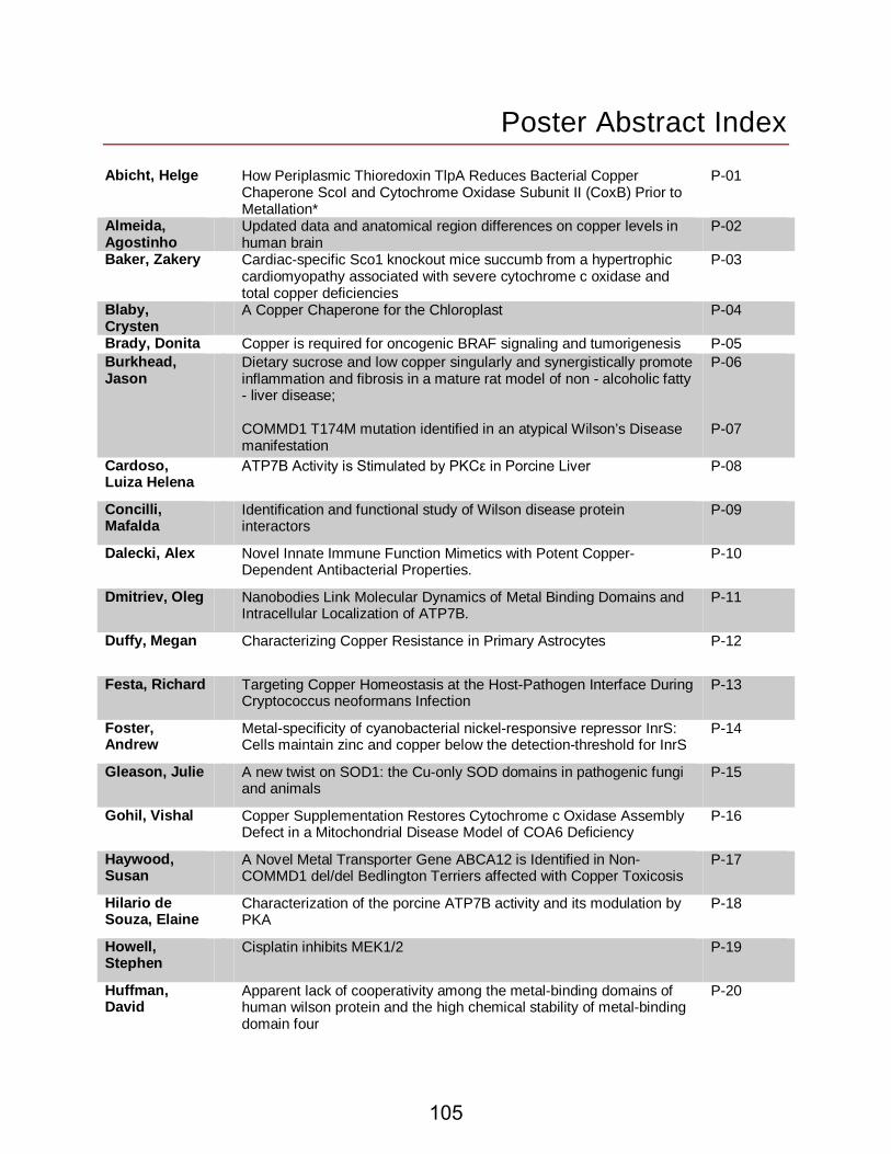
Poster Abstract Index Abicht, Helge How Periplasmic Thioredoxin TlpA Reduces Bacterial Copper
Chaperone ScoI and Cytochrome Oxidase Subunit II (CoxB) Prior to Metallation*
P-01
Almeida, Agostinho
Updated data and anatomical region differences on copper levels in human brain
P-02
Baker, Zakery Cardiac-specific Sco1 knockout mice succumb from a hypertrophic cardiomyopathy associated with severe cytochrome c oxidase and total copper deficiencies
P-03
Blaby, Crysten
A Copper Chaperone for the Chloroplast P-04
Brady, Donita Copper is required for oncogenic BRAF signaling and tumorigenesis P-05 Burkhead, Jason
Dietary sucrose and low copper singularly and synergistically promote inflammation and fibrosis in a mature rat model of non - alcoholic fatty - liver disease; COMMD1 T174M mutation identified in an atypical Wilson’s Disease manifestation
P-06 P-07
Cardoso, Luiza Helena
ATP7B Activity is Stimulated by PKCε in Porcine Liver P-08
Concilli, Mafalda
Identification and functional study of Wilson disease protein interactors
P-09
Dalecki, Alex Novel Innate Immune Function Mimetics with Potent Copper-Dependent Antibacterial Properties.
P-10
Dmitriev, Oleg Nanobodies Link Molecular Dynamics of Metal Binding Domains and Intracellular Localization of ATP7B.
P-11
Duffy, Megan Characterizing Copper Resistance in Primary Astrocytes P-12
Festa, Richard Targeting Copper Homeostasis at the Host-Pathogen Interface During Cryptococcus neoformans Infection
P-13
Foster, Andrew
Metal-specificity of cyanobacterial nickel-responsive repressor InrS: Cells maintain zinc and copper below the detection-threshold for InrS
P-14
Gleason, Julie A new twist on SOD1: the Cu-only SOD domains in pathogenic fungi and animals
P-15
Gohil, Vishal Copper Supplementation Restores Cytochrome c Oxidase Assembly Defect in a Mitochondrial Disease Model of COA6 Deficiency
P-16
Haywood, Susan
A Novel Metal Transporter Gene ABCA12 is Identified in Non-COMMD1 del/del Bedlington Terriers affected with Copper Toxicosis
P-17
Hilario de Souza, Elaine
Characterization of the porcine ATP7B activity and its modulation by PKA
P-18
Howell, Stephen
Cisplatin inhibits MEK1/2 P-19
Huffman, David
Apparent lack of cooperativity among the metal-binding domains of human wilson protein and the high chemical stability of metal-binding domain four
P-20
105
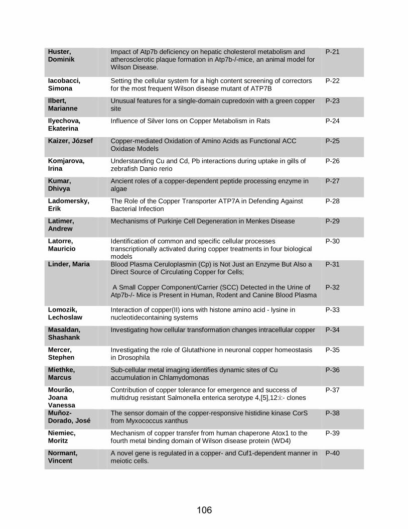
Huster, Dominik
Impact of Atp7b deficiency on hepatic cholesterol metabolism and atherosclerotic plaque formation in Atp7b-/-mice, an animal model for Wilson Disease.
P-21
Iacobacci, Simona
Setting the cellular system for a high content screening of correctors for the most frequent Wilson disease mutant of ATP7B
P-22
Ilbert, Marianne
Unusual features for a single-domain cupredoxin with a green copper site
P-23
Ilyechova, Ekaterina
Influence of Silver Ions on Copper Metabolism in Rats P-24
Kaizer, József Copper-mediated Oxidation of Amino Acids as Functional ACC Oxidase Models
P-25
Komjarova, Irina
Understanding Cu and Cd, Pb interactions during uptake in gills of zebrafish Danio rerio
P-26
Kumar, Dhivya
Ancient roles of a copper-dependent peptide processing enzyme in algae
P-27
Ladomersky, Erik
The Role of the Copper Transporter ATP7A in Defending Against Bacterial Infection
P-28
Latimer, Andrew
Mechanisms of Purkinje Cell Degeneration in Menkes Disease P-29
Latorre, Mauricio
Identification of common and specific cellular processes transcriptionally activated during copper treatments in four biological models
P-30
Linder, Maria Blood Plasma Ceruloplasmin (Cp) is Not Just an Enzyme But Also a Direct Source of Circulating Copper for Cells; A Small Copper Component/Carrier (SCC) Detected in the Urine of Atp7b-/- Mice is Present in Human, Rodent and Canine Blood Plasma
P-31 P-32
Lomozik, Lechoslaw
Interaction of copper(II) ions with histone amino acid - lysine in nucleotidecontaining systems
P-33
Masaldan, Shashank
Investigating how cellular transformation changes intracellular copper P-34
Mercer, Stephen
Investigating the role of Glutathione in neuronal copper homeostasis in Drosophila
P-35
Miethke, Marcus
Sub-cellular metal imaging identifies dynamic sites of Cu accumulation in Chlamydomonas
P-36
Mourão, Joana Vanessa
Contribution of copper tolerance for emergence and success of multidrug resistant Salmonella enterica serotype 4,[5],12:i:- clones
P-37
Muñoz-Dorado, José
The sensor domain of the copper-responsive histidine kinase CorS from Myxococcus xanthus
P-38
Niemiec, Moritz
Mechanism of copper transfer from human chaperone Atox1 to the fourth metal binding domain of Wilson disease protein (WD4)
P-39
Normant, Vincent
A novel gene is regulated in a copper- and Cuf1-dependent manner in meiotic cells.
P-40
106

Öhrvik, Helena
A Role for Copper in Mast Cells: Copper regulates mast cell maturation by affecting the storage and expression of tryptase and proteoglycans
P-41
Pantidos, Nikolaos
Production of stable copper nanoparticles using a biological approach P-42
Pérez, Juana Deepening into the mechanism of action of the CorE-like metal regulators
P-43
Petzoldt, Svenja
Cytoplasmic copper chaperones Atox1 and CCS exhibit cross-reactivity and can be loaded with copper by the Ctr1 C-terminus in vitro.
P-44
Polishchuk, Elena
Wilson disease protein ATP7B utilizes lysosomal exocytosis to maintain copper homeostasis
P-45
Sautron, Emeline
Biochemical characterization of AtHMA8/PAA2, a chloroplast-thylakoid Cu(I)-ATPase
P-46
Saviello, Maria rosaria
Structural properties of the Human Hetero-Complex BolA3-Glrx5 P-47
Scheiber, Ivo Florin
Iron copper interactions in the marine phytoplankton Ostreococcus tauri
P-48
Seigneurin-Berny, Daphné
Functional and biochemical characterization of PIB-type ATPases involved in the regulation of chloroplast copper homeostasis
P-49
Siotto, Mariacristina
Ceruloplasmin specific activity in serum of Alzheimer’s disease patients.
P-50
Solioz, Marc Characterization of a copper-induced quinone degradation pathway in Lactococcus lactis IL1403
P-51
Spampinato, Giorgia
8-Hydroxyquinoline derivatives and anticancer therapy: differential potentiation of cytotoxic effects by copper and zinc.; ATOX1 gene silencing increases susceptibility to anticancer therapy based on copper ionophores or chelating drugs
P-52 P-53
Thomason, RebeccaT.
Role of Peptidylglycine α− Amidating Monooxygenase in the Adaptive Plasticity of Anamniote Hatching
P-54
Vila, Alejandro
Mechanism of Biogenesis of the CuA Center in Human Cytochrome c Oxidase
P-55
Wedd, Anthony
The molecular properties of Atox1, Grx1 and GSH are consistent with a correlation between copper homeostasis and redox sulfur chemistry
P-56
Wiemer, Matthias
Paraquat stress induces a copper deficiency phenotype in the aging model Podospora anserina
P-57
Wittung-stafshede, Pernilla
Mechanisms of metal transfer to and from the human cytoplasmic copper chaperone, Atox1
P-58
Wolschendorf, Frank
Copper Ions in Disguise: A Trojan Horse Approach to Hyper-sensitize Drug-resistant Bacteria to Copper.
P-59
107


