the esters-treated rat at high dose, except for cMP females. Significant increase in apoptotic epithelial cells in the initial segment of the epididymis of high-dose ester-treated males was also observed. The results suggested that although acute renal toxicity was lower than 3-McPD, these three 3-McPD fatty acid esters have the potential to exert subchronic tox-icity to the rat kidneys and epididymis, to a similar degree as 3-McPD under the present conditions. NOAels (no-observed-adverse-effect levels) of cDP, cMP and cDO were suggested to be 14, 8 and 15 mg/kg B.W./day, respectively.
AdditivesNOAel No-observed-adverse-effect levellOel lowest observed effect levelPMTDI Provisional maximum tolerable daily intakeBMDl lower confidence limit of benchmark doseBMD Benchmark dose
Introduction
3-Monochloropropane-1,2-diol (3-McPD), belonging to a group of compounds called chloropropanols, is known to be one of the most common food-processing contaminants. It was first detected in acid-hydrolyzed vegetable proteins
Abstract 3-monochloropropane-1,2-diol (3-McPD), a rat renal and testicular carcinogen, has been reported to occur in various foods and food ingredients as free or esteri-fied forms. Since reports about toxicity of 3-McPD esters are limited, we conducted a 13-week rat subchronic toxic-ity study of 3-McPD esters (palmitate diester: cDP, pal-mitate monoester: cMP, oleate diester: cDO). We admin-istered a carcinogenic dose (3.6 × 10⁻⁴ mol/kg B.W./day) of 3-McPD or these esters at equimolar concentrations and two 1/4 lower doses by gavage with olive oil as a vehicle five times a week for 13 weeks to F344 male and female rats. As a result, five out of ten 3-McPD-treated females died from acute renal tubular necrosis, but none of the ester-treated rats. Decreased HgB was observed in all high-dose 3-McPD fatty acid ester-treated rats, except cDO-treated males. The absolute and relative kidney weights were signifi-cantly increased in the ester-treated rats at medium and high doses. Relative liver weights were significantly increased in
Electronic supplementary material The online version of this article (doi:10.1007/s00204-013-1190-6) contains supplementary material, which is available to authorized users.
S. Onami · y. cho · T. Toyoda · y. Mizuta · M. yoshida · K. Ogawa (*) Pathology, National Institute of Health Sciences, 1-18-1, Kamiyoga, Setagaya-ku, Tokyo 158-8501, Japane-mail: [email protected]
S. Onami · A. Nishikawa Pathogenetic Veterinary Science, united graduate School of Veterinary Sciences, gifu university, 1-1, yanagido, gifu, gifu 501-1193, Japan
A. Nishikawa Biological Safety Research center, National Institute of Health Sciences, 1-18-1, Kamiyoga, Setagaya-ku, Tokyo 158-8501, Japan
(acid-HVP) (Velíšek et al. 1980). But several subsequent studies have also demonstrated its presence in various foodstuffs such as cereal-derived products (bread, biscuits), malt-derived products, coffee, cheese, smoked food, meat and salted fish (Baer et al. 2010; crews et al. 2001).
concerning the toxicological profile, 3-McPD is known to be genotoxic in most in vitro assays (WHO 2002) but not in vivo assays (el Ramy et al. 2007; Robjohns et al. 2003; ScF 2001). In an animal bioassay, no evidence of carcinogenic potential was revealed in B6c3F1 mice receiving drinking water containing 3-McPD at 30, 100 and 300 (changed to 200 at day 100) ppm (equivalent to 4.2, 14.3, and 33.0 mg/kg B.W./day for males and 3.7, 12.2 and 31.0 mg/kg B.W./day for females, respectively) (Jeong et al. 2010). However, in the rat, it is considered to be a carcinogen targeting the kid-neys (renal tubule carcinomas) and testes (leydig cell tumor) from studies of drinking water administration at 25, 100 and 400 ppm (equivalent to 1.97, 8.27 and 29.50 mg/kg B.W./day for males and 2.68, 10.34 and 37.03 mg/kg B.W./day for females, respectively) in the SD strain (cho et al. 2008b) and at 20, 100 and 500 ppm (equivalent to 1.1, 5.2 and 28 mg/kg B.W./day for males and 1.4, 7.0 and 35 mg/kg B.W./day for females, respectively) in F344 animals (Sunahara et al. 1993). 3-McPD is highly suspected to be a non-genotoxic carcinogen, but there have been suggestions that the kidney tumors might be secondary to chronic nephropathy and the leydig cell tumors might be strain-specific and/or associated with hormonal imbalance (lynch et al. 1998; WHO 2002). In 2001, the Joint FAO/WHO expert committee on Food Additives (JecFA) established the provisional maximum tolerable daily intake (PMTDI) of 3-McPD as 2 μg/kg body weight based on the lowest observed effect level (lOel) of 1.1 mg/kg B.W./day found in a long-term study (Sunahara et al. 1993) and a safety factor of 500, which included a factor of five for extrapolation from an lOel to an no-observed-effect level (NOel) (WHO 2002). In this study, the lOel was derived from consideration of renal tubular hyperplasia observed in the low-dose 3-McPD group (1.1 mg/kg B.W./day) (Sunahara et al. 1993).
Importantly, further analyses have revealed that 3-McPD occurs in various foods either as a free form or more com-monly esterified with long-chain fatty acids. High concen-trations of 3-McPD fatty acid esters have been reported in hydrogenated fats, palm oil and palm oil fractions, and solid flying fats (IlSI 2009; Zelinková et al. 2006). Moreover, the occurrence of 3-McPD fatty acid esters in human breast milk has been documented (Zelinková et al. 2008). However, data for toxicity of 3-McPD fatty acid esters is quite limited, in spite of the considerable human exposure to these chemicals.
As evidence of acute oral toxicity of 3-McPD 1-mono-palmitic ester (cMP) and 3-McPD dipalmitic ester (cDP), liu et al. (2012) reported induction of renal tubular necrosis and decrease of spermatids in the seminiferous tubules of
Swiss mice, cMP exerting greater effects than cDP (liu et al. 2012). A ninety-day toxicological study of cDP conducted by Barocelli et al. (2011) at doses of 9.78, 39.19 and 156.75 mg/kg B.W./day by daily oral gavage with corn oil (2 ml/kg B.W.) in Wistar rats demonstrated renal and testicular changes induced by high-dose cDP are similar to that in phe-notype but milder than that with equimolar doses of 3-McPD. For cDP, the authors therefore proposed the benchmark doses (BMD10) as 41.1 and 64.1 mg/kg B.W./day and corresponding lower confidence limits of the benchmark doses (BMDl10) as 17.4 and 44.3 mg/kg B.W./day from severe renal and testicular damage, respectively (Barocelli et al. 2011).
3-McPD fatty acid esters include various forms with different fatty acids thought to be metabolized to 3-McPD in the body (Abraham et al. 2013; Buhrke et al. 2011; See-felder et al. 2008). However, it is unclear whether their tox-icological profiles are all identical to that of 3-McPD. As a further concern, 3-McPD might be metabolized to glycid-ols of a genotoxic carcinogen, though this reaction is char-acteristically observed in bacteria (Wijngaard et al. 1989). Therefore, prompt attention should be paid to potential tox-icity of 3-McPD fatty acid esters (Bakhiya et al. 2011).
To evaluate the toxicological profiles of three 3-McPD fatty acid esters, cDP, cMP and 3-McPD oleate diester (cDO) for comparison with 3-McPD, we conducted the present rat subchronic toxicity study with oral gavage administration using olive oil, which is reported to be mini-mally contaminated by 3-McPD fatty acid esters (Destail-lats et al. 2012; Zelinková et al. 2006).
Materials and methods
Test chemicals
Olive oil, 3-McPD (98 % pure), cDP (98 % pure), (sn1)-cMP (98 % pure) and cDO (98 % pure) were purchased from Wako Pure chemical Industries, ltd (Osaka, Japan). Initially, (sn2)-cMP was also synthesized by Wako for analysis, but was found to be unstable and naturally converting into (sn1)-cMP. For this reason, we analyzed only (sn1)-cMP as a monoester.
Animals, diet and housing conditions
Six-week-old male and female F344 rats were obtained from charles River Japan (Kanagawa, Japan). The ani-mals were housed in polycarbonate cages (three or four rats per cage) with soft chips for bedding in a room with a barrier system, maintained under conditions of controlled temperature (24 ± 1 °c), humidity (55 ± 5 %), air change (12 times per hour) and lighting (12 h light/dark cycle), and were given free access to a cRF-1 basal diet (Oriental yeast, Tokyo, Japan) and tap water.
Arch Toxicol
1 3
experimental design
After a one-week acclimatization period, 120 adult male F344 rats weighing 97.5–128.7 g and 120 adult female F344 rats weighing 81.5–103.8 g were used in this experi-ment. Rats were allocated with body-weight-basis ran-domization to twelve groups per sex, each consisting of 10 males and 10 females. The groups of both sexes were control (non-treatment group), olive oil (vehicle control group: 5 ml/kg B.W.), 3-McPD (40 mg/kg B.W.) and various doses of 3-McPD fatty acid ester groups. Doses of 3-McPD fatty acid esters were determined to be 14, 55 and 220 mg/kg B.W. for cDP, 8, 32 and 130 mg/kg B.W. for cMP and 15, 60 and 240 mg/kg B.W. for cDO, the highest doses in each case being equimolar concentrations to the 40 mg/kg B.W. for 3-McPD, selected on the basis of pre-vious carcinogenicity testing (cho et al. 2008b). 3-McPD and 3-McPD fatty acid esters were dissolved in olive oil at the time of dosing and administered five times a week by intragastric intubation for 13 weeks. During the period of administration, the animals were observed daily for any clinical signs and mortality. Body weights were measured weekly. At the end of the study, after an overnight fast, all the animals were anesthetized with isoflurane, weighed and blood samples were collected from the abdominal aorta for serum biochemistry and hematology. The animals were then killed by exsanguination from the abdominal aorta. The present experimental protocol was basically in accord-ance with guidelines for Designation for Food Additives and for Revision of Standards for use of Food Additives of Japan (1996), with minor changes, and approved by the Animal care and utilization committee of the National Institute of Health Sciences (MHlW 1996).
Serum biochemistry
Parameters for serum biochemistry shown in Supplemen-tary Table S3 and S4 were analyzed at SRl, Inc. (Tokyo, Japan) using sera frozen after centrifugation of whole blood.
Hematological examination
Parameters for hematology shown in Supplementary Table S5 and S6 were analyzed using an automated hematology analyzer, K-4500 (Sysmex corp., Hyogo, Japan). Differen-tial leukocyte and reticulocyte counts were performed with a MIcROx Heg-50S (Sysmex corp.).
Histopathology
All surviving animals were necropsied at 13 weeks. The brain, heart, lungs, liver, kidneys, spleen, thymus, adrenal glands and testes were weighed. Relative organ weights were
calculated as the values relative to body weights. In addition to these organs, the nasal cavity, trachea, aorta, pituitary, thy-roids, parathyroids, adrenal gland, salivary gland, tongue, esophagus, stomach, duodenum, jejunum, ileum, cecum, colon, rectum, pancreas, urinary bladder, epididymides, prostate, seminal vesicles, ovaries, uterus, vagina, mammary gland, skin, lymph node, sternum, femur including bone mar-row, sciatic nerve, trigeminal nerve, spinal cord (cervical, tho-racic and lumber cords), eyes and thigh muscle were fixed in 10 % neutral buffered formalin. Testes were fixed in Bouin’s solution overnight and then transferred into 10 % neutral buffered formalin. eyeballs, nervi opticus and Harderian glands were fixed in Davidson’s solution overnight and then transferred into 10 % neutral buffered formalin. Tissues that needed decalcification, such as the nasal cavity, spinal cord with bones, sternum and femur, were treated with a mixture of 10 % formic acid and 10 % neutral phosphate-buffered formalin. Adequately trimmed tissues were embedded in paraffin, sectioned at 3 μm thick for hematoxylin and eosin staining, and examined under a light microscope. Histopatho-logical examinations were carried out for the highest dose and control groups. If a chemical treatment-related change appeared at the highest dose, the relevant tissue(s) from the lower-dose groups were then also examined. Animals found dead or moribund were also analyzed as far as possible.
Immunohistochemical analysis
For the evaluation of cell proliferation in the kidney, Ki-67 labeling indices were immunohistochemically analyzed with anti-Ki67 rabbit monoclonal antibodies (SP6, 1:500: abcam, cambridge, uK) in the paracortical area of proxi-mal tubular epithelium per at least 2,000 proximal tubular epithelium cells per animal.
Statistical analysis
Variance in the data for body weights, organ weights, serum biochemistry and hematology was checked for homogeneity by Bartlett’s procedure. After analysis of vari-ance, the significance of differences in the multiplicity of lesions was evaluated using Tukey’s test. For incidences of histopathological findings, the Fisher’s exact probability test was applied. p values <0.05 between the olive oil group and treated groups were considered statistically significant.
Results
general condition, body weight
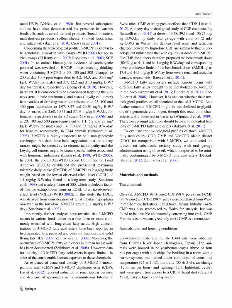
Survival curves are shown in Fig. 1. One male rat treated with 3-McPD died in the third week, one receiving
Arch Toxicol
1 3
14 mg/kg cDP in the first week, one given 220 mg/kg cDP in the 13th week and one administered 15 mg/kg cDO in the ninth week. Four female rats treated with 3-McPD died, one each in the first, second, and fourth weeks. One female given 55 mg/kg cDP died in the first week and one receiving 220 mg/kg cDP on the 13th week. One female rat treated with 3-McPD was observed to be moribund and therefore was immediately killed in the third week.
except for these 12 rats, there was no deterioration in the general conditions observed in any of the groups.
Body weight data are shown in Supplementary Fig. S1. The body weights of all groups increased gradually and progressively with age. Significantly elevated body weights were detected in the male non-treated control group com-pared to the vehicle control (olive oil) group in the 12th and 13th weeks. No significant differences in female body weight gain were detected among the treatment groups.
Organ weights
Final body weights and the absolute and relative organ weights are summarized in Supplementary Tables S1 and S2. Significant increase in absolute and relative kidney weights was observed in male and female rats treated with 3-McPD and with all 3-McPD fatty acid esters at medium
or high doses (Fig. 2). Significant increase in relative liver weights was also noted in 3-McPD and all high-dose 3-McPD fatty acid ester-treated male and female groups, except for cMP females. These changes observed in 3-McPD fatty acid ester groups showed dose dependence (Fig. 2).
The absolute liver and relative heart and spleen weights were significantly increased in high-dose cMP males. The relative heart weight was significantly increased in high-dose cDP and cMP females. The relative brain weights were significantly increased in high-dose cMP and cDO females.
Serum biochemistry
The results of serum biochemical analysis are sum-marized in Supplementary Tables S3 and S4. Signifi-cant increase in chlorine bilirubin (Bil) was observed in medium-dose cDP females. Significant decrease of aspar-tate aminotransferase (AST), alanine aminotransferase (AlT) and alkaline phosphatase (AlP) was observed in medium-dose cMP females, high-dose cMP or cDO females and medium- or high-dose 3-McPD fatty acid ester males, respectively. Significant decrease of blood urea nitrogen (BuN) was observed in cMP females. Sig-nificant decrease of creatinine (cre) was observed in the 3-McPD and 3-McPD fatty acid ester-treated groups, except for low-dose cMP males, and low-dose cDP and cDO females.
Significant decrease of sodium (Na) and chlorine (cl) was observed in low and medium cMP females, while sig-nificant increase was observed in high-dose cMP males.
Hematological examination
The results of hematological examination are summa-rized in Supplementary Tables S5 and S6. Significant decrease of HgB was observed in the groups treated at high dose with all 3-McPD fatty acid esters, except in cOD males (Fig. 3). Additionally, it was observed in low-dose cDP females and medium-dose cMP females. Significant decrease of HcT was observed in high-dose cMP males and high-dose cDP, cMP and cDO females. Significant decrease of RBc was also observed in females of low-dose and high-dose cDP, high-dose cMP and high-dose cOD groups. Significant decrease of McH was observed in females given 3-McPD and high doses of any of the 3-McPD fatty acid esters. Signifi-cant decrease of McHc was observed in females treated with 3-McPD and high-dose cDP and cDO. These changes reflecting anemia were seen with tendencies for dose dependence, especially in 3-McPD fatty acid ester-treated females.
0
20
40
60
80
100
0 1 2 3 4 5 6 7 8 9 10 11 12 13
Control
Olive oil
3-MCPD
CDP 14mg/kg
CDP 55mg/kg
CDP 220mg/kg
CMP 8mg/kg
CMP 32mg/kg
CMP 130mg/kg
CDO 15mg/kg
CDO 60mg/kg
CDO 240mg/kg
(weeks)
(%)
0
20
40
60
80
100
0 1 2 3 4 5 6 7 8 9 10 11 12 13
Control
Olive oil
3-MCPD
CDP 14mg/kg
CDP 55mg/kg
CDP 220mg/kg
CMP 8mg/kg
CMP 32mg/kg
CMP 130mg/kg
CDO 15mg/kg
CDO 60mg/kg
CDO 240mg/kg
(weeks)
a
(%)b
Fig. 1 Survival (%) for F344 rats treated with 3-McPD fatty acid esters for 13 weeks: a males; b females
Arch Toxicol
1 3
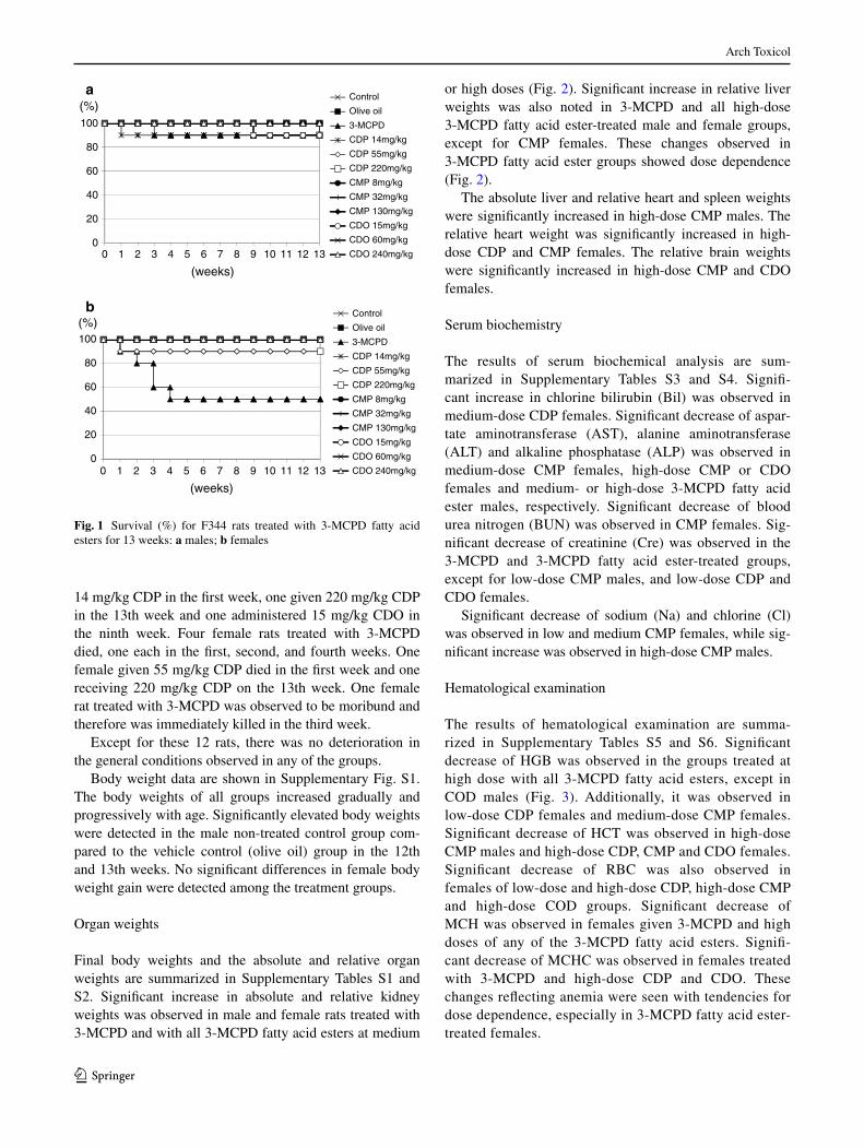
Histopathology
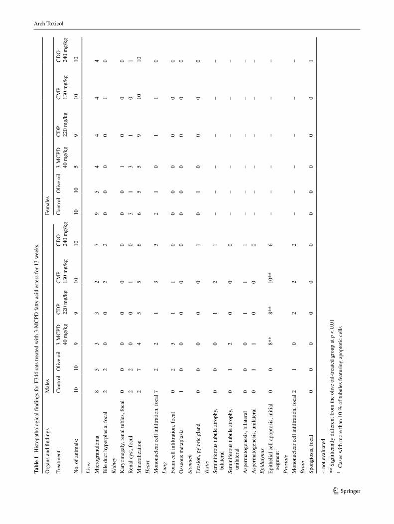
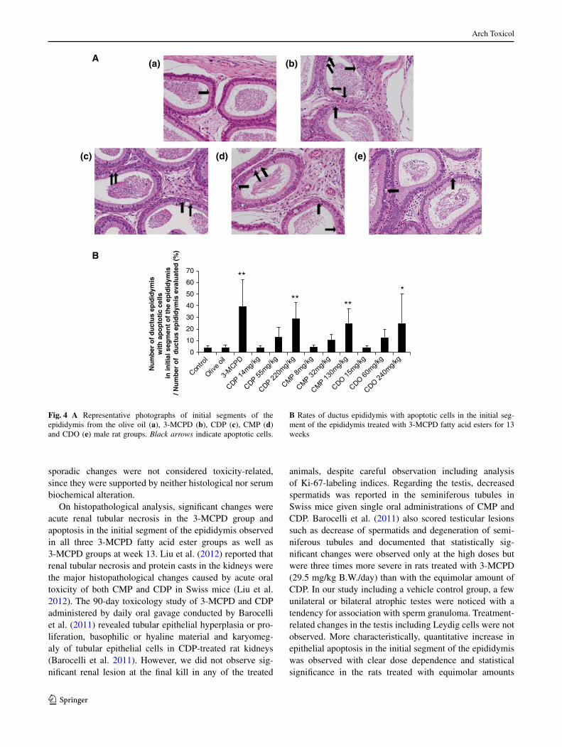
Severe tubular necrosis of the kidney was observed in the dead or moribund rats treated with 3-McPD (Sup-plementary Fig. S2). The dead rats in 3-McPD fatty acid ester groups did not show such changes in the kidney (Supplementary Table S7) but exhibited lesions includ-ing hemorrhage of the esophagus and thoracic cavity. The histopathological findings in the rats treated with high-dose test chemicals and controls for 13 weeks are sum-marized in Table 1. Additional examination of all dose groups was performed for the testis, epididymis and kid-ney. In the testis, focal granuloma, unilateral or bilateral seminiferous tubular atrophy and aspermatogenesis were observed without clear relation to the treatment and/or dose dependence. No treatment-related changes in ley-dig cells were observed. In the epididymis, apoptotic cell death (Fig. 4A) was noted, limited to the columnar epithe-lium of the initial segment, the degree being apparently
related to the dose. For quantitative evaluation, the rates of ducts with apoptotic cells in the initial segment of the epididymis were analyzed and that in male 3-McPD and high-dose 3-McPD fatty acid esters groups was signifi-cantly increased as compared to the vehicle control group (Fig. 4B). In the kidneys, neither proliferative lesions nor fibrosis were observed in the treated groups. The labeling index of Ki-67 in the proximal tubules was not altered in the treated groups (Supplementary Fig. S3). The incidence of renal mineralization in female 3-McPD fatty acid ester groups tended to be increased (Supplementary Fig. S4). In the brain, small spongiotic lesions with slight gliosis were observed in the bilateral nucleus of the diencephalon of one female treated with high-dose cDO (Supplementary Fig. S5). This lesion was not found in other rats, although additional sections for evaluation were carefully exam-ined. In other tissues, there were no significant changes in the incidences of lesions between the vehicle control and treatment groups.
a
c
(g%)
(g%)
(g%)
(g%)
b
d
****
****
****
**
**
**
**
**
**
*
**
** **
****
* * **
Kidney (female)Kidney (male)
Liver (female)Liver (male)
Fig. 2 Relative kidney and liver weights for F344 rats treated with 3-McPD fatty acid esters for 13 weeks: a relative kidney weights for males; b relative kidney weights for females; c relative liver weights
for males; d relative liver weights for females. *, **Significantly dif-ferent from the olive oil group at p < 0.05 and p < 0.01, respectively
Arch Toxicol
1 3
Discussion
In the present experiment, for comparison of toxicity of three 3-McPD fatty acid esters and 3-McPD in rats, we decided a dose level of 3-McPD close to that of the two-year rat carcinogenesis study performed by cho et al. (2008b) in which 400 ppm in drinking water (equivalent to 29.50 and 37.03 mg/kg B.W./day for male and female, respectively) was defined as the high-dose level (cho et al. 2008b). The mortality in our study was higher than that ear-lier at 13 weeks. In our experiment, five out of 10 females and one out of 10 males treated with 3-McPD died or became moribund by the end of week four. Barocelli et al. (2011) also reported that single oral gavage administration of 3-McPD resulted in similar mortality (50 %) but lower mortality (20 %) when 3-McPD was given twice a day at half the dose (Barocelli et al. 2011). Therefore, it was
suggested that peak level of blood 3-McPD concentration might be critical for acute toxicity of 3-McPD by oral gav-age, apparently more in female than in male rats. Indeed, similar to Barocelli’s study, histopathological examination revealed tubular necrosis in all six dead rats treated with 3-McPD. On the other hand, histological evidence of dam-age such as hemorrhage of the esophagus ~ thoracic cav-ity and/or bacterial colony of the lung were noted without any kidney lesions in all dead rats treated with 3-McPD fatty acid esters, thus suggesting accidental death related to gavage.
Abraham et al. (2013) reported oral gavage applica-tion of free 3-McPD with corn oil was followed by rapid absorption resulting in high peak levels in the blood within 30 min. In contrast, elevation of the blood 3-McPD con-centration after administration of cDP was considerably slower and resulted in correspondingly lower peak levels at 2–3 h (Abraham et al. 2013). considering that the mor-tality of rats given 3-McPD in the drinking water (cho et al. 2008b; Sunahara et al. 1993) was also low, our study supports the hypothesis that differences in peak 3-McPD blood levels affect the acute renal toxicity and the survival rate.
In serum biochemistry, none of the changes observed in this study were considered as toxicity-related, because they were opposite to those expected for toxic change or non dose-dependent. Surprisingly, no change related to renal toxicity was noted.
In hematological data, a proclivity for hypochromic ane-mia was observed in all 3-McPD fatty acid ester groups and more prominent in females than males with dose dependence, although histopathological changes in the hematopoietic organs related to hematopoesis or severe hemorrhage resulting in anemia were not observed. In the study conducted by Barocelli et al. (2011), a tendency for anemia was observed in males and females treated with either 3-McPD or cDP (Barocelli et al. 2011).
In organ weight data, the absolute and relative kidney weights in males and females were significantly increased in 3-McPD and in all 3-McPD fatty acid ester groups given at medium and high dose. Furthermore, significant increase in relative liver weights was noted in 3-McPD and high-dose 3-McPD fatty acid esters groups, except for female cMP group, without any related changes in histol-ogy or serum chemistry. cho et al. (2008a) reported similar increase in relative kidney weights with 200 and 400 ppm (equivalent to 36.97 and 76.79 mg/kg B.W./day for male, 30.23 and 61.34 mg/kg B.W./day for female, respectively) 3-McPD administration and increase in relative liver weights in females with 400 ppm 3-McPD administration for 13 weeks in B6c3F1 mice (cho et al. 2008a). Similar effects were also observed at high doses of either 3-McPD or cDP in Wistar rats (Barocelli et al. 2011). Other
a
b
HGB (male)
HGB (female)
(g/dL)
(g/dL)
**
**
**
*
** **
Fig. 3 concentration of HgB for F344 rats treated with 3-McPD fatty acid esters for 13 weeks: a males; b females. *, **Significantly different from the olive oil group at p < 0.05 and p < 0.01, respec-tively
Arch Toxicol
1 3
Tabl
e 1
His
topa
thol
ogic
al fi
ndin
gs f
or F
344
rats
trea
ted
with
3-M
cPD
fat
ty a
cid
este
rs f
or 1
3 w
eeks
-: n
ot e
valu
ated
** S
igni
fican
tly d
iffe
rent
fro
m th
e ol
ive
oil-
trea
ted
grou
p at
p <
0.0
11
cas
es w
ith m
ore
than
10
% o
f tu
bule
s fe
atur
ing
apop
totic
cel
ls
Org
ans
and
findi
ngs
Mal
esFe
mal
es
Tre
atm
ent:
con
trol
Oliv
e oi
l3-
Mc
PD40
mg/
kgc
DP
220
mg/
kgc
MP
130
mg/
kgc
DO
240
mg/
kgc
ontr
olO
live
oil
3-M
cPD
40 m
g/kg
cD
P22
0 m
g/kg
cM
P13
0 m
g/kg
cD
O24
0 m
g/kg
No.
of
anim
als:
1010
99
1010
1010
59
1010
Liv
er
Mic
rogr
anul
oma
85
33
27
95
44
44
Bile
duc
t hyp
erpl
asia
, foc
al2
20
02
20
00
01
0
Kid
ney
Kar
yom
egal
y, r
enal
tubl
es, f
ocal
00
00
00
00
10
00
Ren
al c
yst,
focu
l2
20
01
03
13
10
1
Min
eral
izat
ion
27
45
56
65
59
1010
Hea
rt
Mon
onuc
lear
cel
l infi
ltrat
ion,
foc
al7
22
13
32
10
11
0
Lun
g
Foam
cel
l infi
ltrat
ion,
foc
al0
23
11
00
00
00
0
Oss
eous
met
apla
sia
10
00
00
00
00
00
Stom
ach
ero
sion
, pyl
oric
gla
nd0
00
00
10
10
00
0
Test
is
Sem
inif
erou
s tu
bule
atr
ophy
, bi
late
ral
00
01
21
––
––
––
Sem
inif
erou
s tu
bule
atr
ophy
, un
ilate
ral
01
20
00
––
––
––
Asp
erm
atog
enes
is, b
ilate
ral
00
01
11
––
––
––
Asp
erm
atog
enes
is, u
nila
tera
l0
11
00
0–
––
––
–
Epi
didy
mis
epi
thel
ial c
ell a
popt
osis
, ini
tial
segm
ent1
00
8**
8**
10**
6–
––
––
–
Pro
stat
e
Mon
onuc
lear
cel
l infi
ltrat
ion,
foc
al2
10
22
2–
––
––
–
Bra
in
Spon
gios
is, f
ocal
00
00
00
00
00
01
Arch Toxicol
1 3
sporadic changes were not considered toxicity-related, since they were supported by neither histological nor serum biochemical alteration.
On histopathological analysis, significant changes were acute renal tubular necrosis in the 3-McPD group and apoptosis in the initial segment of the epididymis observed in all three 3-McPD fatty acid ester groups as well as 3-McPD groups at week 13. liu et al. (2012) reported that renal tubular necrosis and protein casts in the kidneys were the major histopathological changes caused by acute oral toxicity of both cMP and cDP in Swiss mice (liu et al. 2012). The 90-day toxicology study of 3-McPD and cDP administered by daily oral gavage conducted by Barocelli et al. (2011) revealed tubular epithelial hyperplasia or pro-liferation, basophilic or hyaline material and karyomeg-aly of tubular epithelial cells in cDP-treated rat kidneys (Barocelli et al. 2011). However, we did not observe sig-nificant renal lesion at the final kill in any of the treated
animals, despite careful observation including analysis of Ki-67-labeling indices. Regarding the testis, decreased spermatids was reported in the seminiferous tubules in Swiss mice given single oral administrations of cMP and cDP. Barocelli et al. (2011) also scored testicular lesions such as decrease of spermatids and degeneration of semi-niferous tubules and documented that statistically sig-nificant changes were observed only at the high doses but were three times more severe in rats treated with 3-McPD (29.5 mg/kg B.W./day) than with the equimolar amount of cDP. In our study including a vehicle control group, a few unilateral or bilateral atrophic testes were noticed with a tendency for association with sperm granuloma. Treatment-related changes in the testis including leydig cells were not observed. More characteristically, quantitative increase in epithelial apoptosis in the initial segment of the epididymis was observed with clear dose dependence and statistical significance in the rats treated with equimolar amounts
(a) (b)
(c) (e)(d)
0
10
20
30
40
50
60
70
Nu
mb
er o
f d
uct
us
epid
idym
is
wit
h a
po
pto
tic
cells
in
init
ial s
egm
ent
of
the
epid
idym
is/ N
um
ber
of
du
ctu
s ep
idid
ymis
eva
luat
ed (
%)
A
B
**
**
**
*
Fig. 4 A Representative photographs of initial segments of the epididymis from the olive oil (a), 3-McPD (b), cDP (c), cMP (d) and cDO (e) male rat groups. Black arrows indicate apoptotic cells.
B Rates of ductus epididymis with apoptotic cells in the initial seg-ment of the epididymis treated with 3-McPD fatty acid esters for 13 weeks
Arch Toxicol
1 3
of all three 3-McPD fatty acid esters as well as 3-McPD (40 mg/kg B.W./day). The initial segment of the epididymis is essential for male fertility (yeung et al. 1998), and after orchidectomy, apoptosis first appears in this site (Fan and Robaire 1998). Blocking lumicrine factors from enter-ing the epididymis results in a wave of apoptosis of epi-thelial cells of the initial segment, which does not occur in other epididymal regions and is not reversed by androgen replacement (Turner and Riley 1999). It is reported that this part of epididymis is uniquely regulated to preven-tion of apoptosis with testicular lumicrine factors through eRK, STST and NFKB pathways (xu et al. 2011). Further studies regarding the effects of 3-McPD and related fatty acid esters on the production of lumicrine factors and male infertility are required.
Our present experiment revealed a basically similar outcome to the study of Barocelli et al. (2011). As a gen-eral difference, the intensity of toxicological effects in rats treated with either cDP, cMP or cDO for 13 weeks were almost equivalent to those with equimolar 3-McPD in our experiment, although the earlier authors mentioned that the effect of cDP was milder than that of equimolar 3-McPD, particularly in the testis where it was one-thirds. As other differences, significant anemia was here observed only in ester-treated but not in 3-McPD-treated rats, apoptosis in the initial segment of the epididymis being the major change instead of testicular damage. Possible reasons for the differences are (1) Higher dose of esters and volume of oil used for vehicle, which might effect on lipase induction and hydrolysis efficacy; (2) The strain difference; and (3) Interval period of two days per week for administration. With the fact of acute renal toxicity seen only by gavage administration, for risk assessment, we should consider the route and type of administration in toxicity studies and whether they are relevant to human exposure. Regard-ing exposure levels for 3-McPD and its esters, based on national estimates from a wide range of foods including soy sauce and soy-sauce-related products provided for 10 countries (Denmark, Finland, France, germany, Ire-land, the Netherlands, Norway, Sweden, Thailand, uK), in 2007, JecFA estimated 3-McPD of 0.7 μg/kg B.W. per day could be taken to represent the average for the general population, and an intake of 2.3 μg/kg B.W. per day could be taken to represent consumers with a high intake (WHO 2007). Recent analysis of 3-McPD esters in food reported that 25.35 and 14.40 mg/kg of overall 3-McPD esters were detected in grape seed oil and palm oil, respectively (yamazaki et al. 2013). It might be more practical to esti-mate total 3-McPD including its esters for safety evalua-tion, because they might be easily interconverted.
In conclusion, no obvious differences of toxicological profile among cDP, cMP and cDO were here observed. compared with 3-McPD, the toxicities of the 3-McPD
fatty acid esters were lower in the acute phase regard-ing tubular necrosis and equivalent in the subchronic phase, as evidenced by kidney organ weight gain in males and females and apoptosis in the initial segment of the epididymis. Based on significant change in absolute and relative kidney weights, no-observed-adverse-effect lev-els (NOAels) in the present experiment in F344 rats were estimated to be 14 mg/kg B.W. for cDP, 8 mg/kg B.W./day for cMP and 15 mg/kg B.W./day for cDO, equimolar to 2.5 mg/kg B.W. 3-McPD in both males and females.
Acknowledgments We thank Ms. A. Saikawa and Ms. y. Komatsu for their expert technical assistance in processing histological mate-rials and Dr. g. Matsui for useful discussion and comments on his-topathological analysis. This study was funded by the Food Safety commission of Japan.
Conflict of interest The authors declare that they have no conflicts of interest regarding this work.
References
Abraham K, Appel Ke, Berger-Preiss e et al (2013) Relative oral bio-availability of 3-McPD from 3-McPD fatty acid esters in rats. Arch Toxicol 87(4):649–659
Baer I, de la calle B, Taylor P (2010) 3-McPD in food other than soy sauce or hydrolysed vegetable protein (HVP). Anal Bioanal chem 396(1):443–456
Bakhiya N, Abraham K, gürtler R, Appel Ke, lampen A (2011) Tox-icological assessment of 3-chloropropane-1,2-diol and glycidol fatty acid esters in food. Mol Nutr Food Res 55(4):509–521
Barocelli e, corradi A, Mutti A, Petronini Pg (2011) compari-son between 3-McPD and its palmitic esters in a 90-day toxi-cological study. ScIeNTIFIc RePORT submitted to eFSA. cFP/eFSA/cONTAM/2009/01. Accepted for publication on 22 August 2011. www.efsa.europa.eu/en/supporting/pub/187e.htm. Accessed 19 Sep 2013
Buhrke T, Weißhaar R, lampen A (2011) Absorption and metabolism of the food contaminant 3-chloro-1,2-propanediol (3-McPD) and its fatty acid esters by human intestinal caco-2 cells. Arch Toxicol 85(10):1201–1208
cho WS, Han BS, lee H et al (2008a) Subchronic toxicity study of 3-monochloropropane-1,2-diol administered by drinking water to B6c3F1 mice. Food chem Toxicol 46(5):1666–1673
cho WS, Han BS, Nam KT et al (2008b) carcinogenicity study of 3-monochloropropane-1,2-diol in Sprague-Dawley rats. Food chem Toxicol 46(9):3172–3177
crews c, Brereton P, Davies A (2001) The effects of domestic cook-ing on the levels of 3-monochloropropanediol in foods. Food Addit contam 18(4):271–280
Destaillats F, craft BD, Sandoz l, Nagy K (2012) Formation mecha-nisms of monochloropropanediol (McPD) fatty acid diesters in refined palm (Elaeis guineensis) oil and related fractions. Food Addit contam A chem Anal control expo Risk Assess 29(1):29–37
el Ramy R, Ould elhkim M, lezmi S, Poul JM (2007) evaluation of the genotoxic potential of 3-monochloropropane-1,2-diol (3-McPD) and its metabolites, glycidol and beta-chlorolactic acid, using the single cell gel/comet assay. Food chem Toxicol 45(1):41–48
Fan x, Robaire B (1998) Orchidectomy induces a wave of apoptotic cell death in the epididymis. endocrinology 139(4):2128–2136
IlSI (2009) (International life Sciences Institute) 3-McPD esters in Food Products, Summary Report of a Workshop held in February 2009 in Brussels, Belgium. http://www.ilsi.org/Publications/Final version 3 McPD esters.pdf. Accessed 19 Sep 2013
Jeong J, Han BS, cho WS et al (2010) carcinogenicity study of 3-monochloropropane-1, 2-diol (3-McPD) administered by drinking water to B6c3F1 mice showed no carcinogenic poten-tial. Arch Toxicol 84(9):719–729
liu M, gao By, Qin F et al (2012) Acute oral toxicity of 3-McPD mono- and di-palmitic esters in Swiss mice and their cytotoxicity in NRK-52e rat kidney cells. Food chem Toxicol 50(10):3785–3791
lynch BS, Bryant DW, Hook gJ, Nestmann eR, Munro Ic (1998) carcinogenicity of monochloro-1,2-propanediol (alpha-chlorohy-drin, 3-McPD). Int J Toxicol 17(47):47–76
MHlW (1996) (Ministry of Health, labour and Welfare, Japan) guidelines for designation for food additives and for revision of standards for use of food additives of Japan. http://www.mhlw.go.jp/topics/bukyoku/iyaku/syokuten/960322/betu.html. Accessed 19 Sep 2013
Robjohns S, Marshall R, Fellows M, Kowalczyk g (2003) In vivo genotoxicity studies with 3-monochloropropan-1,2-diol. Mutagenesis 18(5):401–404
ScF (Scientific committee on Food) (2001) Opinion on 3-mono-chloropropane-1,2-diol (3-McPD), updating the ScF opinion of 1994 adopted on 30 May 2001. http://ec.europa.eu/food/fs/sc/scf/out91_en.pdf. Accessed 19 Sep 2013
Seefelder W, Varga N, Studer A, Williamson g, Scanlan FP, Stadler RH (2008) esters of 3-chloro-1,2-propanediol (3-McPD) in veg-etable oils: significance in the formation of 3-McPD. Food Addit contam A chem Anal control expo Risk Assess 25(4):391–400
Sunahara g, Perrin I, Marchesini M (1993) carcinogenicity study on 3-monochloropropane-1,2-diol (3-McPD) administered in drink-ing water to Fischer 344 rats. un published report No Re-SR 93003 submitted to WHO by Nestec ltd, Research & Develop-ment, Switzerland
Turner TT, Riley TA (1999) p53 independent, region-specific epithe-lial apoptosis is induced in the rat epididymis by deprivation of luminal factors. Mol Reprod Dev 53(2):188–197
Velíšek J, Davídek J, Kubelka V, Janícek g, Svobodová Z, Simicová Z (1980) New chlorine-containing organic compounds in protein hydrolysates. J Agric Food chem 28(6):1142–1144
WHO (2002) 3-chloro-1, 2-Propandiol, WHO Food Add. Ser. 48, WHO, geneva, pp 401–432. http://www.inchem.org/documents/jecfa/jecmono/v48je18.htm. Accessed 19 Sep 2013
WHO (2007) 3-chloro-1,2-propanediol. WHO Food add. Ser. 58, WHO, geneva, pp 239–267
Wijngaard AJVD, Janssen DB, Witholt B (1989) Degradation of epichlorohydrin and halohydrins by bacterial cultures isolated from freshwater sediment. J gen Microbiol 135:2199–2208
xu B, Abdel-Fattah R, yang l, crenshaw SA, Black MB, Hinton BT (2011) Testicular lumicrine factors regulate eRK, STAT, and NFKB pathways in the initial segment of the rat epididymis to prevent apoptosis. Biol Reprod 84(6):1282–1291
yamazaki K, Ogiso M, Isagawa S, urushiyama T, ukena T, Kibune N (2013) A new, direct analytical method using lc-MS/MS for fatty acid esters of 3-chloro-1,2-propanediol (3-McPD esters) in edible oils. Food Addit contam A chem Anal control expo Risk Assess 30(1):52–68
yeung cH, Sonnenberg-Riethmacher e, cooper Tg (1998) Receptor tyrosine kinase c-ros knockout mice as a model for the study of epididymal regulation of sperm function. J Reprod Fertil Suppl 53:137–147
Zelinková Z, Svejkovská B, Velíšek J, Doležal M (2006) Fatty acid esters of 3-chloropropane-1,2-diol in edible oils. Food Addit contam 23(12):1290–1298
Zelinková Z, Novotný O, Schurek J, Velíšek J, Hajšlová J, Doležal M (2008) Occurrence of 3-McPD fatty acid esters in human breast milk. Food Addit contam A chem Anal control expo Risk Assess 25(6):669–676