A 3D localized surface plasmon resonance biosensor for the study of trivalent arsenic binding to the ArsA ATPase Chang Liu a , Vittoria Balsamo b , Dali Sun a , Melodie Naja d , Xuemei Wang e , Barry Rosen c , Chen-Zhong Li a,n a Nanobioengineering/Nanobioelectronics Laboratory, Department of Biomedical Engineering, Florida International University, 10555 W Flagler Street, Miami, FL 33174, USA b Departamento de Ciencia de los Materiales, Universidad Simon Bolivar, Apartado 89000, Caracas1080 A, Venezuela c Department of Cellular Biology and Pharmacology, Herbert Wertheim College of Medicine, Florida International University, 11200 S.W. 8th Street, AHC 2, Miami, FL 33199, USA d Everglades Foundation, 18001 Old Cutler Road, Palmetto Bay, FL 33157, USA e State Key Lab of Bioelectronics (Chien-Shiung Wu Laboratory), Southeast University, No. 2 Sipailou, Nanjing 210096, China article info Article history: Received 11 February 2012 Received in revised form 12 April 2012 Accepted 13 April 2012 Available online 26 April 2012 Keywords: Localized surface plasmon resonance Optical biosensor Arsenic Binding kinetics Nanoparticle 3-D polymer abstract A self-assembled 3D hydrogel–nanoparticle composite integrated surface plasmon resonance (SPR) sensor is reported here. The novel assembled substrate was developed by means of a surface mediated radical co- polymerization process to obtain a highly sensitive hydrogel-based thin film that possesses specific binding sites for target analytes. Initially, amino group modified gold nanoparticles (AuNPs) were covalently linked to acrylic acid monomer. Following this, N-isopropylacrylamide (NIPAAm) and AuNPs linked acrylic acid (AAc) monomers were randomly co-polymerized by the ‘‘grafting from’’ method in the presence of initiator and crosslinker onto the sensing surface. Surface charecterization techniques were utilized to evaluate the thickness and composition of the hydrogel-nanoparticle film. The sensing platform was employed to study the binding kinetics and conformational changes of the ArsA ATPase as a consequence of binding trivalent arsenicals under a variety of conditions. ArsA, the catalytic subunit of the ArsAB arsenite (As(III)) translocating ATPase, is one of the five proteins encoded by the arsenical resistance (ars) operon of plasmid R773 in cells of Escherichia coli, that confers resistance to trivalent and pentavalent salts of the metalloid arsenic. SPR measurements indicate that the 3D hydrogel-nanoparticle coated sensors exhibited a higher sensitivity than that of the 2D AuNPs decorated sensors. Binding of As(III) to ArsA is greatly facilitated by the presence of magnesium ion and ATP. & 2012 Elsevier B.V. All rights reserved. 1. Introduction Surface plasmon resonance has brought a revolutionary change to in vitro study of biological and biochemical processes due to its ability to measure extremely small changes in surface refractive index (RI), binding equilibrium and kinetics (Turner, 2000; Lee et al., 2006; Natsume et al., 2000; Quinn et al., 2000; Gao et al., 2006; Cooper, 2003). Strategies based on plasmonic nanoparticles have been employed to enhance the sensitivity for a variety of applications, such as diagnosis of diseases, environmental analysis, food safety, and chemical threat detection (Shan et al., 2010; Ali Syed et al., 2011). Theoretically, SPR can be induced only in free electron metals, e.g., Au, Ag and Cu, due to the interaction of surface electrons with the electromagnetic wave and the con- tribution from the interband transition of the d-shell electrons (Beyene et al., 2010; Raether, 1988). At the end of the 1990s, several research groups had begun exploring schemes for the development of localized surface plasmon resonance (LSPR) biosensors using noble metal nanoparticles because of the extremely sensitive nature of their electron-rich surfaces to the surrounding environ- ment. Based on the Mie theory, when an electromagnetic wave is directed to the metallic nanoparticle, an induced oscillation of free electrons occurs at the surface, resulting in a characteristic extinc- tion spectrum that depends on the type of metal, the size and shape of the nanoparticles, interparticle distance and most importantly, the RI of the surrounding medium (Jung et al., 1998; Link and El- Sayed, 1999; Kreibig and Vollmer, 1995). Compositional and con- formational changes within the surrounding dielectric medium near a nanoparticle could therefore be detected as shifts in the extinction spectrum. LSPR spectroscopy operates in a manner that is analogous to SPR to induce the extinction spectrum, however, the electromagnetic field of LSPR decays within a much smaller length than in SPR, which gives significant rise to the sensitivity of LSPR sensors. In 2008, Miura’s group demonstrated a prototype field portable LSPR system using monoclonal trinitrotoluene (TNT) antibody modified AuNPs for detection of TNT (Kawaguchi et al., 2008). They achieved a detectable range of 10 ppt to 100 ppb, which is four-fold more sensitive than that in the absence of AuNPs. Contents lists available at SciVerse ScienceDirect journal homepage: www.elsevier.com/locate/bios Biosensors and Bioelectronics 0956-5663/$ - see front matter & 2012 Elsevier B.V. All rights reserved. http://dx.doi.org/10.1016/j.bios.2012.04.026 n Corresponding author. Tel.: þ305 348 0120; fax: þ305 348 6954. E-mail address: licz@fiu.edu (C.-Z. Li). Biosensors and Bioelectronics 38 (2012) 19–26
Transcript
Biosensors and Bioelectronics 38 (2012) 19–26
Contents lists available at SciVerse ScienceDirect
Biosensors and Bioelectronics
0956-56
http://d
n Corr
E-m
journal homepage: www.elsevier.com/locate/bios
A 3D localized surface plasmon resonance biosensor for the study oftrivalent arsenic binding to the ArsA ATPase
Chang Liu a, Vittoria Balsamo b, Dali Sun a, Melodie Naja d, Xuemei Wang e,Barry Rosen c, Chen-Zhong Li a,n
a Nanobioengineering/Nanobioelectronics Laboratory, Department of Biomedical Engineering, Florida International University, 10555 W Flagler Street, Miami, FL 33174, USAb Departamento de Ciencia de los Materiales, Universidad Simon Bolivar, Apartado 89000, Caracas1080 A, Venezuelac Department of Cellular Biology and Pharmacology, Herbert Wertheim College of Medicine, Florida International University, 11200 S.W. 8th Street, AHC 2, Miami, FL 33199, USAd Everglades Foundation, 18001 Old Cutler Road, Palmetto Bay, FL 33157, USAe State Key Lab of Bioelectronics (Chien-Shiung Wu Laboratory), Southeast University, No. 2 Sipailou, Nanjing 210096, China
a r t i c l e i n f o
Article history:
Received 11 February 2012
Received in revised form
12 April 2012
Accepted 13 April 2012Available online 26 April 2012
A self-assembled 3D hydrogel–nanoparticle composite integrated surface plasmon resonance (SPR) sensor
is reported here. The novel assembled substrate was developed by means of a surface mediated radical co-
polymerization process to obtain a highly sensitive hydrogel-based thin film that possesses specific
binding sites for target analytes. Initially, amino group modified gold nanoparticles (AuNPs) were
covalently linked to acrylic acid monomer. Following this, N-isopropylacrylamide (NIPAAm) and AuNPs
linked acrylic acid (AAc) monomers were randomly co-polymerized by the ‘‘grafting from’’ method in the
presence of initiator and crosslinker onto the sensing surface. Surface charecterization techniques were
utilized to evaluate the thickness and composition of the hydrogel-nanoparticle film. The sensing platform
was employed to study the binding kinetics and conformational changes of the ArsA ATPase as a
consequence of binding trivalent arsenicals under a variety of conditions. ArsA, the catalytic subunit of the
ArsAB arsenite (As(III)) translocating ATPase, is one of the five proteins encoded by the arsenical resistance
(ars) operon of plasmid R773 in cells of Escherichia coli, that confers resistance to trivalent and pentavalent
salts of the metalloid arsenic. SPR measurements indicate that the 3D hydrogel-nanoparticle coated
sensors exhibited a higher sensitivity than that of the 2D AuNPs decorated sensors. Binding of As(III) to
ArsA is greatly facilitated by the presence of magnesium ion and ATP.
& 2012 Elsevier B.V. All rights reserved.
1. Introduction
Surface plasmon resonance has brought a revolutionary changeto in vitro study of biological and biochemical processes due to itsability to measure extremely small changes in surface refractiveindex (RI), binding equilibrium and kinetics (Turner, 2000; Leeet al., 2006; Natsume et al., 2000; Quinn et al., 2000; Gao et al.,2006; Cooper, 2003). Strategies based on plasmonic nanoparticleshave been employed to enhance the sensitivity for a variety ofapplications, such as diagnosis of diseases, environmental analysis,food safety, and chemical threat detection (Shan et al., 2010;Ali Syed et al., 2011). Theoretically, SPR can be induced only infree electron metals, e.g., Au, Ag and Cu, due to the interactionof surface electrons with the electromagnetic wave and the con-tribution from the interband transition of the d-shell electrons(Beyene et al., 2010; Raether, 1988). At the end of the 1990s, severalresearch groups had begun exploring schemes for the development
ll rights reserved.
þ305 348 6954.
of localized surface plasmon resonance (LSPR) biosensors usingnoble metal nanoparticles because of the extremely sensitivenature of their electron-rich surfaces to the surrounding environ-ment. Based on the Mie theory, when an electromagnetic wave isdirected to the metallic nanoparticle, an induced oscillation of freeelectrons occurs at the surface, resulting in a characteristic extinc-tion spectrum that depends on the type of metal, the size and shapeof the nanoparticles, interparticle distance and most importantly,the RI of the surrounding medium (Jung et al., 1998; Link and El-Sayed, 1999; Kreibig and Vollmer, 1995). Compositional and con-formational changes within the surrounding dielectric mediumnear a nanoparticle could therefore be detected as shifts in theextinction spectrum. LSPR spectroscopy operates in a manner thatis analogous to SPR to induce the extinction spectrum, however,the electromagnetic field of LSPR decays within a much smallerlength than in SPR, which gives significant rise to the sensitivity ofLSPR sensors. In 2008, Miura’s group demonstrated a prototypefield portable LSPR system using monoclonal trinitrotoluene (TNT)antibody modified AuNPs for detection of TNT (Kawaguchi et al.,2008). They achieved a detectable range of 10 ppt to 100 ppb,which is four-fold more sensitive than that in the absence of AuNPs.
C. Liu et al. / Biosensors and Bioelectronics 38 (2012) 19–2620
Using a similar principle, Kim’s group recently constructed an LSPRbiosensor based on subwavelength 1D and 2D gold nanoarrays builton a thin gold film for the detection of avian influenza DNAhybridization (Kim et al., 2010). Their results showed that 1Dnanogratings exhibited four-fold amplification of the SPR signal,and 2D nanohole arrays exhibited a 2.5-fold increase in amplifica-tion. Although such technologies have successfully demonstratedhigh detection sensitivity in the range of picomolar of analytes,small molecules such as heavy metal ions have had difficulty beingmeasured using this current setup.
The fabrication of the metallic nanostructure on the LSPRsensing layer is traditionally performed by ‘‘2D’’ methods. DebRoy’sgroup immobilized polyclonal antibodies for Escherichia coli (E. coli)
O157:H7 using biotin–neutravidin binding to detect E. coli O157:H7spiked in pasteurized milk (skim-milk), apple juice, and ground beefextract (Waswa et al., 2007). They also functionalized a SPR goldchip with carboxymethylated dextran layer followed by Protein Ato immobilize polyclonal antibodies against E. coli or Salmonella
Enteritidis (Waswa et al., 2006). In these cases, low concentrationtargets in the sample were difficult to detect because of the limitedinteraction time with the sensing surface due to continuous flow.Signal amplification is a common strategy for detection of lowconcentrations of target molecules. Cheng’s research group recentlyreported a novel SPR signal amplification strategy based on in situ
surface-initiated atom transfer radical polymerization (Liu et al.,2010), in which a polymer was used as a label for small molecules.Another feasible strategy is the functionalization of the SPR sensorchip with an absorbtive coating in addition to using bare nanoscalenoble metal structures for amplifying the sensor response. Radicalcopolymers would be ideal materials for this purpose due to theirhigh capacity for absorbing the analyte via a swelling-shrinkingprocess upon interacting with a water based buffer, enabling thesensing surface to capture larger amount of analyte (Zhang et al.,2002). This property allows interparticle distance tuning. Poly(N-isopropylacrylamide) (PNIPAAm) is a promising material thatcan satisfy these requirements (Chang et al., 2009; Lindqvist et al.,2008). Furthermore, copolymers obtained from the combination ofpolymers may result in even more sensing applications, as poly-mers with different functional groups allow the modulation of thematerial’s final properties for recognizing different analytes. Finally,radical copolymers prevent non-specific binding to the remainingfree gold surface in between the probes. This method is comparableto the conventional immunoassay approach in which the surface ismasked with a high concentration of a non-specific protein such asbovine serum albumin or denatured casein (Ehrentreich-Forsteret al., 2003).
Arsenic (As) is one of the most common toxic elements in theenvironment and is introduced by both geochemical and anthropo-genic sources. Exposure to this metalloid leads to cancer, cardiovas-cular and peripheral vascular diseases, diabetes and neurologicaldisease (Abernathy et al., 2003). The field of arsenic detoxification hasbeen studied for many decades (Bhattacharjee and Rosen, 2007).Nearly every organism, from bacteria to humans, has an arsenicdetoxifying system. In bacteria and archaea, the genes for arsenicresistance are usually found in arsenical resistance (ars) operons. Onesuch operon, the ars operon of plasmid R773, produces resistance totrivalent and pentavalent salts of the metalloids arsenic and anti-mony in cells of E. coli catalyzed by an ATP-coupled As(OH)3
extrusion pump (Chen and Rosen, 1997). The operon has five genes,arsRDABC (San Francisco et al., 1990). Among them, ArsR is a trans-acting repressor protein that homeostatically regulates the levels ofars transcription (Wu and Rosen, 1993). ArsD is an As(III) chaperonethat binds and transfers cytosolic arsenite to ArsA, an As(III)-activatedATPase (Lin et al., 2006). Together ArsA and ArsB, a transmembranearsenite antiporter, form the ATP-driven ArsAB As(OH)3 extrusionpump (Chen et al., 1986). In 2006, Tao’s group reported the first
application of SPR sensing for arsenic detection in groundwater(Forzani et al., 2007). However, there are no reports that utilize thistechnique for kinetics study on arsenic transportation and detoxifica-tion to date.
In this study, we integrated in situ radical copolymerizationand AuNPs to construct a novel LSPR sensing system. By growing3D AuNPs-doped PNIPAAm-co-PAAc hydrogel-based coating onthe gold sensing surface, we observed a 6.8-fold enhancement ofLSPR signal comparing to the traditional 2D AuNPs-decoratedgold sensing surface. We applied this LSPR sensor to study thebinding kinetics of the ArsA ATPase for As(III) in the presence ofadenosine-5’-triphosphate and magnesium ion (MgATP) andArsD. In previous studies, binding of As(III) by Ars proteins wasperformed by methods such as extended x-ray absorption finestructure (EXAFS) (Qin et al., 2007). EXAFS is a definitive methodfor analyzing metal ligands but lacks sensitivity and real-timeanalysis. In contrast, our 3D hydrogel based LSPR sensing strategyobtained direct, real-time binding kinetic information (Rich andMyszka, 2000). Thus, in addition to providing a means of amplify-ing the LSPR response, this work presents a novel approach tostudy the kinetic behavior of the arsenic extrusion pathway.Finally, the functionality of our novel 3D hydrogel-nanoparticlecoating can be easily modified by changing or adjusting loading ofmonomers, it has potential to be broadly applied to sensing arange of biological analytes.
2. Experimental
2.1. Materials
Allylmercaptan, AAc, NIPAAm, N,N-methylenebisacrylamide(BIS) were purchased from Acros Organic (New Jersey),2,20azobisisobutyronitrile (AIBN) and diisopropylfluorophosphate(DIFP) were obtained from Sigma-Aldrich (St. Louis, MO). 11-mercaptoundecanoic acid (MUA) was purchased from Asemblon(Redmond, WA). Cystamine dihydrochloride was obtained fromSpectrum (New Brunswick, NJ). 1-ethyl-3-(3-dimethylaminopro-pyl) carbodiimide hydrochloride (EDC), N-Hydroxysuccinamide(NHS) and hydrogen tetrachloroaurate (HAuCl4) were obtainedfrom Alfa Aesar (Ward Hill, MA). Trisodium citrate (Na3C6H5O7)and dimethyl sulfoxide (DMSO) were purchased from FisherScientific (Waltham, MA). All reagents and solvents were usedas received.
2.2. Protein expression and purification
Cells bearing the indicated plasmids were grown in LysogenyBroth (LB) medium overnight at 37 1C and then diluted 50-fold into1 L of the same medium. Proteins were expressed by induction with0.3 mM isopropyl-ß-D-thiogalactopyranoside at A600 of 0.6–0.8 for3 h. ArsA with a six histidine tag at the C-terminus was purifiedfrom cells of strain BL21 (DE3) expressing pAlter-1-dAhB plasmid,as described (Zhou and Rosen, 1997). Cells were harvested bycentrifugation and washed once with a buffer containing 50 mM 3-(N-morpholino)propanesulfonic acid (MOPS), pH 7.5, 0.5 M NaCl,30 mM imidazole and 10 mM 2-mercaptoethanol (Buffer A). Thecells were suspended in 5 mL of Buffer A per gram of wet cells andlysed by a single passage through a French press at 20,000 psi. DIFPwas added at 2.5 mL/g wet cells immediately following Frenchpress. Unbroken cells and membranes were removed by centrifuga-tion at 150,000 xg for 1 h at 4 oC. The supernatant was loaded to10 mL Probond Ni-column (Invitrogen) pre-equilibrated with BufferA. Unbound proteins were washed by 60 mL of buffer A, and ArsAwas eluted with imidazole gradient generated by Buffer A andBuffer B (50 mM MOPS, pH 7.5, 0.5 M NaCl, 300 mM imidazole and
C. Liu et al. / Biosensors and Bioelectronics 38 (2012) 19–26 21
10 mM 2-mercaptoethanol), followed by addition of 0.25 mMEthylenediaminetetraacetic acid (EDTA) and 5 mM Dithiothreitol(DTT) to each fraction. ArsA containing fractions were identified bysodium dodecyl sulfate polyacrylamide gel electrophoresis (SDS-PAGE), pooled, concentrated by Amicon Ultra-15 Centrifugal FilterUnit with Ultracel-10 membrane (Millipore), mixed with 10%glycerol, aliquoted and stored at �80 1C until use. ArsD and itsderivatives with a six histidine tag at the N-terminus were purifiedsimilarly. Purified proteins were stored at �80 1C until use, andtheir concentrations were determined according to the methodof Bradford (Bradford, 1976) or from the absorption at 280 nm(Gill and von Hippel, 1989).
2.3. Preparation of gold nanoparticles
Colloidal gold nanoparticles used in this study were prepared bycitrate reduction of HAuCl4 in aqueous solution (McFarland et al.,2004). The formation of gold nanoparticles can be observed by achange of color. Briefly, HAuCl4 and sodium citrate solutions werefiltered through a 22 mm microporous membrane filter prior to use.
Scheme 1. (A) Cartoon representation of the swelling-shrinking process of the copolym
illustration of the in-situ radical copolymerization of PNIPAAm-co-PAAc hydrogel.
HAuCl4 (40 mL, 1.0 mM) was then added to an Erlenmeyerflask(250 mL), vigorously stirred and brought to a boil on a hotplate.Following this, 3.5 mL of 1% trisodium citrate was added to the vortexof the boiling solution. 100 nm Au particles were formed 2 min afterthe addition of trisodium citrate and 15 nm AuNPs were obtained ifthe solution was stirred for an additional 10 min. A layer of absorbedcitrate anions on the surface of the nanoparticles prevents aggrega-tion. The particle size was determined by a ZEN3600 Zetasizer fromMalvern Instruments, Inc. (Westborough, MA).
2.4. In-situ polymerization
A stock of amino group modified AuNPs was prepared byincubating bare AuNPs in 1 mM cystamine dihydrochloride solu-tion for 12 h at 4 1C. Following this, 400 mmol AAc was added tothe amino modified AuNPs and incubated under the presence ofNHS/EDC for 30 min to form covalent linkage (pH¼7.4). Theresulting solution was then centrifuged at 7000 RCF for 10 minand the sediment was washed and resuspended with a samevolume of DMSO for further use (not shown in Scheme 1). A gold
er for LSPR signal amplification and immobilization of ArsA ATPase. (B) Structural
C. Liu et al. / Biosensors and Bioelectronics 38 (2012) 19–2622
SPR chip was cleaned using piranha solution for 2 min and rinsedwith copious ethanol. The cleaned gold chip was incubated in a10 mM allylmercaptan/ethanol solution afterward for 12 h atroom temperature in the absence of light. Lastly, the chip wasrinsed three times with ethanol and DMSO shortly prior to use toremove all the unbounded allylmercaptan residual. The in situ
polymerization was carried out in the following manner: 5 mLDMSO containing 950 mmol of NIPA, 150 mmol of AuNPs linkedAAc, 250 mmol of AAc, 58 mmol of crosslinking agent BIS and805 mmol of AIBN as the initiator were first added along with thegold chip into a three-neck round bottom flask. The resultingsolution was degassed by passage of a stream of nitrogen for aminimum of 20 min and then heated at 60 1C for 110 min innitrogen environment (Scheme 1). After polymerization, theAuNPs doped PNIPAAm-co-PAAc polymer-coated gold chip waswashed with DMSO, ethanol and water in order to remove anynon-bonded copolymer and unreacted monomers. Finally, thechip was stored under vacuum to remove any water absorbed inthe hydrogel before LSPR experiment. Control chips were alsoprepared by deposition of bare AuNPs on SPR chips and functio-nalized with carboxylic groups using MUA for characterizationpurposes.
2.5. Experimental setup and conditions
2.5.1. Surface characterization
Fourier transform infrared spectroscopy by attenuated totalreflectance (ATR-FTIR) was carried out using a FTIR-4100 spectro-meter (Easton, MD) with a maximum resolution of 0.9 cm. ANanoscope 3A atomic force microscope (AFM) obtained fromVeeco (Plainview, NY) was employed to measure the thicknessof the polymer thin film on the gold chip. Briefly, a 10�10 mm2
area of the polymer layer was scratched and removed using acontact mode AFM cantilever (spring constant 0.2 Nm) under theload of 500 nN force. To determine the depth of the scratchedarea, the morphology of a 30�30 mm2 area covering thescratched area was imaged by AFM under a 25 N force load witha scanning speed of 2 Hz. A JSM6330F field emission scanningelectron microscopy (SEM) from JEOL (Peabody, MA) wasemployed for observing the surface morphology and componentof the LSPR sensing surface.
Fig. 1. FTIR-ATR spectrum of the 3D PNIPAAm-co-PAAc polymer matrix on the
sensing surface.
2.5.2. Preparation of LSPR measurement
A BI-2000 SPR instrument was purchased from BiosensingInstrument (Tempe, AZ). A gold SPR chip (20 mm�20 mm�1 mm),which is a BK7 glass slide coated with a 45 nm layer of gold over a5 nm layer of chromium, was mounted on the upperface of the BK7prism with the gold layer facing upward. A 3–5 mL drop of indexmatching fluid (World Precision Instruments, Inc., FL) was appliedbetween the glass face of the chip and the prism with care so that noair bubbles were trapped at the interface. Following this, a biocom-patible polydimethylsiloxane (PDMS) microfluidic injection cham-ber gasket with a 5 mm�1.7 mm�125 mm channel was clampedto the gold face of the chip. A dual syringe pump was attached to theinjection chamber allowing both sample and reference buffer flowthrough the sensing surface. All buffers and solutions were degassedby vigorously stirring and purging with nitrogen before introductioninto the injection system to avoid oxidation of ArsA ATPase and ArsDmetallochaperone protein by air. Data collection and instrumentcontrol was performed using the Biosensing Instrument SPR ControlProgram running on a PC.
In the LSPR experiment, a 670 nm wavelength laser with a72.21 incident angle was employed as the light source. Thesensing surface was flushed by 50 mM MOPS buffer (pH 7.5)at a 150 mL/min flow rate until a stable SPR baseline was acquired.
In general, a 100–150 mDeg SPR angle shift would be observedduring this process due to the swelling of the PNIPAAm-co-PAAchydrogel upon introduction of water based MOPS buffer. TheMOPS flow rate was then reduced to 50 mL/min for optimizedbinding time and maintained during the whole LSPR measure-ment. All measurements were carried out in MPOS buffer envir-onment unless otherwise stated.
In order to demonstrate the enhanced sensitivity by the polymerhydrogel thin layer as the immobilization and sensing material, aswell as its suitability for binding kinetic study, ArsA ATPase wascoupled to the carboxylic groups of AAc and the specific interactionof As(III) with the immobilized ArsA was observed by LSPR. Initially,carboxylic groups of the polymer layer were activated with asolution of EDC (75 mM) and NHS (15 mM) at an injection rate of20 mL/min (denoted as X in Scheme 1). ArsA in MOPS with aconcentration of 50 mg/mL was injected with a same rate andallowed to react with the activated polymer for 4 min to form acovalent linkage between the surface carboxylic groups and theArsA amino-groups. This was followed by an addition of steppedconcentration of As(III) to study the binding kinetics and capacity ofthe ArsA ATPase. Finally, the above experiment was repeated usingcontrol chipscoated only with AuNPs.
3. Results and discussion
3.1. Surface morphology and composition characterization
To verify the incorporation of NIPAAm and AAc monomericunits in the copolymer grown from the allylmercaptan-modifiedgold surface, ATR-FTIR spectra were taken from the polymercoated gold chip surface. As a result, Fig. 1 shows a region ofthe ATR-FTIR spectrum obtained, which confirms the presence ofboth NIPAm and AAc in the thin film. The main bands at located at1651 and 1551 cm correspond to the carbonyl stretching of theamide group (amide I band) and to the N–H stretching of the
C. Liu et al. / Biosensors and Bioelectronics 38 (2012) 19–26 23
secondary amide (amide II band), respectively. On the other hand,the appearance of a much less intense absorption band at1716 cm, characteristic of the carboxyl group, indicates theincorporation of a minor acrylic acid fraction as expected. Besides,a band at 1460 cm is clearly observed, which can be attributed tothe –CH3 and –CH2– deformation of both monomeric units (Panet al., 2001; Yang et al., 2004).
The thickness of the PNIPAAm-co-PAAc polymer layer underhydrated and dehydrated condition was determined by AFM usingthe aforementioned manner (Hirata et al., 2004). Fig. 2(A) shows themorphologies and line profiles of the scratched areas under bothconditions, with the average thickness of each sample determinedfrom the line profiles. The hydrated polymer layer displays athickness of 40.673 nm, whereas the dehydrated polymer layerexhibited a thickness of 10.171 nm. Fig. 2(B) shows a SEM image inwhich AuNPs (bright dots) can be observed along the backbone of thepolymer hydrogel, indicating that AuNPs were successfully embeddedin the PNIPAAm-co-PAAc hydrogel by the linkage with AAc.
3.2. Optimization of ArsA immobilization conditions
The incubation time required for the binding reaction betweenArsA and polymer matrix is a critical parameter that determines theperformance of our kinetics sensor. Insufficient incubation time
Fig. 2. (A) AFM images of the polymer matrices under hydrated (upper) and dehydrated
LSPR sensor coated with AuNPs doped hydrogel thin film.
Fig. 3. Optimization of immobilization conditions. (A) ArsA immobilization on the 3D po
Maximum SPR angle shift is plotted versus responding injection rate. (B) Different conc
polymer coated LSPR sensor with optimal injection rate (20 mL/min). Maximum SPR an
causes low coverage of ArsA on the surface and leads to lowersensitivity, whereas over-long incubation time may results in multi-ple layers of ArsA and leads to blockage of binding sites and lowerefficiency. To optimize the time required for completeness of thecovalent linking, 80 mg/mL ArsA solution was injected and past thePNIPAAm-co-PAAc modified gold sensing surface at various injectionrates. LSPR angle shift was used to evaluate the surface coverage. Asshown in Fig. 3(A), this study found that ArsA immobilization with asample injection rate of 20 mL/min or slower exhibited a stableresponse demonstrating the complete interaction between ArsA andPNIPAAm-co-PAAc. Therefore, we employed optimized sample injec-tion rate of 20 mL/min for further immobilization and analysis.
In order to evaluate the amount of ArsA ATPase immobilizedon the surface of the 3D polymer matrix, we investigated the LSPRresponse to different concentrations of ArsA injections. Initially,5 polymer modified LSPR chips were activated using 75 mM EDCand 15 mM NHS in MOPS buffer. Following activation, eachactivated sensor was employed to react with different concentra-tions of ArsA solution at an injection rate of 20 mL/min. Theresulting LSPR response for each sensor was plotted against thecorresponding concentration of ArsA as shown in Fig. 3(B). It isevident that the amount of ArsA ATPase bound to the sensingsurface reaches a maximum at 50 mg/mL and remains constant atall concentration beyond. As a result, we employed 50 mg/mL asan optimum concentration for further binding study.
(lower) conditions, indicating the swelling-shrinking process. (B) SEM image of the
lymer coated surface with different injection rate (5, 10, 15, 20, 25, 30, 35 mL/min).
entration of ArsA (10, 20, 30, 40, 50, 60, 70, 80 mg/mL) immobilization on the 3D
gle shift is plotted versus ArsA concentration.
C. Liu et al. / Biosensors and Bioelectronics 38 (2012) 19–2624
3.3. ArsA and As(III) interaction
Sensitivity study was first performed with chips coated byAuNPs doped polymer layer. Prior to the binding test for eachdifferent concentrations of arsenite, ArsA was immobilized ontothe activated carboxylic groups by the aforementioned protocol.Then, three different concentrations of arsenite (8 mM, 4 mM,2 mM) were dosed separately with an injection rate of 20 mL/min.Fig. 4(a) shows the LSPR response to the injection of each arsenitesample. When the test solution was introduced at �50 s, �620 sand �1100 s, a blue shift of 25.8 mDeg, 19.1 mDeg and 16.5 mDegwere observed corresponding to the introduction of 8 mM, 4 mMand 2 mM arsenite sample, respectively. The 2D AuNPs structurewas not expected to induce as much LSPR enhancement as the 3DAuNPs doped polymer matrix according to our previous mentionedtheory. Therefore, we repeated the binding experiment with chipsdecorated by 2D AuNPs structure. Fig. 4(b) indicates that a blue shiftof 3.7 mDeg, 2.9 mDeg and 2.4 mDeg were recorded in response tothe same concentration gradients of arsenite. As a result, the signalenhancement of the 3D polymer matrix was calculated to be about6.8 fold. This increase can be attributed to the unique swelling-shrinking property of the PNIPAAm-co-PAAc hydrogel, yielding agreater capacity for ArsA immobilization than that of the 2D AuNPschip (Scheme 1).
We expected larger enhancement from the 3D polymer matrixsince the previous mentioned AFM measurements demonstratethat the thickness of the hydrated polymer is 40.615 nm, indicat-ing an ideal capacity increase of 180 fold for ArsA (63 kDa,a¼73.34, b¼75.64, c¼223.42 A) compared to the ArsA monolayerformed on the 2D chip. One possible explanation for this dis-crepancy is the heterogeneous distribution of carboxylic groupsduring the random copolymer reaction resulting in a lack ofavailable binding sites for ArsA. We also suspect that the injectedArsA bound to the surface of the polymer matrix has blocked theaccess of the unbound ArsA ATPase to binding sites inside thepolymer matrix.
3.4. Kinetics study of ArsA–As(III) binding
ArsD is a metallochaperone that delivers As(III), as well as othertrivalent metalloids, to the ArsA ATPase. Previous studies haveshown that interaction with ArsD increases the affinity of ArsA forAs(III), conferring resistance to environmental concentrations ofarsenic (Yang et al., 2010). However, there is a lack of research onthe binding affinity of ArsA for ArsD till now. Based on measure-ments on the effect of the As(III) chelator dimercaptosuccinic acid(DMSA) on the transfer reaction, it is suggested that As(III) transfer
Fig. 4. SPR sensorgrams for the covalently immobilized ArsA interacting with
As(III) obtained with (a) 3D AuNPs doped polymer coated sensor and (b) 2D AuNPs
structure modified sensor.
is directly channeling from one protein to the other, rather thandissociating from ArsD and reassociating with ArsA. Therefore,affinity study between ArsA and ArsD is essential to understandingthe structure of ArsA–ArsD complex and improve understanding ofthe transfer reaction.
Initially, ArsA was immobilized to the activated carboxylicgroups of the polymer as previously mentioned. Following this,1% ethanolamine was injected to block the free carbonyl groups.ArsD metallochaperone (50 mg/mL) was then introduced to theflow chamber at 20 mL/min. For comparison, ArsD was substitutedby bovine serum albumin (BSA) in the negative control experi-ment. Fig. S1 (supplementary materials) shows the LSPR sensor-gram for the ArsA–ArsD interaction study. The binding betweenArsA and ArsD (red) results in an angle shift of 356.2 mDeg,whereas the binding between ArsA and BSA (black) only results inan angle shift of 45.5 mDeg. Thus, it is evident that ArsA is able tospecifically bind ArsD without the presence of As(III) and MgATP.This result is consistent with the direct transfer of As(III) in theArsA–ArsD complex.
3.5. Effects of ArsD and MgATP on ArsA–As(III) binding
We have previously shown that ArsD transfers As(III) and Sb(III)to ArsA in the presence of MgATP (Lin et al., 2007). Herein, bindingkinetics of ArsA–As(III) interaction was compared using the 3D LSPRsensing platform under MgATP catalytic, ArsD promotion, andnoncatalytic conditions.
ArsA was bound to the 3D LSPR sensing surface using the samemanner. As(III) samples of 8 different concentrations (1–8 mM)were prepared in MOPS buffer. First, ArsA–As(III) binding kineticswas measured without catalysis as a control experiment. Sampleswith 1–8 mM As(III) were injected in 10 min interval betweeneach other to allow for baseline stabilization. SPR angle shift —
time curve (data not shown) was obtained for each sample inrealtime. In the association phase, the reaction rate, dR/dt, can bedescribed by the following equation:
maxdR
dt
� �¼ ka ArsA½ � AsðIIIÞ½ �
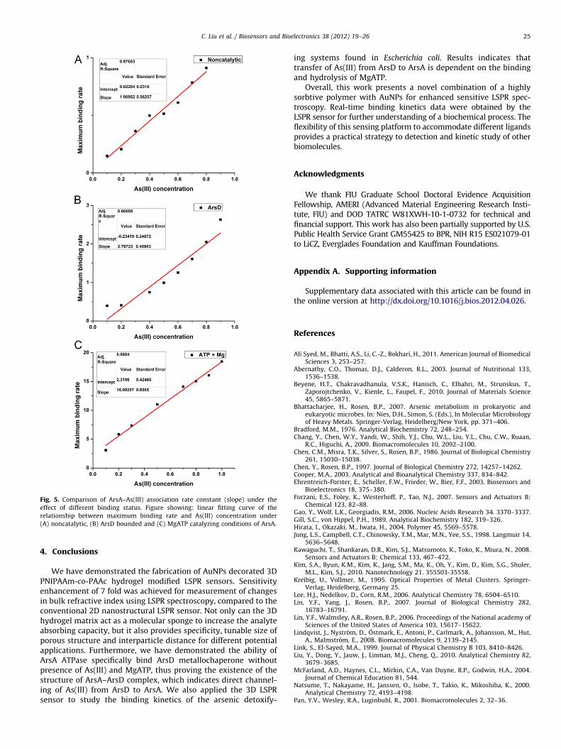
where R is the SPR signal at time t, ka is the association rateconstant which indicates the binding affinity between two reac-tants, [ArsA] and [As(III)] are the concentration of ArsA and As(III),respectively. The maximum value of (dR/dt) for each sample wasdetermined by calculating the maximum slope of the associationcurve using Matlab. Linear fitting was utilized to obtain ka since[ArsA] is constant. Fig. 5(A) shows the ka calculated to be 1.07 M/min for noncatalytic ArsA–As(III) interaction. Based on the afore-mentioned study, interaction with ArsD increases the affinity ofArsA for As(III). Therefore, association rate constant was alsomeasured for interaction under the presence of ArsD in theenvironmental buffer and ka was calculated to be 2.79 M/min asshown in Fig. 5(B). Furthermore, ArsA contains two nucleotide-binding sites (NBSs) and a binding site for arsenic, and crystallizesin the presence of As(III) and MgATP (Zhou et al., 2000). Based onthese structural features, we tested association rate constantunder the MgATP catalysis. Results show that the ka was sig-nificantly raised to 16.09 M/min (Fig. 5(C)). By comparing thesethree sets of experiments, it is obvious that the binding efficiencyis significantly raised under the presence of MgATP, indicatingthat the activity of the arsenic binding site is dependent on thebinding status of the two NBSs for ATP. Based on the result, As(III)transfer occurs only under conditions where ArsA hydrolyzes ATP,suggesting that ArsD transfers As(III) to an ArsA conformationtransiently formed during catalysis and not simply to the closedconformation that ArsA adopts when As(III) and MgATP are bound.
Fig. 5. Comparison of ArsA–As(III) association rate constant (slope) under the
effect of different binding status. Figure showing: linear fitting curve of the
relationship between maximum binding rate and As(III) concentration under
(A) noncatalytic, (B) ArsD bounded and (C) MgATP catalyzing conditions of ArsA.
C. Liu et al. / Biosensors and Bioelectronics 38 (2012) 19–26 25
4. Conclusions
We have demonstrated the fabrication of AuNPs decorated 3DPNIPAAm-co-PAAc hydrogel modified LSPR sensors. Sensitivityenhancement of 7 fold was achieved for measurement of changesin bulk refractive index using LSPR spectroscopy, compared to theconventional 2D nanostructural LSPR sensor. Not only can the 3Dhydrogel matrix act as a molecular sponge to increase the analyteabsorbing capacity, but it also provides specificity, tunable size ofporous structure and interparticle distance for different potentialapplications. Furthermore, we have demonstrated the ability ofArsA ATPase specifically bind ArsD metallochaperone withoutpresence of As(III) and MgATP, thus proving the existence of thestructure of ArsA–ArsD complex, which indicates direct channel-ing of As(III) from ArsD to ArsA. We also applied the 3D LSPRsensor to study the binding kinetics of the arsenic detoxify-
ing systems found in Escherichia coli. Results indicates thattransfer of As(III) from ArsD to ArsA is dependent on the bindingand hydrolysis of MgATP.
Overall, this work presents a novel combination of a highlysorbtive polymer with AuNPs for enhanced sensitive LSPR spec-troscopy. Real-time binding kinetics data were obtained by theLSPR sensor for further understanding of a biochemical process. Theflexibility of this sensing platform to accommodate different ligandsprovides a practical strategy to detection and kinetic study of otherbiomolecules.
Acknowledgments
We thank FIU Graduate School Doctoral Evidence AcquisitionFellowship, AMERI (Advanced Material Engineering Research Insti-tute, FIU) and DOD TATRC W81XWH-10-1-0732 for technical andfinancial support. This work has also been partially supported by U.S.Public Health Service Grant GM55425 to BPR, NIH R15 ES021079-01to LiCZ, Everglades Foundation and Kauffman Foundations.
Appendix A. Supporting information
Supplementary data associated with this article can be found inthe online version at http://dx.doi.org/10.1016/j.bios.2012.04.026.
References
Ali Syed, M., Bhatti, A.S., Li, C.-Z., Bokhari, H., 2011. American Journal of BiomedicalSciences 3, 253–257.
Bhattacharjee, H., Rosen, B.P., 2007. Arsenic metabolism in prokaryotic andeukaryotic microbes. In: Nies, D.H., Simon, S. (Eds.), In Molecular Microbiologyof Heavy Metals. Springer-Verlag, Heidelberg/New York, pp. 371–406.