53
A Biomimetics Approach to Engineering Arterial Conduits for Congenital Heart Surgery Timothy Martens, MD PhD Co-Director, Pediatric Heart Center Loma Linda University Children’s Hospital

A Biomimetics Approach to Engineering Arterial Conduits for Congenital Heart Surgery
Timothy Martens, MD PhDCo-Director, Pediatric Heart Center
Loma Linda University Children’s Hospital

Congenital Heart Disease
• Affects roughly 40,000 births annually in the United States
• 25% of cases considered severe, requiring surgical intervention within the first year of life
• Currently 3.3 million cases of congenital heart disease in the United States alone
https://www.cdc.gov/ncbddd/heartdefects/data.html

Chapter 18, Cardiovascular System 3
External Heart: Anterior View
Figure 18.4b

Truncus Arteriosus

Ross Procedure - Biventricular Outflow Tract Reconstruction

Current Prosthetic and Conduit Options
Vascular Conduit Options
Valve Repair and Replacement

Holy Grail• Engineered replacement that is fully functional upon
implantation• Has the capacity to keep pace with somatic growth
and avoid reoperation• Composed of non-immunogenic materials / cells• Durable

Biomimetic Approach
Grayson et al, Semin Cell Dev Biol (2009)

Scaffold Cells
PerfusionBioreactor

Hypothesis: Mimicking the physiologic milieu through pulsatile perfusion of a cardiovascular progenitor cell populated scaffold will induce vascular smooth muscle differentiation to create an arterial conduit exhibiting native mechanical properties and hemodynamics.

Experimental Overview
Early CPC Selection
Early CPC Expansion

Experimental Overview
In Vitro Static Conditioning
In Vitro Pulsatile Conditioning
Early CPC Expansion

Optimized Detergent Decellularization
• 1% Sodium Dodecyl Sulfate (SDS) – 24 Hour Wash• Anionic surfactant• Lyses cells
• 1% Triton X-100 – 1 Hour Wash• Nonionic surfactant• Solubilizes SDS for removal
• Phosphate Buffered Saline (PBS) – 72 Hour Wash• Removes SDS, Triton X-100, and cellular contents from
decellularized scaffold

Decellularized Pulmonary and Aortic Roots
• Matrix proteins well conserved across species –no shortage of donor scaffolds
• Structural and mechanical properties well preserved• Retention of matrix components• Decellularized Products already in widespread clinical use

Flawed decellularization leaves nuclear tissue or damages the matrix
Nuclei – PurpleExtracellular Matrix – Pink
Scale Bar - 100μm
Inadequate SDS Exposure Excessive SDS Exposure
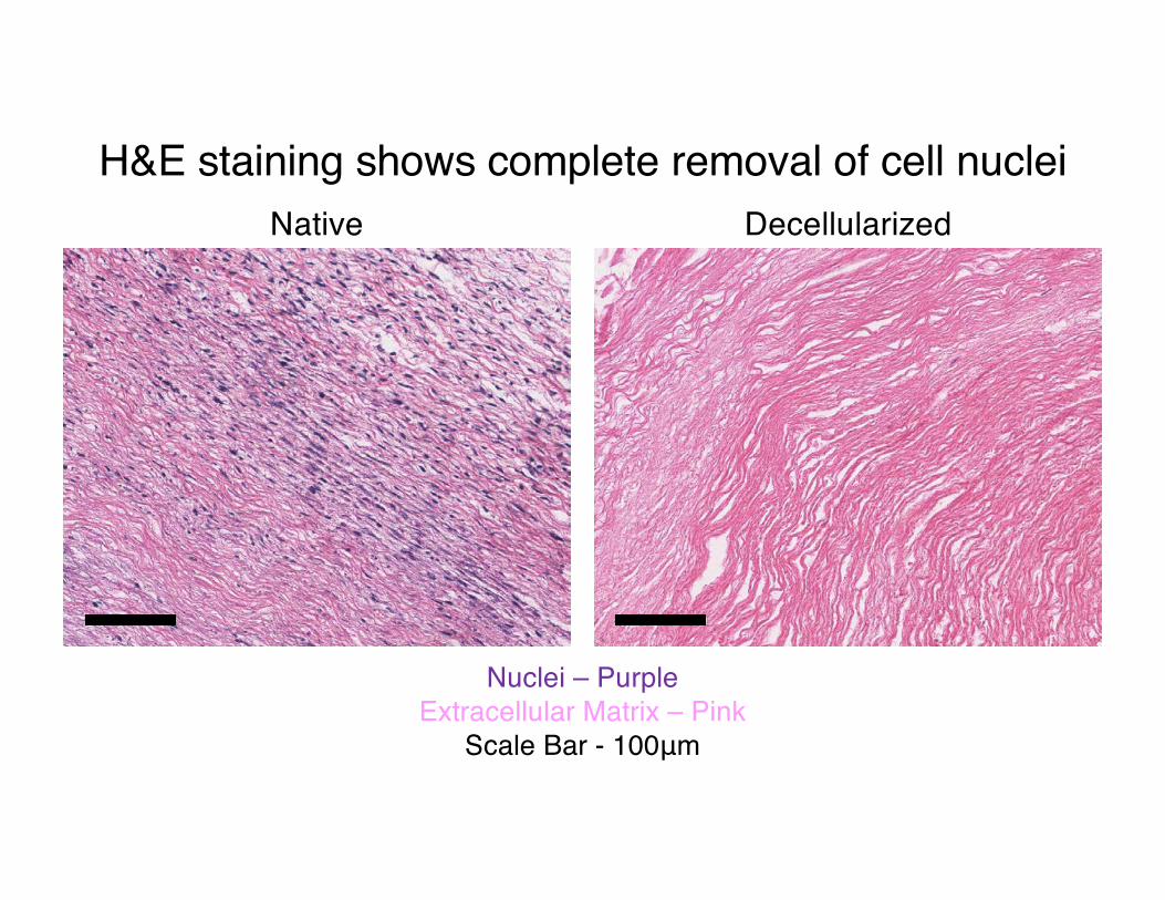
H&E staining shows complete removal of cell nuclei
Nuclei – PurpleExtracellular Matrix – Pink
Scale Bar - 100μm
Native Decellularized

Trichrome staining demonstrates collagen maintenance
Collagen - BlueCytoplasm – Red
Smooth Muscle - BrownScale Bar - 100μm
Native Decellularized

Elastin staining confirms elastin maintenance
Elastin – BlackCollagen - Pink
Scale Bar - 100μm
Native Decellularized

Scanning electron microscopy shows removal of cell bodies
Native Decellularized

Double-stranded DNA isolation showed significant removal of residual DNA
• 93% reduction in DNA content
• Acceptable for recellularization
• Biological activity of residual DNA unknown
• Will include DNase step for transplantation

Experimental Overview
Early CPC Selection
Early CPC Expansion

Cardiovascular Progenitor Lineages and Differentiation
Von Willebrand Factor (vWF) – EndothelialCardiac Troponin T (TropT) – CardiomyocyteSmooth Muscle Actin (SMA) – Smooth Muscle
Baio, J., npj Microgravity, 2018

Manual Recellularization with Tuberculin Syringe
• Cardiovascular progenitor cells (CPCs) suspended in 0.5mL of CPC growth media
• Injected into decellularized scaffolds with 27g tuberculin syringe• Eight linear tracts made with syringe and cells injected as the
needle was retracted• Static or pulsatile culture in CPC
growth media

Optimization of Manual Recellularization
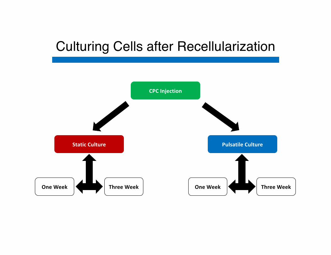
Culturing Cells after Recellularization
CPC Injection
Static Culture Pulsatile Culture
One Week One WeekThree Week Three Week

Static Culture Conditions
• Seeded scaffold submerged in 20mLs of CPC growth media in T-25 flask
• Incubator maintained at 37°C with 5% CO2
• CPC growth media changed every other day

Pulsatile Perfusion Conditions
• Seeded scaffold submerged in 20mLs of CPC growth media in T-25 flask• Allow 24 hours for cell adherence
• Scaffold mounted in perfusion chamber after 24 hours and connected in circuit with centrifugal perfusion pump
• Scaffold perfused with CPC growth media
• Incubator maintained throughout entire process at 37°C with 5% CO2

Pulsatile Perfusion Conditions

Pulsatile Perfusion Conditions

Ex Vivo Pulsatile Perfusion

Desired Cell Lineages

Cardiovascular Progenitor Cell Markers
W

Cardiomyocyte Markers

Endothelial Cell Markers

Smooth Muscle Cell Phenotyping
Synthetic Contractile
ü Smooth Muscle ActinCalponinMyosin Heavy Chain 11
ü Smooth Muscle Actinü CalponinüMyosin Heavy Chain 11

Smooth Muscle Cell Markers

Smooth muscle actin present in recellularized scaffolds at three weeks
SMA – Red DAPI – Blue Scale Bar - 100μm
Positive Control Recellularized

Ki67 presence indicates seeded cells are proliferative at three weeks
Ki67 – Green DAPI – Blue Scale Bar - 100μm
Positive Control Recellularized

Endothelial cells absent in recellularized scaffolds at three weeks
Scale Bar - 200μm vWF – Red DAPI – Blue Scale Bar - 100μm
Positive Control Recellularized

Cardiomyocytes absent in recellularized scaffolds at three weeks
TropT – Red DAPI – Blue Scale Bar - 100μm
Positive Control Recellularized

Native pulmonary arteries express calponin ubiquitously
Calponin - Green DAPI – Blue Scale Bar - 300μm
Positive Control Negative Control

Calponin is present on 3 Week Pulse arterial scaffold.
Calponin - Green DAPI – Blue Scale Bar - 300μm
3 Week Static 3 Week Pulse

Smooth Muscle Cell Markers

Tissue bath contraction studies functionally confirm decellularization of our pulmonary arteries

Functional testing verified contractility of three week pulsatile scaffolds
All scaffolds were tensioned to 1.5 grams prior to contractility testing.

Conclusions
• Detergent decellularization completely removes cells while maintaining native extracellular matrix.
• The decellularized scaffold microenvironment directs mesodermal differentiation into synthetic smooth muscle cells.
• Pulsatile perfusion commits cardiovascular progenitor cells to a contractile smooth muscle fate.
• Pulsatile perfusion may also induce endothelial differentiation with longer duration.


Animal Study
3 month old juvenile sheep
Allogeneic Implant into mPA
Unseeded Control (n=3)vs.
3Week Pulsatile (n=3)
Two week survival
Repeat Echocadiography ans Sacrifice

Echo Data – Pulmonary Artery Diameter (cm)161G 164G 168G 176G 185G Mean p
PRE 1.55 1.14 1.51 1.61 1.81 1.55
POST 1.71 1.17 1.93 1.61 1.88 1.71 N.S.
EXPLANT 1.61 1.29 1.60 1.64 1.60 1.62 N.S.
No pulmonary valve insufficiency
No pulmonary artery stenosis
Normal RV function

Native Tissue Decellularized Graft Recellularized Graft
Smoo
th M
uscl
e Ac
tinCa
lpon
inVo
n W
illeb
rand
Fac
tor

Future Directions
• Longer term implants to study true growth potential
• Pulmonary Root Replacement (valved conduit)
• Allogeneic vs. Autologous
• Pericytes?

Acknowledgements
• Bright Field Microscopy – Jon Hough
• UCR Central Facility for Advanced Microscopy and Microanalysis• Michael Pidgeon
• Funding - 2017 Grants to Promote Collaborative and Translational Research (GCAT)
• Mary Kearns-Jonker• Keren Garcia• Aaron Deatherage• Larry Lopez• Sean Wilson• Monica Romero• Sam Murray• Arlin Blood• Gordana Vunjak-
Novakovic

Questions?


![Biomimetics Report Final Version[1]](https://static.documents.pub/doc/80x56/5556b28dd8b42a9c798b546e/biomimetics-report-final-version1.jpg)


