A C-RING-like Domain Participates in Protein Self-Assembly andMineral NucleationFairland F. Amos, Moise Ndao, Christopher B. Ponce, and John Spencer Evans*
Laboratory for Chemical Physics, New York University, 345 East 24th Street, New York, New York 10010, United States
*S Supporting Information
ABSTRACT: AP7 is a nacre-associated protein of the mollusk shell that formssupramolecular assemblies that nucleate single-crystal aragonite in vitro. AP7possesses two major sequence regions: a random coil 30-amino acid N-terminaldomain (AP7N) and a partially disordered 36-amino acid C-terminal domain(AP7C) that exhibits imperfect sequence homology to the C subclass of theintracellular RING domain family. We report here new findings that implicate theC-RING domain in AP7-mediated supramolecular assembly and single-crystalmineral formation. AP7 protein spontaneously self-assembles over a pH range of4−9 and is monomeric at pH >9.5. AP7N and AP7C both oligomerize over the pH range of 4−9, with the AP7C sequenceclosely resembling AP7 in terms of particle morphology and size. In vitro mineralization experiments demonstrate that bothAP7N and AP7C form supramolecular assemblies that nucleate single-crystal calcium carbonates. Comparison of previouslypublished nuclear magnetic resonance-based structures of AP7C and AP7N reveals the significant presence of complementaryanionic−cationic electrostatic molecular surfaces on AP7C that are not found on AP7N, and this may explain the noteddiscrepancies between the two domains in terms of self-assembly and single-crystal nucleation. We conclude that the C-RING-like sequence is an important site for AP7 self-association and mineral nucleation, and this represents the first known instance ofa RING-like sequence performing these functions within an extracellular protein.
The formation of biominerals in Nature often requires theparticipation of specialized proteins that direct matrix
assembly, nucleation, and crystal growth.1−11 In many cases, thesequences of these specialized proteins are highly unique andoften feature regions that do not correspond to the sequencesof other known globular proteins.1−11 However, imperfectsequence homologies exist between certain regions ofbiomineralization proteins and other non-mineral-associatedproteins.1,3−5 We will refer to these imperfect homologousdomains as “imitator” domains, because they copy certainaspects of the globular sequence with less than perfect fidelity.Some recent examples have been identified in mollusk shellmineralization protein sequences, where regions partiallyhomologous to acetylcholine-binding domains,3 glycineloops,1 and disulfide core domains1 exist. For a number ofreasons, little if any information regarding the true function ofthese imitator domains within biomineralization proteins isavailable. However, there is speculation that these domainsperform functions that are not directly related to nucleation orcrystal interaction.3,5
One interesting example of an imitator domain-containingbiomineralization protein is AP7 (7.5 kDa, Haliotis rufes-cens).5,11,15−17 This intrinsically disordered12−14 nacre layerprotein forms supramolecular complexes that nucleate single-crystal and lamellar aragonite.11 The phenomenon of proteinsupramolecular complexes stabilizing aragonite has also beennoted for other nacre protein sequences.18−20 The AP7sequence consists of a random coil 30-amino acid N-terminalregion (1−30, AP7N)5,11,15−17 and a partially structured 36-amino acid C-terminal region (31−66, AP7C) (Figure 1). It is
this C-terminal domain that is most interesting, for it exhibitspartial homology (<50%)15 to an interesting subset of carboxy-terminal Cys-rich Zn binding motifs known as the C-RING(really interesting new gene domain, C subclass)21−24 and thusrepresents a putative imitator domain. Reported variants of theRING domain family occur in more than 400 proteins, andmany of these domains are typically involved in mediatingprotein−protein interactions within eukaryotic cells.21−24 Thus,the occurrence of a C-RING-like sequence within anextracellular biomineralization protein is highly intriguing, andit has been suggested that this sequence participates in theformation of mineralized AP7 supramolecular assemblies.11
However, this hypothesis has not yet been confirmed.In this paper,we report new experiments that establish the
functional relevance of the C-RING-like imitator sequencewithin AP7. Like other nacre protein sequences,18−20 weconfirm that AP7, AP7N, and AP7C all oligomerize in solutionin the pH range of 4.0−9.5. However, it is the C-RING-likeAP7C that most closely resembles the AP7 protein in terms ofsupramolecular assembly morphology and oligomer particlesize. In vitro mineralization studies confirm that both AP7Nand AP7C assemblies nucleate single-crystal calcium carbonatesin vitro. Solvent accessible surface analysis of published nuclearmagnetic resonance (NMR)-determined AP7N and AP7Cstructures15,17 reveals that AP7C possesses complementary
Received: June 2, 2011Revised: September 16, 2011Published: September 19, 2011
anionic−cationic molecular surfaces that are not found withinAP7N, and this may explain the significant pH-dependentoligomerization of both AP7C and AP7. We conclude that theAP7 C-RING-like sequence contributes to the self-assemblyand nucleation activities of AP7, and this represents the firstknown instance of a C-RING-like sequence performing thesefunctions within an extracellular protein.
■ MATERIALS AND METHODS
Synthesis and Purification of AP7, AP7N, and AP7C.The AP7 protein (Figure 1) was chemically synthesized usingtBoc chemistry and purified as described previously.11,16
Similarly, the protein fragments, AP7N (amino acids 1−30,free N-terminus, C-α amide capped) and AP7C (amino acids31−66, N-acetyl-modified N-terminus, free C-terminus)(Figure 1), were synthesized using FMOC chemistries andpurified as previously described.5,15,17 The purified, thiol-containing AP7 and AP7C polypeptides were subsequentlydissolved in H2- and N2-flushed unbuffered deionized distilledwater (UDDW).11,15,16 Aliquots were then stored at −20 °C inairtight sealed vials that were preflushed with highly pure N2gas. The AP7N peptide was stored in the lyophilized form at−20 °C until needed and was dissolved in UDDW immediatelyprior to the start of the experiments.5,11,17
Nacre Polypeptide Assembly and Calcium CarbonateNucleation. Using supernatant sampling,11,20 we recoveredAP7N, AP7C, and AP7 mineralization clusters from mineraliza-tion assay solutions for TEM visualization and electrondiffraction. These mineralization assays utilized 12.5 mMCaCl2 (>98% pure, Sigma/Aldrich) in UDDW, with solidammonium carbonate vapor (99% pure, Sigma/Aldrich)diffusion at 16 °C for 18 h.8,11,15,18−20 Final peptideconcentrations in these assays were 50 and 100 μM,corresponding to the protein concentrations that give rise tostabilized aragonite−protein complexes.11,18−20 Two controlconditions were utilized: no added peptide (negative control)and 5.4 μM (355 μg/mL) bovine serum albumin (BSA, >96%
pure, 66 kDa, Sigma-Aldrich), a concentration that is consistentwith that employed in previous mineralization studies.19,20 ForTEM studies, an aliquot (∼10 μL) of the assay supernatant wasremoved at the end of the 16 h period and carefully spotted ona copper TEM grid [coated with a Formvar layer that isstabilized by carbon (Ted Pella, Inc.)].11,20 This spot was gentlywashed with ethanol and UDDW, and then the grids wereallowed to air-dry. TEM imaging and electron diffractionmeasurements were performed on grids using a Philips CM12transmission electron microscope at 120 kV. Cropping of TEMimages and adjustment of brightness/darkness and contrastlevels were performed using Adobe Photoshop. For electrondiffraction measurements, we obtained diffraction patterns for10 crystals in each sample.Dynamic Light Scattering. The hydrodynamic radii of
AP7, AP7N, and AP7C were measured over a pH range of 4−11 using a DynaPro MS/X dynamic light scattering instrument(Protein Solutions, Inc.) and experimental protocols developedfor nacre polypeptides.18 The polypeptide samples were mixedin various 10 mM buffers (pH 4.0−6.0, sodium acetate/aceticacid buffer; pH 7.0−8.5, Tris-HCl buffer; pH 9.0−11.3, sodiumcarbonate/bicarbonate buffer), with final polypeptide concen-trations of 100 μM. All samples were filtered using a 0.22 μmpolyvinylidene fluoride syringe filter (Fisher) and thenincubated at 16 °C for 10 min in the cuvette prior tomeasurement. Ten acquisitions were taken per pH point persample. Data regularization analysis was performed usingDynamics version 6.0. By measuring the fluctuations in thelaser light intensity scattered by the sample, the instrument isable to detect the speed (diffusion coefficient) at which theparticles are moving through the medium. This value isconverted to hydrodynamic radius (RH) using the Stokes−Einstein relation:25
where D is the diffusion coefficient, k is Boltzmann's constant,T is the absolute temperature, η is the viscosity, and RH is thesphere-equivalent hydrodynamic radius.25
NMR Studies of Monomeric and Oligomeric AP7. Tocompare the degree of intermolecular association within AP7oligomers, we performed solution NMR experiments on 100μM samples [90% (v/v) UDDW and 10% (v/v) D2O (99.9%pure) (Cambridge Isotope Laboratories, Inc.)] at pH 4.0 and10.0, corresponding to the predominantly assembled andmonomeric states, respectively, as determined by our DLSexperiments. Two-dimensional proton NMR spectroscopy(TOCSY and NOESY) was conducted at 298 K on a 900MHz ultrashield Bruker US2 AVANCE spectrometer (21.14 Tsuperconducting magnet) outfitted with a 5 mm HXY triple-resonance Bruker cryoprobe. The 1H 90° pulses were 8.04 and8.90 μs for AP7 at pH 4 and 10, respectively; the relaxationdelay was 1.5 s, and a spectral width of 12 ppm was used. Watersuppression was achieved using gradient excitation sculpting.TOCSY experiments were performed with mixing times of 60,70, 75, and 80 ms, while NOESY experiments were performedwith mixing times of 50, 100, 150, and 200 ms. The data werecollected for 2048 complex points and transformed andvisualized using NMRPipe (National Institutes of Health,Bethesda, MD) and Sparky (Sparky 3, University of California,San Francisco, CA).
Figure 1. Histogram comparison of particle size distributions obtainedfor apo-AP7, AP7C, and AP7N polypeptides in aqueous buffers overthe pH range of 4−11. The error bars represent the skewedpolydispersity of the particles. The arrows indicate that the RH valuesfor a particular pH value are out of range for the DLS instrument.Results obtained in the presence of 12.5 mM CaCl2 are identical tothat presented here for the apo states. The primary amino acidsequence of AP7 is shown below the graph, along with the AP7N (1−30) and C-RING AP7C (31−66) sequence regions.
Electrostatic Surface and Solvent Accessible AreaCalculations for AP7N and AP7C. To gain further insightinto electrostatic-based self-assembly of AP7N and AP7C, weutilized the NMR-determined lowest-energy structures ofAP7N17 and AP7C15 fragments to determine the correspond-ing Poisson−Boltzmann solvation energies (vacuum to solventtransfer) and molecular surface plots at pH 8.0 using thePDB2PQR charge assignment (CHARMM19)26 and AdaptivePoisson−Boltzmann Solver (APBS) solvation calculationmodule27 within Python Molecular Viewer version 1.5.4(MGL Tools, Scripps Research Institute, La Jolla, CA). Forboth sequences, the solvation parameters utilized a linearizedPoisson−Boltzmann method, single Debye−Huckel boundaryconditions, a spline-based surface smoothing method, a proteindielectric of 2.0, a solvent dielectric of 78.54, a solvent proberadius of 1.4 Å, and a system temperature of 298 K.
■ RESULTSThe Oligomerization of AP7 Is pH-Dependent.
Previously, we observed the formation of aragonite-containingAP7 supramolecular assemblies within in vitro mineralizationassays under nonreducing conditions.11 The starting solutionfor these assays had a pH of approximately 3−4, and after theintroduction of (NH4)2CO3 vapor, the final assay pH wasapproximately 8.1−8.3 after 16 h.5,8,11,19,20 Hence, an alkalinepH shift occurs during the nucleation and supramolecularassembly processes, and thus, like that of other nacresequences,22 the formation of AP7 complexes may be pH-dependent.To assess this, we investigated the oligomerization of the
apo-AP7 protein (100 μM) over a pH range of 4−11 inbuffered solutions under nonreducing conditions using DLS(Figure 1). Note that we utilize the RH values to determine therelative differences in particle sizes under different bufferconditions. We will first discuss the events that occur within the
pH range of 4−9. Here, we found that the AP7 protein exists inan oligomeric state with polydispersity values of >15%,indicating that the AP7 particles are heterodisperse.9,18,25 AtpH 4, the hydrodynamic radius, RH, of AP7 is 100.8 ± 16.3 nm,and this value was found to be concentration-independent overthe range of this study [i.e., 25−100 μM (data not shown)]. Inthe pH range of 4.5−9.0, the RH scattering intensity exceeds themaximum detection limit of the instrument. This is schemati-cally portrayed on corresponding histogram bars as an arrowpointing toward higher RH values, reflecting a dramatic increasein either the number or the average size of the protein particlesin solution.9,18,25 We note that other nacre protein sequenceshave also exhibited out-of-range particle sizes or numbers underidentical conditions.18 The high scattering index at pH ≥5represents a further increase in the level of AP7 oligomeriza-tion25 and correlates with the appearance of micrometer-sizedsupramolecular assemblies in mineralization assays.11
At pH >9, we note a dramatic decrease in AP7 particle size(RH = 20.8 ± 10.1 nm at pH 9.5; RH = 1.2 ± 0.3 nm at pH 10)(Figure 1), which indicates that AP7 oligomers are unstablewithin this pH range. The RH value of 1.2 ± 0.3 nmcorresponds to a molecular mass of approximately 7 kDa, andthus at pH 10, the monomeric form of AP7 is preferred. Weconfirmed these findings by conducting 1H NMR TOCSYexperiments with 100 μM AP7 at pH 4.0 (oligomeric) and pH10 (monomeric) (Figure 2) and searching for evidence ofoligomer-related cross-peak intensity and frequency changes.Overlay TOCSY spectra reveal that there are pH-dependentspectral differences. First, we note differences in 1H chemicalshifts for backbone CHα and side chain methyl, methylene, andaryl ring protons, indicating that conformational differencesexist between oligomeric and monomeric AP7 molecules.9,28−30
Second, we note that there are TOCSY cross-peaks present inthe pH 10 spectra that are absent from the pH 4 spectra. Fromprevious NMR studies,28−30 we know that protein oligomeriza-
Figure 2. Overlay 900 MHz 1H TOCSY spectra of 100 μM apo-AP7 in a 90% (v/v) UDDW/10% (v/v) D2O mixture at 298 K and pH 4.0(oligomeric, yellow) and pH 10 (monomeric, red): (A) backbone Hα, side chain methyl, side chain methylene fingerprint region, (B) side chain aryl,amide, guanidinium fingerprint region, and (C) side chain methylene fingerprint region. 1H NMR chemical shifts are referenced from internal d4-TSP.
tion leads to attenuation of scalar cross-peak intensities due tothe formation of intermolecular contacts and subsequentintermediate time scale broadening. Hence, at pH 4, there aremore intermolecular interactions (oligomeric), and at pH 10,there are far fewer (monomeric), which correlates with ourDLS data (Figure 1). In summary, we find that undernonreducing conditions, AP7 is monomeric at pH 10 andexists as a high-molecular mass oligomer from pH 4 to 9 insolution. The pH dependency of AP7 oligomerization indicatesthat side chain electrostatics18,31−35 play an important role inAP7 protein−protein interactions. Note that line widthbroadening effects arising from large particle sizes and base-catalyzed solvent−backbone amide exchange preclude directNMR structural studies of AP7 in either the oligomeric (pH 4)or the monomeric (pH 10) form (Figure 2) at this time.pH-Dependent Self-Assembly of AP7 Domains. To
assess the participation of AP7N and AP7C in AP7oligomerization, we determined the individual assemblycapabilities of AP7N and AP7C in solution using DLS (Figure1). Here, we utilized the RH values to determine the relativedifferences in particle sizes formed by the three polypeptides.These experiments revealed that both sequences oligomerizeover the pH range of 4−9, but with striking differences. At pH4, both AP7N (RH = 2.3 ± 1.6 nm) and AP7C (1.8 ± 0.3 nm)are nearly identical in terms of RH values and exist as a mixtureof monomeric and oligomeric species. From pH 5 to 9,oligomerization predominates for both sequences, and it is theAP7C sequence that forms larger or more numerous proteinparticles, similar to those observed for AP7 (i.e., out ofdetection range). In contrast, the AP7N RH scatteringintensities remain within the range of detection and reach amaximum value of 79.7 ± 26.8 nm at pH 5 and plateau at 44.1± 20 nm from pH 6 to 9. Both AP7N and AP7C particles havepolydispersity values of >15%, indicating that these particles areheterodisperse.18,25 At pH >9, the RH values for both peptidesdecrease (AP7N RH ∼ 7−9 nm; AP7C RH ∼ 2−13 nm) but do
not attain monomeric values, indicating that both sequencesform smaller oligomers under these conditions. In contrast,under these same conditions, the AP7 protein is primarilymonomeric (Figure 1). From these results, we conclude thatboth AP7N and AP7C exist in an oligomeric state in the pHrange of 5−9, and it is likely that both assembly processes aredriven by side chain electrostatics.18,31−35 However, comparedto AP7N, the C-RING-like AP7C sequence forms larger ormore numerous polypeptide particles in solution, similar towhat we observe for the AP7 protein (Figure 1).Mineralization Activity of AP7 Domains. Having
established the self-assembly capabilities of AP7N and AP7Csequences (Figure 1), we now assess the nucleation capabilitiesof both domains using AP7-based mineralization assays andsupernatant sampling techniques (Figures 3 and 4). Super-natants recovered from negative control assays containedsingle-crystal calcite (inset image, Figure 3A; Figure S1 andTable S1 of the Supporting Information) and chainlikearrangements of round deposits (Figure 3A). As reportedelsewhere, AP7 assay supernatants contain amorphous-appearing complexes that contain electron-dense deposits(Figure 3B).11 AP7N assay samples contained irregularchainlike and clustered assemblies that contained a limitednumber of electron-dense particles (Figure 3C, note arrows). Incontrast, supernatants recovered from AP7C assays containnumerous electron-dense round deposits that are associatedwith a dense matrixlike material (Figure 3D−F), similar to whatis typically recovered from AP7 assays (Figure 3B) as well asother intracrystalline nacre polypeptide assays.20 These TEMimages correlate with our DLS findings: AP7N and AP7C bothself-assemble in solution, with the AP7C C-RING-like sequenceforming particles that are comparable to those formed by AP7(Figure 1).Electron diffraction patterns obtained for single-crystal
deposits nucleated by AP7N and AP7C reveal interestingdifferences compared to control scenarios that yielded only
Figure 3. Transmission electron micrographs of deposits recovered from assay supernatants: (A) negative control (no peptide) [note that typicaldeposits are either chainlike or single-crystal forms (inset image, scale bar of 0.5 μm)], (B) 100 μM AP7 [note electron-dense deposits within theprotein matrix (arrows)], (C) 100 μM AP7N [deposits appear chainlike or clustered and contain electron-dense material (arrows)], and (D−F) 100μM C-RING AP7C. The number of deposits appears greater than the number observed for either control or AP7N scenarios. Images E and Frepresent higher-magnification images of single-crystal mineral deposits and matrixlike material, respectively, formed by AP7C in solution.
calcite (Figure S1 of the Supporting Information). Mineralclusters produced by AP7N were found to be exclusively single-crystal calcite [10 of 10 crystals assayed (Figure 4A and Table1)], whereas crystals produced by AP7C were found to besingle-crystal calcite (9 of 10 crystals assayed) similar to thatshown in Figure 4A and Table 1 and, in limited instances,single-crystal aragonite [1 of 10 crystals assayed (Figure 4B andTable 1)].36 Obviously, the formation of single-crystal aragoniteis not statistically significant in these studies, and we note thatAP7C does not generate ordered lamellar or clustered single-crystal aragonite assemblies that are typical of AP7.11 On thebasis of these observations, we conclude that both AP7N andAP7C sequences nucleate single-crystal calcium carbonates insolution, with the C-RING-like AP7C sequence exhibiting avery limited ability to nucleate aragonite.Differences in pH-Dependent Assembly Are Linked to
Molecular Surface Electrostatics. The observation thatAP7C forms pH-dependent particles similar to those formedby AP7 (Figure 1) suggests that there are molecular features
found within AP7C that are important for AP7 assembly. TheNMR-determined structures of the AP7N17 and AP7C15
polypeptides are available for comparison but have yet to bejointly analyzed with regard to structural features, molecularsurface electrostatics, and how these features might impact self-assembly and nucleation. Thus, we utilized the NMR-determined coordinates of the lowest-energy structures ofAP7N17 and AP7C15 (Figure 5) and calculated the Poisson−
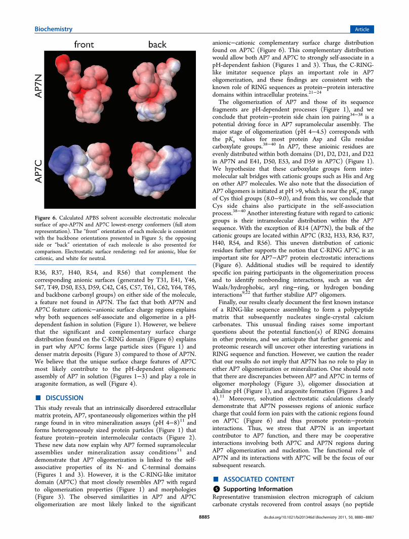
Boltzmann solvent accessible molecular surfaces for bothpolypeptides at pH 8.0 (mineralization assay conditions)(Figure 6). The lowest-energy structure of AP7N (Figure 5)highlights the intrinsically disordered11−14,28−30,40 conforma-tion of this sequence (extended loop random coil structure),and this contrasts with the partially structured AP7C C-RINGdomain15 that consists of three short α-helical segments andone β-turn segment, with each segment linked to another bylooplike random coil intervening segments.15 However, weobserve striking differences between these sequences withregard to solvent accessible electrostatic surfaces (Figure 6).Here, both domains possess cationic and anionic electrostaticregions, with the AP7N featuring greater anionic (carboxylate,Tyr, Ser, Thr-OH, and backbone carbonyl) than cationic (R14guanidinium) surface charge. In contrast, the AP7C sequencefeatures significant cationic surfaces (generated by R32, H33,
Figure 4. Transmission electron micrograph and correspondingelectron diffraction pattern of typical mineral crystals recovered from100 μM AP7N (top) and 100 μM AP7C (bottom) mineralizationassay supernatants. In the top panel, the diffraction pattern taken fromthe protruding crystallite encircled in the main figure unambiguouslymatches that of calcite. The lattice parameters obtained from bothdiffraction patterns can be found in Table 1. For reference, geologiccalcite lattice parameters (WWW-Mincryst Card 70634) are as follows:a = b = 0.49896 nm, c = 1.7061 nm, α = β = 90°, and γ = 120°. In thebottom panel, the diffraction pattern taken from this crystalunambiguously matches that of aragonite. For reference, geologicaragonite lattice parameters (WWW-Mincryst Card 29834) are asfollows: a = 0.49611 nm, b = 0.79672 nm, c = 0.57404 nm, α = β = γ =90°.36
Table 1. Experimental d Spacing and θ Angle ValuesObtained for Mineral Deposits within AP7N and AP7CAssemblies (Figure 4)a
aGeologic aragonite lattice parameters were taken from WWW-Mincryst Card 298: a = 0.49611 nm, b = 0.79672 nm, c = 0.57404 nm,α = β = γ = 90°. Geologic calcite lattice parameters were taken fromWWW-Mincryst Card 706: a = b = 0.49896 nm, c = 1.7061 nm, α = β= 90°, and γ = 120°.36
Figure 5. XPLOR-NIH-simulated annealing/molecular dynamicslowest-energy backbone structures (tube representation) of apo-AP7N and AP7C polypeptides.
R36, R37, H40, R54, and R56) that complement thecorresponding anionic surfaces (generated by T31, E41, Y46,S47, T49, D50, E53, D59, C42, C45, C57, T61, C62, Y64, T65,and backbone carbonyl groups) on either side of the molecule,a feature not found in AP7N. The fact that both AP7N andAP7C feature cationic−anionic surface charge regions explainswhy both sequences self-associate and oligomerize in a pH-dependent fashion in solution (Figure 1). However, we believethat the significant and complementary surface chargedistribution found on the C-RING domain (Figure 6) explainsin part why AP7C forms large particle sizes (Figure 1) anddenser matrix deposits (Figure 3) compared to those of AP7N.We believe that the unique surface charge features of AP7Cmost likely contribute to the pH-dependent oligomericassembly of AP7 in solution (Figures 1−3) and play a role inaragonite formation, as well (Figure 4).
■ DISCUSSIONThis study reveals that an intrinsically disordered extracellularmatrix protein, AP7, spontaneously oligomerizes within the pHrange found in in vitro mineralization assays (pH 4−8)11 andforms heterogeneously sized protein particles (Figure 1) thatfeature protein−protein intermolecular contacts (Figure 2).These new data now explain why AP7 formed supramolecularassemblies under mineralization assay conditions11 anddemonstrate that AP7 oligomerization is linked to the self-associative properties of its N- and C-terminal domains(Figures 1 and 3). However, it is the C-RING-like imitatordomain (AP7C) that most closely resembles AP7 with regardto oligomerization properties (Figure 1) and morphologies(Figure 3). The observed similarities in AP7 and AP7Coligomerization are most likely linked to the significant
anionic−cationic complementary surface charge distributionfound on AP7C (Figure 6). This complementary distributionwould allow both AP7 and AP7C to strongly self-associate in apH-dependent fashion (Figures 1 and 3). Thus, the C-RING-like imitator sequence plays an important role in AP7oligomerization, and these findings are consistent with theknown role of RING sequences as protein−protein interactivedomains within intracellular proteins.21−24
The oligomerization of AP7 and those of its sequencefragments are pH-dependent processes (Figure 1), and weconclude that protein−protein side chain ion pairing34−38 is apotential driving force in AP7 supramolecular assembly. Themajor stage of oligomerization (pH 4−4.5) corresponds withthe pKa values for most protein Asp and Glu residuecarboxylate groups.38−40 In AP7, these anioinic residues areevenly distributed within both domains (D1, D2, D21, and D22in AP7N and E41, D50, E53, and D59 in AP7C) (Figure 1).We hypothesize that these carboxylate groups form inter-molecular salt bridges with cationic groups such as His and Argon other AP7 molecules. We also note that the dissociation ofAP7 oligomers is initiated at pH >9, which is near the pKa rangeof Cys thiol groups (8.0−9.0), and from this, we conclude thatCys side chains also participate in the self-associationprocess.38−40 Another interesting feature with regard to cationicgroups is their intramolecular distribution within the AP7sequence. With the exception of R14 (AP7N), the bulk of thecationic groups are located within AP7C (R32, H33, R36, R37,H40, R54, and R56). This uneven distribution of cationicresidues further supports the notion that C-RING AP7C is animportant site for AP7−AP7 protein electrostatic interactions(Figure 6). Additional studies will be required to identifyspecific ion pairing participants in the oligomerization processand to identify nonbonding interactions, such as van derWaals/hydrophobic, aryl ring−ring, or hydrogen bondinginteractions9,22 that further stabilize AP7 oligomers.Finally, our results clearly document the first known instance
of a RING-like sequence assembling to form a polypeptidematrix that subsequently nucleates single-crystal calciumcarbonates. This unusual finding raises some importantquestions about the potential function(s) of RING domainsin other proteins, and we anticipate that further genomic andproteomic research will uncover other interesting variations inRING sequence and function. However, we caution the readerthat our results do not imply that AP7N has no role to play ineither AP7 oligomerization or mineralization. One should notethat there are discrepancies between AP7 and AP7C in terms ofoligomer morphology (Figure 3), oligomer dissociation atalkaline pH (Figure 1), and aragonite formation (Figures 3 and4).11 Moreover, solvation electrostatic calculations clearlydemonstrate that AP7N possesses regions of anionic surfacecharge that could form ion pairs with the cationic regions foundon AP7C (Figure 6) and thus promote protein−proteininteractions. Thus, we stress that AP7N is an importantcontributor to AP7 function, and there may be cooperativeinteractions involving both AP7C and AP7N regions duringAP7 oligomerization and nucleation. The functional role ofAP7N and its interactions with AP7C will be the focus of oursubsequent research.
■ ASSOCIATED CONTENT*S Supporting InformationRepresentative transmission electron micrograph of calciumcarbonate crystals recovered from control assays (no peptide
Figure 6. Calculated APBS solvent accessible electrostatic molecularsurface of apo-AP7N and AP7C lowest-energy conformers (full atomrepresentation). The “front” orientation of each molecule is consistentwith the backbone orientations presented in Figure 5; the opposingside or “back” orientation of each molecule is also presented forcomparison. Electrostatic surface rendering: red for anionic, blue forcationic, and white for neutral.
FundingSupported by the U.S. Department of Energy, Office of BasicEnergy Sciences, Division of Materials Sciences and Engineer-ing, via Grant DE-FG02-03ER46099.
■ ACKNOWLEDGMENTSThis is contribution 60 from the Laboratory for ChemicalPhysics, New York University.
■ ABBREVIATIONSAP7, aragonite protein 7 from Haliotis rufescens; AP7C, 36-amino acid C-terminal domain of the AP7 protein; AP7N, 30-amino acid N-terminal domain of the AP7 protein; IDP,intrinsically disordered protein; UDDW, unbuffered deionizeddistilled water; DLS, dynamic light scattering; RING, reallyinteresting new gene; C-RING, RING domain, C subclass;ACC, amorphous calcium carbonate.
■ REFERENCES(1) Shen, X., Belcher, A. M., Hansma, P. K., Stucky, G. D., and
Morse, D. E. (1997) Molecular cloning and characterization of LustrinA, a matrix protein from shell and pearl nacre of Haliotis rufescens. J.Biol. Chem. 272, 32472−32481.(2) Samata, T., Hayashi, N., Kono, M., Hasegawa, K., Horita, C., and
Akera, S. (1999) A new matrix protein family related to the nacreouslayer formation of Pinctada fucata. FEBS Lett. 462, 225−232.(3) Ma, Z., Huang, J., Sun, J., Wang, G., Li, C., Xi, L., and Zhang, R.
(2007) A novel extrapallial fluid protein controls the morphology ofnacre lamellae in the pearl oyster, Pinctada fucata. J. Biol. Chem. 282,23253−23260.(4) Suzuki, M., Saruwatari, K., Kogure, T., Yamamoto, Y., Nishimura,
T., Kato, T., and Nagasawa, H. (2009) An acidic matrix protein, Pif, isa key macromolecule for nacre formation. Science 325, 1388−1390.(5) Michenfelder, M., Fu, G., Lawrence, C., Weaver, J. C., Wustman,
B. A., Taranto, L., and Evans, J. S. (2003) Characterization of twomolluscan crystal-modulating biomineralization proteins and identi-fication of putative mineral binding domains. Biopolymers 70, 522−533. (2004) 73, 291 (erratum).(6) Fu, G., Qiu, S. R., Orme, C. A., Morse, D. E., and DeYoreo, J. J.
(2007) Acceleration of calcite kinetics by nacre proteins. Adv. Mater.17, 2678−2683.(7) Falini, G., Albeck, S., Weiner, S., and Addadi, L. (1996) Control
of aragonite or calcite polymorphism by mollusk shell macromolecules.Science 271, 67−69.(8) Ndao, M., Keene, E., Amos, F. A., Rewari, G., Ponce, C. B.,
Estroff, L., and Evans, J. S. (2010) Intrinsically disordered molluskshell prismatic protein that modulates calcium carbonate crystalgrowth. Biomacromolecules 11, 2539−2544.(9) Delak, K., Harcup, C., Lakshminarayanan, R., Zhi, S., Fan, Y.,
Moradian-Oldak, J., and Evans, J. S. (2009) The tooth enamel protein,porcine amelogenin, is an intrinsically disordered protein with anextended molecular configuration in the monomeric form. Biochemistry48, 2272−2281.(10) Mann, K., Siedler, F., Treccani, L., Heinemann, F., and Fritz, M.
(2007) Perlinhibin, a cysteine, histidine, and arginine-rich miniproteinfrom abalone (Haliotis laevigata) nacre, inhibits in vitro calciumcarbonate crystallization. Biophys. J. 93, 1246−1252.
(11) Amos, F. F., and Evans, J. S. (2009) AP7, a partially disorderedpseudo C-RING protein, is capable of forming stabilized aragonite invitro. Biochemistry 48, 1332−1339.(12) Uversky, V. N. (2002) Natively unfolded proteins: A point
where biology waits for physics. Protein Sci. 11, 739−756.(13) Uversky, V. N., Gillespie, J. R., and Fink, A. L. (2000) Why are
“natively unfolded” proteins unstructured under physiologic con-ditions? Proteins 41, 415−427.(14) Meng, J., Romero, P., Yang, J. Y., Chen, J. W., Vacic, V.,
Obradovic, Z., and Uversky, V. N. (2008) The unfoldomics decade:An update on intrinsically disordered proteins. BMC Genomics 9, 1−26.(15) Collino, S., Kim, I. W., and Evans, J. S. (2008) Identification and
structural characterization of an unusual RING-like sequence within anextracellular biomineralization protein, AP7. Biochemistry 47, 3745−3755.(16) Kim, I. W., Collino, S., Morse, D. E., and Evans, J. S. (2006) A
crystal modulating protein from molluscan nacre that limits the growthof calcite in vitro. Cryst. Growth Des. 6, 1078−1082.(17) Collino, S., and Evans, J. S. (2007) Structural features that
distinguish kinetically distinct biomineralization polypeptides. Bio-macromolecules 7, 1686−1694.(18) Amos, F. F., Ponce, C. B., and Evans, J. S. (2011) Formation of
framework nacre polypeptide supramolecular assemblies that nucleatepolymorphs. Biomacromolecules 12, 1883−1889.(19) Keene, E. C., Evans, J. S., and Estroff, L. A. (2010) Silk fibroin
hydrogels coupled with the n16N-β-chitin complex: An in vitroorganic matrix for controlling calcium carbonate mineralization. Cryst.Growth Des. 10, 5169−5175.(20) Amos, F. F., Destine, E., Ponce, C. B., and Evans, J. S. (2010)
The N- and C-terminal regions of the pearl-associated EF handprotein, PFMG1, promote the formation of the aragonite polymorphin vitro. Cryst. Growth Des. 10, 4211−4216.(21) Saurin, A. J., Borden, K. L. B., Boddy, M. N., and Freemont, P.
S. (1996) Does this have a familiar RING? Trends Biochem. Sci. 21,208−215.(22) Freemont, P. S. (2000) Ubiquitination: RING for destruction?
Curr. Biol. 10, R84−R87.(23) Capili, A. D., Edghill, E. L., Wu, K., and Borden, K. L. B. (2004)
Structure of the C-terminal RING finger from a RING-IBR-RING/TRIAD motif reveals a novel zinc-binding domain distinct from aRING. J. Mol. Biol. 340, 1117−1129.(24) Borden, K. L. B. (2000) RING domains: Master builders of
molecular scaffolds? J. Mol. Biol. 295, 1103−1112.(25) Schar̈tl, W. (2007) Light scattering from polymer solutions and
nanoparticle dispersions, 1st ed., Springer-Verlag, Heidelberg, Germany.(26) Dolinsky, T. J., Nielsen, J. E., McCammon, J. A., and Baker, N.
A. (2004) PDB2PQR: An automated pipeline for the setup, execution,and analysis of Poisson-Boltzmann electrostatics calculations. NucleicAcids Res. 32, 665−667.(27) Baker, N. H., Sept, D., Joseph, S., Holst, N. J., and McCammon,
J. A. (2001) Electrostatics of nanosystems: Application to micro-tubules and the ribosome. Proc. Natl. Acad. Sci. U.S.A. 98, 10037−10041.(28) Ndao, M., Dutta, K., Bromley, K., Sun, Z., Lakshminarayanan,
R., Rewari, G., Moradian-Oldak, J., and Evans, J. S. (2011) Probing theself-association, intermolecular contacts, and folding propensity ofamelogenin. Protein Sci. 20, 724−734.(29) Buchko, G. W., Tarasevich, B. J., Bekhazi, J., Snead, M. L., and
Shaw, W. J. (2008) A solution NMR investigation into the early eventsof amelogenin nanosphere self-assembly. Biochemistry 47, 13215−13222.(30) Li, M., Liu, J., Ran, X., Fang, M., Shi, J., Qin, H., Goh, J. M., and
Song, J. (2006) Resurrecting abandoned proteins with pure water: CDand NMR studies of protein fragments solubilized in salt-free water.Biophys. J. 91, 4201−4209.(31) Shera, J. N., and Sun, X. S. (2009) Effect of peptide sequence on
surface properties and self-assembly of an amphipathic pH-responsivepeptide. Biomacromolecules 10, 2446−2450.
(32) Zimenkov, Y., Dublin, S. N., Ni, R., Tu, R. S., Breedveld, V.,Apkarian, R. P., and Conticello, V. P. (2006) Rational design of areversible pH-responsive switch for peptide self-assembly. J. Am. Chem.Soc. 126, 6770−6771.(33) Dalmau, M., Lim, S., and Wang, S. W. (2009) pH-triggered
disassembly in a caged protein complex. Biomacromolecules 10, 3199−3206.(34) Kayser, V., Turton, D. A., Aggeli, A., Beevers, A., Reid, G. D.,
and Beddard, G. S. (2004) Energy migration in novel pH-triggeredself-assembled β-sheet ribbons. J. Am. Chem. Soc. 126, 336−343.(35) Rajagopal, K., Lamm, M. S., Haines-Butterick, L. A., Pochan, D.
J., and Schneider, J. P. (2009) Tuning the pH responsiveness of β-hairpin peptide folding, self-assembly, and hydrogel materialformation. Biomacromolecules 10, 2619−2625.(36) Chichagov, A. V., Varlamov, D. A., Dilanyan, R. A., Dokina, T.
N., Drozhzhina, N. A., Samokhvalova, O. L., and Ushakovskaya, T. V.(2001) MINCRYST: A crystallographic database for minerals, localand network (WWW) versions. Crystallogr. Rep. 46, 876−879.(37) Ndao, M., Keene, E., Amos, F. A., Rewari, G., Ponce, C. B.,
Estroff, L., and Evans, J. S. (2010) Intrinsically disordered molluskshell prismatic protein that modulates calcium carbonate crystalgrowth. Biomacromolecules 11, 2539−2544.(38) Song, Y., Mao, J., and Gunner, M. R. (2009) MCCE2:
Improving protein pKa calculations with extensive side chain rotamersampling. J. Comput. Chem. 30, 2231−2247.(39) Merz, K. M. (1991) Determination of pKas of ionizable groups
in proteins: The pKa of Glu 7 and 35 in hen egg white lysozyme andGlu 106 in human carbonic anhydrase. J. Am. Chem. Soc. 113, 3572−3575.(40) Li, H., Robertson, A. D., and Jensen, J. H. (2005) Very fast
empirical prediction and rationalization of protein pKa values. Proteins:Struct., Funct., Bioinf. 61, 704−721.