University of Kentucky UKnowledge Plant Pathology Faculty Publications Plant Pathology 9-2017 A Comparative Genome Analysis of Cercospora sojina with Other Members of the Pathogen Genus Mycosphaerella on Different Plant Hosts Fanchang Zeng Shandong Agricultural University, China, [email protected]Xin Lian Shandong Agricultural University, China Guirong Zhang University of Illinois - Urbana-Champaign, [email protected]Xiaoman Yu Shandong Agricultural University, China Carl A. Bradley University of Kentucky, [email protected]See next page for additional authors Right click to open a feedback form in a new tab to let us know how this document benefits you. Follow this and additional works at: hps://uknowledge.uky.edu/plantpath_facpub Part of the Genomics Commons , and the Plant Pathology Commons is Article is brought to you for free and open access by the Plant Pathology at UKnowledge. It has been accepted for inclusion in Plant Pathology Faculty Publications by an authorized administrator of UKnowledge. For more information, please contact [email protected]. Repository Citation Zeng, Fanchang; Lian, Xin; Zhang, Guirong; Yu, Xiaoman; Bradley, Carl A.; and Ming, Ray, "A Comparative Genome Analysis of Cercospora sojina with Other Members of the Pathogen Genus Mycosphaerella on Different Plant Hosts" (2017). Plant Pathology Faculty Publications. 73. hps://uknowledge.uky.edu/plantpath_facpub/73
A Comparative Genome Analysis of Cercosporasojina with Other Members of the Pathogen GenusMycosphaerella on Different Plant HostsFanchang ZengShandong Agricultural University, China, [email protected]
Xin LianShandong Agricultural University, China
Guirong ZhangUniversity of Illinois - Urbana-Champaign, [email protected]
Right click to open a feedback form in a new tab to let us know how this document benefits you.Follow this and additional works at: https://uknowledge.uky.edu/plantpath_facpub
Part of the Genomics Commons, and the Plant Pathology Commons
This Article is brought to you for free and open access by the Plant Pathology at UKnowledge. It has been accepted for inclusion in Plant PathologyFaculty Publications by an authorized administrator of UKnowledge. For more information, please contact [email protected].
Repository CitationZeng, Fanchang; Lian, Xin; Zhang, Guirong; Yu, Xiaoman; Bradley, Carl A.; and Ming, Ray, "A Comparative Genome Analysis ofCercospora sojina with Other Members of the Pathogen Genus Mycosphaerella on Different Plant Hosts" (2017). Plant PathologyFaculty Publications. 73.https://uknowledge.uky.edu/plantpath_facpub/73
A comparative genome analysis of Cercospora sojina with other members ofthe pathogen genus Mycosphaerella on different plant hosts
Fanchang Zenga,b,1, Xin Liana,1, Guirong Zhangb, Xiaoman Yua, Carl A. Bradleyc,d, Ray Mingb,e,⁎
a State Key Laboratory of Crop Biology, College of Agronomy, Shandong Agricultural University, Tai'an, Shandong 271018, Chinab Department of Plant Biology, University of Illinois, Urbana, IL 61801, USAc Department of Plant Pathology, University of Kentucky, Princeton, KY 42445, USAd Department of Crop Sciences, University of Illinois, Urbana, IL 61801, USAe FAFU and UIUC-SIB Joint Center for Genomics and Biotechnology, Fujian Agriculture and Forestry University, Fuzhou, Fujian 350002, China
Fungi are the causal agents of many of the world's most serious plant diseases causing disastrous consequencesfor large-scale agricultural production. Pathogenicity genomic basis is complex in fungi as multicellular eu-karyotic pathogens. Here, we report the genome sequence of C. sojina, and comparative genome analysis withplant pathogen members of the genus Mycosphaerella (Zymoseptoria. tritici (synonyms M. graminicola), M. pini, M.populorum and M. fijiensis - pathogens of wheat, pine, poplar and banana, respectively). Synteny or collinearitywas limited between genomes of major Mycosphaerella pathogens. Comparative analysis with these related pa-thogen genomes indicated distinct genome-wide repeat organization features. It suggests repetitive elementsmight be responsible for considerable evolutionary genomic changes. These results reveal the background ofgenomic differences and similarities between Dothideomycete species. Wide diversity as well as conservation ongenome features forms the potential genomic basis of the pathogen specialization, such as pathogenicity towoody vs. herbaceous hosts. Through comparative genome analysis among five Dothideomycete species, ourresults have shed light on the genome features of these related fungi species. It provides insight for under-standing the genomic basis of fungal pathogenicity and disease resistance in the crop hosts.
1. Introduction
A number of genome sequences of plant pathogenic fungi in thegenus Mycosphaerella that cause economically important disease ofmajor crop hosts have been released [1–4]. In addition, the fungusCercosopora sojina is a plant pathogen that threatens global soybeansupplies. The teleomorphs of Cercospora species with identified sexualstages are in the genus Mycosphaerella [5]. Recently, we sequenced andreleased the genome sequence of C. sojina, which would greatly expandthe range for comparative analysis of the closely related members in thegenus Mycosphaerella, and may provide new insight into the genomicbasis of phytopathogenicity biology. It is essential for designing stra-tegies to manage destructive disease in different major crop hosts ef-fectively.
The genus Mycosphaerella and its associated anamorphs compriseone of the largest groups of plant-pathogenic fungi. ManyMycosphaerella species are important pathogens causing leaf spottingdiseases in a wide variety of economically important crops including
cereals, banana, woody plants, citrus, eucalypts, soft fruits and horti-cultural crops. Two of the most important pathogens of wheat andbanana are Z. tritici (formerly known as synonyms M. graminicola) andM. fijensis, which cause Septoria leaf blotch and black Sigatoka leafspot, respectively [6,7]. These diseases occur in most wheat- and ba-nana-producing areas throughout the world every year. Mycosphaerellapini and Mycosphaerella populorum are foliar pathogens of many pineand poplars species respectively, causing serious economic losses onforests and ecological deterioration world-wide. Pines account for themajority of commercial forest products and important members of na-tive forests in many countries. And poplars, as the model organism forforest tree research, are valued as a future source for biofuel. Because ofthe undisputed economic and ecological importance, understandingthese foliar pathogens at the genome level is the basis for developingnew methods to manage the disease. These pathogens together re-present the Mycosphaerella branch of the fungal evolutionary tree.Phylogenetically, species of Mycosphaerella are close relatives of thesoybean frogeye leaf spot pathogen, C. sojina [8]. No sexual
http://dx.doi.org/10.1016/j.gdata.2017.07.007Received 17 February 2017; Received in revised form 17 May 2017; Accepted 7 July 2017
⁎ Corresponding author at: Department of Plant Biology, University of Illinois, Urbana, IL 61801, USA.
(teleomorphic) stage of C. sojina has been identified, but the tele-omorphs of Cercospora species with identified sexual stages are in thegenus Mycosphaerella. However, the host diversity of these pathogenssuggests pathogen specialization besides the common pathogenicitymechanisms in fungi. Therefore, the availability of genome data from C.sojina as well as the closely related species provides the opportunity forcomparative genome analysis. It could be valuable in identifying thegenome features that can be exploited for better control of diseaseepidemics.
Through high-throughput DNA sequencing and large-scale com-parative genomics of phytopathogenic fungi in this report, we present agenome profile of plant pathogenic fungi in the genus Mycosphaerella,and we investigate the genome sequence background of related pa-thogens to reveal the differences on genome features involved in pa-thogenicity to different hosts such as woody vs. herbaceous plants,small crop vs. large trees, tropical and temperate zones crops.
2. Material and methods
2.1. C. sojina whole-genome sequencing and assembly
Genome of C. sojina strain S9, from a soybean field in Georgia, wassequenced using the Illumina GA IIx next generation technology bypaired-end sequencing method to a depth of 239× at Keck Center atUniversity of Illinois Urbana-Champaign. The produced sequences hadread length of 124 base pairs (bp). A total of 29,619,123 reads fromeach end were produced for a total of 59,238,246 reads from one lane.C. sojina genomes were assembled using Velvet algorithm to obtainoptimized results with high quality assembly.
2.2. C. sojina genome annotation
The C. sojina genes were predicted with ab initio gene finders(FGENESH, FGENESH+, and GENEWISE). We referred the gene modelsfrom Z. tritici (M. graminicola) as the closest species with C. sojina totrain the gene finding programs. BlastX against publicly available non-redundant protein and BlastN against ESTs databases are used to vali-date and curate predicted complete coding regions of the gene models.The entire DNA sequence was also compared against the nonredundantprotein databases in all six reading frames, using BlastX with thresholdE < 1e-5 to identify any possible coding sequences previously missedby using ARTEMIS to collate data and facilitate annotation. Finally, anon-redundant set of gene models is produced, in which a single bestgene model per locus is selected, preferring the candidate annotationwith supporting evidence of homolog protein/EST sequence in publicdatabase and complete coding sequence region.
2.3. Genomics synteny mapping, genome wide orthologous genes annotationand evolutionary relationships across multiple species
Comparison of the genome of C. sojina with Joint Genome Institute(JGI) released other four related fungal genomes (M. graminicola v2.0,M. pini v1.0, M. populorum v1.0, M. fijiensis v2.0) was performed usingSynteny Mapping and Analysis Program package (SyMAP v3.4) fordetecting and displaying syntenic relationships between sequencedgenome [9,10] in compliance with instruction from the package.Genome wide comparison and annotation of orthologous genes acrossmultiple species was performed using OrthoVenn [11] with the defaultsettings. Basic cladogram for evolutionary relationships analysis wascarried out with the software Mauve with the default settings fromwhole-genome ortholgous gene sequence data [12].
2.4. Genomics repeat structure profile and mapping
For the genome repeat sequences features of C. sojina and other fourrelated fungal, genome repeat sequences structure were detected and
repeat organization map were generated by Pygram pipeline [13], as anefficient genome repeat analysis tool, which provide an representationof the organization of repeated structures including frequency visuali-zation in multi-genomes for discovering new structure features andspecific repeat properties.
2.5. Genomics functional annotation
All predicted genes are annotated for function and physiologypathway using Blast2Go function annotation system [14,15], accordingto Gene Ontology (GO), eukaryotic orthologous groups (KOGs), andKEGG metabolic pathways. For annotated C. sojina genes, where pos-sible, assigned predicted protein functions using a combination of se-quence comparison with BlastP and domains/motif identification withinterProScan [14] and PFAM [16].
Large-scale genome-wide genes GO functional comparison in allthese related fungi were explored and plotted by program WEGO [17].Comparison histogram were displayed with all items at different GOlevel separately, including the default second level and the third level,as well as level limited to only items with significant relationship for thegenome dataset compared base on Pearson Chi-Square test (Sig-nificance level is below the 0.05, expected item counts are greater than5).
3. Results
3.1. Overall genome comparison
The C. sojina genome was compared with the four fungal genomes ofclose relatives in the same genus. Limited similarity was present amongthese phylogenetically close fungal genomes with the exception of theC. sojina and Z. tritici (M. graminicola) genomes, as shown in dot plotalignment mapping and 3D schematic view (Fig. 1). The homologousgenome blocks between C. sojina and each of the other four speciesdisplayed 81.0% to 85.9% nuclear acid identity with an average of only82.9%, for they are from close relatives (Additional file 1 Table S1).However, if counting the non-homologous regions at whole genomelevel, the sequences similarity decreases dramatically.
Whole-genome orthologous genes were comprehensively in-vestigated in five close species. The overall comparison analysis resultwas displayed as Venn diagram in Fig. 2A. Total 5020 orthologousgenes were shared among all five species, which highlights these fivespecies are close relatives with considerable common coding gene.Whole genome phylogenetic analysis in our study reveals the evolu-tionary relationships of these five additional important close specieswhich were not included in previous study of Goodwin SB, 2001 exceptspecie of C. sojina [8]. Among the five relatives, M. populorum wasfound as the closest specie to C. sojina (Fig. 2B).
3.2. Genome synteny and genomic changes
The availability of these related fungal genome sequences has al-lowed comparisons of synteny among different species. These five fungigenomes provide an opportunity to study eukaryotic genome differ-ences and similarities between the genomes. The genomes of C. sojinashared 92% overall synteny with that of Z. tritici (M. graminicola)(Additional file 1 Table S1). In contrast, only 46%, 63%, 66% of C.sojina genome assembly could be mapped to conserved syntenic blocksof the other three genomes of M. pini, M. fijiensis, and M. populorum,respectively (Additional file 1 Table S1). While the overall syntenybetween C. sojina and Z. tritici (M. graminicola) genomes was conserved,considerable rearrangements were detected among genomes of C. sojinaand other three fungi (Fig. 3 and Additional file 2 Fig. S1). Previous
F. Zeng et al. Genomics Data 13 (2017) 54–63
55
studies suggested that synteny or collinearity were limited amonggenomes from different genera [18–20]. Our results mostly confirmedthis notion with the exception of the C. sojina and Z. tritici (M. grami-nicola) genomes.
While the overall synteny between C. sojina and Z. tritici (M. gra-minicola) genomes is conserved, synteny or collinearity was limitedbetween genomes of C. sojina and M. pini, M. populorum, M. fijiensis.
Consistent with the synteny result, genome-wide gene density andstructure in C. sojina and Z. tritici (M. graminicola) shared higher averageexon numbers per gene and gene density than those of the other threefungal genomes (Table 1). M. fijiensis, which is a pathogen of banana,displayed the unique genome feature with extremely high genome size,genes number but considerably low average exon number per gene andgene density, as well as low GC content (Table 1). For the pathogens ofthe two woody hosts of pine and poplar, M. pini and M. populorum alsohad the unique feature of the highest gene density and lowest averageexon number per gene with smallest genome size among all pathogensin this study (Table 1).
3.3. Genome repeat organization
Eukaryotic genomes contain many repetitive sequences. Many stu-dies on genome sequences have revealed the major role played by re-peated sequences in the structure, function, dynamics and evolution ofgenomes [21–26]. Thus, understanding genome structure dependscrucially on repeat organization and features.
Surprisingly, among the pathogen genomes analyzed in this study,the C. sojina genome displayed the most distinct repeat organizationproperties compared with other relatives, as shown in genome repeatprofile map (Fig. 4). Uniform repeat size patterns present in C. sojinagenome and most of the repeats were less than 100 bp on size. Incontrast, significant variations of repeat size existed on all other fungidisplayed as fluctuations in Fig. 4. Most important, considerable largerepeats were contained in genomes of other species, some of whicheven reached around 10,000 bp.
Furthermore, the pathogen of pine, M. pini represents a specificgenome-wide repeat feature with very low repeat frequency detectedthrough the whole genome as shown in Fig. 4. In contrast, the M. fi-jiensis genome had extremely high repeat density with a large size.
The organization of repeated structures in these five fungal genomeswas de novo detected and generated by the Pygram pipeline. Extensiverepeats present on the fungal genomes with distinct genome-wide re-peat organization pattern. Repeats frequency are indicated by the smallblue boxes proportional in size to the frequency located between themiddle black line, and the plus(+) and minus (−)strand views at eachoccurrence of the repeat. The x-axis corresponds to the sequence strandcoordinate position, the y-axis to repeat size and scale is logarithmic.Each repeat has its own specific color. All occurrences of the same re-peat have the same color on both strands. (For interpretation of thereferences to color in this figure legend, the reader is referred to theweb version of this article.)
M. p
opul
orum
M. p
ini
M. g
ram
inic
ola
M. f
ijien
sis
C. sojinaA B
Fig. 1. Dot plot alignment mapping and 3D view of C. sojina genome comparison with four close pathogen relatives.(A) Genome dot plot alignment between C. sojina and Z. tritici (M. graminicola), M. pini, M. populorum, M. fijiensis. Dots represent anchors (also referred to as “hits”). A blue box indicates aSynteny Block determined by the SyMAP synteny-finding algorithm. (B) 3D schematic view of genome alignment between C. sojina and Z. tritici (M. graminicola),M. pini,M. populorum,M.fijiensis. The synteny blocks are shown as colored ribbons, with direct synteny blocks colored red, and inverted blocks colored green. The five species also differed considerably in genomesize (Table 1). The largest, M. fijiensis (75 Mb), was two and half times bigger than the smallest, M. populorum (30 Mb). This difference seems to be due to an acquisition of sequence in M.fijiensis rather than loss in M. populorum since these two species diverged from a common ancestor. Finally, extensive genome feature divergences were exhibited among them includinggenome size, genes number, gene density, gene structure and GC content. (Table 1, see next section for details). (For interpretation of the references to color in this figure legend, thereader is referred to the web version of this article.)
F. Zeng et al. Genomics Data 13 (2017) 54–63
56
3.4. Comparison of genomic annotation and functional prediction of theseclose fungi species
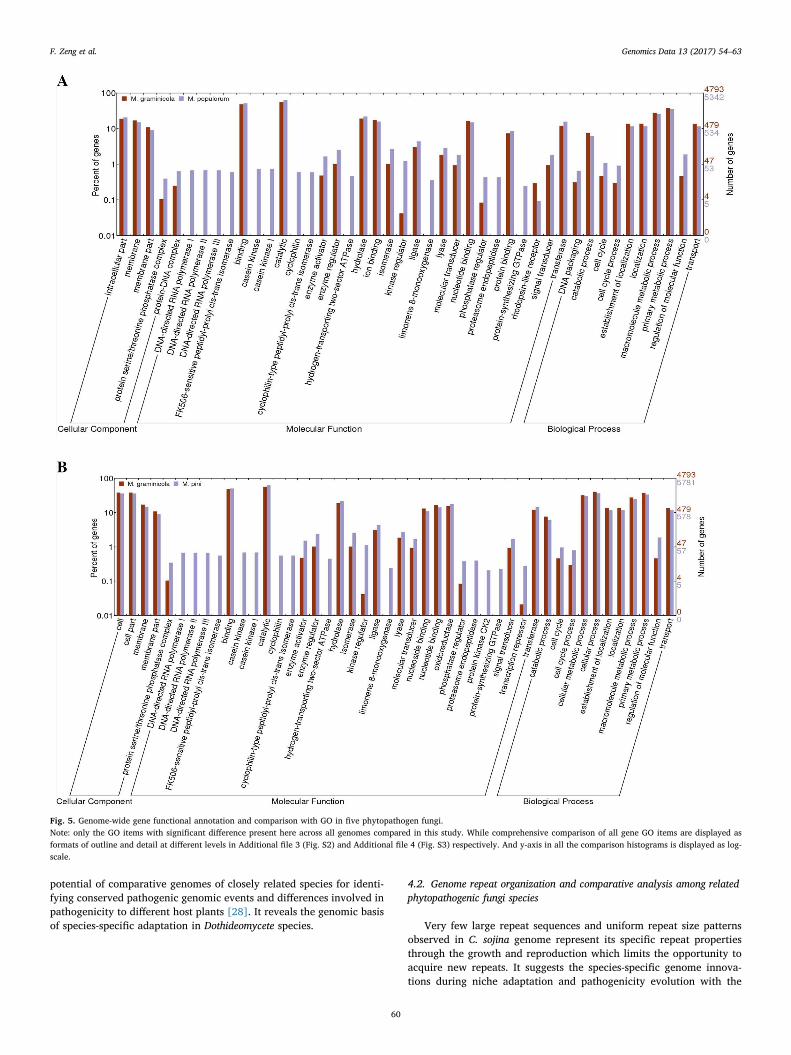
With systematic comparisons of functional prediction across allgenomes in this study, we identified both specific and common genomicelements in these five fungi. A wide diversification of the biologicalassociation processes among these fungal genomes is illustrated inFig. 5. Notably, highly abundant genes involved in cell division andoxidation reduction were identified in the C. sojina genome, comparedwith the other four genomes. At meantime, other fungi also have theirpreferential biological process versus C. sojina such as Cell Commu-nication, Regulation of Cellular process/function, which reflect thegenomic basis of pathogen specialization during evolution. For ex-ample, a higher number of two-component response regulators werefound in the genome of Z. tritici (M. graminicola) and M. fijiensis, sug-gesting that these two fungi could act with more advanced social be-havior than C. sojina.Genetic elements with function of transcriptionrepressor and subtilase that act as “chemical weapons” were not foundin C. sojina but in the other fungal genomes analyzed. And, pepsin A andRhodopsin-like receptor only present in Z. tritici (M. graminicola) and M.fijiensis (Fig. 5A). In particular, as the pathogens of woody plant hosts,
M. pini and M. populorum contain the factor functioned as casein kinasecyclophilin, limonene 8-monoxygenase and endopeptidase, not in thepathogens of herbaceous hosts in this study (Fig. 5B). Variation of se-creted proteins and metabolites features displayed in genome of pa-thogens of woody versus herbaceous hosts suggests gene loss and ac-quisition across these fungi.
Besides the wide divergence of pathogenic functional factors dis-tribution among fungal genomes as mentioned above, genome com-parison of phytopathogenic eukaryotes provides a powerful means ofidentifying conserved pathogenic processes from lineage-specific pa-thogenesis. Therefore, we also identified general functional elementsand biological processes conserved among these species. MolecularTransducers are highly conserved among C. sojina, Z. tritici and M. fi-jiensis, and so do Molecular Catalytic events among C. sojina,M. pini andM. populorum. They are candidates as common targets for diseasecontrol and management. Moreover, Responses to Stimuli are widelyconserved among all these five fungi. Similarly, Antioxidant,Transcription Regulator, Translation Regulator and PigmentationBiology Process showed no significant differences among these speciesexcept C. sojina (Fig. 5 and Additional file 3 Fig. S2).
A
B
Fig. 2. Whole-genome orthologous genes and evolutionary re-lationships among five close species.(A) Venn-diagram of whole-genome orthologous genes in five closespecies. The numbers in the diagram indicate overlapped conservedgenes or un-overlapped specific genes in those five species. (B) Basiccladogram for evolutionary relationships for five close species basedon phylogenomic analysis.
F. Zeng et al. Genomics Data 13 (2017) 54–63
57
4. Discussion
4.1. Genome synteny comparison between C. sojina and four closephytopathogenic relatives
Comparative analysis with Mycosphaerella pathogens indicatedconsiderable rapid rearrangements occur among these related fungal
genomes, which form the genomic basis of the pathogen specialization.Previous studies suggested that synteny or collinearity were limitedamong genomes from different genera [18–20]. As indicated by genomecomparison in our study, considerable conserved synteny between C.sojina and Z. tritici (M. graminicola), but less conserved synteny with thegenomes of the other three fungal species in the genus Mycosphaerella,illuminating genomic basis of the localized polymorphism and pa-thogen specialization through the long term evolution during the in-teraction of fungal pathogens and their specific hosts. Moreover, C.sojina and Z. tritici (M. graminicola) shared higher average exon numbersper gene and gene density than those of the other three fungal genomes.These genome features form the molecular basis of the fact that thehosts of C. sojina and Z. tritici (M. graminicola), soybean and wheat, havesimilar characteristics of growing conditions and pathogen resistance,compared with perennial tree species pine, poplar, and banana as hostsof M. pini, M. populorum and M. fijiensis respectively. Our results mostlyconfirmed previous notion.
There are also large regions lacking synteny as reported in the
Fig. 3. Syntenic relationship of C. sojina genome with four close pathogen relatives.
Table 1General features of fungal genomes compared with that of C. sojina.
fungal genus Aspergillus [27], in which it was thought to play a role inniche adaptation and virulence. Even for small-scale synteny betweenfungal species in some examples, there is no conservation of gene orderor orientation [18,19]. It can be hypothesized that rapid rearrangement
of such regions lacking synteny or collinearity might facilitate species-specific evolution of pathogenicity determinant genes. These sequencedivergence and genome rearrangements may result from selection dueto interactions of fungi and their plant hosts. This study highlights the
C. sojina
M. pini
M. fijiensis
37682220
28819770
29949520
0209620
38528050
38528051
45568642
42048346
49088938
52609234
56129530
59649826
63170122
66690418
70210714
73731010
M. populorum
Fig. 4. Abstract visualization of genome-wide repeat organization pattern in C. sojina and four close pathogen relatives.
F. Zeng et al. Genomics Data 13 (2017) 54–63
59
potential of comparative genomes of closely related species for identi-fying conserved pathogenic genomic events and differences involved inpathogenicity to different host plants [28]. It reveals the genomic basisof species-specific adaptation in Dothideomycete species.
4.2. Genome repeat organization and comparative analysis among relatedphytopathogenic fungi species
Very few large repeat sequences and uniform repeat size patternsobserved in C. sojina genome represent its specific repeat propertiesthrough the growth and reproduction which limits the opportunity toacquire new repeats. It suggests the species-specific genome innova-tions during niche adaptation and pathogenicity evolution with the
Fig. 5. Genome-wide gene functional annotation and comparison with GO in five phytopathogen fungi.Note: only the GO items with significant difference present here across all genomes compared in this study. While comprehensive comparison of all gene GO items are displayed asformats of outline and detail at different levels in Additional file 3 (Fig. S2) and Additional file 4 (Fig. S3) respectively. And y-axis in all the comparison histograms is displayed as log-scale.
F. Zeng et al. Genomics Data 13 (2017) 54–63
60
Fig. 5. (continued)
F. Zeng et al. Genomics Data 13 (2017) 54–63
61
specific host of soybean which, as an herbaceous dicot, differs fromother hosts. The lack of repetitive sequence has been previously thoughtdue to operation of a genome-wide defense system known as the RIP(repeat-induced point mutation) like in some fungi such as F. grami-nearum [26,29,30], in which RIP identifies duplicated sequences [31]that are subject to extensive mutation. RIP could partially account forthe reduced repeat content and apparent low number of paralogousgenes [26], which may occur in C. sojina with fewer repeats and lowergene number. Moreover, three Aspergillus species' fungal genomes havea single predicted DNA methyltransferase gene that is essential for RIPin Neurospora. crassa [29]. Apart from it, no additional DNA methyl-transferase genes were identified, which are required for methylation inthese fungi [29], while a number of putative DNA methyltransferasegenes were predicted in C. sojina genome. Although RIP has not beendemonstrated in C. sojina, above features in the genome suggest thatRIP as well as methylation might be active in C. sojina.
Furthermore, as the only gymnosperm among the hosts, pine differsfrom other hosts. Accordingly, the pathogen of pine,M. pini represents aspecific genome-wide repeat feature with very low repeat frequencydetected through the whole genome. In contrast, M. fijiensis genomepresents extremely high repeat density with large size. It suggests theM. fijiensis specific genome innovation against its unique host bananawhich is a monocot tree in tropical area of the world. These findingsfurther present the profound influences of repeats on genome innova-tion and pathogenic adaptation. Moreover, repeat density is correlatedwith genome AT richness in all these five species (Fig. 4 and Table 1).Extremely high repeat density in M. fijiensis go with remarkably low GCcontent. Consistently, lower abundance of repeat of in C. sojina and M.pini corresponding to higher GC content. The notable specific propertiesof genome-wide repeat profile in these related fungi pathogens re-present the roles that repetitive elements have played, and are con-tinuing to play, in the genome evolution and species-specific adaptationthrough the interactions of pathogens with their specific host.
Pepsin A and Rhodopsin-like receptor only present in Z. tritici (M.graminicola) and M. fijiensis, which implies that these secreted proteinsand response factors to external cues are essential for the ability of thesetwo close pathogens to decay the host material and adapt to fluctuatingand competitive environments. Variety of secreted proteins and meta-bolites features displayed in genome of pathogens of woody versusherbaceous hosts suggests gene loss and acquisition across these fungi.These findings represent and extend the report that variety of fungispecies-specific secreted proteins and metabolites, perhaps as effectorswithin plant cells, play roles in pathogenicity and determining the hostrange of the fungus [32]. Additionally, the relatively different numberof transcription repressors between the wood pathogens M. pini and M.populorum as well as the herbaceous pathogens Z. tritici (M. graminicola)and M. fijiensis suggests the differential regulation action during spe-cies-specific pathogenesis (Fig. 5A, B). It implies that the various eco-logical niches and hosts occupied by different fungal species are re-flected in both the gene family's class and abundance present in thegenomes.
The identified specific and common genomic elements in C. sojinaand other members of the genus Mycosphaerella represent a rich set ofcandidate targets for further investigation. Coupled with large-scalegene functional analysis studies, this will allow functional definition ofthese genetic elements that have the greatest potential in elucidatingthe phytopathogenesis.
5. Conclusions
Comparative genome analysis of C. sojina with M. pini, M.Populorum, Z. tritici (M. graminicola) and M. fijiensis on different planthosts have shed new light on the genomic basis of pathogenicity di-versity of these fungi likely to be common to all eukaryotic phyto-pathogens. The availability of genome data from C. sojina as well as theclosely related species provides new insights into genomic basis of
species-specific phytopathogenicity. From the analysis of comparativegenomes, we identified the differences on genome features involved inpathogenicity to different plant hosts such as woody vs. herbaceousplants, small crop vs. large trees, tropical and temperate zones crops.These results represent the initial step in elucidating the evolution ofpathogenicity. These efforts and ongoing sequencing projects for ad-ditional closely related particular phytopathogen isolates from the samespecies promise to reveal functional diversity of pathogenesis asso-ciated genetic elements, and will also facilitate to unveil the evolutionof eukaryotic microbial pathogenicity. It will, ultimately, change ourunderstanding of this important group of agronomically and scientifi-cally relevant fungi.
Supplementary data to this article can be found online at http://dx.doi.org/10.1016/j.gdata.2017.07.007.
Transparency document
The http://dx.doi.org/10.1016/j.gdata.2017.07.007 associatedwith this article can be found, in the online version.
Acknowledgments
We thank Jianfei Wu, Xinpeng Gao and Chaofan Wang for assistanceon additional data analyses and MS revision. This project was supportedby a grant from National Key R &D Program for Crop Breeding2016YFD0100306 and the USDA-CSREES as part of the SoybeanDisease Biotechnology Center at the University of Illinois. This workwas also supported by NSFC 31401428, Fok Ying-Tong Foundation151024 and Taishan Scholar Talent Project from China. The genomesequences used to compare with C. sojina were produced by the USDepartment of Energy Joint Genome Institute http://www.jgi.doe.gov/in collaboration with the user community.
References
[1] J.K. Hane, A.H. Williams, R.P. Oliver, Genomic and comparative analysis of theclass Dothideomycetes, in: S. Pöggeler, J. Wöstemeyer (Eds.), TheMycota, XIV:Evolution of Fungi and Fungal-Like Organisms, Springer, 2011, pp. 205–229.
[2] E.H. Stukenbrock, T. Bataillon, J.Y. Dutheil, T.T. Hansen, R. Li, M. Zala,B.A. McDonald, J. Wang, M.H. Schierup, The making of a new pathogen: insightsfrom comparative population genomics of the domesticated wheat pathogenMycosphaerella graminicola and its wild sister species, Genome Res. D21 (2011)2157–2166.
[3] R. Oliver, Genomic tillage and the harvest of fungal phytopathogens, New Phytol.196 (2012) 1015–1023.
[4] R.A. Ohm, N. Feau, B. Henrissat, C.L. Schoch, B.A. Horwitz, K.W. Barry,B.J. Condon, A.C. Copeland, B. Dhillon, F. Glaser, et al., Diverse lifestyles andstrategies of plant pathogenesis encoded in the genomes of eighteenDothideomycetes fungi, PLoS Pathog. 8 (2012) e1003037.
[5] C.R. Grau, A.E. Dorrance, J. Bond, J.S. Russin, Fungal diseases, in: H.R. Boerma,J.E. Specht (Eds.), Soybeans: Improvement, Production and Uses, 3rd edition,Madison, ASA, CSSA, SSSA, 2004, pp. 679–763.
[6] C.L. Palmer, W. Skinner, Mycosphaerella graminicola: latent infection, crop deves-tation and genomics, Mol. Plant Pathol. 3 (2002) 63–70.
[7] A.C.L. Churchill, Mycosphaerella fijiensis, the black streak pathogen of banana:progress towards understanding pathogen biology and detection, disease devel-opmentand the challenges of control, Mol. Plant Pathol. 12 (2011) 307–328.
[8] S.B. Goodwin, L.D. Dunkle, V.L. Zismann, Phylogenetic analysis of Cercospora andMycosphaerella based on the internal transcribed spacer region of ribosomal DNA,Phytopathology 91 (2001) 648–658.
[9] C. Soderlund, W. Nelson, A. Shoemaker, A. Paterson, SyMAP: a system for dis-covering and viewing syntenic regions of FPC maps, Genome Res. 16 (2006)1159–1168.
[10] C. Soderlund, M. Bomhoff, W.M. Nelson, SyMAP v3.4: a turnkey synteny systemwith application to plant genomes, Nucleic Acids Res. 39 (2011) e68.
[11] Y. Wang, D. Coleman-Derr, G. Chen, Y.Q. Gu, OrthoVenn: a web server for genomewide comparison and annotation of orthologous clusters across multiple species,Nucleic Acids Res. 43 (2015) W78–W84.
[12] A.C. Darling, B. Mau, F.R. Blattner, N.T. Perna, Mauve: multiple alignment ofconserved genomic sequence with rearrangements, Genome Res. 414 (2001)394–403.
[13] P. Durand, F. Mahé, A.S. Valin, J. Nicolas, Browsing repeats in genomes: pygramand an application to non-coding region analysis, BMC Bioinf. 7 (2006) 477.
[14] S. Götz, J.M. García-Gómez, J. Terol, T.D. Williams, S.H. Nagaraj, M.J. Nueda,M. Robles, M. Talón, J. Dopazo, A. Conesa, High-throughput functional annotation
and data mining with the Blast2GO suite, Nucleic Acids Res. 36 (2008) 3420–3435.[15] A. Conesa, S. Götz, J.M. García-Gómez, J. Terol, M. Talón, M. Robles, Blast2GO: a
universal tool for annotation, visualization and analysis in functional genomicsresearch, Bioinformatics 21 (2005) 3674–3676.
[16] A. Bateman, E. Birney, L. Cerruti, R. Durbin, L. Etwiller, S.R. Eddy, S. Griffiths-Jones, K.L. Howe, M. Marshall, E.L. Sonnhammer, The Pfam protein families da-tabase, Nucleic Acids Res. 30 (2002) 276–280.
[17] J. Ye, L. Fang, H. Zheng, Y. Zhang, J. Chen, Z. Zhang, J. Wang, S. Li, R. Li, L. Bolund,et al., WEGO: a web tool for plotting GO annotations, Nucleic Acids Res. 34 (2006)293–297.
[18] R.A. Dean, N.J. Talbot, D.J. Ebbole, M.L. Farman, T.K. Mitchell, M.J. Orbach,M. Thon, R. Kulkarni, J.R. Xu, H. Pan, et al., The genome sequence of the rice blastfungus Magnaporthe grisea, Nature 434 (2005) 980–986.
[19] J.K. Hane, R.G.T. Lowe, P.S. Solomon, K.C. Tan, C.L. Schoch, J.W. Spatafora,P.W. Crous, C. Kodira, B.W. Birren, J.E. Galagan, Dothideomycete-plant interac-tions illuminated by genome sequencing and EST analysis of the wheat pathogenStagonospora nodorum, Plant Cell 19 (2007) 3347–3368.
[20] D.M. Soanes, T.A. Richards, N.J. Talbot, Insights from sequencing fungal and oo-mycete genomes: what can we learn about plant disease and the evolution of pa-thogenicity? Plant Cell 19 (2007) 3318–3326.
[21] X. Gao, D.F. Voytas, A eukaryotic gene family related to retroelement integrases,Trends Genet. 21 (2005) 133–137.
[23] J. Lai, Y. Li, J. Messing, H.K. Dooner, Gene movement by Helitron transposonscontributes to the haplotype variability of maize, Proc. Natl. Acad. Sci. U. S. A. 102
(2005) 9068–9073.[24] M. Morgante, S. Brunner, G. Pea, K. Fengler, A. Zuccolo, A. Rafalski, Gene dupli-
cation and exon shuffling by helitronlike transposons generate intraspecies diversityin maize, Nat. Genet. 37 (2005) 997–1002.
[25] N. Jiang, Z. Bao, X. Zhang, S.R. Eddy, S.R. Wessler, Pack-MULE transposable ele-ments mediate gene evolution in plants, Nature 431 (2004) 569–573.
[26] C.A. Cuomo, U. Güldener, J.R. Xu, F. Trail, B.G. Turgeon, A. Di Pietro, J.D. Walton,L.J. Ma, S.E. Baker, M. Rep, et al., The Fusarium graminearum genome reveals a linkbetweenlocalized polymorphism and pathogen specialization, Science 317 (2007)1400–1402.
[27] P. Siriputthaiwan, A. Jauneau, C. Herbert, D. Garcin, B. Dumas, Functional analysisof CLPT1, a Rab/GTPase required for protein secretion and pathogenesis in theplant fungal pathogen Colletotrichum lindemuthianum, J. Cell Sci. 118 (2005)323–329.
[28] F. Sillo, M. Garbelotto, M. Friedman, P. Gonthier, Comparative genomics of siblingfungal pathogenic taxa identifies adaptive evolution without divergence in patho-genicity genes or genomic structure, Genome Biol. Evol. 7 (2015) 3190–3206.
[29] J.E. Galagan, E.U. Selker, RIP: the evolutionary cost of genome defense, TrendsGenet. 20 (2004) 417–423.
[30] E.U. Selker, E.B. Cambareri, B.C. Jensen, K.R. Haack, Rearrangement of duplicatedDNA in specialized cells of neurospora, Cell 51 (1987) 741–752.
[31] M.K. Watters, T.A. Randall, B.S. Margolin, E.U. Selker, D.R. Stadler, Action of re-peat-induced point mutation on both strands of a duplex and on tandem duplica-tions of various sizes in neurospora, Genetics 153 (1999) 705–714.
[32] M.J. Sweeney, A.D. Dobson, Molecular biology of mycotoxin biosynthesis, FEMSMicrobiol. Lett. 175 (1999) 149–163.