JFST Issue 6 (2019) 48-61 48 A comparative study of three conventional methods for diagnosis urinary schistosomiasis. Abosalif, K. O 1 ;Ahmed, A. A 2 ; Shammat. I. M 3 ;Aljafari. A. S 4 ; Afifi, A.A* 5 and Clive, J. Shiff 6 1 Department of Medical Parasitology, Faculty of Medical Laboratory Sciences- Omdurman Islamic University 2 Department of Zoology, Faculty of Science, University of Khartoum 3 Department of Molecular Biology, Faculty of Medical Laboratory Sciences- Omdurman Islamic University 4 Department of Medical Parasitology, Faculty of Medical Laboratory Sciences- Alneelain University 5 Department of Zoology, Faculty of Sciences and Technology - Omdurman Islamic University (corresponding Author) 6 Department of Molecular Microbiology and Immunology, Bloomberg School of Public Health, Johns Hopkins university Abstract The general objective of this comparative study was the evaluation of threediagnostictechniques(sedimentation technique using centrifugation, filtration techniques using millipore filter and Ninhydrine stained filter paper) conventional approaches. This was carried out at Alzaidab, River Nile State, Northern Sudan on 93 school children. The study involved the collection of samples from school children of 7 years age and above. A questionnaire was distributed to collect data on gender, age and water contact activities. The examinations on all samples were carried out applying the methods previously mentioned. The results revealed that the overall prevalence rates of Schistosomahaematobium in Alzaidab Nile State were 16.1%, 24.7% and 24.7% using the centrifugation, syringe filtration millipore, ninhydrin – filter paper staining method respectively. According to gender, males showed higher prevalence rates than females using all the techniques mentioned and the highest prevalence rate in males (36.1%) was reported bythesyringe filtration millipore and ninhydrin – filter paper staining method. According to age groups, the highest prevalence rate was reported among the group of children over 13 years of age, using all the techniques. The high prevalence rate (64.3%) among the above mentioned group (over 13 year’s of age) was shown by the syringe filtration millipore and ninhydrin – filter paper staining method. The highest prevalence rate
Transcript
JFST Issue 6 (2019) 48-61
48
A comparative study of three conventional methods for diagnosis urinary
schistosomiasis.
Abosalif, K. O
1;Ahmed, A. A
2 ; Shammat. I. M
3 ;Aljafari. A. S
4 ; Afifi, A.A*
5and Clive, J. Shiff
6
1Department of Medical Parasitology, Faculty of Medical Laboratory Sciences- Omdurman Islamic
University 2Department of Zoology, Faculty of Science, University of Khartoum 3Department of Molecular Biology, Faculty of Medical Laboratory Sciences- Omdurman Islamic
University 4Department of Medical Parasitology, Faculty of Medical Laboratory Sciences- Alneelain University 5Department of Zoology, Faculty of Sciences and Technology - Omdurman Islamic University
(corresponding Author) 6Department of Molecular Microbiology and Immunology, Bloomberg School of Public Health, Johns Hopkins university
Abstract The general objective of this comparative study was the evaluation of
threediagnostictechniques(sedimentation technique using centrifugation, filtration techniques
using millipore filter and Ninhydrine stained filter paper) conventional approaches. This was
carried out at Alzaidab, River Nile State, Northern Sudan on 93 school children. The study
involved the collection of samples from school children of 7 years age and above. A
questionnaire was distributed to collect data on gender, age and water contact activities. The
examinations on all samples were carried out applying the methods previously mentioned. The
results revealed that the overall prevalence rates of Schistosomahaematobium in Alzaidab Nile
State were 16.1%, 24.7% and 24.7% using the centrifugation, syringe filtration millipore,
ninhydrin – filter paper staining method respectively. According to gender, males showed higher
prevalence rates than females using all the techniques mentioned and the highest prevalence rate
in males (36.1%) was reported bythesyringe filtration millipore and ninhydrin – filter paper
staining method. According to age groups, the highest prevalence rate was reported among the
group of children over 13 years of age, using all the techniques. The high prevalence rate
(64.3%) among the above mentioned group (over 13 year’s of age) was shown by the syringe
filtration millipore and ninhydrin – filter paper staining method. The highest prevalence rate
JFST Issue 6 (2019) 48-61
49
amongthose who had water contact (32.9%) was shown by the syringe filtration millipore and
ninhydrin – filter paper staining method.It was found that the syringe filtration millipore and
ninhydrin – filter paper staining methods gave identical and reproducible results, yet the
ninhydrin – filter paper staining does not fit for field practice since it can only be carried out in
the laboratory.
Introduction
Bilharzia (schistosomiasis) occurs in the
tropics and subtropics and is one of the most
important parasitic diseases of humans.
Human disease is caused by
Schistosomahaematobium,
Schistosomamansoni, Schistosomajaponicum,
less frequently, Schistosomamekongi and
Schistosomaintercalatum(1, 2).These flukes
reside in the blood vessels of the gut or the
bladder, causing fever, pain and bleeding.
Bladder cancer or oesophagealvarices may
follow. Diagnosis is difficult, requiring
detection of parasite eggs in stool, urine or
gut/bladder biopsies.
Infection with cercariae occurs through intact
skin via contact with infested water. The
penetration of the cercariae is followed by
Katayama syndrome; an acute syndrome with
fever, rash and eosinophilia. The syndrome is
thought to be caused by antigen excess due to
the presence of schistosomulae in the blood
and the beginning of egg
deposition(Gryseelset al., 2006 and Ross et
al., 2007).After maturation, the adult male
and female worms mate and actively migrate
to their target organs(Gryseelset al., 2006 and
Ross et al., 2007).Schistosomahaematobium
resides in the wall of the bladder, sacral and
pelvic blood vessels surrounding the urinary
tract. The other mentioned species reside in
mesenteric veins. After deposition of eggs in
the capillary system, eggs penetrate the
mucosa of target organs and are excreted in
urine or faeces.
Sequelae of acute and chronic infection
include hepato-splenic diseases, portal
hypertension with varices, pulmonary
hypertension, squamous cell cancer of the
bladder, liver fibrosis, and less common
conditions such as myelo-radiculitis and
female genital schistosomiasis. Co-infections
with Hepatitis C virus (HCV) and
Schistosoma may also modify the course of
hepatitis C (Ross et al., 2002 and Quack et
al., 2006).Historically speaking, the ancient
kingdoms of the Nile basin have always been
in close touch with the disease. This
happened through trade, invasion,
immigration due to political oppression, or
natural disasters such as floods, drought and
famines(Hammamet al., 1933).The disease
was well known and documented amongst
the ancient Egyptians. It is still endemic
amongst the rural population. The history of
schistosomiasis in other parts of the Nile
JFST Issue 6 (2019) 48-61
50
valley is not as yet clear. However, it is
known to occur along the shores of Lake
Victoria, Lake Tana and also all along the
course of the Nile down to the Egyptian delta.
It is well known that the Egyptians have
invaded and dominated the Nile valley during
ancient times and also during the 19th
century as a part of the Ottoman
Empire(Eltayeb, 1998).The variation in
transmission patterns in different endemic
areas of urinary schistosomiasismakes it
almost impossible to set up a "standard"
control strategy. In fact, real and meaningful
control requires recognition of the importance
of this disease. Perhaps the impact of this
disease on the financial and socioeconomical
level as well as the productivity of
handicapped individuals in the affected
community, not to speak of the numerous
fatalities, can be an indicator of facing an
important foe who can never be overlooked.
Moreover the recent researches show that the
complication of an acute and chronic
infection of urinary schistosomiasis includes
hepato-splenic disease, portal hypertension
with varices, pulmonary hypertension,
squamous cell cancer of the bladder and liver
fibrosis. Indeed, the recognition of the
importance of appropriate investigation and
diagnostic methods become evident and
essential for the identification of the parasite
and lead to determining the precise treatment.
All that necessitates the selection of the most
accurate investigation techniques, which can
yield reproducible and reliable results. This is
an attempt to investigate the readability,
sensitivity and specificity of three
conventional methods (sedimentation
technique using centrifugation, filtration
techniques using millipore filter and
Ninhydrine stained filter paper) in detecting
urinary schistosomiasis.
Materials and methods:
Study design:This is a cross-sectional and
descriptive community-based study of
qualitative and quantitative approach.
Study area: The study was conducted in
Alzaidab, River Nile state, Northern Sudan.
Alzaidab area is located between
longitudes17 ° 20 `N-17 ° 39` N and 33 ° 46
`E-33 ° 57` E in the River Nile state on the
west bank of the River Nile.
Sample size and study population:
In this study, 93 individuals were enrolled.
Participants of both genders living at the
study area territories selected were of the age
between 5 and 18 years old (school age
children). Cohort children participants were
selected according to the WHO criteria.
These criteria required the categorization of
the school children into three groups; from
1st to 3rd, 4th to 6th, and from 7th to the 8th
grade. From each group 50 pupils were
selected.
Sampling technique was randomized and
non-probability hypothesis was considered.
JFST Issue 6 (2019) 48-61
51
Samples collection:
From each participant 60 ml of fresh voided
urine was collected. 10 ml sample of this
specimen was transferred into a centrifuge
tube and subjected to centrifugation. Another
10 ml sample was filtered through Millipore
filter. The rest of the specimen was passed
through Whatman paper No. 3 and another
commercial filter paper for further
Ninhydrin-filter paper staining.
Methods:
Microscopic examinations:
For microscopic examination, urine specimen
was prepared with the following methods.
Centrifugation technique:
Procedure:
10 ml of the collected urine sample was
placed into centrifuge tubes. The tube was
centrifuged at 2000 rpm for 3 minutes. The
supernatant was discarded the deposit was
placed on a slide, covered with a cover-slip
and examined under a binocular microscope.
Eggs were counted, and the result was
recorded as eggs count per 10 ml of urine.
Syringe filtration technique:
Procedure:
The Isopore™ Membrane Filter, a product of
Merck Millpore Company®, Ireland, is
composed of polycarbonate film, which has a
smooth, glass-like surface for clearer sample
observation.
A polycarbonate filter was carefully placed
on the filter holder, using a blunt–ended
forceps. A 10ml syringe was inserted into the
upper opening of the filter holder and the
plunger was removed. The syringe was filled
to the 10 ml mark with the well-mixed urine
sample. The plunger was replaced again and
the whole setting was held over a 40 ml
beaker to let the urine pass into it through the
polycarbonate filter. The filter holder was
removed and unscrewed. The filter was
carefully removed using forceps and
transferred face upward onto a slide. A drop
of physiological saline was added, and
covered with a cover glass. The preparation
was examined microscopically; using the 10x
objective lens for search and the 40x was
then used for identification. The number of
eggs was recorded as egg counts per 10 ml of
urine.
Nin-hydrin – filter paper staining method:
Procedure:
Approximately 40 ml urine was passed
through a 12.5 cm Whatman No. 3,
(Whatman International, Maidstone,
England) filter paper , folded in a cone. This
grade of paper was selected because it was
coarse, maintains a cone shape when folded,
and it retained both schistosome eggs.
Data management and statistical analysis
The data collected in this study through the
different methodologies was tabulated and
then converted into an electronic form. The
analysis was of a descriptive and parametric
nature. Statistical processing was achieved
JFST Issue 6 (2019) 48-61
52
quantitatively and qualitatively using
Statistical Package for Social Sciences
(SPSS) software. The data analysis was
referenced to standard values of the different
statistical methods. The computation of the
probability (P-value) was carried out through
the Chi Square test, whereas the sensitivity
and specificity were determined via cross-
tabulation of data according to the
relationship.
Sensitivity =True positive
True positive + false positive× 100%
Specifity =True negative
True negative + false negative× 100%
Results
The results revealed that the overall
prevalence rates of Schistosomahaematobium
in Alzaidab, River Nile State were 16.1%,
24.7% and 24.7% using the centrifugation,
syringe filtration millipore and ninhydrin –
filter paper staining method respectively. As
shown in table (1), the differences in
prevalence rates were found to be highly
significant at P< 0.001.
According to gender, males showed higher
prevalence rates than females using all the
techniques mentioned. The difference in rates
for all techniques were statistically significant
(P< 0.05). The highest prevalence rate in
males (36.1%) was reported bysyringe
filtration millipore and ninhydrin – filter
paper staining method (table 2) while the
lowest prevalence rate in female (3.1%) was
reported by the centrifugation, syringe
filtration millipore and ninhydrin – filter
paper staining method techniques (table 2).
According to age groups, the highest
prevalence rate was reported among the
group of children over 13 years of age, using
all the techniques. It was nil (0%) among the
group of children up to 7 years of age (table
3). The differences in rates were statistically
significant for the centrifugation, syringe
filtration millipore and ninhydrin – filter
paper staining method techniques.
The highest prevalence rate (64.3%) among
the above mentioned group (over 13 year’s of
age) is shown by the syringe filtration
millipore and ninhydrin – filter paper staining
method techniques (table 3) and the lowest
prevalence rate (12.8%) was reported by the
centrifugation technique (table 3). The result
demonstrated that all the positive cases were
observed in those who had water contact
activities (table 4). Not a single positive case
was reported among those who had no
contact with water, hence the statistical
difference was found to be highly significant
at P-values = 0.018, 0.002 and 0.002 for
centrifugation, syringe filtration millipore and
ninhydrin – filter paper staining method
techniques, respectively (table4). The highest
prevalence rate among those who had contact
with water (32.9%) was shown with the
syringe filtration millipore and ninhydrin –
JFST Issue 6 (2019) 48-61
53
filter paper staining method (table 4) while
the lowest prevalence rate (21.4%) was
reported with the centrifugation technique
(table 4). Assuming the centrifugation
technique as the gold standard technique, the
urine samples were firstly examined by the
centrifugation technique. Of the total 93
samples examined, 15 samples were found to
be positive for Schistosomahaematobium.
The positive cases were examined by the
syringe filtration millipore technique showing
also 15 positive cases. The 78 urine samples
found negative by the centrifugation
technique were also subjected to examination
by the syringe filtration millipore. Eight
samples proved positive, while 70 were
negative, constituting a 100% sensitivity rate
of syringe filtration millipore technique and a
specificity of rate 90%. Yet, the sensitivity
and specificity of the centrifugation technique
were 65% and 100% respectively when
considering the syringe filtration millipore
technique the gold standard technique (table
5). Assuming the centrifugation technique as
the gold standard technique, the urine
samples were firstly examined by the
centrifugation technique. Of the total 93
samples examined, 15 samples were found to
be positive for Schistosomahaematobium.
The positive cases were examined by the
ninhydrin – filter paper staining method
showing also 15 positive cases. The rest 78
urine samples found negative by the
centrifugation technique were also subjected
to examination by the ninhydrin – filter paper
staining method. Eight samples proved
positive, while 70 were negative, constituting
a 100% sensitivity rate of ninhydrin – filter
paper staining method and a specificity of
90%. Yet, the sensitivity and specificity rates
of the centrifugation technique were 65% and
100% respectively when considering the
ninhydrin – filter paper staining method as
the gold standard technique(table6).
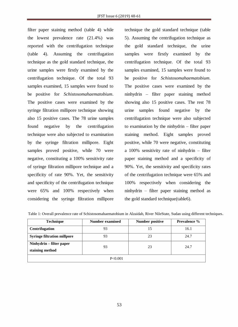
Table 1: Overall prevalence rate of Schistosomahaematobium in Alzaidab, River NileState, Sudan using different techniques.
Technique Number examined Number positive Prevalence %
Centrifugation 93 15 16.1
Syringe filtration millpore 93 23 24.7
Ninhydrin – filter paper
staining method 93 23 24.7
P<0.001
JFST Issue 6 (2019) 48-61
54
Table 2: Prevalence rate of Schistosomahaematobium in Alzaidab, River Nile State, Sudan according to gender using
centrifugation, Syringe filtration millpore and Ninhydrin – filter paper staining method techniques.
Technique Gender Number
Examined
Number
positive
Prevalence % Pvalue
Centrifugation
Male 61 14 22.9 <0.001
Female 32 1 3.1
Syringe filtration
millpore
Male 61 22 36.1 <0.001
Female 32 1 3.1
Ninhydrin – filter
paper staining method
Male 61 22 36.1
<0.001 Female 32 1 3.1
P < 0.05
Table 3: Prevalence rate of Schistosomahaematobium in Alzaidab, River Nile State, Sudan according to age group using
centrifugation, Syringe filtration millpore and Ninhydrin – filter paper staining methodCentrifugation techniques.
Technique Age Group Number
Examined
Number
positive
Prevalence % P value
Centrifugation
Up to 7 years 1 0 0
<0.001 8 – 13 years 78 10 12.8
More than 13
years 14 5 35.7
Syringe filtration
millpore
Up to 7 years 1 0 0
<0.001 8 – 13 years 78 14 17.9
More than 13
years 14 9 64.3
Ninhydrin – filter
paper staining
method
Up to 7 years 1 0 0
<0.001 8 – 13 years 78 14 17.9
More than 13
years 14 9 64.3
P < 0.001
Table 4: Prevalence rate of Schistosomahaematobium in Alzaidab, River Nile State, Sudan according to contact with water
using centrifugation, Syringe filtration millpore and Ninhydrin – filter paper staining methodCentrifugation techniques.