Published: September 01, 2011 r2011 American Chemical Society 2252 dx.doi.org/10.1021/mp200346y | Mol. Pharmaceutics 2011, 8, 2252–2261 ARTICLE pubs.acs.org/molecularpharmaceutics A Compound That Inhibits the HOP Hsp90 Complex Formation and Has Unique Killing Effects in Breast Cancer Cell Lines Genaro Pimienta, Kristina M. Herbert, and Lynne Regan* Department of Molecular Biophysics and Biochemistry, Yale University, New Haven, Connecticut, United States b S Supporting Information ’ INTRODUCTION The chaperone Hsp90 is a well-established anticancer drug target. 1,2 Hsp90 is responsible for the folding of numerous proteins into their mature, functional state. 3 Many such Hsp90 “client proteins” are involved in key oncogenic pathways includ- ing proliferation, cell cycle progression, inhibition of apoptosis and metastasis. 4,5 When Hsp90 is inhibited, Hsp90-dependent client proteins are ubiquitinated and thus targeted for proteasomal degradation. 6 9 Inhibitors of Hsp90 therefore have the potential to be effective against a broad range of different cancer types. 7 The natural product geldanamycin and its derivatives such as 17-AAG inhibit Hsp90 by binding to its N-terminal domain at the ATP-binding site. 10,11 Treatment of cells with such com- pounds indeed results in the degradation of a large number of client proteins that are involved in oncogenic pathways. 12 15 The mechanism of action of 17-AAG has been well studied. 1,2 Recently however, various other Hsp90 ATP-binding site in- hibitors, such as PU-H71 and NVP-AUY922, have been de- scribed and are together with 17-AAG in clinical trials. 1,2 A component of resistance common to these Hsp90 ATP-binding site inhibitors is thought to be the transcriptional induction of a heat shock response: overproduction of Hsp27 and Hsp70, which have strong antiapoptotic activity. 16 19 We have recently described a new class of chaperone inhibi- tors that prevent the interaction of Hsp90 with the protein HOP (Hsp70-Hsp90 organizing protein) in vitro. 20 In the folding pathway of Hsp90 client proteins, folding is initiated on Hsp70, and the partially folded protein is then transferred to Hsp90, where the final stages of maturation are completed (Figure 1). 21 These reactions occur on the Hsp70 HOP Hsp90 complex. 4,5 If Hsp90 is prevented from interacting with HOP, this complex can no longer form (Figure 1). 4,5 Here we investigate how one of these compounds (1,6-dimethyl-3- propylpyrimido[5,4-e][1,2,4]triazine-5,7-dione), which for sim- plicity we refer to as C9 (Supplementary Figure 1a in the Supporting Information), kills the highly metastatic triple nega- tive breast cancer (TNBC) cell line MDA-MB-231. We have also extended the investigation of the most significant findings in other cancer cell lines. We found that C9 induces a change in cell morphology and causes cell cycle arrest of MDA-MB-231 cells, with a consequent inhibition of spheroid formation and cell migration. The killing activity of C9 against MDA-MB-231 cells is not via a pathway that involves the activation of the apoptosis mediators Caspase 3/7. This result is unique because the currently available Hsp90 Received: March 13, 2011 Accepted: September 1, 2011 Revised: August 28, 2011 ABSTRACT: The chaperone Hsp90 is required for the correct folding and matura- tion of certain “client proteins” within all cells. Hsp90-mediated folding is particularly important in cancer cells, because upregulated or mutant oncogenic proteins are often Hsp90 clients. Hsp90 inhibitors thus represent a route to anticancer agents that have the potential to be active against several different types of cancer. Currently, various Hsp90 inhibitors that bind to Hsp90 at its ATP-binding site are in preclinical and clinical trials. Some of the most promising Hsp90 ATP-binding site inhibitors are the well characterized geldanamycin derivative 17-AAG and the recently described compounds PU-H71 and NVP-AUY922. An undesirable characteristic of these compounds is the transcriptional upregulation of Hsp70 that has prosurvival effects. Here we characterize the activity of a new type of chaperone inhibitor, 1,6-dimethyl- 3-propylpyrimido[5,4-e][1,2,4]triazine-5,7-dione (named C9 for simplicity). Using purified protein components in vitro, C9 prevents Hsp90 from interacting with the cochaperone HOP and is thus expected to impair the Hsp90-dependent folding pathway in vivo. We show that this compound is effective in killing various breast cancer cell lines including the highly metastatic MDA-MB-231. An important property of this compound is that it does not induce the transcriptional upregulation of Hsp70. Moreover, when cells are treated with a combination of C9 and either 17-AAG or NVP-AUY922, the overexpression of Hsp70 is counteracted considerably and C9’s lethal-IC50 decreases compared to its value when added alone. KEYWORDS: triple negative breast cancer, chaperone inhibitor, Hsp70, p38, JNK1/2
Transcript
Published: September 01, 2011
r 2011 American Chemical Society 2252 dx.doi.org/10.1021/mp200346y |Mol. Pharmaceutics 2011, 8, 2252–2261
ARTICLE
pubs.acs.org/molecularpharmaceutics
A Compound That Inhibits the HOP�Hsp90 Complex Formation andHas Unique Killing Effects in Breast Cancer Cell LinesGenaro Pimienta, Kristina M. Herbert, and Lynne Regan*
Department of Molecular Biophysics and Biochemistry, Yale University, New Haven, Connecticut, United States
bS Supporting Information
’ INTRODUCTION
The chaperone Hsp90 is a well-established anticancer drugtarget.1,2 Hsp90 is responsible for the folding of numerousproteins into their mature, functional state.3 Many such Hsp90“client proteins” are involved in key oncogenic pathways includ-ing proliferation, cell cycle progression, inhibition of apoptosisand metastasis.4,5 When Hsp90 is inhibited, Hsp90-dependentclient proteins are ubiquitinated and thus targeted for proteasomaldegradation.6�9 Inhibitors of Hsp90 therefore have the potentialto be effective against a broad range of different cancer types.7
The natural product geldanamycin and its derivatives such as17-AAG inhibit Hsp90 by binding to its N-terminal domain atthe ATP-binding site.10,11 Treatment of cells with such com-pounds indeed results in the degradation of a large number ofclient proteins that are involved in oncogenic pathways.12�15
The mechanism of action of 17-AAG has been well studied.1,2
Recently however, various other Hsp90 ATP-binding site in-hibitors, such as PU-H71 and NVP-AUY922, have been de-scribed and are together with 17-AAG in clinical trials.1,2
A component of resistance common to these Hsp90 ATP-bindingsite inhibitors is thought to be the transcriptional inductionof a heat shock response: overproduction of Hsp27 and Hsp70,which have strong antiapoptotic activity.16�19
We have recently described a new class of chaperone inhibi-tors that prevent the interaction of Hsp90 with the protein HOP
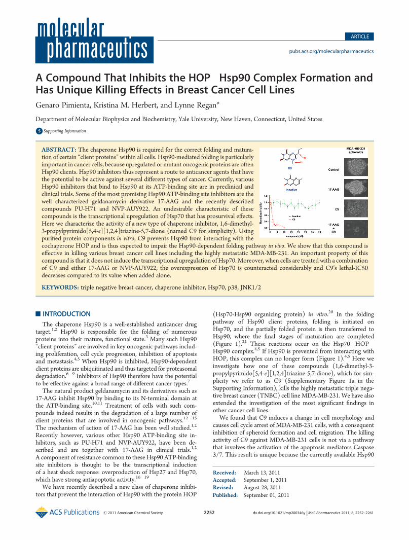
(Hsp70-Hsp90 organizing protein) in vitro.20 In the foldingpathway of Hsp90 client proteins, folding is initiated onHsp70, and the partially folded protein is then transferred toHsp90, where the final stages of maturation are completed(Figure 1).21 These reactions occur on the Hsp70�HOP�Hsp90 complex.4,5 If Hsp90 is prevented from interacting withHOP, this complex can no longer form (Figure 1).4,5 Here weinvestigate how one of these compounds (1,6-dimethyl-3-propylpyrimido[5,4-e][1,2,4]triazine-5,7-dione), which for sim-plicity we refer to as C9 (Supplementary Figure 1a in theSupporting Information), kills the highly metastatic triple nega-tive breast cancer (TNBC) cell lineMDA-MB-231. We have alsoextended the investigation of the most significant findings inother cancer cell lines.
We found that C9 induces a change in cell morphology andcauses cell cycle arrest of MDA-MB-231 cells, with a consequentinhibition of spheroid formation and cell migration. The killingactivity of C9 against MDA-MB-231 cells is not via a pathwaythat involves the activation of the apoptosis mediators Caspase3/7. This result is unique because the currently available Hsp90
Received: March 13, 2011Accepted: September 1, 2011Revised: August 28, 2011
ABSTRACT: The chaperone Hsp90 is required for the correct folding and matura-tion of certain “client proteins” within all cells. Hsp90-mediated folding is particularlyimportant in cancer cells, because upregulated or mutant oncogenic proteins are oftenHsp90 clients. Hsp90 inhibitors thus represent a route to anticancer agents that havethe potential to be active against several different types of cancer. Currently, variousHsp90 inhibitors that bind to Hsp90 at its ATP-binding site are in preclinical andclinical trials. Some of the most promising Hsp90 ATP-binding site inhibitors are thewell characterized geldanamycin derivative 17-AAG and the recently describedcompounds PU-H71 and NVP-AUY922. An undesirable characteristic of thesecompounds is the transcriptional upregulation of Hsp70 that has prosurvival effects.Here we characterize the activity of a new type of chaperone inhibitor, 1,6-dimethyl-3-propylpyrimido[5,4-e][1,2,4]triazine-5,7-dione (named C9 for simplicity). Usingpurified protein components in vitro, C9 prevents Hsp90 from interacting with thecochaperone HOP and is thus expected to impair the Hsp90-dependent folding pathway in vivo. We show that this compound iseffective in killing various breast cancer cell lines including the highly metastatic MDA-MB-231. An important property of thiscompound is that it does not induce the transcriptional upregulation of Hsp70. Moreover, when cells are treated with a combinationof C9 and either 17-AAG or NVP-AUY922, the overexpression of Hsp70 is counteracted considerably and C9’s lethal-IC50decreases compared to its value when added alone.
KEYWORDS: triple negative breast cancer, chaperone inhibitor, Hsp70, p38, JNK1/2
ATP-binding site inhibitors, such as 17-AAG, PU-H71 andNVP-AUY922, induce Caspase 3/7-mediated apoptotic cell death.18,22
At the molecular level C9 like 17-AAG induces the degrada-tion of the well-establishedHsp90 client proteins CDK4 and Raf-1. An unexpected yet interesting result is the C9-mediatedprotein downregulation of the mitogen activated protein kinases(MAPKs) JNK1/2 and p38, also known as stress-activatedprotein kinases (SAPKs), for which a dependency on theHsp90 folding pathway is not yet fully established.23�27 Thedecrease in the protein levels of these kinases is significant,because when overactive they are known to drive the tumorigenicphenotype in malignant cell lines.
Remarkably, treatment of cells with C9 does not result in theundesirable increase in the protein levels of Hsp27 and Hsp70,which is characteristic of the aforementioned ATP-bindingsite Hsp90 inhibitors. Furthermore, when cells are treated witha combination of C9 and either 17-AAG or NVP-AUY922, theeffect of C9 is dominant and the upregulation of Hsp27 andHsp70 is suppressed considerably.
’MATERIALS AND METHODS
Cell Culture. All cell lines were cultured in high glucoseDulbecco’s modified Eagle’s medium (DMEM) supplementedwith L-glutamine and sodium pyruvate (Invitrogen), plus 10%v/v fetal bovine serum (FBS) heat inactivated (Gibco), 1% v/vof penicillin/streptomycin 10,000 units (Gibco) and 10 μM ofrecombinant insulin (Sigma). Cells were grown to about 80%confluency, and then synchronized by serum starvation for 24 hprior to adding inhibitory compounds.Caspase 3/7 Activity and Viability Assays. Intracellular
Caspase 3/7 activity and cell viability in the form of metabolicATP content were quantified using the CellTiter Caspase 3/7(Promega) or the CellTiter-Glow (Promega) luminescent as-says, respectively. In both assays, cells were seeded in 96-wellopaque plates (Corning), serum starved for about 24 h so thatmost of the cells were synchronized in a resting population, andthen treated with 10% FBS and insulin, plus either C9, 17-AAG,PU-H71, NVP-AUY922 or H2O2, typically for 24 h.For these and all experiments (unless noted otherwise) the
concentration of C9 was 3 μMand the concentration of 17-AAG,PU-H71, and NVP-AUY922 was 1 μM.After 24 h of drug treatment, the tissue culture plates were
treated with either CellTiter Caspase 3/7 (Promega) or CellTi-ter-Glo (Promega) reagent following the manufacturer’s proto-col. Caspase 3/7 activity and viability (metabolic ATP content)
as a function of relative fluorescent intensity were measured onan ENVISION plate reader using a luminescence filter.Poly-HEMA Coating of Plates To Prevent Cell Attachment.
6-Well plates were treated with 1 mL/per well of 0.5% w/v ofPoly-HEMA (Sigma) dissolved in 95% ethanol. This solutionwas left overnight to dry completely in a sterile cell culture hood,and the procedure was repeated 5 more times, to ensure ahomogeneous coating of the plate surface. Before use, the platewas washed twice with sterile phosphate buffered saline (PBS)solution (Gibco) to remove any traces of chemical contaminants.3D-Spheroid Generation. Matrigel was used to assist the
spheroid formation of MDA-MB-231cells. A cell suspension inDMEM growth medium, with 2.5% v/v cold Matrigel matrix(BD Biosciences), was added to a plate previously treatedwith poly-HEMA and allowed to grow nonattached. In the caseof MCF7 and BT-474 cells, the addition of Matrigel was notnecessary, because unlikeMDA-MB-231 cells, these two cell linesform spontaneous 3D spheroids even in the absence of Matrigel.Microscope Cell Imaging. Cell images were obtained by
phase contrast with an inverted microscope (Olympus CKX41),at 20�40� magnification. Pictures were taken with a digitalcamera interfaced to a PC desktop computer. Each image wascropped and its brightness/contrast adjusted in ImageJ.Wound-Healing/Scratch Assay. Cells were grown to about
80% confluency and then synchronized by serum starvation for24 h. A sterile 200 μL pipet tip was used to scratch acrossthe plate, making a “wound” in the cell layer. The medium wasthen removed by aspiration and the cellular monolayer washed5 times with serum free medium to remove the detached cellsfrom the scratch. FBS-containing DMEM medium plus eitherDMSO (control), C9 or 17-AAG was added to each plate,and the samples were left to grow. The width of the scratchwas documented by photographing the cells as described aboveevery 12 h.FACS Cell Cycle Analysis. For DNA-cell cycle analysis,
confluent cells from a 10 cm plate were detached with trypsin-EDTA, washed with cold PBS and harvested in FACS poly-propylene tubes. Cell pellets were resuspended in prechilled 70%ethanol and incubated at 4 �C for at least an hour to allow DNAfixing. Next, the samples were washed 3 times with cold PBS andtreated with 100 μL of DNase free ribonuclease 100 μg/mL(Sigma) for 10 min at room temperature. This was followed bythe addition of 400 μL of propidium iodide dissolved in PBS at50 μg/mL and protected from light. Stained samples wereanalyzed on a FACScan instrument at the Yale University Schoolof Medicine Cell Sorter Facility.
Figure 1. C9 and 17-AAG inhibit Hsp90 through different mechanisms. Cartoon representation of the Hsp90-mediated folding pathway and the pointsat which 17-AAG and C9 act to inhibit it. 17-AAG binds directly to Hsp90, at the ATP binding site of the N-terminal domain (middle). C9 binds to theTPR2A domain of HOP, thus preventing the C-terminus of Hsp90 from binding to HOP (right).
Western Blots (WB). Cells were harvested by centrifugationand lysed with a solution containing 50 mM Tris-Base (pH 7.6),150 mM NaCl, 1.5 mM EDTA, 1% v/v Nonidet-P40 and 10%v/v glycerol. To inhibit proteases and phosphatases, 50 mL ofcold lysis solution was freshly mixed with a tablet of a cocktail ofprotease inhibitors (Sigma) and 100 μL of phosphatase inhibitorcocktails I and II (Sigma).Total protein concentration in each extract was determined
using the Bradford protein assay (Bio-Rad). Based on theseconcentrations, equal amounts of total protein from each extractwere separated by SDS polyacrylamide gel electrophoresis,followed by the electrotransfer of proteins onto a nitrocellulosemembrane. The transferred blot was blocked with either 5% fat-free milk or 5% bovine serum albumin in a Tween20/Tris-Basesaline solution (TTBS), at 4 �C overnight and probed withspecific antibodies purchased from Cell Signaling Technology,also at 4 �C overnight. The blots were then washed with TTBSand incubated with a species-specific IgG secondary antibodycoupled to horseradish peroxidase (HRP), for an hour at roomtemperature. To detect the HRP signal, an acridan-based reagent(ECL-Plus from Amersham) was added to each blot followed byautoradiography on a film.qRT-PCR. RNA was isolated using TRIzol (Invitrogen),
DNase treated (RNase-Free DNase Set, Qiagen), and thenpurified on RNeasy Mini cleanup columns (Qiagen). The HighCapacity cDNA Reverse Transcription Kit (Applied Biosystems)was used according to the manufacturer’s protocol. The qRT-PCR experiments were performed using the 7900HT Fast Real-Time PCR System using Taqman assays (Applied Biosystems)for the individual genes. Taqman assay IDs were as follows:RPL30 control (Hs00265497_m1), Hsp27 (Hs00356629_g1)and Hsp70 (Hs00271229_s1). RPL30 was determined to be anappropriate normalization control in an initial experiment inwhich isolated cDNA was run on a TaqMan Array GeneSignature 96-well human endogenous control plate. Of the 32control genes present on this plate, RPL30 showed the leastvariability in amplification cycle number between cells treatedwith DMSO, C9, or 17AAG. The mRNA levels of each gene werenormalized to the value obtained for the RPL30 mRNA tran-script in the same sample. The normalized values obtained fromMDA-MB-231 cells treated with C9 or 17-AAG, were normalized once more with respect to values obtained in the DMSO-treated sample.
’RESULTS
We have previously described compounds with a commonpyrimidotriazinedione structural core that inhibit the Hsp90-dependent folding pathway by a novel mechanism. In vitro, thesecompounds inhibit the interaction of the TPR2A domain ofHOP with the C-terminus segment of Hsp90, and are thusexpected to impair the formation of a functional Hsp70�HOP�Hsp90 complex in vivo.20,28 Here we present a characterizationof the effects of one of these compounds, C9 (1,6-dimethyl-3-propylpyrimido[5,4-e][1,2,4]triazine-5,7-dione) (SupplementaryFigure 1a in the Supporting Information), on the highly meta-static TNBC cell line MDA-MB-231. The TNBC subtype doesnot express detectable levels of estrogen and progesteronereceptors (ER and PR, respectively) or the human epidermalgrowth factor receptor 2 (HER2).29
Furthermore, MDA-MB-231 cells have been reported tobe relatively insensitive to 17-AAG.30 So far, PU-H71 and
NVP-AUY922 are the only Hsp90 ATP-binding site inhibitorsthat kill MDA-MB-231 cells.18,22 However, the killing effects ofPU-H71 andNVP-AUY922 against MDA-MB-231 cells can onlybe observed at 72 h.18,22
Using 24 h treatments, we compare and contrast the effects ofC9 with those of 17-AAG, which is anHsp90 inhibitor with a wellestablished mechanism of action. Furthermore, because 17-AAGand C9 are proposed to inhibit Hsp90 by completely differentmechanisms (Figure 1), we also investigate the effects of treatingcells with a combination of both compounds. To better demon-strate the most important features of C9, we also show compar-isons with PU-H71 and NVP-AUY922, which, like 17-AAG, areATP-binding site inhibitors of Hsp90 that induce an increase inthe mRNA and protein levels of Hsp70.Specificity of Action: In Vivo Activity of C9 vs a Derivative
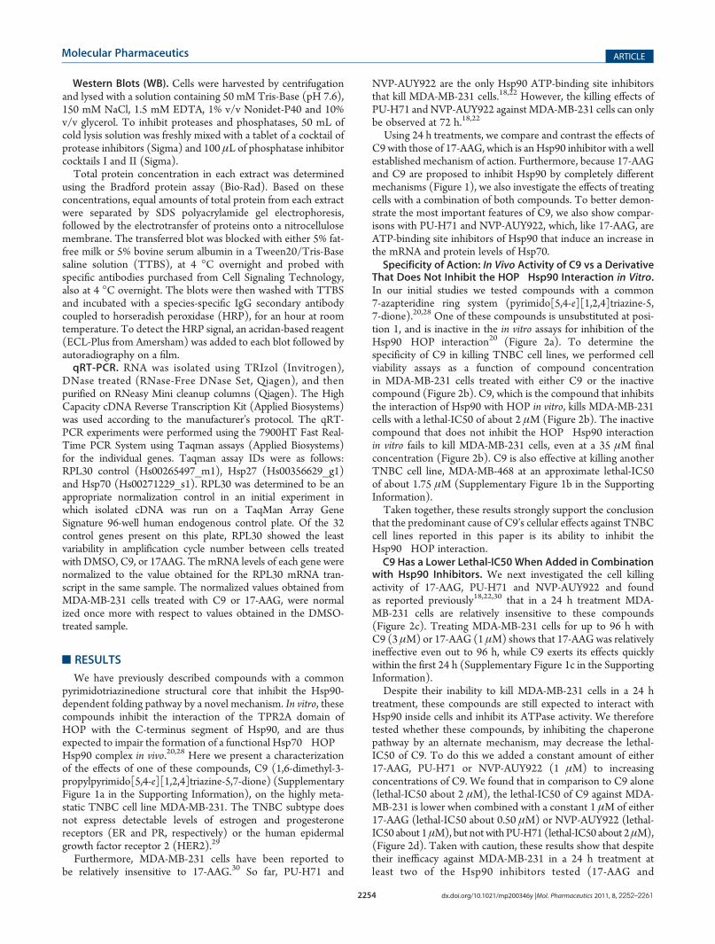
That Does Not Inhibit the HOP�Hsp90 Interaction in Vitro.In our initial studies we tested compounds with a common7-azapteridine ring system (pyrimido[5,4-e][1,2,4]triazine-5,7-dione).20,28 One of these compounds is unsubstituted at posi-tion 1, and is inactive in the in vitro assays for inhibition of theHsp90�HOP interaction20 (Figure 2a). To determine thespecificity of C9 in killing TNBC cell lines, we performed cellviability assays as a function of compound concentrationin MDA-MB-231 cells treated with either C9 or the inactivecompound (Figure 2b). C9, which is the compound that inhibitsthe interaction of Hsp90 with HOP in vitro, kills MDA-MB-231cells with a lethal-IC50 of about 2 μM (Figure 2b). The inactivecompound that does not inhibit the HOP�Hsp90 interactionin vitro fails to kill MDA-MB-231 cells, even at a 35 μM finalconcentration (Figure 2b). C9 is also effective at killing anotherTNBC cell line, MDA-MB-468 at an approximate lethal-IC50of about 1.75 μM (Supplementary Figure 1b in the SupportingInformation).Taken together, these results strongly support the conclusion
that the predominant cause of C9’s cellular effects against TNBCcell lines reported in this paper is its ability to inhibit theHsp90�HOP interaction.C9 Has a Lower Lethal-IC50 When Added in Combination
with Hsp90 Inhibitors. We next investigated the cell killingactivity of 17-AAG, PU-H71 and NVP-AUY922 and foundas reported previously18,22,30 that in a 24 h treatment MDA-MB-231 cells are relatively insensitive to these compounds(Figure 2c). Treating MDA-MB-231 cells for up to 96 h withC9 (3 μM) or 17-AAG (1 μM) shows that 17-AAGwas relativelyineffective even out to 96 h, while C9 exerts its effects quicklywithin the first 24 h (Supplementary Figure 1c in the SupportingInformation).Despite their inability to kill MDA-MB-231 cells in a 24 h
treatment, these compounds are still expected to interact withHsp90 inside cells and inhibit its ATPase activity. We thereforetested whether these compounds, by inhibiting the chaperonepathway by an alternate mechanism, may decrease the lethal-IC50 of C9. To do this we added a constant amount of either17-AAG, PU-H71 or NVP-AUY922 (1 μM) to increasingconcentrations of C9. We found that in comparison to C9 alone(lethal-IC50 about 2 μM), the lethal-IC50 of C9 against MDA-MB-231 is lower when combined with a constant 1 μM of either17-AAG (lethal-IC50 about 0.50 μM) or NVP-AUY922 (lethal-IC50 about 1μM), but not with PU-H71 (lethal-IC50 about 2μM),(Figure 2d). Taken with caution, these results show that despitetheir inefficacy against MDA-MB-231 in a 24 h treatment atleast two of the Hsp90 inhibitors tested (17-AAG and
NVP-AUY922) potentiate the killing effects of C9, by reducingits lethal-IC50. Finally, while this data highlights the potentialof C9 in combination therapies aimed at disrupting the Hsp90folding pathway in cancer cells, further experimentation isrequired to truly establish quantitatively a synergistic effectbetween C9 and either 17-AAG or NVP-AUY922.C9, but Not 17-AAG, Inhibits MDA-MB-231 Spheroid
Formation. We next investigated the effects of C9, 17-AAG,and a combination of the two compounds onMDA-MB-231 cellsgrown as nonadherent 3-dimensional (3D) spheroids. C9 treat-ment inhibits spheroid formation and kills MDA-MB-231 cellscultured in the 3D spheroid form, whereas 17-AAG has little orno effect (Figure 3a). WhenMDA-MB-231 spheroids are treatedwith a combination of C9 and 17-AAG, the effect of C9 isdominant. That is, spheroid formation is inhibited and cellsare killed (Figure 3a). The observation that C9 kills MDA-MB-231 cells when cultured as 3D-spheroids alone or incombination with 17-AAG is important, because the cultureof breast cancer cell lines as nonattached aggregates in thepresence of Matrigel extract is a better mimic of the tissueenvironment in vivo.31,32
C9 Destroys the Spheroids Formed by BT-474 and MCF7Cells. We further corroborated the ability of C9 to kill breastcancer cell lines cultured as spheroids by testing two additionalbreast cancer cell lines, BT-474 and MCF7, both of which form
3D-spheroids even in the absence of Matrigel.33,34 We showedin our previous work that C9 is effective at killing these non-TNBC cell lines when cultured as monolayers.20 BT-474 cellsbelong to the HER2-positive breast cancer subtype and MCF7cells are ER-positive.29 We found that C9 disrupts spheroidformation and kills both of these two cell lines effectively(Figure 3b,c). 17-AAG is also effective in disruption and killingof these cell lines (Figure 3b,c). These results show that C9 onthe one hand is effective in killing spheroids of different breastcancer subtypes, while 17-AAG on the other hand is ineffectiveagainst the TNBC MDA-MB-231 cells, but can kill the non-TNBC cell lines BT-474 and MCF7.C9, but Not 17-AAG, Induces Cell-Shape Changes and
Decreases Cell Migration. MDA-MB-231 cells have a spindle-like shape, have a mesenchymal phenotype, and are highlyinvasive.35 Treatment of MDA-MB-231 cells with C9 causesthem to acquire a cobblestone-like morphology, whereas treat-ment with 17-AAG does not (Figure 4a). This result suggeststhat C9 may decrease the migration properties of MDA-MB-231cells. To follow up on this observation, we investigated the effectsof C9 and 17-AAG in a “wound healing” scratch assay, as a proxyfor metastatic potential. C9 impaired the migration propertiesof MDA-MB-231 cells, while 17-AAG did not (Figure 4b).The change in cell shape and the inhibition of migration in vitroare important observations because they suggest that C9 may be
Figure 2. In vitro and in vivo activity of C9. (a) Plot showing inhibition of the interaction of TPR2A (the Hsp90-binding domain of HOP) with theC-terminal peptide of Hsp90 in an in vitro AlphaScreen assay.16,24 Fraction bound is plotted versus the concentration of either C9 or the inactivederivative. Adapted from ref 16. (b) Dose�response killing assays of MDA-MB-231 cells cultured as adherent monolayers when treated 24 h with C9(0�10 μM) or the inactive compound (0�35 μM). (c) Dose�response killing assays of MDA-MB-231 cells cultured as adherent monolayers whentreated 24 h with C9, 17-AAG, PU-H71 or NVP-AUY922. (d) Dose�response killing assays of MDA-MB-231 cells cultured as adherent monolayerswhen treated 24 h with C9 or combination of C9 plus a constant amount (1 μM) of either 17-AAG, PU-H71 or NVP-AUY922. In the dose�responsekilling assays, each data point is the average of 6 biological replicates normalized to the average viability measured in cells treated with DMSO.Cell viability was determined by measuring cellular ATP content using the CellTiter-Glow (Promega) luminescent assay.
Figure 4. Effects C9 and 17-AAG on the cell shape and in vitromigration of MDA-MB-231 cells. (a) Cell shape of MDA-MB-231 cells observed at 40�magnification when treated 24 h with DMSO, C9 (3 μM) or 17-AAG (1 μM). (b)Wound-healing/scratch assay documented at 20�magnification after0, 12, and 24 h of drug treatment. MDA-MB-231 cells were treated with DMSO, C9 (3 μM) or 17-AAG (1 μM).
Figure 3. Effects of C9 and 17-AAG in breast cancer cell lines cultured as 3D spheroids. (a) Matrigel-assisted 3D spheroid formation of MDA-MB-231cells on poly-HEMA coated plates, documented at 20�magnification (b) Spontaneous 3D spheroid formation of BT-474 cells on poly-HEMA coatedplates, documented at 20� magnification. (c) Spontaneous 3D spheroid formation of MCF7 cells on poly-HEMA coated plates, documented at 20�magnification. In all cases, cell were treated 24 h with either C9 (3 μM), 17-AAG (1 μM) or a combination of C9 (3 μM) plus 17-AAG (1 μM).
reversing the metastatic potential of this cell line, which is clearlya desirable characteristic.Both C9 and 17-AAG Inhibit Cell Cycle Progression. The
currently available Hsp90 ATP-binding site inhibitors such as17-AAG commonly induce cell cycle arrest.1,2 We thereforedetermined the effect of C9 and 17-AAG on the progressionthrough the cell cycle, by monitoring the cellular DNA contentusing FACS. Serum depletion of MDA-MB-231 cells for 24 harrests about 70% of cells in G1 (Supplementary Figure 2 inthe Supporting Information). Addition of serum allows cells toprogress through the cell cycle as seen in the DMSO-treatedcontrol sample where only about 45% of cells are in G1.Treatment of cells with either C9 or 17-AAG following serumstarvation however prevents cell cycle progression and resultsin about 60% of cells in G1 (Supplementary Figure 2 in theSupporting Information). The ability of C9 to arrest cell cycleprogression is a desirable property of an anticancer compound.C9 Induces Cell Death without Activating the Apoptosis
Mediators Caspase 3/7. To obtain mechanistic details abouthow C9 kills breast cancer cells, we investigated the abilityof C9 to induce apoptosis via Caspase 3/7 activation, which is atypical effect of 17-AAG and other Hsp90 ATP-binding siteinhibitors.1,2 We found that even at a 5 μM final concentrationC9 did not activate Caspase 3/7 (Figure 5a). The three Hsp90ATP-binding site inhibitors tested (17-AAG, PU-H71 and NVP-AUY922) induced Caspase 3/7 activity (Figure 5a), to levelssimilar to those reported previously.18,22 These results indicatethat C9 not only inhibits the Hsp90-dependent folding pathway
in a manner different from the ATP-binding site Hsp90 inhibi-tors like 17-AAG (Figure 1) but also induces a differentmechanism of cell death. To further support this result, wecorroborated the absence of Caspase 3/7 activation in C9-treatedMDA-MB-231 cells with the PARP-cleavage assay (Supplemen-tary Figure 3a in the Supporting Information).Effects of C9 and 17-AAG on Nontumorigenic Cells in
Culture (Human Fibroblasts). In the absence of Caspase 3/7activation, an undesirable property of C9 would be that its killingeffects are conducive to massive toxicity like in the case ofunspecific anticancer drugs used in chemotherapy. To obtainan in vitro estimation of the toxicity of C9 in nontumorigenic cells,we compared the killing effects of C9 and 17-AAG on humanfibroblasts cultured in vitro, as a surrogate for nontumorigenictissue. We found that neither C9 nor 17-AAG kill humanfibroblasts considerably even when added at a final concentrationof 5 μM (Figure 5b). While it is premature to claim that C9 isuniversally not toxic to nontumorigenic cells, these resultssuggest great promise for C9 in future in vivo investigations.Changes in the Levels of Hsp90 “Client” Proteins as a
Consequence of C9 or 17-AAG Treatment. We investigatedthe effect of treating cells with C9, 17-AAG or a combination ofthe two compounds on the levels of the kinases AKT, CDK4,EGFR and Raf-1, which are well-documented Hsp90-dependent“client” proteins expressed at detectable levels in MDA-MB-231cells. We also investigated the effects of C9 and 17-AAG onthe levels of the mitogen activated protein kinases (MAPK) -ERK1/2, JNK1/2 and p38, for which the role of Hsp70 andHsp90 has been suggested but is not yet fully established.23�27
Finally, we used BT-474 cells to investigate the effects of C9 onthe estrogen receptor (ER), which is a steroid receptor and a wellcharacterized 17-AAG target.Neither C9 nor 17-AAG nor a combination of the two
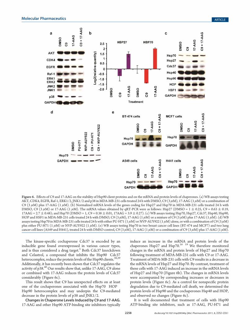
compounds has an effect on AKT (Figure 6a). By contrast bothC9 and 17-AAG cause a clear decrease in CDK4 levels, and a lessdrastic diminution in the case of EGFR and Raf-1 (Figure 6a).A combination of C9 and 17-AAG induced a more pronounceddecrease in CDK4, EGFR and Raf-1 levels than either compoundalone (Figure 6a).ERK1/2 was not affected by C9, 17-AAG or a combination
of the two compounds. In contrast, C9 and less dramatically17-AAG decreased JNK1/2, and only C9 had a clear effect on thelevels of p38 (Figure 6a).The inability of 17-AAG to affect the protein stability of
ERK1/2 has been extensively documented.7 Additionally, ithas been reported that the inhibition of Hsp90 by 17-AAGcauses a decrease in HER2 levels more effectively than it inducesa decrease in the levels of EGFR/HER1.36 Also, the degree ofAKT downregulation by Hsp90 inhibitors has been reported tovary in different cell lines.37
In the case of the ER we found that 17-AAG decreased theprotein levels of this steroid receptor, while C9 did not affect thisprotein (Supplementary Figure 3b in the Supporting In-formation), further suggesting that in agreement with theirdifferent proposed mechanism of action (Figure 1), C9 and 17-AAG affect a different subset ofHsp90 client proteins (Figure 6a).An Unexpected Effect of C9 Treatment Is the Decrease of
the Protein Cdc37. In order to determine a possible mechanismfor the unexpected decrease of the two structurally related SAPKstested here (JNK1/2 and p38), we next investigated the proteinlevels of Cdc37, which is a cochaperone implicated in theregulation of Hsp90 client proteins with kinase activity.
Figure 5. Cancer cell killing mode of C9. (a) Dose�response Caspase3/7 activation assays of MDA-MB-231 cells cultured as adherentmonolayers when treated 24 h with C9, 17-AAG, PU-H71 or NVP-AUY922. (b) Dose�response killing assays of human fibroblast cellscultured as adherent monolayers when treated 24 h with C9 or 17-AAG.
The kinase-specific cochaperone Cdc37 is encoded by aninducible gene found overexpressed in various cancer types,and is thus considered a drug target.5 Both Cdc37 knockdownand Celastrol, a compound that inhibits the Hsp90�Cdc37heterocomplex, reduce the protein levels of theHsp90 clients.38,39
Additionally, it has recently been shown that Cdc37 regulates theactivity of p38.40 Our results show that, unlike 17-AAG, C9 aloneor combined with 17-AAG reduces the protein levels of Cdc37considerably (Figure 6c).This result shows that C9 has unexpected effects on at least
one of the cochaperones associated with the Hsp70�HOP�Hsp90 heterocomplex and may underpin the C9-mediateddecrease in the protein levels of p38 and JNK1/2.Changes in Chaperone Levels Induced by C9 and 17-AAG.
17-AAG and other Hsp90 ATP-binding site inhibitors typically
induce an increase in the mRNA and protein levels of thechaperones Hsp27 and Hsp70.16�19 We therefore monitoredchanges in the mRNA and protein levels of Hsp27 and Hsp70following treatment of MDA-MB-231 cells with C9 or 17-AAG.Treatment ofMDA-MB-231 cells with C9 results in a decrease inthe mRNA levels of Hsp27 and Hsp70. By contrast, treatment ofthese cells with 17-AAG induced an increase in the mRNA levelsof Hsp27 and Hsp70 (Figure 6b). The changes in mRNA levelswere accompanied by corresponding increases or decreases inprotein levels (Figure 6c). As a control for nonspecific proteindegradation due to C9-mediated cell death, we determined theprotein levels of Hsp90 and the cochaperones Hsp40 and HOP,and observed no changes (Figure 6c).It is well documented that treatment of cells with Hsp90
ATP-binding site inhibitors, such as 17-AAG, PU-H71 and
Figure 6. Effects of C9 and 17-AAG on the stability of Hsp90 client proteins and on the mRNA and protein levels of chaperones. (a) WB assays testingAKT, CDK4, EGFR, Raf-1, ERK1/2, JNK1/2 and p38 inMDA-MB-231 cells treated 24 h with DMSO, C9 (3 μM), 17-AAG (1 μM) or a combination ofC9 (3 μM) plus 17-AAG (1 μM). (b) Normalized mRNA levels of the genes coding for Hsp27 and Hsp70 in MDA-MB-231 cells treated 24 h withDMSO, C9 (3 μM) or 17-AAG (1 μM). The mRNA values obtained by qRT-PCR were as follows: Hsp27 (DMSO = 1 ( 0.25, C9 = 0.63 ( 0.19,17AAG = 2.7( 0.48); and Hsp70 (DMSO = 1, C9 = 0.38( 0.01, 17AAG = 3.9( 0.27). (c) WB assays testing Hsp70, Hsp27, Cdc37, Hsp40, Hsp90,HOP andHSF1 inMDA-MB-231 cells treated 24 h with DMSO, C9 (3 μM), 17-AAG (1 μM) or a mixture of C9 (3 μM) plus 17-AAG (1 μM). (d)WBassays testingHsp70 inMDA-MB-231 cells treated 24 hwith either PU-H71 (1 μM)orNVP-AUY922 (1 μM) alone, or with a combination of C9 (3 μM)plus either PU-H71 (1 μM) or NVP-AUY922 (1 μM). (e) WB assays testing Hsp70 in two breast cancer cell lines (BT-474 and MCF7) and two lungcancer cell lines (A549 andH441), treated 24 h with DMSO-control, C9 (3 μM), 17-AAG (1 μM) or a combination of C9 (3 μM) plus 17-AAG (1 μM).
NVP-AUY922, causes an increase in the cellular levels ofHsp70.16�19 This side effect is undesirable, because Hsp70 hasantiapoptotic properties.41 We therefore investigated the effectof combining 17-AAG treatment, which causes an increase inHsp70 levels, with C9 treatment, which causes a decrease inHsp70 levels. We observed that in MDA-MB-231 cells treatedwith C9 plus 17-AAG the overexpression of Hsp70 is dampened,compared to the levels induced by 17-AAG (Figure 6c). C9 hada similar effect on Hsp70 when combined with NVP-AUY922,but not with PU-H71 (Figure 6d). Interestingly, these resultscorrelate with the finding that a combination of C9 plus 17-AAGor NVP-AUY922, but not PU-H71, decreases C9’s lethal-IC50(Figure 2d).To better demonstrate the effects of C9 when combined with
an Hsp90 ATP-binding site inhibitor such as 17-AAG, we nextdetermined Hsp70's protein levels in various other cell lines,besides MDA-MB-231. While the decrease in the levels of Hsp70induced by C9 varied between cell lines, a combination of C9with 17-AAG prevented the upregulation of Hsp70, normallyinduced by 17AAG alone, in all cases (Figure 6e).The ability of C9 to counterbalance the upregulation of Hsp70
induced by Hsp90 ATP-binding site inhibitors is an importantresult because it suggests that an added advantage to a combina-tion of C9 and either 17-AAG or NVP-AUY92 in cancer therapywould be to antagonize their prosurvival side effects, by counter-acting the increase in Hsp70 levels induced by these compoundswhen added alone.Furthermore, while a 3 μM C9 final concentration is lethal
against the breast cancer cell lines BT-474 andMCF7 (Figures 3band 3c), this dose does not have an obvious killing effect againstthe two lung cancer cell lines A549 and H441 as shown bypictures taken of these cells after C9 treatment (SupplementaryFigure 4 in the Supporting Information). Considering that thecombination of two anticancer drugs may enhance toxicity,our results highlight the potential use of C9 at sublethal dosesin combination with Hsp90 inhibitors like 17-AAG or NVP-AUY922 to suppress the transcriptional upregulation of Hsp70.HSF1, the Transcription Factor That Regulates the Expres-
sion of Hsp27 and Hsp70, Decreases in C9-Treated MDA-MB-231 Cells. HSF1 is a transcription factor that induces theexpression of genes with an upstream heat shock element, suchas Hsp27 and Hsp70.42 To further investigate the origin of thedecrease in Hsp27 and Hsp70 levels as a consequence of C9treatment, we compared the protein levels of heat shock factor 1(HSF1) in MDA-MB-231 cells treated with either C9, 17-AAGor a combination of C9 and 17-AAG (Figure 6c).We found that treatment of MDA-MB-231 cells with C9 alone
or in combination with 17-AAG results in a substantial decreaseof HSF1, while 17-AAG alone has a less drastic effect (Figure 6c).The decrease in the protein levels of HSF1 is most probably not aconsequence of a change in its phosphorylation pattern becausethe WB does not show a slow migrating band (SupplementaryFigure 5 in the Supporting Information). It is important to pointout that WB assays coupled to SDS�PAGE do not necessarilyinform about the oligomeric state of HSF1. In the absence of athorough picture of how HSF1 is being affected by C9, ourresults nevertheless clearly show that 17-AAG has differenteffects on this transcription factor, and this is what is relevantin our data.In conclusion, treatment of MDA-MB-231 cells with C9 alone
or combined with 17-AAG induces a marked decrease in theprotein levels of HSF1, an event that probably leads to the
downregulation of Hsp27 and Hsp70. Clearly, the decrease ofHSF1, like that of Cdc37 mentioned above, is an unexpected yetbeneficial effect of C9.The Anticancer Effects of C9 Are Different from Those of a
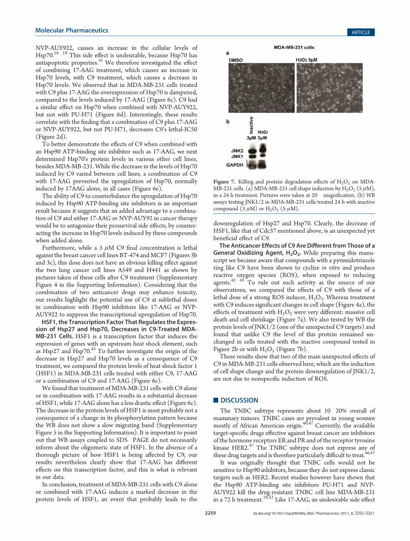
General Oxidizing Agent, H2O2. While preparing this manu-script we became aware that compounds with a pyrimidotriazolering like C9 have been shown to cyclize in vitro and producereactive oxygen species (ROS), when exposed to reducingagents.43�45 To rule out such activity as the source of ourobservations, we compared the effects of C9 with those of alethal dose of a strong ROS inducer, H2O2. Whereas treatmentwith C9 induces significant changes in cell shape (Figure 4a), theeffects of treatment with H2O2 were very different: massive celldeath and cell shrinkage (Figure 7a). We also tested by WB theprotein levels of JNK1/2 (one of the unexpected C9 targets) andfound that unlike C9 the level of this protein remained un-changed in cells treated with the inactive compound tested inFigure 2b or with H2O2 (Figure 7b).These results show that two of the main unexpected effects of
C9 inMDA-MB-231 cells observed here, which are the inductionof cell shape change and the protein downregulation of JNK1/2,are not due to nonspecific induction of ROS.
’DISCUSSION
The TNBC subtype represents about 10�20% overall ofmammary tumors. TNBC cases are prevalent in young womenmostly of African American origin.46,47 Currently, the availabletarget-specific drugs effective against breast cancer are inhibitorsof the hormone receptors ER and PR and of the receptor tyrosinekinase HER2.47 The TNBC subtype does not express any ofthese drug targets and is therefore particularly difficult to treat.46,47
It was originally thought that TNBC cells would not besensitive to Hsp90 inhibitors, because they do not express classictargets such as HER2. Recent studies however have shown thatthe Hsp90 ATP-binding site inhibitors PU-H71 and NVP-AUY922 kill the drug-resistant TNBC cell line MDA-MB-231in a 72 h treatment.18,22 Like 17-AAG, an undesirable side effect
Figure 7. Killing and protein degradation effects of H2O2 on MDA-MB-231 cells. (a) MDA-MB-231 cell shape induction by H2O2 (5 μM),in a 24 h treatment. Pictures were taken at 20� magnification. (b) WBassays testing JNK1/2 in MDA-MB-231 cells treated 24 h with inactivecompound (3 μM) or H2O2 (5 μM).
of PU-H71 and NVP-AUY922 treatment is that it induces anincrease in the mRNA and protein levels of the antiapoptoticchaperone Hsp70.18,22
We have demonstrated that the novel chaperone inhibitor C9is effective in killing breast cancer cell lines (BT-474 andMCF7),including the drug-resistant mammary TNBC subtype (MDA-231 and MDA-MB-468). Remarkably, treatment of MDA-MB-231 and MDA-MB-468 cells with C9 kills these TNBC cell linesin a 24 h exposure (Figure 2b and Supplementary Figure 1b inthe Supporting Information), in contrast to the 72 h neededfor PU-H71 and NVP-AUY922 to be lethal against MDA-MB-231 cells.18,22
Furthermore, C9 induces a change in cell shape, together withan inhibition of the characteristic migration properties character-istic of these cells, in a “scratch/wound-healing” assay (Figures 4aand 4b). An interesting feature of C9 is its ability to kill TNBCcell lines in a Caspase 3/7-independent manner (Figure 5a).
We also show that treating cells with C9 alone or combinedwith 17-AAG results in a decrease in the levels of a set of Hsp90-dependent client proteins (Cdk4, Raf-1, JNK1/2 and p38)(Figure 6a). The protein downregulation of these kinases inC9-treated cells is particularly interesting because in mammarycell lines the individual inactivation of JNK1/2 or p38 has beencorrelated with inhibition of cell migration.48�50 Additionally,activated JNK1/2 is proapoptotic in a context-dependentmanner7 and has been shown to participate in the onset ofapoptosis in breast cancer cell lines, under conditions of cellularstress.51
Our hypothesis is that the downregulation by C9 of thekinases p38 and JNK1/2, which are affected less drasticallyby 17-AAG, may explain at least in part the migration inhibi-tion and Caspase 3/7-independent killing properties of C9.An alternative hypothesis is that the downregulation of Cdc37is expected to have an effect on the total phosphorylationlevels, which may in turn induce cytoskeletal rearrangementsand thus account for the cell migration inhibition and changein cell shape induced by C9.
Finally, the decrease in the mRNA levels of Hsp27 and Hsp70is most probably due to the drastic diminution in the proteinlevels of HSF1 induced by C9 (Figure 6c). Treating cells with acombination of C9 and either 17-AAG or NVP-AUY922 sup-presses substantially the undesirable overexpression of Hsp70(Figures 6c and 6d), which is a hallmark of these Hsp90inhibitors. Another potential benefit of combining C9 with twoof these compounds (17-AAG and NVP-AUY922) is that in a24 h treatment the lethal-IC50 of C9 against MDA-MB-231 cellsis slightly lower in comparison to that determined for C9 alone(Figure 2d).
Interestingly, C9 fails to counteract PU-H71’s transcriptionalupregulation of Hsp70, despite sharing a common binding sitewith the other HSP-90 inhibitors, 17-AAG or NVP-AUY922.Each of these Hsp90 inhibitors has a unique chemical structure,which in turn induces different allosteric conformational changesin the Hsp90 N-terminus.1 The Hsp90 allosteric changes in-duced by PU-H71 are the most drastic, compared to 17-AAG orNVP-AUY922.1 These differences are proposed to underpin thedistinct anticancer properties of these Hsp90 inhibitors despitetheir common binding site in Hsp901 and may explain theinability of C9 to suppress PU-H71’s transcriptional upregulationof HSP-70.
To summarize, our results suggest that C9 alone, or incombination with at least two Hsp90 ATP-binding site inhibitors
currently in clinical trials (17-AAG and NVP-AUY922), could bean effective route to novel anticancer therapeutics, with apotential activity against the TNBC subtype. To define globallythe effects and direct targets of C9 in cancer cells, future studieswill include, respectively, a whole-cell quantitative profile of thetranscript and protein levels; and a systematic investigation of theprotein interactome associated with the Hsp70�HOP�Hsp90chaperone complex.
’ASSOCIATED CONTENT
bS Supporting Information. Five additonal figures as dis-cussed in the text. This material is available free of charge via theInternet at http://pubs.acs.org.
’AUTHOR INFORMATION
Corresponding Author*Yale University, Department of Molecular Biophysics andBiochemistry, New Haven, CT, United States. E-mail: [email protected].
’ACKNOWLEDGMENT
We are grateful to Ewa Menet at the Cell Sorting Facility, YaleUniversity, who performed and analyzed the FACS experiments.Michael Salcius operated the plate reader at the Yale UniversityGenomics and Proteomics Center. MDA-MB-231 cells were akind gift of Dr. Anthony Koleske. This work was funded in partby grants from Joan’s Legacy Lung Cancer Foundation andMaryKay Ash (to L.R.). K.M.H. is an HHMI Fellow of the DamonRunyon Cancer Research Foundation.
’REFERENCES
(1) Biamonte, M. A.; et al. Heat shock protein 90: inhibitors inclinical trials. J. Med. Chem. 2010, 53 (1), 3–17.
(2) Janin, Y. L. ATPase inhibitors of heat-shock protein 90, secondseason. Drug Discovery Today 2010, 15 (9�10), 342–53.
(3) Pearl, L. H.; Prodromou, C. Structure and mechanism of theHsp90 molecular chaperone machinery. Annu. Rev. Biochem. 2006,75, 271–94.
(4) Pratt, W. B.; Toft, D. O. Regulation of signaling protein functionand trafficking by the hsp90/hsp70-based chaperone machinery. Exp.Biol. Med. (Maywood) 2003, 228 (2), 111–33.
(5) Caplan, A. J.; Mandal, A. K.; Theodoraki, M. A. Molecularchaperones and protein kinase quality control. Trends Cell Biol. 2007,17 (2), 87–92.
(6) Powers, E. T.; et al. Biological and chemical approaches todiseases of proteostasis deficiency. Annu. Rev. Biochem. 2009, 78,959–91.
(7) Powers, M. V.; Clarke, P. A.; Workman, P. Death by chaperone:HSP90, HSP70 or both? Cell Cycle 2009, 8 (4), 518–26
(8) Kundrat, L.; Regan, L. Identification of residues on Hsp70 andHsp90 ubiquitinated by the cochaperone CHIP. J. Mol. Biol. 2010, 395(3), 587–94.
(9) Kundrat, L. Regan, L.; , Balance between folding and degradationfor Hsp90-dependent client proteins: a key role for CHIP. Biochemistry.49(35): p. 7428-38.
(10) Stebbins, C. E.; et al. Crystal structure of an Hsp90-geldana-mycin complex: targeting of a protein chaperone by an antitumor agent.Cell 1997, 89 (2), 239–50.
(11) Roe, S. M.; et al. Structural basis for inhibition of the Hsp90molecular chaperone by the antitumor antibiotics radicicol and gelda-namycin. J. Med. Chem. 1999, 42 (2), 260–6.
(12) Solit, D. B.; et al. 17-Allylamino-17-demethoxygeldanamycininduces the degradation of androgen receptor and HER-2/neu andinhibits the growth of prostate cancer xenografts. Clin. Cancer Res. 2002,8 (5), 986–93.(13) Ge, J.; et al. Design, synthesis, and biological evaluation of
hydroquinone derivatives of 17-amino-17-demethoxygeldanamycin aspotent, water-soluble inhibitors of Hsp90. J. Med. Chem. 2006, 49 (15),4606–15.(14) Solit, D. B.; et al. Phase I trial of 17-allylamino-17-demethox-
ygeldanamycin in patients with advanced cancer. Clin. Cancer Res. 2007,13 (6), 1775–82.(15) Solit, D. B.; et al. Phase II trial of 17-allylamino-17-demethox-
ygeldanamycin in patients with metastatic melanoma. Clin. Cancer Res.2008, 14 (24), 8302–7.(16) McCollum, A. K.; et al. Up-regulation of heat shock protein 27
induces resistance to 17-allylamino-demethoxygeldanamycin through aglutathione-mediated mechanism. Cancer Res. 2006, 66 (22), 10967–75.(17) Maloney, A.; et al. Gene and protein expression profiling of
human ovarian cancer cells treated with the heat shock protein 90inhibitor 17-allylamino-17-demethoxygeldanamycin. Cancer Res. 2007,67 (7), 3239–53.(18) Caldas-Lopes, E.; et al. Hsp90 inhibitor PU-H71, a multimodal
inhibitor of malignancy, induces complete responses in triple-negativebreast cancer models. Proc. Natl. Acad. Sci. U.S.A. 2009, 106 (20),8368–73.(19) Gaspar, N.; et al. Mechanistic evaluation of the novel HSP90
inhibitor NVP-AUY922 in adult and pediatric glioblastoma.Mol. CancerTher. 2010, 9 (5), 1219–33.(20) Yi, F.; Regan, L. A novel class of small molecule inhibitors of
Hsp90. ACS Chem. Biol. 2008, 3 (10), 645–54.(21) Hartl, F. U.; Hayer-Hartl, M. Converging concepts of protein
folding in vitro and in vivo. Nat. Struct. Mol. Biol. 2009, 16 (6), 574–81.(22) Eccles, S. A.; et al. NVP-AUY922: a novel heat shock protein
90 inhibitor active against xenograft tumor growth, angiogenesis, andmetastasis. Cancer Res. 2008, 68 (8), 2850–60.(23) Lu, Z.; Hunter, T. Degradation of activated protein kinases by
ubiquitination. Annu. Rev. Biochem. 2009, 78, 435–75.(24) Lopez-Otin, C.; Hunter, T. The regulatory crosstalk between
kinases and proteases in cancer. Nat. Rev. Cancer 2010, 10 (4), 278–92.(25) Ota, A.; et al. Specific regulation of noncanonical p38alpha
activation by Hsp90-Cdc37 chaperone complex in cardiomyocyte.Circ. Res. 2010, 106 (8), 1404–12.(26) Nieto-Miguel, T.; et al. Proapoptotic role of Hsp90 by its
interaction with c-Jun N-terminal kinase in lipid rafts in edelfosine-mediated antileukemic therapy. Oncogene 2008, 27 (12), 1779–87.(27) Gutierrez, G. J.; et al. Interplay between Cdh1 and JNK activity
during the cell cycle. Nat. Cell Biol. 2010, 12 (7), 686–95.(28) Yi, F.; et al. An AlphaScreen-based high-throughput screen
to identify inhibitors of Hsp90-cochaperone interaction. J. Biomol.Screening 2009, 14 (3), 273–81.(29) Neve, R. M.; et al. A collection of breast cancer cell lines for the
study of functionally distinct cancer subtypes. Cancer Cell 2006, 10 (6),515–27.(30) Munster, P. N.; et al. Inhibition of heat shock protein 90
function by ansamycins causes the morphological and functionaldifferentiation of breast cancer cells. Cancer Res. 2001, 61 (7), 2945–52.(31) Pickl, M.; Ries, C. H. Comparison of 3D and 2D tumor models
reveals enhanced HER2 activation in 3D associated with an increasedresponse to trastuzumab. Oncogene 2009, 28 (3), 461–8.(32) Weigelt, B.; Lo, A. T.; Park, C. C.; Gray, J. W.; Bissell, M. J.
HER2 signaling pathway activation and response of breast cancer cells toHER2-targeting agents is dependent strongly on the 3D microenviron-ment. Breast Cancer Res. Treat. 2010, 122 (1), 35–43.(33) Ivascu, A.; Kubbies, M. Diversity of cell-mediated adhesions in
breast cancer spheroids. Int. J. Oncol. 2007, 31 (6), 1403–13.(34) Korkaya, H.; et al. HER2 regulates the mammary stem/
progenitor cell population driving tumorigenesis and invasion.Oncogene2008, 27 (47), 6120–30.
(35) Wolf, K.; et al. Compensation mechanism in tumor cellmigration: mesenchymal-amoeboid transition after blocking of pericel-lular proteolysis. J. Cell Biol. 2003, 160 (2), 267–77.
(36) Xu, W.; et al. Sensitivity of mature Erbb2 to geldanamycin isconferred by its kinase domain and is mediated by the chaperone proteinHsp90. J. Biol. Chem. 2001, 276 (5), 3702–8.
(37) Theodoraki, M. A.; et al. Akt shows variable sensitivity to anHsp90 inhibitor depending on cell context.Exp. Cell Res. 2007, 313 (18),3851–8.
(38) Smith, J. R.; et al. Silencing the cochaperone CDC37 destabi-lizes kinase clients and sensitizes cancer cells to HSP90 inhibitors.Oncogene 2009, 28 (2), 157–69.
(39) Zhang, T.; et al. A novel Hsp90 inhibitor to disrupt Hsp90/Cdc37 complex against pancreatic cancer cells.Mol. Cancer Ther. 2008,7 (1), 162–70.
(40) Ota, A.; et al. Specific regulation of noncanonical p38alphaactivation by Hsp90-Cdc37 chaperone complex in cardiomyocyte. Circ.Res. 2010, 106 (8), 1404–12.
(41) Garrido, C.; et al. Heat shock proteins 27 and 70: anti-apoptoticproteins with tumorigenic properties.Cell Cycle 2006, 5 (22), 2592–601.
(42) Voellmy, R. On mechanisms that control heat shock transcrip-tion factor activity in metazoan cells. Cell Stress Chaperones 2004, 9 (2),122–33.
(43) Johnston, P. A.; et al. Development of a 384-well colorimetricassay to quantify hydrogen peroxide generated by the redox cycling ofcompounds in the presence of reducing agents. Assay Drug Dev. Technol.2008, 6 (4), 505–18.
(44) Johnston, P. A. Redox cycling compounds generate H2O2 inHTS buffers containing strong reducing reagents—real hits or promis-cuous artifacts? Curr Opin. Chem. Biol. 2011, 15 (1), 174–82.
(45) Soares, K. M.; et al. Profiling the NIH Small MoleculeRepository for compounds that generate H2O2 by redox cycling inreducing environments. Assay Drug Dev. Technol. 2010, 8 (2), 152–74.
(46) Stockmans, G.; et al. Triple-negative breast cancer. Curr. Opin.Oncol. 2008, 20 (6), 614–20.
(47) Carey, L.; et al. Triple-negative breast cancer: disease entity ortitle of convenience? Nat Rev. Clin. Oncol. 2010, 7 (12), 683–92.
(48) Huang, C.; et al. JNK phosphorylates paxillin and regulates cellmigration. Nature 2003, 424 (6945), 219–23.
(49) Shin, I.; et al. H-Ras-specific activation of Rac-MKK3/6-p38pathway: its critical role in invasion and migration of breast epithelialcells. J. Biol. Chem. 2005, 280 (15), 14675–83.
(50) Kaoud, T. S.; et al. Development of JNK2-Selective PeptideInhibitors That Inhibit Breast Cancer Cell Migration. ACS Chem. Biol.2011, 6 (6), 658–66.
(51) Mingo-Sion, A. M.; et al. Inhibition of JNK reduces G2/Mtransit independent of p53, leading to endoreduplication, decreasedproliferation, and apoptosis in breast cancer cells.Oncogene 2004, 23 (2),596–604.