No. 2079. JULY 4, 1863. A Course of Twelve Lectures ON THE STRUCTURE AND DEVELOPMENT OF THE VERTEBRATE SKELETON. Recently delivered at the Royal College of Surgeons of England, BY PROFESSOR HUXLEY, F.R.S. LECTURE IX. MR. PRESIDENT AND GENTLEMEN,--At the conclusion of my 6ijdea"tiing to identify in th skull of the turtle the majority or the most important of thos( bo - i’e3itl"iideid so tsach at length in the othej verMta.. ’I ’oted t %i the base of the skull a distinct basi-occipital, and a basilar ossification, which in the adull skull is coalesced with a proper basi-sphenoid. Then, in the interior of the skull, a complete cartilaginous septum, a carti- laginous partition between the two orbits, which represents the pre-sphenoid ; a cartilaginous continuation of this process be’ tween the nasal capsules, which represents the ethmoid. In the side walls of the skull, again, we had the ex-occipitals well developed, perfectly recognizable; and in the roof the supra-occipital in its ordinary place. But in the front walls of the skull there was no distinctly ossified ali- sphenoid, at least none apparently proceeding from a cartilaginous basis, nor was there any trace of an orbito-sphenoid. In the roof of the skull, as usual, there were the parietals and frontals, but there were no distinct nasal bones-at least in ordinary chelonians,-and their place appears to be taken very much by pre-frontals arching in the front of the skull over the nasal capsules. Then the bones which surround the organ of hear- ing were in an extremely interesting condition. We identified at once the prootic bone, having just the same relations that we found in the fish, lying immediately behind the third divi. sion of the fifth nerve, allowing the entrance of the auditory and of the portio dura, as usual, and lodging the anterior parts of the organs of hearing-that is to say, part of the vestibule and the greater part of the anterior vertical semicircular canal: there is no doubt whatever about that bone. Then between the organ of hearing and the ex-occipital there was a bone of which only a very small part is shown in the interior of the skull, the bone itself, however, being a large one as seen on the exterior of the skull. This bone lies in front of the exit of the eighth nerve, and immediately behind the posterior part of the organ of hearing ; it has the fenestra rotunda in it, -and also part of the external semicircular canal. By its relations to the eighth nerve and the external semicircular canal, we were able to identify it with that bone which I pointed out in the ephippus, and that bone called the petrosum and other names in the perch, the cod, and elsewhere; and, on the other hand, by its relations to the fenestra rotunda and the rudi- mentary cochlea we were equally able to identify it with that opisthotic ossification which we found in man. The uppermost part of the case of the organ of hearing in the adult turtle is formed by what appears to be a continuation of the supra-occipital bone. But there is reason to believe that in the young state that is a distinct bone; it is, in fact, a separate epiotic bone, and has all the relations of the true epiotic ele- ment. It results from this arrangement of the bones that there is in the skull a tri-radiate suture: one narrow rim of it running between the epiotic and the opisthotic; then one leg running between the epiotic and the prootic; and a third leg-which is exceedingly broad, and in the dry skull constitutes a large open space, the cartilage having disappeared-between the prooti and the opisthotic. That tri-radiate suture we shall find to b an extremely persistent structure in the higher oviparous ver tebrata of which I am now about to speak. The fenestr: I rotunda is a structure which we have not hitherto met with il the ascending series of vertebrata: it is a distinct hole forme( in the opisthotic element, and remaining covered over by ! mass of dense membrane like that which covers over t fenestra ovalis, and bringing the external medium into direc contact with the cochlea itself. This aperture makes its appear ance in correspondence with the increasing complexity of the organ of hearing. There are rudiments of the fenestra rotund -traces of it-in some of the higher amphibia, but not lowe] in the vertebral series than that. Then as to the inferior arches of the turtle’s skull, we have, as usual, the pre-maxilla perfectly recognizable in its place. though but a small bone, and, like the maxilla, containing nc teeth, all these parts in the turtle and in the chelonia generally being covered by a large horny shield. Then there are twc palatine bones in their ordinary position, and two pterygoid bones; and these pterygoid bones abut posteriorly, as in the lower oviparous vertebrata, on the quadrate bone, and the quadrate bone, as usual, gives articulation to the articulare of the lower jaw; and from the articulare there is continued- forming an axis round which the other bony elements of the lower jaw are disposed-a cartilage corresponding with Meckel’s cartilage in the chelonia. The anterior nares are formed in the ordinary way between the prolongation of the pre-frontals (which represent the nasals), the pre-maxillse, and the vomer in the middle line. But there is a very interesting approxima- tion towards the formation of a posterior nares as distinguished from what we call the median nares. You will recollect that the median nares lay between the palatine bones upon each side, the vomer in front, and the maxilla at the sides - or the inter-maxilla, as the case might be. But when, as in this case, the maxillary bones are very much enlarged, and throw lateral processes inwards towards the middle line, there is a production of posterior nares in the sense that we find in man, and by exactly the same process; that is to say, the proper median nares are more or less shut off from the cavity of the mouth, though slightly so, in the turtle, and the true commu- nication between the nose and the mouth is thrown much far- ther back than it would be supposing the internal processes of the maxillary bones not.to exist. . You will observe, as a very interesting feature of the che- Ionian skull, that all the anterior facial arches, if I may so call them, are firmly fixed to the base of the skull, partly to the basilar ossification, and partly to the cartilage which is in front. There is no mobility at all about the parts of the upper jaw or of the palate; and inasmuch as these parts are firmly fixed, by means of the pterygoid, to the quadrate bone, that also is practically rendered immovable, or would be so if it were not, in addition, fixed to the sides of the skull by a large and extensive surface of union. The quadrate bone becomes very large and expanded; all the posterior part gives attach- ment most especially, in an indirect manner, to the tympanic membrane ; but the arch which it forms is almost completely closed up by bone, so that there is only a small hole left through which the columella passes (that is, the columella of the organ of hearing), the columella being on its way to the fenestra ovalis. In consequence of the manner in which the quadrate bone has become expanded and fixed to the prootic and to the opisthotic, and other bones in the vicinity, there is a sort of chamber formed between the sides of the skull on the one hand and the tympanic membrane on the other, and these lateral boundary walls in front and behind. That chamber is the tympanum or drum of the ear, of which a more or less well-marked rudiment already exists in the frog; but in the frog it is by no means thoroughly shut off from the cavity of the mouth; so that that communication which exists in us, and which is a long narrow tube, the Eustachian tube, is in frogs a wide and patent aperture; and you will only regard the tympanum as a sort of recess, if one may so say, of the roof of the mouth. In the chelonians, on the other hand, in consequence of the large development of the bone3 about the A
Transcript
No. 2079.
JULY 4, 1863.
A Course of Twelve LecturesON THE
STRUCTURE AND DEVELOPMENTOF THE
VERTEBRATE SKELETON.
Recently delivered at the Royal College of Surgeons of England,
BY PROFESSOR HUXLEY, F.R.S.
LECTURE IX.
MR. PRESIDENT AND GENTLEMEN,--At the conclusion of my6ijdea"tiing to identify in thskull of the turtle the majority or the most important of thos(bo - i’e3itl"iideid so tsach at length in the othejverMta.. ’I ’oted t %i the base of the skull a distinctbasi-occipital, and a basilar ossification, which in the adullskull is coalesced with a proper basi-sphenoid. Then, in theinterior of the skull, a complete cartilaginous septum, a carti-laginous partition between the two orbits, which represents thepre-sphenoid ; a cartilaginous continuation of this process be’tween the nasal capsules, which represents the ethmoid.In the side walls of the skull, again, we had the ex-occipitalswell developed, perfectly recognizable; and in the roofthe supra-occipital in its ordinary place. But in the frontwalls of the skull there was no distinctly ossified ali- sphenoid,at least none apparently proceeding from a cartilaginous basis,nor was there any trace of an orbito-sphenoid. In the roof ofthe skull, as usual, there were the parietals and frontals, butthere were no distinct nasal bones-at least in ordinarychelonians,-and their place appears to be taken very much bypre-frontals arching in the front of the skull over the nasalcapsules. Then the bones which surround the organ of hear-
ing were in an extremely interesting condition. We identifiedat once the prootic bone, having just the same relations thatwe found in the fish, lying immediately behind the third divi.sion of the fifth nerve, allowing the entrance of the auditoryand of the portio dura, as usual, and lodging the anterior partsof the organs of hearing-that is to say, part of the vestibuleand the greater part of the anterior vertical semicircular canal:there is no doubt whatever about that bone. Then betweenthe organ of hearing and the ex-occipital there was a bone ofwhich only a very small part is shown in the interior of theskull, the bone itself, however, being a large one as seen onthe exterior of the skull. This bone lies in front of the exit ofthe eighth nerve, and immediately behind the posterior part ofthe organ of hearing ; it has the fenestra rotunda in it, -andalso part of the external semicircular canal. By its relationsto the eighth nerve and the external semicircular canal, wewere able to identify it with that bone which I pointed out inthe ephippus, and that bone called the petrosum and othernames in the perch, the cod, and elsewhere; and, on the otherhand, by its relations to the fenestra rotunda and the rudi-mentary cochlea we were equally able to identify it with thatopisthotic ossification which we found in man.The uppermost part of the case of the organ of hearing in the
adult turtle is formed by what appears to be a continuation ofthe supra-occipital bone. But there is reason to believe that inthe young state that is a distinct bone; it is, in fact, a separateepiotic bone, and has all the relations of the true epiotic ele-ment. It results from this arrangement of the bones that thereis in the skull a tri-radiate suture: one narrow rim of it runningbetween the epiotic and the opisthotic; then one leg runningbetween the epiotic and the prootic; and a third leg-which isexceedingly broad, and in the dry skull constitutes a large open
space, the cartilage having disappeared-between the prootiand the opisthotic. That tri-radiate suture we shall find to ban extremely persistent structure in the higher oviparous vertebrata of which I am now about to speak. The fenestr:
I rotunda is a structure which we have not hitherto met with ilthe ascending series of vertebrata: it is a distinct hole forme(in the opisthotic element, and remaining covered over by !mass of dense membrane like that which covers over tfenestra ovalis, and bringing the external medium into direccontact with the cochlea itself. This aperture makes its appearance in correspondence with the increasing complexity of theorgan of hearing. There are rudiments of the fenestra rotund-traces of it-in some of the higher amphibia, but not lowe]in the vertebral series than that.Then as to the inferior arches of the turtle’s skull, we have,
as usual, the pre-maxilla perfectly recognizable in its place.though but a small bone, and, like the maxilla, containing ncteeth, all these parts in the turtle and in the chelonia generallybeing covered by a large horny shield. Then there are twc
palatine bones in their ordinary position, and two pterygoidbones; and these pterygoid bones abut posteriorly, as in thelower oviparous vertebrata, on the quadrate bone, and thequadrate bone, as usual, gives articulation to the articulare ofthe lower jaw; and from the articulare there is continued-forming an axis round which the other bony elements of thelower jaw are disposed-a cartilage corresponding with Meckel’scartilage in the chelonia. The anterior nares are formed in the
ordinary way between the prolongation of the pre-frontals(which represent the nasals), the pre-maxillse, and the vomerin the middle line. But there is a very interesting approxima-tion towards the formation of a posterior nares as distinguishedfrom what we call the median nares. You will recollect thatthe median nares lay between the palatine bones upon eachside, the vomer in front, and the maxilla at the sides - orthe inter-maxilla, as the case might be. But when, as in thiscase, the maxillary bones are very much enlarged, and throwlateral processes inwards towards the middle line, there is aproduction of posterior nares in the sense that we find in man,and by exactly the same process; that is to say, the propermedian nares are more or less shut off from the cavity of themouth, though slightly so, in the turtle, and the true commu-nication between the nose and the mouth is thrown much far-ther back than it would be supposing the internal processes ofthe maxillary bones not.to exist..
You will observe, as a very interesting feature of the che-Ionian skull, that all the anterior facial arches, if I may so callthem, are firmly fixed to the base of the skull, partly to thebasilar ossification, and partly to the cartilage which is infront. There is no mobility at all about the parts of the upperjaw or of the palate; and inasmuch as these parts are firmlyfixed, by means of the pterygoid, to the quadrate bone, thatalso is practically rendered immovable, or would be so if itwere not, in addition, fixed to the sides of the skull by a largeand extensive surface of union. The quadrate bone becomesvery large and expanded; all the posterior part gives attach-ment most especially, in an indirect manner, to the tympanicmembrane ; but the arch which it forms is almost completelyclosed up by bone, so that there is only a small hole left
through which the columella passes (that is, the columella ofthe organ of hearing), the columella being on its way to thefenestra ovalis. In consequence of the manner in which thequadrate bone has become expanded and fixed to the prooticand to the opisthotic, and other bones in the vicinity, there isa sort of chamber formed between the sides of the skull on theone hand and the tympanic membrane on the other, and theselateral boundary walls in front and behind. That chamber isthe tympanum or drum of the ear, of which a more or lesswell-marked rudiment already exists in the frog; but in thefrog it is by no means thoroughly shut off from the cavity ofthe mouth; so that that communication which exists in us,and which is a long narrow tube, the Eustachian tube, is infrogs a wide and patent aperture; and you will only regardthe tympanum as a sort of recess, if one may so say, of theroof of the mouth. In the chelonians, on the other hand, inconsequence of the large development of the bone3 about the
A
2
outer parts of the organ of hearing, the tympanic cavity be-comes much better defined, and more constricted from theaperture, by means of which the tympanum communicateswith the mouth, and this aperture now becomes a definiteEustachian tube. In the turtle, as in the lower vertebrata,there is only a single rod serving as a bone of the organ ofhearing-a single rod, which is expanded at its inner extremity,and there fitted into the fenestra ovalis in the same way as thestapes is fitted in us, and which, on the other hand, is con-tinued out as a straight rod, and, becoming expanded at theextremity, is fixed into the tympanic membrane itself, beingoccasionally connected indirectly with the sides of the quadratebone as well. Then upon the upper part of the chamber of theorgan of hearing you have a well-marked squamosal appliedupon the outer sides of the skull, and there is in front of theorgan of hearing a post-frontal, these bones taking quite thesame position as they do in the fish. The hyoidean apparatusbecomes much simplified in the chelonia to what we find it infrogs and perenni-branchial amphibia, consisting of a broadplate with two cornua inside; but into the details of thisstructure, as we have so much to do, it is not worth while nowto enter.The development of the chelonian skull is quite comparable
to that of other vertebrata. There is the same sort of memobranous skull formed in the first place; the same rudimentarycartilaginous skull with its two trabecu]2a; and ossifications inthe main take place in the corresponding bones in the sameway as in the lower vertebrata.
Leaving this type of the skull, then, I may now pass on tospeak of the lizard’s skull, which is in principle constructed invery much the same fashion as the chelonian skull, although inits details it has an exceedingly different appearance. In thechelonian skull, particularly in that of the turtle, with whichI commenced my description, not only are all the bones of theface fixed to one another, and all the different parts of the skullperfec ly immobile, but there is a sort of additional fixity givento these different bones by a very remarkable expansion of theupper part of the frontal and of the sides of the parietal bones;and that expansion extends to the squamosal, post frontal, andall these bones; the expansion from the median line extendingoutwards, and the expansion from the sides extending upwardsand towards the median line-all these unite together, and forma kind of second arch above the top of the skull. If you takethe skull of the trionyx, you observe all the bones which form ’,the limits of the periotic capsule, the prootic, and so on, arevisible at the top of the skull; but in the turtle the occipital Iarch has thrown out a great lateral expansion, and the other Iupper bones have also thrown out a lateral expansion : thusyou have formed that great chamber which lies on the top ofthe turtle’s skull, and gives it such a helmet-like appearance.In the lacertian skull everything is changed in this respect.All the parts have become singularly. mobile, so that not onlyis the quadrate bone movable upon its articulation in mostcases (I speak now of the typical lacertian skull, for there areexceptions), but even the front part of the skull is movable inmost lizards from the back part in consequence of a mechanismwhich I will explain by-and-by.
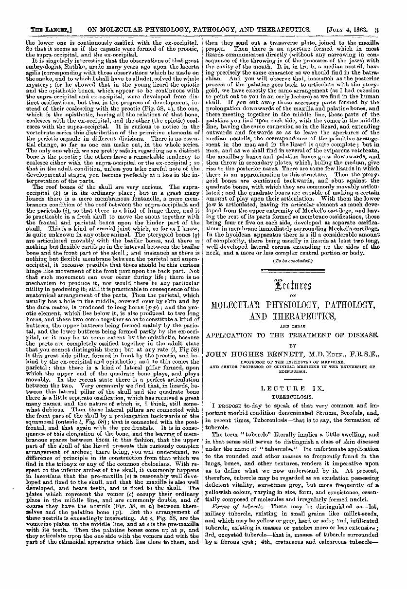
In a vertical section of a lizard’s skull (Fig. 58) you find, in
Section of lizard’s skull. a, Basi-occipital. b, Basi-sphenoid ;below b is the basi-temporal. c, Right vomer. e, Pre-maxillary. f, Nasal. h, Frontal. pl f, Parietal fontanelle.i, Parietal p, Process of parietal. k, Supra-occipital.1, Wing of ex-occipital. m, Median part of ex-occipital.8, Situation of foramen for vagus nerve. ", Prootic.5, Foramen ovale. o, A1i’Fpbenoiùal " columella." p, Pala-tine. MM, Median nares. px, Os transversum. q, Pterygoid.r, Descending plate of parietal, s, Jugal. t, Quadratum.? r, Turbinal. 2, Optic nerve.
the first place, that the ull is constructed upon the same plan I
as that of the turtle and of the pike-that is to say, that thereis in the front part a laterally-compressed inter-orbital septum(Fig. 58, in front of 2), composed in great measure of cartilageor membrane, so that the front part of the cranial cavity isextremely narrow; at the back part the basi cranial axis (a) isas usual; and in front of that there is a basilar ossification(below b) to a very considerable extent, but nothing like somuch as it is in the crocodiles and chelonians. In front of thatpoint almost the whole of the basi cranial axis is unossified, butthere are certain small ossifications placed within it, whichhave a very considerable interest from their correspondencewith the ossifications that we find in the corresponding regionin the pike, for example. If you examine the skull (Fig. 59)
of the ignana, for instance, you will find in front of that bonewhich 1 called the basilar bone, commonly called the basi-sphenoid, a little Y-shaped bone (Fig. 59, c), consisting of twolittle crura which lie behind the exits of the optic nerves, andof a little stem which comes down in the middle line, and abutsupon the beak like prolongation forwards of the basilar bone.Fig. 59, a, is the basi-occipital; b is the basilar bone, lodgingthe pituitary body, and continued from it the forward beak ofthe basilar bone; and c is the -shaped bone of which I spoke;the optic nerves pass out at 2. Fig. 59, d, is the septum,which is composed mainly of cartilage, or, indeed, hardly ofcartilage, but mere membrane; you may find in front certainirregular ossifications. As this V-shaped ossification lies im-mediately in front of the pituitary fossa, and behind the exitof the optic nerves, I think there can be no doubt that it repre-sents the body of the proper basi-sphenoid-that is, correspond-ing with the corresponding-shaped bone in the pike, and somany other Rshes; and the ossification, d, which is placed infront, corresponds with the imperfectly-ossified pre-spbenoid,which you may also find in many fishes. It runs in the same
way under the peduncles of the olfactory nerves, and has thesame relation to the optic nerves, lying, as it does, in front ofthem; so that I suspect the great mass of that bone, which hasbeen called the basi-sphenoid in lizards, is merely that peculiarbasilar ossification which, arising sometimes from one andsometimes from two centres, is so characteristic of the oviparousvertebrata. The continuation forward of the cranio-facial axisof the lizard presents hardly any ossifications at all-none thatmay be called distinct bones. The lateral walls of the skull,again, are hardly more complete. There is a distinct ex-
occipital (Fig. 58, l) in its place, quite recognizable byall those characteristics by which we have identified the
ex-occipital throughout. But then there is no ali sphenoid;at least, I do not imagine that the bone which seems to repre-sent it is ossified from cartilage. I am not aware that therehas been any demonstration at all that it is so ossified. Butthere is a very curious bone (Fig. 58, o), coming down like alittle column from the parietal bone. This bone is. unfor-tunately, called the columella; I say unfortunately, becauseprecisely the same anatomical term is appiied to the little bonewhich represents the stapes, which extends from the fenestrarotunda to the tympanic membrane. That, however, is thename which it has now, and it has been supposed, and with aconsiderable degree of probability, that it represents the ali-
spheroid in the lizards. But it must be remembered that itdiffers from it in many important respects. Of anything likethe orbito,spbenoid there is hardly a trace ; at most, somelittle irregular ossifications here and there. The bones whichsurround the organ of hearing, however, are very well marked,and easily recognizable by the tri-radiate suture. Fig. 58, n,shows the bone which lodges the front part of the organ ofhearing, and 5, Fig. 58, the exit of the third division of the
fifth. The lower part of k is, of course, the epiotic; and theanterior part of m the opisthotic. But the lower division andthe upper one are not distinct bones; the upper one is con-tinued in the supra. occipital by a continuous ossification, whilst
3
the lower one is continuously ossified with the ex-occipital.So that it seems as if the capsule were formed of the prootic,the supra, occipital, and the ex-occipital.
It is singularly interesting that the observations of that greatembryologist, Rathke, made many years ago upon the lacertaagilis (corresponding with those observations which he made onthe snake, and to which I shall have to allude), solved the wholemystery; for he showed that in the young lizard the epioticand the opisthotic bones, which appear to be continuous withthe supra-occipital and ex-occipital, were developed from dis-tinct ossifications, but that in the progress of development, in-stead of their coalescing with the prootic (Fig. 58, n), the one,which is the opisthotic, having all the relations of that bone,coalesces with the ex-occipital, and the other (the epiotic) coal-esces with the supra occipital. It is curious to notice in thevertebrate series this distribution of the primitive elements ofthe periotic capsule in different divisions. There is no essen-tial change, so far as one can make out, in the whole series.The only one which we are pretty safe in regarding as a distinctbone is the prootic; the others have a remarkable tendency tocoalesce either with the supra-occipital or the ex-occipital ; sothat in the adult condition, unless you take careful note of thedevelopmental stages, you become perfectly at a loss in the in-terpretation of the parts.The roof bones of the skull are very curious. The supra-
occipital (k) is in its ordinary place; but in a great manylizards there is a mere membranous fontanelle, a mere mem-branous condition of the roof between the supra-occipitals andthe parietals (i), so that there is a kind of hinge there, and itis practicable in a fresh skull to move the snout together withthe frontal and parietal bones upon the hinder part of theskull. This is a kind of cranial joint which, so far as I know,is quite unknown in any other animal. The pterygoid bones (q)are articulated movably with the basilar bones, and there isnothing but flexible cartilage in the interval between the basilarbone and the front part of the skull ; and inasmuch as there isnothing but flexible membrane between the parietal and supra-occipital, it becomes possible that there should be this curioushinge-like movement of the front part upon the back part. Notthat such movement can ever occur during life ; there is nomechanism to produce it, nor would there be any particularutility in producing it; still it is practicable in consequence of theanatomical arrangement of the parts. Then the parietal, whichusually has a hole in the middle, covered over by skin and bythe dura mater, is produced to long horns (p p) and the pro-otic element, which lies below it, is also produced to two longhorns, and these two come together so as to constitute a kind ofbuttress, the upper buttress being formed mainly by the parie-tal, and the lower buttress being formed partly by the ex-occi.pital, or it may be to some extent by the opisthotic, becausethe parts are completely ossified together in the adult statethat you cannot distinguish them; but at any rate (l, Fig 58)is this great side pillar, formed in front by the prootic, and be-hind by the ex-occipital and opisthotic ; and to this comes theparietal : thus there is a kind of lateral pillar formed, uponwhich the upper end of the quadrate bone plays, and playsmovably. In the recent state there is a perfect articulationbetween the two. Very commonly we find that, in lizards, be-tween this lateral pillar of the skull and the quadrate bonethere is a little separate ossification, which has received a greatmany names, and the nature of which is, I think, still some-what dubious. Then these lateral pillars are connected withthe front part of the skull by a prolongation backwards of thesquamosal (outside l, Fig. 58) ; that is connected with the post-frontal, and that again with the pre-frontals. It is in conse-
quence of this elongation of the bone, and the leaving of mem-branous spaces between them in this fashion, that the upperpart of the skull of the lizard presents this curiously complexarrangement of arches; there being, you will understand, nodifference of principle in its construction from that which wefind in the trionyx or any of the common chelonians. With re-spect to the inferior arches of the skull, it commonly happensin lacertians that the pre-maxilla (e) is reasonably well deve-loped and fixed to the skull. and that the maxilla is also welldeveloped, and bears teeth, and is fixed to the skull. Theplates which represent the vomer (c) occupy their ordinaryplace in the middle line, and are commonly double, and ofcourse they have the nostrils (Fig. 58, m n) between them-selves and the palatine bone (p). But the arrangement ofthese nostrils is exceedingly interesting. At c, Fig. 58, are thevomerine plates in the middle line, and at e is the pre-maxillawith its teeth. Then the palatine bones come up at p, andthey articulate upon the one side with the vomers and with thepart of the ethmoidal apparatus which lies close to them, and
then they send out a transverse plate, joined to the maxillaproper. Then there is an aperture formed which in mostlizards communicates directly (without any narrowing in con-sequence of the throwing in of the processes of the jaws) withthe cavity of the mouth. It is, in truth, a median nostril, hav-ing precisely the same character as we should find in the batra-chian. And you will observe that, inasmuch as the posteriorprocess of the palatine goes back to articulate with the ptery-goid, we have exactly the same arrangement (as I had occasionto point out to you in an early lecture) as we find in the humanskull. If you cut away those accessory parts formed by theprolongation downwards of the maxilla and palatine bones, andthere meeting together in the middle line, those parts of thepalatine you find upon each side, with the vomer in the middleline, having the same connexion as in the lizard, and extendingoutwards and forwards so as to leave the apertures of themedian nostrils, the correspondence of the primitive arrange-ment in the man and in the lizard is quite complete ; but inman, and as we shall find in several of the oviparous vetebrata,the maxillary bones and palatine bones grow downwards, andthen throw in secondary plates, which, hiding the median, giverise to the posterior nares. There are some few lizards in whichthere is an approximation to this structure. Then the ptery-goid bones are continued backwards, and abut against thequadrate bones, with which they are commonly movably articu-lated ; and the quadrate bones are capable of making a certainamount of play upon their articulation. With them the lowerjaw is articulated, having its articular element as much deve-loped from the upper extremity of Meckel’s cartilage, and hav-ing the rest of its parts formed as membrane ossifications, thosebeing four or five on each side, developed as separate ossifica-tions in membrane immediately surrounding Meckel’s cartilage.In the hyoidean apparatus there is still a considerable amountof complexity, there being usually in lizards at least two long,well-developed lateral cornua extending up the sides of theneck, and a more or less complex central portion or body.
(To be concluded.)
LecturesON
MOLECULAR PHYSIOLOGY, PATHOLOGY,AND THERAPEUTICS,
AND THEIR
APPLICATION TO THE TREATMENT OF DISEASE.
BY
JOHN HUGHES BENNETT, M.D.EDIN., F.R.S.E.,PROFESSOR OF THE INSTITUTES OF MEDICINE,
AND SENIOR PROFESSOR OF CLINICAL MEDICINE IN THE UNIVERSITY OFEDINBURGH.
LECTURE IX.TUBERCULOSIS.
’I PROPOSE to-day to speak of that very common and im-
portant morbid condition denominated Struma, Scrofula, and,in recent times, Tuberculosis-that is to say, the formation oftubercle..
The term " tubercle" literally implies a little swelling, andin that sense still serves to distinguish a class of skin diseasesunder the name of " tuberculse." Its unfortunate applicationto the rounded and other masses so frequently found in thelungs, bones, and other textures, renders it imperative uponus to define what we now understand by it. At present,therefore, tubercle may be regarded as an exudation possessingdeficient vitality, sometimes grey, but more frequently of ayellowish colour, varying in size, form, and consistence, essen-tially composed of molecules and irregularly formed nuclei.forms of tubercle. -These may be distinguished as-1st,
miliary tubercle, existing in small grains like millet-seeds,and which may be yellow or grey, hard or soft; nd, infiltratedtubercle, existing in masses or patches more or less extensive ;3rd, encysted tubercle-that is, masses of tubercle surroundedby a fibrous cyst ; 4th, cretaceous and calcareous tubercle-