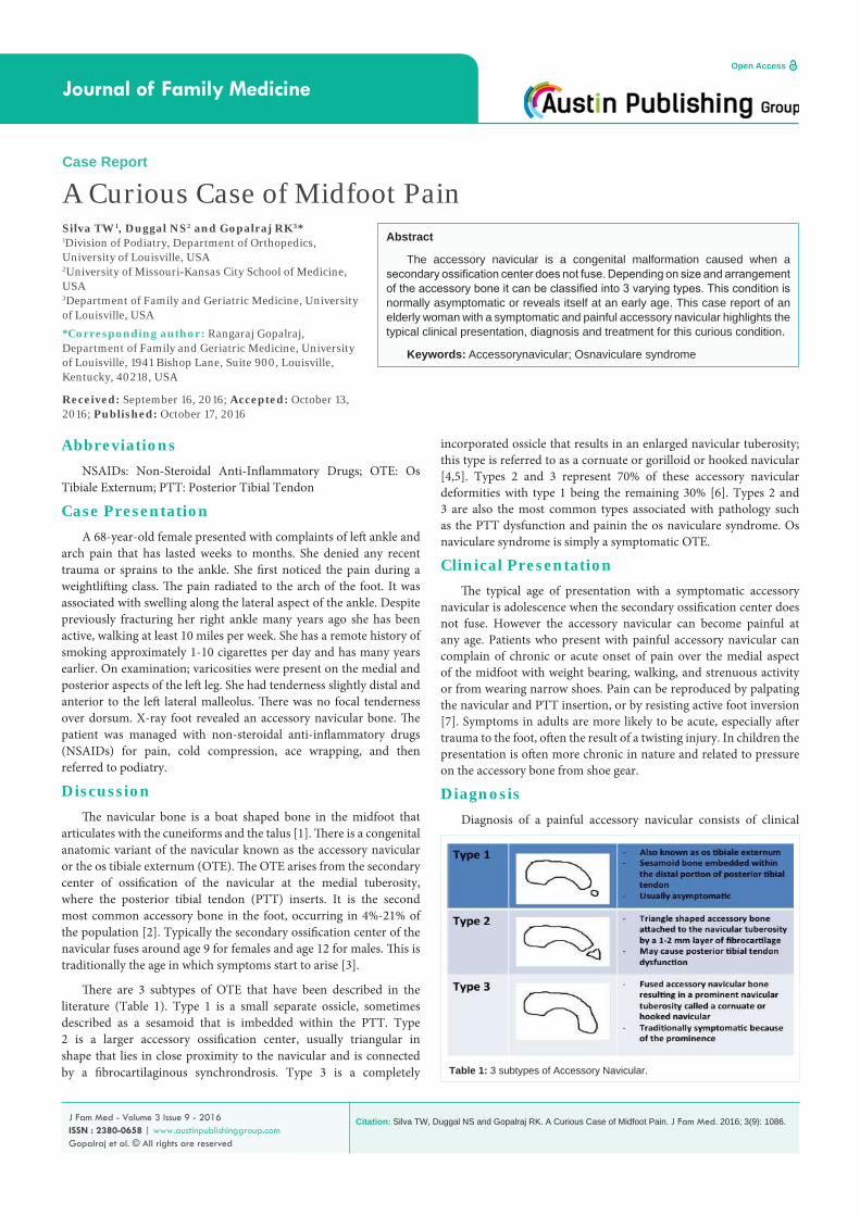
+ All Categories
Home > Documents > A Curious Case of Midfoot Pain - austinpublishinggroup.com

A Curious Case of Midfoot Pain - austinpublishinggroup.com

Date post: 12-Jan-2022
Category:
Upload: others
View: 4 times
Download: 0 times
Share this document with a friend
2
Citation: Silva TW, Duggal NS and Gopalraj RK. A Curious Case of Midfoot Pain. J Fam Med. 2016; 3(9): 1086. J Fam Med - Volume 3 Issue 9 - 2016 ISSN : 2380-0658 | www.austinpublishinggroup.com Gopalraj et al. © All rights are reserved Journal of Family Medicine Open Access Abstract The accessory navicular is a congenital malformation caused when a secondary ossification center does not fuse. Depending on size and arrangement of the accessory bone it can be classified into 3 varying types. This condition is normally asymptomatic or reveals itself at an early age. This case report of an elderly woman with a symptomatic and painful accessory navicular highlights the typical clinical presentation, diagnosis and treatment for this curious condition. Keywords: Accessorynavicular; Osnaviculare syndrome incorporated ossicle that results in an enlarged navicular tuberosity; this type is referred to as a cornuate or gorilloid or hooked navicular [4,5]. Types 2 and 3 represent 70% of these accessory navicular deformities with type 1 being the remaining 30% [6]. Types 2 and 3 are also the most common types associated with pathology such as the PTT dysfunction and painin the os naviculare syndrome. Os naviculare syndrome is simply a symptomatic OTE. Clinical Presentation e typical age of presentation with a symptomatic accessory navicular is adolescence when the secondary ossification center does not fuse. However the accessory navicular can become painful at any age. Patients who present with painful accessory navicular can complain of chronic or acute onset of pain over the medial aspect of the midfoot with weight bearing, walking, and strenuous activity or from wearing narrow shoes. Pain can be reproduced by palpating the navicular and PTT insertion, or by resisting active foot inversion [7]. Symptoms in adults are more likely to be acute, especially aſter trauma to the foot, oſten the result of a twisting injury. In children the presentation is oſten more chronic in nature and related to pressure on the accessory bone from shoe gear. Diagnosis Diagnosis of a painful accessory navicular consists of clinical Abbreviations NSAIDs: Non-Steroidal Anti-Inflammatory Drugs; OTE: Os Tibiale Externum; PTT: Posterior Tibial Tendon Case Presentation A 68-year-old female presented with complaints of leſt ankle and arch pain that has lasted weeks to months. She denied any recent trauma or sprains to the ankle. She first noticed the pain during a weightliſting class. e pain radiated to the arch of the foot. It was associated with swelling along the lateral aspect of the ankle. Despite previously fracturing her right ankle many years ago she has been active, walking at least 10 miles per week. She has a remote history of smoking approximately 1-10 cigarettes per day and has many years earlier. On examination; varicosities were present on the medial and posterior aspects of the leſt leg. She had tenderness slightly distal and anterior to the leſt lateral malleolus. ere was no focal tenderness over dorsum. X-ray foot revealed an accessory navicular bone. e patient was managed with non-steroidal anti-inflammatory drugs (NSAIDs) for pain, cold compression, ace wrapping, and then referred to podiatry. Discussion e navicular bone is a boat shaped bone in the midfoot that articulates with the cuneiforms and the talus [1]. ere is a congenital anatomic variant of the navicular known as the accessory navicular or the os tibiale externum (OTE). e OTE arises from the secondary center of ossification of the navicular at the medial tuberosity, where the posterior tibial tendon (PTT) inserts. It is the second most common accessory bone in the foot, occurring in 4%-21% of the population [2]. Typically the secondary ossification center of the navicular fuses around age 9 for females and age 12 for males. is is traditionally the age in which symptoms start to arise [3]. ere are 3 subtypes of OTE that have been described in the literature (Table 1). Type 1 is a small separate ossicle, sometimes described as a sesamoid that is imbedded within the PTT. Type 2 is a larger accessory ossification center, usually triangular in shape that lies in close proximity to the navicular and is connected by a fibrocartilaginous synchrondrosis. Type 3 is a completely Case Report A Curious Case of Midfoot Pain Silva TW 1 , Duggal NS 2 and Gopalraj RK 3 * 1 Division of Podiatry, Department of Orthopedics, University of Louisville, USA 2 University of Missouri-Kansas City School of Medicine, USA 3 Department of Family and Geriatric Medicine, University of Louisville, USA *Corresponding author: Rangaraj Gopalraj, Department of Family and Geriatric Medicine, University of Louisville, 1941 Bishop Lane, Suite 900, Louisville, Kentucky, 40218, USA Received: September 16, 2016; Accepted: October 13, 2016; Published: October 17, 2016 Table 1: 3 subtypes of Accessory Navicular.
Transcript

Citation: Silva TW, Duggal NS and Gopalraj RK. A Curious Case of Midfoot Pain. J Fam Med. 2016; 3(9): 1086.J Fam Med - Volume 3 Issue 9 - 2016ISSN : 2380-0658 | www.austinpublishinggroup.com Gopalraj et al. © All rights are reserved

Journal of Family MedicineOpen Access

Abstract

The accessory navicular is a congenital malformation caused when a secondary ossification center does not fuse. Depending on size and arrangement of the accessory bone it can be classified into 3 varying types. This condition is normally asymptomatic or reveals itself at an early age. This case report of an elderly woman with a symptomatic and painful accessory navicular highlights the typical clinical presentation, diagnosis and treatment for this curious condition.

Keywords: Accessorynavicular; Osnaviculare syndrome

incorporated ossicle that results in an enlarged navicular tuberosity; this type is referred to as a cornuate or gorilloid or hooked navicular [4,5]. Types 2 and 3 represent 70% of these accessory navicular deformities with type 1 being the remaining 30% [6]. Types 2 and 3 are also the most common types associated with pathology such as the PTT dysfunction and painin the os naviculare syndrome. Os naviculare syndrome is simply a symptomatic OTE.

Clinical PresentationThe typical age of presentation with a symptomatic accessory

navicular is adolescence when the secondary ossification center does not fuse. However the accessory navicular can become painful at any age. Patients who present with painful accessory navicular can complain of chronic or acute onset of pain over the medial aspect of the midfoot with weight bearing, walking, and strenuous activity or from wearing narrow shoes. Pain can be reproduced by palpating the navicular and PTT insertion, or by resisting active foot inversion [7]. Symptoms in adults are more likely to be acute, especially after trauma to the foot, often the result of a twisting injury. In children the presentation is often more chronic in nature and related to pressure on the accessory bone from shoe gear.

DiagnosisDiagnosis of a painful accessory navicular consists of clinical

AbbreviationsNSAIDs: Non-Steroidal Anti-Inflammatory Drugs; OTE: Os

Tibiale Externum; PTT: Posterior Tibial Tendon

Case PresentationA 68-year-old female presented with complaints of left ankle and

arch pain that has lasted weeks to months. She denied any recent trauma or sprains to the ankle. She first noticed the pain during a weightlifting class. The pain radiated to the arch of the foot. It was associated with swelling along the lateral aspect of the ankle. Despite previously fracturing her right ankle many years ago she has been active, walking at least 10 miles per week. She has a remote history of smoking approximately 1-10 cigarettes per day and has many years earlier. On examination; varicosities were present on the medial and posterior aspects of the left leg. She had tenderness slightly distal and anterior to the left lateral malleolus. There was no focal tenderness over dorsum. X-ray foot revealed an accessory navicular bone. The patient was managed with non-steroidal anti-inflammatory drugs (NSAIDs) for pain, cold compression, ace wrapping, and then referred to podiatry.

DiscussionThe navicular bone is a boat shaped bone in the midfoot that

articulates with the cuneiforms and the talus [1]. There is a congenital anatomic variant of the navicular known as the accessory navicular or the os tibiale externum (OTE). The OTE arises from the secondary center of ossification of the navicular at the medial tuberosity, where the posterior tibial tendon (PTT) inserts. It is the second most common accessory bone in the foot, occurring in 4%-21% of the population [2]. Typically the secondary ossification center of the navicular fuses around age 9 for females and age 12 for males. This is traditionally the age in which symptoms start to arise [3].

There are 3 subtypes of OTE that have been described in the literature (Table 1). Type 1 is a small separate ossicle, sometimes described as a sesamoid that is imbedded within the PTT. Type 2 is a larger accessory ossification center, usually triangular in shape that lies in close proximity to the navicular and is connected by a fibrocartilaginous synchrondrosis. Type 3 is a completely

Case Report

A Curious Case of Midfoot PainSilva TW1, Duggal NS2 and Gopalraj RK3*1Division of Podiatry, Department of Orthopedics, University of Louisville, USA2University of Missouri-Kansas City School of Medicine, USA3Department of Family and Geriatric Medicine, University of Louisville, USA

*Corresponding author: Rangaraj Gopalraj, Department of Family and Geriatric Medicine, University of Louisville, 1941 Bishop Lane, Suite 900, Louisville, Kentucky, 40218, USA

Received: September 16, 2016; Accepted: October 13, 2016; Published: October 17, 2016

Table 1: 3 subtypes of Accessory Navicular.

J Fam Med 3(9): id1086 (2016) - Page - 02

Gopalraj RK Austin Publishing Group

Submit your Manuscript | www.austinpublishinggroup.com

presentation and examination, plain radiography, MRI, and CT scans. Anterior-posterior, lateral, and external oblique weight bearing radiographs are the most important views in the identification of the accessory navicular. In most cases plain film imaging is able to diagnose what category of accessory navicular your patient falls into. If there is a concern for insertional PTT dysfunction in addition to the accessory navicular, then a non contrast MRI would be indicated in addition to the plain films. If you need more detail a CT scan may demonstrate degenerative changes at the synchodrosis (in a type 2) and abnormal osseous density in greater detail [8].

TreatmentNon-operative treatment, regardless of classification, consists of

shoe wear modification to relieve pressure over medial prominence, NSAIDs, rest, braces, and foot orthoses. Some advocate the use of a corticosteroid injection into the synchondrosis [7]. In cases that have an acute onset following an injury, immobilization in a short leg-walking cast for 3-6 weeks is indicated. Persistent symptoms warrant surgical treatment, which normally involves removing the accessory

Figure 1: Radiograph of Type 2 Accessory Navicular.

bone and advancing the insertion of the PTT. Grogan et al found that non-operative treatment was only successful in 33% of patients and that surgical treatment is necessary in most cases [9].

ConclusionIn our patient, she presented with a primary complaint of pain

in her left ankle and arch. She denied any trauma to the area but did state the pain started after taking a weight lifting class. Upon physical examination and clinical evaluation it was decided to proceed with radiographs of the left foot and ankle. The plain film radiographs revealed a type 2 accessory navicular (Figure 1). She was set up with a podiatrist to pursue conservative measures.

Physicians should consider the painful accessory navicular as an explanation of medial foot pain. Its diagnosis is easy and traditionally only needs plain radiographs. If doubt persists then a MRI or CT scan may be ordered. The initial treatment is always conservative and if they fail then surgical correction will have to be pursued.

References1. Sarrafian SK. Osteology. In: Sarrafian SK, editor. Anatomy of the foot and

ankle. 2nd ed. Philadelphia: Lippincott. 1993; 89–112.

2. Romanowski CA, Barrington NA. The accessory navicular--an important cause of medial foot pain. Clin Radiol. 1992; 46: 261-264.

3. Baker BJ, Dupras L, Tocheri W. Bones of the hand and feet. The osteology of infants and children: Texas AM Univ Press. 2005; 142.

4. Bencardino JT, Rosenberg ZS. MR imaging and CT in the assessment of osseous abnormalities of the ankle and foot. Magn Reson Imaging Clin N Am. 2001; 9: 567-578, xi.

5. Lawson JP, Ogden JA, Sella E, Barwick KW. The painful accessory navicular. Skeletal Radiol. 1984; 12: 250-262.

6. Sella EJ, Lawson JP. Biomechanics of the accessory navicular synchondrosis. Foot Ankle. 1987; 8: 156-163.

7. Coughlin MJ. Sesamoids and accessory bones of the foot. In: Coughlin MJ MR, editor. Surgery of the Foot and Ankle. 7th ed. St. Louis, MO: Mosby. 1999; 437-499.

8. Miller TT. Painful accessory bones of the foot. Semin Musculoskelet Radiol. 2002; 6: 153-161.

9. Grogan DP, Gasser SI, Ogden JA. The painful accessory navicular: a clinical and histopathological study. Foot Ankle. 1989; 10: 164-169.

Citation: Silva TW, Duggal NS and Gopalraj RK. A Curious Case of Midfoot Pain. J Fam Med. 2016; 3(9): 1086.J Fam Med - Volume 3 Issue 9 - 2016ISSN : 2380-0658 | www.austinpublishinggroup.com Gopalraj et al. © All rights are reserved


Recommended